a cytological and ultrastructural study on the maturation ... · 193 annamaria vercesi 1, renata...
TRANSCRIPT
193
ANNAMARIA VERCESI1, RENATA TORNAGHI2, SILVIO SANT1, SANTELLA BURRUANO3
AND FRANCO FAORO2
" Istituto di Patologia Vegetale, Universita[ di Milano
#CNR, Centro Miglioramento Sanitario Colture Agrarie, Via Celoria 2, 20133 Milano
$ Istituto di Patologia Vegetale, Universita[ di Palermo, Via delle Scienze 2, 90128 Palermo, Italy
Observations on cytological and ultrastructural changes in Plasmopara viticola oospores were carried out during the overwintering
period. Three types of oospores were observed. Type I, characterized by a thin inner oospore wall (IOW), large lipid globules and
two nuclei, was recovered only in samples collected in October. These oospores were considered to be immature. Maturation
occurred during November and involved a noticeable increase in thickness of the IOW, fusion of nuclei, formation of an ooplast and
break up of large lipid globules into smaller ones (type II oospores). A few oospores (type III) showed abnormal organization with
very large lipid globules and less frequently discernible nuclei. IOW solubilization, dissolution of the ooplast and lipid globules and
nuclear division were the first detectable events during oospore germination. Germinating oospores produce a germ tube which was
terminated by a sporangium. In its young stage, the sporangium had a thick wall and an unusual multi-layered membrane. During
this phase, nuclear divisions took place in the sporangium. While sporangium development progressed, the ribosome density in the
cytoplasm decreased and mitochondria, initially roundish with evident cristae, became their usual tubular profile. The plasma
membrane had a typical structure and storage organelles, such as finger print vacuoles and lipid globules, became more numerous in
the cytoplasm. Larger vacuoles contained the flagella of differentiating zoospores.
Plasmopara viticola (Berk. & M. A. Curtis) Berl. & De Toni, the
causal agent of grapevine downy mildew, forms oospores as
a result of sexual reproduction. In temperate climates, oospores,
differentiated in infected leaf tissues, are the only surviving
structures of the pathogen during the winter. The inoculum
for new infections during the spring is, therefore, provided
exclusively by oospore germination.
Due to the important epidemiological role of P. viticola
oospores, their germination dynamics have been investigated
throughout the overwintering period in different grapevine
growing areas (Arens, 1929 ; Zachos, 1959 ; Burruano &
Ciofalo, 1990 ; Vavassori, 1994). These studies showed that no
germination occurs until November. Germination rates then
increase and attain their highest value from February to April.
Differences in germination dynamics observed from year to
year and from various viticultural areas have been attributed
to the climatic conditions occurring during the overwintering
period (Ronzon, 1987 ; Serra & Borgo, 1995). In particular it
seems that abundant rainfall and, to a lesser extent, low
temperatures, have a positive effect on oospores germination.
Since germination assays do not discriminate between
immature and mature non-germinating oospores, it is still
unknown if these climatic factors primarily influence oospore
maturation or the germination of already mature oospores.
Cytological and ultrastructural studies may provide useful
information on oospore development during overwintering.
Unfortunately the well known difficulties encountered in
processing oospores for cytological and ultrastructural obser-
vations, arising from the poor penetration of fixatives and
resin through the oospore walls (Hemmes & Bartnicki-Garcia,
1975 ; Beakes, El-Hamalawi & Erwin, 1986), have hampered
such investigations. Furthermore, since P. viticola does not
grow on synthetic media, observations must be carried out on
oospores naturally differentiated in grapevine leaves and
overwintered in vineyards.
Preliminary investigations demonstrated acceptable oospore
preservation when using recently introduced low viscosity
resins (Vercesi, Tornaghi & Faoro, 1991). This provided the
basis for an extensive cytological and ultrastructural exam-
ination of naturally occurring oospores of P. viticola, described
here, which was carried out during the overwintering period,
from their differentiation until the sporangium formation, with
the aim of identifying the main steps in oospore development.
MATERIALS AND METHODS
Source of overwintering and germinating P. viticola
oospores
From October 1992 to May 1995, grapevine leaves showing
mosaic symptoms of downy mildew infection were collected
from a naturally infected vineyard of cv. Corvina located near
Mycol. Res. 103 (2) : 193–202 (1999) Printed in the United Kingdom
A cytological and ultrastructural study on the maturation andgermination of oospores of Plasmopara viticola fromoverwintering vine leaves
Maturation and germination of P. viticola oospores 194
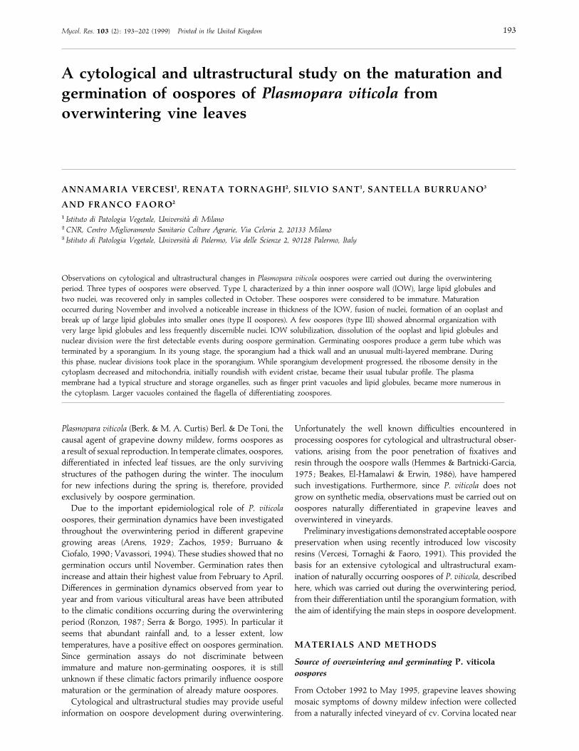
Figs 1–7. P. viticola oospores sampled in October. Fig. 1. Differential interference contrast microscopy of a cleared leaf fragment
showing three oospore types (I, II, III). Figs 2–7. Median ultra-thin sections of the above oospore types. Fig. 2. Type I : lipid globules
(L) of different sizes and electron-densities fill the whole cell lumen ; the periplasmic space (S) between the oogonium wall (OG) and the
outer oospore wall (OOW) contains electron-dense material ; the detail of the oospore wall is visible in Fig. 3. Fig. 3. Type I oospore
wall is formed by a thick inner layer (IOW) separated from a thin outer layer (OOW) by a third layer (arrowheads) which is probably
the oosphere membrane (P¯ plasma membrane). Fig. 4. Type II : small and uniform lipid globules (L) surround a very electron-dense
ooplast (OS). IOW is very thick and formed by a series of intergrading layers with different electron-density. Fig. 5. A quiescent
mitochondrion (M) is visible among lipid globules of the oospore in Fig. 4.
Verona, Italy. Infected leaves were examined by light
microscopy in order to excise the zones containing significant
numbers of oospores. Twenty-four nylon (pore size¯100 µm#) bags per year were each filled with ten oospore rich
leaf fragments. Nylon was chosen since it would not interfere
with environmental influences on the oospores and allowed
rapid and easy recovery of leaf fragments from the field site.
Oospore overwintering was carried out by laying the nylon
bags side by side on the soil surface of the vineyard where the
leaf samples were collected. Each month, from October to
May, a nylon bag was randomly removed ; some leaf fragments
were processed directly for microscopy and some others
utilized for oospore isolation. Oospore isolation was carried
out by finely grinding the leaf fragments as described by
Ronzon (1987).
In order to follow the different germination stages, from
production of the germination tube onwards, 400 isolated
oospores were incubated in Petri dishes on water agar (Noble
Annamaria Vercesi and others 195
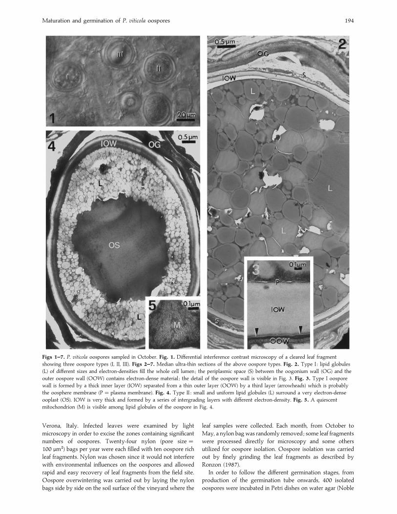
Fig. 6. Detail of the oospore walls in Fig. 4 (P¯ plasma membrane). Fig. 7. Type III : a few, very large lipid globules (L) almost
completely occupy the cell lumen. The IOW is very thick but rather homogeneous.
Agar, Difco, 1% w}v) at 20 °C for 14 d and examined daily
by light microscopy.
Light microscopy
Some leaf fragments were cleared with commercial sodium
hypochloride (5% active sodium) and observed by differential
interference contrast microscopy, according to Beakes et al.
(1986), to assess the type of protoplasm organization in the
oospores. Nuclear staining was carried out on isolated
oospores, either immediately after isolation or at different
incubation times (1–14 d) on water agar. The oospores were
fixed with 30% ethanol for 5 min, washed repeatedly with
distilled H#O, treated for 15 min with 3 µg ml−" DAPI
fluorochrome in 0±05 Tris HCl buffer, pH 7 (Brunk, Jones &
James, 1979) and examined under epifluorescence. Semi-thin
sections (1–2 µm) processed for TEM as described in the next
section, but not treated with OsO%, were stained with 1%
toluidine blue in 0±5% Na#CO
$at 60° for a few seconds or
0±3% Sudan black B in 70% ethanol for 30 min (Bronner,
1975).
Electron microscopy
Overwintering oospores were processed for TEM directly
within the leaf fragments, while germinating oospores were
fixed after removal from the germination medium up to 48 h
after the onset of germination (first detection of germination
tube). Leaf fragments were fixed following different pro-
cedures ; (i) 3% glutaraldehyde in 0±1 phosphate buffer
(pH 7) at room temperature for at least 4 h ; (ii) 3%
glutaraldehyde in 0±1 phosphate buffer (pH 7), at 60° for
2 h followed by 2 h at room temperature, with or without
vacuum using a water pump (Fineran, 1994) (iii) a mixture of
2±5% glutaraldehyde and 2±5% formaldehyde in 0±1
cacodylate buffer (pH 7) under vacuum, at room temperature
for 4–48 h ; (iv) 1% KMnO%in 0±1 cacodylate buffer (pH 7)
for at least 48 h under vacuum.
Germinating oospores were covered with 2% Bacto-agar
(Difco). After agar solidification, 1 mm$ blocks containing a
single oospore were cut out of the agar and fixed using pro-
cedure (iii), but without vacuum, as these structures are much
more easily penetrated by fixatives.
In most cases, samples were post-fixed in 1% OsO%
for
2–4 h. Samples were dehydrated in ethanol and embedded in
either Epon-Araldite, L. R. White (London Resin Company,
U.K.) and Bio-Acryl resin (BioCell, U.K.), infiltrated for
12–48 h under vacuum and polymerized for 24 h at 60°.Ultra-thin sections were cut, stained with 0±5% aqueous
uranyl acetate and lead citrate and examined under a Jeol 100-
SX electron microscope.
RESULTS
The best results were achieved when samples were fixed in a
mixture of 2±5% paraformaldehyde and 2±5% glutaraldehyde
in 0±1 cacodylate buffer for at least 24 h under a slight
vacuum at room temperature and post-fixed in 1% OsO%
in
the same buffer for 4 h. Thus all the figures, unless otherwise
stated, are from samples processed in this way. Fixation
carried out at 60° resulted in easier recognition of dehydrated
organelles, particularly mitochondria. This method is not,
however, generally recommended as it induced significant
artefacts such as swelling of the mitochondria.
KMnO%fixation did not result in acceptable preservation of
oospore protoplasm. As cell wall layers were more easily
distinguishable following this treatment, however, this was
routinely utilized in addition to aldehyde fixation. L. R. White
resin proved to be the most suitable resin for embedding
overwintering oospores, infiltrated over a period of 48 h
under vacuum at 4° and cured for 24 h at 60°. No significant
problems were encountered in fixing and embedding ger-
mination oospores.
Overwintering oospores
In the samples processed in October, three different types of
oospore organization were recognizable by light microscopy
Maturation and germination of P. viticola oospores 196
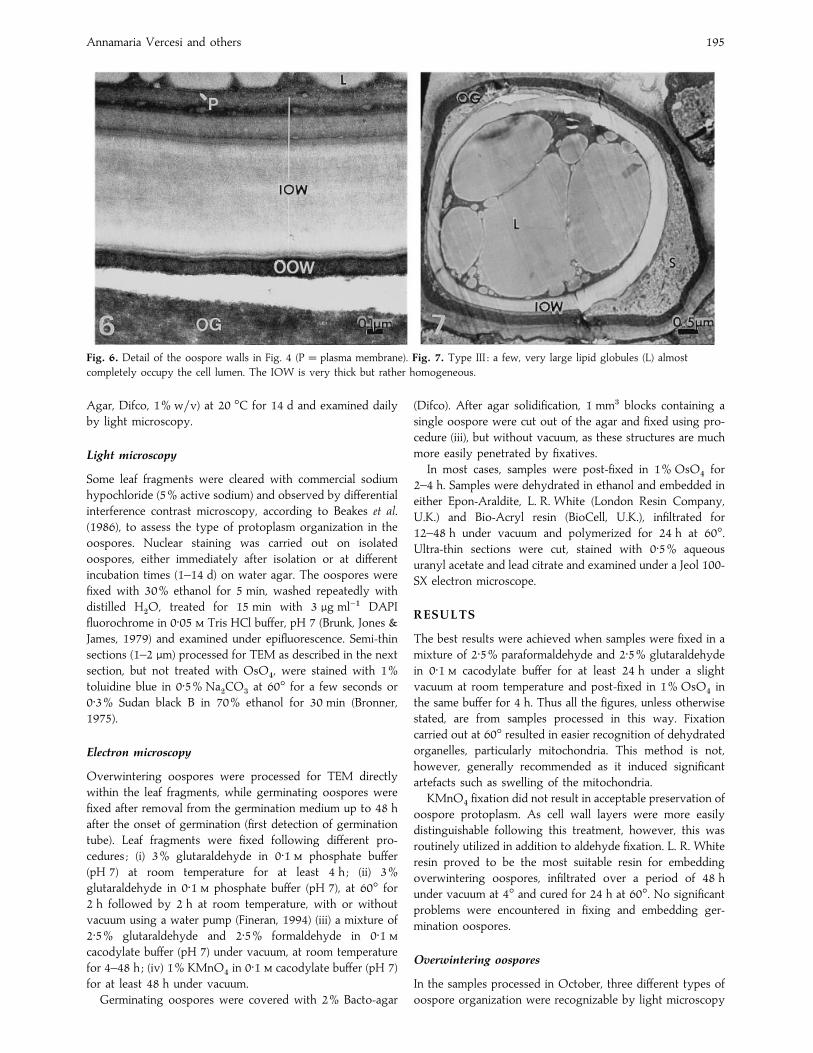
Figs 8–9. Ultra-thin sections of P. viticola oospores collected in November. Fig. 8. Detail of the inner (IOW) and outer (OOW)
oospore walls in a sample fixed with KMnO%. The IOW is formed by intergrading numerous layers, some of which are discretely
corrugated, probably because of inadequate fixation. Fig. 9. A nucleus (N), recognizable by the negatively-stained nuclear cisternae
(arrowhead), among lipid globules (L) at cell periphery.
Figs 10–11. Ultrathin sections of P. viticola oospores collected in May. Fig. 10. An oospore showing incipient germination. The IOW
inner layers (asterisks) are being digested, while mitochondria (M) are now easily recognizable. Fig. 11. Another oospore in incipient
germination : numerous nuclei (N) appear at the cell periphery and the fragmented ooplast (OS) shows many already digested areas.
Annamaria Vercesi and others 197
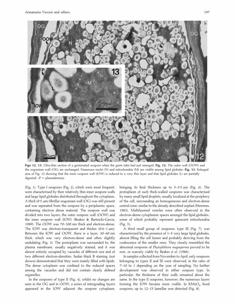
Figs 12, 13. Ultra-thin section of a germinated oospore when the germ tube had just emerged. Fig. 12. The outer wall (OOW) and
the oogonium wall (OG) are unchanged. Numerous nuclei (N) and mitochondria (M) are visible among lipid globules. Fig. 13. Enlarged
area of Fig. 12 showing that the inner oospore wall (IOW) is reduced to a very thin layer and that lipid globules (L) are partially
digested ; (P¯ plasmalemma).
(Fig. 1). Type I oospores (Fig. 2), which were most frequent,
were characterised by their relatively thin inner oospore walls
and large lipid globules distributed throughout the cytoplasm.
A thick (0±5 µm) fibrillar oogonium wall (OG) was still present
and was separated from the oospore by a periplasmic space,
containing electron dense material. The oospore wall was
divided into two layers, the outer oospore wall (OOW) and
the inner oospore wall (IOW) (Beakes & Bartnicki-Garcia,
1989). The OOW was 70–100 nm thick and electron-dense.
The IOW was electron-transparent and thicker (0±6–1 µm).
Between the IOW and OOW, there is a layer, 30–40 nm
thick, which was very electron-dense and often slightly
undulating (Fig. 3). The protoplasm was surrounded by the
plasma membrane, usually negatively stained, and it was
almost entirely occupied by vacuoles of different size and of
two different electron-densities. Sudan black B staining (not
shown) demonstrated that they were mainly filled with lipids.
The dense cytoplasm was confined to the reduced spaces
among the vacuoles and did not contain clearly defined
organelles.
In the oospores of type II (Fig. 4), whilst no changes are
seen in the OG and in OOW, a series of intergrading layers
appeared in the IOW adjacent the oospore cytoplasm,
bringing its final thickness up to 3–3±5 µm (Fig. 6). The
protoplasm of such thick-walled oospores was characterised
by many small lipid droplets, usually localized at the periphery
of the cell, surrounding an homogeneous and electron-dense
central zone, similar to the already described ooplast (Hemmes,
1983). Multilayered vesicles were often observed in the
electron-dense cytoplasmic spaces amongst the lipid globules ;
some of which probably represent quiescent mitochondria
(Fig. 5).
A third small group of oospores, type III (Fig. 7), was
characterized by the presence of 1–5 very large lipid globules,
almost filling the cell lumen and probably deriving from the
coalescence of the smaller ones. They closely resembled the
abnormal oospores of Phytophthora megasperma proved to be
non- or scarcely viable by Beakes et al. (1986).
In samples collected from November to April, only oospores
belonging to types II and III were observed, in the ratio of
7–10 to 1 depending on the year of sampling. No further
development was observed in either oospore type. In
particular, the thickness of their walls remained about the
same. In the type II oospores, however, the numerous layers
forming the IOW became more visible. In KMnO%
fixed
oospores, up to 12–15 lamellae wee detected (Fig. 8).

Maturation and germination of P. viticola oospores 198
Figs 14–16. Light microscopy of germinated oospores (G). Fig. 14. Fluorescence microscopy of an oospore (G) stained with DAPI
soon after the appearance of the germ tube : the bright spots are nuclei, often coupled possibly as the result of a recent division ; some
nuclei are possibly migrating through the germ tube (arrowheads). Fig. 15. Interference contrast microscopy of a germinated oospore
showing a mature sporangium (SP). Fig. 16. The same sporangium as in Fig. 15 stained with DAPI and observed with UV light and
at the same stage as Fig. 20.
Under TEM nuclei, usually localized at the cell periphery,
were rarely visible in overwintering oospores (Fig. 9).
Observations of a large number of oospores stained with the
nuclear fluorochrome DAPI revealed the presence of two
nuclei (probably prefusion, haploid nuclei) in type I oospores,
while only a single nucleus was found in type II. In type III
oospores, the nucleus was frequently not detected, although
sometimes a diffuse fluorescence was observed in the
cytoplasm, possibly due to the breakdown of the nuclear
membrane.
Germinating oospores
In May, a small number of oospores type II ultrastructure
showed signs of incipient lysis of the IOW zone near the
protoplasm (Fig. 10) indicative of the onset of germination.
Moreover, the whole IOW appeared more electron-trans-
parent and the number of visible layers apparently decreased.
At the same time, two or more nuclei could be seen in the
spore indicating nuclear divisions (Fig. 11). Numerous
mitochondria were present in the interstices among the lipid
globules (Fig. 10). The ooplast vacuole contents were less
electron-dense and contained prominent electron-transparent
spaces. As soon as the germ tube emerged, the overall
ultrastructure of the oospore changed markedly. The thickness
of IOW rapidly became greatly reduced and in some cases this
wall layer had almost completely disappeared. The OG did
not show appreciable change in comparison with resting
oospores while the OOW appeared very crinkled (Fig. 12). In
the oospore cytoplasm the first functional organelles, notably
mitochondria, became evident (Fig. 13). The lipid globules
became more electron-transparent at their periphery, with a
more electron-dense central core (Fig. 12). This, together with
loss of IOW was indicative of utilization of storage material.
Numerous nuclei were observed in germinating oospores and
they were readily detected with DAPI staining while probably
migrating through the germ tube (Fig. 14). Germinating
oospores always produced a germ tube (29–56 µm long) at
the end of which a pyriform sporangium (27¬45 µm) soon
appeared (Figs 15 and 16).
Ten to twelve hours after germ tube emergence, the
sporangium wall was almost electron-transparent and rather
thick (Fig. 17). The protoplasm was surrounded by a multi-
layered layer underlying the sporangial cell wall. The
cytoplasm contained many ribosomes, very often organized in
polysome structures, suggesting enhanced synthetizing ac-
tivity (Fig. 18). Vesicles, surrounded by a well-defined
membrane, were distributed in the cytoplasm and showed a
granular content (Fig. 18). At this stage, nuclear divisions were
readily observed in the sporangium (Fig. 16). Pairs of
centrioles, aligned end to end, were detected in the cytoplasm
(Fig. 19). A very large number of mitochondria with a rather
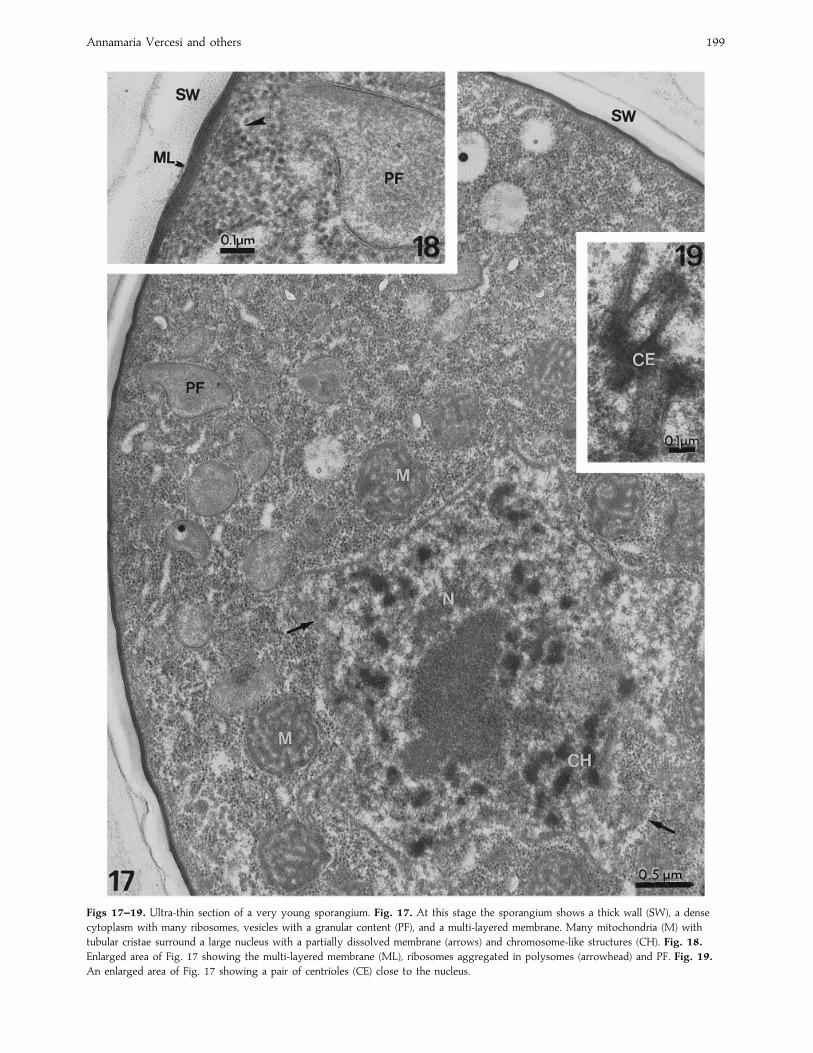
Annamaria Vercesi and others 199
Figs 17–19. Ultra-thin section of a very young sporangium. Fig. 17. At this stage the sporangium shows a thick wall (SW), a dense
cytoplasm with many ribosomes, vesicles with a granular content (PF), and a multi-layered membrane. Many mitochondria (M) with
tubular cristae surround a large nucleus with a partially dissolved membrane (arrows) and chromosome-like structures (CH). Fig. 18.
Enlarged area of Fig. 17 showing the multi-layered membrane (ML), ribosomes aggregated in polysomes (arrowhead) and PF. Fig. 19.
An enlarged area of Fig. 17 showing a pair of centrioles (CE) close to the nucleus.
Maturation and germination of P. viticola oospores 200
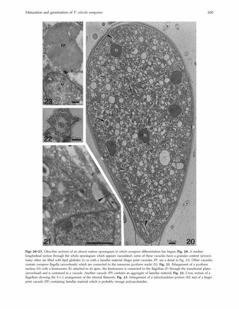
Figs 20–23. Ultra-thin sections of an almost mature sporangium in which zoospore differentiation has begun. Fig. 20. A median
longitudinal section through the whole sporangium which appears vacuolated ; some of these vacuoles have a granular content (arrows),
many other are filled with lipid globules (L) or with a lamellar material (finger print vacuoles, FP, see a detail in Fig. 23). Other vacuoles
contain zoospore flagella (arrowheads) which are connected to the numerous pyriform nuclei (N). Fig. 21. Enlargement of a pyriform
nucleus (N) with a kinetosome (K) attached to its apex ; the kinetosome is connected to the flagellum (F) through the transitional plates
(arrowhead) and is contained in a vacuole. Another vacuole (FP) contains an aggregate of lamellar material. Fig. 22. Cross section of a
flagellum showing the 92 arrangement of the internal filaments. Fig. 23. Enlargement of a mitochondrion portion (M) and of a finger
print vacuole (FP) containing lamellar material which is probably storage polysaccharides.
Annamaria Vercesi and others 201
unusual round shape were localized very close to the nuclei
and were characterized by prominent cristae (Fig. 17).
In a later stage of sporangium development, its wall became
thinner and the multi-layered membrane was replaced by a
typical plasma membrane. The nuclei showed an interphase
status (Fig. 20). Staining with DAPI revealed that some of
them appeared coupled. Close to the apex of the pyriform
nuclei, kinetosomes, originating from the centrioles, were
connected to the zoospore flagella (Fig. 21). The flagella in
cross section appeared to consist of a 92 arrangement of
internal filaments (Fig. 22) and were contained in a vacuole
(Fig. 20). Mitochondria assumed their usual elongate tubular
profile and the cristae were less evident. Also in these
developing sporangia the ribosome density decreased and the
cytoplasm appeared much more vacuolated. Other typical
structures could be observed ; lipid globules, sometimes with
a dark core (Fig. 20) and finger print vacuoles (Fig. 23), which
are thought to contain storage polysaccharides (Hemmes,
1983).
A septum (not shown) separated the mature sporangium
from the germination tube. At this stage of the sporangium
differentiation, the oospore appeared completely empty and
recognizable only by the OG and OOW. The IOW and the
cytoplasm were completely utilised for the formation of the
sporangium and only a few residues of the previous structure
remained in the oospore.
Unfortunately it was not possible to detect sporangium
cleavage, probably because of the rapidity of its occurrence.
DISCUSSION
Difficulties encountered in investigating oospore ultrastructure
have been pointed out by many authors and concern all the
steps involved in sample preparation, particularly embedding
(Hemmes & Bartnicki-Garcia, 1975 ; Beakes, 1980a, b ; Beakes,
1981 ; Hemmes & Stasz, 1984). Hydrophilic low viscosity
methacrylate resins such as L. R. White seem to be the best
embedding media for the oospores of P. viticola. Epon-Araldite
and Bio-Acryl, which is a mixture of epoxy and methacrylate
resins, do not easily penetrate the wall layers even when
infiltration is conducted under a moderate vacuum. Fur-
thermore, use of L. R. White, instead of other uv-polymerizing
methacrylate resins, allowed post fixation with OsO%, which
is essential for good preservation of the oospores.
Ultrastructural studies on the sexual structures of Oomycota
have been carried out almost exclusively on species able to
grow on synthetic media. The only exceptions being the
studies on Albugo candida, an obligate parasite of Brassica
campestris (Tewari & Skoropad, 1977) and Bremia lactucae, the
causal agent of lettuce downy mildew (Sargent, Ingram &
Tommerup, 1977). Other obligate biotrophs belonging to the
Oomycota have been surprisingly neglected, in spite of the
important role played by oospores in assuring survival of the
pathogen (Populer, 1981).
Recently it has been pointed out that systematics of downy
mildews needs to be defined on the basis of more accurate
and precise criteria (Hall, 1996) but unfortunately numerous
important species are still inadequately described (Dick, 1995).
The ultrastructural approach has been recommended and
seems to be particularly interesting both for phylogenetic and
systematic purposes. The present study is the first report on
the oospore ultrastructure of the grapevine downy mildew
fungus, P. viticola, belonging to a genus with very poor
information as to its morphological characteristics.
Wall layers of P. viticola oospores are more similar to those
of Pythiaceae (Hemmes, 1983 ; Hemmes & Stasz, 1984 ; Beakes
& Bartnicki-Garcia, 1989) than to the oospore wall complex of
the other already described obligate parasites A. candida and
B. lactucae (Tewari & Skoropad, 1977 ; Sargent et al., 1977).
Furthermore, the IOW of P. viticola oospores appears
sometimes thicker and less homogeneous, being formed by
numerous layers, characterized by different electron densities.
Such wall organization has not been observed in other
oomycetes which show a rather uniform appearance of the
IOW, with the exception of S. ferax and S. furcata (Beakes &
Gay, 1978 ; Beakes, 1980b). In these two species, however, the
inner part of IOW is characterized by the presence of patches
of electron-dense material.
The multi-layered structure of IOW in P. viticola might be
related to the in situ production of the oospores. In fact, almost
all investigations on the oospore ultrastructure have been
carried out on samples differentiated on culture media or
inoculated hosts under controlled conditions (Sargent et al.,
1977 ; Beakes, 1980b). By contrast, P. viticola oospores were
formed under fluctuating conditions of temperature and
wetness and it is possible that sudden environmental changes
during differentiation induce a less homogenous development
of the IOW. Also the genetic variation in the wild P. viticola
population may have been greater than in the population
maintained under controlled conditions.
Dense cytoplasmic appearance, the presence of membrane
delimited components among the lipid globules and the
ooplast in the centre of the cell are common characteristics of
quiescent oospores (Hemmes, 1983). The inactive status of the
oospore cytoplasm is confirmed by the erratic observation of
constitutive organelles such as quiescent mitochondria (Beakes
et al., 1986).
Nuclear staining with DAPI allowed detection of two
nuclei in type I oospores that were unable to germinate
(Vercesi, Cortesi & Zerbetto, 1993). Fusion of nuclei and
achievement of a definitive structure, together with ger-
mination capability, are particular characteristics of type II
oospores. Sporangium formation by type II oospores supports
the hypothesis of Jiang et al. (1989) that the presence of a
single nucleus in oospores of Phytophthora spp. is an
indispensable prerequisite for germination and that the fusion
of gametangial nuclei is a useful criterion to distinguish mature
oospores.
Diffuse fluorescence observed in oospores characterized by
large lipid globules (type III) can be interpreted as a nuclear
degeneration. Abnormal oospore structure and no discernible
nuclei have already been associated, respectively, with scarce
viability and absence of germination (Beakes et al., 1986 ; Jiang
et al., 1989). It can, therefore, be assumed that type III
oospores represent dead or degenerating P. viticola over-
wintering structures.
Oospores germinated at variable times after collection,
depending on the year and the sampling month. Generally,
Maturation and germination of P. viticola oospores 202
oospores isolated from samples collected in January or
February germinated in 6–7 d, whilst those collected in April
germinated in 3–4 d. Early germination events are represented
by the rehydration of mitochondria, nuclear divisions and the
progressive enzymic breakdown of the IOW which appears
uniformly eroded in contrast with the large erosion cavities
observed in P. megasperma (Beakes & Bartnicki-Garcia, 1989).
The following unusual features were observed during the
formation of P. viticola sporangia. At the beginning of
germination, the sporangium wall appeared rather thick and
divided into layers characterized by different electron
transparency. In the same developmental stage a multi-layered
structure was evident between the wall and the protoplasm
and could be interpreted as a multi-stratification of plasma
membrane. Though it is difficult to understand the significance
of this stratification it could be the result of an intense
synthetic activity of membrane and wall materials to allow the
rapid enlargement of the sporangium. The same explanation
could be put forward for the unusual thickness of the
sporangium wall.
As already pointed out, P. viticola oospores play a very
important role in grapevine downy mildew epidemiology.
Germination of oospores provides the vital inoculum required
for primary infections of susceptible hosts during the spring.
In this season, the absence of primary foci in vineyards not
treated against downy mildew, even after the occurrence of
climatic conditions suitable for oospore germination, has been
attributed to the still immature state of oospores.
The unchanged ultrastructure of P. viticola oospores from
November onwards demonstrates that they are mature by the
middle of November. Oospore maturation is not, therefore,
the limiting factor for their germination and cannot account
for the fluctuations in the germination rates reported by many
authors and attributed to the effect of climatic factors (Zachos,
1959 ; Ronzon, 1987 ; Burruano & Ciofalo, 1990 ; Serra &
Borgo, 1995). Following from these observations and the new
results presented in this paper, it seems likely that the
influence of rainfall and temperature occurring during
overwintering are exerted on the physiological and}or
biochemical processes leading to germination in already
morphologically mature oospores.
REFERENCES
Arens, K. (1929). Untersuchungen u$ ber Keimung and Zitologie der Oosporen
von Plasmopara viticola (Berl. et de Toni). Jahrbucher fuX r Wissenschaftliche
Botanik 70, 57–92.
Beakes, G. W. (1980a). Electron microscopy study of oospore maturation and
germination in an emasculate isolate of Saprolegnia ferax. 1. Gross changes.
Canadian Journal of Botany 58, 182–194.
Beakes, G. W. (1980b). Electron microscopy study of oospore maturation and
germination of an emasculate isolate of Saprolegnia ferax. 2. Wall
differentiation. Canadian Journal of Botany 58, 195–208.
Beakes, G. W. (1981). Ultrastructural aspects of oospore differentiation. In
The Fungal Spore : Morphogenetic Controls (ed. G. Turian & H. R. Hohl),
pp. 71–90. Academic Press : London, New York, Toronto, Sydney.
(Accepted 22 April 1998)
Beakes, G. W. & Gay, J. L. (1978). Light and electron microscopy of oospore
maturation in Saprolegnia furcata : wall development. Transactions of the
British Mycological Society 71, 25–35.
Beakes, G. W. & Bartnicki-Garcia, S. (1989). Ultrastructure of mature
oogonium-oospore wall complexes in Phytophthora megasperma : a com-
parison of in vivo and in vitro dissolution of the oospore wall. Mycological
Research 93, 321–334.
Beakes, G. W., El-Hamalawi, Z. A. & Erwin, D. C. (1986). Ultrastructure of
mature oospores of Phytophthora megasperma f.sp. medicaginis : preparation
protocols and effects of MTT vital staining and permanganate pre-
treatment. Transactions of the British Mycological Society 86, 195–206.
Bronner, R. (1975). Simultaneous demonstration of lipids and starch in plant
tissues. Stain Technology 50, 1–4.
Brunk, C. F., Jones, K. C. & James, T. W. (1979). Assays for nanograms
quantities of DNA in cellular homogenates. Analytical Biochemistry 92,
497–500.
Burruano, S. & Ciofalo, G. (1990). Studio sulla dinamica di germinazione delle
oospore di Plasmopara viticola (Berk. et Curt.) Berl. et De Toni. Notiziario
Malattie Piante 111, 284–286.
Dick, M. W. (1995). Sexual reproduction in the Peronosporomycetes (chromistan
fungi). Canadian Journal of Botany 73 (Suppl. 1), S712–S724.
Fineran, B. A. (1994). Hot fixation of fungal spores for transmission electron
microscopy : application to thick-walled spores of the smut fungus
Entorrhiza. Mycological Research 98, 799–899.
Hall, G. S. (1996). Modern approach to species concepts in downy mildews.
Plant Pathology 45, 1009–1026.
Hemmes, D. E. (1983). Cytology of Phytophthora. In Phytophthora, Its Biology,
Taxonomy, Ecology and Pathology (ed. D. C. Erwin, S. Bartnicki-Garcia &
P.H. Tsao), pp. 9–40. The American Phytopathological Society : St Paul.
Hemmes, D. E. & Bartnicki-Garcia, S. (1975). Electron microscopy of
gametangial interaction and oospore development in Phytophthora capsici.
Archives of Microbiology 103, 91–112.
Hemmes, D. E. & Stasz, T. E. (1984). Ultrastructure of dormant, converted and
germinating oospores of Pythium ultimum. Mycologia 76, 924–935.
Jiang, Jiping, Stephenson, L. W., Erwin, D. C. & Leary, J. V. (1989). Nuclear
changes in Phytophthora during oospore maturation and germination.
Mycological Research 92, 463–469.
Populer, C. (1981). Epidemiology of downy mildews. In The Downy Mildews
(ed. D. M. Spencer), pp. 57–105. Academic Press : London, New York, San
Francisco.
Ronzon, C. (1987). Mode! lisation du comportement e! pide! mique du mildiou de
la vigne ; e! tude du ro# le de la phase sexue! e de Plasmopara viticola. The' se de
doctorat, Universite! de Bordeaux II.
Sargent, J. A., Ingram, D. S. & Tommerup, I. C. (1977). Oospore development
in Bremia lactucae Regel : an ultrastructural study. Proceedings of the Royal
Society London B 198, 129–138.
Serra, S. & Borgo, M. (1995). Indagini sulla maturazione e germinazione delle
oospore di Plasmopara viticola svernate in condizioni naturali. Petria 5,
91–103.
Tewari, J. P. & Skoropad, W. P. (1977). Ultrastructure of oospore development
of Albugo candida on rapeseed. Canadian Journal of Botany 55, 2354–2357.
Vavassori, A. (1994). Indagini sulla dinamica di germinazione delle oospore di
Plasmopara viticola (Berk et Curt.) Berl. and De Toni. PhD Thesis, University
of Milan.
Vercesi, A., Cortesi, P. & Zerbetto, F. (1993). La previsione del rischio
epidemico da Plasmopara viticola : risultati e prospettive. Atti del Convegno
Nazionale ‘Protezione delle colture : osservazioni, previsioni, decisioni ’, pp. 301–
324.
Vercesi, A., Tornaghi, R. & Faoro, F. (1991). Preliminary investigations on the
ultrastructure of Plasmopara viticola oospores. Giornale Botanico Italiano 125,
1012–1013.
Zachos, D. G. (1959). Recherches sur la biologie et l’e! pide! miologie du mildiou
de la vigne en Gre' ce. Annales de l’Institut Phytophatologique Benaki, nouvelle
seU rie 2, 193–355.