clinical and cytological correlations in pericardial ... · romanian journal of morphology and ......
TRANSCRIPT
Romanian Journal of Morphology and Embryology 2009, 50(2):251–256
OORRIIGGIINNAALL PPAAPPEERR
Clinical and cytological correlations in pericardial effusions with cardiac
tamponade D. P. PETCU1), C. PETCU2), CARMEN FLORINA POPESCU3),
C. BĂTĂIOSU4), D. ALEXANDRU1)
1)University of Medicine and Pharmacy of Craiova 2)“Fundeni” Institute of Cardiovascular Diseases, Bucharest
3)Emergency County Hospital, Craiova 4)Cardiology Center, Craiova
Abstract We studied 27 patients diagnosed with pericardial effusion with cardiac tamponade on which pericardiocentesis was performed. The purpose of the study was to evaluate the benefits and limits of the cytological examination of the pericardial liquid in the etiological diagnosis and the treatment of patients with cardiac tamponade. The pericardial liquid taken was examined macroscopically, biochemically (content of proteins, glucose, cholesterol, and LDH), cytologically (MGG stained smears from pericardial liquid) and bacteriologically. The obtained results were compared to the clinical data, the laboratory and paraclinical tests, to differentiate the cause and therapeutically procedure. The cardiac tamponade remitted after pericardiocentesis in all patients. The pericardial liquid was exudate (Ligth criteria) in 82% of all patients. The cytological examination of the pericardial liquid showed malignant smear in 40.74% of the patients, smear of the TBC specific inflammation type in 7.40% patients, smear of non-specific inflammation type in 25.94% of patients, reactive type smear in 25.9% of patients. Keywords: pericardiocentesis, cytological examination, etiology.
Introduction
The prevalence of pericardial effusions is 0.1% from the hospitalized pathology in the specialized medical centers in diagnosis and treatment of cardiovascular diseases [1].
Pericardial effusions with a cardiac tamponade constitute a surgical emergency and, after a clinical, biochemical, electrocardiographic, echocardiographic and radiological evaluation, the pericardiocentesis is to be imposed as a therapeutically goal.
The pericardiocentesis promptly carried out and according to the recommendations in the pericardic diseases, treatment guide (Maisch B et al., 2004), represents the first class IA therapeutically recommendation [2].
After pericardiocentesis practice, the patients need an etiopathogenic treatment. In spite of all the progresses made by invasive and non-invasive investigations (Cheema MA et al., 2007), the etiopathology of pericardial effusions still remains unknown in 20–40% of the cases [3].
The causes of pericardial effusions differ depending on the patients grouping, geographical zone, having been meanwhile modified.
The diagnosis value of the cytological, biochemical and bacteriological examination in the pericardial liquid is differently presented in the specialty literature [4, 5].
The purpose of this study is to evaluate the benefits
and limits of the cytological examination carried out from the pericardial liquid in establishing the etiological diagnosis and the treatment of patients with cardiac tamponade.
Patients and Methods
We have included in our study allotment a number of 27 patients, eight women and 19 men suffering from pericardial effusions with cardiac tamponade, aged between 21 and 81-years-old, hospitalized between June 2007–June 2008 in the Cardiology Center of Craiova.
The study is both a prospective and a retrospective one.
The diagnosis with pericardial effusion with cardiac tamponade was established by:
A. Clinical criteria (arterial hypotension, systolic blood pressure <90 mmHg, paradoxal pulse systolic blood pressure decreases by more than 10 mmHg in inspiration, increase in jugular venous pressure);
B. Echocardiographic criteria (diastolic collapse of right cardiac cavities, presence of circumferential pericardial liquid >20 mm).
The pericardiocentesis was therapeutically performed in all patients. The subxiphoidian approa-ching was used for all the patients. The technique and materials used for pericardiocentesis are described in the standard protocol [2].
The patients were examined clinically (medical
D. P. Petcu et al.
252
history and complete physical examination), electro-cardiographically, echocardiographically, radiologically (cardiothorax radiography), by general echography and laboratory tests (hemoleucogram, T-troponin, CKMB, hepatic functional tests, creatinine, urea, glycemia, cholesterol, TSH-ionogram, T3, anti-DNA, lupic cells, rheumatoid factors), HIV tests, cutaneous testing for tuberculin.
There were excluded from the study the patients with cardiac tamponade and acute myocardial heart failure, cardiac trauma, aorta dissection and hemorrhagic diseases.
Aspirative needle-punction (Marfan procedure) and pericardial drainage guided with a drain tube percutaneously introduced (Seldinger technique) were performed in the angiography room or in ICU with radioscopic or echocardiographic guidance. The Seldinger technique was performed after a local anesthesia with 1% Lidocaine, following the aseption and antiseption conditions of teguments and after administration of i.v. 5–10 mg [2].
The pericardial liquid taken was examined macroscopically, biochemically (content of proteins, glucose, cholesterol, LDH), cytologically, and bacteriologically (TBC, anaerobus, aerobus and fungi bacilli).
For the cytological examination, the adequate reaping, the correct preservation and processing of serous effusions were essential for a correct cytological interpretation.
Choosing the cytoprocessing method was made according to physical properties of the respective liquid sample and to the patient’s history. An accompanying card, containing reliable clinical information for the cytopathologist to correlate with the cytological aspects, accompanied each sample.
For preventing coagulation of the serous effusions, in each sample, after reaping, there was added 1 ml of EDTA solution, after which the samples were sent to the Laboratory of Pathological and Cytological Anatomy of the Emergency County Hospital, Craiova, where they were processed by centrifugation.
All samples were centrifuged at 2500 rot/min., for 10 minutes, and then the supernatant was eliminated and there were performed smears from the sediment. Depending on the sediment quantity, we performed from minimum three smears (in samples with minimum sediment) until 10 smears (in samples with abundant sediment).
All smears were stained through the May-Grünwald–Giemsa method; the result of the staining was the achieving of blue-violet nuclei and the cytoplasm in variable shades of pink to light blue-dark blue.
For the establishing of the cytodiagnosis, we evaluated the cellular and malignant criteria of malignity.
The obtained results after the cytological examination from the pericardial liquid were compared to the clinical, paraclinical and laboratory data.
In order to establish a relationship between these data we used the Student t-test.
Results
Results of the clinical study
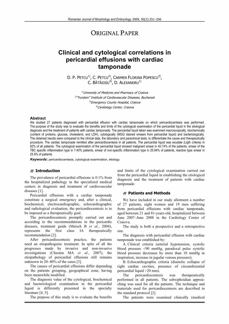
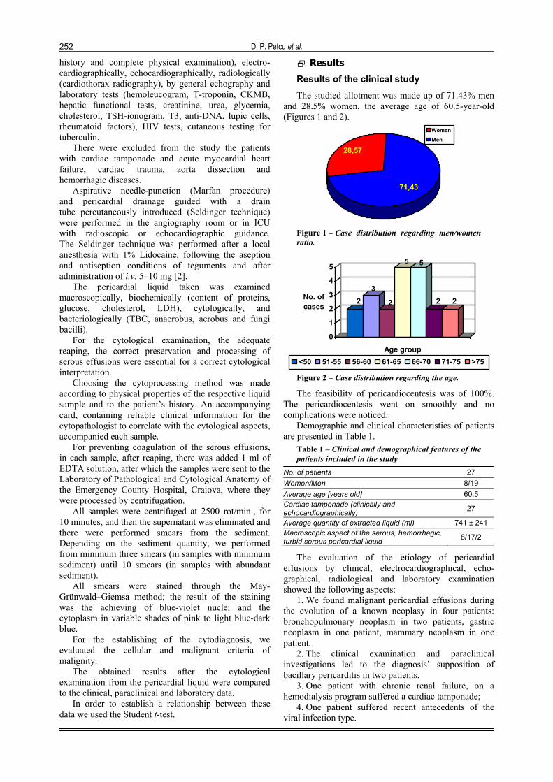
The studied allotment was made up of 71.43% men and 28.5% women, the average age of 60.5-year-old (Figures 1 and 2).
71,43
28,57
Women
Men
Figure 1 – Case distribution regarding men/women ratio.
23
2
5 5
2 2
0
1
2
3
4
5
No. of cases
Age group<50 51-55 56-60 61-65 66-70 71-75 >75 Figure 2 – Case distribution regarding the age.
The feasibility of pericardiocentesis was of 100%. The pericardiocentesis went on smoothly and no complications were noticed.
Demographic and clinical characteristics of patients are presented in Table 1.
Table 1 – Clinical and demographical features of the patients included in the study
No. of patients 27 Women/Men 8/19 Average age [years old] 60.5 Cardiac tamponade (clinically and echocardiographically) 27
Average quantity of extracted liquid (ml) 741 ± 241 Macroscopic aspect of the serous, hemorrhagic, turbid serous pericardial liquid 8/17/2
The evaluation of the etiology of pericardial effusions by clinical, electrocardiographical, echo-graphical, radiological and laboratory examination showed the following aspects:
1. We found malignant pericardial effusions during the evolution of a known neoplasy in four patients: bronchopulmonary neoplasm in two patients, gastric neoplasm in one patient, mammary neoplasm in one patient.
2. The clinical examination and paraclinical investigations led to the diagnosis’ supposition of bacillary pericarditis in two patients.
3. One patient with chronic renal failure, on a hemodialysis program suffered a cardiac tamponade;
4. One patient suffered recent antecedents of the viral infection type.
Clinical and cytological correlations in pericardial effusions with cardiac tamponade
253
5. In 19 patients the causes of cardiac tamponade were not clinically and paraclinically distinguished (Table 2).
Table 2 – The distribution of the cases regarding the etiology
Etiology No. of cases Neoplasm 4
TBC 2 Chronic renal failure 1
Viral infection 1 Data free 19
The macroscopic aspect of the pericardial effusion was:
▪ hemorrhagic in 17 patients; ▪ serous in eight patients; ▪ turbid-serous in two patients. The biochemical examination of the pericardial
effusion pointed out exudates in 24 patients. The biochemical alterations in the pericardial liquid are presented in Table 3.
Table 3 – The biochemical characteristics of the pericardial fluid
CharacteristicsMalignant pericardial
effusion
Bacillary pericardial
effusion
Reactive pericardial
effusion
Idiopathic pericardial
effusion Proteins in pericardial liquid [g/dL]
0.6 (0.5–0.7)
0.8 (0.7–0.9)
0.6 (0.5–0.7)
0.6 (0.5–0.7)
Glucose in pericardial liquid [g/dL]
0.8 (0.6–1.2)
0.7 (0.6–0.9)
0.9 (0.8–1.0)
1.1 (0.7–1.5)
LDH in pericardial liquid [IU/L]
4.3 (0.6–8)
8.2 (4.2–12.2)
2.6 (0.6–4.6)
2.3 (0.1–4.5)
Results of the cytological study Depending on the microscopic aspect of the
cellularity of smears, they were sub classified in the following types: inflammatory smear (seven cases), smear with reactive benign cellular alterations induced by a non-specific infection (two cases) or specific (eight cases) and malignant smear (in 11 patients) (Table 4).
Table 4 – Correlation between the cytological alterations and established etiology by clinical data Cytological diagnosis The etiology established by
clinical data Malignant smear Specific reactive smear Reactive smear Inflammatory smear Neoplasm 3 0 0 1
TBC 0 1 1 0 Chronic renal failure 0 0 0 0
Viral infection 0 0 1 0 Clinical data free 8 1 5 6
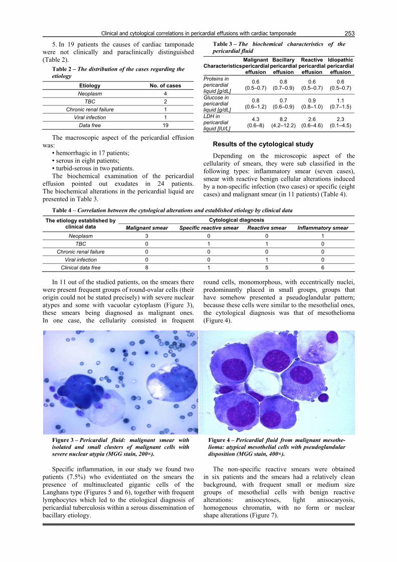
In 11 out of the studied patients, on the smears there
were present frequent groups of round-ovalar cells (their origin could not be stated precisely) with severe nuclear atypes and some with vacuolar cytoplasm (Figure 3), these smears being diagnosed as malignant ones. In one case, the cellularity consisted in frequent
round cells, monomorphous, with eccentrically nuclei, predominantly placed in small groups, groups that have somehow presented a pseudoglandular pattern; because these cells were similar to the mesothelial ones, the cytological diagnosis was that of mesothelioma (Figure 4).
Figure 3 – Pericardial fluid: malignant smear with isolated and small clusters of malignant cells with severe nuclear atypia (MGG stain, 200×).
Figure 4 – Pericardial fluid from malignant mesothe-lioma: atypical mesothelial cells with pseudoglandular disposition (MGG stain, 400×).
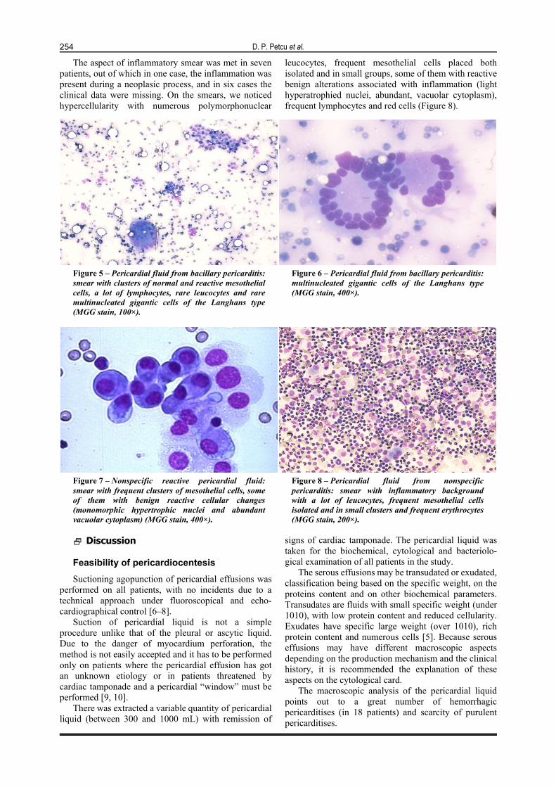
Specific inflammation, in our study we found two
patients (7.5%) who evidentiated on the smears the presence of multinucleated gigantic cells of the Langhans type (Figures 5 and 6), together with frequent lymphocytes which led to the etiological diagnosis of pericardial tuberculosis within a serous dissemination of bacillary etiology.
The non-specific reactive smears were obtained in six patients and the smears had a relatively clean background, with frequent small or medium size groups of mesothelial cells with benign reactive alterations: anisocytoses, light anisocaryosis, homogenous chromatin, with no form or nuclear shape alterations (Figure 7).
D. P. Petcu et al.
254
The aspect of inflammatory smear was met in seven patients, out of which in one case, the inflammation was present during a neoplasic process, and in six cases the clinical data were missing. On the smears, we noticed hypercellularity with numerous polymorphonuclear
leucocytes, frequent mesothelial cells placed both isolated and in small groups, some of them with reactive benign alterations associated with inflammation (light hyperatrophied nuclei, abundant, vacuolar cytoplasm), frequent lymphocytes and red cells (Figure 8).
Figure 5 – Pericardial fluid from bacillary pericarditis: smear with clusters of normal and reactive mesothelial cells, a lot of lymphocytes, rare leucocytes and rare multinucleated gigantic cells of the Langhans type (MGG stain, 100×).
Figure 6 – Pericardial fluid from bacillary pericarditis: multinucleated gigantic cells of the Langhans type (MGG stain, 400×).
Figure 7 – Nonspecific reactive pericardial fluid: smear with frequent clusters of mesothelial cells, some of them with benign reactive cellular changes (monomorphic hypertrophic nuclei and abundant vacuolar cytoplasm) (MGG stain, 400×).
Figure 8 – Pericardial fluid from nonspecific pericarditis: smear with inflammatory background with a lot of leucocytes, frequent mesothelial cells isolated and in small clusters and frequent erythrocytes (MGG stain, 200×).
Discussion
Feasibility of pericardiocentesis Suctioning agopunction of pericardial effusions was
performed on all patients, with no incidents due to a technical approach under fluoroscopical and echo-cardiographical control [6–8].
Suction of pericardial liquid is not a simple procedure unlike that of the pleural or ascytic liquid. Due to the danger of myocardium perforation, the method is not easily accepted and it has to be performed only on patients where the pericardial effusion has got an unknown etiology or in patients threatened by cardiac tamponade and a pericardial “window” must be performed [9, 10].
There was extracted a variable quantity of pericardial liquid (between 300 and 1000 mL) with remission of
signs of cardiac tamponade. The pericardial liquid was taken for the biochemical, cytological and bacteriolo-gical examination of all patients in the study.
The serous effusions may be transudated or exudated, classification being based on the specific weight, on the proteins content and on other biochemical parameters. Transudates are fluids with small specific weight (under 1010), with low protein content and reduced cellularity. Exudates have specific large weight (over 1010), rich protein content and numerous cells [5]. Because serous effusions may have different macroscopic aspects depending on the production mechanism and the clinical history, it is recommended the explanation of these aspects on the cytological card.
The macroscopic analysis of the pericardial liquid points out to a great number of hemorrhagic pericarditises (in 18 patients) and scarcity of purulent pericarditises.
Clinical and cytological correlations in pericardial effusions with cardiac tamponade
255
Clinical and paraclinical evaluation of patients, anterior to pericardiocentesis, identified an apparent cause in eight patients, more frequently a neoplasia and rarely a tuberculosis, metabolic disease or viral pericarditis.
In 19 patients with cardiac tamponade, the identification of a cause was impossible.
Cardiac tamponade having tuberculosis as its cause
The clinical signs of the cardiac tamponade of tuberculosis or non-tuberculosis etiology are the same. Elucidating the tuberculosis etiology is important in a case of a patient with cardiac tamponade because without specific treatment, in six months, the mortality will be about 85% [11, 12].
On the other hand, the high incidence of tuberculosis, 8.8 million of new cases in 2003 (WHO), the high number of deaths by this illness, 1.7 millions (WHO) and association with HIV-infection are important elements for the elucidation of the etiology of a pericardic liquid [11].
There are important for the diagnosis of tuberculosis three clinical criteria: fever, nocturnal perspirations, weight loss, personal data and hereditary-collateral antecedents.
The cytological analysis of tuberculosis effusions showed numerous lymphocytes, isolated and in small clusters of mesothelial cells, some of them having reactive nuclear modifications, as well as a variable number of polymorph nuclear lymphocytes (Figure 3). On smears from a patient, we identified rare multinucleated giant cells of the Langhans type.
M. tuberculosis was not identified at the bacteriological exam in our patients, because fibrin and hemoglobin are bacillus inhibitors (the exudate was hemorrhagic) and pericardial liquid has only few bacilli.
In the present study, we found in a case, a female patient of 27 years old, association between HIV-infection and tuberculosis. The literature data showed that this association might be present up to 100% in the African population [3].
Malignant cardiac tamponade
Our study is important for showing the role of cytological examination in malignant pericardial fluids. The cytological examination revealed malignant pericardial exudates as the first manifestation of a cancer in 29.5% of the studied patients. The literature data showed that pericardial effusions might appear in 21% of the patients with cancer [13]. 50% of these patients may have cardiac tamponade, and in another 50% of patients cardiac tamponade is the first manifestation of the cancer. The cytology of pericardial fluid has an 80–90% diagnostic accuracy for malignant etiology and one study from 2002 shows the improvement of the prognosis in 40% of the patients due to administration of an adequate therapy [13, 14].
The cytological evaluation might be false negative at the patients with lymphoma or mesothelioma, with 100% specificity but a variable sensitivity between
57–100% [14]. The cytological examination may show the presence of atypical cells into the pericardial fluid, but within some limits:
▪ Sometimes we cannot differentiate the reactive mesothelial cells from the malignant metastatic cells;
▪ Very rarely the cytological assessment may establish exactly the origin of malignant cells from the pericardial fluid. For this last purpose, it is important to perform cellular blocks by embedding the sediment into paraffin and by immunostaining for specifically markers through immunocytochemical techniques.
We included in our study one patient with malignant mesothelioma. Malignant causes of the pericardial liquid may appear in 42–62% out of cancer patients. Flowcytometry with DNA-identification and pericardial biopsy increase the sensitivity and specificity of the malignant pericardial exudate diagnosis [15].
Idiopathic cardiac tamponade 25.9% out of the patients assessed in this study
had an idiopathic pericardial fluid. It is possible that a lot of the idiopathic effusions to have viral etiology. The possibilities for virusologic diagnosis increase the accuracy of the diagnosis. The smears obtained from a pericardial fluid of viral etiology do not have any specific feature. A good evolution with nonspecific anti-inflammatory treatment is an argument for viral etiology.
Pericardial effusions in metabolic diseases
Our study included one patient with cardiac tamponade and chronic renal failure undergoing a dialysis program. The pericardial effusion did not decrease in spite of the intensification of the dialysis and he needed a pericardiocentesis for the improvement of the clinical manifestations.
The differential diagnosis often includes a primary tumor, such as mesothelium or interesting of myocardium by a mediastinal tumor (lymphoma, thymoma, seminoma, and malignant teratoma), a metastasized tumor or a chronic inflammatory process like: rheumatic pericarditis or post heart attack pericarditis [9]. All benign disorders determine the atypical feature of mesothelial cells that require a special examination.
The cytological analysis of pericardial liquid increased the certainty of the etiological diagnosis, seven patients were diagnosed with malignant pericardial effusions, with no antecedents or clinical data of neoplasia. In seven patients there were found alterations of the non-specific inflammation type, being suggestive from the clinical data from one patient.
Cytological alterations of the non-specific reactive type were found in seven patients, with no etiopathogenic involvement in six patients. The incidence of pericardial effusions with etiology free of cardiac tamponade in studied patients was of 25.9%.
The data in the specialized literature describe an incidence of idiopathic pericardial effusions between 20 and 32% [16].
D. P. Petcu et al.
256
Conclusions
Echocardiographically or radioscopically guided pericardiocentesis is safe and efficient for the initial treatment of pericardial effusions with cardiac tamponade. Initial evaluation by clinical, imagistic and serological examination does not decelate the etiology of pericardial effusion in 15 patients (55.55%).
Cytological examination of the pericardial liquid increases the possibility of the etiology precision, 74.9% patients being diagnosed etiopathogenically.
The pericardial effusions with cardiac tamponade have a malignant origin in 40.74%, TBC-specific infection in 7.40% and non-specific pericardial inflammation in 25.94%.
By a cytological, biochemical, bacteriological, clinical and imagistic evaluation in 74.1% out of the patients with cardiac tamponade, one cause was identified.
Other invasive investigations are necessary for confirmation of the etiological diagnosis in 25.9% of the studied patients.
References [1] TSANG T. S., ENRIQUEZ-SARANO M., FREEMAN W. K.,
BARNES M. E., SINAK L. J., GERSH B. J., BAILEY K. R., SEWARD J. B., Consecutive 1127 therapeutic echocardio-graphically guided pericardiocenteses: clinical profile, practice patterns, and outcomes spanning 21 years, Mayo Clin Proc, 2002, 77(5):429–436.
[2] MAISCH B., SEFEROVIĆ P. M., RISTIĆ A. D., ERBEL R., RIENMÜLLER R., ADLER Y., TOMKOWSKI W. Z., THIENE G., YACOUB M. H.; TASK FORCE ON THE DIAGNOSIS AND MANAGEMENT OF PERICARDIAL DISEASES OF THE EUROPEAN SOCIETY OF CARDIOLOGY, Guidelines on the diagnosis and management of pericardial diseases executive summary; The Task Force on the diagnosis and management of pericardial diseases of the European Society of Cardiology, Eur Heart J, 2004, 25(7):587–610.
[3] CHEEMA M. A., GHALIB M. B., SHATOOR A. S., SULIMAN F. A., AL-HORUB S. S., KARDASH M., AHMED M. E., Pattern of pericardial disease in the Asir Region of Saudi Arabia, Ann Saudi Med, 2007, 19:171–173.
[4] SARGISTÀ-SAULEDA J., MERCÉ J., PERMANYER-MIRALDA G., SOLER-SOLER J., Clinical clues to the causes of large pericardial effusions, Am J Med, 2000, 109(2):95–101.
[5] BEN-HORIN S., BANK I., SHINFELD A., KACHEL E., GUETTA V., LIVNEH A., Diagnostic value of the biochemical composition of pericardial effusions in patients undergoing pericardio-centesis, Am J Cardiol, 2007, 99(9):1294–1297.
[6] MAGGIOLINI S., OSCULATI G., VITALE G., Utility and safety of diagnostic pericardiocentesis, Eur Heart J, 2005, 26(10):1046–1047.
[7] SIDAWY M. K., ALI S. Z., Fine needle aspiration cytology, Churchill Livingstone–Elsevier, Philadelphia, 2007.
[8] WANG Z. J., REDDY G. P., GOTWAY M. B., YEH B. M., HETTS S. W., HIGGINS C. B., CT and MR imaging of pericar-dial disease, Radiographics, 2003, 23 Spec No:S167–S180.
[9] KOSS G. L., MELAMED M. R., Effusions in the absence of cancer and effusions in the presence of cancer. In: KOSS G. L., MELAMED M. R. (eds), Koss’ diagnostic cytology and its histopathologic bases, 5th edition, Lippincott Williams & Wilkins, Philadelphia, 2006.
[10] PARK J. S., RENTSCHLER R., WILBUR D., Surgical manage-ment of pericardial effusion in patients with malignancies. Comparison of subxiphoid window versus pericardiectomy, Cancer, 1991, 67(1):76–80.
[11] REUTER H., BURGESS L., VAN VUUREN W., DOUBELL A., Diagnosing tuberculous pericarditis, QJM, 2006, 99(12):827–839.
[12] MEYERS D. G., MEYERS R. E., PRENDERGAST T. W., The usefulness of diagnostic tests on pericardial fluid, Chest, 1997, 111(5):1213–1221.
[13] FIOCCO M., KRASNA M. J., The management of malignant pleural and pericardial effusions, Hematol Oncol Clin North Am, 1997, 11(2):253–265.
[14] WANG P. C., YANG K. Y., CHAO J. Y., LIU J. M., PERNG R. P., YEN S. H., Prognostic role of pericardial fluid cytology in cardiac tamponade associated with non-small cell lung cancer, Chest, 2000, 118(3):744–749.
[15] SPANGLER S., LYNCHARD G. S., GENTLESK P. J., Pericarditis, acute, eMedicine from WebMD, http://emedicine.medscape. com/article/156951-overview-…-media, 2009.
[16] SEFEROVIĆ P. M., RISTIĆ A. D., MAKSIMOVIĆ R., TATIĆ V., OSTOJIĆ M., KANJUH V., Diagnostic value of pericardial biopsy: improvement with extensive sampling enabled by pericardioscopy, Circulation, 2003, 107(7):978–983.
Corresponding author Dorel Petruş Petcu, Associate Professor, MD, PhD, University of Medicine and Pharmacy of Craiova, 2–4 Petru Rareş Street, 200349 Craiova, Romania; Phone +40251–522 458, Fax +40251–593 077, e-mail: [email protected] Received: January 7th, 2009
Accepted: March 25th, 2009