2015 astro radiation/cancer biology practice examination ... · 2015 astro radiation/cancer biology...
TRANSCRIPT
1
2015 ASTRO RADIATION/CANCER BIOLOGY PRACTICE EXAMINATION AND
STUDY GUIDE
Produced by the Radiation/Cancer Biology Practice Examination and Study Guide Subcommittee of the ASTRO Radiation and Cancer Biology Committee
Please address all correspondence to: American Society for Radiation Oncology 8280 Willow Oaks Corporate Drive, Suite 500 Fairfax VA, 22031
© 2015 American Society for Radiology Oncology. All rights reserved. Reproduction or dissemination of any portion of this publication is strictly prohibited unless express written authorization is first obtained from the American Society for Radiology Oncology, 8280 Willow Oaks Corporate Drive, Suite 500, Fairfax, VA 22031.
Editor-in-Chief: Gayle Woloschak, PhD.
Co-Editor: Michael Joiner, MA, PhD Subcommittee members: Christopher Barker, MD, Elizabeth Balcer-Kubiczek, PhD, MS, Beth Beadle, MD, PhD, Ranjit Bindra, MD, PhD, Deborah Citrin, MD, Elizabeth Ester, MD, David Gius, MD, PhD, Michael Philip Hagan, MD, PhD, Sunil Krishnan, MD, Yaacov Lawrence, MB, MD, Brian Marples, PhD, Stephen Sapareto, PhD, Zhiyuan Shen, PhD, Phuoc Tran, MD, PhD Contributors: Anum Habib, MSHS, Stephanie Stevens, MPH
TABLE OF CONTENTS TOPIC PAGE NUMBER
Table of Contents 4 Preface 6
Questions
I. Interaction of Radiation with Matter 9 II. Molecular Mechanisms of DNA Damage 11 III. Molecular Mechanisms of DNA Repair 13 IV. Chromosome and Chromatid Damage 17 V. Mechanisms of Cell Death 19 VI. Cell and Tissue Survival Assays 22 VII. Models of Cell Survival 23 VIII. Linear Energy Transfer 26 IX. Oxygen Effect 27 X. Repair at the Cellular Level 29 XI. Solid Tumor Assay Systems 31 XII. Tumor Microenvironment 33 XIII. Cell and Tissue Kinetics 37 XIV. Molecular Signaling 40 XV. Cancer 42 XVI. Total Body Irradiation 47 XVII. Clinically Relevant Normal Tissue Responses to Radiation 50 XVIII. Mechanisms of Normal Tissue Radiation Responses 56 XIX. Therapeutic Ratio 60 XX. Time, Dose, Fractionation 63 XXI. Brachytherapy 66 XXII. Radiobiological Aspects of Alternative Dose Delivery Systems 68 XXIII. Chemotherapeutic Agents and Radiation Therapy 70 XXIV. Radiosensitizers, Bioreductive Drugs, Radioprotectors 73 XXV. Hyperthermia 75 XXVI. Radiation Carcinogenesis 77 XXVII. Heritable Effects of Radiation 80
XXVIII. Radiation Effects in the Developing Embryo 81 XXIX. Radiation Protection 83 XXX. Molecular Techniques used in Radiation and Cancer Biology 85
Answers, Explanations and References General References 89
I. Interaction of Radiation with Matter 96 II. Molecular Mechanisms of DNA Damage 99 III. Molecular Mechanisms of DNA Repair 100 IV. Chromosome and Chromatid Damage 104 V. Mechanisms of Cell Death 106 VI. Cell and Tissue Survival Assays 110

VII. Models of Cell Survival 111 VIII. Linear Energy Transfer 113 IX. Oxygen Effect 114 X. Repair at the Cellular Level 115 XI. Solid Tumor Assay Systems 118 XII. Tumor Microenvironment 119 XIII. Cell and Tissue Kinetics 123 XIV. Molecular Signaling 125 XV. Cancer 127 XVI. Total Body Irradiation 131 XVII. Clinically Relevant Normal Tissue Responses to Radiation 134 XVIII. Mechanisms of Normal Tissue Radiation Responses 140 XIX. Therapeutic Ratio 145 XX. Time, Dose, Fractionation 147 XXI. Brachytherapy 150 XXII. Radiobiological Aspects of Alternative Dose Delivery Systems152 XXIII. Chemotherapeutic Agents and Radiation Therapy 153 XXIV. Radiosensitizers, Bioreductive Drugs, Radioprotectors 157 XXV. Hyperthermia 160 XXVI. Radiation Carcinogenesis 162 XXVII. Heritable Effects of Radiation 165
XXVIII. Radiation Effects in the Developing Embryo 166 XXIX. Radiation Protection 168 XXX. Molecular Techniques used in Radiation and Cancer Biology 170

PREFACE TO 2015 ASTRO PRACTICE EXAM AND STUDY GUIDE The ASTRO Radiobiology Practice Exam and Study Guide was set-up as an approach to help residents and other trainees prepare for their Board exams. The questions have been designed to be used with the answers to aid the student in studying and to provide additional resources that the student can use to learn more about the area of radiation biology. The exam itself is divided into sections that correspond to those areas that are listed within the curriculum of ASTRO and the ABR for training of residents in radiation biology. The guide is updated each year in hopes of keeping the information updated and relevant to our current understanding of radiobiology. Thanks are extended to the committee that worked so carefully on compiling, checking and re-checking this exam/study guide and to the ASTRO staff who worked to format and prepare the document for use as a web-based exam and also as a downloadable study guide.

QUESTIONS

I. Interaction of Radiation with Matter
I-1) Which one of the following sequences correctly orders portions of the electromagnetic spectrum in terms of increasing photon energy?
A. Radiowaves, infrared, visible light, UV, X-rays. B. UV, X-rays, microwaves, infrared, radiowaves. C. Visible light, UV, X-rays, radiowaves, infrared. D. Radiowaves, UV, X-rays, visible light, infrared radiation. E. UV, infrared, visible light, X-rays, radiowaves.
I-1) A Portions of the electromagnetic spectrum show the following order with increasing
photon energy: radio waves, microwaves, infrared radiation, visible light, UV and X-rays. This corresponds to decreasing wavelength and increasing frequency. Through the process of ionization, only X-ray and gamma ray photons have sufficient energy to disrupt atomic structure and break chemical bonds.
I-2) Which of the following processes represents the principal interaction with tissue for X-rays used in radiotherapy?
A. Pair production. B. Photoelectric effect. C. Compton process. D. Photodisintegration. E. Coherent scattering.
I-2) C For photons in the energy range used typically in radiotherapy, the Compton
process is predominant. In the Compton process, a high-energy photon interacts with an atom to cause ejection of an outer shell electron (referred to as a recoil electron) and a scattered photon. The energy of the incident photon is distributed between the scattered photon and the kinetic energy of the recoil electron. The Compton interaction may occur when photon energies range from 150 keV to 3 MeV although it also occurs to some extent at lower energies of 100-150 keV. Pair production occurs when a photon of greater than 1.02 MeV interacts with a nucleus to form an electron-positron pair. This amount of energy is just sufficient to provide the rest mass of the electron and positron, 0.51 MeV each. Excess of energy above 1.02 MeV will be possessed by these two particles, which produce ionizations as they travel in the material. As the positron comes to rest, it interacts with an electron in an annihilation reaction and is replaced by two photons, each having an energy of 0.51 MeV and moving in opposite directions. Pair production becomes an important form of interaction above about 10 MeV. The photoelectric effect is predominant for photons that have energies less than approximately 100-150 keV, typical of X-rays used in diagnostic radiology. In the photoelectric process, a photon interacts with an inner orbital electron and is completely absorbed. The electron is ejected from the atom becoming a free photoelectron. The kinetic energy of the ejected electron is equal to the energy of the incident photon minus the binding energy of the electron that has been ejected.
The vacancy left in the shell by the ejected electron is filled in by the transition of an electron from an outer shell and is accompanied by the emission of a characteristic X-ray, whose energy represents the difference in the energy levels of the shells involved in the electron transition. When the excess energy derived from the transition of the electron from the higher to the lower energy state is transferred to an orbital electron that is ejected, this is referred to as an Auger electron. Photodisintegration occurs at photon energies much higher than those used in either diagnostic radiology or radiation therapy. In this process, a high-energy photon interacts with the nucleus of an atom resulting in the emission of one or more nucleons. An electron is not ejected through coherent scattering and no energy is transferred in this type of interaction, only the direction of the incident photon is altered.
I-3) An atom or molecule that has an unpaired electron in its outer shell is referred to as a(n):
A. Spallation product. B. Heavy ion. C. Ion pair. D. Recoil proton. E. Free radical.
I-3) E A free radical is an atom or molecule with an unpaired electron, making it highly reactive with other atoms and molecules. Spallation products are the result of nuclear fragmentation; for example, when high energy particles, such as neutrons, strike a target nucleus. Nuclear reaction products include nuclear fragments called spallation products in addition to nucleons (protons and neutrons) and alpha particles. Conventionally, ionized atoms with an atomic number less than or equal to 10 are called light ions, whereas those with an atomic number greater than 10 are termed heavy ions. In the case of water radiolysis produced from an X-ray interaction, an electron is produced in addition to a positively charged water ion radical. This is referred to as an ion pair. For neutrons with energies less than 6 MeV, the main type of interaction is elastic scattering, which in soft tissue involves interaction of the neutron with a hydrogen nucleus causing the formation of a recoil proton that goes on to cause ionizations.
I-4) Which of the following statements concerning photons is correct?
A. Exposure to a particular dose of 1 MeV gamma rays compared with mono-energetic 1 MeV X-rays will produce significantly different biological effects.
B. Compton scattering results in the release of characteristic X-rays. C. Electromagnetic radiations travel at less than the speed of light. D. The annihilation reaction involves an interaction between a positron and an
electron. E. Higher energy photons have longer wavelengths than lower energy photons.
I-4) D The positron formed through pair production combines with an electron on a
separate atom to form two photons, each with energy of 0.511 MeV and moving

in the exactly opposite directions. This process is termed the annihilation
reaction. 1 MeV -rays and mono-energetic 1 MeV X-rays are identical as they are both photons with an energy of 1 MeV and will therefore have the same relative biological effectiveness (RBE). The photoelectric effect results in the production of characteristic X-rays. All forms of electromagnetic radiation travel at 3 x 10⁸ m/sec, the speed of light. The probability of a photoelectric interaction is proportional to the atomic number, Z3. The wavelength is inversely proportional to photon energy.
I-5) Which of the following statement regarding photon interaction with matters is TRUE?
A. When the photon energy is low and equivalent to the binding energy of an orbit election, it has the highest probability for photoelectric effect to occur.
B. When the photon energy is less than the mass of two electrons (1.022Mev), it has a probability for electron pair production to occur.
C. During a Compton scattering event, the entering photon will be completely abolished to transfer the energy to a free electron that is knocked out of an orbit.
D. Annihilation radiation is an electron produced by a pair of photons.
I-5) A. Photoelectric effect is most efficient when the photon energy is equivalent to the
binding energy of an orbit electron. The photon energy needs to be more than 1.022 MeV to be able to produce electron pair. During a Compton scattering event, the entering photon does not vanish, but exit with a reduced energy and likely an altered direction. Annihilation radiation refers to the new photon produced by the interaction between a positively charge electron (a positron or an antielectron) with a negatively charged electron.
I-6) Which of the following statements concerning the interaction of radiation with
matter is TRUE?
A. Both X- and gamma rays are produced by nuclear disintegration. B. Auger electrons are a product of pair production. C. Free radicals have half-lives on the order of seconds. D. Free radicals carry a net electrical charge. E. There is complete photon absorption in the photoelectric effect.
I-6) E There is complete absorption of the incident photon during the photoelectric
process. Although ga mma rays, which represent energy released from the nucleus of an atom, are produced during nuclear disintegration, X-rays are
produced from physical processes that occur outside of the nucleus. Auger electrons may be produced through the photoelectric effect, not pair production.
Free radicals have half-lives on the order of micro- to milliseconds. Free radicals do not necessarily possess charge. An atom with an unpaired electron that is
charged is referred to as an ion radical.
I-7) Which one of the following particles has the smallest mass?
A. Neutron. B. Positron. C. Alpha-particle. D. Proton. E. Carbon ion.
I-7) B A positron has a mass approximately 1,840 times smaller than either a neutron or proton. An alpha particle is the nucleus of a helium atom and therefore consists of 2 protons and 2 neutrons. A carbon ion is the nucleus of a carbon atom and therefore consists of 6 protons and 6 neutrons.
I-8) Which of the following statements concerning photons is TRUE?
A. Ideally, photons used for radiotherapy should interact with matter through the
photoelectric effect. B. Photons can be produced by the annihilation reaction, which involves an
interaction between a positron and an electron. C. X-rays travel faster than visible light. D. The probability of a photoelectric interaction is inversely proportional to the
atomic number of the absorber.
I-8) B The annihilation reaction involves an interaction between a positron and an electron to produce two photons, each with energy of 0.511 MeV and moving in the exactly opposite directions. Photon energies in the range that would result in the photoelectric effect are suboptimal for radiotherapy since there would be undesirable, preferential absorption by bone, which contains a disproportionate amount of higher atomic number elements (such as calcium and phosphorus) than soft tissues. This is because, unlike the Compton process, the probability of a photoelectric interaction is proportional to the third to fourth power of the atomic number of the absorber. In addition, the relatively low photon energies associated with the photoelectric effect result in poor penetration through tissue and therefore result in large doses to skin and superficial tissues. All forms of the electromagnetic radiation spectrum (i.e. radio waves, infrared radiation,
visible light, ultraviolet light, X-rays, etc.) travel at 3 x 10⁸ m/sec in a vacuum. The different types of electromagnetic radiation are categorized not by their speed, but by their frequency or wavelength. The Auger effect is seen as the result of the movement of an electron from an atom’s outer shell to a vacant more tightly bound, inner orbital. An Auger electron may be produced through the photoelectric effect because the excess energy that results when an electron moves to a lower energy state during replacement of the ejected photoelectron may cause ejection of a second electron, which is referred to as an Auger electron.
I-9) Which of the following statement concerning energy deposit of charged particles is
TRUE? A. For a proton beam, the majority of the energy tends to deposit at the beginning
of the track, which forms the Bragg peak.
B. For a proton beam, the majority of the energy tends to deposit uniformly along the track, thus there is no Bragg peak.
C. For a proton beam, the majority of the energy tends to deposit at the end of the track.
D. For an alpha-particle beam, the energy deposit pattern is totally different from a proton beam.
I-9) C. For charged particles, they tend to loss their energy at the end of the track. When the amount of energy deposited to the absorbing material (or the energy lost by the particle) is plotted again the distance of the particle traveled, it will display a peak at the end of the track on the plot, which is called Bragg peak. Alpha particles are positively charged, they have a similar pattern as a proton beam.

13
II. Molecular Mechanisms of DNA Damage
II-1) The biological effects resulting from exposure to ultraviolet (UV) radiation are due primarily to the formation of:
Thymine glycols. Ionizations. Pyrimidine dimers. Heat. Oxidized guanine.
II-1) B Ionizing radiation is radiation that carries enough energy to liberate
electrons from atoms or molecules - ionizing them. Ionizing radiation comprises subatomic particles, ions or atoms moving at fast speeds, and electromagnetic waves on the high-energy end of the electromagnetic spectrum. Lower energy ultraviolet (UV), visible light, infrared, microwaves, and radio waves are considered non-ionizing radiation; the boundary however is not sharply defined. Even non-ionizing radiation such as UV can cause biological damage by breaking and making chemical bonds, a process named "a photochemical reaction". The major types of DNA damage produced in cells by exposure to UV radiation include cyclobutane pyrimidine dimers and pyrimidine (6-4) pyrimidone photoproducts. In both cases, two pyrimidines, located next to each other, react to form a dimer. DNA-protein crosslinks are also important lesions in cells exposed to UV radiation. Crosslinks are particularly disruptive, as they occur mostly in the area of the chromosome that is undergoing replication. Thymol glycol [5,6-dihydroxy-5,6-dihydrothymine] and oxidized guanine [8-oxo- 7,8-dihydroguanine (8-oxo-G)] are DNA base lesions present in clustered DNA damage induced in cells by ionizing radiation. Heat is a form of energy associated with the motion of atoms and molecules.
II-2) Which type of radiation-induced DNA lesion is most important for cell killing caused by exposure to ionizing radiation?
Base damage. DNA double-strand break (DSB). DNA single-strand break (SSB). DNA-DNA interstrand crosslink. DNA-protein crosslink.
II-2) B The type of radiation-induced DNA damage most implicated in cell killing is

14
the double-strand break. It should be noted that IR induces SSBs much more commonly than DSBs in cells (e.g., a good approximate rule of thumb is that 1 Gy of IR induces approximately 40 DSBs, and 1000 SSBs; recognizing this is an approximation and will vary in different cell lines and tissues types). SSBs are repaired rapidly by the cell because the complementary DNA strand often can be readily accessed in most cases. DSBs, in contrast, are lethal because they require a sister chromatid as a template for repair, since both strands of DNA have been broken (it is possible that the homologous chromosome can be used as well, but this process is poorly understood). Alternatively, a cell can use NHEJ to repair the DSB without a template, but this often is mutagenic. A single unrepaired DSB is lethal because during mitosis, entire chromosomal arms would fail to migrate properly, causing a massive imbalance of DNA content in the daughter cells.
II-3) Which of the following is NOT produced by exposure to ionizing radiation?
A. DNA double-strand break (DSB). B. DNA single-strand break (SSB). C. Alkylation damage. D. D. 8-oxo-guanine. E. DNA-protein crosslink.
II-3) C IR induces a broad spectrum of DNA and cellular lesions. Base damage is
common, and 8-oxoG is a common lesion. SSBs and DSBs are induced by DSBs, as are crosslinks between proteins and DNA. Alkylation damage, however, is not common, and is more commonly associated with nitrogen mustards and other
chemotherapeutics that directly modify DNA bases.
II-4) The number of DSBs induced by ionizing radiation is directly related to the follow factors, EXCEPT:
A. Ionizing radiation dose. B. The amount of oxygen present during irradiation. C. The presence of free radical scavengers. D. The presence of chromatin around the DNA. E. The expression of homologous recombination genes such as Rad51.
II-4) E Rad51 is a protein involved in HR repair of IR-induced DSBs. As such,
loss of this gene would only lead to impaired repair of the DSBs, but it would not affect the initial numbers of induced DSBs. The number of

15
double-strand breaks produced increases with radiation dose. A lack of oxygen will decrease the number of initial breaks because the free radicals formed through interactions with oxygen that may result in the formation of DNA double-strand breaks will not be created if oxygen is at a diminished level. Similarly, free radical scavengers would reduce potentiation of IR-induced DNA damage, and thus would reduce the levels of DSB formation. Nuclear proteins play a critical role in protecting DNA from radiation damage. Thus removal of histones/ c h r o m a t i n greatly enhances the sensitivity of mammalian cells to radiation damage.
II-5) Non-targeted, radiation-induced bystander effects are associated with:
A. Production of pyrimidine dimers. B. Effects in non-irradiated cells co-cultured with irradiated cells. C. Induction of mutations in BCL2. D. Radiation-induced heat. E. Induction of miRNA.
II-5) B Non-targeted, radiation-induced bystander effects are effects that appear in unirradiated cells in the presence of irradiated cells. The original observation of the bystander effect regarded chromatin damage in hamster ovary cells in response to very low dose alpha-radiation. Chromatin damage was noted in 30% cells despite the fact that less than 1% of cell nuclei were actually traversed by an alpha-particle (Cancer Res. 1992 Nov 15;52(22):6394-6.). It is likely that multiple pathways are involved in the bystander phenomenon, including direct cell–cell communication by gap junction intercellular communication and release of factors into the medium. Pyrimidine dimers are produced in DNA by UV. There is no evidence that the product of the antiapoptotic gene, BCL2, is involved in bystander responses. The average heat input from the absorption of ionizing radiation is very small. For example, the temperature rise in the tissues irradiated with 5 Gy is only about 0.001C.
II-6) The majority of ionizing radiation-induced DNA DSBs typically are
repaired in cells within the following timeframe:
A. 1-5 seconds. B. 2-5 minutes. C. 2-24 hours. D. 2-3 days. E. 1-2 weeks.
II-6) C Most studies indicate that while most DSBs are induced nearly
16
instantaneously after IR exposure, the repair of these lesions occurs over a longer period, typically over a period of 1-24 hours post-IR. The initial recognition of DSBs, as assessed by H2AX phosphorylation on Serine 139 at sites flanking the DSB in the chromatin, peaks between 30 min to 2 hours after IR, and slowly resolves over 24 hours and correlates with DSB repair. There appears to be an initial rapid repair of DSBs within the first 6-8 hours post-IR, which is thought to represent repair by canonical NHEJ, with a more gradual slope between 8-24 hours. It should be noted that persistent DSBs remain at 24 hours and beyond, which appear to represent either unrepaired or complex lesions; however, these represent only a minority of the events. These kinetics were determined in vitro in cell lines, using well-established techniques to quantify DSBs, including pulsed-field electrophoresis, comet assays, and the H2AX foci assays described above.
II-7) The following represent appropriate assays to measure the most
common forms of ionizing radiation-induced DNA damage, EXCEPT:
A. Alkaline comet assay. B. Alkaline eloution assay. C. Neutral comet assay. D. Pulsed-field gel electrophoresis. E. Microsatellite instability.
II-7) D The most common DNA lesions induced by IR include SSBs and DSBs.
The neutral comet assay is a sensitive and selective assay that is widely employed to directly measure the levels of DSBs in genomic DNA after a give treatment. Briefly, IR-treated cells are fixed in agarose, lysed, placed in a neutral unwinding solution, and subjected to neutral electrophoresis. The DNA is visualized with a stain and DSBs are assessed by measuring the level of broken DNA fragments that migrate away from the nuclei during electrophoresis. Rates of DSBs are expressed as the mean tail moment, which is defined as the product of the tail length and the fraction of DNA in the tail. A similar assay is performed under alkaline conditions to measure the amounts of SSBs. Pulsed-field gel electrophoresis is a related procedure to measure DSBs, which is based on the differential migration of fragmented DNA in response to electric fields (e.g., created by IR-induced DSBs). As noted above in question (6), the initial recognition of DSBs, as assessed by H2AX phosphorylation on Serine 139 at sites flanking the DSB in the chromatin, peaks between 30 min to 2 hours after IR, and slowly resolves over 24 hours and correlates with DSB repair. Thus, H2AX phosphorylation resolution can be used to monitor DNA damage and repair. Microsatellite instability assays are used to assess the status of another DNA repair pathway, mismatch repair (MMR). MMR defects induce expansions and contractions at specific microsatellite repeats in the

17
genome, which can be measured by PCR amplification of these loci. IR may induce low levels of mismatches in the genome, indirectly via base damage, but this is not thought to be a common event.
II-8) Which of the following is incorrect regarding DNA structure?
A. Primary structure of DNA consists of a linear sequence of nucleotides that are linked together by phosphodiester bonds.
B. The most common tertiary structure of DNA consists of an elongated helix with a wide major groove and a narrow minor groove.
C. DNA structure plays little role in controlling gene expression. D. The role of topoisomerase enzymes is to wind/unwind the DNA
(termed supercoiling). E. DNA wraps around the histone proteins forming nucleosome.
II-8) C All these statements except C support the concept that DNA is the primary
target of ionizing radiation (Mutat Res. 2010 Apr-Jun;704(1-3):68-77). The existence of the bystander effect suggests that factors such as membrane signaling and intercellular communication play a role in determining the response to ionizing radiation.
II-9) Which of the following findings does not support the concept that DNA is the primary target of ionizing radiation?
A. Thymidine analogues modify radiosensitivity when incorporated into
chromatin. B. Cells with deficient DNA repair mechanisms are more sensitive to
ionizing radiation. C. The existence of the bystander effect. D. High-dose radiation delivered to the cytoplasm is better tolerated than
low dose radiation delivered to the nucleus. E. The number of double-strand DNA breaks correlates with cell death in
many systems.
II-9) E There are several levels to DNA’s structure: Primary structure: a linear sequence of nucleotides that are linked together by phosphodiester bonds. Secondary structure: the two strands of DNA are held together by hydrogen bonds. Tertiary structure: The DNA double helix. There are various arrangements of the double helix, the most common in-vivo is ‘B-DNA’ in which there is a wide major groove and a narrow minor groove. Quaternary structure: Refers to the association of DNA with other macromolecules – in particular how DNA is wound around histone proteins forming nucleosomes, these nucleosomes are themselves wrapped into a tightly wound structure

18
(http://en.wikipedia.org/wiki/Nucleic_acid_structure). It is thought that DNA structure plays an important role in controlling gene expression, for instance tightly wound DNA is less accessible to polymerase enzymes to perform transcription hence answer E is incorrect.
III. Molecular Mechanisms of DNA Repair
III-1) Which of the following is NOT a characteristic of DNA-dependent protein kinase (DNA-PK)?
A. Consists of a catalytic subunit and two smaller accessory proteins, Ku70 (XRCC6) and Ku80 (XRCC5).
B. Participates in the repair of DNA double strand breaks primarily through the mechanism of homologous recombination.
C. Loss in mice results in altered radiation sensitivity. D. Phosphorylates histone H2AX at sites of double strand breaks. E. Belongs to the phosphatidyl inositol 3-kinase-like protein kinase
(PIKK) family.
III-1) B DNA-PK is involved with non-homologous end-joining repair, not homologous recombination. The major pathway for repairing DSBs in mammalian cells is the non-homologous endjoining (NHEJ), which is mediated by a set of cellular proteins that include
Entrez Gene: PRKDC protein kinase, DNA-PK, Ku70/80, DNA ligase IV and
XRCC4.activated, catalytic polypeptide [ Homo sapiens (human) ]. http://www.ncbi.nlm.nih.gov/gene/5591/
Wang C and Lees-Miller SP. Detection and repair of ionizing radiation-induced DNA
double strand breaks: new developments in nonhomologous end joining. Int J Radiat Oncol Biol Phys. 2013 Jul 1;86(3):440-9.
Collis SJ, DeWeese TL, Jeggo PA, Parker AR. The life and death of DNA-PK.
Oncogene. 2005 Feb 3;24(6):949-61.
III-2) Which of the following proteins is NOT directly involved in repairing DNA double strand breaks?
A. Artemis. B. RAD51. C. DNA-PKcs. D. CDK4. E. BRCA1.
19
III-2) D CDK4 is a cyclin dependent kinase that plays an important role in the progression of cells through G1 and into S phase. Artemis and DNA-PKcs play important roles in non-homologous end-joining of DNA double strand breaks, whereas RAD51 and BRCA1 are involved in the repair of double strand breaks through homologous recombination.
Jeggo PA, Löbrich M. Artemis links ATM to double strand break rejoining. Cell Cycle.
2005 Mar;4(3):359-62. Jeggo PA, Geuting V, Löbrich M. The role of homologous recombination in radiation-
induced double-strand break repair. Radiother Oncol. 2011 Oct;101(1):7-12.
Entrez Gene: CDK4 cyclin-dependent kinase 4 [ Homo sapiens (human) ] http://www.ncbi.nlm.nih.gov/gene/1019
III-3) SCID mice are often used in cancer research because they:
A. Are radioresistant. B. Exhibit high levels of non-homologous end joining. C. Have efficient immune systems. D. Are better able to repair radiation damage. E. Are useful host animals for growing human tumor xenografts.
III-3) E SCID mice are immune deficient, making them good hosts for growing xenografts of human tumors. SCID mice are deficient in DNA-PK and are therefore radiosensitive. Cells from these mice have low levels of non-homologous end- joining.
III-4) Cells derived from individuals diagnosed with xeroderma pigmentosum are deficient in:
A. Nucleotide excision repair. B. Methyl-guanine transferase. C. Mismatch repair. D. Base excision repair. E. Homologous recombination.
III-4)A People with xeroderma pigmentosum are deficient in one of the several
proteins involved in nucleotide excision repair. They are therefore extremely sensitive to UV irradiation because they are unable to repair the pyrimidine dimers produced in DNA, but they are not sensitive to ionizing radiation.
Lehmann AR, McGibbon D, Stefanini M. Xeroderma pigmentosum. Orphanet J Rare Dis. 6:70, 2011. PMID: 22044607

20
III-5) The repair of DNA double strand breaks by homologous recombination is most likely to occur:
A. In G0
B. In G1
C. As a cell enters S phase. D. In late S phase. E. Throughout the cell cycle.
III-5) D Homologous recombinational repair requires the presence of a
homologous DNA template, and is therefore most likely to occur following DNA replication in late S phase (when a sister chromatid is available as a template).
III-6) Which syndrome is caused by a deficiency in the repair-associated protein MRE11?
A. Werner’s syndrome. B. Ataxia-telangiectasia-like disorder. C. Xeroderma pigmentosum. D. Bloom’s syndrome. E. Cockayne’s syndrome.
III-6) B A deficiency in MRE11 results in an ataxia telangiectasia-like disorder. Werner's syndrome: caused by mutation in WRN gene Xeroderma pigmentosum: caused by one or more mutations in nucleotide excision repair (NER) enzymes Cockayne syndrome: caused by mutations in the ERCC6 and ERCC8 genes Bloom's syndrome: caused by mutations in the BLM gene leading to mutated DNA helicase protein formation
Taylor AM, Groom A, and Byrd PJ. Ataxia-telangiectasia-like disorder (ATLD)-
its clinical presentation and molecular basis, DNA Repair 3:1219-1225, 2004. PubMed Link
III-7) All the following statements about homologous recombination repair of DNA double strand breaks are true, EXCEPT:
A. H2AX phosphorylation represents an initial important step in the
formation of repair foci. B. The BLM protein serves to coat single stranded DNA regions to
prevent their degradation. C. The MRN complex relocates to sites of DNA double strand breaks
to process DNA resulting in production of single stranded ends. D. RAD51 is a recombinase and forms a nucleoprotein filament that
facilitates strand invasion for homologous recombination. E. ATM is activated following irradiation by auto-phosphorylation and
21
conversion from an inactive dimer to an active monomer.
III-7) B The BLM protein is a helicase. RPA serves to coat single stranded DNA regions generated during homologous recombination to prevent their degradation.
Moynahan ME, Jasin M. Mitotic homologous recombination maintains genomic stability and suppresses tumorigenesis, Nature Reviews Molecular Cell Biology, 11:196-207, 2010. PubMed link
Branzei D, Foiani M. Maintaining genome stability at the replication fork, Nature Reviews Molecular Cell Biology, 11:208-219, 2010. PubMed link
Negrini S, Gorgoulis VG, Halazonetis TD. Genomic instability — an evolving hallmark of cancer, Nature Reviews Molecular Cell Biology 11:220-228, 2010. PubMed link
Powell SN, Kachnic LA. Therapeutic exploitation of tumor cell defects in homologous recombination, Anticancer Agents Med Chem 8:448-460, 2008. PubMed link
Li X, Heyer WD. Homologous recombination in DNA repair and DNA damage tolerance. Cell Res 18:99-113, 2008. PubMed link
O'Driscoll M, Jeggo PA. The Role of Double-Strand Break Repair - Insights from Human Genetics. Nat Rev Genet 7:45-54, 2006. PubMed link
III-8) All of the following statements about non-homologous end joining (NHEJ) are true, EXCEPT:
A. Artemis is primarily responsible for ligating broken DNA ends. B. DNA ligase IV forms a tight complex with XRCC4. C. DNA-PKcs associates with Ku70/80 to form the DNA-PK holo-enzyme. D. The Ku70/80 heterodimer has a high affinity for DNA ends and
forms a close-fitting asymmetrical ring that threads onto a free end of DNA.
E. NHEJ is an error-prone process.
III-8) A The main role for Artemis is to cleave (through its nuclease activity) any residual
DNA loops or hairpins that form during non-homologous end-joining. Lieber MR. The mechanism of human nonhomologous DNA end joining, J Biol Chem
283:1-5, 2008. PubMed link
Weterings E, Chen DJ. The endless tale of non-homologous end-joining. Cell Res

22
18:114-124, 2008. PubMed link
Collis SJ, DeWeese TL, Jeggo PA, et al. The Life and Death of DNA-PK. Oncogene 24:949- 961, 2005. PubMed link
Jeggo PA , Lobrich M. Artemis Links ATM to Double Strand Break Rejoining. Cell Cycle 4:359-362, 2005. PubMed link
III-9) Which of the following is NOT a known substrate for ATM?
A. Ku70/80 (XRCC6/XRCC5). B. BRCA1. C. NBS1. D. p53 (TP53). E. CHK2 (CHEK2).
III-9) A All of the proteins listed are substrates for ATM except for Ku70/80.
Kurz EU, Lees-Miller SP. DNA damage-induced activation of ATM and ATM-dependent signaling pathways. DNA Repair (Amst). 2004 Aug-Sep;3(8-9):889-900.
Jeggo PA, Löbrich M. Artemis links ATM to double strand break rejoining. Cell Cycle. 2005
III-10) Which of the following statements is TRUE concerning DNA repair processes?
A. Between 10-20% of the population is thought to be heterozygous for
the types of mutations that are responsible for causing ataxia telangiectasia (AT).
B. Non-homologous end-joining (NHEJ) repair requires the involvement of a sister chromatid.
C. Mutations in the genes that encode proteins involved in translesion DNA synthesis are typically present in people who develop hereditary non- polyposis colon cancer.
D. The most common types of DNA damage induced by ionizing radiation are repaired through base excision repair.
E. Sublethal damage repair (Elkind Repair) is significant for the repair of both x-rays and neutrons damage.
III-10) D The most common alterations produced in the DNA by radiation are base damages which are repaired by base excision repair, a repair process that is usually rapid and accurate. The proportion of the population that is heterozygous for the types of mutations that are found in people with AT – typically protein truncation mutations – is roughly 1-2%. Non-

23
homologous end joining does not require a sister chromatid. Mutation of the genes involved with mismatch repair, primarily MSH2 and MLH1, are often present in people who develop hereditary non-polyposis colon cancer. Homologous recombination is a relatively error-free process. Sublethal damage repair is nearly non-existent following neutron- irradiation.
III-11) Normal tissue complications are most likely to be exhibited following conventional radiotherapy in patients suffering from:
A. Ataxia telangiectasia. B. Systemic lupus erythematosus. C. Bloom’s syndrome. D. Xoderma pigmentosum. E. Klinefelter syndrome.
III-11) A Radiation injury would most likely occur in a person with ataxia telangiectasia. People with this syndrome are very sensitive to ionizing radiation due to the absence of functional ATM protein, which plays a central role in the repair of DNA double strand breaks and regulation of the cell cycle following irradiation.
Derheimer FA, Kastan MB. Multiple roles of ATM in monitoring and maintaining DNA integrity. FEBS Lett. 584:3675-8, 2010. PMID: 20580718
III-12) Repair of DNA double-strand breaks can be accomplished by which one of the following pathways?
A. Mismatch repair. B. Non-homologous end joining. C. Base excision repair. D. Nucleotide excision repair. E. Photoreactivation.
III-12) B Non-homologous end-joining represents the principal means by which human
cells repair DNA double strand breaks. Mismatch repair is primarily responsible for correction of errors made during DNA replication. Base excision repair removes base damages. Nucleotide excision repair mainly removes bulky adducts from DNA such as UV-induced pyrimidine dimers and chemical adducts. Photoreactivation involves the action of DNA photolyase which is activated by long wavelength UV and visible light to split the cyclobutyl bond of a pyrimidine dimer restoring it back to its original state.

24
Huen, MS, Sy SM, Chen J. BRCA1 and its toolbox for the maintenance of genome integrity, Nat Rev Mol Cell Bio, 11:138-148, 2010. PubMed link
Caldecott KW. Single-strand break repair and genetic disease. Nat Rev
Genet,9:619-631, 2008. PubMed link Helleday T, Petermann E, Lundin C, Hodgson B, Sharma RA. DNA repair
pathways as targets for cancer therapy, Nat Rev Cancer, 8:193-204, 2008. PubMed Link
Bonner WM, Redon CE, Dickey JS, et al. GammaH2AX and cancer, Nat Rev
Cancer, 8:957- 967, 2008. PubMed link Pollard JM, Gatti RA. Clinical radiation sensitivity with DNA repair disorders:
an overview, Int J Radiat Oncol Biol Phys, 74:1323-31, 2009. PubMed link
Lavin MF. Ataxia-telangiectasia: from a rare disorder to a paradigm for cell
signaling and cancer, Nat Rev Mol Cell Biol, 9:759-769, 2008. PubMed link
Branzei D, Foiani M. Regulation of DNA repair throughout the cell cycle, Nat
Rev Mol Cell Biol, 9:297-308, 2008. PubMed link Helleday T, Petermann E, Lundin C, Hodgson B, Sharma RA. DNA repair
pathways as targets for cancer therapy, Nat Rev Cancer, 8:193-204, 2008. PubMed
Wang W. Emergence of a DNA-damage response network consisting of
Fanconi anaemia and BRCA proteins, Nat Rev Genet, 8:735-748, 2007. PubMed link
III-13) RAD51 and BRCA2 function together:
A. As inhibitors of cyclin dependent kinases. B. To phosphorylate H2AX and NBS1. C. To enhance apoptosis by inhibiting p53 (TP53). D. In the initial steps of homologous recombination. E. To play a central role in nucleotide excision repair.
III-13) D RAD51 and BRCA2 function together in homologous recombinational repair of DNA double strand breaks. Jensen RB, Carreira A, Kowalczykowski SC. Purified human BRCA2 stimulates

25
RAD51-mediated recombination. Nature. 467:678-83, 2010. PMID: 20729832

26
IV. Chromosome and Chromatid Damage
IV-1) Radiation-induced anaphase bridges generally result from:
A. Dicentric chromosomes. B. Ring formed between two different chromosomes. C. Acentric fragment. D. Isochromatid breaks and subsequent union between the sister
chromatids. E. A single chromosome break.
IV-1) D Anaphase bridges generally result from the induction of isochromatid breaks, which are breaks induced in both chromatids of a sister chromatid pair following
DNA replication in late S phase or G2 of the cell cycle. They undergo an illegitimate union resulting in a bridge-like structure during anaphase of mitosis due to the inability of the sister chromatids to separate normally. Dicentric chromosomes result from breaks induced in two chromosomes in which the two broken chromosomes possessing the centromere join, resulting in a dicentric chromosome. An acentric fragment is produced following the breakage of a chromosome or chromosomes in which a portion of the chromosome that does not include the centromeric region is detached from the remainder of the chromosome. A single chromosome break would result in a terminal deletion.
IV-2) The minimum whole body radiation dose that can be detected through the measurement of dicentric chromosomes in peripheral blood lymphocytes is approximately:
A. 0.0005 Gy. B. 0.015 Gy. C. 0.25 Gy. D. 3.5 Gy. E. 10 Gy.
IV-2) C The minimum whole body dose that can be detected through measurement of dicentric chromosomes in peripheral blood lymphocytes is approximately 0.25 Gy.
Leonard A, Rueff J, Gerber GB, et al. Usefulness and limits of biological
dosimetry based on cytogenetic methods, Radiat Prot Dosimetry, 115:448-454, 2005. PubMed link
Rodrigues AS, Oliveira NG, Gil OM, et al. Use of cytogenetic indicators in radiobiology.
Radiat Prot Dosimetry, 115:455-460, 2005. PubMed link

27
IV-3) Which one of the following radiation-induced chromosome aberrations is a single hit type?
A. Terminal deletion. B. Acentric ring. C. Dicentric. D. Anaphase bridge. E. Inversion.
IV-3) A A terminal deletion is produced when a single chromosomal break results in deletion of a portion of the chromosome, that is, a ―one-hit‖ aberration. An acentric ring results from two chromosomal breaks within the same arm of a chromosome. A dicentric results from breaks in two different chromosomes, while an inversion is produced by two breaks in the same chromosome. An anaphase bridge is produced by breaks produced in two sister chromatids.
IV-4) Which of the following types of chromosomal aberrations is most likely
to cause lethality?
A. Insertion. B. Dicentric. C. Translocation. D. Inversion. E. Micronuclei.
IV-4) B The formation of a dicentric chromosome is most likely to trigger the
events during mitosis that lead to mitotic catastrophe and the death of the cell (although it should be noted that some dicentrics are stable and long-lived). The other chromosomal aberrations listed are not as likely to result in cellular death (for example, inversions, translocations and insertions do not produce acentric fragments) although they could play an important role in carcinogenesis if the portion of the chromosome altered results in the inactivation of a tumor suppressor gene or activation of an oncogene.
IV-5) An accidental exposure to a radiation source is reported one month following irradiation of a person not wearing a dosimeter. Which of the following assays would represent the best method to estimate the radiation dose received by this person?
A. alkaline elution. B. staining with a monoclonal antibody to gamma- H2AX. C. karyotyping peripheral blood lymphocytes.

28
D. pulsed-field gel electrophoresis. E. neutral comet assay.
IV-5) C The most reliable approach to estimate dose one month following a radiation exposure is to karyotype peripheral blood lymphocytes to detect chromosomal aberrations, particularly dicentric chromosomes, which are normally not found in unirradiated people. Alkaline elution would detect single strand DNA breaks while gamma-H2AX, pulsed-field gel electrophoresis and the neutral comet assay can all measure DNA double strand breaks. These would not be useful assays to measure a dose that had been received one month prior to tissue being obtained as virtually all DNA single and double-strand breaks would be repaired by this time.
IV-6) Which of the following statements concerning chromosome translocation is TRUE?
A. It takes at least two DNA double strand breaks to produce a chromosome translocation.
B. The dose-response curve for chromosome translocation is always linear.
C. It is only produced in S phase. D. It can be detected by Comet assay but not by fluorescent Spectral
Karyotyping (SKY) technology. E. Chromosome damage is most often detected in G0 phase of the cell
cycle IV-6) A The formation of chromosome translocation between two chromosomes
needs two DSB. The dose response curve is linear quadratic because the two DSB can be produced by a single event or two independent events. It can be formed in G1 phase, and detected by SKY technology. Comet assay measures strand breaks in agarose but not metaphase chromosome aberrations.
IV-7) Which of the following statements concerning chromosome aberrations is FALSE?
A. Ring chromosomes are induced as a linear function of dose for
high LET radiation. B. The induction of radiation-induced terminal deletions is a linear
function of dose. C. An anaphase bridge is a chromatid aberration. D. For a given dose of X-rays, the yield of dicentrics decreases with
decreasing dose rate. E. Symmetrical translocations are unstable chromosome aberrations.

29
IV-7) E Symmetrical translocations are stable chromosome aberrations as they generally do not interfere with the ability of the cell to replicate its DNA nor proceed through mitosis, although they may play a role in carcinogenesis, e.g., such as with the BCR-ABL fusion.
30
V. Mechanisms of Cell Death
V-1) Which of the following tissue culture experimental assay would NOT be a useful method /assay for the detection of cells undergoing apoptosis?
A. TUNEL. B. DNA ladder formation. C. Annexin V labeling. D. DAPI. E. Staining with pimonidazole.
V-1) E Pimonidazole detects hypoxic cells, whereas all the other assays listed would be useful for the identification of apoptotic cells. During the execution phase of apoptosis, nucleases are activated which cleave DNA into 180-200 base pair. increments. Several assays are available to measure this phenotype. The TUNEL method identifies apoptotic cells by using terminal deoxynucleotidyl transferase (TdT) to transfer biotin-dUTP to strand breaks of cleaved DNA. The Annexin V Assay, a classical technique for detecting apoptosis, is the most commonly used method for detecting apoptosis by flow cytometry. Annexin V is a calcium-dependent phospholipid binding protein that has a high affinity for the phophatidylserine (PS), a plasma membrane phospholipid. One of the earliest features of apoptosis is the translocation of PS from the inner to the outer leaflet of the plasma membrane, thereby exposing PS to the external environment. Annexin V binds to PS exposed on the cell surface and identifies cells at an earlier stage of apoptosis than assays based on DNA fragmentation. DNA ladder formation is detected by gel electrophoresis of pooled DNA. Diamidino-2- phenylindole (DAPI) is DNA-specific dye that displays a blue fluorescence. This dye could be used to assess the nuclear morphology of normal versus apoptotic cells by fluorescence microscopy.
V-2) Which of the following tissue culture experimental assay/methods would represent the most established radiobiological method to assess the ability of radiation to decrease the total overall tumor cell survival of actively dividing cells following irradiation?
A. clonogenicClonogeniccell survival assay. B. Dcell proliferation or cell division delay. C. Apoptosis levels at 24 hours. D. Giant cell formation. E. Detection of necrotic cells.
V-2) A The most appropriate approach to assess cellular survival to radiation for an actively dividing population of cells is to determine what fraction of the irradiated cells is capable of clonogenic survival (colony formation). Division delay would measure the amount of cell cycle perturbation caused by radiation, but occurs in all actively dividing cells regardless of whether they ultimately live or die. Apoptosis is just one form of death, and can occur at many different times after irradiation. The formation of giant cells with multiple nuclei is a manifestation of

31
cells undergoing mitotic catastrophe following the formation of chromosome aberrations, but is not the only mechanism of radiation-induced cell death. Likewise, detection of necrotic cells would only provide the fraction of cells that undergo this form of cell death, and would not give an overall sense of cellular lethality that could also occur through either apoptosis, autophagy, mitotic catastrophe or senescence.
V-3) The primary mechanism accounting for cell death in most solid tumors following exposure to ionizing irradiation treatment is due to:
A. Activation of apoptosis by the DNA damage response. B. DNA damage induced senescence. C. Mitotic catastrophe following incorrect segregation of genetic material. D. DNA damage induced senescence. E. Mitotic catastrophe following incorrect segregation of genetic material. F. Oxidative damage to cellular proteins. G. Generation of ceramide through the action of sphingomyelinase.
V-3) C Mitotic catastrophe is caused by the mis-segregation of genetic material into daughter cells resulting from radiation-induced chromosome aberrations and/or damage to the replication machinery of the cell. Apoptosis is a form of programmed cell death and can occur in response to initial radiation induced damage. However, this is rare and limited to specific tumor types such as low- grade lymphoma. Even when cells die by apoptosis, this usually occurs after mitotic catastrophe. In this case mitotic catastrophe is the reason for cell death, and apoptosis is just the mode of cell death. Oxidative damage to proteins can
32
occur, but is not significant at doses that are lethal to cells due to DNA damage. The generation of ceramide through the action of sphingomyelinase plays a role in the intrinsic pathway leading to apoptosis, and may be important in endothelial cells, but is not a major mechanism for the lethality of irradiation in solid tumors.
V-4) Following radiotherapy-relevant doses of ionizing radiation, apoptosis: only one answer is correct
A. is the primary mechanism of cell death for most cell types. B. is manifested primarily in cells of myeloid and lymphoid lineage and in
some epithelial cell types. C. take places when p53 blocks BAK and BAX. D. generally only happens during mitosis. E. occurs only in tumor cells, not in normal tissue cells.
V-4) B Apoptosis predominates in some normal tissue cells derived from
lymphoid tissues. In addition, radiation-induced apoptosis occurs in some normal epithelial tissues, such as the salivary gland and intestinal epithelium. However, apoptosis is not the most frequent mode of death for most cancer cells following radiation. Instead, mitotic death is more common. Apoptosis often occurs during interphase prior to mitosis. p53 plays a large role in regulation of the apoptotic program by increasing pro-death proteins like PUMA that block anti-death Bcl-2 proteins, which allow pro-death Bcl-2 proteins like BAX and BAK to kill the cell via apoptosis.
V-5) Which of the following mechanisms would be the least likely to significantly contribute to reduced colony-forming ability of irradiated tumor cells?
A. Presence of chromosomal inversions. B. Apoptosis. A. Senescence. B. Autophagy. C. Apoptosis. C. Necrosis.
V-5) A Two breaks in a single chromosome can cause inversion, deletion or ring structure. Inversion is a chromosomal abnormality in which the segment between two breakpoints is inverted before sealing the breaks. Chromosomal inversions are stable aberrations and cells may continue to go through many divisions in their presence. Apoptosis and necrosis are forms of cell death and would reduce clonogenic survival. Autophagy, in some but not all circumstances can also lead to cell death.

33
Senescence does not result in lethality per se, however senescent cells do not divide and therefore would not be able to contribute to colony formation.
Wouters BG. Cell death after irradiation: how, when and why cells die. Chapter 3
in: Basic Clinical Radiobiology. M Joinner and A van der Kogel, Eds, Fourth Edition (2009), Hodder Arnold, London UK.
V-6) Which of the following statements concerning tumor cells undergoing radiation-induced apoptosis is TRUE?
A. Loss of plasma membrane integrity is one of the first steps in the
apoptotic process. B. Caspases become active, move to the nucleus and degrade DNA. C. Cells susceptible to undergoing apoptosis tend to be radioresistant. D. Annexin V is able to bind to phosphatidyl serine on the outer
membrane. E. Apoptotic cells usually appear in clusters in irradiated tissues.
V-6) D Annexin V stains phosphatidyl serine, a phospholipid, which is normally located on the inner leaflet of the cell membrane, but flips to the outer portion of the membrane during apoptosis. Plasma membrane integrity is maintained until the final stages of apoptosis, when the membrane blebs and pinches off to form apoptotic bodies. Cleavage of nuclear DNA at linker regions between nucleosomes is carried out by a DNAase, which is activated by caspases. Cells, such as lymphocytes and serous acinar cells that have a pro-apoptotic tendency, are generally radiosensitive, not radioresistant. In irradiated tissues, apoptotic cells often appear singly and in isolation.
V-7) Which of the following statements regarding radiation-induced tumor cell death is TRUE?
A. The majority of tumor cells undergoing radiation-induced cell
death do so following mitotic catastrophe. B. The cells that will undergo mitotic catastrophe can be identified
immediately post-irradiation by their characteristic morphological features.
C. Apoptosis occurs exclusively through a p53-dependent pathway. D. Cells that undergo necrosis can be identified by blebbing of their cell
membrane, shrinking of the cytoplasm and development of specific DNA fragmentation patterns.
E. At sublethal doses, most cells undergo permanent growth arrest.
V-7) A The majority of both normal and tumor cells die by mitotic catastrophe

34
following one, or no more than a few, abortive mitotic cycles. However, until these cells attempt their first division post-irradiation, there is no morphological evidence of injury. In comparison to cells undergoing apoptosis, those undergoing necrosis demonstrate a loss of membrane integrity, a swelling of the cytoplasm and mitochondria, and random degradation of DNA (leading to a smear following agarose gel electrophoresis). An alternate pathway by which cells cease to proliferate following lethal doses of radiation is permanent growth arrest (also called replicative senescence); cells acquire a senescent-like morphology, characterized by increased granularity within the nucleus,
accompanied by increased levels of p16INK4A (Cdkn2a) and SA- beta- galactasidase. A number of pathways can be activated that lead to apoptosis, only some of which are p53- dependent.
Vandenabeele P, Balluzzi L, Vanden Berghe T, Kroemer G. Molecular mechanisms of
necroptosis: an ordered cellular explosion, Nature Reviews Molecular Cell Biolog, 11:700-714, 2010. PubMed link
Cotter TG. Apoptosis and cancer: the genesis of a research field, Nat Rev Cancer 9:501-7, 2009. PubMed
Eisenberg A, Bialik S, Simon HU and Kimchi A. Life and death partners: apoptosis, autophagy and the cross-talk between them, Cell Death Diff, 16: 966-975, 2009. PubMed link
Ohtani N, Mann DJ, Hara E. Cellular senescence: its role in tumor suppression and aging, Cancer Sci, 100:792-7, 2009. PubMed
Letai AG. Diagnosing and exploiting cancer's addiction to blocks in apoptosis. Nat Rev Cancer, 8:121-132, 2008. PubMed
Kroemer G, Levine B. Autophagic cell death: the story of a misnomer, Nat Rev Mol Cell Biol, 9:1004-1010 2008. PubMed
Ow YP, Green DR, Hao Z, et al. Cytochrome c: functions beyond respiration. Nat Rev Mol Cell Biol, 9:532-542, 2008. PubMed
Taylor RC, Cullen SP, Martin SJ. Apoptosis: controlled demolition at the cellular level, Nat Rev Mol Cell Biol, 9:231-241, 2008. PubMed
Youle RJ, Strasser A. The BCL-2 protein family: opposing activities that mediate cell death, Nat Rev Mol Cell Biol, 9:47-59, 2008. PubMed
Wouters BG, Brown JM. Apoptosis, p53, and tumor cell sensitivity to anticancer agents, Cancer Res, 59(7):1391-9, 1999. PubMed link

35
V-8) Tumor cells undergoing apoptosis following irradiation:
A. Elicit a strong inflammatory response. B. Display enhanced expression of the gene encoding MSH2. C. Exhibit nuclear fragmentation. D. Prematurely enter into mitosis. E. Only initiate this process upon entry into mitosis.
V-8) C Cells undergoing apoptosis exhibit nuclear fragmentation. Apoptosis does not induce an inflammatory response, unlike necrosis. Apoptotic cells do not exhibit an increased expression of the MSH2 gene, whose product is involved in mismatch repair. Apoptotic cells do not swell, but exhibit condensation and fragment into apoptotic bodies. Apoptosis can take place during interphase.
V-9) Which of the following statements concerning apoptosis is TRUE?
A. Caspase 8 is an important downstream effector once apoptosis is initiated.
B. p53 activation down-regulates apoptosis. C. The extrinsic apoptosis mechanism involves stimulation of TNFR
family members. D. BAD is an anti-apoptotic protein. E. A distinguishing feature of the extrinsic mechanism is the
release of mitochondrial cytochrome c.
V-9) C The extrinsic apoptotic pathway involves stimulation of TNFR family members. Caspase 8 is an important initiator caspase for the extrinsic mechanism. p53 upregulates apoptosis. BAD is a pro-apoptotic protein. Leakage of cytochrome c from the mitochondrial membrane is a central aspect of the intrinsic apoptotic pathway.
V-10) Bcl-xL (BCL2L1) inhibition of apoptosis takes place at the:
A. Mitochondrion. B. Ribosome. A. Cell membrane. C. Nucleus. B. Lysosome.
V-10) A Bcl-xL prevents apoptosis primarily through inhibition of cytochrome c release from the mitochondria.

36
Youle RJ, Strasser A. The BCL-2 protein family: opposing activities that mediate cell
death, Nat Rev Mol Cell Biol, 9:47-59, 2008. PubMed link
V-11) Which of the following statements is TRUE concerning the irradiation of a series of cell lines derived from breast carcinomas with an X-ray dose of 4 Gy?
A. Most cells will die within several hours. B. Annexin V staining will be detectable in the majority of cells within
minutes. C. A majority of cells will undergo apoptosis before completing mitosis. D. Many cells will continue to divide for several days.
V-11) E It is likely that following a dose of 4 Gy, many cells that may be reproductively dead will still be able to divide for several days following irradiation until they undergo mitotic catastrophe. It would be anticipated that a minority of carcinoma cells would undergo apoptosis and exhibit annexin V staining. Possession of a mutation in p53 would likely not substantially affect the radiosensitivity of carcinoma cells. It is only tumor cells, such as lymphomas that have a pronounced pro-apoptotic capacity, for which a p53 mutation results in radioresistance since the apoptotic pathway is inhibited in these mutant cells.
V-12) Which of the following statements concerning necroptosis is FALSE?
A. Apoptotic cell death induced by loss of cell-cell or cell-matrix interactions.
B. Triggers innate and adaptive immune responses. C. Caspase-independent programmed cell death. D. Necrotic cell death dependent on receptor-interacting kinases. E. Triggered by death receptors when the function of caspase 8 is
inhibited or disrupted. V-12) A The anchorage-dependent mode of cell death by apoptosis induced
abnormal detachment from the stratum or and/or loss of cell-to-cell contact is termed anoikis (from a Greek word meaning “homelessness”). Choices B through E describe different features of active (molecularly programmed) necrosis or necroptosis. This active or regulated necrosis can be defined as cell death mediated through a pathway that depends on the receptor-interacting protein kinase 1 (RIP1)-RIP3 complex (necrosome). Necroptotic cell death is characterized by plasma membrane rupture, lack of specific apoptotic markers such as caspase activation and chromatic condensation, spillage of cell content including externalization of intact nuclei, oxidative

37
burst and inflammation. In contrast, apoptosis is immunologically silent, and we observe caspase activation, chromatin condensation, nuclear fragmentation, and DNA cleavage. Different cellular stimuli are shown to induce necroptosis, including much-investigated tumor necrosis factor (TNF). Whereas, TNF-triggered apoptosis is mediated by caspases, TNF-mediated necroptotosis requires inhibition of caspase-8 and assembly of RIP1/RIP3 complex. Following its stabilization via cross phosphorylation of the two kinases, necrosome is able to initiate downstream pronecroptotic signals. Recall, that caspase-8 inhibition blocks extrinsic apoptotic pathway after extracellular TNF ligands to bind to death receptors. Necroptosis can be viewed as a “trap door” that opens when caspase 8 is inhibited or disrupted and, as a consequence, apoptotic pathway is inaccessible. A shift from apoptotic to necroptotic cell death is often observed following infection with some viruses that inhibit caspase-8 (e.g. cowpox virus and cytomegalovirus). In these cases, inhibition of apoptosis is means viruses avoid immune clearance and propagate in the infected cell. Paradoxically, necroptosis is also means viruses control own pathogenicity, but some viruses may co-opt autophagy as an alternative mode of death. In the cancer therapy context, necroptosis may serve as an “Achilles’ heel” in tumor cells. Tumor cells have evolved numerous strategies to evade apoptosis but may be sensitive to necroptosis. For example, BCR-ABL-positive leukemia cells exhibit caspase-independent cell death consistent with necroptosis in response to treatment with the ABL kinase inhibitor, Gleevec (imatinib).
Linkermann A, Green DR. Necroptosis, N Engl J Med, 370:455-465, 2014.
Okada M, Adachi S, Imai T, et al, A novel mechanism for imatinib mesylate-induced cell death of BCR-ABL-positive human leukemic cells: caspase-independent, necrosis-like programmed cell death mediated by serine protease activity. Blood, 103:2299-2307, 2004.

38
V-13) Of the following, the best method to determine the apoptotic index (AI) in irradiated cell cultures is:
A. Trypan Blue exclusion assay. B. DNA ladder detection by gel electrophoresis. C. Detection of the 85 kDa caspase-cleaved fragment of poly(ADP-Ribose)
polymerase by Western blot. D. Detection of phosphorylation of the histone 3 at serine 10 by a flow cytometer. E. Analysis of cells double-labeled with annexin V and a DNA stain (e.g.,
propidium iodide) by a flow cytometer.
V-13) E The apoptotic index (AI) for a population of cells, a fraction of apoptotic versus non-apoptotic cells, can be measured based on an apoptotic marker. In the method E, apoptotic cells are made visible by assaying the reporter molecule (Annexin V) conjugated to a reporter molecule (phosphatidylserine) in two-parameter flow cytometry. A DNA stain (propidium iodide, PI) is added to distinguish necrotic cells (permeable, PI stained) from apoptotic cells (impermeable, PI non-stained). A proportion of annexin V-positive and PI-negative cells is proportional to the apoptotic index. One-parameter flow cytometry can be used to determine AI based on DNA fragmentation using cells stained with a DNA-binding dye, such as propidium iodide. At a later stage of apoptosis, endonucleases break the linkers between the nucleosomes. Consequently, large number of small fragments of DNA, whose sizes are the multiples of about 180 bp, accumulate in the cell. If cells are fixed with ethanol and subsequently rehydrated, some of the lower molecular weight fragments leach out, lowering the DNA content. These cells are observed as a “sub-G1” peak in a DNA histogram. Trypan Blue permeable cells have damaged membrane and are dead. Gel electrophoresis uses pooled DNA content from a cell population and a proportion of fragmented DNA versus total DNA is difficult, if not impossible, to quantify.The cleavage of PARP is one of the earliest hallmarks of apoptosis the antibody for cleaved PARP makes an excellent tool for detecting apoptosis in a cell population. Anti-PARP p85 fragment (p85) antibody specifically detects the 85 kDa fragment, but not the 116 kDa intact PARP and would be useful for determining AI in individual cells (stained versus non-stained) by immunocytochemistry or immunohistochemistry. Western blot use pooled protein extracts. Phosphorylation of the histone 3 at serine 10 (H3 S10P) first appears in prophase and persists until anaphase and thus is a specific mitotic marker. Flow cytometry analysis of cells stained with anti-H3 S10P antibody would determine a proportion of cells in mitosis (the mitosic index).
Martinez MM, Reil RD, Pappas D, Detection of apoptosis: A review of conventional and novel techniques. Anal Methods 2: 996-1004, 2010.
Prigent C, Dimitrov S, Phosphorylation of serine 10 in histone H3, what for? J Cell Sci 116: 3677- 3685, 2003
39
V-14) Which of the following statements concerning autophagy is CORRECT?
A. Caspase 8 is essential to initiating the “eat-me”signal and the subsequent formation of autophagosomes.
B. Autophagy does not occur unless cells are exposed to extreme genotoxic conditions.
C. Endoplastic reticulum (ER)-stress-elicited autophagy is caused by the accumulation of incorrectly folded proteins in the ER lumen.
D. Binding of beclin 1 (BECN1) to BCL2 proteins stimulates autophagy and inhibits necroptosis.
E. DNA fragmentation is a morphological marker of both apoptosis and autophagy.
V-14) C Autophagy is a homeostatic process that take place in all eukaryotic cells. Beyond this homeostatic function, autoghagy is an adaptive process used by the cell when deprived of nutrients. By the catabolism of macromolecules and whole organelles, autophagy may correct energy imbalance and generate precursors for protein synthesis. For example, tumor cells often exposed to limiting nutrient conditions as they grow beyond their blood supply use autophagy as a temporary survival strategy until new blood vessels are formed. In general, autophagy acts as a survival mechanism under condition of stress. Autophagy pathway involves the sequestration of parts of the cytoplasm and intracelullar organelles in double or multi-membrane autophagic vacuoles (named autophagosomes), which are delivered to lysosomes for bulk degradation and re-cycling. This process requires autophagic proteins encoded by the ATG genes. Autophagy does not require caspase 8 but may depend on other caspases, according to some recent reports (e.g, Ryter et al. 2014).The disruption of the autophagy inhibiory complex containing beclin 1 bound to antiapoptotic BCL2 and BCL-Xl proteins, followed by the liberation of beclin1, stimulates autophagy. ER-stress-elicited autophagy (known as reticulophagy) is triggered by the accumulation of incorrectly folded proteins in the ER lumen and results in the degradation of unfolded proteins as well as in the removal of superfluous ER membranes. In some cells, a mixed phenotype of apoptosis and autophagy can be detected at the single cell level. Typically, while partial chromatin condensation appears in autophagic cells, DNA fragmentation does not occur. Finally, there is cross-talk between autophagy and apoptosis. Finally, similar stressors can induce either autophagy or apoptosis in a context-dependent fashion, but the two processes are to some degree mutually inhibited. Although autophagy mostly allows cells to adapt to stress, massive autophagy can also kill cells.
Choi AMK, Ryter SW, Levine B, Autophagy in human health and disease. N Engl J Med 368:651-662, 2013. Edinger AL, Thompson CB, Death by design: apoptosis, necrosis and autophagy. Curr Opin Cell Biol 16: 663-669, 2004. Ryter SW, Mizumura K, Choi AMK, The impact of autophagy on cell death modalities. Int J Cell Biol, Article ID 502676, 2014. V-15) The important feature(s) of autophagy include:
40
A. BCL2–dependent formation of pores in mitochondria. B. Caspase-independent process that could be prevented by inhibiting the activity
of the protein kinase RIP. C. Formation of vacuoles in the cytoplasm and subsequent fusion with the
lysosomes. D. Formation of multiple binucleated giant cells. E. Loss of regulation of ion homeostasis.
V-15) C The process of autophagy involves formation of a double (or multiple) membrane
vesicles in the cytosol that encapsulates whole organelles and bulk cytoplasm (autophagosome). This autophagosome then fuses with the lysosome (called autolysosomal vesicles or vacuoles).where its contents are degraded and recycled. Vacuolation in the cytoplasm is observed in both autophagic and necroptotic cells, but not in “accidental” necrosis characterized by no vesicles formation and complete lysis. Choice A describes one of morphological features of apoptosis; other morphological features of apoptosis are: Membrane blebbing without loss of its integrity, aggregation of chromatin at the nuclear membrane, shrinkage of cytoplasm and condensation of the nucleus, and formation of membrane bound vesicles (apoptotic bodies). Choice B describes programmed (or regulated or active) necrosis, or necroptosis. This caspase-independent necrotic cell death can be prevented by eliminating the activity of receptor-interacting protein (RIP) kinases or by treatment with antioxidants. Choice D describes one feature of mitotic catastrophe. Mitotic catastrophe occurs during or as a result of aberrant mitosis. Aberrant mitosis produces atypical chromosome segregation and cell division, and leads to the formation of giant cells with abrrant nuclear morphology, multiple nuclei and/or several micronuclei. Choice E describes one of biochemical features of necrosis (“accidental” cell death, not necroptosis); other biochemical features of necrosis are: No energy requirement (passive process) and random digestion of DNA (smear of DNA on aragose gel electrophoresis). Necrosis is a passive form of cell death and is the end result of bioenergetics catastrophe from ATP depletion to a level incompatible with cell survival following “accidents” such as toxic insult or physical damage.
V-16) Which of the following markers differentiates “accidental” necrosis from apoptosis?
A. Affects groups of contiguous cells rather than individual single cells. B. Induced by physiological stimuli (e.g., lack of growth factors or hormonal
environment). C. Phagocytosis by adjacent cells or macrophages. D. Does not occur at 4 degrees C. E. No significant inflammatory response.
V-16) A Necrosis occurs when cells are exposed to extreme non-physiological conditions,
which may result in damage to the plasma membrane; examples include: hypoxia, ischemia and metabolic poisons. Under physiological conditions direct damage to the plasma membrane may be caused by agents such as complement or lytic

41
viruses. Necrosis begins with the impairment of the cell’s ability to maintain homeostasis, leading to an influx of water and extracellular ions. Intracellular organelles, most notably the mitochondria, and the entire cell swell and rupture (cell lysis). Necrosis is a passive process, does not require energy (ATP) and therefore does occur at 4 degrees C. Due to the ultimate breakdown of the plasma membrane, the cytoplasmic contents, including lysosomal enzymes, are released into the extracellular fluid. Therefore, in vivo, necrotic cell death is often associated with extensive tissue damage resulting in an intense inflammatory response. By contrast, apoptosis is a mode of cell death that occurs under normal physiological conditions and the cell is an active participant in its own demise (“cellular suicide” or self-killing). Apoptotic death is energy (ATP)-dependent (active) process and does not occur at 4 degrees C. Morphological feature of apoptosis include partition of cytoplasm and nucleus into membrane bound-vesicles (apoptotic bodies), which contain ribosomes, morphologically intact mitochondria and nuclear material. In vivo, these apoptotic bodies are rapidly recognized and phagocytized by either macrophagages or adjacent cells. Due to this efficient mechanism for removal of apoptotic cells in vivo no inflammatory response is elicited. In vitro,, the apoptotic bodies ultimately swell and finally lyse.
42
VI. Cell and Tissue Survival Assays VI-1) Which one of the following is NOT a fundamental assumption underlying the use of the jejunal crypt cell assay to measure cell survival in vivo?
A. All crypts contain approximately the same number of stem cells. B. Surviving stem cells (and their progeny) in the irradiated volume do not
migrate between crypts during regeneration. C. Stem cells from outside the irradiated volume do not migrate into the area
and contribute to the regeneration of the crypts. D. Stem cells can be identified morphologically and distinguished from
differentiated cells. E. Jejunal cells can be differentially affected by low-dose-rate compared to high-
dose-rate exposures.
VI-1) D This and other in vivo clonogenic assays do not require that the investigator be able to unambiguously identify the stem cell or distinguish it from its differentiated progeny. Instead, the stem cell is identified functionally, by its ability to produce progeny; its survival is assayed by the ability to repopulate the depleted crypt after irradiation. All of the other factors would compromise the accuracy of the assay. A wide variation in the number of stem cells (e.g., 1 in some crypts, 10 or 50 in others) would result in large variations in the extrapolation number, n, of the radiation survival curve, and therefore in the vertical position of the exponential region of the survival curve. Such variability would make the assay unusable. The clonogenic assay also assumes that the presence of one (or more) surviving stem cells in an irradiated crypt leads to the regeneration of that crypt, and that a crypt where no stem cells survive does not regenerate. The migration of surviving stem cells from one regenerating crypt into a neighboring crypt that had no surviving stem cells or the repopulation/survival of dying crypts as a result of the migration of unirradiated stem cells from outside of the irradiated volume would result in an overestimation of the survival of the irradiated crypt stem cells. Conversely, if some stem cells survived, but did not proliferate for several days after irradiation, their crypts would not regenerate during the relatively short observation period used in this assay and the stem cells would erroneously be scored as dead. Stem cell survival would be underestimated in this case.
VI-2) Till and McCulloch’s studies of the radiation response of murine hematological colony forming units (CFU’s) represent the first:
A. Demonstration of the presence of rare, pluripotent stem cells in a normal tissue B. Clonogenic assay of mammalian cell survival after irradiation C. Attempt at bone marrow transplantation D. Demonstration that pre-irradiation of a bone marrow recipient could
enhance the ―take rate‖ of donated marrow E. Demonstration that protection of the bone marrow can affect survival of other
stem cells in the body

43
VI-2) A During the early 1960s, Till and McCulloch performed a series of experiments in which bone marrow cells were injected into lethally-irradiated mice, some of which went on to form colonies/nodules of bone marrow cells in the spleens of the recipient mice. This was the first demonstration that normal tissues possess pluripotent stem cells. In addition, for some experiments, they irradiated the donor mice and showed that with increasing dose, greater numbers of cells were necessary to produce spleen nodules in the recipient mice. This represented the first in vivo radiation dose response curve for a normal tissue (although radiation survival assays for cells in vitro had been developed a few years earlier). In recognition of this work, they were awarded the 2005 Albert Lasker Prize for Basic Medical Research.
Till JE, McCulloch EA. A direct measurement of the radiation sensitivity of normal mouse bone
marrow cells, Radiation Res, 14:213-222, 1961.

44
VII. Models of Cell Survival
VII-1) A set of data defining the survival of cells irradiated with graded doses of X-rays is well-fitted by the mathematical expression for a single-hit
survival curve having an SF2 of 0.37. The best estimate for the alpha parameter that describes this survival response is:
A. 0.1 Gy-1
B. 0.01 Gy-1
C. 0.05 Gy-1
D. 0.5 Gy-1
E. 2.0 Gy-1
VII-1) D The formula for a single-hit survival curve is S = e-alphaD. Because the SF2
(the surviving fraction following a dose of 2 Gy) is 0.37, 0.37 = e-alphaD (note: e-
1 = 0.37) or alpha D = 1 = alpha(2 Gy). Hence, alpha= 0.5 Gy-1
VII-2) According to classical target theory, D0 is a measure of the:
A. Amount of sublethal damage a cell can accumulate before lethality occurs B. Total number of targets that must be inactivated to kill a cell C. Dose required to produce an average of one lethal lesion per irradiated cell D. Width of the shoulder region of the cell survival curve E. Total number of hits required per target to kill a cell
VII-2) C In classical target theory, the D0 is the dose that reduces cell survival to 37% of some initial value, as measured on the exponential portion of the radiation survival curve. The D0 dose also produces an average of one lethal lesion per cell in a population of irradiated cells; this can be derived from a Poisson distribution in which there is an average of one lethal hit in a series of targets. In this instance, while some targets will receive multiple hits, 37% of the targets will not receive any lethal hits and will survive. It is the quasi-threshold dose, Dq, which is an approximation of the total amount of sublethal damage that a cell can accumulate before lethality occurs. The Dq would be a manifestation of the width of the shoulder of a survival curve. The extrapolation number, n, represents the total number of targets that must be inactivated (or hits that must be received in a single target) for a cell to be killed. The Dq would be a manifestation of the width of the shoulder of a survival curve.
VII-3) The D0 for most mammalian cells irradiated with X-rays in vitro under well- aerated conditions falls in the range of:
A. 0.1 - 0.2 Gy B. 0.2 - 1 Gy C. 1 - 2 Gy D. 2 - 4 Gy E. 4 - 8 Gy

45
VII-3) C The D0 for most oxygenated, mammalian cells falls in the range of 1 - 2 Gy.
VII-4) For a particular cell line characterized by a D0 of 1 Gy and n equal to 1, what would be the approximate percentage of cells killed by a dose of 3 Gy?
A. 5 B. 10 C. 37 D. 50 E. 95
VII-4) E A cell survival curve characterized by an extrapolation number equal to 1 is exponential. Therefore, if the D0 is 1 Gy, then a dose of 3 Gy would yield a surviving fraction of (0.37) x (0.37) x (0.37) or approximately 0.05 (5%). Thus, 95% of the cells would be killed. Alternative solution can be obtained by applying the single hit single target equation S = exp(-D/Do) with D0=1 Gy and D = 3 Gy; the surviving fraction will be S = e-3/1 = 0.05.
VII-5) A multifraction protocol for cells exposed to x-rays produces an effective survival curve that is:
A. Linear-quadratic B. Bell-shaped C. Linear D. Parabolic E. Exponentially linear
VII-5) E The survival curve resulting from a fractionated protocol is referred to as the effective survival curve. It is exponential and therefore appears as a straight line when plotted on a log-linear scale. Thus, the effective survival curve is not literally “―linear”‖ mathematically-speaking, but only takes on this appearance when the data are plotted in this manner. A linear-quadratic curve would start out looking straight on a log-linear scale but then begin to curve. Bell-shaped implies that survival first increases with dose and then decreases, which does not occur. A parabolic dose response also does not occur.
VII-6) For a cell line whose single-dose survival curve is characterized by an n of 10, increasing fraction size causes the effective D0 to:
A. Remain the same B. Increase C. Decrease D. Decrease over a low dose range, but increase at high doses E. Increase over a low dose range, but decrease at high doses

46
VII-6) C For a cell line that exhibits significant curvature of its acute dose survival curve (as suggested by an n of 10), the effective D0 would decrease with increasing fraction size since the killing per fraction would be greater compared to a multifraction survival curve employing smaller-sized dose fractions.
VII-7) Following an X-ray dose of 8 Gy, a clonogenic assay revealed that 20 colonies arose from an initial cell population of 2,000 cells. When 200 unirradiated cells were assayed for clonogenic survival, 40 colonies grew. What is the percent survival following the 8 Gy dose?
A. 0.1 B. 0.5 C. 1 D. 5 E. 10
VII-7) D 20 colonies/2,000 cells plated = 0.01 absolute surviving fraction (1% survival). However, this value must be corrected for the plating efficiency of unirradiated cells, which is 40 colonies/200 cells plated = 0.2 (20% survival). Thus the normalized percent survival is 0.01/0.2 = 0.05 = 5%.
VII-8) What would be the estimated surviving fraction of V79 Chinese hamster cells
irradiated with an X-ray dose of 5 Gy delivered acutely? (Assume alpha =0.2 Gy-
1 and
=0.05 Gy-2)
A. 0.01 B. 0.10 C. 0.37 D. 0.50 E. 0.90
VII-8) B Since the data given is used in a linear-quadratic formula, the dose response
curve for X- irradiated V79 Chinese hamster cells can be 2
modeled using the expression S = e(DD ) . Using the parameters provided, the

47
surviving fraction following a dose of 5 Gy would be S = e-[(0.2)(5)+(0.05)(25)] =
e-(1+1.25)
= e-2.25 ~ 0.1.
VII-9) Referring back to the previous question, what would the approximate surviving fraction be if the 5 Gy dose had been delivered over a 10 hour period?
A. 0.01 B.0.10 C. 0.37 D. 0.50 E. 0.90
VII-9) C If the 5 Gy dose is delivered over a 10 h period, then the dose rate equals 5 Gy/10 h = 0.5 Gy/h. Assuming that relatively few cells divide during the 10 hour irradiation interval, the surviving fraction will increase due to repair of sublethal damage and the beta parameter value will
approach zero. (beta= 0 means that all repairable damage has been
repaired). Thus, the surviving fraction will equal
e-(0.2)(5) = e-1 = 0.37
VII-10) What is the approximate surviving fraction following 5 doses of 0.5 Gy of carbon ions, assuming that the surviving fraction following one dose is 0.4?
A. 0.01 B. 0.10 C. 0.37 D. 0.50 E. 0.90
VII-10) A Since the survival curve for high LET carbon ions is exponential, the
surviving fraction following 5 irradiations with a dose that results in a
surviving fraction of 0.4 would be (0.4)5 = 0.01.
VII-11) Which of the following is the most plausible explanation for the decreased clonogenic survival observed among the progeny of cells that survived a prior irradiation?
A. Increased expression of genes which encode repair enzymes B. Genomic instability C. Increased synthesis of glutathione D. Adaptive response
48
E. Decreased expression of caspase 8
VII-11) B Genomic instability can be induced in cells surviving a prior irradiation, and this would be inherited by those cells’ progeny, which may contribute to their showing a decreased clonogenic survival. All of the remaining explanations have the potential to increase, not decrease, survival.
VII-12) The X-ray survival curve for a particular cell line is characterized by
alpha = 0.4 Gy-1 and beta= 0.2 Gy-2. What is the dose at which the amount of single-hit cell killing equals the amount of multi-hit cell killing?
A. 0.08 Gy B. 0.16 Gy C. 0.4 Gy D. 0.6 Gy E. 2.0 Gy
VII-12) E The dose at which the level of single-hit equals the multi-hit killing is
equal to the alpha/beta ratio, which in this case is 0.4 Gy-1/0.2 Gy-2 = 2 Gy.
VII-13) Which of the following statement about the shoulder of a radiation survival curve is true:
A. The width of the shoulder reflects a delayed cell death after irradiation. B. The width of the shoulder reflects a repair of sub-lethal damage. C. The width of the shoulder reflects a repair of potential lethal damage. D. The width of the shoulder has no influence on overall cell sensitivity to
fractionated irradiation. E. Broad shoulders are common in survival curves from M phase cells
VII-13) B The width of should reflect the repair of sub-lethal damage. It has
significant implication on how effective the dose fractionation can impact
the overall survival. VII-14) For a population of patients with identical tumor, assuming that: 1) each tumor has 2x10⁹ cells, 2) after 25 doses of 2Gy treatment, the effective survival is 10-9; 3) there is no regrowth of the tumor during the treatment, 4) the tumor may recur even if there is only one viable tumor cell survive. What is a patient’s probability to be recurrence-free from the original tumor?
A. 50.0% (or 1/2) B. 13.5% (or e-2)

49
C. 0.01 (or 10-2) D. 0%, because the each of the patients will have 2 survived cells E. 100%
VII-14) B Among the identical patients treated, there will be a distribution for the
actual number of survived cells, although the average number of cells to survive should be 2 (or 2x109 * 10-9). The probability of having no cell to survival (thus recurrence-free) fits the Poison distribution. It can be calculated as: p=e-n, where n is the average number of survived cells. Because the average number of survived tumor cells is 2, the probability to have no tumor cells survived should be: e-2.
VII-15) Assuming that: 1) a tumor has 3x10⁹ cells; 2) a single D0 dose of 1.8 Gy results in 36.78% survival; 3) there is no tumor re-growth. After 20 cycles of treatment with the D0 dose, average of how many tumor cells will likely to survive?
A. 6.96x10⁻⁷ [or: 3*109* (e-1.8)20 = 3*109
*e-36] B. 0.0184 [or 0.3678/20] C. 6.18 [or: 3*109
* (e-1)20 = 3*109*e-20]
D. 5.55x10⁷ [or: 3x10 9 x 0.37/20]
VII-15) C The survival fraction for each of the D0 fractionation will be: e-1 (36.78%). Thus the overall survival after 20 fractionations will be: (e-1)
20, or e-20. This results in an average number of survived cells of 6.18 (or: 3*109
*e-20) Using the cell survival curves provided above determine the values
requested in the next two questions

50
VII-16) What is n for Curve A
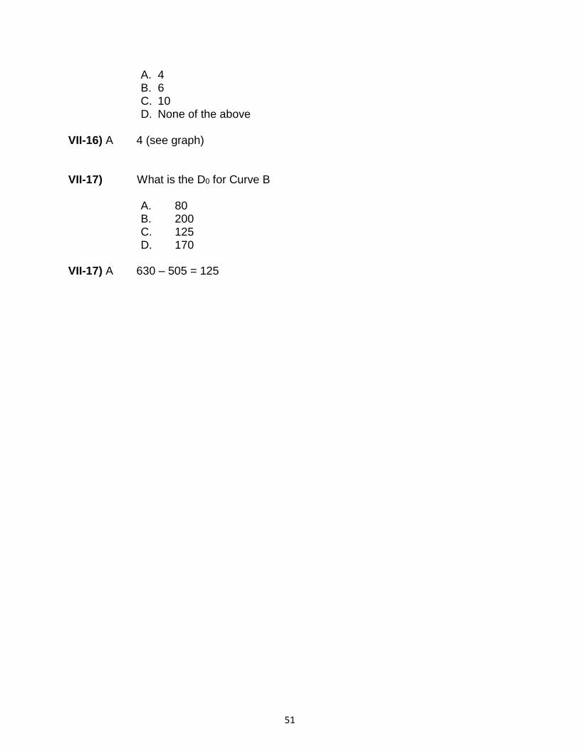
51
A. 4 B. 6 C. 10 D. None of the above
VII-16) A 4 (see graph) VII-17) What is the D0 for Curve B
A. 80 B. 200 C. 125 D. 170
VII-17) A 630 – 505 = 125

52
VIII. Linear Energy Transfer
VIII-1) Which of the following types of ionizing radiation has the highest maximum LET?
A. alpha particles B. argon ions C. 1 GeV/nucleon carbon ion D. 18 MeV/nucleon carbon ions E. 150 MeV protons
VIII-1) B Clinically-relevant 75 MeV per nucleon argon ions have an LET 250 keV/µm. 1 GeV/nucleon and 18 MeV/nucleon carbon ions have LET values of approximately 10 keV/µm and 108 keV/µm, respectively. 2.5 MeV alpha particles have an LET value of approximately 170 keV/µm. 150 MeV protons are considered low LET, with values in the range of 0.5 keV/µm.
VIII-2) The carbon ion RBE for hypoxic cells compared with that for aerated cells is:
A. equal B. lower C. greater D. dependent upon the endpoint being measured E. the same as the OER
VIII-2) C The carbon ion RBE is the dose required to produce a certain effect in X- irradiated cells divided by the carbon ion dose to produce the same biological effect. This ratio will be greater for cells irradiated under hypoxic conditions because of the much greater dose required to produce the effect in the X- irradiated cells where an oxygen effect is present, compared to the high LET irradiated cells where the oxygen effect is absent. The oxygen enhancement ratio, OER, for carbon ions would be lower than that for low LET radiations, but the absolute value of the OER is not related to the value of the RBE.
VIII-3) Which of the following statements concerning LET is FALSE?
A. The highest RBE occurs for radiations with LET values of
approximately 100 keV/m.
B. High LET radiations yield survival curves with low D0 values. C. The OER increases with increasing LET. D. High LET radiations often produce exponential survival curves.

53
E. LET is an average energy (in keV) transferred from a charged particle traversing a distance of 1 μm in the medium.
VIII-3) C OER decreases with increasing values of LET. Maximum effectiveness and therefore RBE reaches a peak for radiations whose LET is approximately 100 keV/µm. The RBE of high LET radiations is generally high, resulting in low values for D0. The survival curves resulting from irradiation of cells with high LET radiations are typically exponential. LET is the term that describes the density of ionization or the average amount of energy lost (in keV) to the medium per unit of track length (μm).
VIII-4) What is the effect on both RBE and the alpha/beta ratio as the LET
for the type of radiation increases up to 100 keV/m?
A. both remain the same B. both increase C. both decrease
D. the RBE decreases while the alpha/betaincreases
E. the RBE increases while the alpha/betadecreases Modifiers of Cell Survival: Oxygen Effect
VIII-4) B As the LET for different forms of radiation increases to about 100
keV/m, both the RBE and the alpha/beta ratio for the corresponding cell survival curves increase due primarily to an increase in the alpha parameter.
IX. Modifiers of Cell Survival
IX-1) In irradiated cells, oxygen:
A. acts as a radical scavenger by converting free radicals to non-reactive species
B. acts as a radioprotector C. reacts with hydrogen radicals to form water, thus reducing the
number of free radicals formed D. modifies the level and spectrum of free radical damage produced in
DNA E. is unlikely to play a role in the indirect effect of radiation
IX-1) D In irradiated cells, oxygen increases the number and/or type of free radicals and thereby acts as a radiosensitizer, effectively increasing the level of damage produced. Oxygen reacts with free radicals resulting in the production of different radical species, which may be longer lived, and therefore more damaging than the original radicals. For example,

54
oxygen may react with hydrogen radicals to produce peroxyl radicals. Through its reaction with free radicals formed from the radiolysis of water, oxygen plays a role in the indirect effect of radiation.
IX-2) Which one of the following statements regarding radiation and hypoxia is TRUE?
A. Irradiation under hypoxic conditions yields more DNA strand
breaks than under aerated conditions. B. Irradiation under aerated conditions leads to less overall cellular
damage than irradiation under hypoxic conditions. C. The presence of oxygen reduces radiation toxicity. D. Oxygen must be present either during or within microseconds
following irradiation to act as a radiosensitizer. E. The effect of oxygen on radiation-induced damage varies most
between 2% and 5% oxygen
IX-2) D In order for oxygen radiosensitization to be observed, oxygen must be
present either during or within microseconds following the irradiation. Irradiation under hypoxic conditions results in fewer DNA strand breaks than irradiation under aerated conditions. Irradiation in air results in more cellular damage and cell killing than irradiation under hypoxic conditions. The effect of oxygen upon radiobiologic response changes most between 0.05%-2%, with a half-maximum effect around 1%
IX-3) For large, single doses of low LET radiation, the OER is typically in the
range of:
A. 0-1 B. 1-2 C. 2-3.5 D. 3.5-5 E. 5-10
IX-3) C The OER for most forms of low LET radiation delivered acutely is in the range of 2-3.5.
IX-4) The oxygen enhancement ratio is:
A. Equal to the survival of cells irradiated under hypoxic conditions divided by the survival under aerobic conditions for a fixed radiation dose
B. Greater at low radiation doses than at high radiation doses C. the same regardless of radiation quality (LET)
55
D. equal to the dose of radiation under hypoxic conditions divided by the dose of radiation under aerobic conditions that results in the same biological effect
E. All of the above
IX-4) D The OER is calculated as a ratio of doses, not effect. It is equal to the ratio of doses under hypoxic and aerobic conditions that yields the identical level of a biologic effect. For low LET radiation this value is approximately 2.5-3, and is somewhat lower at low doses (high levels of survival). The OER decreases with radiation of increased LET, due to an increased proportion of direct DNA damage.
IX-5) Which of the following statements is true about HIF-1? A. HIF-1 is a kinase that phosphorylates p53 and ATM B. HIF-1 is turned on by hyperthermia C. HIF-1 is a transcription factor that induces VEGF and GLUT-1 D. HIF-1 is regulated by VEGF and binds to VEGF when it is hydroxylated E. HIF-1 binds to the HSE to turn on transcription of HSP70 and HSP90
IX-5) C HIF-1 or Hypoxia Indudicble Factor-1 is a transcription factor that binds to
the HRE (Hypoxia Response Element) and turns on transcription of a variety of genes including VEGF, GLUT-1, and CA9 (involved in angiogenesis, glycolysis, and metastasis, respectively). It does not have any kinase function and is not affected by hyperthermia. When hydroxylated, it binds to Von Hippel Lindau protein (VHL) which targets it for ubiquitination and degradation in the proteasome. HIF-1 does not bind to Heat Shock Element (HSE). : Fig. 16.10 in Basic Clinical Radiobiology, Fourth Edition.
Edited by Michael Joiner & Albert van der Kogel; Fig. 6.7 in Hall and Giaccia IX-6) Which of the following factors has pro-angiogenic function?
A. FGF-2 B. Endostatin C. Heparin D. IL2 E. IL1
IX-6) A FGF2 or bFGF is a proangiogenic agent that is synergistic with VEGF and
may reduce endothelial cell apoptosis; endostatin and heparin are both inhibitors of angiogenesis. IL2 and IL1 are cytokines that are not known to have an effect on angiogenesis.
The Biology of Cancer, Table 13-4, Gelfand Science, 2007

56
IX-7) What is the evidence that there are hypoxic regions in tumors in humans?
A. Nitroimidazoles bind to regions of the tumor upon histological examination.
B. Hemoglobin levels pretreatment are not good prognostic indicators for response to therapy.
C. Survival curves in vivo and in vitro are different from each other showing a single D0 dose.
D. Neutrons have not been shown to be effective in human tumors. E. Amifostine has been shown to bind to regions of the tumor that surround the necrotic regions.
IX-7) A Nitroimidazoles, which are markers of hypoxia have been shown to bind to
regions in the tumor that surround necrotic regions and demarcate hypoxia. Amifostine is a radioprotector and has no role in oxygenation/hypoxia. Survival curves in vivo and in vitro are similar to those in which hypoxia has been shown to play a role; these curves have two components, one that is more radiosensitive (the oxic region) and one that is more radioresistant (the hypoxic region). Neutrons are not influenced by oxygen to the same extent as low LET radiations, but they are not used in human tumors for a variety of reasons that have nothing to do with hypoxia. Hemoglobin levels pretreatment ARE prognostic indicators in a variety of tumors.
IX-8) On the basis of in vitro clonogenic survival assays in cancer cell lines which radiotherapeutic technique would overcome hypoxic cell radioresistance?
A. Standard fractionated intensity modulated radiotherapy B. Hadron therapy with proton beam C. Therapy with carbon ion beam D. Low dose rate brachytherapy with a beta emitting source E. Stereotactic radiosurgery with a single high dose
IX-8) A In vitro clonogenic survival assays have demonstrated that the oxygen
enhancement ratio of cancer cells exposed to ionizing radiation is greater with larger doses. In high-dose (10-30 Gy) assays, the OER has been estimate to be 3.5, where as in low dose assays (1-3 Gy), the OER has been estimated to be 2.5. Palcic B et al. Reduced oxygen enhancement ratio at low doses of ionizing radiation. Rad Res 100: 328-333, 1985 and Hall EJ Radiobiology for the Radiologist, Oxygen Effect at Reoxygenation, Chapter 6).
IX-9) Which of the following statements regarding tumor hypoxia is FALSE?

57
A. Hypoxia is associated with differentiation and apoptosis. B. Chronic hypoxia is diffusion-limited C. Tumor hypoxia is associated with nodal metastases D. Early cervical cancer patients can be assigned to prognostic groups based upon hypoxia D. Early cervical cancer patients with increased Hif-1a expression show
better outcomes IX-9) E With an increase in tumor size, there are more viable tumor cells, more
necrotic cells, and more hypoxic cells. This is primarily a function of geometry and distance from of a cell in a tumor from oxygen-rich capillaries. Necrotic cells are already dead and are therefore not a component of the biologic response to radiation therapy. Hypoxic cells are relatively more radioresistant than viable tumor cells because of the effect of the oxygen effect. Therefore, as tumors get larger and have a larger fraction of hypoxic cells, this is the likely reason for relative difference in radiosensitivity. The fraction of stem cells that make up a tumor as it increases in size is not well characterized, and may or may not be associated with radiosensitivity.
IX-10) For a given biological system, the D37 in the presence of O2 was
determined to be 2 Gy for a particulate radiation of energy A and 1 Gy for the same particle with energy B. Under hypoxic conditions, the D37 was 6 Gy for A and 1.5 Gy for B. Which of the following statements best describes the relationship between the two radiations?
A. Radiation A has a higher LET than type B. B. The OER for radiation A is 2. C. If a given dose of radiation B was delivered at a low dose rate, the
amount of cell killing would not differ markedly from that produced at a high dose rate.
D. Radiation B likely has a higher energy than radiation A.
IX-10 C The OER for radiation A is 3 and the OER for radiation B is 1.5. Therefore the LET of radiation B is higher than the LET for radiation A. Answers (A) and (B) and (D) are therfore incorrect, remembering that higher particle energies correspond with lower LET and vice-versa. Since radiation B is much higher LET than radiation A, judging by the high RBE and the much lower OER, the dose-rate effect for radiation B will be much less, or even non-existant, thus (C) is the correct answer.

58
X. Modifiers of Cell Survival: Repair
X-1) An X-ray dose of 10 Gy delivered at 1 Gy/min has a greater biologic effect than the same dose delivered at 1 Gy/day because:
A. Fewer free radicals are produced B. Apoptosis predominates as the major form of cell death when
radiation is delivered at a high dose rate C. The normal ATM-mediated inhibition of cell cycle progression is
inhibited at the higher dose rate D. Cell proliferation may occur during irradiation at the high dose rate E. There is less repair of the sublethal damage during the course of
irradiation at a high dose rate
X-1) E An X-ray dose delivered at a high dose rate results in greater cellular lethality since less repair of sublethal damage occurs during irradiation at a high dose rate. In contrast, during the course of irradiation at a low dose rate, many sublethal damages will be repaired and therefore will no longer be available to interact and form lethal damage. The fraction of cells undergoing apoptosis is primarily a reflection of the apoptotic tendency of the cell type rather than a reflection of the rate at which the dose was delivered. Activation of ATM, which in turn stimulates the production of molecules that cause inhibition of cell cycle progression, occurs regardless of whether the radiation is delivered at high or low dose rates. Cell proliferation is inhibited in cells irradiated at a high dose. Furthermore, cell proliferation would increase survival.
X-2) What would be the expected effect of a drug that inhibits repair of X-ray-induced chromosome breaks? It would:
A. Decrease the yield of terminal deletions B. Increase the dose rate effect C. Stimulate the repair of sublethal damage D. Enhance repair of potentially lethal damage E. Sensitize cells to low dose rate irradiation
X-2) E A drug that inhibits the rejoining of chromosome breaks in irradiated cells would be expected to decrease the amount of sublethal damage repair (itself a manifestation of the rejoining of chromosome breaks), and therefore, to sensitize cells to low dose rate irradiation where sublethal damage would otherwise be repaired. The repair of potentially lethal damage would also be inhibited. The yield of terminal deletions would be expected to increase as there would be less repair of chromosomal breaks. The dose rate effect, manifested as increased cell survival for irradiation at low, compared to high, dose rates, would also be diminished.
59
X-3) Generally, the sparing effect of dose fractionation increases with increasing time between fractions. Under certain irradiation conditions however, an increase in the interval between fractions results in decreased cell survival. This occurs because of:
A. Reassortment B. Repopulation C. Repair D. Reoxygenation E. Adaptive response
X-3) A Generally, increasing the time between fractions in a split dose treatment results in a higher cell surviving fraction due to repair at relatively short interfraction intervals of a few hours, or due to repopulation for longer times between fractions. However, under certain irradiation conditions and depending on the cell line, the initial dose may cause inhibition of progression from G2 into M phase. Therefore, the second dose may be delivered when the majority of the surviving cells have reassorted into G2, a radiosensitive phase of the cell cycle. Thus, even though repair of sublethal damages has occurred in these cells, which by itself would lead to a greater surviving fraction, this may be more than counterbalanced by reassortment sensitization, resulting in lower cell survival. Hypoxic conditions would not be expected for cells grown in tissue culture, so reoxygenation, which could lead to greater cell killing if it were to occur, is unlikely. The adaptive response in which cells treated with an initial low ―priming‖ dose of radiation exhibit greater resistance to a second, higher, ―challenge‖ dose, would increase, not decrease, cell survival.
X-4) Which of the following statements is TRUE concerning SLDR and PLDR?
A. As the X-ray dose rate is reduced and therefore the exposure time increases, the biological effectiveness of a given dose of radiation for killing cells increases.
B. PLDR is best demonstrated with a split dose experiment.
C. There is an inverse correlation between the alpha/beta ratio of an acute dose X-ray survival curve and the amount of SLDR in a fractionated irradiation.
D. The magnitude of PLDR and SLDR is greater following exposure to high LET compared to low LET radiation.
E. PLDR plays an important role in the decreased survival seen with fractionated
F. Irradiation to the normal lung as compared to lung cancer cells
60
X-4) C Cell lines whose X-ray survival curves have low / ratios generally display a large capacity for SLDR, whereas cells whose X-ray survival curves have high
alpha/beta ratios show relatively little SLDR. As the dose rate is lowered and exposure time increased, the biological effect of an X-ray dose diminishes due to SLDR. PLDR is best demonstrated with a ―delayed plating‖ experiment, and is operationally defined as an increase in the surviving fraction resulting from prolonged incubation of cells under non-growth conditions following irradiation. There is little or no SLDR or PLDR following exposure to high LET radiation. Fractionated irradiation would be expected to increase survival (not decrease it) in normal lung tissue compared to lung cancer cells, and this would result from SLDR, not PLDR.
X-5) Sublethal damage recovery is best demonstrated by:
A. Determining the TCD50
B. A cell synchronization experiment C. A split dose experiment D. A delayed plating experiment E. The paired survival curve technique
X-5) C Sublethal damage recovery is operationally defined as an increase in cell survival when a total dose is split into two fractions separated by a time interval compared with delivery of the same dose in one large fraction. The TCD50 is the total dose that locally controls, on average, 50% of tumors in laboratory animals, but by itself does not directly demonstrate sublethal damage recovery. A cell synchronization experiment would not demonstrate sublethal damage recovery per se, although it could be used to show variations in SLDR capacity in cells in different phases of different cell cycle. An increase in cell survival when cells are maintained under a non-growth state after irradiation is the operational definition of potentially lethal damage recovery. The paired survival curve technique is used to determine a tumor’s radiobiological hypoxic fraction.
X-6) The dose rate range over which SLDR most contributes to the dose rate effect for X-rays is:
A. 0.001 - 0.01 Gy/min B. 0.01 – 1 Gy/min C. 1 - 5 Gy/min D. 5 - 10 Gy/min E. 10-20 Gy/min

61
X-6) B As the dose rate decreases from about 1 Gy/min to 0.01 Gy/min, the greatest increase in cell survival due to SLDR is observed for most X-irradiated cell lines. Decreasing the dose rate further may permit an even greater increase in the surviving fraction, but this further increase would be due to repopulation that may take place if the dose is delivered at a very low dose rate over a long interval.
X-7) Chinese hamster cells were irradiated with 10 Gy X-ray dose given singly or with 10 Gy divided into two 5 Gy fractions separated by 5 hrs at 24 degree C. The surviving fraction after 5 Gy was 0.08. The survival fraction after a single dose of10 Gy was 0.002. The recovery factor is approximately equal to:
A. 0.16 B. 0.025 C. 4 D. 3 E. 6.25
X-7) D The recovery factor is the ratio of the surviving fraction resulting from two-
dose fractionation (5 Gy + 5 Gy) to the survival from a single total dose (10 Gy). If cells surviving a first dose of 5 Gy completely repaired their sublethal damage, they would survive the second exposure according to the original single dose survival curve. Hence, the survival to 5 Gy plus 5 Gy with “full” recovery between exposure would be 0.08 x 0.08 = 0.0064. The recovery factor is then equal to 0.0064/0.002 or approximately 3.

62
X-8) The survival curve is characterized by the extrapolation number (N) of 5
and the quasithreshold dose (Dq) of 2 Gy. The final slope D0 calculated
from these Dq and N values is 1.2 Gy. The cells were first irradiated at 5
Gy X-rays. The preirradiated cells at 5 Gy were next irradiated with graded X-rays doses (0 Gy to 8 Gy) after holding the cells for 6 hrs at 37 degree C. What are the parameters of the fractionated survival curve?
A. The parametrs of the fractionated survival curve and the parameters of
the single-dose survival curve of cells will be statistically the same because 6-h is long enough for cells to fully recover after the 5 Gy exposure. .
B. N = 5, but Dq < 2 Gy and D0 < 1.2 Gy, because of incomplete repair
between fractions.
C. N > 5, Dq > 2 Gy and Do > 1.2 Gy, because of the population of cells
surviving the first dose will be more radiation resistant than the starting

63
population.
D. N > 5, Dq = 2 Gy, Do = 1.2 Gy, because the fractionated survival
curve is not normalized to the survival resulting from the first 5 Gy exposure.
E. Do = 1.2 Gy, but N and Dq cannot be determined because the effect
of cell progression (growth and division) during a 6-h recovery period was not quantified.
X-8) D Diagrammatic representation of the relationship between the parameters related to cell survival and recovery, and the doses is shown in the above figure (modified from GH Fletcher Editor: Textbook of Radiotherapy 3rd edition. Philadelphia, Lea & Febiger, 1980, p.2 and from Power Show.com). If sublethal damage is fully repaired, then the slope of the second-dose survival curve (survival curve B) is the same as that for the single-dose survival curve (survival curve A. The first dose D1 = 5 Gy has killed a proportion of cells (~ 92% in this example) but surviving cells respond as if they had never been irradiated. The survival parameters Do and Dq of curves A and B are the same. The split-dose survival curve B starts at the
survival level corresponding to 5 Gy (5 x 𝑒−5/1.2 or 0.078 in this example) and is displaced to the right by Dq = 2 Gy relative a single dose curve A.
The apparent extrapolation number of split-dose curve B (denoted n’) is
greater than the extrapolation number of the single-dose curve A. Note that however, when the second-dose survival curve B is normalized to the survival corresponding to D1 = 5 Gy (0.078 by preceding calculations), as shown in the figure, curve B becomes statistically indistinguishable from the single dose survival curve A of cells which had no previous radiation.
Elkind MM, Sutton H, X-ray damage and recovery in mammalian cells in culture. Nature 184:1293-1295, 1959. Elkind MM, Sutton H, Radiation response of mammalian cells grown in culture. I. Repair of X-ray damage in surviving Chinese hamster cells, Radiat Res 13: 556-593, 1960.
X-9) The X-ray survival curve is characterized by the quasithreshold dose (Dq) of 3 Gy in a target model description or the alpha/beta ratio of 3 Gy in the linear-quadratic model description. Cells were kept at 37 degree C during protracted exposures. Which of the following experimental approaches is best to demonstrate sublethal damage repair in this cell line?
A. Use two 2 Gy fractions separated by 6 h. B. Use two 5 Gy fractions separated by 6 h. C. Irradiate the cells at 1 Gy/min.

64
D. Use 0.5 Gy of α-irradiation followed by 5 Gy of X rays with 6 h interval between the two doses. F. Use two 5 Gy fractions separated by 6 hrs and stationary cultures
instead of proliferating cultures. X-9) B Sublethal damage repair is best demonstrated in split-dose experiments
using “off shoulder” doses (greater than Dq), because in the shoulder region most damage is sublethal. Using the linear-quadratic description with alpha/beta= 3 Gy, 60% of damage is irreparable and 40% of damage is repairable at 2 Gy, whereas 40% of damage is irreparable and 60 % of damage is reparable at 5 Gy. This means that the amount of reparable damage increases with dose. Cells which survive a pre-exposure to α-particles do not suffer any sublethal damage and a first exposure to α-particle does not alter the response of surviving cells to subsequent doses of X-rays. A reduction in cell killing (or an increase in cell survival) due to repair of sublethal damage) require much lower dose rates of about 0.01 Gy/min. The amount of sublethal damage and repair kinetics are similar in proliferating (log-phase) versus stationary (lag-phase) cultures. This means that repair of sublethal damage does not depend on proliferation.
65
XI. Solid Tumor Assay Systems
XI-1) Tumor-bearing mice are randomized into a control group and groups treated with localized irradiation of the tumor alone, an anticancer drug alone, or radiation in combination with the drug. Which of the following represents the most rigorous, reliable and informative approach to comparing the effectiveness of the different treatments?
A. Killing the mice at a predetermined time after treatment, removing
and weighing the tumors, and calculating the ratio of the volumes of the treated and control tumors
B. Measuring three diameters of the tumors with calipers at a predetermined time after treatment, calculating the volume and computing the ratio of the volumes of the treated and control tumors
C. Measuring the tumors 3 times per week until the treated tumors return to their pre- irradiation volume and calculating the mean time needed for each group to reach that volume
D. Measuring the tumors 3x per week until the control tumors reach 4 times the volume at the time of treatment, and comparing the mean volume of the tumors in each treatment group at that time
E. Measuring the tumors 33 times per week until each tumor reaches 4 times the volume at the time of treatment and calculating the mean time needed for the tumors in each group to reach that volume
XI-1) E The tumor regrowth delay assay measures the average time necessary for a treated tumor to reach a pre-determined size compared to the time it takes for control tumors. Of the assays listed, the tumor regrowth assay is most informative as to the effect of radiation and/or drug treatment. The techniques described in A and B are still used to measure the relative effectiveness of experimental chemotherapy drugs, however they provide only very limited information. The technique described in C is effective only if the treated tumors shrink immediately and dramatically following treatment, but is less effective with agents such as radiation that produce a delayed cell death or with drugs that have cytostatic effects, because these agents will not produce rapid and sizeable shrinkage of the tumors. The approach described in D may not allow meaningful comparisons between the different treatment groups if some or all treatments have been relatively successful and tumors in several groups have not even begun to regrow by the time the control tumors become large.
XI-2) For a group of tumors identical in size and homogeneous with respect to cellular radiosensitivity, what would be the general shape of the

66
curve in a linear-linear graph defining the increase in tumor control probability with increasing radiation dose?
A. Step function from 0 – 100% at the dose that kills all of the cells B. Linear increase from 0 – 100% over a narrow range of doses C. Logarithmic increase from 0 – 100% over a wide range of doses D. Sigmoidal increase from 0 – 100% over a narrow range of doses E. Exponential increase from 0 – 100% over a narrow range of doses
XI-2) D Radiation-induced cell killing is random, and the probability follows a
Poisson distribution; a tumor will be controlled only when no clonogenic cells remain. The dose at which a specific tumor is controlled will be determined by the probability of killing the last clonogenic cell in that tumor. However, this will not be the same for each tumor because of the random nature of radiation damage and of cell death. The result of this, statistically, is that the tumor control probability plotted as a function of dose on a linear scale will yield a steep, sigmoid-shaped curve that reflects only the random variation in the dose needed to kill the last clonogenic cell in the tumor. Heterogeneity between the tumors (e.g., differences in size/cell number) or heterogeneity within the tumor cell population (e.g., heterogeneity in the radiosensitivity of the cells because of their position in the cell cycle, oxygenation status, or genotype) would broaden the dose range over which the sigmoidal increase in tumor control probability occurred, and the resulting tumor control probability curve would be shallower.
XI-3) The following graph shows data for the percent of local tumor control by different doses of radiation therapy. Based upon the data provided in this graph, which of the following statements is correct?

67
A. TCD50 is 70 Gy B. NTCP50 is 60 Gy C. The additional dose required to increase the probability of tumor
control from 50 to 60% is larger than the dose required to increase the probability of tumor control from 90 to 100%
D. The impact of a radiosensitizer upon tumor control will be most readily detected for experimental protocols that evaluate TCD50.
E. TCD50 is 40 Gy
XI-3) D The TCD50 is the dose resulting in 50% cure or control rate. The
impact of a radiosenisitizer upon tumor control will be most readily detected for experimental protocols that result in a 50% rate of tumor cure since even a small level of sensitization will significantly decrease the TCD50 in this steep portion of the curve. This curve demonstrates that 50% of the tumors will be controlled by a dose of 60 Gy (ie. TCD50 = 60 Gy), whereas 70 Gy increase tumor cure to >90%. NTCP stands for normal tissue complication probability and is not indicated on this graph. The additional dose to increase cures from 50 to 60% is relatively small because the TCD50 is on the steep part of the dose-response curve, but to increase cure rates from 90 to 100%, a much larger dose is required. At 50 Gy, approximately 10% of the tumors will be controlled.

68
XII. Tumor Microenvironment
XII-1) Which of the following statements concerning the tumor microenvironment is true?
A. Hypoxia is found primarily at the core of large tumors.
B. Cellular oxygenation status in solid tumors is expected to remain relatively constant over a 24 hr period. C. Tumors of a similar size have similar hypoxic fractions. D. Acute changes in blood flow contribute to tumor hypoxia. E. Hypoxic tumors are resistant to irradiation due to expression of HIF1.
XII-1) D Acute changes in blood flow cause acute, or perfusion limited hypoxia. This can be partial or total occlusion of blood vessels that causes hypoxia in cells being fed by the vessel in question. Acute hypoxia can change over a period of 15 min- 2 hrs and will thus alter overall tumor oxygenation during a period of 24 hrs between radiation fractions. Hypoxia can be distributed throughout the entire tumor mass, and can potentially be present surrounding any blood vessel in the tumor. No relationship has been observed between tumor hypoxia and tumor size. Tumor hypoxia varies widely amongst patient tumors and is thought to be an important contributor to the overall variation in response to therapy.
Cao Y. Off-tumor target –beneficial site for antiangiogenic cancer therapy? Nat Rev Clin Oncol,
7:604-609, 2010. PubMed link
Dewhirst MW, Cao Y, Moeller B. Cycling hypoxia and free radicals regulate angiogenesis and radiotherapy response, Nature Reviews Cancer, 8, 425-437, 2008. PubMed link
XII-2) Tumor hypoxia has been specifically associated with all of the following, EXCEPT:
A. Reduced radiosensitivity B. Large tumors C. Increased genomic instability D. Poor patient prognosis E. Increased metastasis
XII-2) B Clinical studies indicate that hypoxia does not show a strong relationship with tumor size. This is due to the fact that hypoxia arises through deficiencies in blood vessel perfusion and from a limited ability to diffuse through metabolically active tissue. Hypoxia is observed at distances from 100-200 microns from functional vessels. Thus, even very small tumors may have high hypoxic fractions. Hypoxic cells are radioresistant and exhibit genomic instability and increased probability of metastasis. Each of these factors translates into a worse patient prognosis.

69
Bristow RG, Hill RP. Hypoxia and metabolism. Hypoxia, DNA repair and genetic instability, Nat Rev Cancer, 8:180-92, 2008. PubMed link
XII-3) In a respiring tissue, the maximum diffusion distance of oxygen from a capillary is:
A. 1-2 m
B. 100-200 m C. 1-2 mm D. independent of cellular respiration rate E. independent of hemoglobin concentration
XII-3) B The maximum distance that oxygen can diffuse from a capillary before hypoxia is
detected is roughly 150 m and is determined by the oxygen consumption rate of cells surrounding blood vessels. This distance also depends on the starting concentration in the blood, which can be influenced by hemoglobin levels. An increase in oxygen utilization would reduce the diffusion limit.
XII-4) Which of the following statements concerning tumor hypoxia is FALSE?
A. Chronically hypoxic cells are generally more radiation resistant than acutely hypoxic cells
B. For a tumor containing only 1% radiobiological hypoxic fraction, essentially all hypoxic cells would be eliminated by the end of a typical course of radiotherapy, even in the absence of any reoxygenation
C. Hypoxic regions in tumors may be detected using pimonidazole
D. Regions of acute hypoxia may develop in tumors due to the temporary closing/blockage of a blood vessel
E. Reoxygenation during fractionation in radiotherapy reduces the influence of hypoxic cells on tumor response.
XII-4) B Without reoxygenation, it is unlikely that a tumor comprised of any significant proportion of hypoxic cells (even as low as 1%), would be controlled following total doses used in typical radiotherapy protocols.
XII-5) HIF-1 activity is increased primarily during hypoxia as a consequence of:
A. Increased transcription of HIF-1α B. Reduced stability of HIF-1beta C. Increased turnover of HIF-1 α D. Reduced hydroxylation of HIF-1α E. Increased activity of VHL
XII-5) D Under normoxic conditions, HIF-1α is hydroxylated in a reaction that uses oxygen as a co-factor. When hydroxylated HIF-1α is recognized by VHL and degraded.

70
During hypoxia, hydroxylation is prevented and HIF-1α becomes stabilized and active as a transcription factor.
XII-6) With regard to the radiobiological influence of oxygen, which of the following statements is FALSE?
A. Reoxygenation of human tumors during fractionated radiation therapy reduces
the impact of both chronically and acutely hypoxic cells on overall response. B. Metabolically-active hypoxic cells in human tumors can be identified through
preferential binding of administered nitroimidazole compounds. C. Hypoxia induces pro-angiogenic factors such as VEGF. D. HIF-1alphais stabilized under hypoxic conditions and dimerized with
constitutive HIF-1beta. E. The oxygen enhancement ratio (OER) for X-rays is higher for doses < 2 Gy
than for doses > 10 Gy.
XII-6) E The oxygen enhancement ratio (OER) for X-rays is lower (~1.5-2.0) for X-ray doses <2 Gy and higher (~3.0) for doses >10 Gy. Chronic hypoxia develops in regions of a tumor distant from blood vessels, which is mainly due to the limited diffusion distance of oxygen. Acute hypoxia is associated with transient changes in vascular perfusion that could be due to either temporary blockage in a blood vessel, vascular status or other factors. Reoxygenation of both chronically and acutely hypoxic cells has been demonstrated in experimental tumors, however, both the mechanisms and the time course of reoxygenation are different for chronic and acute hypoxia. Hypoxia plays a role in tumor control through the induction of hypoxia-inducible factor alpha (HIF-1alpha) (HIF1A). HIF-1alpha is degraded under well-oxygenated conditions, but is stabilized under hypoxic conditions. In hypoxia, stabilized HIF-1apha dimerizes with constitutively expressed HIF-1beta (ARNT) to form a transcription factor that regulates expression of pro-angiogenic genes, including that for vascular endothelial growth factor (VEGF).
Dewhirst MW. Relationships between cycling hypoxia, HIF-1, angiogenesis and oxidative
stress, Radiat Res, 172:653-665, 2009. PubMed link
Kaelin WG Jr. The von Hippel-Lindau tumour suppressor protein: O2 sensing and cancer. Nat Rev Cancer, 8:865-873, 2008.PubMed link
Bertout JA, Patel SA, Simon MC. The impact of O2 availability on human cancer, Nat Rev Cancer, 8:967-975, 2008. PubMed link
Bristow RG, Hill RP. Hypoxia and metabolism. Hypoxia, DNA repair and genetic instability, Nat Rev Cancer, 8:180-192, 2008. PubMed link
Dewhirst MW, Cao Y, Moeller B. Cycling hypoxia and free radicals regulate angiogenesis and radiotherapy response, Nat Rev Cancer, 8:425-437, 2008. Erratum in: Nat Rev Cancer, 8:654, 2008. PubMed link

71
Galanis A, Pappa A, Giannakakis A, Lanitis E, Dangaj D, Sandaltzopoulos R. Reactive oxygen species and HIF-1 signalling in cancer, Cancer Letters, 266:12-20, 2008. PubMed link
XII-7) Which of the following markers and imaging approaches would be least useful for measuring tumor hypoxia non-invasively?
A. [18F]-fluorodeoxyglucose (FDG) – PET
B. [18F]-fluoromisonidazole (FMISO) – PET
C. [123I] radioiodinated azomycin arabinosides – SPECT
D. [64Cu]-Cu-ATSM – PET
XII-8) Which of the following statements concerning hypoxia is TRUE?
A. Hypoxic cell radiosensitizers produce a greater increase in the therapeutic index when used with conventional fractionated radiotherapy than for treatment with one or a few large radiation doses
B. A biphasic survival curve would result from low LET irradiation of a mixed population of both aerated and hypoxic cells
C. The OER is defined as the dose to produce a given effect in aerated cells divided by the dose to produce the same effect in hypoxic cells
D. The diffusion distance of oxygen in air is typically less than 100 µm E. For low LET radiation, the maximum OER is typically observed only when the
tissue oxygen concentration reaches about 20% XII-8) B A breaking survival curve in which the survival curve initially displays a steep
slope followed by a shallower response would be predicted for irradiation of a mixed population of aerated and hypoxic cells. This type of dose response would be anticipated since the initial portion of the survival curve reflects the killing of radiosensitive aerated cells whereas the survival curve obtained at higher doses primarily reflects the killing of radioresistant hypoxic cells. A fractionated protocol would be expected to decrease the effect of a hypoxic cell radiosensitizer to enhance tumor control compared with an acute treatment since reoxygenation would cause aeration of many of the hypoxic cells between fractions thereby diminishing the apparent effectiveness of the radiosensitizer. The OER is the dose to produce an effect in hypoxic cells divided by the dose to produce the same effect in aerated cells. The diffusion distance in air for oxygen is much greater than 100 µm. Maximum OER is typically observed only when the oxygen concentration reaches about 3-5%.
XII-9) Which of the following would best be used to estimate the proportion of radiation resistant viable hypoxic cells in an experimental tumor model?
A. Comparison of radiation response with and without breathing of hyperbaric oxygen
B. Paired survival curve analysis in vitro following irradiation in vivo under standard conditions and conditions where blood flow to the tumor has been stopped

72
C. Extrapolation of the initial exponential portion of the cell survival curve for cells comprising the tumor
D. Comparison of radiation responses with and without misonidazole administration
XII-9) B The paired survival curve technique is used to determine the proportion of viable clonogenic cells in a tumor that is hypoxic. In this assay, animals possessing tumors are irradiated while breathing either room air (typical tumor response), or where they are clamped to block blood flow. The ratio of the surviving fractions for the cells under aerated to fully anoxic conditions provides an estimate of the fraction of the cells in the tumor that are hypoxic under normal conditions.
XII-10) Stereotactic body radiation therapy (SBRT) has proven to be a highly effective local treatment for a variety of cancer histologies. Proposals to further optimize SBRT efficacy include the concurrent use of hypoxic cell
radiosensitizers. The most correct answer below describes the:
A. Conventional fractionation of radiation greatly mitigates the protection
afforded by tumor hypoxia because of the phenomenon of reoxygenation which could be further augmented with a hypoxic cell radiosensitizer.
B. Tumor hypoxia is a major negative factor in limiting the curability of tumors by SBRT due to loss of the phenomenon of reoxygenation and this negative effect of hypoxia could be overcome by the addition of a hypoxic cell radiosensitizer.
C. Hypoxic cell radiosensitization with SBRT causes acute damage to the endothelial cells of the tumor vasculature.
D. Only a small proportion of tumor cells are clonogenic cancer stem cells and these could be preferentially killed by a hypoxic cell radiosensitizer.
E. An active immune response is needed to eradicate microscopic residual tumor following SBRT that could be augmented by a hypoxic cell radiosensitizer.
XII-10) B Answer “B” is the most correct which is detailed nicely in ref. 20832663. Answer “A” is true, but SBRT is defined by 1-5 fractions so this answer does not apply well to the question. SBRT is thought to cause acute damage to endothelial cells of the tumor vasculature (ref. 1275052) with or without a hypoxic cell radiosensitizer. Cancer stem cells (CSCs) have been hypothesized to reside in hypoxic regions (ref. 19249645), but hypoxic cell radiosensitizer targeting of CSCs has not been tested thoroughly. Answer “E” is not correct.
XII-11) Which of the following statements best describes the “abscopal effect”?
A. Localized irradiation of a tumor causes not only a shrinking of the irradiated tumor, but growth of tumors far from the irradiated area
B. Localized irradiation of a tumor causes not only a shrinking of the irradiated tumor, but also a shrinking of tumors far from the irradiated area
73
C. When cells are irradiated and the medium is transferred to unirradiated cells, these unirradiated cells show increased clonogenic survival
D. The avid response of irradiated low-grade lymphomas to low-dose radiotherapy such as 2 Gy x 2
E. Ability of transformed cells to transfer death signals to neighboring tumor cells
XII-11) B The “abscopal effect” is a phenomenon in the treatment of metastatic cancer where localized irradiation of a tumor causes, not only a shrinking of the irradiated tumor, but also a shrinking of tumors far from the irradiated area. While this phenomenon is extremely rare, its effect on the cancer can be stunning, leading to the disappearance of malignant growths throughout the entire body. Such success has been described for a variety of cancers, including melanoma, lymphoma, and kidney cancer. Studies in mice suggest that the effect may depend upon activation of the immune system (Refs. 4706791 and 22397654). Answers “C” and “E” are the bystander effect.
XII-12) Which of the following targeted agents is an immune checkpoint inhibitor?
A. Bevacizumab B. Imatinib C. Crizotinib D. Ipilimumab E. Cetuximab
XII-12) D Antitumor immunity is often ineffective due to the tight regulation associated with the maintenance of immune homeostasis. One of the major limitations results from chronic exposure to antigens and is characterized by the upregulation of inhibitory immune checkpoint receptors in order to prevent uncontrolled immune reactions. Blocking of one or several of these immune checkpoints with monoclonal antibodies (mAbs) has been shown to rescue otherwise exhausted antitumor T cells, and most importantly, has been associated with objective clinical responses in cancer patients. The first immune-checkpoint inhibitor to be tested in a clinical trial was ipilimumab (Yervoy, Bristol-Myers Squibb), an anti-cytotoxic T-lymphocyte antigen 4 (CTLA-4) mAb. CTLA-4 belongs to the immunoglobulin superfamily of receptors, which also includes programmed cell death protein 1 (PD-1), B and T lymphocyte attenuator, T-cell immunoglobulin and mucin domain-containing protein 3 (TIM-3), and V-domain immunoglobulin suppressor of T cell activation. In 2011, the US Food and Drug Administration approved the use of ipilimumab in patients with metastatic melanoma, either as initial therapy or after relapse (Refs. 24161671). Bevacizumab is the humanized monoclonal antibody that inhibits vascular endothelial growth factor A (VEGF-A). Imantinib is a small molecular inhibitor of receptor tyrosine kinases most selective for Bcr-Abl, but also less so against c-kit and PDGF-R. Cetuximab is a monoclonal antibody against EGFR. Crizotinib is a small molecular inhibitor of ALK and ROS1 kinases.

74
XIII. Cell and Tissue Kinetics
XIII-1) Which of the following statements is FALSE concerning the cell cycle?
A. Irradiation of cells causes a delay in progression from G1 into S phase of the cell cycle
B. Cells in M phase typically have X-ray survival curves with low alpha/beta ratios
C. Cells are most resistant in late S phase of the cell cycle
D. G1 is the cell cycle phase most variable in duration
E. The G0 phase of resting cells is within G1
XIII-1) B Mitotic cells generally possess little or no survival curve ―shoulder‖ and therefore
are characterized by high /ratios.
XIII-2) The diameter of a tumor was found to double in 18 days. Assuming that all of the cells in the tumor are proliferating and no cells are lost, the tumor cell doubling time is closest to:
A. 1 day B. 3 days C. 6 days D. 12 days E. 18 days
XIII-2) C Assuming that all cells are proliferating, the number of cells in a tumor that doubled in diameter would increase approximately 8-fold as the number of cells
can be approximated from the volume of a sphere which is equal to d3/6. An 8- fold increase in the cell number would require three cell doublings. Since it took 18 days to achieve this increase, the cell cycle time can be estimated at 6 days.
XIII-3) The CDK1/cyclin B complex plays an important role in the transition of cells from:
A. G0 into G1 B. G1 into S C. S into G2 D. G2 into M E. M into G1
XIII-3) D The CDK1/Cyclin B complex plays an important role in the transition of cells from G2 phase into mitosis. Cyclin B binds to cyclin-dependent kinase (CDK) 1. The resulting complex becomes active during the prophase stage of mitosis. It is involved in centromere separation as well as other mitotic events. Once activated, the CDK1/Cyclin B complex also inactivates the molecules which inhibit its expression, allowing a large amount CDK1/Cyclin B to become active quickly. CDK1/Cyclin B is destroyed by the anaphase promoting complex (APC).

75
XIII-4) Which of the following statements is TRUE concerning the cell cycle kinetics of human tumors?
A. The growth fraction of a tumor represents the proportion of cells associated
with tumor recurrence after treatment B. Cell loss is often the major factor that determines the tumor volume doubling
time C. The growth rate generally increases with increasing tumor size D. Volume doubling times are shorter than the value that would be predicted
from the cell cycle time of individual cells E. The volume doubling time is largely determined by the cell cycle time
XIII-4) B Cell loss is often the main factor that determines the tumor volume doubling time
since tumors with a low cell loss factor will grow more rapidly than tumors with a high cell loss factor. The growth fraction of a tumor is the number of proliferating cells in a tumor divided by the number of proliferating and quiescent cells in the tumor. The growth rate generally decreases with increasing tumor size. Volume doubling times are longer than the value that would be predicted from the cell cycle time of individual cells because the growth fraction is usually less than one and the cell loss factor may be large. The volume doubling time is a reflection primarily of the growth fraction and cell loss factor.
XIII-5) A tumor is characterized by a cell cycle time of 10 days, a growth fraction of 0.5 and a cell loss factor of 1.0. Assuming these kinetic parameters remains constant over a one month-period, how much would the tumor volume have increased during that time?
A. Increase 2-fold
B. Increase 3-fold
C. Increase 4-fold
D. Increase 5-fold
E. Remain the same
XIII-5) E The cell loss factor since the cell loss factor is equal to 1.0, the tumor would remain the same size since for every new cell produced, one existing cell would die.
XIII-6) The Tpot for a tumor can be calculated from the cell cycle time of the cells comprising the tumor, the tumor’s growth fraction and with the assumption that the cell loss factor is:
A. 0 B. 1.) C. 0.2 D. 0.6
76
E. Nearly 1.0 when the tumor is small, but decreasing exponentially as the tumor grows
XIII-6) A Tpot represents the time it would take a tumor to double its cell number in the
absence of cell loss (i.e., phi= 0).
XIII-7) The Tpot for most head and neck tumors is in the range of:
A. 1-2 days B. 2-6 days C. 6-24 days D. 24-100 days E. Greater than 100 days
XIII-7) B The Tpot for most head and neck tumors is in the range of 2-6 days.
XIII-8) If the TS, LI and lambda (the correction factor for the non-linear distribution of cells through the cell cycle) were determined for a tumor to be 10 hours, 0.2 and 0.7, respectively, then the Tpot is:
A. 2 hours B. 10 hours C. 18 hours D. 25 hour E. 35 hours
XIII-8) E The Tpot is equal to lambda TS /LI = (0.7)(10 hours)/0.2 = 35 hours
XIII-9) Two patients are diagnosed on the same day with tumors of approximately the same size. However, the Tpot for patient A’s tumor was determined to be 5 days while the Tpot for patient B’s tumor was calculated as 20 days. Assuming that there was no cell loss taking place and the tumor’s growth fractions did not change, if treatment had been initiated 20 days earlier, the ratio of the number of cells in the tumors of patient A to patient B would have been approximately:
A. 16:1 B. 8:1 C. 1:1 D. 1:8 E. 1:20
XIII-9) D If the treatment had been initiated 20 days earlier, then the number of cells in the tumor in patient B would have been one-half of the number of cells present when the cancer was diagnosed. In contrast, the tumor in patient A is growing more rapidly with a Tpot of 5 days. Therefore, if diagnosed 20 days earlier, the cancer in this patient would be only one-sixteenth as many cells. Thus, the ratio of the

77
number of cells in the tumors in patients A and B would be 1:8, if the cancers had been diagnosed 20 days earlier.
XIII-10) The most likely explanation for why a tumor composed of cells with short cell cycle times would have a long volume doubling time, is a:
A. high cell loss factor B. large percentage of cells entering G0 following mitosis C. low growth fraction D. large hypoxic fraction E. abnormally long S phase
XIII-10) A Although a low growth fraction would contribute to a long volume doubling time, the most likely reason why a tumor made up of cells with a short cell cycle time would grow slowly is most likely due to a high cell loss factor.
XIII-11) The volume doubling time (in days) for a tumor with a cell loss factor of 90 and a Tpot of 20 days would be estimated as:
A. 5 B. 20 C. 50 D. 100 E. 200
XIII-11) E The volume doubling time can be estimated from the equation phi= 1-(Tpot/Tvol) where phi is the cell loss factor, Tpot is the potential doubling time and Tvol is the measured volume doubling time. Therefore, 0.9 = 1 - (20 days/Tvol) or Tvol = 200 days.
XIII-12) Which of the following proteins does NOT participate in the p53 pathways involved in cell cycle regulation?
A. SMC1 B. CAK C. P21 (CDKN1A) D. GADD45A E. 14-3-3-sigma
XIII-12) A SMC1 is a substrate for ATM and plays a role in regulation of progression through S phase. It is not part of the p53 pathway. In contrast, p53 regulates p21, which in turn associates with CDK1/cyclin B complexes and inhibits their phosphorylation by CAK or cyclin activating complex. GADD45 is a p53 target gene whose product binds CDK1, preventing cyclinB/CDK1 complex formation. 14-3-3sigma is induced by p53 and plays a role in G2 arrest.
Hurley PJ, Bunz F. ATM and ATR: components of an integrated circuit, Cell Cycle, 6:414-7,

78
2007. PubMed link
Kitagawa R, Kastan MB. The ATM-dependent DNA damage signaling pathway, Cold Spring Harb Symp Quant Biol, 70:99-109, 2005. PubMed Link
Lukas J, Lukas C, Bartek J. Mammalian cell cycle checkpoints: signalling pathways and their organization in space and time, DNA Repair (Amst) 3:997-1007, 2004. PubMed Link
XIII-13) The cell cycle time of cells in a tumor is 2 days and 20% of cells are proliferating. What is the potential doubling time, assuming no cell loss?
A. 20 days B. 10 days C. 6 days D. 1 day E. 11 days
XIII-13 B The potential doubling time is the time taken for the total population of cells
comprising a tumor to double in size. The total population is the sum of the number
of proliferating and quiescent cells. In the absence of cell loss, Tpot = 𝑇𝑐
𝐺𝐹, where Tc is
the average time taken by the cell to traverse the complete cell cycle, and GF is the fraction of all cells moving through the cell. With Tc = 2 d and GF=0.2, Tpot = 10 days.
XIII-14) The cell cycle checkpoints are controlled by a group proteins which (in general) inhibit the production or function of the cyclin-dependent kinases.
Which of the following cyclin dependent kinase inhibitors block transition from the quiescent phase to the proliferative phase of the cell cycle?
A. p21Cip1 (CDKN1A) B. p27Kip1 (CDKN1B) C. p57Kip2 (CDKN1C) D. p68INK2 (CDKN2A) E. p19INK4 (CDKN2D)
XIII-14 E The cyclin-dependent kinase inhibitor (CDKI) p19INK4 is involved in the regulation of quiescence by inhibition G0 to G1 transition. INK4 (inhibitor of cyclin dependent kinase CDK4) is a member of the INK family of CDKIs (p16INK4a, p15INK4b, p18INK4c, and p19INK4) bind and inhibit cyclin D-associated kinases (CDK2, -4, and -6). The CIP/KIP family (p21Cip1, p27Kip1, and p57Kip2) family, inactivate the cyclin A/CDK1 (also known as CDK2), cyclin E/CDK2, cyclin B/CDK1complexes. These complexes regulate each step of the cycle. Thus, cyclinD/CDK2, -4, -6 drive progression through G1; cyclin E/CDK2 controls entry into S phase; cyclin A/CDK1 (also known as CDK2) controls G2; and CDK1/cyclin B facilitates mitosis. The p68INK2 does not exist.

79
Malubresh M, Barbacid M. Cell cycle, CDKs and cancer: a changing paradigm. Nature Reviews Cancer 9153-166, 2010. Teixeira LK, Reed SI. Ubiquitin ligases and cell cycle control. Annu Rev Biochem 82: 387-414, 2013.

80
81
XIV. Molecular Signaling
XIV-1) Mutations in growth factor receptors are common alterations in cancer that may:
A. Signal cancer cells to enter senescence B. Directly inhibit protein translation C. Result in constitutive kinase activity that signals cells to proliferate D. Stimulate ubiquitination of caspase 3 to induce apoptosis E. Lead to enhanced cell death
XIV-1) D Growth factor receptors generally have three domains: an extracellular ligand- binding domain, a trans-membrane domain that spans the plasma membrane of the cell, and an intracellular kinase domain. Mutations can occur in all three domains in ways that contribute to cancer development. The resultant changes in the protein often lead to constitutive kinase activity, which signals the cancer cell to proliferate, not to undergo senescence. Kinases are proteins that attach phosphate groups to other molecules. Such receptor mutations have not been shown to stimulate general protein translation, cause DNA damage that would stimulate formation of gamma H2AX foci or affect caspase 3, which is normally activated by cleavage, not ubiquitination, to cause apoptosis.
XIV-2) Mutations in the Ras proto-oncogene have been associated with what common
human malignancy?
A. Head and neck tumors. B. Cervical cancers C. Pancreas tumors D. Sarcomas E. Lymphomas
XIV-2) C Human pancreas tumors commonly have a mutation in either amino acid 12 or 13
and these mutations are present in 95% of pancreas malignancies and roughly 35% of lung adenocarcinomas.
XIV-3) The transcriptional activity of the tumor suppressor p53 has been shown to be regulated by all of the following, EXCEPT:
A. Phosphorylation of p53 (TP53) by ATM B. Changes in the subcellular localization of p53 C. Changes in the ubiquitination of MDM2 D. Binding of FAS ligand (FASLG/CD95-L) to FAS (CD95/APO-1)
XIV-3) D All of the processes listed, except binding of FAS ligand to FAS receptor on the plasma membrane, have been associated with p53 activation. Binding of FAS ligand to FAS receptor activates the extrinsic pathway to apoptosis, which does

82
not appear to involve p53.
XIV-4) RAS functions as a:
A. GTPase B. Protein kinase C. Phosphatidyl inositol kinase D. Phosphatase E. Transcription factor
XIV-4) A RAS is a GTPase. Vigil D, Cherfils J, Rossman KL, Der CJ. Ras superfamily GEFs and GAPs: validated and
tractable targets for cancer therapy?, Nature Reviews Cancer 10:842-857, 2010. PubMed link
Schubbert S, Shannon K, Bollag G. Hyperactive Ras in developmental disorders and cancer, Nat Rev Cancer, 7:295-308, 2007. PubMed link
XIV-5) Which one of the following is NOT a part of the RAS pathway that stimulates cell proliferation following irradiation?
A. RAF1 B. MEK C. MAPK (ERK) D. FADD
XIV-5) D FADD (FAS-associated death domain) protein plays an important role in the extrinsic apoptotic pathway through activation of caspase 8. Activated RAS stimulates cellular proliferation through activation of multiple pathways including the RAF, MEK, JNK, RAC/RHO, PLC and PI3K/AKT pathways.
XIV-6) Epigenetic modification of DNA-associated histones can occur through all of the following mechanisms, EXCEPT:
A. Phosphorylation B. Acetylation C. Glycosylation D. Methylation
XIV-6) C Epigenetic regulation of genes can occur at the level of the histone proteins intimately associated with the DNA. Modification of the histones that surround the DNA can lead to complex signaling that directs the packing and unpacking of the DNA double helix. Epigenetic regulation of histones can occur through acetylation, phosphorylation, methylation and ubiquitination. Glycosylation does not occur.

83
Camphausen K, Tofilon PJ. Inhibition of histone deacetylation: a strategy for tumor
radiosensitization, J Clin Oncol, 25:4051-6, 2007. PubMed Link
Lohrum M, Stunnenberg HG, Logie C. The new frontier in cancer research: deciphering cancer epigenetics, Int J Biochem Cell Biol, 39:1450-61, 2007. PubMed Link
XIV-7) Which of the following is the most likely consequence of EGFR activation?
A. Increased proliferation B. Apoptosis C. Cell cycle arrest D. Stabilization of microtubules E. Endocytosis
XIV-7) A EGFR is activated in tumors by overexpression or mutation and functions to induce proliferation. The pathways activated by EGFR may also stimulate DNA repair, and promote angiogenesis. As such it is an important target for therapy.
XIV-8) The phenomenon of “oncogene addiction” most correctly refers to which of the following clinical scenarios?
A. A CML patient treated with imatinib B. An EGFR-mutant lung adenocarcinoma patient treated with bevacizumab C. A BRAF-mutant melanoma patient treated with ipilimumab D. An EML4-ALK positive lung adenocarcinoma patient treated with erlotinib E. A CML patient treated with interferon
XIV-8) A Oncogene addiction was first coined by Bernard Weinstein. Oncogene addiction is the phenomenon that despite the diverse array of genetic lesions typical of cancer – some tumors rely on one single dominant oncogene for growth and survival, so that inhibition of this specific oncogene product is sufficient to halt the neoplastic phenotype (ref. 21953712). Answer “A” is correct as imatinib targets BCR-ABL. The other answers are all examples of oncogene addicted cancers that are treated with agents that do not target the dominant oncogene product.
XIV-9) The two most frequently activated signaling pathways in prostate cancer are the
androgen receptor (AR) and PI3K-AKT pathway. Inhibitors of the PI3K pathway are in early clinical trials and AR inhibitors confer clinical
responses in most patients. Which statement most correctly describes the relationship between these two pathways and explains mechanistically why single inhibition of AR and PI3K-AKT pathways rarely induce tumor regression in preclinical models?
A. ADT represses an androgen receptor gene expression program governing
DNA repair and inhibits repair of ionizing radiation–induced DNA damage

84
B. AR and PI3K pathways regulate each other by reciprocal negative feedback, such that inhibition of one activates the other.
C. ADT represses the PI3K-AKT-mTOR pathway D. ADT activates the unfolded protein response
XIV-9) B Prostate cancer is characterized by its dependence on androgen receptor and frequent activation of PI3K signaling. AR transcriptional output is decreased in human and murine tumors with PTEN deletion and that PI3K pathway inhibition activates AR signaling by relieving feedback inhibition of HER kinases. Similarly, AR inhibition activates AKT signaling by reducing levels of the AKT phosphatase PHLPP. Thus, these two oncogenic pathways cross-regulate each other by reciprocal feedback. Inhibition of one activates the other, thereby maintaining tumor cell survival. However, combined pharmacologic inhibition of PI3K and AR signaling causes near complete prostate cancer regressions in a Pten-deficient murine prostate cancer model and in human prostate cancer xenografts, indicating that both pathways coordinately support survival (Ref. 21575859).

85
86
XV. Cancer
XV-1) Which of the following statements concerning telomerase is TRUE? Telomerase: A. Is activated when telomeres decrease below a critical size B. Plays a central role in base excision repair C. Is present at high levels in senescent cells relative to normal cells D. adds DNA sequence repeats onto the ends of chromosomes
E. activation in tumor cells represents a promising cancer treatment strategy
XV-1) D Telomerase adds specific repeat sequences onto and caps the ends of chromosomes, thereby creating telomeres. This both prevents the ends of chromosomes from shortening with each cell division as well as from unraveling and/or inappropriate identification by the cellular DNA repair enzymes as double strand breaks. Telomerase is generally active in normal stem cells and many tumor cells, but not other differentiated, normal cells, which confers on them unlimited replicative potential, i.e., ―immortality‖. Telomerase does not play a central role in base excision repair and tends to be present at low levels in senescent cells. Inhibition, not stimulation, of telomerase represents a potential means to inhibit proliferation of cancer cells.
O’Sullivan RJ, Karlseder J. Telomeres: protecting chromosomes against genome instability, Nature Reviews Molecular Cell Biology, 11:171-181, 2010. PubMed link
Harley CB. Telomerase and cancer therapeutics, Nat Rev Cancer, 8:167-79, 2008. PubMed link
Gilson E, Géli V. How telomeres are replicated, Nat Rev Mol Cell Biol, 8:825-38, 2007. PubMed link
XV-2) Which of the following statements regarding p53 (TP53) is FALSE? p53:
A. Is targeted by MDM2 for degradation B. Mutation in lymphoma cells usually renders these cells radiosensitive C. Is a substrate for the ATM protein kinase D. Serves as a transcription factor and upregulates p21 (CDKN1A) E. Upregulates the pro-apoptotic factors BAX and PUMA
XV-2) B Since p53-mediated apoptosis is the main way lymphoma cells die following irradiation, possession of a mutation in the p53 gene renders these cells radioresistant, not radiosensitive.
Murray-Zmijewski F, Slee EA, Lu X. A complex barcode underlies the heterogeneous response
of p53 to stress, Nat Rev Mol Cell Biol, 9:702-12, 2008. PubMed link

87
Szumiel I. Intrinsic radiation sensitivity: Cellular signaling is the key, Radiat Res, 169:249-258, 2008. PubMed link
Sengupta S and Harris CC, p53: Traffic cop at the crossroads of DNA repair and recombination. Nat Rev Mol Cell Biol 6:44-55, 2005. PubMed link
Gudkov AV and Komarova EA. The role of p53 in determining sensitivity to radiotherapy, Nat Rev Cancer, 3:117-129, 2003. PubMed link
XV-3) Which of the following statements concerning retinoblastoma and the RB (RB1) protein is TRUE?
A. The RB protein suppresses cell proliferation by binding to the E2F
transcription factor, thereby inhibiting gene expression. B. Cell cycle dependent kinases add hydroxyl groups to the RB gene product
causing it to release E2F. C. A mutant RB gene is inherited from one parent in the sporadic form of
retinoblastoma. D. The RB protein product is phosphorylated by CDK1. E. In the familial form of retinoblastoma, patients are only at elevated risk for
retinoblastoma, and not other cancers.
XV-3) A The RB protein suppresses cell growth by binding to the E2F transcription factor, preventing it from activating the transcription of cell cycle-related proteins that allow the cell to transition from G1 to S phase. Cell cycle dependent kinases add phosphate, not hydroxyl, groups to the RB gene product causing it to release E2F. A mutant RB gene is inherited from one parent in the familial form of retinoblastoma, not the sporadic form. The RB protein product is phosphorylated by CDK4, not CDK1. In the familial form, people who inherit a mutated copy of the RB gene exhibit an increased incidence not only of retinoblastoma, but also osteosarcomas, as well as carcinomas of the lung, kidney and bladder.
Mittnacht S. The Retinoblastoma Protein--from Bench to Bedside, Eur J Cell Biol, 84(2-3):97-
107, 2005. PubMed link
Massague J. G1 Cell-Cycle Control and Cancer, Nature, 432:298-306, 2004. PubMed link XV-4) The importance of DNA repair in preventing carcinogenesis is demonstrated by
all of the following clinical/experimental findings, EXCEPT:
A. People suffering from hereditary non-polyposis colon cancer often exhibit mutations in DNA mismatch repair genes.
B. Mutations in caretaker genes may play an important role in cancer progression.
C. Xeroderma pigmentosum patients show an elevated incidence of skin cancers.
D. All tumor cell lines analyzed have been found to have one or more

88
DNA repair deficiencies E. Alteration of a mutator gene may be an early step in carcinogenesis
XV-4) D Although a DNA repair deficiency may lead to greater cancer proneness, it is not true that cells derived from all human tumors have such deficiencies.
XV-5) Oncogenes:
A. Can be activated by epigenetic silencing
B. Are inherited in familial cancers
C. Are induced by gene loss
D. Can be activated by point mutation
E. Are important barriers to prevent tumor formation
XV-5) D Oncogenes are frequently activated by point mutations. Examples include single nucleotide mutations in K-ras or in receptor tyrosine kinases that result in constitutive activation. Oncogene activation drives tumor proliferation and carcinogenesis. Epigenetic silencing and gene loss are events that inactivate tumor suppressors. Familial cancers are caused by inheritance of defective tumor suppressors.
XV-6) p16INK4A (CDKN2A):
A. Is an oncogene B. Is a CDK inhibitor C. Is rarely found mutated in tumors D. Over-expression is associated with metastatic potential E. Is inactivated in hypoxic cells
XV-6) B p16INK4A is an inhibitor of CD4 and CDK6. The gene coding for it is a tumor suppressor that is found mutated in many cancers, particularly melanomas and pancreatic cancers. Inactivation of the gene is associated with an increased metastatic potential, but presumably plays no role vis-à-vis tumor hypoxia.
XV-7) Which of the following disorders associated with chromosomal instability does NOT predispose to cancer?
A. Cockayne’s syndrome B. Bloom’s syndrome C. Fanconi’s anemia D. Nijmegen breakage syndrome E. Ataxia telangectasia
XV-7) A People with Cockayne’s syndrome are deficient in transcription-coupled nucleotide excision repair and are characterized by stunting of growth, impaired

89
development of the nervous system, photosensitivity and premature aging. However, there is no evidence for cancer proneness. The other syndromes are associated with the following cancers:
Bloom’s syndrome – leukemia and lymphoma Fanconi’s anemia – leukemia Nijmegen breakage syndrome - lymphoma ataxia telangiectasia – leukemia, lymphoma
XV-8) Following irradiation, which of the following events involving ATM occurs? ATM:
A. Activation is inhibited by the MRN complex B. Is phosphorylated and undergoes dimerization resulting in its activation C. Causes phosphorylation of MDM2, stimulating its inhibitory action against p53 D. Phosphorylates CHEK2 and inhibits CDC25C activity E. Dephosphorylates gamma H2AX.
XV-8) D Following irradiation, ATM activates CHEK2 which then phosphorylates CDC25C phosphatase, preventing it from dephosphorylating CDK1, a step necessary for progression from G2 into M. Although the mechanism for activation of ATM following irradiation is not clear, it has been suggested that the MRN complex stimulates, not inhibits, its activation. Following irradiation, ATM is autophosphorylated and converted from an inactive dimer to an active monomer. ATM causes phosphorylation of MDM2 and inhibits its inhibitory activity against p53. H2AX is a substrate for ATM kinase activity causing addition of phosphate groups resulting in gamma H2AX.
Boutros R, Lobjois V, Ducommun B. CDC25 phosphatases in cancer cells: key players? Good
targets?, Nat Rev Cancer, 7:495-507, 2007. PubMed link
Bode AM, Dong Z. Post-translational modification of p53 in tumorigenesis, Nat Rev Cancer, 4:793-805, 2004. PubMed Link
XV-9) All of the following are phosphatidyl inositol 3-kinase like kinases, EXCEPT:
A. ATM B. BRCA1 C. ATR D. RAD3 E. DNA-PK (PRKDC)
XV-9) B BRCA1 is not a phosphatidyl inositol 3-kinase like kinase, whereas ATM, ATR, RAD3 and DNA-PK all fall into this category of protein.
XV-10) Which of the following statements is FALSE concerning NFB? NFB:

90
A. Inhibits non-homologous end-joining of DNA double strand breaks
B. Typically inhibits apoptosis
C. Is a transcription factor
D. Activation is associated with tumor progression
E. Can be activated following irradiation
XV-10) A NFB does not inhibit non-homologous end-joining of DNA double strand breaks. NF-κB (nuclear factor kappa-light-chain-enhancer of activated B cells) is a protein complex that controls transcription of DNA . Active NF-κB turns on the expression of genes that cause cell proliferation and inhibit apoptosis.
Gilmore TD ntroduction to NF-κB: players, pathways, perspectives". Oncogene 25: 6680–4,
2006. PMID 17072321.
XV-11) Which of the following statements concerning tumor suppressor genes is FALSE?
A. Loss of heterozygosity is a mechanism for the inactivation of tumor
suppressor genes B. The products of tumor suppressor genes generally accelerate cell growth C. One or more tumor suppressor genes are typically mutated or absent in
human cancers D. The most commonly altered tumor suppressor gene is p53 (TP53) E. The first tumor suppressor gene discovered was RB (RB1)
XV-11) B The products of tumor suppressor genes generally inhibit cell growth, not stimulate it.
XV-12) Which one of the following is a tumor suppressor gene?
A. NEU B. RET C. BRAF D. RAS E. PTEN
XV-12) E PTEN is a tumor suppressor gene. Vogelstein B and Kinzler KW, Cancer Genes and the Pathways They Control, Nat Med 10(8):
789-799, 2004. PubMed link
XV-13) Which of the following is NOT a phenotypic characteristic of a person diagnosed with ataxia telangiectasia?
A. Neurodegeneration

91
B. Abnormalities in ocular blood vessels C. Immune system defects D. Sensitivity to UV induced cancers E. Radiosensitivity
XV-13) D People with ataxia telangiectasia do not exhibit an increased sensitivity to UV induced damage which is repaired by nucleotide excision repair.
McKinnon PJ. ATM and ataxia telangiectasia. EMBO Rep 5: 772-6, 2004. PMID: 15289825
XV-14) Which statement regarding oncogenes and tumor suppressor genes is TRUE?
A. A gain of function mutation of an oncogene would be recessive on a cellular level.
B. A gain of function mutation in a tumor suppressor gene would stimulate malignant progression of a tumor
C. Cancer susceptibility due to inheritance of a mutated tumor suppressor gene behaves in a dominant fashion
D. A loss of function mutation in a tumor suppressor gene would be dominant on a cellular level
E. A loss of function mutation in an oncogene would be dominant in a pedigree in regard to cancer susceptibility
XV-14) C A loss of function mutation in a tumor suppressor gene would be dominant in a pedigree. This is observed because inheritance of a mutated copy of a tumor suppressor would result in the inactivation of one copy of the tumor suppressor gene in all cells in the body. It is likely that, during the course of such an individual’s life, the other copy of the tumor suppressor gene would be lost through loss of heterozygosity in at least some cells thereby creating conditions to promote malignant transformation. A gain of function mutation of an oncogene would be dominant on a cellular level since the protein encoded by the oncogene would then be overexpessed and stimulate malignant progression. A gain of function mutation in a tumor suppressor gene may, if anything, inhibit malignant progression of a tumor since it would likely be inhibitory of cell growth. A loss of function mutation in a tumor suppressor gene is recessive on a cellular level since the remaining normal copy of the gene should encode sufficient protein. A loss of function mutation in an oncogene would probably have either no effect or potentially inhibit cancer susceptibility since there may be a diminished level of the gene product which could reduce cell growth.
XV-15) What of the following statements is FALSE? A. BRCA1 and BRCA2 are tumor suppressor genes B. BRCA1 and BRCA2 mutations account for the majority of breast cancers
92
C. BRCA1 and/or BRCA2 mutations are linked to increased risk of breast and ovarian cancers, as well as peritoneal cancers, fallopian cancers, prostate cancers, and pancreatic cancers D. BRCA1 and BRCA2 mutations are more common in people of Ashkenazi Jewish descent E. BRCA1 and BRCA2 testing is recommended by the United States Preventive Services Task Force (USPSTF) for certain high-risk women, but not for all women XV-15) B BRCA1 and BRCA2 mutations do NOT account for the majority of breast cancers.
BRCA1 and BRCA2 are tumor suppressor genes. Together mutations in these two genes account for only 5-10% of all breast cancers. In addition to breast and ovarian cancers, abnormalities in these genes are related to peritoneal, fallopian tube, prostate, and pancreatic cancers. Genetic abnormalities are more common in patients of Ashkenazi Jewish descent. In 2013, the USPSTF did provide screening recommendations for high-risk women. However, they do not recommend BRCA screening for all women.
Campeau PM, Foulkes WD, Tischkowitz MD. Hereditary breast cancer: New genetic
developments, new therapeutic avenues. Human Genetics 2008; 124(1):31–42. [PubMed Abstract]
Brose MS, Rebbeck TR, Calzone KA, et al. Cancer risk estimates for BRCA1 mutation carriers identified in a risk evaluation program. Journal of the National Cancer Institute 2002; 94(18):1365–1372. [PubMed Abstract]
U.S. Preventive Services Task Force. Risk Assessment, Genetic Counseling, and Genetic Testing for BRCA-Related Cancer in Women: Clinical Summary of USPSTF Recommendation. AHRQ Publication No. 12-05164-EF-3. December 2013. XV-16) Which of the following statements is FALSE?
A. RAS stimulates the MAPK pathway B. CDK1/cyclin B constitute the mitosis promoting factor (MPF) C. The first oncogene discovered was in a retrovirus (Rous sarcoma virus) D. p21 (CDKN1A) levels decrease in irradiated cells. E. ATM acts upstream of p53
XV-16) D Park J, Carter S, Reardon DB, Schmidt-Ullrich R, Dent P, Fisher PB. Roles for Basal and Stimulated p21Cip-1/WAF1/MDA6
Expression and Mitogen-activated Protein Kinase Signaling in Radiation-induced Cell Cycle
Checkpoint Control in Carcinoma Cells. Mol Biol Cell 10(12): 4231–4246, 1999. PMCID: PMC25755
XV-17) Which of the following processes is NOT a typical mechanism for the activation of a proto-oncogene to an oncogene?
93
A. Loss of heterozygosity B. Point mutation C. Retroviral insertion D. Chromosomal rearrangement E. Gene amplification
XV-17) A Loss of heterozygosity of a tumor suppressor gene, not an oncogene, often occurs during malignant progression, and involves the loss of a protein that otherwise would play a role in inhibiting cell proliferation.
Anderson MW, Reynolds SH, You M, Maronpot RM. Role of proto-oncogene activation in carcinogenesis. Environ Health Perspect. Nov 1992; 98: 13–24. PMCID: PMC1519627
XV-18) Defects in mismatch repair proteins have been associated with which one of the following tumors?
A. Hereditary non-polyposis colorectal cancer B. Neurofibromatosis C. Ovarian carcinoma of the serous type D. Glioblastoma E. Retinoblastoma
XV-18) A Hereditary non-polyposis carcinomas of the colon have displayed mutations in mismatch repair genes. Neurofibromatosis and retinoblastoma are associated with the loss of tumor suppressor genes. Ovarian cancers and glioblastomas have been reported to harbor numerous gene defects.
XV-19) Overexpression of BCL2 promotes tumorigenesis because BCL2 over- expressing cells:
A. Exhibit diminished levels of apoptosis B. Proliferate more rapidly than their normal counterparts C. Have increased angiogenesis D. Are more likely to be hypoxic E. Have a decreased ability to repair DNA double strand breaks
XV-19) A BCL2 is an anti-apoptotic protein that counters the release of cytochrome c from the mitochondria, a necessary step in the intrinsic apoptotic pathway. Therefore, BCL2 over-expressing cells are resistant to apoptosis. BCL2 over-expressing cells do not proliferate rapidly, do not have increased angiogenesis, are not necessarily hypoxic, and do not have decreased DNA double strand break repair.
94
XVI. Total Body Irradiation
XVI-1) Which of the following statements concerning whole body effects of radiation is TRUE?
A. The time to death from the hematopoietic syndrome is 1-2 months. B. The time to death from the cerebrovascular syndrome is 2-4 weeks. C. The time to death from gastrointestinal syndrome is 2-4 months. D. The threshold dose for the gastrointestinal syndrome is 1 Gy. E. The threshold dose for the hematopoietic syndrome is 10 Gy.
XVI-1) A The time to death from the hematopoietic syndrome is about 1-2 months. The latent period before death from the cerebrovascular syndrome is 1-2 days (after doses in excess of 100 Gy). The threshold doses (the minimum dose at which these syndromes may be detectable in some people in an irradiated population), for hematopoietic and gastrointestinal syndromes are approximately 1 Gy and 5 Gy, respectively. However, it should be noted that doses of approximately 2.5 Gy and 8 Gy are necessary before a substantial portion of an irradiated population would exhibit pronounced symptoms of hematopoietic and gastrointestinal syndromes, respectively. The latent period until death from GI syndrome is about 5-12 days.
XVI-2) The main cause of death from the hematopoietic syndrome is:
A. Hypotension arising from microvascular destruction B. Hemolytic anemia C. Infection and hemorrhage resulting from loss of white cells and platelets D. Loss of erythrocytes resulting in organ ischemia E. Dehydration due to extravasation of fibrin from blood vessels
XVI-2) C Death from the hematopoietic syndrome usually results from infection and hemorrhage due to radiation-induced loss of white cells and platelets.
XVI-3) Which of the following statements is TRUE concerning a female worker at a radioactive waste reprocessing facility who accidentally receives an estimated 3
Gy acute whole body -ray dose?
A. Antibiotic treatment should not be initiated until signs of infection. B. Tissue typing should be done for a possible bone marrow transplant. C. Within one week she will become dehydrated, suffer infections, develop
bloody diarrhea and likely die. D. She should be sent home and advised to schedule an appointment with a
physician about 6 months later, as this represents the minimum latency period prior to the manifestation of radiation injury.
E. She should be monitored carefully to watch for symptoms of infection
XVI-3) E A person exposed to 3 Gy of gamma-rays should be carefully watched for

95
symptoms of infection and hemorrhage resulting from loss of white blood cells and platelets, with the critical period being 2-4 weeks following irradiation. Prophylactic administration of antibiotics and cytokine therapy with G-CSF should be initiated immediately following the accident rather than waiting for overt signs of infection. A bone marrow transplant would likely be of no value at this dose, so tissue typing is not necessary, since use of antibiotics and transfusion of blood components, as necessary, would substantially enhance the probability for survival without the use of a transplant. The dose the worker received was too low for her to develop symptoms of the GI syndrome, which include dehydration and bloody diarrhea, likely culminating in death. If the dose received was less than 2 Gy, it would be reasonable to be monitored from home, but following a dose of 3 Gy a person should be hospitalized in reverse air flow isolation with supportive care, including antibiotic administration immediately.
ACR Disaster Preparedness for Radiology Professionals, A Primer for Radiologists, Radiation
Oncologists and Medical Physicists, Government Version 3.0 available through the ASTRO website at: Astro pdf link
Planning Guidance for Nuclear Detonation, first edition Jan 2009, Homeland Security Council Interagency Policy Coordination Subcommittee for Preparedness and Response to Radioological and Nuclear Threats. Available on the ASTRO website at: Astro pdf link
Goans RE, Waselenko JK. Medical management of radiological casualties, Health Phys 89:505-512, 2005. PubMed link
Turai I, Veress K, Gunalp B, et al. Medical response to radiation incidents and radionuclear threats, BMJ, 328:568-72, 2004. PubMed Link

96
Waselenko JK, MacVittie TJ, Blakely WF, et al. Medical Management of the Acute Radiation Syndrome: Recommendations of the Strategic National Stockpile Radiation Working Group, Ann Intern Med, 140:1037-1051, 2004. PubMed link
Dainiak N, Gent RN, Carr Z, et al. First Global Consensus for Evidence-Based Management of the Hematopoietic Syndrome Resulting From Exposure to Ionizing Radiation. Disaster Med Public Health Prep, 5(3):202-212, 2011.
XVI-4) Which of the following would probably NOT be noted in an individual who received an acute, whole body dose of 5 Gy of X-rays and received no medical care?
A. Infection B. Nausea C. Bleeding D. Death within 1 week following irradiation E. Epilation
XVI-4) D Death from the gastrointestinal syndrome could occur within one week following irradiation, but is unlikely following a whole body dose of 5 Gy. However, a person irradiated with this dose who did not receive appropriate medical care has a greater than 50% chance of dying within a 1-2 month period from bone marrow syndrome. Following a whole body dose of 5 Gy, infections are likely due to loss of white blood cells and lack of treatment with antibiotics. Nausea would be observed during the early prodromal period. Epilation and bleeding would occur during the period before the person dies from hematopoietic syndrome.
XVI-5) A detectable change in blood count would be expected following a minimum whole body dose of approximately:
A. 0.001 Gy B. 0.01 Gy C. 0.1 Gy D. 1 Gy E. 10 Gy
XVI-5) D A drop in the level of white cells and platelets may be observed following a whole body dose of approximately 1 Gy, although it has been detected at doses as low as 0.5Gy.
XVI-6) Within 4 days of an accidental whole body radiation exposure at a nuclear power plant, 8 workers develop severe diarrhea. Assuming that 3 of the workers are female and 5 male, what is their likely prognosis?
A. All will live, but will likely develop radiation-induced cancers.

97
B. Approximately 50% will survive. C. All will live, but with an increased risk of cataracts. D. They will all die in less than a month following the irradiation. E. The men will be sterilized, but the women will remain fertile.
XVI-6) D A whole body dose that results in severe diarrhea within 4 days of irradiation is likely to be lethal (probably 8 Sv or higher). Therefore, all of the people would be expected to die within 1-2 weeks following irradiation due to GI syndrome.
XVI-7) Which of the following radiation-induced effects could be a cause of death one year after total body irradiation of a patient being prepared for a bone marrow transplant?
A. Hematopoietic syndrome B. Gastrointestinal syndrome C. Cerebrovascular syndrome D. Brain necrosis E. Lung fibrosis
XVI-7) E A person who dies one year following total body irradiation would not die from any of the conventional whole-body radiation syndromes. These syndromes cause death at about 1-2 days (cerebrovascular), 1-2 weeks (gastrointestinal) or 1-2 months (hematopoietic), respectively, following irradiation. Since the dose received was not sufficiently high to cause death from the GI syndrome (i.e., at least 8 Sv), it would likewise not be high enough to cause brain necrosis. However, the treatment dose may have been high enough to cause lung fibrosis, which may result in death, within one year after irradiation.
XVI-8) Immunosuppression observed within 24 hours following exposure to a whole body dose of 5 Gy X-rays would be due primarily to:
A. Death of hematopoietic progenitor cells B. Apoptosis of peripheral blood lymphocytes C. A loss of circulating granulocytes D. Decreased activity of NK cells E. Inactivation of circulating antibodies
XVI-8) B Immunosupression observed within 24 hours after irradiation would be the
consequence of the rapid death of lymphocytes due to radiation-induced apoptosis. A much longer period than 24 hours would be required for the death of progenitor cells and a loss of granulocytes. Doses much greater than 5 Gy would be necessary to cause decreased activity of NK cells and inactivation of circulating antibodies.
XVI-9) Which of the following statements concerning the human LD50 is TRUE?
A. The LD50/60 associated with an acute whole body irradiation is
approximately 3.5 Gy for people who do not receive appropriate medical
98
care following irradiation. B. Even with optimal medical care, the LD50/60 cannot be increased.
C. The most common cause of death in people who receive a dose close to the LD50/60 is severe anemia.
D. A person who received a whole body dose close to the LD50/60 would
exhibit severe diarrhea within 24 hours. E. The LD50/60 is the dose that leads to the death within 50 days of 60% of
the population.
XVI-9) A The LD50/60 for an acute, whole body irradiation is estimated to be 3.5 Gy without medical intervention and approximately 7 Gy with optimal medical care. The principal causes of death for people who receive a dose close to the LD50/60 are infections and hemorrhage. A person who received a dose of about 3.5 Gy would not exhibit the symptoms associated with the GI syndrome, such as severe diarrhea. The LD50/60 is the dose that leads to the death within 60 days of 50% of the population.
XVI-10) Total body irradiation in preparation for a bone marrow transplant is delivered at a low dose rate in order to reduce injury to the:
A. Parotid glands B. Pung C. Skin D. Oral mucosa E. Lymphocytes
XVI-10) B The use of low dose rate irradiation in preparation for a bone marrow transplant results in substantial sparing of the lung with respect to the development of radiation fibrosis. In contrast, there is more modest sparing of either serous acinar cells in the parotid glands, basal cells in the skin, the oral mucosa, or lymphocytes.
XVI-11) Which of the following is the correct temporal sequence for the appearance of the stated radiation effect on peripheral blood components?
A. Lymphocytopenia, granulocytopenia, thrombocytopenia, anemia B. Anemia, lymphocytopenia, granulocytopenia, thrombocytopenia C. Granulocytopenia, thromobocytopenia, anemia, lymphocytopenia D. Lymphocytopenia, anemia, granulocytopenia, thrombocytopenia E. Lymphocytopenia, thrombocytopenia, granulocytopenia, anemia
XVI-11) A The chronological sequence over which the components of peripheral blood decline after irradiation are lymphocytes, granulocytes, platelets and erythrocytes.
XVI-12) A person has just been exposed to 6Gy of total body irradiation. What cytokines would you consider administering?

99
A. GM-CSF and G-CSF B. TNF-alpha C. TGF-beta and IFN-alpha D. SCF E. IL-11 and IL-6
XVI-12) A Shirley Lenhert, Biomolecular Action of Ionizing Radiation, Table 13.3. G-CSF and
GM-CSF have bone marrow restorative function. TNF-alpha and IL-12 are only protective and therefore would not work following radiation exposure. TGF-beta and interferons alpha and beta are radiosensitizers for the BM. While IL-6 has some restorative function, it is also a radiosensitizer.
XVI-13) Which of the cell types is thought to be the main target of radiation injury in the CNS following total body exposure?
A. Neural stem cells B. Neuronal cells C. Microglia D. Ependymal cells E. Meninges
XVI-13) B Hall’s textbook indicates that most damage to the CNS is caused by neuronal death and possibly vascular damage. Neural stem cells appear to be less sensitive targets, microglia are relatively radioresistant even at very high doses of 100Gy. Other cell types have not been identified as possible targets.
XVI-14) Which of the following cells in the gut is the most sensitive following exposure to total body single dose of 10Gy of radiation?
A. Absorptive enterocytes B. Mesenchymal cells C. Goblet cells D. Crypt paneth cells E. Proliferating progenitor cells in the gut
XVI-14) D Hall’s textbook, Fig. 8.3 shows that the cells at the base of the crypt (the paneth cell) is the single most sensitive cell in the gut. Although others are sensitive, the paneth cell is by definition the precursors of others in the villus.

100

101
XVII. Clinically Relevant Normal Tissue Responses to Radiation
XVII-1) Which of the following statements concerning radiation cataractogenesis is TRUE?
A. The lens of the eye is capable of eliminating cells damaged by
radiation, which has the net effect of decreasing the incidence of cataracts.
B. There is a shorter latency period for the development of cataracts following a large radiation dose than a small one.
C. The neutron RBE for cataract formation following irradiation with a series of small doses is approximately 3.0.
D. For an acute exposure, the threshold dose for the induction of an X-ray- induced cataract is 15 Gy.
E. As is true for most radiation-induced injuries, there are no pathognomonic characteristics specific for a radiation-induced cataract.
XVII-1) B Increasing the radiation dose decreases the latent period for cataract formation.
The lens does not have the ability to eliminate damaged fibers. The RBE for cataract formation following irradiation with a series of small doses is in the range of 50-100 since there is substantial sparing associated with the X-irradiation, thereby substantially increasing the threshold dose to induce a cataract by X- rays. In contrast, the neutron dose to induce a cataract is relatively unaffected by the magnitude of the individual doses. Hence, the RBE, which is the ratio of the X-ray dose divided by the test radiation (neutrons) dose to induce an effect (cataract formation), increases with decreasing fraction size. The threshold dose for the induction of a radiation-induced cataract following an acute X-ray dose is 2 Gy or less. A radiation-induced cataract is one of the few examples of a radiation injury which does have distinct pathognomonic characteristics that identify it as having been induced by ionizing radiation; radiation-induced cataracts typically begin in the posterior portion of the lens, unlike the case of age-related cataracts.
Ainsbury A, Bouffler SD, Dörr W, et al. Radiation cataractogenesis: A review of recent studies, Radiat Res, 172:1-9, 2009. PubMed link
XVII-2) Which of the following statements is TRUE concerning irradiation of the testes?
102
A. Spermatids and spermatozoa and relatively radiosensitive, whereareas spermatogonia tend to be radioresistant
B. A substantial drop in testosterone levels can be detected following a scattered X-ray dose of 0.1 Gy to the testes of an adult man
C. If sterility in the male is not observed within one month following irradiation, it is unlikely to occur at a later time
D. Dose fractionation increases the risk for sterility in the male E. Full recovery of a normal sperm count following radiation-induced
azoospermia caused by exposure to a dose of 6 Gy of X-rays generally occurs within 6 months
XVII-2) A Only about 1% of the children develop severe restrictive pulmonary disease, although the majority develop some symptoms.
Faraci M, Barra S, Cohen A et al. Very late nonfatal consequences of fractionated TBI
in children undergoing bone marrow transplant, Int J Radiat Oncol Biol Phys, 63:1568-75 2005 PubMed Link

103
XVII-3) Concerning irradiation of the small and large intestine, which of the following statements is FALSE?
A. Chronic radiation injury is attributable primarily to fibrosis and
vascular insufficiency (chronic ischemia) B. The most common portions of the intestinal tract that display
radiation damage include the cecum, terminal ileum, rectum and distal sigmoid
C. Acute radiation injury is most prominent in the mucosa, whereas late effects tend to manifest themselves in the submucosa
D. Compared to other hierarchical tissues, the gastrointestinal mucosa is a slowly renewing system
E. Killing of the stem cells in the gut crypts and the resulting failure to replace mature cells causes the gastrointestinal syndrome following acute radiation exposure
XVII-3) D Similar to other hierarchical tissues, the gastrointestinal mucosa is considered a rapidly renewing system, with the transit time from a gut stem cell to a terminally differentiated epithelial cell being lost from the tip of a villus being on the order of a week.
XVII-4) Which of the following statements concerning complications arising from pelvic irradiation is FALSE?
A. Diarrhea is the most common manifestation of radiation injury to the
bowel B. Diarrhea usually does not appear until at least 6 months following
the completion of radiotherapy C. Late bowel reactions include mucosaI atrophy, stenosis, ulceration,
obstruction, adhesions and perforation D. Bowel stenosis can develop in the absence of severe mucosal
atrophy or ulceration E. Adhesions following irradiation contribute to late bowel injury and
usually develop 2-7 months after irradiation
XVII-4) B Diarrhea usually occurs about 3 weeks after the start of fractionated radiotherapy.
XVII-5) Which of the following statements concerning irradiation of the spinal cord is FALSE?
A. Early radiation myelopathy differs from transient demyelination in that
it is less severe B. One of the main manifestations of transient demyelination is
Lhermitte's sign.

104
C. White matter necrosis starts as focal demyelination that develops into focal necrosis
D. The clinical syndrome resulting from white matter necrosis is "early myelopathy", which has a latency period of 3-6 months following irradiation
E. Late vascular injury causes a chronic, progressive myelopathy that develops gradually and progresses slowly over several years after a latency period of 3-6 months
XVII-5) A Early myelopathy differs from transient demyelination in that it is more severe and progressive, not less so.
XVII-6) Which of the following statements concerning late radiation effects in the brain is FALSE?
A. The classical late radiation effect in the brain is localized necrosis
generally limited to the involved white matter, with focal coagulative necrosis and demyelination as dominant features.
B. Symptoms of late radiation effects include motor, sensory and/or speech/receptive deficits, seizures and symptoms of increased intracranial pressure.
C. The "somnolence syndrome" is observed 1-6 months post-irradiation. D. During the 3-6 month period following completion of RT, a general
neurologic deterioration may occur that results from transient, diffuse demyelination.
E. Arterial cerebrovasculopathy is commonly observed.
XVII-6) E Arterial cerebrovasculopathy is an infrequent, not common, occurrence. Kelsey CR, Marks LB. Somnolence syndrome after focal radiation therapy to the
pineal region: case report and review of the literature, J Neurooncol, 78(2):153-6., 2006. PubMed link
Ryan J. Radiation somnolence syndrome, J Pediatr Oncol Nurs, 17(1):50-3, 2000. PubMed link
Johannesen TB, Lien HH, Hole KH, et al. Radiological and Clinical Assessment of Long-Term Brain Tumour Survivors after Radiotherapy, Radiother Oncol, 69:169-176, 2003. PubMed link
Tofilon PJ, Fike JR. The Radioresponse of the Central Nervous System: A Dynamic Process, Radiat Res, 153:357-370, 2000. PubMed link
XVII-7) Which of the following statements is TRUE concerning the response of the kidney to radiation? The kidney:

105
A. Is considered a relatively radiosensitive organ because of the
marked sensitivity of cells that comprise the nephron B. Exhibits no sparing with increasing dose fractionation C. Has a relatively low tolerance dose because of the limited number
of clonogens within each functional subunit D. Displays substantial re-treatment tolerance E. Manifests symptoms of radiation nephropathy generally within 3
months following the completion of radiotherapy
XVII-7) C The kidney has a relatively low tolerance dose because of the limited number of clonogens within each nephron, although the cells comprising the functional subunits of the kidney are not particularly radiosensitive. The kidney exhibits substantial sparing with fractionation and displays little or no tolerance to re- irradiation. A much longer latent period than 3 months is required before the appearance of radiation nephropathy.
Cohen EP, Robbins ME. Radiation Nephropathy, Semin Nephro, 23(5):486-499, 2003.
PubMed link
Stewart FA, Luts A, Lebesque JV. The lack of long-term recovery and reirradiation tolerance in the mouse kidney, Int J Radiat Biol, 56:449-462, 1989. PubMed Link
XVII-8) Concerning radiation induced liver disease (RILD), all of the following statements are true, EXCEPT:
A. RILD is a clinical syndrome of anicteric hepatomegaly, ascites and
elevated liver enzymes B. RILD is rarely observed earlier than six months following the
completion of radiotherapy C. Suprahepatic vein obstruction and veno-occlusive liver disease are
seen in RILD D. Pathologic changes in RILD include marked venous congestion in the
central portion of each lobule – with sparing of the larger veins – and atrophy of hepatocytes adjacent to the congested veins
E. Killing of vascular endothelial cells appears to be of greater importance than hepatocyte lethality in the pathologic changes observed in RILD
XVII-8) B RILD typically occurs between 2 weeks and 3 months after completion of radiotherapy.

106
Fajardo LF, Berthrong M, Anderson RE: Radiation Pathology. University Press, Oxford, 2001.
Lawrence TS, Robertson JM, Anscher MS, et al. Hepatic toxicity resulting from cancer
treatment, Int J Radiat Oncol Biol Phys, 31:1237-1248, 1995. PubMed Link
107
XVII-9) Which of the following effects is typically observed within a week following irradiation of the small intestine?
A. Hypertrophic villi B. Lymphocyte infiltration C. Atrophic villi D. Mucosal atrophy E. Bowel stenosis
XVII-9) C Atrophic villi would likely be observed within a week following the start of irradiation of the small intestine, since the cells lining the villi have relatively short life spans.
XVII-10) The best way to spare the parotid gland is to:
A. Use hyperfractionated radiotherapy B. Decrease the volume irradiated C. Increase the overall treatment time D. Use hypofractionated radiotherapy E. Accelerate treatment
XVII-10) B The best way to spare the parotid gland is to decrease the volume of the gland irradiated. The parotid exhibits relatively little sparing with fractionation so use of either a hyperfractionated or hypofractionated protocol would have only a modest impact. Prolongation or acceleration of treatment would have little effect on the parotid.
Konings AW, Coppes RP, Vissink A. On the mechanism of salivary gland
radiosensitivity, Int J Radiat Oncol Biol Phys, 62(4):1187-94, 2005. Review. Erratum in: Int J Radiat Oncol Biol Phys, 64:330, 2006. PubMed Link
XVII-11) Of the following, the organ/tissue least able to tolerate re-irradiation is the:
A. Spinal cord B. Oral mucosa C. Kidney D. Lung E. Liver
XVII-11) C The kidney exhibits little or no re-irradiation tolerance, whereas the other organs, including the spinal cord, exhibit at least some recovery following irradiation.

108
Cohen EP and Robbins ME, Radiation Nephropathy, Semin Nephrol, 23, 5:486-499, 2003.
PubMed link
XVII-12) A drug used to treat fibrosis and osteoradionecrosis is:
A. Amifostine B. Tirapazamine C. Nicotinamide D. Pentoxifylline E. Misonidazole
XVII-12) D There is clinical evidence that pentoxifylline may be helpful for the treatment of radiation fibrosis and osteoradionecrosis.
Delanian S, Lefaix JL. Current management for late normal tissue injury: radiation-
induced fibrosis and necrosis, Semin Radiat Oncol, 17:99-107, 2007. PubMed Link
XVII-13) The lacrimal gland is comparable to which of the following organs/glands in terms of its radioresponse?
A. Parotid B. Heart C. liver D. Sebaceous E. Skin
XVII-13) A The lacrimal gland is comparable to the parotid in terms of both its structure and the tendency of secreting cells to undergo radiation-induced interphase death.

109
XVII-14) Which of the following has the highest radiation tolerance dose (TD5/5) for whole organ irradiation?
A. Kidney B. Ureter C. Colon D. Stomach E. Liver
XVII-14) B The ureter has a TD5/5 of 70 Gy. In contrast, the TD5/5 values for whole organ irradiation of the kidney, colon, stomach and liver are 23 Gy, 45 Gy, 50 Gy and 30 Gy, respectively.
XVII-15) Which of the following series of skin reactions match the acute single dose and time to elicit the reaction?
A. Temporary erythema - 1 Gy - 7 days B. Permanent epilation - 7 Gy - 3 months C. Moist desquamation - 3 Gy - 4 weeks D. Dry desquamation - 14 Gy - 1 week E. Temporary epilation - 3 Gy - 3 weeks
XVII-15) E Temporary epilation can be caused by a 3 Gy acute exposure, and is observed around 3 weeks after irradiation. The doses and times to appearance for the other skin reactions are:
temporary erythema - 2 Gy - 1 day permanent epilation - 7 Gy - 3 weeks moist desquamation - 18 Gy - 4 weeks dry desquamation - 14 Gy - 4 weeks
Geleijns J, Wondergem J. X-ray imaging and the skin: radiation biology, patient
dosimetry and observed effects, Radiat Prot Dosimetry, 114(1-3):121-5, 2005. PubMed Link
XVII-16) Renal irradiation can lead to the development of radiation nephropathy, which is characterized by proteinuria, anemia, hypertension and a chronic, progressive decrease in renal function. The decline in kidney function characteristic of radiation nephropathy can be;
A. Treated with anti-hypertensive agents such as beta blockers. B. Prevented using anti-inflammatory agents. C. Reversed using calcium channel blockers. D. Mitigated using drugs that block the renin-angiotensin system.

110
E. Accelerated at lower radiation doses.
XVII-16) D There is an extensive series of laboratory studies that have established a clear role for the renin-angiotensin system in the pathogenesis of radiation nephropathy. Administration of angiotensin-converting enzyme inhibitors (ACEI), such as captopril, and angiotensin type 1 receptor antagonists (AT1RA), such as L-158,809, have been shown to be effective as prophylactic agents and as mitigators of injury when administered after irradiation. The decline in renal function observed in a patient presenting with radiation nephropathy following TBI was reported to be prevented by administration of losartan, an AT1RA. At present, there are no data to suggest that renal function will improve following treatment with ACEI or AT1RA. The ability of these agents to modulate radiation nephropathy is not due to a reduction in blood pressure; ACEI are effective at doses that do not affect blood pressure. Moreover, administration of antihypertensive agents does not ameliorate radiation nephropathy. The decline in kidney function is not accelerated at low radiation doses.
Cohen EP, Robbins ME. Radiation nephropathy, Semin Nephrol, 23:486-99, 2003.
PubMed link
Cohen EP, Hussain S, Moulder JE. Successful treatment of radiation nephropathy with angiotensin II blockade. Int J Radiat Oncol Biol Phys, 55:190-3, 2003. PubMed link
Zhao W, Diz DI, Robbins ME. Oxidative damage pathways in relation to normal tissue injury, Br J Radiol, 80 Spec No 1:S23-31, 2007. PubMed link
Moulder JE, Cohen EP. Future strategies for mitigation and treatment of chronic radiation- induced normal tissue injur, Semin Radiat Oncol, 17:141-8, 2007. PubMed link
Robbins ME, Diz DI. Pathogenic role of the renin-angiotensin system in modulating radiation- induced late effects, Int J Radiat Oncol Biol Phys, 64:6-12, 2006. PubMed link
XVII-17) Which of the following is NOT a delayed effect following head and neck radiation therapy?
A. Dysphagia B. Mucositis C. Persistent xerostomia D. Telangiectasia E. Ulcer

111
XVII-17) B Mucositis is an acute response, not a late effect, and is one of the main dose- limiting toxicities in the management of head and neck and digestive track carcinomas with radiation therapy. The remaining toxicities are some of the chief late complications seen in these patients.
Mantini G, Manfrida S, Cellini F, et al. Impact of dose and volume on radiation-induced
mucositis, Rays, 30:137-144, 2005. PubMed link
Cooper JS, Fu K, Marks J, Silverman S. Late effects of radiation therapy in the head
and neck region, Int J Radiat Oncol Biol Phys, 31:1141-1164, 1995. PubMed link

112
XVII-19) Which of the following statements is TRUE concerning radiation effects on lymphoid tissues?
A. T cells are generally more radiosensitive than B cells B. Filter function in lymph nodes is unaffected by radiation C. Altered immunity is an important factor in gastrointestinal syndrome following
whole body irradiation D. Morphologically, the spleen shows few late effects E. The thymus appears almost fully functional following irradiation with doses in
the range typically used in radiotherapy for cancers in which this organ is in the radiation field
XVII-19) C Although increased permeability of the mucosa in the GI tract is also a key determinant, the altered immunity associated with effects on the lymphoreticular system plays a leading role in the infection that characterizes mortality from the gastrointestinal syndrome. B cells, those that mature in the bone marrow, are more radiosensitive than T cells, due to the sensitivity of the progenitor cells. However, there can be a persistent depression in T cell numbers. Localized radiation to the thymus can predispose a patient to a series of late effects due to the radiation sensitivity of both thymocytes and other thymic cell populations. There is a decrease in spleen size following radiation, as well as marked fibrosis, thickened capsule, and obliteration of the sinusoids.
113
XVIII. Mechanisms of Normal Tissue Radiation Responses
XVIII-1) The tolerance dose for xerostomia resulting from treatment of a head and neck tumor with 3 Gy fractions compared to 2 Gy fractions would be expected to:
A. Increase substantially B. Increase slightly C. Decrease substantially D. Remain about the same E. Either increase or decrease depending upon the particular patient
XVIII-1) D Killing of serous cells in the parotid gland, which causes xerostomia in many head and neck cancer survivors who received radiotherapy, would not be substantially affected by fraction size.
Eisbruch A, Rhodus N, Rosenthal D, et al. The prevention and treatment of radiotherapy-
induced xerostomia, Semin Radiat Oncol, 13:302-308, 2003. PubMed link
Chao KS, Majhail N, Huang CJ, et al. Intensity-modulated radiation therapy reduces late salivary toxicity without compromising tumor control in patients with oropharyngeal carcinoma: a comparison with conventional techniques, Radiother Oncol, 61:275-280, 2001. PubMed
XVIII-2) Assuming that the target cells do not have a pro-apoptotic tendency, the time to the expression of radiation damage in early-responding tissues typically correlates best with the:
A. Radiosensitivity of the cells B. Lifespan of the mature functional cells of the tissue C. Ability of the cells to perform homologous recombinational repair of DNA
damage D. Lifespan of the stem cells comprising that tissue E. Type of radiation used to irradiation the organ
XVIII-2) B Assuming that the mature differentiated cells comprising a tissue do not have a pro-apoptotic tendency, the time to expression of radiation damage in an early- responding tissue correlates best with the lifespan of the mature functional cells. This occurs because tissues with a hierarchical structure (i.e., most early- responding tissues) depend on the constituent stem cells to reproduce and supply new cells to replace the mature ones when they reach the end of their lifespan. However, because stem cells are likely to be killed by radiation, there is a lack of ―replacement cells when the mature cells reach the end of their lifespan. Therefore, the time scale for the appearance of the radiation injury mimics to a first approximation the lifespan of the mature cells.
XVIII-3) Which of the following statements is FALSE concerning cytokines?
114
A. bFGF (FGF2) enhances radiation-induced apoptosis of endothelial cells. B. High levels of TGF 1 (TGFB1) have been reported to be associated with an
increased risk of pulmonary fibrosis following radiotherapy. C. IL-1 is a bone marrow radioprotector. D. VEGF transcription is stimulated by hypoxia as a result of hypoxia inducible
factor (HIF-1) binding to a hypoxia responsive element (HRE) within the VEGF promoter.
E. A paracrine response involves production of cytokines in which the target cells are located in the vicinity of the expressing cell.
XVIII-3) A bFGF protects against, rather than enhances, radiation-induced apoptosis of endothelial cells.
Brush J, Lipnick SL, Phillips T, et al. Molecular mechanisms of late normal tissue injury, Semin
Radiat Oncol, 17:121-130, 2007. PubMed Link
Fleckenstein K, Gauter-Fleckenstein B, Jackson IL, et al. Using biological markers to predict risk of radiation injury, Semin Radiat Oncol, 17:89-98, 2007. PubMed Link
Milano MT, Constine LS, Okunieff P. Normal tissue tolerance dose metrics for radiation therapy of major organs, Semin Radiat Oncol, 17:131-140, 2007. PubMed Link
Bentzen SM. Preventing or reducing late side effects of radiation therapy: radiobiology meets molecular pathology, Nature Rev Cancer, 6:702-713, 2006. PubMed Link
Denham JW, Hauer-Jensen M. The radiotherapeutic injury - a complex "wound", Radiother Oncol, 63:129-145, 2002. PubMed Link

115
Anscher MS, Vujaskovic Z. Mechanisms and Potential Targets for Prevention and Treatment of Normal Tissue Injury after Radiation Therapy, Semin Oncol, 32(2)Suppl 3:S86-91, 2005. PubMed link
Robbins ME, Zhao W. Chronic Oxidative Stress and Radiation-Induced Late Normal Tissue Injury: A Review, Int J Radiat Biol, 80:251-259, 2004. PubMed link
XVIII-4) As the dose to an organ increases, the latency period prior to the development of a late complication generally:
A. Increases B. Decreases C. Remains the same D. Increases, but only for an accelerated protocol E. Decreases, but only for a hyperfractionated protocol
XVIII-4) B Insulin-like growth factor (IGF) is a polypeptide protein hormone. Its primary action is mediated by binding to specific IGF receptors present on many cell types in many tissues. IGF-1 is a potent activator of the AKT signaling pathway, a stimulator of cell growth and inhibitor of apoptosis. TGFbeta, bFGF, CTGF and PDGF all appear to play a role in radiation-induced lung fibrosis.
XVIII-5) The shape of the dose response curve for the induction of late effects is best described as:
A. Gompertzian B. Linear C. Threshold D. Sigmoidal E. Linear-quadratic
XVIII-5) D The dose response for the induction of late normal tissue damage is sigmoidal in shape.
XVIII-6) Which of the following statements regarding the development of radiation- induced lung damage is TRUE?
A. The volume of lung irradiated has relatively little effect on the
tolerance dose B. Radiation-induced pneumonitis is always limited to the treatment field. C. The majority of patients who develop radiation pneumonitis go on to
develop pulmonary fibrosis D. The TD5/5 for whole lung irradiation with a single dose is
116
approximately 17.5 Gy E. Fractionation has little or no effect on lung tolerance
XVIII-6) C The majority of patients who develop clinically-detectable pneumonitis will progress to fibrosis. It is strongly suspected that many of the patients who develop lung fibrosis in the apparent absence of pneumonitis did, in fact, have pneumonitis, but that it was asymptomatic and had gone unrecognized. Lung is a very sensitive, dose-limiting organ with a steep dose response curve for single dose, whole organ irradiation, characterized by a TD5/5 of 7 Gy (the TD5/5 for fractionated radiotherapy using a conventional dose per fraction is about 17.5 Gy). Both volume irradiated and fractionation pattern have large effects on the tolerance dose. A number of investigators have identified regions of pneumonitis that extend outside of the treatment field, known as abscopal effects, however the mechanism for their development remains unclear.
Roberts KB, Rockwell S. Radiation pneumonitis. In: Fishman’s Pulmonary Diseases
& Disorder, 4th Ed, (A.P. Fishman, Ed.) McGraw-Hill, New York, 2009. Werner-Wasik M, Yu X, Marks LB, et al. Normal-tissue toxicities of thoracic radiation
therapy: esophagus, lung, and spinal cord as organs at risk, Hematol Oncol Clin N Am, 18:131- 160, 2004. PubMed link
McDonald S, Rubin P, Phillips TL, Marks LB. Injury to the lung from cancer therapy: clinical syndromes, measurable endpoints, and potential scoring systems, Int J Radiat Oncol Biol Phys, 31:1187-1203, 1995. PubMed link
XVIII-7) In normal tissues, the radiation tolerance dose is hypothesized to depend on the ability of tissue clonogens to maintain an adequate number of mature functioning cells. The relationship between organ function and clonogenic cell survival is dependent on the structural organization of functional subunits (FSUs) within the particular tissue. Which of the following statements concerning FSUs is TRUE? FSUs:
A. Contain a relatively set number of clonogens B. Cannot be repopulated from a single surviving clonogen C. Are defined as units with clear anatomical demarcation D. Are usually dependent on one another in a functional sense E. Cannot be repopulated from an adjacent FSU
XVIII-7) A FSUs contain a relatively constant number of clonogens. FSU’s can be repopulated from a single surviving clonogen, and, for certain tissues, from clonogens that migrate from an adjacent FSU. For some tissues, FSUs are anatomically discrete structures (such as the nephron in the kidney), although for other tissues, there may not be any clear structural or anatomical unit that corresponds to an FSU (such as in the CNS and

117
skin). FSUs are thought to be functionally independent of each other, even though they may be structurally interdependent.
Stewart FA, Van Der Kogel AJ. Proliferative and cellular organization of normal
tissues. In: Basic Clinical Radiobiology, Third Edition, Ed. GG Steel, Arnold, London, 2002.
Wheldon TE, Michalowski AS. Alternative models for the proliferative structure of
normal tissues and their response to irradiation, Br J Cancer Suppl, 7:382-385, 1986. PubMed link
Withers HR, Taylor JM, Maciejewski B. Treatment volume and tissue tolerance, Int J Radiat Oncol Biol Phys, 14:751-759, 1988. PubMed Link
XVIII-8) With the increasingly sophisticated refinements in radiation therapy technique, more attention is now being paid to normal tissue dose and volume factors as they relate to the probability of treatment-associated late effects. Which of the following statements concerning the volume dependence of late complications is FALSE?
A. The parameter that best predicts for lung complications after radiotherapy is the V20/V30.
B. Length irradiated is a critical factor in determining the tolerance dose for the esophagus.
C. The percent volume of rectal wall that receives 40-50 Gy positively correlates with the likelihood of rectal bleeding.
D. The Veff for the liver is 0.94.
E. Small volume irradiation of the brain can lead to focal radiation necrosis.
XVIII-8) B The volume of normal tissue included in the irradiation field can have significant effects in the subsequent development of late effects. Despite being a serially arranged tissue like rectum and spinal cord, several recent studies have shown that increasing the length of esophagus in the treatment field does not predict for severity or duration of radiation-induced esophagitis. The morphological structure of the lung makes it difficult to define precise threshold limits. However, the best predictor for late effects has been found to be the V20/V30, that is, the percentage of normal lung volume that receives 20 Gy or 30 Gy, respectively. In contrast, in the rectum, it is the percentage of the wall that has received 40-50 Gy that determines the likelihood of rectal bleeding, although the extent of reserve, unirradiated tissue is also a factor. Liver is deemed an organ whose FSUs are arranged in parallel. Early estimates of Veff gave a value of 0.32, but with changes in the standard of care over time, this value has risen to 0.94, emphasizing

118
the importance of treatment volume in the probability of late complications. In brain, the complex structure and morphology allows for focal radiation necrosis to be distinguished from diffuse white matter changes. The latency period for cerebral necrosis ranges from 6 months to several years post- radiation.
Kong FM, Pan C, Eisbruch A, Ten Haken RK. Physical models and simpler dosimetric
descriptors of radiation late toxicity, Semin Radiat Oncol, 17:108-20, 2007. PubMed link
XVIII-9) Which of the following statements about TGF(TGFB1) is FALSE? TGF:
A. Is a chemo-attractant for granulocytes B. Is a suppressor of T lymphocytes C. Increases proliferation of fibroblasts and smooth muscle cells D. Increases proliferation of epithelial cells E. Requires activation to be biologically active
XVIII-9) D TGFgenerally has an inhibitory effect on epithelial cell proliferation.
TGF is an important fibrogenic cytokine, increases proliferation of mesenchymal cells and extracellular matrix deposition, and appears to be mechanistically involved in radiation fibrosis. It is secreted as a biologically inactive (latent) homodimer that is complexed with latency-associated peptide (LAP), and requires activation in order to exert its
biological activities. TGF is one of the strongest known chemotactic factors for granulocytes, and on a molar basis, has been estimated to be about 1000-fold more potent than cyclosporine as a T-cell suppressor.
Ikushima H, Miyazono K. TGFβ signalling: a complex web in cancer progression,
Nature Reviews Cancer, 10:415-424, 2010. PubMed link
Travis EL. Genetic susceptibility to late normal tissue injury, Semin Radiat Oncol, 17:149-55, 2007. PubMed link
Bentzen SM. Preventing or reducing late side effects of radiation therapy: radiobiology meets molecular pathology, Nat Rev Cancer, 6:702-13, 2006. PubMed link
Bierie B, Moses HL. Tumour microenvironment: TGFbeta: the molecular Jekyll and Hyde of cancer, Nat Rev Cancer, 6:506-20, 2006. PubMed Link
Anscher MS, Vujaskovic Z. Mechanisms and potential targets for prevention and treatment of normal tissue injury after radiation therapy, Semin Oncol, 32:S86-91, 2005. PubMed link
Robbins ME, Zhao W. Chronic oxidative stress and radiation-induced late normal

119
tissue injury: a review, Int J Radiat Biol, 80:251-259, 2004. PubMed link
Dent P, Yacoub A, Contessa J, et al. Stress and radiation-induced activation of multiple intracellular signaling pathways, Radiat Res, 159:283-300, 2003. PubMed link
XVIII-10) Which of the following statements concerning the tolerance of normal tissues to re-irradiation is TRUE?
A. Evidence from animal studies suggests that the spinal cord can be
re- irradiated to at least partial tolerance provided at least 6 months have passed since an initial course of treatment
B. Soft tissue or bone necrosis has not been observed in patients receiving re- irradiation of recurrent or new primary head and neck tumors
C. Mouse lungs appear incapable of tolerating a second course of fractionated radiation, regardless of the total dose given during the initial course of radiotherapy
D. Rapidly dividing mucosal tissues cannot be re-irradiated, even several years after completion of the initial treatment
E. Animal experiments show that the kidney can be re-irradiated to 80-90% of a full tolerance dose as long as 3 months have elapsed since the initial treatment
XVIII-10) A Evidence from animal studies suggests that at least a partial recovery and re- irradiation tolerance occurs in the spinal cord provided at least 6 months have passed since an initial course of treatment. Soft tissue or bone necrosis has been observed in clinical studies involving re-irradiation of recurrent or new primary head and neck tumors. Mouse lungs are capable of tolerating a second course of fractionated irradiation, depending on the total dose given during the first course (the higher the initial total dose, the less tolerance to re-irradiation, and vice versa). Full re-irradiation tolerance for acute damage in rapidly dividing mucosal tissues is generally observed provided at least a month or two has passed since the initial treatment course. Animal experiments have shown that the kidney does not appear to recover from radiation injury, as it will not tolerate re-irradiation even after a period of several years following the original treatment course.
Ang KK, Price RE, Stephens LC, et al. The tolerance of primate spinal cord to re-
irradiation, Int J Radiat Oncol Biol Phys, 25:459-464, 1993. PubMed link
XVIII-11) The TD5 as a function of length of spinal cord irradiated:
A. Decreases as a linear function of increasing cord length.
120
B. Initially decreases with increasing cord length, and then remains relatively constant for higher total doses.
C. Increases steeply for lengths greater than approximately 10 cm. D. Decreases with decreasing cord length. E. Increases with cord length before reaching a plateau.
XVIII-11) B The TD5 as a function of length irradiated for the spinal cord decreases with increasing cord length and then remains relatively constant.
XVIII-12) Radiation-induced epilation occurs before dermatitis because:
A. Basal cells in the epidermis have shorter cell cycle times than the germinal matrix of the hair bulb
B. Cells in the germinal matrix of the hair bulb have shorter cell cycle times than the basal cells of the epidermis
C. Of the exquisite sensitivity of sebaceous glands D. Of vascular endothelial cells death in the connective tissue at the
distal end of the hair follicle E. Of keratin synthesis inhibition in the hair follicle
XVIII-12) B Radiation-induced epilation occurs before dermatitis due to the short cell cycle time of the cells in the germinal matrix of the hair bulb, compared to that of the basal cells of the epidermis.
XVIII-13) All of the following organs can tolerate 70 Gy (delivered in 2 Gy fractions) to 5% of their volume, except the:
A. Spinal cord B. Kidney C. Lung D. Liver E. Heart
XVIII-13) A Irradiation of a small volume of the spinal cord to 70 Gy can cause myelopathy because of the serial arrangement of the FSUs in this organ (i.e., inactivation of a single FSU can compromise the function of the entire organ), whereas the FSUs in the other organs are arranged in parallel, meaning that these organs have a large functional reserve and therefore can tolerate high doses provide the irradiated volume is small.
121
XIX. Therapeutic Ratio
XIX-1) A tumor contains 106 clonogenic cells. Its effective dose response curve has been determined for dose fractions of 2 Gy/day, and is characterized by no shoulder and a D0 of 2.5 Gy. What is the total dose required to give a 37% chance of tumor cure, assuming sufficient time between fractions to allow full repair of sublethal damage and no cell proliferation between doses.
A. 5 Gy B. 14 Gy C. 21 Gy D. 28 Gy E. 35 Gy
XIX-1) E In order to achieve a 37% tumor control probability, the total dose delivered must reduce the number of surviving clonogenic cells to an average of 1. This is based on the equation P = e-(M)(SF), where P is the probability of tumor cure (37% or 0.37 in this case), M is the initial number of tumor clonogens (106) and SF is the surviving fraction resulting from the irradiation protocol. Thus, for 106 clonogenic cells, a total dose that reduces the surviving fraction to 10-6 (i.e., 1 surviving clonogen) must be used to achieve a 37% control rate. Since the survival curve is exponential with a D10 of 5.75 Gy (D10, = DD X ln 10 = 2.5 X 2.3 = 5.75 Gy) it would be necessary to use a dose of 34.5 Gy.
XIX-2) Based on the same parameters as provided in the previous question, what additional dose must be added to still achieve a 37% chance of tumor cure, if the clonogens in the tumor went through three cell divisions during treatment (assuming that there is no cell loss)?
A. 1 Gy B. 2 Gy C. 5 Gy D. 10 Gy E. 20 Gy
XIX-2) C Three cell divisions would result in an 8-fold increase in the number of cells.
Therefore, the dose would need to be increased by a dose D, where e(D/D0) = 8. Therefore D = 2.5 x ln 8 = 5.2 Gy of additional dose would be needed to achieve the same level of tumor control. It is also worth remembering that 3.3 times the number of cell doublings corresponds to

122
one log10 of cell kill.
XIX-3) Suppose a chemotherapeutic agent that killed tumor cells independently of radiation was also employed during the aforementioned course of treatment. It is known from previous data that this drug regimen results in a surviving fraction of 10-4 for the tumor under treatment. Now what is the total radiation dose required to produce a 37% chance of tumor cure (still assuming that the cells go through three cell divisions)?
A. 12 Gy B. 17 Gy C. 24 Gy D. 36 Gy E. 48 Gy
XIX-3) B Since the chemotherapy results in a surviving fraction of 10-4, the number of clonogens in the tumor would be reduced from 8x10⁶ to 8x10². Since the D10 for this tumor is 5.75 Gy, then a dose of approximately 17 Gy would produce a 37% control rate. Another way to more precisely determine the answer to this problem is to recognize that since the chemotherapy results in a surviving fraction of 10-4, the amount of radiation dose, D, NOT needed is given by SF = e(-D/D0) = 10-4. Therefore –D/D0 = ln 10-4 or D = -(D0)(-ln 10-4)= -(2.5)(-9.2) = 23 Gy and so the final dose required is 34.5 + 5.2 – 23 = 16.7 Gy. Alternatively, with use of chemotherapy, the number of clonogens is reduced from 8x10⁶ to 8x10², so the dose D now required for 37% cure is given by D = (2.5)[ln(8 x 10²)] = (2.5)(6.7) =16.8 Gy.
123
XIX-4) Tumors A and B have identical single dose TCD50 values. However, the cell survival dose response curve for tumor A is characterized by an
/ratio of 2 Gy, while the curve for tumor B has an / ratio of 30 Gy. If these tumors are both treated with a fractionated protocol using daily dose fractions of approximately 2 Gy in the same overall treatment time, the total dose to yield a TCD50 for tumor A compared with tumor B will be:
A. Lower B. Greater C. Equal D. Less for a lower probability of tumor control and greater for a higher
probability of control E. Impossible to determine from the information provided
XIX-4) B Tumor A has a low alpha/beta ratio and therefore this tumor will exhibit a high degree of sparing with dose fractionation. In contrast, tumor B, which has a high alpha/beta ratio will exhibit correspondingly less sparing with fractionation. Thus, the TCD50 for a fractionated protocol will be higher for tumor A compared with tumor B.
XIX-5) The cells comprising a patient’s tumor are characterized by an SF2 of 0.3 and a doubling time of 3 days. Due to an unexpectedly severe skin reaction, the patient is put on a 3 week break during treatment to allow some healing to occur. How much extra dose would be required to achieve the same probability of tumor control if the treatment had not been interrupted? (Assume that treatment is given as daily, 2 Gy fractions, the multifraction survival curve for the cells comprising this tumor is exponential, and that radiation-induced cell cycle perturbations are negligible.)
A. 2 Gy B. 4 Gy C. 6 Gy D. 8 Gy E. 10 Gy
XIX-5) D During a 3 week (21 day) break, cells with a 3 day doubling time will undergo 7 additional doublings, leading to an increase in the number of tumor cells by a factor of 128. Solving for x in the equation (0.3)x = 1/128, where x is the number of fractions, yields x ≈ 4. (Taking the logarithm of both sides of the equation gives x log 0.3 = - log 128, so x = 2.10/0.52). Thus, ―compensating‖ for the extra cells produced by proliferation would require an additional 4 fractions of 2 Gy, or 8 Gy.
XIX-6) Assuming that the D10 for a tumor cell population is 4 Gy and the
124
extrapolation number n equals 1, the single dose to achieve a TCD9D
for a tumor containing 100 million clonogenic cells is closest to:
A. 18 Gy B. 24 Gy C. 28 Gy D. 36 Gy E. 44 Gy
XIX-6) D In order to achieve a 90% tumor control probability, it is necessary to reduce the number of tumor cells to 0.1 (on average). Since the extrapolation number is 1 for the cells comprising the tumor, it can be assumed that there is little or no ―shoulder‖ on the survival curve. Thus, for a tumor with 108 cells initially, the surviving fraction would need to be 10-9. Thus would be achieved by a dose of 4 Gy x 9 logs = 36 Gy.

125
XIX-7) What is the typical shape of a tumor growth curve?
A. Gompertzian B. Exponential C. Parabolic D. Linear E. Linear-quadratic
XIX-7) A If a tumor increases its volume by a constant fraction per unit time, then it would
display exponential growth as per the equation V = e(.693)(T/Tv), where T is the total elapsed time and Tv is the tumor’s volume doubling time. In practice however, this is rarely observed because as a tumor grows, generally the growth fraction decreases and cell loss increases. This type of progressively slowing growth
curve is best fit using the Gompertz equation, V V0e A/ B (1eBt ) , where V0 is the

126
volume at time zero and A and B are growth parameters specific for the particular tumor. At small times for t, the equation is exponential with V = V0e
At. At long times, e-Bt become very small, so the volume reaches a maximum of V0e
A/B.
Joiner M and van der Kogel A, Eds. Basic Clinical Radiobiology, 4th Ed. Hodder Arnold, London, 2009; page 79.
XIX-8) For conventional fractionation, the tolerance dose for a particular normal tissue complication is found to be 30 Gy. If a patient is treated with a drug that has a dose reduction factor of 1.3, then the new tolerance dose for this tissue should be roughly:
A. 23 Gy B. 30 Gy C. 33 Gy D. 36 Gy E. 39 Gy
XIX-8) E The dose reduction factor or DRF is a parameter used to measure the effectiveness of a radioprotector. The DRF equals the dose to produce a certain effect in the presence of a radioprotector divided by the dose to produce the same effect in the absence of the protector. Thus, 1.3 = x/30 Gy, so x = 39 Gy.
XIX-9) The TCD9D for a series of 0.1 cm diameter tumors receiving fractionated radiotherapy in 1.8 Gy daily fractions was determined to be 56 Gy.
Assuming that the 0.1 cm diameter tumors each contained 106
clonogenic cells, what dose would be necessary to maintain the 90% control rate if the tumors were allowed to continue growing until they reached a 1 cm diameter? (Assume that the growth fraction remained constant during the course of treatment.)
A. 48 Gy B. 56 Gy C. 64 Gy D. 71 Gy E. 80 Gy
XIX-9) E To produce a TCD90 for a series of tumors containing 106 clonogenic cells would require a total dose that would reduce the surviving fraction to 10-7. Since 56 Gy produced this level of control, the D10 for these cells must be approximately 56 Gy/7 logs = 8 Gy. The relative increase in the number of clonogens resulting from an increase in tumor diameter from

127
0.1 cm to 1 cm is (1/0.1)3 = 103, so the number of cells would increase from 106 to 109. To produce 90% control would then require 8 Gy x 10 logs = 80 Gy. Depending on the normal tissue(s) of concern in the radiation field, its tolerance dose, and how much of its volume would need to be irradiated, delivering a total dose of 80 Gy may or may not be Feasible.

128
XX. Time, Dose, Fractionation
XX-1) Which of the following statements concerning the /ratio for tumors and normal tissues is TRUE?
A. The alpha/beta ratio is generally low for early responding tissues and high
for late responding tissues. B. The alpha/beta ratio corresponds to the dose at which the survival curve
begins to bend and deviate from its initial slope. C. In vivo, alpha/beta ratios for normal tissues and tumors are derived from an
analysis of isoeffect data derived from multi-fraction experiments. D. The alpha/beta ratio tends to be low for cells with a pro-apoptotic tendency.
E. The alpha/beta ratio represents the surviving fraction at which the linear and quadratic contributions to cell killing are equal.
XX-1) C An analysis of multifraction isoeffect data for normal tissues and tumors in vivo
forms the basis for the determination of the alpha/beta ratio. This is accomplished by generating a so-called reciprocal dose plot (―Feplot‖), a type of isoeffect curve in which the reciprocal of the total dose to produce an isoeffect is plotted as a function of the dose per fraction used in multifractionation experiments. Based on such an isoeffect curve (which should be linear in shape assuming the linear- quadratic model provides a good fit to the data), the
/ ratio would be equal to the intercept of the curve extrapolated to zero dose divided by its slope. The alpha/beta is generally high for early responding tissues and low for late responding tissues. The flexure dose, not the alpha/beta ratio, is the dose at which the survival curve first begins to bend away from its initial
slope. The /ratio tends to be high, not low, for cell types with a pro-apoptotic
tendency. The alpha/beta ratio is the dose at which the linear and quadratic contributions to cell killing are equal.
XX-2) A treatment schedule consisting of 25 daily fractions of 1.8 Gy was found to be biologically equivalent to a schedule consisting of 17 daily fractions of 2.5 Gy with
respect to complication probability in a critical normal tissue. The /ratio for this tissue is closest to:
A. 1 Gy B. 3 Gy C. 6 Gy D. 10 Gy E. 20 Gy

129
XX-2) D The alpha/beta ratio for this tissue can be determined by setting n1d1 [1 + d1/(
alpha/beta)] = [1 + d2/( alpha/beta)], where n1 and n2 are the number of fractions
and d1 and d2 are doses per fractions used for the first and second protocols,
respectively. Thus, (25)(1.8 Gy)(1+1.8 Gy/ Gy2/2.5 Gy= 10.1 Gy.) = 45 Gy + 81
Gy2/ alpha/beta=) = (17)(2.5 Gy)(1+2.5 Gy/42.5 Gy + 106.25 Gy2/ alpha/beta or
alpha/beta = 25.25 Gy2/2.5 Gy= 10.1 Gy.) = 45 Gy + 81 Gy2/ alpha/beta = 42.5 Gy
+ 106.25 Gy2/ alpha/beta or alpha/beta = 25.25 Gy2/2.5 Gy= 10.1 Gy.
XX-3) The slopes of isoeffect curves for late responding tissues compared to early responding tissues and tumors are typically (assume data are plotted on a log- log scale):
A. Variable, depending upon the specific tissue B. Comparable C. Shallower D. Steeper E. Flat
XX-3) E Local regional control censored at five years was significantly superior only in the HFX arm. There was a non-significant trend for better local regional control in the other two experimental arms. Likewise only the HFX arm demonstrated improved overall survival with the data censored at five years. All arms had equivalent distant control (approx 20-25%). Regarding side effects: all three experimental arms demonstrated increased acute side effects compared to standard fractionation. Although there were numerically more late side effects in all three experiential arms compared to control arm these differences were not statistically significant. Of note the results of this trial have been reported several times, with the most important publications being "A Radiation Therapy Oncology Group (RTOG) phase III randomized study to compare hyperfractionation and two variants of accelerated fractionation to standard fractionation radiotherapy for head and neck squamous cell carcinomas: first report of RTOG 9003. "Int J Radiat Oncol Biol Phys. 2000 Aug 1;48(1):7-16 and "Final Results of Local-Regional Control and Late Toxicity of RTOG 9003: A Randomized Trial of Altered Fractionation Radiation for Locally Advanced Head and Neck Cancer", Int J Radiat Oncol Biol Phys, Volume 89, Issue 1, Pages 13-20, 2014. Although in the 2000 report the HFX and AFX-C arms were reported to be joint 'winners', in the final 2014 report the HFX arm came out on top. These issues are discussed in "RTOG 9003: The Untold Story", Int J Radiat Oncol Biol Phys, Volume 90, Issue 2, Pages 251-252, 2014.
130
XX-4) Two isoeffect curves, one corresponding to a given level of tumor control and the other for a given probability of a late complication in a critical normal tissue, are found to intersect. If the curves were plotted as total dose on the Y-axis and dose per fraction on the X-axis, the most important application of this information would be to predict the:
A. Tumor control probability B. Optimal range of fraction sizes to use for treatment C. Optimal overall treatment time D. Outcomes when split course treatment is used E. Normal tissue complication probability
XX-4) D When plotted as the log of the total dose to produce a given isoeffect as a function of the log of the dose per fraction (plotted on a reverse scale), most late responding normal tissues are characterized by steep isoeffect curves, whereas those for early responding normal tissues and most tumors tend to be shallow.
Joiner M and van der Kogel A, Eds. Basic Clinical Radiobiology, 4th Ed. Hodder Arnold, London, 2009; page 103.
XX-5) If the dose-limiting, normal tissue toxicity of interest is characterized by an alpha/beta ratio of 6 Gy, and the corresponding tumor possesses an alpha/beta
ratio of 2 Gy, it is most likely that a patient being treated for this type of cancer would benefit from:
A. Split course treatment B. Accelerated treatment C. Hypofractionation D. Hyperfractionation E. Low dose rate brachytherapy
XX-5) B The dose per fraction at which the isoeffect curves for tumor control and late effects intersect helps to define the range over which the desired tumor control probability can be achieved while also staying at or below the tolerance dose for the late responding normal tissue. Since the use of smaller-than-conventional fraction sizes generally results in greater sparing of late effects relative to tumor control, treatment protocols involving the use of fraction sizes smaller than the point of intersection between the two isoeffect curves would yield the desired level of tumor control while not exceeding normal tissue tolerance. This type of
analysis would not provide any information as to the actual extent of tumor control or the extent of normal tissue damage anticipated since these are already specified by the chosen isoeffect. (It would be necessary to determine TCP and NTCP curves to obtain this information, independent of any isoeffect analysis.) Also, these isoeffect curves provide no information as to the effects of changing overall treatment time, since the type of isoeffect curve plot as stated evaluates the influence of dose per fraction and not time (and further, it is assumed that overall time remains fairly constant in this analysis, and that it is only dose per fraction that changes). Likewise, the effect of a split course treatment could not be evaluated in this case, because data as to the tumor’s potential doubling time
131
are not provided.
XX-6) Tumor cell repopulation during treatment causes the BED value to:
A. Increase B. Decrease C. No effect D. Increase, but only if Tpot is greater than 5 days
E. Increase, but only if the alpha/beta ratio for the tumor is large
XX-6) C If the alpha/beta ratio is less for a patient’s tumor than their dose-limiting normal tissue, such a patient may benefit from the use of large fraction sizes, because the tumor would be more sensitive to fraction size than the dose limiting normal tissue and would be preferentially damaged by hypofractionation.
XX-7) Accelerated fractionation is used to:
A. Counteract the inherent radioresistance of some tumor cells. B. Overwhelm DNA repair processes in tumor cells. C. Overcome the radioresistance of hypoxic tumor cells. D. Increase the potential for repopulation by cells in normal tissues. E. Reduce the potential for tumor cell repopulation.
XX-7) B Tumor cell repopulation during treatment would cause a decrease in the BED, since the cell divisions that take place during the course of therapy could counteract some, if not all, of the toxicity of the radiation. This can be calculated from the equation BED = nd[1+d/( alpha/beta)] – [(0.693)(T)/(alpha)(Tpot)] where n is the number of fractions, d is the dose per fraction, alpha and beta are the parameters characterizing the underlying dose response curve for the tumor, T is the length of time during treatment that repopulation occurs and Tpot
is the potential doubling time, that is, the time it would take the tumor to double its cell number in the absence of cell loss.
XX-8) A treatment prescription of 72 Gy delivered in 2 Gy fractions is changed to deliver 3 Gy fractions, with the total dose adjusted accordingly so that the new prescription would be isoeffective with respect to late complications in a normal tissue
characterized by an alpha/betaratio of 2 Gy. If the alpha/beta ratio for the tumor is 10 Gy, what is the approximate change in biologically effective dose to the tumor, assuming no change in overall treatment time?
A. +14% B. +7% C. 0 D. -7% E. -14%
132
XX-8) E An accelerated treatment schedule is used primarily to limit the amount of tumor cell repopulation that may occur before the completion of radiotherapy. The repopulation that may occur, particularly for tumors with short Tpot values, can severely limit the effectiveness of treatment.
XXI. Brachytherapy
XXI-1) Which of the following isotopes is most commonly used for HDR brachytherapy?
A. I r-192 B. Pd-103 C. Iodine-125 D. Co-60 E. Y-90
XXI-1) A Ir-192 is most commonly used for HDR brachytherapy. Pd-103 and I-125 are used in LDR brachytherapy. Co-60 is used in external beam radiotherapy. Y-90 is used in radioimmunotherapy.
XXI-2) Accelerated partial breast irradiation can be performed using either interstitial multicatheter brachytherapy or intracavitary balloon brachytherapy. Which of the following is NOT a potential advantage associated with these treatments?
A. Because smaller normal tissue volumes are irradiated in a more conformal
manner, toxic side effects may be reduced B. Ease of implantation of the catheters or balloons makes these techniques
highly desirable C. The overall treatment times for partial breast irradiation are much shorter than
for more conventional, external beam radiotherapy of the whole breast D. A higher dose per fraction can be used because of the limited volume of
normal tissue irradiated E. High dose rate after-loading systems such as these reduce the radiation
exposure of medical personnel
XXI-2) B Interstitial multicatheter and intracavitary balloon brachytherapy techniques for partial breast irradiation are designed to treat only that portion of the breast at highest risk for harboring subclinical disease following breast-conserving surgery. Because of the more limited treatment volumes irradiated with these techniques compared to external beam therapy, higher radiation doses can be given in a shorter treatment course (typically 3.4 Gy per fraction, 10 fractions, in one week) with minimal toxicity and good cosmesis. HDR brachytherapy with Ir-192 after- loading also reduces radiation exposure of medical personnel. However, interstitial multicatheter placement is considered technically challenging and the balloon applicator has a more limited ability to adapt to the tumor bed.
Dickler A. Technology insight: MammoSite--a new device for delivering brachytherapy
following breast-conserving therapy, Nat Clin Pract Oncol, 4:190-6, 2007. PubMed link

133
Patel RR, Arthur DW. The emergence of advanced brachytherapy techniques for common malignancies, Hematology/Oncology Clinics of North America, 20:97-118, 2006. PubMed link
Patel RR, Das RK. Image-guided breast brachytherapy: an alternative to whole-breast radiotherapy, Lancet Oncology, 7:407-415, 2006. PubMed link
XXI-3) Iodine-131 tositumomab (Bexxar) is:
A. A radiolabeled small molecule tyrosine kinase inhibitor used to treat lung cancer
B. Used to treat thyroid cancer C. Of limited clinical utility because of its high toxicity to the GI tract D. A radiolabeled antibody against the CD20 antigen over-expressed in non-
Hodgkin’s lymphoma cells
E. Highly effective at cell killing because of the high LET -particle emissions
from the I-131
XXI-3) D I-131 tositumomab (Bexxar) is a radiolabeled antibody against the CD20 cell surface antigen found in a very high percentage of B cell non-Hodgkin’s lymphomas. The beta- and gamma- emitting (not alpha-emitting) radioisotope I-131 is used for treatment of thyroid cancer, and is administered singly, not attached to any antibody. The primary clinical toxicity from I-131 tositumomab is a dose-related, reversible, hematopoietic suppression.
Macklis RM. Iodine-131 tositumomab (Bexxar) in a radiation oncology environment, Int J
Radiat Oncol Biol Phys, 66:S30-S34, 2006. PubMed link
Pohlman B, Sweetenham J, Macklis RM. Review of Clinical Radioimmunotherapy, Expert Rev Anticancer Ther, 6:445-461, 2006. PubMed link
XXI-4) A primary advantage of HDR brachytherapy for the treatment of prostate cancer is that:
A. The OER is expected to be lower for HDR than for LDR brachytherapy. B. The probability of late normal tissue damage decreases with increasing fraction
size. C. Tumor response should be improved by using larger fraction sizes because of
the lower alpha/beta ratio associated with prostate cancer compared with that for the surrounding normal tissues.
D. Radiation safety issues are generally of less concern for the radioisotopes used for HDR brachytherapy than for those used for LDR brachytherapy.
XXI-4) C Most clinical evidence now indicates that prostate cancers have unusually low alpha/beta ratios, possibly as low as 1.5 Gy, and significantly less than the

134
/ ratio of roughly 3 Gy assumed for late complications in the normal tissues surrounding the prostate. This low alpha/beta ratio suggests that prostate tumors should be especially sensitive to the large fraction sizes used for HDR brachytherapy. Since the OER usually increases with dose and dose rate, it would be expected to be greater for HDR than LDR brachytherapy. The probability of late normal tissue complications could increase with HDR because of the high doses per fraction used, but the high conformality of the dose makes this less of an issue compared with the use of external beam irradiation. The radioisotopes such as I- 125 and Pd-103 used for LDR brachytherapy require relatively little shielding (HVLs of 0.025 mm and 0.008 mm lead, respectively), and are generally delivered as a permanent seed implant. In contrast, Ir-192, an isotope commonly used for HDR brachytherapy, has an HVL of 2.5 mm lead and is typically administered through a catheter-based after-loading technique.
XXI-5) Which of the following is TRUE regarding Radium-223? A. Radium-223 is a pure beta emitter B. A randomized trial of Radium-223 vs. placebo in patients with metastatic prostate cancer demonstrated significant rates of myelosuppresion with Radium-223 C. A randomized trial of Radium-223 vs. placebo in patients with metastatic prostate cancer demonstrated an overall survival benefit in patients treated with Radium-223 D. Radium-223 cannot be combined with other agents in the treatment of bone metastases E. Radium-223 is only indicated for pain reduction in patients with metastatic
prostate cancer XXI-5) C A randomized trial of Radium-223 vs.placebo in patients with metastatic prostate
cancer (ALSYMPCA) demonstrated improved overall survival in patients treated with Radium-223. Radium-223 is a pure alpha emitter, and hence toxicity was expected to be low due to limited depth of penetration. The trial confirmed this, with low rates of myelosuppression in patients treated with Radium-223. In the randomized trial, patients previously treated with docetaxel were eligible. Current studies involving combining Radium-223 with docetaxel concurrently. Given the survival benefit, Radium-223 is indicated for treatment of patients with metastatic prostate cancer, not just for palliation of pain.
Parker C et al. Alpha emitter radium-223 and survival in metastatic prostate cancer,l NEJM, 369(3):213-23. PMID: 23863050 Clinicaltrials.gov NCT01106352.

135
XXII. Radiobiological aspects of alternative dose delivery systems
XXII-1) The use of one or a few large radiation doses is generally contraindicated for radiotherapy because of an increased likelihood of late normal tissue complications compared to more conventional fractionation. However, special procedures such as stereotactic radiosurgery and intraoperative radiotherapy employ large doses, apparently without an increase in late effects. The best explanation for this finding is that:
A. these special procedures have not been in use long enough for all of the
anticipated late complications to manifest themselves B. normal tissue radioprotectors are usually administered along with the high
dose treatments C. radioresistance caused by tissue hypoxia is more pronounced when large
doses are used rather than small doses D. extra care is taken in these procedures to produce the most conformal
treatment plan possible, so as to minimize the amount of late-responding normal tissue irradiated
E. DNA repair systems in tumor cells are more easily saturated following one or a few large doses than in the surrounding normal tissue cells incidentally irradiated
XXII-1) D Although stereotactic radiosurgery or intraoperative radiotherapy employ large fraction sizes in which the entire treatment dose may be delivered in one irradiation, the incidence of late complications from these regimens has generally not been significantly elevated compared with a standard protocol because the dose is delivered so as to avoid irradiation of normal tissue. In addition, the biologic mechanisms for achieving tumor control and production of normal tissue damage may differ substantially for very large dose fractions compared with standard 2 Gy dose fractions. For many trials, a sufficient follow-up period has been realized so that most late effects, if they were to develop, would have appeared. Normal tissue radioprotectors are not routinely used in conjunction with these procedures. Although it would be correct that radioresistance by tissue hypoxia is more pronounced when large doses are used and there is less opportunity for hypoxic tissue to reoxygenate with only one or a small number of fractions, in most instances, normal tissues do not contain hypoxic regions. There is no evidence that DNA repair systems would saturate more readily in tumor cells than normal cells, if at all.
XXII-2) For which of the following matches of radiation quality and characteristics is NOT correct:
A. Carbon ions – have both depth-dose and biological advantages for
radiotherapy B. Electrons – useful for the treatment of deep-seated tumors C. Protons – dose distribution advantages, but with an RBE approximately equal

136
to 1.0. D. Photons – most common type of radiation used for radiotherapy E. Neutrons – relatively poor dose distributions, but with greater biologic
effectiveness
XXII-2) B Electrons are useful only for relatively superficial treatments since at the energies used for radiotherapy, they are not capable of penetrating very far into tissue.
XXII-3) Which statement comparing carbon ion with proton beam radiotherapy is FALSE? Both carbon ions and protons:
A. Provide the type of precision radiotherapy needed to treat certain tumors
located near critical structures B. Display a lower OER compared with X-rays C. Exhibit a Bragg peak. D. Represent particulate forms of radiation
XXII-3) B Although carbon ions exhibit a reduced OER, the OER for protons is high and similar to that for X-rays.
XXII-4) Protons used for cancer radiotherapy:
A. Show the greatest potential in the treatment of tumors with high hypoxic fractions and/or poor reoxygenation rates
B. Are typically in the 1 - 10 MeV range
C. Exhibit LET values <10 keV/m D. Exhibit an RBE of approximately 5 E. Have radiobiological properties that are similar to neutrons
XXII-4) C Protons used for radiotherapy must be of a very high energy (> 100 MeV) in
order to be sufficiently penetrating and are therefore of relatively low LET, typically less than 10 keV/µm. Since radiotherapy protons are low LET, they exhibit an OER in the range of 2-3 and therefore, like X-rays, would not be particularly effective at eradicating hypoxic tumor cells. Protons are only slightly more biologically effective than X-rays and have an RBE of ~1.1.

137
XXIII. Chemotherapeutic agents and radiation therapy
XXIII-1) Irinotecan:
A. Acts directly on RNA polymerase B. Is activated intracellularly to camptothecin C. Is a proteasome inhibitor D. Acts by stabilizing the topoisomerase I cleavable complex E. Is a derivative of cyclophosphamide
XXIII-1) D Topoisomerase inhibitors general can be divided up into two clases of drugs, based on the type of enzyme they inhibit (i.e., Topoisomerase I versus II). Topoisomerase I inhibitors include irinotecan, topotecan and camptothecin. Toposimerase II inhibitors include etoposide (VP-16), doxorubicin and teniposide. Generally, topoisomerase I inhibitors induce single-strand breaks, while topoisomerase II inhibitors induce double-strand breaks.
Irinotecan, specifically, is a synthetic analogue of camptothecin (CPT) and inhibits topoisomerase I by trapping the cleavable complex formed between this enzyme and DNA. CPT is a natural product derived from the bark and stem of Camptotheca (Happy Tree) with remarkable anticancer activity, but also low solubility and high adverse reactions. Because of these disadvantages, synthetic derivatives have been developed. Proteasome inhibitors are drugs that block the action of proteasomes, the cellular complexes that break down proteins, such as p53. Examples of proteasome inhibitors include bortezomib, the first proteasome approved for use in the US, and salinosporamide A currently in clinical trials for multiple myeloma. Cyclophosphamide (Cytoxan) is an alkylating agent.
Helleday T, Petermann E, Lundin C, Hodgson B, Sharma RA. DNA repair pathways as targets for cancer therapy, Nat Rev Cancer, 8:193-204, 2008. PubMed link
Pommier Y. Topoisomerase I inhibitors: camptothecins and beyond, Nat Rev Cancer, 6:789- 802, 2006. PubMed Link
XXIII-2) The epidermal growth factor receptor (EGFR) is a target of which of the following agents?
A. Bevacizumab B. Cetuximab C. Celecoxib D. Sirolimus E. Bortezomib
XXIII-2) B Cetuximab is a monoclonal antibody that blocks the epidermal growth factor
receptor. The combination of cetuximab and radiation has been shown to be an effective treatment for cancers of the head and neck. Bevacizumab is a monoclonal

138
antibody against VEGF and acts by interfering with angiogenesis. Celecoxib is a nonsteroidal anti-inflammatory drug that inhibits the cyclo- oxygenase 2 enzyme. Sirolimus is an immunosuppressant whose mode of action is to bind the FK-binding protein 12 (FKBP12), which in turn inhibits the mammalian target of rapamycin (mTOR) pathway. Bortezomib is a proteasome inhibitor that is used to treat multiple myeloma.
Murphy JD, Spalding AC, Somnay YR, et al. Inhibition of mTOR radiosensitizes soft tissue sarcoma and tumor vasculature, Clin Cancer Res,15(2):588-596, 2009. PubMed link Atkins M, Jones CA, Kirkpatrick P. Sunitinib maleate, Nat Rev Drug Discov, 5:279-80, 2006. PubMed link Bonner JA, et al. Radiotherapy plus cetuximab for squamous-cell carcinoma of the head and neck, NEJM 354:567-578, 2006. PubMed link Chinnaiyan P, Allen GW, Harari PM. Radiation and new molecular agents, part II: targeting HDAC, HSP90, IGF-1R, PI3K, and RAS, Semin Radiat Oncol, 16:59-64, 2006. PubMed link Mendelsohn J, et al. Epidermal growth factor receptor targeting in cancer, Semin Oncol, 33: 369-385, 2006. PubMed link Mesa RA. Tipifarnib: Farnesyl transferase inhibition at a crossroads, Expert Rev Anticancer Ther, 6:313-319, 2006. PubMed link Minucci S, Pelicci PG. Histone deacetylase inhibitors and the promise of epigenetic (and more) treatments for cancer, Nat Rev Cancer, 6:38-51, 2006. PubMed link Sabatini DM. mTOR and Cancer: Insights into a complex relationship, Nature Reviews Cancer, 6: 729-734, 2006. PubMed link Spalding AC, Lawrence TS. New and emerging radiosensitizers and radioprotectors, Cancer Invest, 24:444-56, 2006. PubMed Link Wilhelm S, Carter C, Lynch M, et al. Discovery and development of sorafenib: a multikinase inhibitor for treating cancer, Nat Rev Drug Discov, 5:835-844, 2006. PubMed link Sartor CI, Raben D, O'Neil B. Biologicals and their interactions with radiation. In: G. Tepper (ed.), Clinical Radiation Oncology, 2nd edition, pp. 99-109: Churchill Livingstone Elsevier, 2006. Hynes NE, Lane HA. ERBB receptors and cancer: the complexity of targeted inhibitors, Nat Rev Cancer, 5:341-354, 2005. PubMed link
XXIII-3) Herceptin (trastuzumab) is a:
A. mTOR/FRAP inhibitor B. FLT-3 inhibitor C. siRNA that targets ATM

139
D. inhibitor of RAS E. anti-HER2 antibody
XXIII-3) E Herceptin is an anti-HER2 antibody. An example of a mTOR/FRAP inhibitor is Rapamycin, which inhibits translation initiation. Activating mutations of FMS-like tyrosine kinase 3 (FLT3) are present in approximately 30% of patients with de novo acute myeloid leukemia (AML) and are associated with lower cure rates from standard chemotherapy-based treatment. Targeting the mutation by inhibiting the tyrosine kinase activity of FLT3 is cytotoxic to cell lines and primary AML cells harboring FLT3 mutations. An example of FLT3 inhibitor is CEP-701. RAS mutations may result in constitutive activation of the RAS/RAF/MEK/ERK kinase signaling pathway, and have been found to occur frequently in human tumors. Multiple kinase inhibitors of this pathway are being evaluated.
XXIII-4) Iressa (gefitinib) is a(n):
A. Monoclonal antibody against VEGF B. Analog of nitrogen mustard C. COX-2 inhibitor D. EGFR inhibitor E. Anti-HER2 antibody
XXIII-4) D Iressa is a small molecule EGFR-tyrosine kinase inhibitor. Monoclonal antibodies
directed against vascular endothelial growth factor (VEGF) such as Avastin may benefit some patients with colorectal, breast and lung cancers. Nitrogen mustards are used as antineoplastic agents in cancer therapy as nonspecific DNA alkylating agents. The antitumor activity of nitrogen mustards has been connected with their ability to cross-link the twin strands of DNA which if not repaired, can inhibit DNA replication and transcription, eventually leading to cell cycle arrest, apoptosis, and the inhibition of tumor growth. Cyclooxygenase (COX) inhibitors are compounds that block the action of COX enzymes, which are produced in response to inflammation and by precancerous and cancerous tissues.. An example of a COX inhibitors is Celecoxib. Anti-bodies against HER-2 receptor, which is overexpressed in some breast cancers, include trastuzumab (Herceptin).
Pao W, Chmielecki J. Rational, biologically based treatment of EGFR-mutant non-small-cell
lung cancer, Nature Reviews Cancer, 10:760-774, 2010. PubMed link
XXIII-5) Cyclooxygenase (COX)-2:
A. Tends to be down-regulated in tumors B. Is constitutively produced by most normal tissues C. Inhibits prostaglandin synthesis D. Mediates synthesis of eicosanoids from arachidonic acid E. Is specifically inhibited by erlotinib
XXIII-5) D Cyclooxygenase (COX)-2 mediates synthesis of eicosanoids from arachidonic acid.
140
It tends to be over-expressed in tumors, is not constitutively produced in most normal tissues and stimulates, rather than inhibits, prostaglandin synthesis. EGFR is inhibited by erlotinib.
XXIII-6) Which of the following agents is most active in a particular cell cycle phase?
A. Cisplatin B. Ifosfamide C. 5-FU D. BCNU E. Epirubicin
XXIII-6) C 5-FU affects thymidylate synthase and inhibits the synthesis of nucleotides required or DNA synthesis. Accordingly, it primarily affects cells in S phase of the cell cycle. All of the other agents can create damage throughout the cell cycle, and generally do not have any phase specificity.It should be noted that some drugs may have enhanced rates of DNA damage potentiation and/or induced cytotoxicity in specific cell cycle phases (e.g., cisplatin in S-phase)

141
XXIII-7) Which of the following drugs is an anti-metabolite?
A. Melphalan B. Gemcitabine C. Etoposide D. Taxol E. Mitomycin C
XXIII-7) B Gemcitabine is a nucleoside analog of deoxycytidine in which the hydrogens at the 2' carbons in the sugar are replaced by fluorines. Once incorporated into DNA, the presence of this analog inhibits further DNA synthesis. In contrast, the other drugs listed cause toxicity either due to damage they produce or by interfering with normal cellular processes. Both melphalan and mitomycin c are alkylating agents, etoposide is a topoisomerase II poison, and taxol stabilizes microtubule formation.
XXIII-8) Which of the following pairs of chemotherapeutic agents and their mechanism of action is FALSE?
A. Chlorambucil – DNA alkylator B. Gleevec – tyrosine kinase inhibitor C. Etoposide – topoisomerase II poison D. Doxyrubicin – DNA intercalator E. Methotrexate – thymidylate synthase inhibitor
XXIII-8) E Methotrexate is a competitive inhibitor of dihydrofolate reductase
(DHFR) and thus prevents the formation of reduced folate. Reduced folate is required for transfer of methyl groups in the biosynthesis of purines and in the conversion of deoxyuridine monophosphate (dUMP) to thymidine monophosphate (dTMP). Reduced folate becomes oxidized to folic acid in this reaction and its regeneration is dependent on DHFR for reduction to its active form.
XXIII-9) Which of the following agents has a mechanism of action similar to that of paclitaxel (Taxol)?
A. Methotrexate B. Camptothecin C. Carboplatin D. Dactinomycin E. Vincristine

142
XXIII-9) E Both vincristine and paclitaxel affect microtubules. However, vincristine binds to tubulin dimers, inhibiting assembly of microtubule structures, whereas Taxol affects microtubule formation through hyper-stabilization.
XXIII-10) Cisplatin causes cell lethality due to:
A. Microtubule depolymerization B. Formation of DNA-protein crosslinks C. Inhibition of ribonucleotide reductase D. The formation of cyclobutyl bonds between adjacent bases E. Production of DNA crosslinks
XXIII-10) E Cisplatin causes cellular lethality due to the formation of crosslinks
between the two DNA strands. This prevents normal DNA replication. Kelland L. The resurgence of platinum-based cancer chemotherapy, Nat Rev Cancer,
7:573- 84, 2007. PubMed link
XXIII-11) Bortezomib (Velcade) inhibits the activity of:
A. Tyrosine kinases B. KIT C. mTOR (FRAP1) D. Proteasomes E. VEGF
XXIII-11) D Bortezomib is a proteasome inhibitor. Richardson PG, Mitsiades C, Hideshima T, et al. Bortezomib: proteasome inhibition as
an effective anticancer therapy, Annu Rev Med, 57:33-47, 2006. PubMed Link
Schwartz R, Davidson T. Pharmacology, Pharmacokinetics, and Practical Applications of Bortezomib, Oncology, 18:14-21, 2004. PubMed link
XXIII-12) Avastin (bevacizumab) is a monoclonal antibody that targets:
A. ERBB3 B. DNA-PK C. VEGF D. sphingomyelinase E. caspase 3
XXIII-12) C Avastin is a monoclonal antibody against VEGF. It should be noted that

143
the agent binds VEGF secreted in the tumor microenvironment, versus more recently developed VEGF receptor small molecule inhibitors (VEGFR).
Ellis LM, Hicklin DJ. VEGF-targeted therapy: mechanisms of anti-tumour activity, Nat
Rev Cancer, 8:579-591, 2008. PubMed link
Jain RK, Duda DG, Clark JW, et al. Lessons from Phase III Clinical Trials on Anti-VEGF Therapy for Cancer, Nat Clin Pract Oncol, 3:24-40, 2006. PubMed link
144
XXIV. Radiosensitizers, Radioprotectors and Bioreductive Drugs
XXIV-1) Treatment with an antiangiogenic agent may cause a tumor to exhibit increased sensitivity to a subsequent radiation dose. It has been hypothesized by some investigators that this reflects the fact that:
A. Most antiangiogenic agents are also chemical radiosensitizers B. Vascular damage decreases tumor perfusion and results in longer
retention of the toxic, radiation-induced free radicals C. Vascular damage increases hypoxia, which increases expression of
HIF-1 in tumor cells, which in turn increases cellular radiosensitivity D. Some antiangiogenic agents transiently ―normalize the tumor
vasculature, resulting in increased oxygenation of the tumor and thus Increased radiosensitivity
E. Transient normalization of the tumor vasculature can occur after treatment with some antiangiogenic agents, resulting in a more uniform radiation dose delivery.
XXIV-1) D It has been suggested that the transient increase in radiation response reflects the transient normalization of the tumor vasculature, which results in increased perfusion and increased oxygen delivery, leading to a decrease in tumor hypoxia and decreased hypoxia-induced radioresistance.
Jain RK. Normalization of tumor vasculature: an emerging concept in antiangiogenic
therapy, Science, 307:58-62, 2005. PubMed Link
Jain RK. Antiangiogenic therapy for cancer: current and emerging concepts, Oncology, 19(4 Suppl 3):7-16, 2005. PubMed Link
XXIV-2) Which of the following statements is TRUE concerning the DAHANCA trial testing the effectiveness of nimorazole with radiotherapy for the treatment of supraglottic and pharyngeal tumors?
A. Nimorazole radiosensitizes through depletion of natural sulfhydryl
compounds present in the cell. B. No significant improvement was noted with respect to either loco-
regional tumor control or disease-free survival. C. Nimorazole has greater radiosensitizing efficiency than other
compounds in its chemical class. D. The toxicity produced by nimorazole was relatively mild. E. Due to the negative results, the authors of this trial concluded that
nimorazole has no role in the treatment of head and neck cancers.
145
XXIV-2) D The DAHANCA trial of nimorazole reported that this 5-nitroimadazole hypoxic cell radiosensitizer can be delivered without serious, dose-limiting side effects. Because nimorazole has its NO2 group at the 5, rather than the 2, position on the imidazole ring, it is a less efficient radiosensitizer than either misonidazole or etanidazole. Loco-regional failure and disease-specific mortality were more frequent in patients assigned to the radiation plus placebo arm of the trial than for those patients given radiation plus nimorazole. Thus, it was the recommendation of the authors of this study that nimorazole should be used routinely in the treatment of these types of head and neck cancer.
Rockwell S, Dobrucki IT, Kim EY, et al. Hypoxia and radiation therapy: Past history,
ongoing research, and future promise, Current Mol Med, 9:441-459, 2009. PubMed link
Overgaard J, Eriksen JG, Nordsmark M et al. Danish Head and Neck Cancer Study Group.Plasma osteopontin, hypoxia, and response to the hypoxia sensitiser nimorazole in radiotherapy of head and neck cancer: results from the DAHANCA 5 randomised double-blind placebo-controlled trial, Lancet Oncol, 6:757-64, 2005. PubMed Link
XXIV-3) A new biological response modifier will be of value in combination with radiotherapy only if it:
A. Acts synergistically with radiation in any cell type B. Selectively modulates the radiation response of all proliferating cells C. Disables key DNA repair pathways in cells D. Has minimal cytotoxicity to cells in normal tissues without radiation E. Produces a “therapeutic gain” (or enhanced “therapeutic index”
XXIV-3) E The critical factor in determining whether a new agent will be clinically valuable when combined with radiation is whether it produces a therapeutic gain, that is, it increases tumor toxicity or reduces normal tissue toxicity without a commensurate increase or decrease, respectively, in the other tissue. Synergy with radiation per se will not produce a therapeutic gain if it occurs equally in both tumor and critical normal tissues. Similarly, a therapeutic gain will not be produced unless the proliferation in tumors and critical normal tissues show significant differences that result in the modulator producing a selective increase in the radiation response of the tumor, nor will a therapeutic gain be achieved unless the vasculature in tumors and critical normal tissues show differences that result in the modulator producing a selective increase in the radiation response of tumor. The cytotoxicity of most biological response modulators is minimal and manageable; their

146
efficacy as cancer treatments results primarily from their ability to modulate radiation sensitivity. Minimal normal tissue toxicity alone does not necessarily lead to a therapeutic gain; in fact, a therapeutic gain can be obtained despite significant toxicity in normal tissue, provided the relative cytotoxic effect is greater in the tumor.
147
XXIV-4) Overgaard has published a meta-analysis of clinical trials in which agents such as oxygen and hypoxic cell radiosensitizers were used to address the problem of radioresistant hypoxic cells. He concluded that the overall effect of these hypoxia-directed interventions on tumor control and patient survival was that:
A. Tumor control remained the same, but survival improved B. Tumor control improved, but survival remained the same C. Tumor control decreased, but survival improved D. Neither tumor control nor survival were affected E. There was an improvement in both tumor control and survival
XXIV-4) E Overgaard has published a meta-analysis using data obtained from over 10,000
patients in 86 randomized trials who received radiotherapy and either oxygen or nitroimidazoles as hypoxic cell radiosensitizers, compared to radiotherapy alone. His findings were that these attempts at modification of tumor hypoxia significantly improved the effect of radiotherapy, with an odds ratio of 0.77 for loco-regional tumor control and an associated significant survival benefit (with an odds ratio of 0.87).
Overgaard J. Hypoxic radiosensitization: adored and ignored, J Clin Oncol, 10;25(26):4066-74,
2007. PubMed Link
XXIV-5) One of the mechanisms by which gemcitabine is thought to act as a radiosensitizer is through an effect on:
A. RAD50 B. Ribonucleotide reductase C. ATM D. DNA pol alpha (POLA1) E. DNA topoisomerase II alpha (TOP2A)
XXIV-5) B dFdCDP formed in cells treated with gemcitabine interferes with ribonucleotide reductase, causing depletion of deoxynucleotide triphosphates necessary for DNA synthesis. This is thought to be a mechanism leading to radiosensitization.
Shewach DS, Lawrence TS. Antimetabolite radiosensitizers, J Clin Oncol, 25:4043-50, 2007.
PubMed link
XXIV-6) Which one of the following treatment modifications would NOT be expected to alter the radiation response of normal tissues to fractionated radiotherapy?
A. Changing the fraction size B. Step down in field size C. Scheduling a gap D. Co-administration of nimorazole E. Administration of amifostine

148
XXIV-6) D The use of an hypoxic cell radiosensitizer such as nimorazole would not be expected to affect the response of normal tissues to radiotherapy since normal tissues generally do not possess regions of hypoxia. A change in fraction size may affect both the incidence and the severity of the radiation response in normal tissues, particularly late-responding tissues. A step down in field size would spare at least some normal tissues the full treatment dose. A gap in treatment may lessen the severity of the response in acutely-responding normal tissue, as repopulation of surviving cells during the gap would compensate to some extent for the damage caused by the radiation. Administration of amifostine, a radioprotector, also may protect normal tissues.
XXIV-7) Which of the following is true regarding the radioprotector amifostine?
A. It does not protect normal tissues from cytotoxic chemotherapy. B. Amifostine is delivered orally. C. Amifostine itself does not cause side effects. D. Amifostine’s mechanism of action in protecting tissues from radiation includes
being a free radical scavenger. E. Amifostine is more active in-vitro than in-vivo since it is inactivated in the
plasma membrane of the normal endothelium. XXIV-7) D Amifostine most often is considered to be a free radical scavenger, but it may have
Other mechanisms as well. It does protect normal tissue but not the tumo as effectively, it is not delivered appropriately if given orally. The major side effect of amifostine is hypotension, and the drug itself is inactive when given to the patient but is activated by the liver enzyme alkaline phosphatase that removes the phosphate group from the amifostine prodrug.

149

150
XXV. Hyperthermia
XXV-1) Which of the following statements concerning methods to produce local tumor heating is FALSE?
A. Microwaves produce uniform temperature distributions at shallow
depths, but treatment of more deeply-seated tumors leads to hotspots on the body surface that can limit treatment.
B. The presence of bone or air cavities during ultrasound heating compromises thermal dosimetry.
C. Uniform temperature distributions may be achieved in soft tissues through use of ultrasound for heating.
D. For readily accessible tumors, the use of implanted microwave or radio- frequency sources results in good temperature distributions.
E. Radiofrequency ablation combined with radiotherapy produces radiosensitization.
XXV-1) E Radiofrequency ablation is accomplished by inserting a RF probe into or near a tumor mass, and then heating it to temperatures that produce frank tissue necrosis. RF ablation is typically used singly, not simultaneously with radiation therapy.
XXV-2) The optimal theoretical time to deliver heat (relative to radiation) in order to achieve the greatest radiosensitization is:
A. two hours prior to RT B. one hour prior to RT C. during RT D. one hour after RT E. two hours after RT
XXV-2) C The greatest heat radiosensitization is produced when the heat is delivered as close to the time of irradiation as possible, since a likely mechanism for the sensitizing effect is heat denaturation of the proteins (enzymes) associated with the repair of radiation damage.
Moyer HR, Delman KA. The role of hyperthermia in optimizing tumor response to
regional therapy, Int J Hyperthermia, 24:251-261, 2008. PubMed link
XXV-3) Which of the following statements concerning hyperthermia is TRUE?
A. There is little or no age response through the cell cycle for hyperthermia

151
B. Hyperthermia is thought to enhance the effect of radiation primarily by creating additional DNA damage
C. Once thermotolerance develops, it becomes a permanent, heritable phenotype in the heated cells
D. Step-up heating may be useful clinically because it inhibits the development of thermotolerance
E. The thermal enhancement ratio is the dose of radiation to produce a given effect in cells or tissues irradiated at normal physiologic temperature, divided by the dose of radiation for cells or tissues irradiated at elevated temperature to produce the same effect
XXV-3) E The thermal enhancement ratio (TER) is defined as the radiation dose to produce an effect in cells or tissues irradiated at normal physiologic temperature divided by the dose of radiation for cells or tissues irradiated at elevated temperature to produce the same effect. There are large differences in the sensitivity of cells to heat depending on their position in the cell cycle (―age response‖), with S phase cells being most sensitive. This is the opposite of radiation’s age response, in which S phase cells exhibit the greatest resistance. This ―complementarity‖ of toxicities of heat and radiation forms part of the basis for combining the two modalities. A second justification for combining radiation and heat is that heat enhances radiation injury by denaturing proteins/enzymes needed for the repair of radiation damage; heat does not create additional DNA damage in and of itself. Thermotolerance is an acquired resistance to heat, and is thought to be mediated by so-called heat shock proteins, cellular chaperones that help stabilize structures damaged by heating (membranes, proteins, cytoskeleton, etc.). The time course for the appearance, maintenance and eventual disappearance of heat shock proteins in cells undergoing hyperthermia mirrors the time course for the development and decay of thermotolerance. The development of thermotolerance is not a genetic change and therefore is not heritable in the progeny of previously-heated cells. Step-up heating may be useful clinically only if it can be used to protect normal tissues selectively; the procedure involves a pre-heating at mild hyperthermic temperatures so as to induce thermotolerance, followed by high temperature heating sufficient to produce cytotoxicity. Step- down heating has also been attempted for the purposes of sensitizing tumors to hyperthermia. In this case, a tumor is pre-heated at a very high temperature – which temporarily inhibits the development of thermotolerance – followed by heating at a somewhat lower, but still cytotoxic temperature.

152
XXV-4) Which of the following statements concerning hyperthermia is TRUE?
A. G2 cells are the most resistant with respect to both heat and X-rays
B. Cells maintained in a low pH microenvironment tend to be more sensitive to heat than cells maintained at physiologic pH
C. Acutely hypoxic tumor cells are more sensitive to hyperthermia than chronically hypoxic ones
D. In laboratory rodents, hyperthermia tends to increase blood flow in tumors, but decrease blood flow in most normal tissues
E. The amount of killing produced in a population of cells heated at 43 degree C for 10 minutes will be greater than for cells heated at 46 degree C for 5 minutes
XXV-4) B Tissues maintained under conditions of low pH tend to be sensitive to heat. G2 cells are quite radiosensitive, but somewhat more resistant to heat killing, comparatively speaking. It is the chronically hypoxic cells in tumors (that typically exist in acidic microenvironments) that tend to be more sensitive to heat than acutely hypoxic cells. In laboratory rodents, hyperthermia usually results in increased blood flow in normal tissues and decreased blood flow in most tumors, not vice versa. Because the vasculature in normal tissues is generally more ―mature‖ and responsive to external stimuli than tumor vasculature, it can more readily respond to elevated temperatures by dilating and increasing blood flow so as to carry away excess heat and restore normal physiologic temperature. The amount of cytotoxicity produced by a hyperthermic treatment at 43 degree C for 10 minutes would be less, not more, than that produced by 46 degree C for 5 minutes. This would be predicted from the thermal dose calculation (applicable for heat exposures at 43 degree C and above) t2/t1 = 2T1-T2, where t1 and t2 are the exposure times at temperatures T1 and T2 to produce equal biological effects. Thus, if T1 is 46 degree C and T2 is 43 degree C, then the treatment at the lower temperature would need to be 8 times as long as at the higher temperature to produce the same amount of cell killing.

153
XXVI. Radiation Carcinogenesis
XXVI-1) Thymic irradiation during infancy has been shown to increase the incidence of:
A. Breast cancer B. Leukemia C. Thyroid cancer D. Bone tumors E. Head and neck cancers
XXVI-1) C Individuals treated as infants with radiation therapy for an enlarged thymus were found to have an increased incidence of thyroid cancer.
Boice JD. Radiation-induced thyroid cancer -- what's new?, J Natl Cancer Inst, 97:703-705, 2005. PubMed link
XXVI-2) Different tissues have different sensitivities with respect to radiation
carcinogenesis. For risk estimation and radiation protection purposes, tissues are assigned weighing factors (WT) that correct the absorbed dose a tissue receive for biological equivalence. Which of the following organs has the highest tissue weighting factor (WT)?
A. Breast B. Bladder C. Brain D. Gonads E. Kidney
XXVI-2) A Because different tissues have different sensitivities with respect to radiation carcinogenesis, for risk estimation and radiation protection purposes, tissues are assigned ―weighting factors‖ (WT) that correct the absorbed dose a tissue receives for biological equivalence. For example, the breast is assigned a WT = 0.12, whereas bladder and gonads have WT’s = 0.05, and brain and kidney, 0.01.
XXVI-3) What is the most common type of cancer identified in children who were in the vicinity of the Chernobyl nuclear power plant when it exploded in 1986?
A. Osteosarcoma B. Leukemia
154
C. Thyroid cancer D. Glioma E. Mesothelioma
XXVI-3) C Thyroid cancer was the most common cancer observed among children who lived in the Chernobyl area at the time of, and subsequent to, the accidental radiation release, a result of the high level of environmental contamination with radioactive iodine which homed to the thyroid.
XXVI-4) In the Childhood Cancer Survivor Study, the incidence of which of the following cancers was NOT elevated in irradiated children compared to those who did not receive radiotherapy as part of their cancer treatment?
A. Skin cancer B. Sarcoma C. Meningioma D. Pancreatic E. Thyroid cancer
XXVI-4) D In the Childhood Cancer Survivor Study, there was no evidence of an increase in pancreatic cancer, however increased incidences of skin cancer, sarcoma, meningioma and thyroid cancer were observed in childhood cancer survivors who received radiotherapy as part of their treatment.
Armstrong GT, Stovall M, Robison LL. Long-term effects of radiation exposure among
adult survivors of childhood cancer: results from the childhood cancer survivor study, Radiat Res, 174:840-50, 2010. PubMed link
Sadetzki S, Mandelzweig L. Childhood exposure to external ionising radiation and solid cancer risk, Br J Cancer, 7;100(7):1021-5, 2009. Review. PubMed link
155
XXVI-5) Which of the following statements is FALSE concerning radiation carcinogenesis?
A. The use of prenatal X-rays during the 1950’s and 1960’s was
associated with an increased risk for the development of childhood cancer among children who received these diagnostic examinations while in utero
B. For radiation protection purposes, it is assumed that the shape of the dose response curve for radiation-induced solid tumors is linear with no threshold
C. Evidence for radiation-induced leukemia comes from epidemiological studies of children irradiated in utero and from the Japanese A-bomb survivors
D. A radiation oncologists with lifetime dose equivalent of 250 mSv have about a 10% chance of developing a fatal radiation-induced cancer
E. Radiologists in the 1930’s had a lower risk of developing skin cancer than radiologists of today.
XXVI-5) D Using a low dose rate risk estimate for the working population of 0.04 radiation- induced fatal cancers per Sv, and assuming a linear extrapolation of the risk estimate to 0.25 Sv, it would be anticipated that this person would have a 1% excess risk for the development of a cancer resulting from his/her activities as a radiation oncologist.
Preston DL, Cullings H, Suyama A, et al. Solid cancer incidence in atomic bomb
survivors exposed in utero or as young children, J Natl Cancer Inst, 100:428-36, 2008. PubMed link
Health Risks from Exposure to Low Levels of Ionizing Radiation: BEIR VII Phase 2 (2006) National Research Council, National Academies Press, 2006.
Charles MW. LNT -- an apparent rather than a real controversy? J Radiol Prot 26:325-
329, 2006. PubMed link
Tubiana M, Aurengo A, Averbeck D, et al. The debate on the use of linear no threshold for assessing the effects of low doses, J Radiol Prot, 26:317-324, 2006. PubMed link
Wakeford R, Little MP. Risk coefficients for childhood cancer after intrauterine irradiation: a review, Int J Radiat Biol, 79:293-309, 2003. PubMed link
Preston DL, Ron E, Tokuoka S, Funamoto S, Nishi N, Soda M, Mabuchi K, Kodama K. Solid cancer incidence in atomic bomb survivors: 1958-1998, Radiat Res, 168:1-64, 2007. PubMed link

156
XXVI-6) Approximately how many excess, fatal cancers would be induced by the use of CT scanning if 10 million people receiving this type of radiologic examination got an average effective dose equivalent of 10 mSv?
A. 25 B. 150 C. 800 D. 5,000 E. 20,000
XXVI-6) D Using the risk estimate of 0.05/Sv for a general population exposed to X-rays from CT scanning, it would be anticipated that (107 people) x (0.01 Sv per person) x (0.05 radiation-induced fatal cancer deaths) = 5,000 excess cancer deaths.
XXVI-7) Which of the following radiation-induced malignancies has the shortest median latent period?
A. Colorectal cancer B. Leukemia C. Bone sarcoma D. Breast cancer E. Lung cancer
XXVI-7) B Radiation-induced leukemias have a medium latent period of 3-7 years, whereas solid tumors do not appear for at least 10 years following irradiation, if not several decades later.
Finch SC. Radiation-induced leukemia: lessons from history, Best Pract Res Clin
Haematol 20(1):109-18, 2007. Review. PubMed link
Nakachi K, Hayashi T, Hamatani K, et al. Sixty years of follow-up of Hiroshima and Nagasaki survivors: current progress in molecular epidemiology studies, Mutat Res, 659:109-17, 2008. PubMed link
XXVI-8) The EPA estimates that the fraction of the total number of U.S. lung cancer deaths annually caused by indoor radon is approximately:
A. zero for non-smokers B. 0-0.1% C. 1-2% D. 10-20% E. 40-60%

157
XXVI-8) D The EPA has estimated that approximately 20,000 of the annual 160,000 lung cancer deaths in the U.S. each year are due to exposure to
indoor radon through the production of -particles resulting from the
decay of radon to -emitting daughter products. http://epa.gov/radon/healthrisks.html

158
XXVI-9) Which one of the following conditions treated with radiation is associated with an increased incidence of leukemia?
A. Breast cancer B. Ankylosing spondylitis C. Cervical cancer D. Brain tumors E. Enlarged thymus
XXVI-9) B Treatment of ankylosing spondylitis, which at one time involved radiation therapy, has been associated with an increased incidence of leukemia.

159
XXVII. Heritable Effects of Radiation
XXVII-1) The probability of a hereditary disorder in the first generation born to parents exposed to radiation is estimated to be approximately:
A. 0.02/mSv B. 0.2/mSv C. 0.002/Sv D. 0.02/Sv E. 0.2/Sv
XXVII-1) C The current estimate for the development of a hereditary disorder in the children of an irradiated person is 0.002/Sv.
Fujiwara S, Suyama A, Cologne JB, et al. Prevalence of adult-onset multifactorial
disease among offspring of atomic bomb survivors, Radiat Res,170:451-457, 2008. PubMed link
Boice JD Jr, Tawn EJ, Winther JF, et al. Genetic effects of radiotherapy for childhood cancer.
Health Phys 85:65-80, 2003. PubMed link
Schull WJ. The children of atomic bomb survivors: a synopsis, J Radiol Prot, 23: 369-84, 2003.
PubMed link
XXVII-2) The genetically significant dose (GSD) resulting from diagnostic radiology procedures performed in the U.S. annually has been estimated to be:
A. 0.3 uSv
B. 0.3 mSv C. 0.3 cSv D. 0.3 Sv E. 3 Sv
XXVII-2) B The GSD or genetically significant dose, which represents the average dose to the gonads weighted to reflect the child-bearing potential of the people that comprise that population, is estimated at 0.3 mSv for radiation exposures from imaging procedures in the US.
XXVII-3) A 22-year-old man completed a course of radiation therapy for

160
Hodgkin’s lymphoma one year ago. For the previous 6 months, he and his wife tried unsuccessfully to conceive a child. He expressed concern to his radiation oncologist that the radiation exposure (gonadal dose of 0.83 Gy) may have left him sterile. How should the radiation oncologist respond?
A. The radiation dose likely caused permanent sterility. B. The dose of radiation should have had no effect on the patient’s
sperm count and probably isn’t the cause of the couple’s fertility problems.
C. The patient should not even be attempting to conceive a child due to a significantly increased risk for radiation-induced mutations in the offspring of irradiated individuals.
D. Hormonal dysfunction caused by the radiation, and not lowered sperm count per se, probably accounted for the couple’s fertility problems.
E. This dose should interfere with fertility for no more than about a year, so the patient should keep trying to conceive a child.
XXVII-3) E A dose of 0.83 Gy will cause a significant drop in the sperm count that may result in oligospermia and infertility for about a year following the irradiation. After a period of about six months following irradiation, the more differentiated members of the spermatogenic series that were susceptible to mutation will have all matured and been lost. Based on studies with laboratory rodents, this period of time should also be adequate to permit a return to the baseline population risk for mutations in offspring. Also, a dose of 0.83 Gy would be too low to cause a hormonal dysfunction.
XXVII-4) Which of the following statements is TRUE concerning heritable genetic effects of radiation in humans?
A. Risk estimates use human data on rates of germ cell mutations in an
irradiated person. B. Heritable effects in humans are inferred from the experimental data on
radiation-induced genomic instability (RIGI) C. The doubling dose method is the method of choice for predicting the
human risk of genetic disease in the children of an irradiated person. D. Risk estimates are calculated based on rates of genetic diseases in
children of the survivors of Hiroshima and Nagasaki. E. The BEIR VII committee reduced the earlier estimate of the doubling
dose of 1 Gy to 0.2 Gy by subtracting the risk estimates for Mendelian diseases.
XXVII-4) C The doubling dose method of genetic risk estimation enables predicting the

161
additional risk of genetic diseases relative to the baseline frequency of such diseases in the population. The doubling dose is the amount of radiation required to produce in a post-irradiation generation as many mutations as those that arise spontaneously, i.e., the dose required to double the spontaneous mutation incidence. Although there is a vast amount of evidence for radiation-induced mutations in diverse biological systems (e.g., plants and mice), there is no evidence for radiation-induced germ cell mutations that cause genetic disease in humans. Children of the survivors of Hiroshima and Nagasaki have been studied for several hereditary effects including sex chromosome abnormalities and no genetic indication was found. The current estimate of the doubling dose in humans of 1 Gy is based on the spontaneous and the induced rates of mutations at several specific loci in the mouse genome; none of these genes is essential for viability. In contrast, only a small proportion of human genes, when mutated, would result in live birth. The categories of hereditary diseases considered in the estimate of the doubling dose include h Mendelian diseases (e.g., myotonic dystrophy, Bloom syndrome, and ataxia telangiectasia) and multifactorial diseases (congenital and chronic diseases, excluding cancer) which are known to have genetic component and transmission pattern more complex than simple Mendelian. Radiation induced genomic instability (RIGI) is manifested in the progeny of an irradiated cell. RIBI’s are not observed in animal model systems.
XXVII-5) The shape of dose-response curve for the heritable effects is:
A. Linear-quadratic, with a threshold B. Concave downward C. Convex upward D. Sigmoid with a threshold E. Linear, no threshold.
XXVII-5) E The heritable genetic effects are a subset of stochastic effects of radiation. The stochastic effects are relevant mainly to low dose radiation levels such occupational exposure of radiation workers, exposure of patients during radio-diagnostic examinations or the exposure of the public from various radiation sources including natural background. Since stochastic effects, heritable effects and cancer) are all-or-nothing responses to mainly to low radiation doses dose-response is without a threshold. While potential models of non-linearity at low doses for both damaging and protective responses have been proposed in the past decades, the linear-no-threshold (LNT) model continues to be most relevant to radiation protection. The LNT model assumes a linear dose-response relationship between dose and risk and is regarded an acceptable compromise with most experimental and epidemiological data (for example, Pearce et al, 2013). Since stochastic effects have no threshold, heritable effects and carcinogenesis cannot be

162
entirely prevented by simply observing the dose limits recommended by regulatory agencies. In contrast, the threshold dose of non-stochastic is so high that they cannot occur if radiation protection standards are observed.
Pearce MS, Salotti JA, Little MP, et al. Radiation exposure from CT scans in childhood and subsequent risk of leukemia and brain tumors: a retrospective cohort study. Lancet 380:499-505, 2012. BEIR VII Phase 2 Committee to Asses Health Risks from Exposure to Low Levels of Ionizing Radiation, Board on Radiation Effects Research, Division of Earth and Life Studies, National Research Council, Health Effects from Exposure to Low Levels of Ionizing Radiation, National Academies Press, 2006.
XXVII-6) What is the latent period for induction of permanent sterility in the female? A. 2-4 days B. 2-4 weeks C. 2-4 months D. 2-4 years E. None of the above
XVII-6) E Hall’s textbook, Table 11.1: There is no latent period nor is there temporary sterility following exposure of the female to radiation
XXVII-7) Assuming 4.3 x10⁶ live births per year, GSD = 0.3 mSv and the probability of a hereditary disorder in the first generation of 0.2 %/Sv, the number of the genetic disorders per year from medical X rays (in equilibrium) in the United States is estimated to be:
A. 77 B. 25 C. 3 D. 10 E. 9

163
XVII-7) A The genetic disorders produced per generation by medical X rays is: Risk x population x exposure (genetically significant dose GSD) per generation Given the probability of a hereditary disorder in the generation in Problem XXVII-1 and the genetically significant dose resulting from diagnostic radiology procedures performed in the US,0.002 (0.2%) disorders/Sv x 4.3 x10⁶ births x 0.3 mSv/year x 30 year generation = 77 genetic disorders in equilibrium.
XXVII-8) Which of the following is true about radiation-induced mutation induction?
A. Low dose-rate exposures give a higher number of mutations than the same dose given at a high dose-rate exposure
B. The female mouse is more radiosensitive to the induction of mutations than the male mouse
C. As the time from radiation to conception is increased, the number of mutations induced is decreased
D. The dose needed to double the mutation rate in mice is 10 cGy E. Each gene is equally susceptible to radiation damage regardless of
size
XVII-8) C Much of this is described in chapter 11 of Hall’s textbook. Low dose-rate exposures give a lower number of mutations in general than the same dose given at a high dose-rate. While the female mouse is more susceptible to the induction of cancer, the male is more susceptible to radiation-induced mutations when the sperm are irradiated. The dose needed to double the mutation rate in mice is approximately 1Gy. Different genes have different susceptibilities to damage depending on size; larger genes are larger targets for DNA damage by radiation. C which states that as the time from radiation to conception is increased, the number of induced mutations is decreased is correct; the longer one waits to allow for repair, the lower the chance of mutations.
XXVII-9) Epigenetics is defined as the study of:
A. Changes in gene frequency in one population compared with another population of cells
B. Changes in gene expression caused by mechanisms other than underlying DNA sequences
C. Accumulation of mutations in a cell or animal following exposure to a DNA-damaging agent such as radiation
D. Transgenerational mutations that occur as a result of radiation exposure
E. Transgenic animals that have been knocked out for specific genes XVII-9) B This is the definition of epigenetics in most textbooks of molecular biology.

164

165
XXVIII. Radiation Effects in the Developing Embryo and Fetus
XXVIII-1) Midway through the course of a standard external beam treatment for breast cancer, the patient discovered she was pregnant and near the end of her first trimester. Which of the following statements about this situation is TRUE?
A. The woman should be advised to discontinue treatment until she gives
birth. B. The fetus is quite resistant to radiation during this gestational stage,
so there is no need to discuss options with the patient. C. The scattered dose already delivered to the fetus is sufficiently
high that a miscarriage or stillbirth is probable. D. The fetus will be at an increased risk for the development of a
radiation- induced cancer later in life, even if the scattered dose is relatively small.
E. The fetus probably received less than 0.01 cGy, so no remedial action is necessary.
XXVIII-1) D Prenatal irradiation puts individuals at a dose-dependent, increased risk for the development of a radiation-induced cancer at some time later in life. The woman should not be advised to discontinue treatment until reaching term as the scattered dose to her fetus is likely small. In contrast, her personal risk in delaying therapy while her cancer continues to progress would effectively present a much greater concern. In addition to carcinogenesis, the fetus would also be at (an even higher) risk for radiation-induced congenital abnormalities, because irradiation took place during the first trimester of pregnancy when most of the organs are undergoing active development. The scattered dose to the fetus would certainly not be large enough to result in death and miscarriage or stillbirth, however it is likely greater than 0.01 cGy.
XXVIII-2) The thyroid of a developing fetus will incorporate radioactive iodine:
A. At no point during gestation B. At any point during gestation C. From about the 10th week of gestation onward D. Only during the first trimester of gestation E. Only during the third trimester of gestation
XXVIII-2) C The thyroid of a developing fetus will incorporate radioactive iodine from about the 10th week of gestation onward.
XXVIII-3) A young woman is concerned about ovarian irradiation secondary to a

166
screening mammogram she had received with respect to possible deleterious effects on her future offspring. The radiologist should inform her that:
A. Transient changes in hormonal balance will likely result from the
ovarian dose received during mammography, but these should not Affect future offspring
B. Mature ova are highly radiosensitive and those present at the time of irradiation were probably killed, so future offspring cannot be affected
C. Her ovaries receive no scattered dose from screening mammography D. Her ovaries received the equivalent of a genetic doubling dose for
mutations E. Effects on possible future offspring cannot be excluded but are highly
unlikely
XXVIII-3) E The dose to the breasts associated with a screening mammogram is on the order of 10 mSv, with the scattered dose to the ovaries being only a small fraction of this dose. The estimated risk for a mutation being produced in the child of an irradiated individual is only about 0.2% per Sv, so the probability that this woman’s future children would inherit a radiation-induced mutation is very small. For this low a dose, no hormonal effects would be expected and no ova should be killed. It would be incorrect to tell the woman that her ovaries received no dose since there would always be some amount of scattered radiation, although the total dose received would be extremely low. The dose to her ovaries would also be far lower than the estimated 1-2 Sv assumed to be the approximate ―genetic doubling dose‖ for humans, i.e., that dose which would double the spontaneous incidence of mutations among offspring of irradiated parents.

167
XXVIII-4) Temporary growth inhibition would most likely be observed for a developing mouse irradiated during which stage of gestation?
A. Preimplantation B. Organogenesis C. Early fetal period D. Mid fetal period E. Late fetal period
XXVIII-4) B Temporary growth inhibition would most likely be observed if a developing mouse was irradiated during the organogenesis period of gestation. Mice irradiated during this gestational stage tend to have low birth weights, however they usually catch up in size during infancy.
XXVIII-5) Many types of congenital abnormalities, and of varying severity, have been noted in laboratory animals irradiated during the organogenesis period of gestation. This wide spectrum of effects is due primarily to:
A. The sex of the irradiated fetus B. Which organs were actively developing at the time of irradiation C. The type of ionizing radiation to which the fetus was exposed D. Innate differences in radiosensitivity of the different cell types E. Maternal age at conception
XXVIII-5) B The organs that are actively undergoing development (i.e., those that have high rates of cell division and ongoing differentiation) at the time of irradiation are the most susceptible to radiation injury during gestation.
XXVIII-6) What dose to an embryo or fetus during the 10 day to 25 week period of gestation is considered the threshold above which a physician should discuss with a pregnant patient the risk of radiation-induced birth defects, and possible actions to be taken?
A. Gy B. Gy C. Gy D. Gy E. 10 Gy
XXVIII-6) C A dose of 0.1 Gy to an embryo or fetus at the 10 day to 25 week period of gestation is generally accepted as the minimum dose above which a physician should discuss with a pregnant patient the risk of radiation-induced birth defects (including possible congenital abnormalities and mental retardation), and possible actions to be taken,

168
including therapeutic abortion. XXVIII-7) Which of the following statements is true about irradiation during pregnancy in
mice?
A. A radiation dose of 0.2Gy to the mouse during organogenesis results in 100% developmental abnormalities.
B. A single 2Gy dose of radiation in utero on day 19 of gestation in the mouse results in exencephaolopathy and anencephalopathy.
C. CNS damage in mice is reported to have a threshold at 1Gy. D. Prenatal death is usually observed when mice are irradiated during the
fetal period. E. Permanent growth retardation occurs when mice are irradiated in the
preimplantation period.
XXVIII-7) B According to Hall’s textbook chapter 12, a dose of 2Gy results in 100% developmental abnormalities in the mouse, but exencephaolopathy and anenecephalopathy resulted from a 2Gy dose. The threshold for CNS damage in the mouse is reported to be 0.1Gy when exposed in utero. Prenatal death usually occurs when animals are irradiated in the preimplantation period. Permanent growth retardation occurs when animals are irradiated during the fetal period.

169
XXIX. Radiation Protection
XXIX-1) Which of the following statements is FALSE concerning exposure to radiation?
A. The largest contributor to background radiation exposure in the
United States is radon. B. The average annual effective dose from natural background
radiation is approximately 3 mSv.. C. The annual occupational limit for total effective dose i s 5 0 m S v
a n d 500mSv for any organ other than the lens o f t h e e ye . D. The effective equivalent radiation dose the general public receives
from medical testing is much less than that received from the natural background in the US. .
E. Background radiation exposure increases with increasing altitude at which an individual resides.
XXIX-1) D Historically, the annual dose equivalent received from medical diagnostic tests in the US is quoted as approximately 0.4-0.5 mSv per year, which constitutes about 15% of average yearly radiation exposure. This is in comparison to the 3 mSv received from natural background radiation sources (including radon), and the 0.1 mSv from other sources. However, as a result of the large increase in the use of CT scanning in the U.S. over the past 25 years, for which the doses are higher than for most other diagnostic tests, the average annual dose equivalent resulting from use of medical X-rays may now be as high as 3 mSv (or closer to 50% of the total average annual dose). Also, background radiation exposure generally increases with increasing altitude since there would be less atmosphere to attenuate the cosmic rays from space.
Mettler FA Jr, Bhargavan M, Faulkner K, et al. Radiologic and nuclear medicine
studies in the United States and worldwide: frequency, radiation dose, and comparison with other radiation sources -- 1950-2007, Radiology, 253:520-531, 2009. PubMed link
XXIX-2) The ratio of the human genetic doubling dose to the average annual genetically significant dose (GSD) resulting from diagnostic X-ray procedures performed in the U.S. is closest to:
A. 1 B. 20 C. 3,000 D. 100,000

170
E. 600,000
XXIX-2) C It is estimated that an average of 0.3 mSv to the gonads are received each year resulting from use of diagnostic X-rays, although this value may now be somewhat greater due to the increased use of CT scanning. In contrast, the human genetic doubling dose is estimated at 1-2 Gy. Thus the ratio of these values is closest to 3,000.
XXIX-3) It is important for radiologists to use medical X-rays judiciously and avoid ordering unnecessary tests for all of the following reasons, EXCEPT:
A. Radiation-induced cancers caused by diagnostic X-ray procedures
are thought to account for at least 1% of all cancer deaths each year B. There is no dose of radiation that can be considered ―safe‖ C. According to Medicare regulations, an order for a diagnostic X-ray
examination may be based not only upon medical need, but also for the purpose of limiting legal liability
D. The use of X-rays for medical diagnosis has been increasing E. Diagnostic X-rays are the greatest source of man-made background
radiation exposure in the human population XXIX-3) C An order for a diagnostic X-ray examination may only be based upon
medical need and not for the purpose of limiting legal liability for the radiologist. While it may be surprising, using the current estimate that the average annual effective dose equivalent associated with diagnostic radiology is 3 mSv, calculations suggest that (3 x 10⁻³ Sv)(5 x 10⁻²
radiation-induced fatal cancers/Sv)(3 x 108 people) = 45,000 fatal, radiation-induced cancers would be produced per year from imaging procedures. This would constitute about 8% of all cancer deaths each year in the U.S. This risk estimate is based on the currently accepted, linear, no threshold model of radiation carcinogenesis. There is reason to believe that this number may be an over-estimate since the majority of people receiving these medical exposures tend to be older adults who are less susceptible to radiation carcinogenesis than young people. Nevertheless, even accounting for age differences in sensitivity to radiation carcinogenesis, the risk estimate for radiation-induced cancers still would suggest that more than 1% of fatal cancers are induced by medical radiation. However, not all scientists agree that use of the linear, no threshold model is appropriate in the case of such small radiation doses, especially given the amount of extrapolation necessary, and therefore that these risk estimates are probably over-estimates. Nevertheless, how much of an over-estimate remains to be seen.

171
XXIX-4) The Nuclear Regulatory Commission licenses activites related to the nuclear power industry. One mSv, which is the maximum permissible dose per year for a member of the general population, 1mSv, from all licensed activities includes dose contributions received from:
A. Storage of radioactive waste material B. Radioactive elements in the earth’s crust C. A course of radiotherapy D. Exposure to radon E. Mammography
XXIX-4) A According to NCRP guidelines, a member of the public may receive a maximum of 1 mSv per year resulting from exposure to radioactive waste materials. Background radiation and the radiation exposure resulting from medical exposures that are performed to either diagnose or treat disease in that individual do not count towards this annual limit.
NCRP Report 116. Limitation of Exposure to Ionizing Radiation, 1993 PubMed link
ICRP (1991). 1990 Recommendations of the International Commission on Radiological Protection. ICRP Publication 60. Ann of the ICRP 21, 1-3 Pergamon Press, Oxford.
XXIX-5) Which one of the following effects that may be caused by irradiation, represents a deterministic effect?
A. Breast cancer B. Phenylketonuria C. Mental retardation D. Leukemia E. Galactosemia
XXIX-5) C Radiation-induced mental retardation resulting from in utero irradiation is a deterministic effect that has a threshold dose below which the effect is not observed. However, some forms of mental retardation induced by mutations to the egg or sperm would be stochastic. In contrast, cancer (breast and leukemia) and inherited genetic disorders (phenylketonuria and galactosemia) are stochastic effects, characterized by a no dose threshold and endpoints that are ―all or nothing‖.
XXIX-6) The term stochastic is used to describe an effect of radiation in which the:
172
A. Severity of the effect depends on the dose above a threshold. B. Severity of the effect depends on the dose without a threshold. C. Probability of occurrence is a function of dose, with no threshold. D. Probability of occurrence is a function of dose above a threshold. E. Dependency is on age at exposure.
XXIX-6) C The term stochastic is used to describe an effect of radiation in which the probability of occurrence is a function of dose, with no threshold.

173
XXX. Molecular Techniques used in Radiation and Cancer Biology
XXX-1) Which of the following statements concerning molecular techniques is FALSE?
A. Fluorescence in situ hybridization (FISH) can be used to
identify the chromosome location of a gene of interest B. A restriction fragment length polymorphism (RFLP) may result if
the copy number of a particular DNA fragment varies C. An exonuclease produces a cut in the middle of an RNA strand D. A Western blot can be used to detect and characterize a particular
protein E. A restriction endonuclease typically cuts DNA at a specific sequence
XXX-1) C An exonuclease is an enzyme that hydrolyzes the phosphodiester bonds of DNA to cleave nucleotides sequentially from the end of a polynucleotide chain.
XXX-2) Which one of the following reagents is NOT used for a reporter gene assay?
A. Chloramphenicol acetyltransferase (CAT). B. Firefly luciferase. C. RNA polymerase.
D. -galactosidase.
E. Green fluorescent protein (GFP).
XXX-2) C RNA polymerase is an enzyme that transcribes a copy of a DNA template into RNA. This would likely not serve as a useful reporter gene since it does not produce a product that can be detected easily.
XXX-3) An antibody would be used to screen which type of library?
A. Genomic. B. Expression. C. cDNA. D. Intronic. E. Endonuclease.
XXX-3) B An antibody would be useful to screen an expression library, which synthesizes the protein encoded by each gene in the library. If nucleotide sequences are not available as probes for library screening (eg. sequence is not known) antibodies could be used for screening, if
174
available. However, to do this one must create an expression library ie. a library that not only contains the DNA fragments of interest but one that can actually manufacture the protein coded by the fragment so that it may be detected by the antibody. This requires that the cDNA fragment within the vector be inserted downstream of a bacterial promoter which will cause the inserted fragment to be expressed.
XXX-4) The sequence of temperatures (in Celsius) used in a round of PCR to amplify a particular DNA fragment would most likely be:
A. 95, 72, 57 B. 57, 95, 72 C. 72, 57, 95 D. 95, 57, 72 E. 72, 95, 57
XXX-4) D The temperature sequence used in PCR would be first to incubate the sample at 95 degree C to denature the DNA, then decrease the temperature to 57 degree C to permit binding of primers to the DNA (depending on the primers and amplicon, the temperature may vary around this range) and then incubation at 72 degree C, the optimal temperature for synthesis of DNA by Taq polymerase.
175
XXX-5) Which of the following assays would NOT be used for the detection of single nucleotide polymorphisms (SNPs)?
A. TaqMan assay. B. Subtractive hybridization. C. Single-stranded conformation polymorphism (SSCP). D. Invader assay. E. Molecular beacons.
XXX-5) B Subtractive hybridization is a technique that compares amounts of mRNA in different samples. All the other assays are used to analyze genomic alterations. Single nucleotide polymorphisms are ancestral genetic variations that occur when a single nucleotide in a genome is altered. Variations in the DNA sequences of humans can affect how humans develop diseases, respond to pathogens, radiation, chemicals, drugs, etc. This research is generally performed by comparing regions of the genome between matched cohorts with and without a disease or reaction. The increased interest in SNPs has been reflected by the development of a diverse range of SNP genotyping methods, including the single-strand conformation polymorphism (SSCP) assay, TaqMan assay, invader assay and the use of molecular beacons. TaqMan is based on PCR and is limited to applications that involve a small number of SNPs since optimal probes and PCR reaction conditions must be designed for each SNP. Molecular beacons make use of a specially engineered probe. If the probe encounters a complementary sequence, it undergoes a conformational change, which allows the molecule to fluoresce. Alternatively, if the probe encounters a target

176
sequence with as little as one non-complementary nucleotide, the molecular beacon will remain in its original state and no fluorescence will be observed. The invader assay utilizes a specific endonuclease that catalyzes structure-specific cleavage. This cleavage is highly sensitive to mismatches and can be used to interrogate SNPs with a high degree of specificity. Single strand conformation polymorphism (SSCP) involves the electrophoretic separation of single-stranded nucleic acids based on subtle differences in sequence (often a single base pair) which results in a different secondary structure and a measurable difference in mobility through a gel. The mobility of double-stranded DNA in gel electrophoresis is dependent on strand size and length but is relatively independent of the particular nucleotide sequence. The mobility of single strands, however, is noticeably affected by very small changes in sequence, possibly one changed nucleotide out of several hundred. Small changes are detectable because of the relatively unstable nature of single-stranded DNA; in the absence of a complementary strand, the single strand may experience intrastrand base pairing, resulting in loops and folds that give the single strand a unique 3D structure, regardless of its length. A single nucleotide change could dramatically affect the strand's mobility through a gel by altering the intrastrand base pairing and its resulting 3D conformation
Abravaya K, Huff J, Marshall R, Merchant B, Mullen C, Schneider G, and Robinson J.
Molecular beacons as diagnostic tools: Technology and applications, Clin Chem Lab Med, 41: 468-474, 2003. PubMed link
McGuigan FE, Ralson SH. Single nucleotide polymorphism detection: alleic discrimination using TaqMan, Psychiatr Genet, 12: 133-136. PubMed link
Olivier M. The Invader assay for SNP genotyping, Mutat Res, 573:103-110, 2005. PubMed link
Orita M, Iwaha H, Kanazawa H, Hayashi K, and Sekiya T. Detection of polymorphism of human DNA by gel electrophoresis as single-strand polymorphism conformation, PNAS 66:2766-2770, 1989. PubMed link
XXX-6) Which of the following statements is TRUE concerning the structure of eukaryotic genes?
A. Most exons can be identified by their lack of in-frame stop codons. B. Introns represent only a small percentage of the total genome. C. Most human genes do not contain intronic regions. D. Introns represent the coding sequences of genes. E. The RNA transcribed from a DNA template is translated directly
on the ribosomes.

177
XXX-6) A Exons can generally be identified by their lack of stop codons, since only a single one appears per mature mRNA strand. Exons are coding regions of a gene and introns are intervening sequences whose function is unknown. It is estimated that up to 99% of DNA is intronic, non-coding DNA. The primary transcript (RNA) is the exact copy of the entire gene, including introns as well as exons. The difference between the primary transcript and DNA is that T (DNA)U (RNA). The process of splicing removes the introns from the RNA and joins the exons together to create the messenger RNA (mRNA).The mRNA contains the coding sequence (CDS), which is translated into a string of amino acids based on the three-letter mRNA genetic code. CDS starts with the start codon, AUG (methionine). The mRNA also includes an untranslated region on each end, the 5’UTR and 3’UTR. The 3’UTR sequence starts with one of three stop codons (UAG, UAA, or UGA) that end the process of translation.
XXX-7) A DNA ligase:
A. Performs the resynthesis step of nucleotide excision repair. B. Is responsible for the initial step in non-homologous end joining
of DNA double strand breaks. C. Recognizes a particular type of DNA damage and produces
single strand breaks on either side of the damaged nucleotide. D. Recognizes and removes a damaged base from DNA. E. Rejoins simple strand breaks.
XXX-7) E A DNA ligase rejoins simple strand breaks. A DNA polymerase performs the resynthesis step during nucleotide excision repair. DNA ligase IV plays an important role in the final step of non-homologous end joining repair of DNA double strand breaks. During nucleotide excision repair, DNA endonuclease recognizes a particular type of damage and produces single strand cuts on either side of the damaged nucleotide to remove it. An AP endonuclease recognizes and removes a damaged base from DNA as an initial step in base excision repair.
XXX-8) Which technique would best be used to investigate gene expression?
A. Western blot. B. EMSA (Eletrophoretic Mobility Shift Assay). C. Southern blot. D. DNase I footprinting. E. Northern blot.
XXX-8) E A Northern blot, in which RNA is subjected to gel electrophoresis and
178
screened with a probe for a particular RNA transcript, would best be used to study the expression of a particular gene. A Western blot is used to examine an SDS gel for the presence of a particular protein, using an antibody to detect it. The electrophoretic mobility gel shift assay, or EMSA, is used to map transcription factor binding sites in the regulatory portions of genes, and is based on the reduced electrophoretic mobility of a DNA-protein complex compared to unbound DNA. For a Southern blot, DNA run on a gel is screened with a probe for a particular DNA sequence. DNAase I footprinting is used to identify a protein binding site in DNA.
179
XXX-9) The best method to locate a gene on a chromosome is:
A. promoter deletion and/or mutagenesis studies. B. ELISA. C. two-hybrid screen. D. FISH. E. RFLP.
XXX-9) D Fluorescent in situ hybridization or FISH involves the use of a fluorescently- labeled probe for a particular gene in order to identify the location of that gene on a chromosome. Promoter bashing is used to identify that portion of a promoter where a transcription factor binds. The Enzyme-Linked ImmunoSorbent Assay, or ELISA, is used to detect the presence of an antibody or an antigen in a sample. A two-hybrid screen is used to characterize protein-protein interactions. A restriction fragment length polymorphism or RFLP results when the location cut by restriction enzymes varies between individuals, due to insertions, deletions or transversions.
Braselmann H, Kulka U, Baumgartner A, et al. SKY and FISH analysis of radiation-
induced chromosome aberrations: a comparison of whole and partial genome analysis, Mutat Res, 578:124-33, 2005. PubMed Link
Tucker JD, Cofield J, Matsumoto K, et al. Persistence of Chromosome Aberrations Following Acute Radiation: I, Paint Translocations, Dicentrics, Rings, Fragments, and Insertions, Environ Mol Mutagen, 45:229-248, 2005. PubMed link
XXX-10) Which of the following statements is TRUE?
A. A mature mRNA contains the information present only in the DNA introns.
B. Sequencing of a cDNA can be used to predict the amino acid sequence of the protein encoded by the gene.
C. A functional complementation assay involves hybridization of a probe to its complementary sequence in genomic DNA.
D. A cDNA library is created using whole genomic DNA. E. A unique oligonucleotide probe for a particular gene can be
‖backwards engineered‖ from the amino acid sequence of the protein encoded by that gene.
XXX-10) B Sequencing of a cDNA can be used to predict the amino acid sequence of the protein encoded by the original gene since this represents the expressed portion of a gene. The cDNA is synthesized from the mature, processed mRNA, and therefore contains only the information

180
from the DNA’s exons. A functional complementation assay involves the transfer of a gene to a mutant cell in order to determine whether doing so restores the normal phenotype. A cDNA library is created from mature mRNAs not whole genomic DNA. A unique oligonucleotide probe for a particular gene cannot be backwards engineered from the amino acid sequence of the protein encoded by that gene due to the redundancy in the genetic code, i.e., a particular amino acid can be designated by more than one triplet codon.
XXX-11) What assay can be used to indirectly detect radiation-induced double strand breaks in cells and also in situ in tissues?
A. Immunohistochemical staining for proteins associated with repair of
double-strand breaks such as gamma-H2AX or phospho-53BP also called foci staining.
B. Terminal uridine nucleotide end labeling (TUNEL) assay. C. Alkaline comet assay. D. Pulsed-field gel electrophoresis. E. Northern blot.
XXX-11) A A Gamma-H2AX is an indirect measure of DNA damage because it is actually monitoring repair, a consequence of DNA damage rather than DNA damage itself. The tunel assay measures DNA damage that results from apoptosis. The alkaline comet assay and pulsed-field gel electrophoresis both measure DNA damage that occurs in cells. Northern blots detect mRNA levels.
