13 - napa valley college pages 105/_start_here...–blood –heart . title: slide 1 author: jason...
TRANSCRIPT

Copyright © 2010 Pearson Education, Inc.
C h a p t e r
13
Blood Vessels and Circulation
PowerPoint® Lecture Slides
prepared by Jason LaPres
Lone Star College - North Harris
Copyright © 2010 Pearson Education, Inc.

Copyright © 2010 Pearson Education, Inc.
13-1 Arteries, arterioles,
capillaries, venules, and veins
differ in size, structure, and
function

Copyright © 2010 Pearson Education, Inc.
Classes of Blood Vessels
• Arteries
– Carry blood away from the heart
• Arterioles
– Are the smallest branches of arteries
• Capillaries
– Are the smallest blood vessels
– Location of exchange between blood and interstitial fluid
• Venules
– Collect blood from capillaries
• Veins
– Return blood to heart

Copyright © 2010 Pearson Education, Inc.
The Structure of Vessel Walls
• Tunica Intima
– Innermost endothelial lining and connective tissue
• Tunica Media
– Is the middle layer
– Contains concentric sheets of smooth muscle in loose
connective tissue
• Tunica Externa
– Contains connective tissue sheath

Copyright © 2010 Pearson Education, Inc.
Typical Artery and a Typical Vein
Figure 13-1

Copyright © 2010 Pearson Education, Inc.
Arteries
• From heart to capillaries, arteries change
– From elastic arteries
– To muscular arteries
– To arterioles

Copyright © 2010 Pearson Education, Inc.
Arteries
• Elastic Arteries
– Also called conducting arteries
– Large vessels (e.g., pulmonary trunk and
aorta)
– Tunica media has many elastic fibers and few
muscle cells
– Elasticity evens out pulse force

Copyright © 2010 Pearson Education, Inc.
Arteries
• Muscular Arteries
– Also called distribution arteries
– Are medium sized (most arteries)
– Tunica media has many muscle cells

Copyright © 2010 Pearson Education, Inc.
• Arterioles
– Are small
– Have little or no tunica externa
– Have thin or incomplete tunica media
Arteries

Copyright © 2010 Pearson Education, Inc.
Blood Vessels
Figure 13-2

Copyright © 2010 Pearson Education, Inc.
A Plaque within an Artery
Figure 13-3

Copyright © 2010 Pearson Education, Inc.
Capillaries
• Are smallest vessels with thin walls
• Microscopic capillary networks permeate all
active tissues
• Capillary function
– Location of all exchange functions of cardiovascular
system
– Materials diffuse between blood and interstitial fluid

Copyright © 2010 Pearson Education, Inc.
Capillaries
• Capillary Structure
– Endothelial tube, inside thin basal lamina
– No tunica media
– No tunica externa
– Diameter is similar to that of red blood cell

Copyright © 2010 Pearson Education, Inc.
Capillaries
• Capillary Beds (Capillary Plexus)
– Connect one arteriole and one venule

Copyright © 2010 Pearson Education, Inc.
Organization of a Capillary Bed
Figure 13-4a

Copyright © 2010 Pearson Education, Inc.
Organization of a Capillary Bed
Figure 13-4b

Copyright © 2010 Pearson Education, Inc.
Capillaries
• Collaterals
– Multiple arteries that contribute to one
capillary bed
– Allow loose circulation if one artery is blocked
– Arterial anastomosis:
• Fusion of two collateral arteries

Copyright © 2010 Pearson Education, Inc.
Veins
• Collect blood from capillaries in tissues
and organs
• Return blood to heart
• Are larger in diameter than arteries
• Have thinner walls than arteries
• Have lower blood pressure

Copyright © 2010 Pearson Education, Inc.
Veins
• Vein Categories
– Venules:
• Very small veins
• Collect blood from capillaries
– Medium-sized veins:
• Thin tunica media and few smooth muscle cells
• Tunica externa with longitudinal bundles of elastic fibers
– Large veins:
• Have all three tunica layers
• Thick tunica externa
• Thin tunica media

Copyright © 2010 Pearson Education, Inc.
Veins
• Venous Valves
– Folds of tunica intima
– Prevent blood from flowing backward
– Compression pushes blood toward heart

Copyright © 2010 Pearson Education, Inc.
Valves in the Venous System
Figure 13-5

Copyright © 2010 Pearson Education, Inc.
13-2 Pressure and resistance
determine blood flow and
affect rates of capillary
exchange

Copyright © 2010 Pearson Education, Inc.
Factors Affecting Blood Flow
• Total Capillary Blood Flow
– Equals cardiac output
– Is determined by:
• Pressure and resistance in the cardiovascular
system

Copyright © 2010 Pearson Education, Inc.
Factors Affecting Blood Flow
• Pressure (P)
– The heart generates P to overcome resistance
– Absolute pressure is less important than pressure
gradient
• The Pressure Gradient ( P)
• Circulatory Pressure = Pressure Gradient
– The difference between:
• Pressure at the heart
• And pressure at peripheral capillary beds

Copyright © 2010 Pearson Education, Inc.
Factors Affecting Blood Flow
• Force (F)
– Is proportional to the pressure difference ( P)
– Divided by R

Copyright © 2010 Pearson Education, Inc.
Factors Affecting Blood Flow
• Measuring Pressure
– Blood pressure (BP):
• Arterial pressure (mm Hg)
– Capillary pressure (CP):
• Pressure within the capillary beds
– Venous pressure:
• Pressure in the venous system

Copyright © 2010 Pearson Education, Inc.
Factors Affecting Blood Flow
• Circulatory Pressure
– ∆P across the systemic circuit (about
100 mm Hg)
– Circulatory pressure must overcome total
peripheral resistance:
• R of entire cardiovascular system

Copyright © 2010 Pearson Education, Inc.
Factors Affecting Blood Flow
• Vascular Resistance
– Due to friction between blood and vessel walls
– Depends on vessel length and vessel diameter:
• Adult vessel length is constant
• Vessel diameter varies by vasodilation and vasoconstriction:
– R increases exponentially as vessel diameter decreases

Copyright © 2010 Pearson Education, Inc.
Factors Affecting Blood Flow
• Viscosity
– R caused by molecules and suspended
materials in a liquid
– Whole blood viscosity is about four times that
of water

Copyright © 2010 Pearson Education, Inc.
Factors Affecting Blood Flow
• Turbulence
– Swirling action that disturbs smooth flow of
liquid
– Occurs in heart chambers and great vessels
– Atherosclerotic plaques cause abnormal
turbulence

Copyright © 2010 Pearson Education, Inc.
• Systolic Pressure
– Peak arterial pressure during ventricular systole
• Diastolic Pressure
– Minimum arterial pressure during diastole
• Pulse Pressure
– Difference between systolic pressure and diastolic
pressure
Cardiovascular Pressures within the Systemic Circuit

Copyright © 2010 Pearson Education, Inc.
• Elastic Rebound
– Arterial walls:
• Stretch during systole
• Rebound (recoil to original shape) during diastole
• Keep blood moving during diastole
Cardiovascular Pressures within the Systemic Circuit

Copyright © 2010 Pearson Education, Inc.
Figure 13-6
Cardiovascular Pressures within the Systemic Circuit

Copyright © 2010 Pearson Education, Inc.
• Capillary Pressures and Capillary Exchange
– Vital to homeostasis
– Moves materials across capillary walls by:
• Diffusion
• Filtration
• Osmosis
Cardiovascular Pressures within the Systemic Circuit

Copyright © 2010 Pearson Education, Inc.
• Filtration
– Driven by hydrostatic pressure
– Water and small solutes forced through
capillary wall
– Leaves larger solutes in bloodstream
Cardiovascular Pressures within the Systemic Circuit

Copyright © 2010 Pearson Education, Inc.
• Osmosis
– Blood osmotic pressure:
• Equals pressure required to prevent osmosis
• Caused by suspended blood proteins that are too
large to cross capillary walls
Cardiovascular Pressures within the Systemic Circuit

Copyright © 2010 Pearson Education, Inc.
• Interplay between Filtration and Osmosis
– Hydrostatic pressure:
• Forces water out of solution
– Osmotic pressure:
• Forces water into solution
– Both control filtration and reabsorption
through capillaries
Cardiovascular Pressures within the Systemic Circuit

Copyright © 2010 Pearson Education, Inc.
• Net Filtration Pressure (NFP)
– The difference between:
• Net hydrostatic pressure
• And net osmotic pressure
NFP = (CHP – IHP) – (BCOP – ICOP)
Cardiovascular Pressures within the Systemic Circuit

Copyright © 2010 Pearson Education, Inc.
• Capillary Exchange
– At arterial end of capillary:
• Fluid moves out of capillary
• Into interstitial fluid
– At venous end of capillary:
• Fluid moves into capillary
• Out of interstitial fluid
– Transition point between filtration and reabsorption:
• Is closer to venous end than arterial end
– Capillaries filter more than they reabsorb:
• Excess fluid enters lymphatic vessels
Cardiovascular Pressures within the Systemic Circuit

Copyright © 2010 Pearson Education, Inc.
Forces Acting across Capillary Walls
Figure 13-7

Copyright © 2010 Pearson Education, Inc.
• Venous Pressure
– Determines the amount of blood arriving at right atrium each minute
– Low effective pressure in venous system
– Low venous resistance is assisted by:• Muscular compression of peripheral veins:
– compression of skeletal muscles pushes blood toward heart (one-way valves)
• The respiratory pump:– thoracic cavity action
– inhaling decreases thoracic pressure
– exhaling raises thoracic pressure
Cardiovascular Pressures within the Systemic Circuit

Copyright © 2010 Pearson Education, Inc.
13-3 Cardiovascular
regulation involves
autoregulation, neural
mechanisms, and endocrine
responses

Copyright © 2010 Pearson Education, Inc.
Cardiovascular Regulation
• Tissue Perfusion
– Blood flow through the tissues
– Carries O2 and nutrients to tissues and organs
– Carries CO2 and wastes away
– Is affected by:
• Cardiac output
• Peripheral resistance
• Blood pressure

Copyright © 2010 Pearson Education, Inc.
Cardiovascular Regulation
• Cardiovascular regulation changes blood
flow to a specific area
– At an appropriate time
– In the right area
– Without changing blood pressure and blood
flow to vital organs

Copyright © 2010 Pearson Education, Inc.
Cardiovascular Regulation
• Controlling Cardiac Output and Blood Pressure
– Autoregulation:
• Causes immediate, localized homeostatic adjustments
– Neural mechanisms:
• Respond quickly to changes at specific sites
– Endocrine mechanisms:
• Direct long-term changes

Copyright © 2010 Pearson Education, Inc.
Figure 13-9

Copyright © 2010 Pearson Education, Inc.
Autoregulation of Blood Flow within Tissues
• Adjusted by peripheral resistance while cardiac
output stays the same
– Local vasodilators:
• Accelerate blood flow at the tissue level
– Local vasoconstrictors:
• Decrease blood flow at the tissue level

Copyright © 2010 Pearson Education, Inc.
Neural Control of BP and Flow
• Cardiovascular (CV) Centers of the Medulla Oblongata
– Cardiac centers:• Cardioacceleratory center: increases cardiac
output
• Cardioinhibitory center: reduces cardiac output
– Vasomotor center:• Vasoconstriction:
– controlled by adrenergic nerves (NE)
– stimulates smooth muscle contraction in arteriole walls
• Vasodilation:– controlled by cholinergic nerves (NO)
– relaxes smooth muscle

Copyright © 2010 Pearson Education, Inc.
Neural Control of BP and Flow
• Baroreceptor Reflexes
– Stretch receptors in walls of:
• Carotid sinuses: maintain blood flow to brain
• Aortic sinuses: monitor start of systemic circuit
• Right atrium: monitors end of systemic circuit
– When blood pressure rises, CV centers:
• Decrease cardiac output
• Cause peripheral vasodilation
– When blood pressure falls, CV centers:
• Increase cardiac output
• Cause peripheral vasoconstriction

Copyright © 2010 Pearson Education, Inc.
Baroreceptor Reflexes
Figure 13-10

Copyright © 2010 Pearson Education, Inc.
Neural Control of BP and Flow
• Chemoreceptor Reflexes
– Peripheral chemoreceptors in carotid bodies and aortic bodies
monitor blood
– Central chemoreceptors below medulla oblongata:
• Monitor cerebrospinal fluid
– Changes in pH, O2, and CO2 concentrations
– Produced by coordinating cardiovascular and respiratory
activities

Copyright © 2010 Pearson Education, Inc.
The Chemoreceptor Reflexes
Figure 13-11

Copyright © 2010 Pearson Education, Inc.
Hormones and Cardiovascular Regulation
• Hormones have short-term and long-term
effects on cardiovascular regulation
– For example, E and NE from suprarenal
medullae stimulate cardiac output and
peripheral vasoconstriction

Copyright © 2010 Pearson Education, Inc.
Figure 13-12a

Copyright © 2010 Pearson Education, Inc.
The Hormonal Regulation of Blood
Pressure and Blood Volume
Figure 13-12b

Copyright © 2010 Pearson Education, Inc.
13-4 The cardiovascular
system adapts to
physiological stress

Copyright © 2010 Pearson Education, Inc.
Cardiovascular Adaptation
• Blood, heart, and blood vessels
– Work together as a unit
– Respond to physical and physiological
changes (for example, exercise, blood loss)
– Maintains homeostasis

Copyright © 2010 Pearson Education, Inc.
Exercise and the Cardiovascular System
• The Cardiovascular Response to Exercise
– Extensive vasodilation occurs:
• Increasing circulation
– Venous return increases:
• With muscle contractions
– Cardiac output rises:
• Due to rise in venous return (Frank–Starling
principle) and atrial stretching

Copyright © 2010 Pearson Education, Inc.
Exercise and the Cardiovascular System
• Heavy Exercise
– Activates sympathetic nervous system
– Cardiac output increases to maximum:
• About four times resting level
– Restricts blood flow to ―nonessential‖ organs (e.g.,
digestive system)
– Redirects blood flow to skeletal muscles, lungs, and
heart
– Blood supply to brain is unaffected

Copyright © 2010 Pearson Education, Inc.
The Cardiovascular Response to
Hemorrhage
• Entire cardiovascular system adjusts to
– Maintain blood pressure
– Restore blood volume

Copyright © 2010 Pearson Education, Inc.
The Cardiovascular Response to
Hemorrhage
• Short-Term Elevation of Blood Pressure
– Carotid and aortic reflexes:
• Increase cardiac output (increasing heart rate)
• Cause peripheral vasoconstriction
– Sympathetic nervous system:
• Triggers hypothalamus
• Further constricts arterioles
• Venoconstriction improves venous return

Copyright © 2010 Pearson Education, Inc.
The Cardiovascular Response to
Hemorrhage
• Short-Term Elevation of Blood Pressure
– Hormonal effects:
Increase cardiac output
Increase peripheral vasoconstriction (E, NE,
ADH, angiotensin II)

Copyright © 2010 Pearson Education, Inc.
The Cardiovascular Response to
Hemorrhage
• Long-Term Restoration of Blood Volume
– Recall of fluids from interstitial spaces
– Aldosterone and ADH promote fluid retention
and reabsorption
– Thirst increases
– Erythropoietin stimulates red blood cell
production

Copyright © 2010 Pearson Education, Inc.
13-5 The pulmonary and
systemic circuits of the
cardiovascular system exhibit
three general functional
patterns

Copyright © 2010 Pearson Education, Inc.
Pulmonary and Systemic Patterns
• Three General Functional Patterns
– Peripheral artery and vein distribution is the same on
right and left, except near the heart
– The same vessel may have different names in
different locations
– Tissues and organs usually have multiple arteries and
veins:
• Vessels may be interconnected with anastomoses

Copyright © 2010 Pearson Education, Inc.
A Schematic Overview of the Pattern of
Circulation
Figure 13-13

Copyright © 2010 Pearson Education, Inc.
13-6 In the pulmonary
circuit, deoxygenated blood
enters the lungs in arteries,
and oxygenated blood leaves
the lungs in veins

Copyright © 2010 Pearson Education, Inc.
The Pulmonary Circuit
1. Deoxygenated blood arrives at heart from
systemic circuit
– Passes through right atrium and right ventricle
– Enters pulmonary trunk
2. At the lungs
– CO2 is removed
– O2 is added
3. Oxygenated blood
– Returns to the heart
– Is distributed to systemic circuit

Copyright © 2010 Pearson Education, Inc.
The Pulmonary Circuit
• Pulmonary Vessels
– Pulmonary arteries:
• Carry deoxygenated blood
• Pulmonary trunk:
– branches to left and right pulmonary arteries
• Pulmonary arteries:
– branch into pulmonary arterioles
• Pulmonary arterioles:
– branch into capillary networks that surround alveoli

Copyright © 2010 Pearson Education, Inc.
The Pulmonary Circuit
• Pulmonary Vessels
– Pulmonary veins:
• Carry oxygenated blood
• Capillary networks around alveoli:
– join to form venules
• Venules:
– join to form four pulmonary veins
• Pulmonary veins:
– empty into left atrium

Copyright © 2010 Pearson Education, Inc.
The Pulmonary Circuit
Figure 13-14

Copyright © 2010 Pearson Education, Inc.
13-7 The systemic circuit
carries oxygenated blood from
the left ventricle to tissues
other than the lungs’
exchange surfaces, and
returns deoxygenated blood to
the right atrium

Copyright © 2010 Pearson Education, Inc.
The Systemic Circuit
• Contains 84% of blood volume
• Supplies entire body
– Except for pulmonary circuit

Copyright © 2010 Pearson Education, Inc.
Systemic Arteries
• Blood moves from left ventricle
– Into ascending aorta
• Coronary arteries
– Branch from aortic sinus

Copyright © 2010 Pearson Education, Inc.
Systemic Arteries
Figure 13-15

Copyright © 2010 Pearson Education, Inc.
Systemic Arteries
Figure 13-15

Copyright © 2010 Pearson Education, Inc.
Arteries of the Chest and Upper Limb
Figure 13-16

Copyright © 2010 Pearson Education, Inc.
Systemic Arteries
Figure 13-17

Copyright © 2010 Pearson Education, Inc.
Systemic Arteries
• Branches of the Aortic Arch
– Deliver blood to head and neck:
• Brachiocephalic trunk
• Left common carotid artery
• Left subclavian artery

Copyright © 2010 Pearson Education, Inc.
Systemic Arteries
• The Subclavian Arteries
– Leaving the thoracic cavity:
• Become axillary artery in arm
• And brachial artery distally

Copyright © 2010 Pearson Education, Inc.
Systemic Arteries
• The Brachial Artery
– Divides at coronoid fossa of humerus:
• Into radial artery and ulnar artery:
– fuse at wrist to form:
» superficial and deep palmar arches
» which supply digital arteries

Copyright © 2010 Pearson Education, Inc.
Arteries of the Chest and Upper Limb
Figure 13-16

Copyright © 2010 Pearson Education, Inc.
Systemic Arteries
• The Common Carotid Arteries
– Each common carotid divides into:
• External carotid artery — supplies blood to
structures of the neck, lower jaw, and face
• Internal carotid artery — enters skull and delivers
blood to brain:
– divides into three branches:
» ophthalmic artery
» anterior cerebral artery
» middle cerebral artery

Copyright © 2010 Pearson Education, Inc.
Arteries of the Neck and Head
Figure 13-18a

Copyright © 2010 Pearson Education, Inc.
Arteries of the Brain
Figure 13-18b

Copyright © 2010 Pearson Education, Inc.
Systemic Arteries
• The Descending Aorta
– Thoracic aorta:
• Supply organs of the chest:
– bronchial arteries
– pericardial arteries
– esophogeal arteries
– mediastinal arteries
• Supply chest wall:
– intercostal arteries
– superior phrenic arteries

Copyright © 2010 Pearson Education, Inc.
Systemic Arteries
Figure 13-19

Copyright © 2010 Pearson Education, Inc.
Figure 13-19

Copyright © 2010 Pearson Education, Inc.
Systemic Arteries
• The Descending Aorta
– Abdominal aorta:
• Divides at terminal segment of the aorta into:
– left common iliac artery
– right common iliac artery
• Unpaired branches:
– major branches to visceral organs
• Paired branches:
– to body wall
– kidneys
– urinary bladder
– structures outside abdominopelvic cavity

Copyright © 2010 Pearson Education, Inc.
Systemic Arteries
Figure 13-19

Copyright © 2010 Pearson Education, Inc.
Figure 13-19

Copyright © 2010 Pearson Education, Inc.
The Systemic Circuit
• Arteries of the Pelvis and Lower Limbs
– Femoral artery:
• Deep femoral artery
– Becomes popliteal artery:
• Posterior to knee
• Branches to form:
– posterior and anterior tibial arteries
– posterior gives rise to fibular artery

Copyright © 2010 Pearson Education, Inc.
Systemic Veins
• Complementary Arteries and Veins
– Run side by side
– Branching patterns of peripheral veins are more variable
• In neck and limbs
– One set of arteries (deep)
– Two sets of veins (one deep, one superficial)
• Venous system controls body temperature

Copyright © 2010 Pearson Education, Inc.
Systemic Veins
Figure 13-20

Copyright © 2010 Pearson Education, Inc.
Systemic Veins
Figure 13-20

Copyright © 2010 Pearson Education, Inc.
Systemic Veins
• The Superior Vena Cava (SVC)
– Receives blood from the tissues and organs of:
• Head
• Neck
• Chest
• Shoulders
• Upper limbs

Copyright © 2010 Pearson Education, Inc.
Major Veins of the Head and Neck
Figure 13-21

Copyright © 2010 Pearson Education, Inc.
Figure 13-22
Venous Drainage of the Upper Limb
and Thorax

Copyright © 2010 Pearson Education, Inc.
The Superior and Inferior Venae Cavae
Figure 13-23a

Copyright © 2010 Pearson Education, Inc.
The Superior and Inferior Venae Cavae
Figure 13-23b

Copyright © 2010 Pearson Education, Inc.
Systemic Veins
• The Hepatic Portal System
– Connects two capillary beds
– Delivers nutrient-laden blood:
• From capillaries of digestive organs
• To liver sinusoids for processing

Copyright © 2010 Pearson Education, Inc.
Systemic Veins
Tributaries of the Hepatic Portal Vein
1. Inferior mesenteric vein: drains part of large
intestine
2. Splenic vein: drains spleen, part of stomach, and
pancreas
3. Superior mesenteric vein: drains part of stomach,
small intestine, and part of large intestine
4. Left and right gastric veins: drain part of stomach
5. Cystic vein: drains gallbladder

Copyright © 2010 Pearson Education, Inc.
Systemic Veins
• Blood Processed in Liver
– After processing in liver sinusoids (exchange
vessels), blood collects in hepatic veins and
empties into inferior vena cava

Copyright © 2010 Pearson Education, Inc.
The Hepatic Portal System
Figure 13-24

Copyright © 2010 Pearson Education, Inc.
13-8 Modifications of fetal and
maternal cardiovascular
systems promote exchange of
materials, and independence
is achieved at birth

Copyright © 2010 Pearson Education, Inc.
Fetal and Maternal Circulation
• Embryonic lungs and digestive tract
nonfunctional
• Respiratory functions and nutrition
provided by placenta

Copyright © 2010 Pearson Education, Inc.
Placental Blood Supply
• Blood flows to the placenta
– Through a pair of umbilical arteries
– Which arise from internal iliac arteries
– And enter umbilical cord
• Blood returns from placenta
– In a single umbilical vein
– Which drains into ductus venosus
• Ductus venosus
– Empties into inferior vena cava
Copyright © 2010 Pearson Education, Inc.
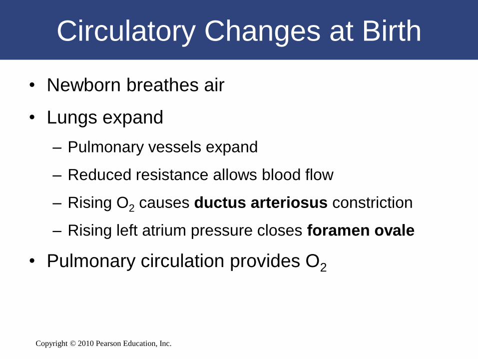
Circulatory Changes at Birth
• Newborn breathes air
• Lungs expand
– Pulmonary vessels expand
– Reduced resistance allows blood flow
– Rising O2 causes ductus arteriosus constriction
– Rising left atrium pressure closes foramen ovale
• Pulmonary circulation provides O2

Copyright © 2010 Pearson Education, Inc.
Fetal Circulation
Figure 13-25a

Copyright © 2010 Pearson Education, Inc.
Fetal Circulation
Figure 13-25b
Copyright © 2010 Pearson Education, Inc.
13-9 Aging affects the blood,
heart, and blood vessels

Copyright © 2010 Pearson Education, Inc.
Aging and the CV System
• Cardiovascular capabilities decline with
age
• Age-related changes occur in
– Blood
– Heart
– Blood vessels

Copyright © 2010 Pearson Education, Inc.
Aging and the CV System
• Three Age-Related Changes in Blood
– Decreased hematocrit
– Peripheral blockage by blood clot (thrombus)
– Pooling of blood in legs
• Due to venous valve deterioration

Copyright © 2010 Pearson Education, Inc.
Aging and the CV System
• Five Age-Related Changes in the Heart
– Reduced maximum cardiac output
– Changes in nodal and conducting cells
– Reduced elasticity of cardiac (fibrous) skeleton
– Progressive atherosclerosis
– Replacement of damaged cardiac muscle cells by
scar tissue

Copyright © 2010 Pearson Education, Inc.
Aging and the CV System
• Three Age-Related Changes in Blood Vessels
– Arteries become less elastic:
• Pressure change can cause aneurysm
– Calcium deposits on vessel walls:
• Can cause stroke or infarction
– Thrombi can form:
• At atherosclerotic plaques

Copyright © 2010 Pearson Education, Inc.
13-10 The cardiovascular
system is both structurally and
functionally linked to all
other systems

Copyright © 2010 Pearson Education, Inc.
CV System Linked to All Systems
• There are many categories of
cardiovascular disorders:
– Disorders may:
• Affect all cells and systems
• Be structural or functional
• Result from disease or trauma

Copyright © 2010 Pearson Education, Inc.
The Cardiovascular System
in Perspective
Functional Relationships Between
the Cardiovascular System and Other Systems
Copyright © 2010 Pearson Education, Inc.

Copyright © 2010 Pearson Education, Inc.
The Integumentary System’s
stimulated mast cells produce
localized changes in bloodflow
and capillary permeability
The Cardiovascular System
delivers immune system cells to
injury sites; clotting response
seals breaks in skin surface;
carries away toxins from sites of
infection; provides heat
The Integumentary System

Copyright © 2010 Pearson Education, Inc.
The Skeletal System
• The Skeletal System provides calcium needed for normal cardiac muscle contraction; protects blood cells developing in bone marrow
• The Cardiovascular System provides calcium and phosphate for bone deposition; delivers EPO to bone marrow, parathyroid hormone and calcitonin to osteoblasts and osteoclasts

Copyright © 2010 Pearson Education, Inc.
The Nervous System
• The Nervous System controls patterns of circulation in peripheral tissues; modifies heart rate and regulates blood pressure; releases ADH
• The Cardiovascular System’s endothelial cells maintain bloodbrain barrier; help generate CSF

Copyright © 2010 Pearson Education, Inc.
The Endocrine System
• The Endocrine System’s
hormone EPO regulates
production of RBCs; several
hormones elevate blood
pressure; epinephrine stimulates
cardiac muscle, elevating heart
rate and contractile force
• The Cardiovascular System
distributes hormones throughout
the body; heart secretes ANP

Copyright © 2010 Pearson Education, Inc.
The Muscular System
The Muscular System assists in moving blood through veins; protects superficial blood vessels, especially in neck and limbs
The Cardiovascular System delivers oxygen and nutrients, removes carbon dioxide, lactic acid, and heat during skeletal muscle activity

Copyright © 2010 Pearson Education, Inc.
The Lymphatic System
• The Lymphoid System defends against pathogens or toxins in blood; fights infections of cardiovascular organs; returns tissue fluid to circulation
• The Cardiovascular System distributes WBCs; carries pathogen-attacking antibodies; restricts spread of pathogens with clotting response

Copyright © 2010 Pearson Education, Inc.
The Respiratory System
• The Respiratory System provides oxygen to cardiovascular organs and removes carbon dioxide
• The Cardiovascular System’s RBCs transport oxygen and carbon dioxide between lungs and peripheral tissues

Copyright © 2010 Pearson Education, Inc.
The Digestive System
• The Digestive System provides
nutrients to cardiovascular organs;
absorbs water and ions to maintain
normal blood volume
• The Cardiovascular System
distributes digestive tract hormones;
carries absorbed nutrients, water, and
ions away; delivers nutrients and
toxins to liver

Copyright © 2010 Pearson Education, Inc.
The Urinary System
• The Urinary System releases renin to elevate blood pressure and EPO to accelerate red blood cell production
• The Cardiovascular System delivers blood to kidneys, where filtration occurs; accepts fluids and solutes reabsorbed during urine production

Copyright © 2010 Pearson Education, Inc.
The Reproductive System
• The Reproductive System maintains
healthy vessels and slows
development of atherosclerosis with
age by estrogens
• The Cardiovascular System
distributes reproductive hormones;
provides nutrients, oxygen, and
waste removal for developing fetus;
local blood pressure changes
responsible for physical changes
during sexual arousal