rombio.eu · web viewhalga p., pop i.m., avarvarei teona, pop viorica, animal nutrition and diet....
TRANSCRIPT

Romanian Biotechnological Letters Vol.22, No. x, 2017Copyright © 2017 University of Bucharest Printed in Romania. All rights reserved
ORIGINAL PAPER
Influence of the diet structure on the development of certain organs of the domestic rabbit male genital system
DANIEL SIMEANU1*, ȘTEOFIL CREANGĂ1, IOAN-MIRCEA POP1, VALERICA GÎLCĂ1 GHEORGHE STAN1, CRISTINEL ȘONEA2
1 University Agricultural Science & Veterinary Medicine Iasi, Animal Science Faculty, Iasi, Romania; 2 Bioterra University Bucharest, Faculty Rural Tourism Management, Bucharest, Romania
*Address correspondence to: University Agricultural Science & Veterinary Medicine Iasi, Animal Science Faculty, Iasi, Romania, Tel: +40232407591, Email: [email protected]
AbstractThe purpose of our study was to evaluate the effect of feeding on the male gonads development
and functionality. Our research was carried using four animal batches (a control group – CG, and three experimental ones - EG), each one of five males belonging to the commercial hybrid "Cunirom-PF-310". All of the subjects were bred from the weaning age up to the age of 120 days, when they reach a medium weight of 2800 grams. The feed administrated to the investigated rabbits was represented by complete mixed fodders, including more types of ingredients which provide 2500-3000 Kcal energetic content /kg dry matter, 183-188 g gross protein /kg dry matter and a different content of gross fiber. All of the males were slaughtered at the age of sexual maturity (120 days), and the testicles (including epididymis) were harvested in order to make histological slides. For the males which were fed with 12% or 16% gross fibers content in mixed fodder, the testicles and the related epididymis were normally developed, with many seminiferous tubules; the seminal epithelium have characteristic cellular elements of seminal line, such as spermatogonia, spermatocyte I and II, and spermatids. There were also observed many Sertoli cells with large, fringe and granulated nuclei, numerous mitoses that indicating a very active spermatogenesis process in these animals. The statistical interpretation of the data related to the structure of the testes shown insignificant differences, being possible to conclude that the feeding of the rabbit males with this kind of mixed fodder did not negatively influenced the male gonadal development and function, and the installation time of the puberty status.For the males which were fed with a mixed fodder which contain 20% or 24% gross fiber, there were found large differences, in negative terms for testicles and epididymis development and functionality, and on regard to the installation of puberty status; the statistical interpretation of the data related to the structure of the testes shown, with few exceptions, significant differences; therefore, it is possible to conclude that the feeding with a mixed fodder which contain 20% or 24%, gross fiber, had a negative influence on the male gonadal development and function, and the installation time of the puberty status.
Keywords: diet structure, domestic rabbit, genital system, gross fibers, mixed fodder
1. IntroductionAt the males of Oryctolagus Cuniculus Domesticus, in normal conditions of feeding and
maintenance, the puberty take place at the age of 110-120 days (RUNCEANU & al. [13] [14]. As in other males (CIORNEI & al. [4], [5]; PĂDURARU & al. [14], up to the puberty time, the

items of seminal line in rabbit are already formed and the spermatocytogenesis and spermiogenesis events are productive. The testicles, the seminiferous tubules and the seminal epithelium developments fit to the normal. VAISSAIRE [20] noticed that on the rabbits, the spermatocytogenesis time is ranging from 40 to 50 days, but the spermiogenesis time is only of 6 or 7 days. Considering the rabbits as unruminant herbivores which take different levels of macro- and microelements from the feed, the purpose of our study was to evaluate the effect of feeding on the male gonads development and functionality, this fact being demonstrated for females in a previous our research (RUNCEANU & al. [13]; SIMEANU & al., [18]; de BLAS & al. [3].
2. Materials and MethodsThe present study was carried using four animal batches (a control group – CG, and
three experimental ones - EG), each one of five males belonging to the commercial hybrid "Cunirom-PF-310", which was formed by P231 and P232 parental lines crossing. All of the subjects were bred from the weaning age up to the age of 120 days, when they reach a medium weight of 2800 grams.
The feed administrated to the investigated rabbits was represented by a complete mixed fodders, including more types of ingredients which provides 2500-3000 Kcal energetic content (EC) /kg dry matter (DM), 183-188 g gross protein (GP) /kg DM and a different content of gross fiber (GF) (Table 1).
Table 1. The development protocol and the nutritional parameters of feed
Male batches
Number of males Type of feed
Some nutritional parameters for 1 kg DM of feedEC GP GF Ca2+ P3-
(Kcal) (g) (g) (%) (g) (g)CG 5 R1 2919 183,0 137,4 100,0 19,85 11,67EG1 5 R2 3000 187,5 183,3 133,4 14,40 8,40EG2 5 R3 2653 183,0 227,4 165,5 12,73 7,43EG3 5 R4 2493 183,6 274,4 199,6 12,00 7,00
CG – control group; EG – experimental group; EC – energetic content ; GP – gross protein; GF – gross fiber;
Since an appropriate digestion level is achieved only if the feed comprises between 12 and 15% raw fibber (HALGA & al. [12]; de BLAS [2]; POP & al. [15]; GIDENNE & al. [10]; STAN & al. [19], we decided to increase this content, in order to test its impact on the development of the male genital system in the domestic rabbit - Oryctolagus Cuniculus Domesticus. Therefore, the rabbits in the experimental groups received diets containing feedstuffs that increased the raw fibber level with proportions comprised between 33.4% and 99.6% (GOMEZ-CONDE & al. [11]; de VRIES & al. [6].
In order to keep the normal levels of the energy and proteins in the combined feed and to vary the raw fibber level as well, between the groups, we opt out to replace the soy meal with sunflower meal; part of the wheat bran was substituted by flour of alfalfa hay, while the wheat straw proportion was also increased (FALCAO & al. [7], [8]; de BLAS & al. [5].
The purpose of testing this feeding program was to assess the optimal level of raw fibber that could be ingested by any Oryctolagus Cuniculus Domesticus male in order to preserve the normal development rate of the genital organs. An increased level of raw fibber means in the

combined feed fact a less expensive feed price. Therefore, finding out the maximum tolerable level of ingestible raw fibber in domestic male rabbits contributes in decrease the feeding costs of this category, while the normal physiological development will not be affected (NICODEMUS & al. [13]; GARCIA &al. [9].
The four feeding mixture compositions (each one for each batch of males) are shown in Table 2, and their nutritional characterization is presented in Table 3.
Table 2. The structure of mixed fodders used in our study
Ingredients UM Type of feed: Their content in:R1 R2 R3 R4 GP GF
Corn % 10,0 11,0 18,7 8,0 8,9 2,5Barley % 10,0 11,0 2,0 - 11,7 4,3Oat % 17,0 15,0 2,0 - 11,8 10,7Soybean meal % 9,2 2,5 9,0 7,5 45,8 7,6Sunflower meal % - 10,0 9,0 10,0 37,4 18,0Fodder yeast % 2,0 2,0 2,0 1,0 43,5 2,5Fish meal % 1,0 1,0 1,0 2,0 65,0 -Wheat bran % 29,0 13,0 1,0 1,0 16,0 10,2Flour of alfalfa hay % 13,0 31,0 39,0 51,0 11,3 33,5Ground wheat straw % 3,8 - 9,5 10,7 3,2 38,6Fat feed % - 0,3 3,8 6,0 - -Chalk feed % 1,0 1,2 1,0 - - -Monocalcium phosphate % 3,0 1,0 1,0 1,8 - -Zoofort A-i type % 1,0 1,0 1,0 1,0 - -TOTAL % 100,0 100,
0 100,0 100,0 - -
UM - unit of measurement; GP – gross protein; GF – gross fiber.
Table 3. The nutritional characterization of used feed
The batch
Type of feed
The raw chemical content(g/kg DM) DOM
%
EC
GOM GP GF MJ/ Kg SU %
CG R1 892,5 183,0 137,4 72,5 12,221 100, 0EG1 R2 960,5 187,5 183,3 72,0 12,561 102, 8EG2 R3 826,5 183,0 227,4 61,5 11,108 90,9EG3 R4 920,7 183,6 274,3 54,3 10,438 85,4
CG – control group; EG – experimental group; GOM – gross organic matter; GP – gross protein; GF – gross fiber;DOM - digestibility of organic matter; EC – energetic content.
All of the males were slaughtered at the age of sexual maturity (120 days). The testicles (including epididymis) were harvested and histological samples were done after their fixation on Boiun solution for 72 hours. After dehydration, clearing and impregnation, all of the histological samples were included on paraffin and subsequently sectioned using SARTORIUS-MF-23-77 microtome. There were performed serial sections of 5|i thickness, which were displayed on blades. A trichromic staining (the “HEA” type) was executed for these sections. All of the histological slides were examined using a photonic binocular microscope (MC3 type), which was previously adjusted and calibrated. Using a special micrometer scale fitted in the eyepiece unit of MC3 microscope, there were measured: the diameters of seminiferous tubules and epididimar

channel; the wall thickness of the seminiferous tubules (seminal epithelium); the cell size of the seminal series (spermatogonia, spermatocytes) and the Leydig gland; the thickness of the epididymis epithelium. The most representative aspects observed in the microscopic field were photographed with EX-1-A camera type, fitted with a special device on the photonic microscope. All of the pictures were done in three eyepiece (EP) and lens (L) associations, including: 16,8 x 10 (168 times magnification); 16,8 x 20 (336 times magnification); 16,8 x 40 (672 times magnification).
All of the microscopic structures details that have been measured in the microscopic field were statistically processed and interpreted.
3. Results and ConclusionsThe results of our investigation, including the diameter of the testis seminal tubules (μ),
the seminal epithelium thickness of the seminiferous tubules (μ), the diameter of the epididimar channel (μ), the thickness of the epididimar channel (μ), the Leydig cell diameter (µ), according to the structured batches and their feeding components, are presented in Table 4.
Table 4. The statistical significance of differences between male batches, for some structural features of the testicles
Specification
n1The means of the compared
batches
The differences
between means (µ)
F calculated at 1; 58
DOF
Fa at 1; 58 DOF for: Tukey
w=0,01 Significancen2n3 p<0,05 p<0,01 p<0,001n4
The diameter of the testis seminal
tubules (μ)
30CG=173,85 6,20 2,01
4,01 7,10 12,03
11,62 n.sEG1=180,05CG=173,85 65,46 48,65 24,99 ***
30EG2=239,31CG=173,85 44,03 58,43 15,34 ***EG3=129,82
30EG1=180,05 59,26 41,68 24,44 ***EG2=239,31EG1=180,05 50,23 85,90 14,43 ***
30EG3=129,82EG2=239,31 10,49 121,83 26,41 ***EG3=129,82
The seminal epithelium
thickness of the seminiferous
tubules(μ)
30CG=56,67 4,92 5,35
4,01 7,10 12,03
5,66 n.s.EG1=61,59CG=56,67 24,99 230,52 4,38 ***
30EG2=31,68CG=56,67 27,21 233,42 4,74 ***EG3=29,46
30EG1=61,59 29,91 261,05 4,93 ***EG2=31,68EG1=61,59 32,13 265,44 5,25 ***
30EG3=29,46EG2=31,68 2,22 2,37 3,84 n.s.EG3=29,46
The diameter of the epididimar
channel 30 CG=193,27 3,76 0,51 4,01 7,10 12,03 14,03 n.s.
EG1=197,03

(μ)
CG=193,27 4,50 0,66 14,75 n.s
30EG2=188,77CG=193,27 36,37 54,21 13,16 ***EG3=156,90
30EG1=197,03 8,26 2,41 14,15 n.s.EG2=188,77EG1=197,03 40,13 73,29 12,48 ***
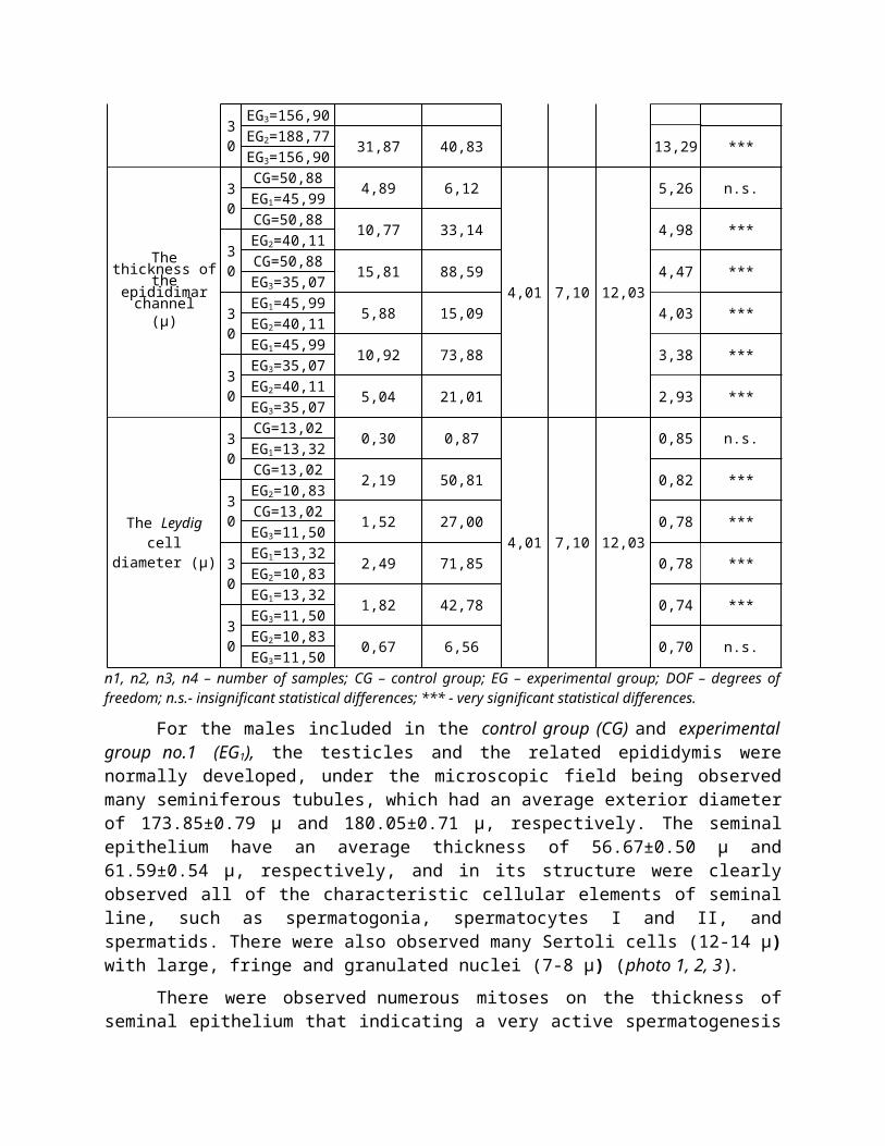
30EG3=156,90EG2=188,77 31,87 40,83 13,29 ***EG3=156,90
The thickness of the epididimar
channel (μ)
30CG=50,88 4,89 6,12
4,01 7,10 12,03
5,26 n.s.EG1=45,99CG=50,88 10,77 33,14 4,98 ***
30EG2=40,11CG=50,88 15,81 88,59 4,47 ***EG3=35,07
30EG1=45,99 5,88 15,09 4,03 ***EG2=40,11EG1=45,99 10,92 73,88 3,38 ***
30EG3=35,07EG2=40,11 5,04 21,01 2,93 ***EG3=35,07
The Leydig cell diameter (µ)
30CG=13,02 0,30 0,87
4,01 7,10 12,03
0,85 n.s.EG1=13,32CG=13,02 2,19 50,81 0,82 ***
30EG2=10,83CG=13,02 1,52 27,00 0,78 ***EG3=11,50
30EG1=13,32 2,49 71,85 0,78 ***EG2=10,83EG1=13,32 1,82 42,78 0,74 ***
30EG3=11,50EG2=10,83 0,67 6,56 0,70 n.s.EG3=11,50
n1, n2, n3, n4 – number of samples; CG – control group; EG – experimental group; DOF – degrees of freedom; n.s.- insignificant statistical differences; *** - very significant statistical differences.
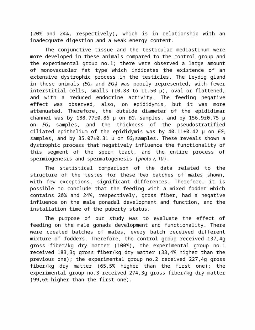
For the males included in the control group (CG) and experimental group no.1 (EG1), the testicles and the related epididymis were normally developed, under the microscopic field being observed many seminiferous tubules, which had an average exterior diameter of 173.85±0.79 μ and 180.05±0.71 μ, respectively. The seminal epithelium have an average thickness of 56.67±0.50 μ and 61.59±0.54 μ, respectively, and in its structure were clearly observed all of the characteristic cellular elements of seminal line, such as spermatogonia, spermatocytes I and II, and spermatids. There were also observed many Sertoli cells (12-14 µ) with large, fringe and granulated nuclei (7-8 µ) (photo 1, 2, 3).
There were observed numerous mitoses on the thickness of seminal epithelium that indicating a very active spermatogenesis process in these organs. The testis interstitial gland was normal developed, including numerous Leydig cells, which were relatively large on their size (13.02±0.21 µ ÷ 13.32±0.2 µ), oval or round shaped, mono- or binucleated, and with a diameter average of their nuclei ranged from 7.70±0.19 to 7.72±0.19 μ. Considering the epididymis of these males, the average outer diameter of the epididimar channel was 193.27±0.84 μ for the

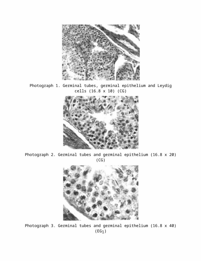
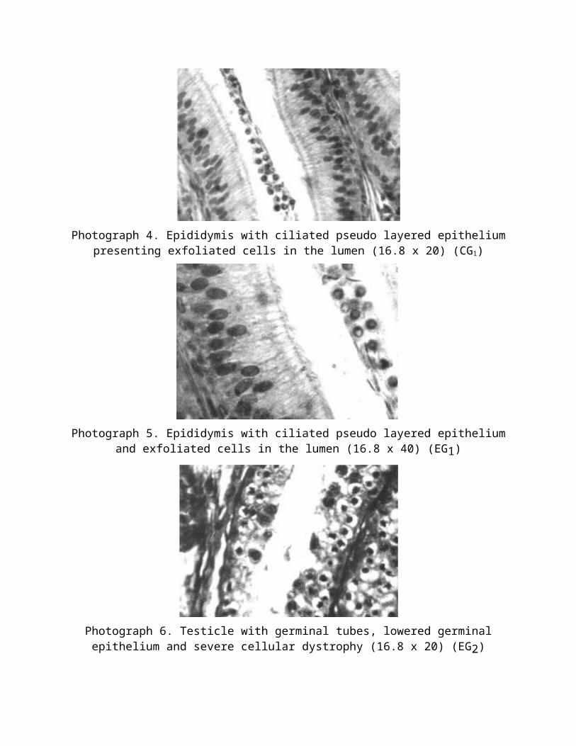
individuals included in CG, and 197.03±0.81 μ for the males of EG1. The wall of the epididimar channel, which is a pseudostratified ciliated epithelium, had an average thickness of 50.88±0.54 μ for CG males, and 45.99±0.46 μ for EG1 ones, which we consider that it is a normal thickness. In the lumen of the epididimar channel there were observed many peeling cells, sperm, and a small amount of fluid (photo 4, 5).
The statistical comparison of the data related to the structure of the testes for these two batches of males shown insignificant differences. Therefore, it is possible to conclude that the feeding with a mixed fodder which contain 12% and 16%, respectively, gross fiber, did not negatively influenced the male gonadal development and function, and the installation time of the puberty status.
For the males included in the experimental group no.2 (EG2) and experimental group no.3 (EG3), which were fed with a mixed fodder which contain 20% and 24%, respectively, gross fiber, there were found large differences, in negative terms for testicles and epididymis development and functionality, and on regard to the installation of puberty status. Therefore, the seminiferous tubules were deformed, with an external diameter of 239.31±1.26 µ for the males included in EG2, and of 129.82±0.92 µ for the males included in EG3. The thickness of seminal epithelium was by 31.68±0.40 μ for EG2 samples, and by 29.46±0.45 μ for EG3 samples, and in its structure there was fewer Sertoli cells, flattened and reduced spermatogonia cells, rare spermatocytes I, as, in fact, the mitosis; the spermatocytes II and the spermatids were completely missing (photo 6, 7, 8, 9). This histological picture shows that the spermatocytogenesis process was seriously disturbed and retarded, due to the higher content of gross fiber (20% and 24%, respectively), which is in relationship with an inadecquate digestion and a weak energy content.
The conjunctive tissue and the testicular mediastinum were more developed in these animals compared to the control group and the experimental group no.1; there were observed a large amount of monovacuolar fat type which indicates the existence of an extensive dystrophic process in the testicles. The Leydig gland in these animals (EG2 and EG3) was poorly represented, with fewer interstitial cells, smalls (10.83 to 11.50 μ), oval or flattened, and with a reduced endocrine activity. The feeding negative effect was observed, also, on epididymis, but it was more attenuated. Therefore, the outside diameter of the epididimar channel was by 188.77±0,86 μ on EG2 samples, and by 156.9±0.75 μ on EG3 samples, and the thickness of the pseudostratified ciliated epithelium of the epididymis was by 40.11±0.42 μ on EG2 samples, and by 35.07±0.31 μ on EG3 samples. These reveals shown a dystrophic process that negatively influence the functionality of this segment of the sperm tract, and the entire process of spermiogenesis and spermatogenesis (photo 7, 10).
The statistical comparison of the data related to the structure of the testes for these two batches of males shown, with few exceptions, significant differences. Therefore, it is possible to conclude that the feeding with a mixed fodder which contains 20% and 24%, respectively, gross fiber, had a negative influence on the male gonadal development and function, and the installation time of the puberty status.
The purpose of our study was to evaluate the effect of feeding on the male gonads development and functionality. There were created batches of males, every batch received different mixture of fodders. Therefore, the control group received 137,4g gross fiber/kg dry matter (100%), the experimental group no.1 received 183,3g gross fiber/kg dry matter (33,4% higher than the previous one); the experimental group no.2 received 227,4g gross fiber/kg dry
matter (65,5% higher than the first one); the experimental group no.3 received 274,3g gross fiber/kg dry matter (99,6% higher than the first one).
Photograph 1. Germinal tubes, germinal epithelium and Leydig cells (16.8 x 10) (CG)
Photograph 2. Germinal tubes and germinal epithelium (16.8 x 20) (CG)
Photograph 3. Germinal tubes and germinal epithelium (16.8 x 40) (EG1)

Photograph 4. Epididymis with ciliated pseudo layered epithelium presenting exfoliated cells in the lumen (16.8 x 20) (CG1)
Photograph 5. Epididymis with ciliated pseudo layered epithelium and exfoliated cells in the lumen (16.8 x 40) (EG1)
Photograph 6. Testicle with germinal tubes, lowered germinal epithelium and severe cellular dystrophy (16.8 x 20) (EG2)
Photograph 7. Epididymis, cross section through the epididymial channel, with reduced epithelium (16.8 x 20) (EG2)
Photograph 8. Testicle with low developed germinal tubes, reduced germinal epithelium and severe cell dystrophy (16.8 x 10) (EG3)
Photograph 9. Testicle, germinal tube (fragment) presenting very reduced germinal epithelium and acute cell dystrophy (16.8 x 40) (EG3)
Photograph 10. Epididymis presenting reduced epithelium and agglutinated cilia (16.8 x 20) (EG3)
The feeding of the rabbit males with a mixed fodder which contain 12% or 16% gross fiber (the control and the experimental group no.1), did not negatively influenced the gonadal development and function, and the installation time of the puberty status. On the other hand, the feeding with a mixed fodder which contain 20% or 24% gross fiber (the experimental group no.2 and the experimental group no.3), had a negative influence on the male gonadal development and function, and the installation time of the puberty status.
References1. CACHALDORA P., NICODEMUS N., GARCIA J., CARABANO R., DE BLAS J.C., Efficacy of
Amylofeed® in growing rabbit diets. World Rabbit Science, 12: 23 – 31 (2004)
2. CARLOS DE BLAS AND JULIAN WISEMAN, Nutrition of the rabbit, 2nd Edition. CABI Int. Wallingford, UK, 2010
3. CARLOS DE BLAS, JAVIER GARCIA, ROSA CARABANO, Role of fiber in rabbit diets. A review. Annales de zootechnie, INRA/EDP Sciences, 48 (1), pp.3-13 (1999)
4. CIORNEI ŞT.G., ROŞCA P., DRUGOCIU D., Bacterial and fungal burden in boar semen, Research Journal of Biotechnology, Vol. 7 (4), pp. 23 – 27 (2012)
5. CIORNEI ŞT.G., ROŞCA P., DRUGOCIU D., MAREȘ M., NECHIFOR F, IBĂNESCU I., Reproduction biotechnologies in Mangalita breed boars. Res. J. Biotech. Vol. 8 (11), 51-56 (2013)
6. DE BLAS J.C., MATEOS G.G., Feed formulation. In: de Blas J.C., Wiseman J. (Eds). The Nutrition of the Rabbit. CAB Int., Wallingford, UK, 1998, pp. 241-254.
7. DE VRIES J.W. AND RADER J.I., Historical perspective as a guide for identifying and developing applicable method for dietary fiber. Journal of AOAC International. 88, 1349 – 1366 (2005)
8. FALCAO E CUNHA L., JORGE J., FEREIRE J.P.B. AND PEREZ H., Fat addition to feeds for growing rabbits differing in fiber level and nature: effects of growing rate, digestibility and caecal patterns. World Rabbits Science 8 (Suppl. 1, vol. C), 191 – 198 (2000)
9. FALCAO E CUNHA L., PEREZ H., FEREIRE J.P.B. AND CASTRO-SOLLA L., Effect of alfalfa, wheat bran or beet pulp, with or without sunflower oil, on caecal fermentation and on digestibility on the rabbits. Animal Feed Science and Technology 117, 131 – 149 (2004)
10.GARCIA J., NICODEMUS N., PEREZ-ALBA L., CARABANO R., DE BLAS J.C., Characterization on fiber digestion of grape seed meal and sunflower hulls in rabbits. I. Fiber digestibility and rate of passage. World Rabbit Science 8 (Suppl. 1, vol. C), 225 – 231 (2000)
11.GIDENNE T., CARABANO R., GARCIA J. AND DE BLAS C., Fiber digestion in Nutrition of the rabbit, 2nd Edition. CABI Int. Wallingford, UK, 2010 pp. 66 - 82
12.GOMEZ-CONDE M.S., PEREZ DE ROZAS A., BADIOLA I., PEREZ.ALBA L., DE BLAS C., CARABANO R., GARCIA J., Effect of neutral detergent soluble fiber on digestion, intestinal micro biota and performance in twenty five day old weaned rabbits. Livestock Science, 125, 192 – 198 (2009)
13.HALGA P., POP I.M., AVARVAREI TEONA, POP VIORICA, Animal nutrition and diet. Editura Alfa, Iaşi, 2005 (in romanian)
14.NICODEMUS N , GARCÍA J, CARABANO R, DE BLAS J.C., Effect of substitution of a soybean hull and grape seed meal mixture for traditional fiber sources on digestion and performance of growing rabbits and lactating does. Journal of Animal Science 2007 Jan; 85(1):181-7 (2007)
15.PĂDURARU V.D., DRUGOCIU D., CIORNEI ȘT.G., COJOCARU OANA, Correlations between age, body weight and scrotal circumference of two bulls in preparation from angus breed, and comparison of results with data from literature, „Lucrari stiintifice de Medicină Veterinară”, Ed. ,,Ion Ionescu de la Brad’’ vol. 54 (13), 396 – 401 (2011)
16.POP I.M., HALGA P., AVARVAREI TEONA, Animal nutrition and feeding. Ed. Tipo Moldova, Iaşi, 2006 (in romanian)
17.RUNCEANU L., CIORNEI ŞT.G., DRUGOCIU D., ROŞCA P., NECHIFOR F., Pathophysiology maternal-fetal veterinary, Ed. Ion Ionescu de da Brad, Iaşi, 2014 (in romanian).
18.RUNCEANU L., DRUGOCIU D., ROŞCA P., NECHIFOR FL., CIORNEI ŞT., ANTONETA RUSU, Elements of normal and pathological development of animal embryo. Ed Ion Ionescu de la Brad, Iaşi, 2013 (in romanian).
19.SIMEANU D., STAN GH., SIMEANU CRISTINA, Research regarding the influence of cellulose on the morphological and structural development of rabbit ovary, Annals of the University of Oradea, Ecotoxicology, Animal Husbandry and Food Industrial Technologies, XI/A, (11), pp. 147-158 (2012)
20. STAN GH. and SIMEANU D., Animal nutrition. Ed. Alfa, Iaşi, 2005
21. VAISSAIRE J. P., La reproduction des animaux domestiques, Vigot-Freires, Paris, 1972