archive.lstmed.ac.ukarchive.lstmed.ac.uk/6400/1/marco manuscript_gene… · web...
TRANSCRIPT

MARCO variants are associated with phagocytosis, pulmonary tuberculosis susceptibility
and Beijing lineage
Nguyen Thuy Thuong Thuong1,2, Trinh Thi Bich Tram1,2, Tran Dinh Dinh1,2, Phan Vuong Khac
Thai3, Dorothee Heemskerk1,2, Nguyen Duc Bang3, Tran Thi Hong Chau4, David Russell5, Guy
E. Thwaites1,2, Thomas R. Hawn6, Maxine Caws7, Sarah J. Dunstan8
1. Oxford University Clinical Research Unit, Ho Chi Minh City, Viet Nam
2. Centre for Tropical Medicine and Global Health, Nuffield Department of Medicine, University of
Oxford, Oxford, United Kingdom
3. Pham Ngoc Thach Hospital for Tuberculosis and Lung Disease, Ho Chi Minh City, Viet Nam
4. Hospital for Tropical Diseases, Ho Chi Minh City, Viet Nam
5. College of Veterinary Medicine, Cornell University, Ithaca, New York, United States of America
6. School of Medicine, University of Washington Seattle, Washington, United States of America
7. Liverpool School of Tropical Medicine, Pembroke Place, Liverpool, United Kingdom
8. Peter Doherty Institute for Infection and Immunity, The University of Melbourne, Australia
Running title: MARCO and tuberculosis
Word count of the abstract: 177
Word count of the text: 4011
Correspondence should be addressed to: Dr Nguyen Thuy Thuong ThuongOxford University Clinical Research Unit, Hospital for Tropical Diseases764 Vo Van Kiet, W.1, Dist.5, Ho Chi Minh CityTel: 84 8 924 1761; Fax: 84 8 923 8904; Email: [email protected]
Abstract

Macrophage receptor with collagenous structure (MARCO) plays an important role in the
phagocytosis of Mycobacterium tuberculosis (M. tuberculosis). We hypothesized that MARCO
polymorphisms are associated with phagocytosis, tuberculosis (TB) disease susceptibility and
presentation, and infecting lineage.
We used a human cellular model to examine how MARCO genotype mediates the immune
response; a case-control study to investigate tuberculosis host genetic susceptibility; and a host-
pathogen genetic analysis to study host-pathogen interactions.
Two MARCO heterozygous (AG) genotypes (SNPs rs2278589 and rs6751745) were associated
with impaired phagocytosis of M. tuberculosis TDM-cord factor and β-glucan coated beads in
macrophages. The heterozygous genotypes of rs2278589 and rs6751745 were also associated
with increased risk of pulmonary TB (rs2278589, p=0.001, OR=1.6; rs6751745, p=0.009,
OR=1.4), and with severe chest X-ray abnormalities (p=0.007, OR=1.6). These two genotypes
were also associated with the Beijing lineage (rs2278589, p=0.001, OR=1.7; rs6751745, p=0.01,
OR=1.5).
Together, these results suggest that MARCO polymorphisms may regulate phagocytosis of M.
tuberculosis and susceptibility and severity of pulmonary tuberculosis. They also suggest
MARCO genotype and Beijing strains may interact to increase the risk of pulmonary
tuberculosis.
Introduction
Although tuberculosis (TB) can be cured, it is still one of the most devastating diseases, and
globally causes active TB in 9.6 million and kills 1.5 million people annually (1). Variation in
the host and pathogen are involved in disease susceptibility and determine disease development
and outcome. Studies suggest that polymorphisms in host immunity genes influence
susceptibility to TB (2, 3), especially in genes encoding Toll-like receptors, C-type lectin and
scavenger receptors, which are involved in recognizing, binding, and phagocytosing M.
tuberculosis.
Scavenger receptors are cell surface receptors, which bind a variety of ligands, and have an
important function in clearance of many foreign microorganisms. Class A and class B scavenger
receptors are involved in the cytokine response to mycobacterial lipoarabinomannans (4) and
lipopeptides (5). Macrophage receptor with collagenous structure (MARCO) is a member of the
class A scavenger receptor family. MARCO, on the cell surface of macrophages, binds bacteria
to facilitate phagocytosis and activates immune responses (6-8). As such, MARCO-deficient
mice have a reduced ability to clear bacteria in pneumonia (7, 9). Class A scavenger receptors
and MARCO participate in phagocytosis of mycobacterial species, including M. leprae (10), M.
bovis Bacille Calmette-Guérin (11), M. marinum (6) and M. tuberculosis (12, 13). More
specifically, it has been demonstrated that M. tuberculosis is captured by MARCO in vivo via its
cell wall cord factor (trehalose 6,6'-dimycolate or TDM), which increases pro-inflammatory
cytokine response through the interaction with Toll-like receptors and CD14 (14).
The genetic diversity of M. tuberculosis is another factor which contributes to the clinical
consequences of TB (15-17). The emergence of Beijing strains, which account for approximately
50% of strains in East Asia and 13% of strains worldwide (18), may contribute to disease

susceptibility, drug resistance and treatment outcome. There is a possibility of human-
mycobacterial co-evolution based on the genetic interactions of genes in the host and pathogen
(18, 19). This would help to explain the interactions between host and pathogen factors in the
development of TB.
Altogether, due to the role of phagocytosis and the potential function of MARCO in the immune
response against M. tuberculosis, we hypothesized that (i) Phagocytic activity is associated with
developing different clinical phenotypes of TB, such as latent, pulmonary or extra-pulmonary
TB; (ii) Polymorphisms in MARCO regulate macrophage phagocytic activity; (iii)
Polymorphisms in MARCO, that contribute to the impairment of macrophage phagocytic
activity, are associated with susceptibility to tuberculosis and influence clinical presentations and
treatment failure; (iv) host and pathogen genotypes combined influence tuberculosis
susceptibility.
Results
Phagocytosis and TB clinical phenotypes
We examined phagocytosis in human MDMs by bead-based internalization assays. Alexa 594-
beads coated with IgG, TDM or β-glucan were added to MDMs and the percentage of
macrophages with or without beads was measured using flow cytometry to assess phagocytic
ability. Phagocytosis was assessed in macrophages isolated from patients with latent (N = 56),
pulmonary (N = 52) or meningeal TB (N = 55). No association was observed between
phagocytic activity and different clinical forms of TB (Figure 1).
There was a wide range of phagocytic activities, with up to 50% of beads coated with M.
tuberculosis TDM in macrophages from latent, pulmonary and meningeal TB (Figure 1). To
investigate how MARCO influences the heterogeneity of phagocytic activity, we next examined
the association of MARCO variants and phagocytosis.
Association of MARCO SNPs with macrophage phagocytosis, mRNA expression and
cytokines in response to M. tuberculosis
MARCO is a phagocytic receptor on macrophages which binds bacteria and facilitates
phagocytosis to control and clear pathogens (6, 8). TDM from M. tuberculosis is a ligand of
MARCO whereas β-glucan is not known to be a MARCO ligand. Scavenger receptors on human
monocytes have been found to bind to β-glucan (20), and MARCO (on CpG-ODN-pretreated
macrophages) has been found to participate in the uptake of zymosan (which is derived from β-
glucan) (21); therefore β-glucan was used in this study to address the question of whether it
might be a ligand for MARCO and to explore possible interaction between MARCO,
tuberculosis and β-glucan.
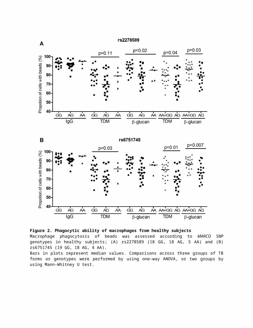
We genotyped twelve MARCO haplotype-tagging SNPs from 41 healthy subjects and performed
phagocytosis assays. The genotypes of two SNPs rs2278589 and rs6751745, were associated
with phagocytosis of either TDM or β-glucan beads, but were not associated with phagocytosis
of IgG beads (Figure 2A, 2B). The remaining 10 SNPs in MARCO were not associated with
phagocytosis of any beads (Figure S1). Furthermore, the results show the heterozygous
genotypes of both SNPs were associated with reduced phagocytosis of TDM and β-glucan beads
(rs2278589, p = 0.04 and 0.03; rs6751745, p = 0.01 and 0.007) (Figure 2A, 2B).
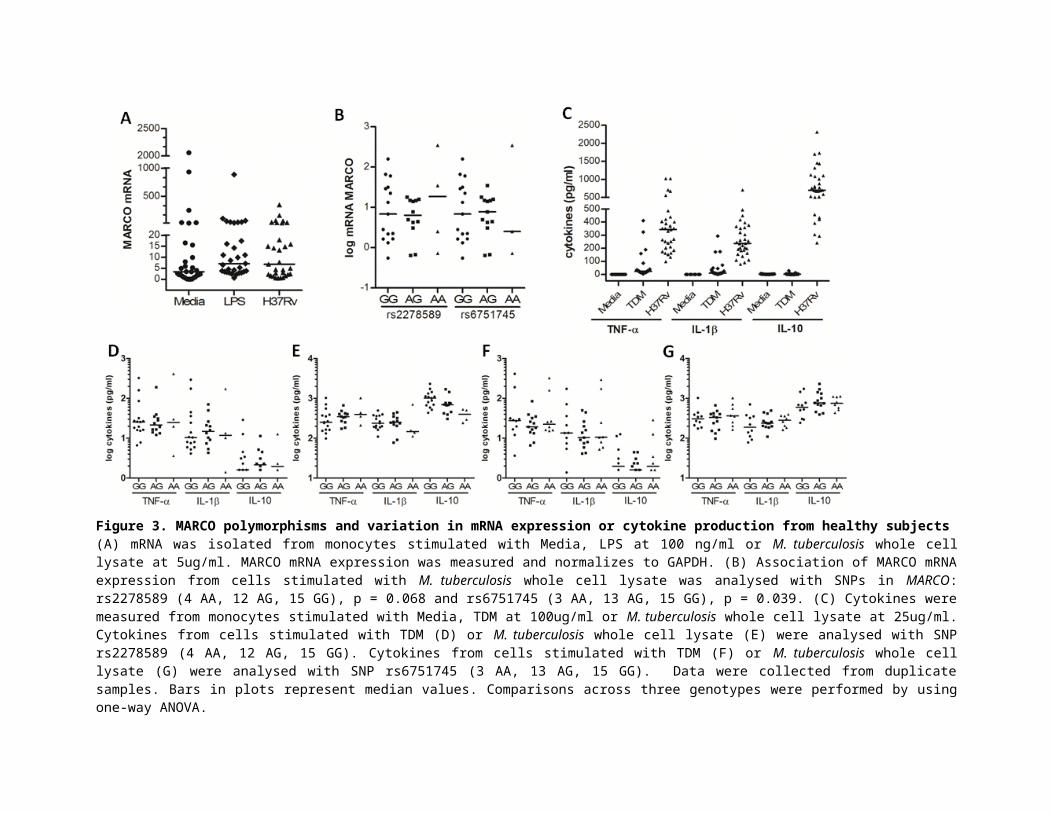
We also examined the association between MARCO SNPs rs2278589 and rs6751745 and mRNA
expression or cytokines in PBMCs from 31 healthy subjects. MARCO mRNA levels were up-
regulated approximately 2 fold in PBMCs stimulated with LPS or M. tuberculosis whole cell
lysate compared with un-stimulated cells (Figure 3A). The genotypes of rs2278589 and

rs6751745 were marginally associated with MARCO mRNA expression in cells stimulated with
M. tuberculosis (Figure 3B) (ANOVA, p = 0.068 and 0.039 respectively). For the heterozygous
model, the AG genotype of these two SNPs was not significantly associated with reduced levels
of MARCO mRNA in PBMCs stimulated with M. tuberculosis. For cytokine production,
PBMCs were activated and produced pro-inflammatory cytokines TNF-α and IL-1β in response
to both TDM and M. tuberculosis lysate. The anti-inflammatory cytokine IL-10 was induced by
M. tuberculosis lysate stimulation, but not TDM (Figure 3C). In TDM or M. tuberculosis lysate
stimulated cells, there was no association between the two SNP genotypes and TNF-α, IL-1β and
IL-10 levels (Figure 3 D-E for rs2278589, and F-G for rs6751745).
Collectively, these data showed that the AG genotype of rs2278589 and rs6751745 in MARCO
was not associated with MARCO mRNA expression or cytokine concentrations in PBMCs, but it
was associated with reduced phagocytosis activated via TDM and β-glucan in macrophages.
MARCO polymorphisms are associated with susceptibility to pulmonary TB, but not with TB
meningitis
We used a case–control study to determine whether MARCO polymorphisms SNPs rs2278589
and rs6751745 are associated with susceptibility to TB, as macrophages with the heterozygote
genotypes of these SNPs displayed reduced phagocytosis of M. tuberculosis ligands (Figure 2).
Therefore we applied the heterozygote advantage model to analyse the relationship between
MARCO SNPs and clinical TB, both pulmonary and meningeal. The heterozygote genotypes of
rs2278589 and rs6751745 are associated with susceptibility to PTB (rs2278589; p = 0.001, OR =
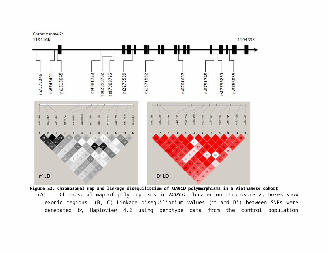
1.6 and rs6751745; p = 0.009, OR = 1.4; Table 1) and figure S2 shows that the two SNPs are in
high LD (D' = 1, r2 = 0.88) in our Vietnamese Kinh control population. Associations between
these 2 SNPs and PTB remained significant after Bonferroni correction (p values x 2) (Table 1).
Interestingly, the genotype frequencies of these two SNPs were different between PTB and TBM
under the heterozygote advantage model [(rs2278589; PTB 0.55, TBM 0.46; p = 0.005, OR =
1.4) (rs6751745; PTB 0.50, TBM 0.40; p = 0.003, OR = 1.5]. However the genotype frequencies
of rs2278589 and rs6751745 in TBM patients were not different compared to the control groups
using the genotypic model (p > 0.05).
To thoroughly examine the association between MARCO SNPs and TB a further 10 SNPs, within
and upstream of the MARCO gene, were analyzed. Apart from the two associated SNPs
described above, rs6748401 (1.5 kb upstream) was associated with PTB in a genotypic
comparison (p = 0.039; Table 2), and none of others were associated with susceptibility to TB.
Collectively, two SNPs in the MARCO gene were associated with PTB, but not with TBM. The
heterozygote genotypes of rs2278589 and rs6751745, which were associated with reduced
phagocytic activity, were also associated with susceptibility to PTB.
MARCO polymorphisms are associated with CXR presentation
To investigate whether MARCO polymorphisms influence clinical presentation or disease
outcome, we examined the relationship between the two associated SNPs (rs2278589 and
rs6751745), pre-treatment CXR abnormalities, and 8-month treatment outcomes. Patients
enrolled in this study were sputum smear-positive for pulmonary TB before treatment. Pre-
treatment CXR showed 427/429 (99.5%) were abnormal with evidence of nodules (139, 32.4%),
infiltrates (407, 94.9%), consolidation (40, 9.3%), cavities (139, 32.4%) and miliary disease (0,
0%).
SNPs rs2278589 and rs6751745 were associated with severity of CXR abnormality. SNP
rs2278589 was associated with intermediate and severe CXR abnormality in the heterozygote
model (p = 0.008 intermediate; p = 0.007 severe, OR = 1.6; Table 3). SNP rs6751745 was
associated with severe CXR abnormality in the heterozygote model (p = 0.007, OR = 1.6; Table
3).
There was no association between rs2278589 and rs6751745 genotype and poor treatment
outcome (29/429, 6%), which was defined by death, or failure to convert to sputum smear
negativity, however this may be due to the lack of events in this dataset and consequent lack of
power.
MARCO polymorphisms are associated with Beijing lineage
Our previous studies reported associations between lineages of M. tuberculosis, particularly the
modern Beijing lineage, and TB clinical phenotypes (15, 17). Given a worldwide emergence of
the M. tuberculosis Beijing strains (18), we hypothesized that variation in the scavenger receptor
MARCO, which binds to Mycobacterium and enables phagocytosis by macrophages, might be
preferentially associated with a specific lineage. Therefore, we next examined whether the
rs2278589 and rs6751745 genotypes are associated with infection caused by a particular
bacterial lineage and whether this relationship influences disease phenotype.
The genotypic frequencies of rs2278589 and rs6751745 in all PTB patients (N = 445) and in
those patients where the lineage of the infecting M. tuberculosis isolate was determined (N =
370), were compared with controls (Table 4). There was no significant association between the
two SNPs and infection with either Indo-Oceanic or Euro-American lineage, or when combined
as non-Beijing lineages. However, we found a significant association between these SNPs and
infection with Beijing isolates in a genotypic comparison (rs2278589, p = 0.005; rs6751745, p =
0.033; Table 4), and in a heterozygous model (rs2278589, p = 0.001, OR=1.7; rs6751745, p =
0.012, OR=1.5; Table 4).
Discussion
The primary finding of our study was that MARCO genotypes were associated with a reduction
of phagocytosis of beads coated with pathogen-derived ligands, TDM from M. tuberculosis.
These genotypes were also associated with increased susceptibility to pulmonary TB and severe
chest radiography abnormality. Our results suggest that these polymorphisms may regulate
phagocytosis of M. tuberculosis, and impairment of phagocytic ability could increase
susceptibility to, and severity of, pulmonary TB. The MARCO genotypes were preferentially
associated with Beijing rather than Indo-Oceanic or Euro-American lineages, which implies
MARCO genotype may increase susceptibility to tuberculosis particularly of the Beijing lineage.
MARCO plays a key role in bacterial phagocytosis and clearance (6, 7, 9). Recognition of TDM
by MARCO, in conjunction with TLR2/CD4, activates transcriptional expression of immunity
genes (22) and cytokine production (14). However, no studies have yet shown the influence of
MARCO genetic variation on the antimicrobial activity of macrophages such as phagocytosis or
immune response. In this study, we found that MARCO SNPs rs2278589 and rs6751745 were
not associated with gene expression and cytokine production in PBMCs but were associated with
reduced phagocytosis of beads coated with pathogen-derived ligands, TDM or β-glucan in
macrophages. We found that MARCO polymorphisms were not associated with cytokine
production. In murine studies, MARCO-deficient macrophages were associated with a reduction
of TNFα, IL-6 and IL-1β cytokine production in macrophages from MARCO knockout mice.
The difference in study design could account for the differences seen in cytokine production
between these two studies. We used human PBMCs, whereas in Bowdish et al (2009; 14)
murine macrophages were used. In the macrophages from knockout mice, MARCO was absent,
potentially having a major impact. In our study, MARCO was still produced, albeit a variant of

MARCO with an unknown and potentially smaller impact. Another reason for the difference
may be the limited numbers of samples in our study once stratified by genotype.
MARCO is involved in phagocytosis of bacteria, a step in pathogenesis which may be important
in the development of pulmonary TB in the early phase of infection. The heterozygous genotypes
of two MARCO SNPs were associated with reduced macrophage phagocytic function. The
impairment of phagocytosis at the beginning of infection reduces the number of macrophages
infected with M. tuberculosis, which then limits microbial killing and antigen presentation to
lymphocytes (23, 24). The consequence of this could be the inadequate induction of innate and
adaptive immune responses against M. tuberculosis, potentially increasing susceptibility to active
disease. Deficient responses could also lead to increased microbial replication, which could
manifest as severe abnormalities on CXR, such as was observed in TB patients carrying the
heterozygous genotypes. Together, our data suggest that TB susceptibility and disease severity in
patients with the MARCO AG genotype may be due to impairment of M. tuberculosis
phagocytosis.
Our results show that variation in human MARCO is associated with susceptibility to pulmonary
TB in the Vietnamese Kinh population. The associated intronic SNPs rs6748401 and rs2278589
are part of a wide haplotype block, suggesting they are markers in high LD with the unknown
causative SNP(s). Two MARCO SNPs (rs17009726 or rs4491733) were previously associated
with TB susceptibility in the Han Chinese Beijing and Gambian populations (25, 26) however
we did not observe any association with these SNPs in our TB population. Conversely, the
associated SNPs (rs6748401 and rs2278589) described in this study were not associated in the
Gambian population and were not genotyped in the Chinese population. The discrepancy in our
results may be due to different population LD structure. The frequencies of the associated SNPs

found in the three studies were very different based on the 1000 Genomes Project
(http://www.ncbi.nlm.nih.gov/projects/SNP; rs17009726 minor allele frequency in African
0.0008, Ad Mixed American 0.0014, European 0.0060, East Asian 0.1210 and South Asian
0.1483 super populations) and overall linkage (D' plots) across the MARCO SNPs in three
populations are visually different (figure S2 and (25, 26)). LD in the Vietnamese Kinh
population across this gene region contains larger haplotype blocks with more SNPs compared to
both the Han Chinese (HCB) and Gambian populations. The differing population structures in
this gene region may account for the inability to replicate individual SNP associations, however
the accumulated evidence across these populations suggests that MARCO variation contributes to
pulmonary TB susceptibility.
The remarkable emergence of Beijing lineage worldwide, including Vietnam, supports the
hypothesis that the variation in the scavenger receptor MARCO, which binds Mycobacterium and
promotes macrophage internalization, might support the emergence of the Beijing lineages. Our
data shows associations of both MARCO variants and M. tuberculosis lineage with TB
susceptibility suggesting potential for host-pathogen co-evolution, as reported previously with
TLR2, NRAMP1 and EREG (15, 27, 28). Our associated SNPs may be markers of non-
synonymous structural variants of MARCO that effects ability to bind ligands from Beijing
lineage strains, reducing phagocytosis and increasing susceptibility to TB. Host-pathogen co-
evolution in tuberculosis needs to be studied on a larger scale with respect to patients and genes,
coupled with functional studies to determine the underlying mechanisms.
The role of MARCO in macrophage phagocytosis is important in clearance of pathogens. Our
results suggest that MARCO polymorphisms may regulate phagocytosis of M. tuberculosis and
thus influence susceptibility to and severity of pulmonary tuberculosis. The results also suggest
that MARCO genotype and Beijing strains may interact to increase the risk of pulmonary
tuberculosis.
Materials and Methods
Cellular studies
Ex vivo isolation of Peripheral blood mononuclear cells (PBMCs) and Monocyte-derived
macrophages (MDMs)
PBMCs were separated from heparinized whole blood by Lymphoprep (Asix-Shield, Norway)
gradient centrifugation according to the manufacturer’s protocol. From 20 ml of blood we
obtained approximately 1-1.5x10⁷ PBMCs. To isolate monocytes by adherence, PBMCs were
plated in cell-culture treated 48-well plates (Nunc, Denmark) with 9x10⁵ cells per well in media
without serum, containing RPMI-1640 (Sigma, Germany), 2mM L-glutamine and 100 units of
penicillin. Cells were incubated at 370C, 5% CO2 for 2 hours and the non-adhered cells were
washed off gently two times by warm phosphate buffered saline (PBS) with 3% fetal calf serum
(FCS; Sigma, Germany). Cells were re-suspended in 0.4 ml complete media, containing RPMI-
1640, 2 mM L-glutamine, 100 units of penicillin, 10% FCS and 10 ng/ml human mCSF (R&D
Systems, USA), and incubated at 370C, 5% CO2. To derive monocytes, adhered cells were
incubated for 5 to 7 days. Complete media was changed at day 4, and phagocytosis assay was
performed at day 7.
PBMC stimulation for MARCO mRNA and cytokine measurement
For MARCO mRNA expression, PBMCs were plated in 24-well plates (106 cell/well). Cells
were subsequently stimulated with either whole cell lysate (WCL) of M. tuberculosis H37Rv at 5
µg/ml (Colorado State University, USA), LPS at 100 ng/ml (Sigma-Aldrich, USA) or media. For
cytokine measurement, PBMCs were plated in 96-well plates (105 cell/well) and then stimulated
with either WCL at 25 µg/ml, TDM at 100 µg/ml (Enzo Life Sciences, USA), LPS 100 ng/ml or
media. For stimulation, TDM was coated on the plate and air-dried in the cabinet the day before,
while other ligands were added directly into media after cell plating.
For real-time quantitative PCR (RTqPCR), after stimulation for 5 hours, RNA was extracted by
Trizol according to the manufacturer’s protocol (Invitrogen, USA), dissolved in RNase-free
water and stored at -700C until use. Taqman real time PCR was performed to measure the
expression level of MARCO gene using primers Forward 5'-GGGCTCACCTGGTGGTTATC-3'
and Reverse 5'-TCGACAACCTGGTCTGACAGT-3' (Sigma-Aldrich Pte Ltd, Singapore), and
probe 5'-Cyan500-CTCCGGGTCCTGGAGATGTATTTCCTCA-BHQ1-3' (TIB MOLBIOL
Syntheselabor GmbH, Berlin, Germany). Samples were normalized to GAPDH and analyzed by
using LightCycler 480 release 1.5.1.62 Relative Quantification software.
For Cytokine measurement, after stimulation for 24 hours, supernatants were collected for
cytokine measurement, including TNF-α, IL-1β and IL-10. Cytokine levels were determined
with a sandwich ELISA technique by the Luminex multiplex bead array technology (Bio-Rad
Laboratories, Inc., Hercules, CA, USA).
Preparation of beads for phagocytosis
The procedure of coating beads was adopted from Yates et al (29). Carboxylate-modified silica
particles (25 mg or 500 µl of 3 µm; Kisker Biotech, Germany) were washed three times in 1ml
of PBS by vortexing and centrifugation at 2000 g for 1 min. Beads were incubated at room
temperature in 25 mg/ml cyanamide (Sigma-Aldrich, USA), which works as a cross-linker, in
PBS with agitation for 15 minutes. Beads were washed twice in 1 ml of coupling buffer (0.1 M
borate buffer, pH 8.0) and then incubated in 0.5 ml coupling buffer with 1.0 mg defatted bovine

serum albumin (Sigma-Aldrich, USA) and 0.1 mg human IgG (Molecular Probes, USA) or 0.25
mg ligands [TDM (Enzo Life Sciences) or β-glucan/ whole glucan particles (Invivogen, USA)]
then dispersed for 12 hours with agitation. The coated beads were washed three times in 1 ml of
quench buffer (250 mM glycine, Sigma-Aldrich) to quench unreacted cyanamide. The beads
were re-suspended in 1 ml coupling buffer with 10 µl of 5 mg/ml Alexa Fluor 594 succidinimyl
ester (Molecular Probes) in dimethyl sulfoxide (DMSO, Sigma-Aldrich) and agitated for 1 hour.
The beads were washed three times in 1 ml quench buffer, re-suspended in 1 ml PBS with 0.02%
sodium azide and stored at 40C.
Phagocytosis assays At day 7, MDMs from healthy subjects or TB patients were checked by microscope to ensure a
monolayer with 80-90 % confluence had been achieved. After removing media, new media was
added immediately with 200 µl per well in the 48 well plates. Stored beads were washed three
times in PBS, then 10 µl of beads coated with Alexa Fluor 594 SE and IgG/ligand was added
into each well with concentration to achieve an average of 1–2 beads internalized per
macrophage. Binding and uptake of the beads was performed by incubating of macrophages with
a suspension of the beads at 370C, 5% CO2 for 10 minutes. Cells were washed three times in PBS
with 3% FBS to remove unbound beads, harvested by scraping in cold PBS with 1% para-
formaldehyde and transferred into a tube for flow cytometry analysis.
Phagocytosis of macrophages was determined by the percentage of phagocytes which have
internalized beads coated with Alexa Fluor 594 SE. Samples were run using BD FACSCanto II
and FACSDiva acquisition software, and fluorescence intensity analyzed using FlowJo analysis
software (BD Biosciences, USA). Bead ingestion was confirmed by the presence of bright green
fluorescence in the proteolysis assay (Figure S3). The green fluorescence indicated that almost
all of the MDM associated beads were internalized following 10 minute incubation.
Haplotype-tagging SNPs
We identified haplotype-tagging SNPs from the Han Chinese in Beijing (CHB) population from
the International HapMap Project (http://www.hapmap.org) and obtained functional information
of SNPs from UCSC Genome Bioinformatics (http://genome.ucsc.edu/). We searched a region
on chromosome 2p14.2 encompassing MARCO and 10 kb upstream for tagged SNPs using an r2
cutoff of 0.8 for linkage disequilibrium and a minor allele frequency cut-off of 10%. Haploview
4.2 (Broad Institute of MIT and Harvard, USA) was used to calculate r2 and D' for linkage
disequilibrium. Twelve haplotype-tagging SNPs including one SNP in the promoter region (3 bp
upstream) and one missense SNP (in exon 10) were genotyped. SNPs in the MARCO gene were
genotyped using the GoldenGate genotyping assay (Illumina, San Diego, USA).
Human Subjects
For cellular studies, in addition to the tuberculous meningitis (TBM) and pulmonary tuberculosis
(PTB) patients described below, latent TB (LTB) subjects (N = 56) were recruited from healthy
Vietnamese volunteers working at Oxford University Clinical Research Unit (OUCRU),
Vietnam. They were diagnosed for LTB infection using T-SPOT.TB test (Oxford Immunotec,
UK). Another group of healthy Vietnamese volunteers (N = 31) from OUCRU was enrolled in
cellular studies in which we genotyped SNPs on MARCO and examined macrophage
phagocytosis, mRNA expression and immune response.
The case-control genetics association study cases comprised of 900 HIV negative adults with TB
treated from 2008 to 2011 for PTB (N=450), and in several clinical trials and observational
studies from 2001 to 2013 for TBM (N=450). PTB patients were recruited through the network

of district TB control units, and TBM patients were recruited from either Pham Ngoc Thach
Hospital for Tuberculosis and Lung Disease or the Hospital for Tropical Diseases, in Ho Chi
Minh City, Vietnam. PTB patients had acid fast bacilli in sputum and TBM patients had clinical
meningitis in addition to specific clinical requirements that have been previously described(30).
Population controls were 450 newborn babies born at Hung Vuong Obstetric Hospital, Ho Chi
Minh City. Peripheral blood samples from TB patients or umbilical cord blood from babies were
collected. All samples came from unrelated individuals who were ethnic Vietnamese Kinh.
Written informed consent was obtained from each volunteer. Protocols were approved by human
subjects review committees at the Hospital for Tropical Diseases and Pham Ngoc Thach Hospital
for TB and Lung Disease, HCMC, Vietnam. Ethical approval was also granted by the Oxford
Tropical Research Ethics Committee (UK).
Case-control genetic association study and quality checking
Fourteen genotyped SNPs were tested for Hardy-Weinberg equilibrium (HWE) in control
subjects using a Chi-square test. SNPs were excluded if they had >5% missing genotype calls, a
minor allele frequency of < 10 % or a HWE p value of < 0.05.
Chest radiography
Chest X-rays (CXR) were examined at the time of TB diagnosis, with reports provided by
clinicians from district TB control units. Abnormal features on a chest radiograph were recorded
comprising of nodules, infiltrates, consolidation, cavities and miliary TB. To grade chest
radiograph severity, the abnormal features were assessed and classified as mild if abnormal
features were present in one lobe, intermediate if abnormal features were present in one lung,
and severe if abnormal features were present in both lungs.
Statistical analysis

Host genetic analysis was performed using a Chi-squared test with two degrees of freedom in
genotypic comparisons, or one in genotypic models (ie. dominant, recessive or heterozygous
advantage model). A significance threshold of p < 0.05 was used. For multiple SNP
comparisons, Bonferroni correction was applied.
Comparisons across three clinical groups (PTB, TBM, and controls) or genotypes were
performed by using one-way Analysis of Variance (ANOVA) or two groups by using Mann-
Whitney U test. Analyses were performed using SPSS version 14.0, USA. mRNA, cytokine and
phagocytosis graphs were generated using GraphPad Prism version 6.04 for Windows,
(GraphPad Software, La Jolla California USA, www.graphpad.com).
Figure 1. Phagocytic ability of macrophages from individuals with different TB phenotypes Monocyte derived macrophages from patients at day 6 were treated with Alexa 547-beads coated with either IgG, TDM or β-glucan. Phagocytic ability was determined by the percentage of macrophages with beads. (A) Phagocytic ability of macrophages isolated from individuals with three TB phenotypes (55 TB meningitis, 52 pulmonary TB and 56 latent TB). Bars in plots represent median values. Comparisons across three groups of TB forms or genotypes were performed by using one-way ANOVA. On these comparisons, p values > 0.05.

Figure 2. Phagocytic ability of macrophages from healthy subjects Macrophage phagocytosis of beads was assessed according to MARCO SNP genotypes in healthy subjects; (A) rs2278589 (18 GG, 18 AG, 5 AA) and (B) rs6751745 (19 GG, 18 AG, 4 AA). Bars in plots represent median values. Comparisons across three groups of TB forms or genotypes were performed by using one-way ANOVA, or two groups by using Mann-Whitney U test.

Figure 3. MARCO polymorphisms and variation in mRNA expression or cytokine production from healthy subjects(A) mRNA was isolated from monocytes stimulated with Media, LPS at 100 ng/ml or M. tuberculosis whole cell lysate at 5ug/ml. MARCO mRNA expression was measured and normalizes to GAPDH. (B) Association of MARCO mRNA expression from cells stimulated with M. tuberculosis whole cell lysate was analysed with SNPs in MARCO: rs2278589 (4 AA, 12 AG, 15 GG), p = 0.068 and rs6751745 (3 AA, 13 AG, 15 GG), p = 0.039. (C) Cytokines were measured from monocytes stimulated with Media, TDM at 100ug/ml or M. tuberculosis whole cell lysate at 25ug/ml. Cytokines from cells stimulated with TDM (D) or M. tuberculosis whole cell lysate (E) were analysed with SNP rs2278589 (4 AA, 12 AG, 15 GG). Cytokines from cells stimulated with TDM (F) or M. tuberculosis whole cell lysate (G) were analysed with SNP rs6751745 (3 AA, 13 AG, 15 GG). Data were collected from duplicate samples. Bars in plots represent median values. Comparisons across three genotypes were performed by using one-way ANOVA.

Supplementary Figures
Figure S1. Phagocytic ability of macrophages from healthy subjects Macrophage phagocytosis of IgG, TDM and β-glucan beads was plotted by MARCO SNP genotype. Data is shown for 10 tagging-SNPs in 41 healthy subjects. Bars in plots represent median values. Comparisons across three groups of TB forms or genotypes were performed by using one-way ANOVA. For all these comparisons the p value > 0.05.

Figure S2. Chromosomal map and linkage disequilibrium of MARCO polymorphisms in a Vietnamese cohort (A)Chromosomal map of polymorphisms in MARCO, located on chromosome 2, boxes show exonic regions. (B, C) Linkage
disequilibrium values (r2 and D') between SNPs were generated by Haploview 4.2 using genotype data from the control population (Vietnamese Kinh). Empty squares indicate complete linkage disequilibrium (r2 or D'=1).
Figure S3. Assessment of monocyte derived macrophage (MDM) phagocytosis by flow cytometric analysis. Beads were coated with (1) Alexa Fluor 594 [y axis] and (2) DQ Green BSA substrate (Molecular Probes) [x axis]. Bright green fluorescence is achieved when beads are internalized by MDMs and DQ Green BSA substrate is cleaved by proteolysis to release green fluorescent protein fragments. MDMs were incubated with beads for 10 minutes to allow uptake, then cells were washed three times to remove loosely attached or unbound beads. Green fluorescence was measured at early (10 minutes) and late (180 minutes) time points.
Table 1: MARCO SNPs rs2278589 and rs6751745 are associated with pulmonary TBSNP rs2278589 rs6751745Control genotype (11, 12, 22) (N, %) 194(0.45) 190 (0.44) 48 (0.11) 210 (0.48) 181 (0.42) 43 (0.10)PTB genotype (11, 12, 22) (N, %) 165(0.37) 245 (0.55) 35 (0.08) 194 (0.43) 225 (0.50) 27 (0.06)Genotypic (p, p*) 0.004 0.008 0.011 0.022 Dominant [p, p*, OR (95%CI)] 0.101 0.202 1.5 (0.9-2.3) 0.035 0.070 1.7 (1.0-2.8)Recessive [p, p*, OR (95%CI)] 0.018 0.036 0.7 (0.5-0.9) 0.146 0.292 0.8 (0.6-1.1)Heterozygous [p, p*, OR (95%CI)] 0.001 0.002 1.6 (1.2-2.0) 0.009 0.018 1.4 (1.1-1.8)
1: majority allele; 2: minority allele; Dominant is the comparison of 22 vs. (11+12)p = p valuep* = corrected p value, Bonferoni correction by 2 SNPs (p value x 2)OR (95%CI): odds ratio (95% confidence interval)

Table 2: Summary of genotyped SNPs in MARCOrs ID Cases Controls GenotypicLocation PTB/TBM 11/12/22 11/12/22 HWE-p prs7573346 PTB 131/233/84 122/213/105 0.524 0.1754.9 Kb upstream TBM 127/216/101 0.913rs6748401 PTB 108/245/90 120/215/100 0.845 0.0391.5 Kb upstream TBM 115/223/109 0.788rs1318645 PTB 109/246/92 119/215/100 0.879 0.0573 bp upstream TBM 115/223/109 0.815rs4491733 PTB 104/240/102 114/228/93 0.290 0.593intron 1 TBM 120/222/101 0.782rs12998782 PTB 228/184/34 243/160/31 0.510 0.345intron 1 TBM 239/168/33 0.883rs17009726 PTB 331/110/8 340/94/6 0.863 0.456intron 1 TBM 342/99/5 0.911rs2278589 PTB 165/245/35 194/190/48 0.885 0.004intron 3 TBM 194/203/47 0.871rs1371562 PTB 289/141/15 284/138/15 0.724 0.998intron 6 TBM 286/140/15 0.998rs6761637 PTB 323/114/8 335/89/9 0.289 0.202exon 10 TBM 333/101/6 0.519rs6751745 PTB 194/225/27 210/181/43 0.663 0.011intron 13 TBM 223/178/39 0.752rs17796260 PTB 293/139/13 283/135/18 0.708 0.622intron 13 TBM 292/136/14 0.739rs3765035 PTB 145/251/49 152/220/60 0.164 0.210intron 15 TBM 183/199/60 0.149
1: majority allele; 2: minority allele
HWE: Hardy-Weinberg equilibrium, p: p value

Table 3. MARCO SNPs rs2278589 and rs6751745 are associated with level of CXR abnormality in PTB patients
Abnormality level Genotype Genotypic Heterozygous GG AG AA p p OR (95%CI)rs2278589
Controls 194 (0.45) 190 (0.44)48
(0.11) Mild 26 (0.38) 39 (0.57) 4 (0.06) 0.112 0.052 1.7 (1.0-2.7)
Intermediate67 (0.37) 101 (0.56) 13
(0.07) 0.022 0.008 1.6 (1.1-2.3)
Severe60 (0.35) 96 (0.56) 15
(0.09) 0.026 0.007 1.6 (1.1-2.3)rs6751745
Controls 210 (0.48) 181 (0.42)43
(0.10) Mild 31 (0.44) 35 (0.50) 4 (0.06) 0.314 0.193 1.4 (0.8-2.3) Intermediate 82 (0.45) 90 (0.50) 9 (0.05) 0.055 0.068 1.4 (1.0-2.0)
Severe68 (0.40) 92 (0.54) 11
(0.06) 0.026 0.007 1.6 (1.1-2.3)
24

Table 4. MARCO SNPs rs2278589 and rs6751745 are associated with the Beijing strainGroup Genotype Genotypic Heterozygous GG AG AA p p OR (95%CI)rs2278589 Controls 194 (0.45) 190 (0.44) 48 (0.11) PTB 165 (0.37) 245 (0.55) 35 (0.08) 0.004 0.001 1.6 (1.2-2.0) All isolates 135 (0.36) 205 (0.55) 30 (0.08) 0.005 0.001 1.6 (1.2-2.1) Non-Beijing 61 (0.42) 77 (0.53) 8 (0.05) 0.060 0.066 1.4 (1.0-2.1) East Asian/Beijing 74 (0.33) 128 (0.57) 22 (0.10) 0.005 0.001 1.7 (1.2-2.3)rs6751745 Controls 210 (0.48) 181 (0.42) 43 (0.10) PTB 194 (0.43) 225 (0.50) 27 (0.06) 0.011 0.009 1.4 (1.1-1.9) All isolates 161 (0.47) 187 (0.48) 23 (0.05) 0.021 0.014 1.4 (1.1-1.9) Non-Beijing 68 (0.47) 70 (0.48) 8 (0.05) 0.174 0.188 1.3 (0.9-1.9) East Asian/Beijing 93 (0.41) 117 (0.52) 15 (0.07) 0.033 0.012 1.5 (1.1-2.1)
25

Acknowledgements
We would like to acknowledge the work of the clinical and laboratory staff from the
Hospital of Tropical Diseases, Pham Ngoc Thach Hospital and the District TB units in
HCMC who initially diagnosed and studied the patients with TBM and PTB. We would
like to thank Dr Nguyen Thi Hieu from Hung Vuong Obstetric Hospital and all
Vietnamese individuals who were recruited into this study. This work was supported by a
Wellcome Trust Training Fellowship in Public Health and Tropical Medicine awarded to
NTTT (097124/Z/11/Z).
Conflict of interest
The authors declare no conflict of interest.
26

References
1. WHO. Gobal tuberculosis report 2015. 20th ed. Geneva: WHO; 2015.
2. Berrington WR, Hawn TR. Mycobacterium tuberculosis, macrophages, and the
innate immune response: does common variation matter? Immunol Rev.
2007;219:167-86. Epub 2007/09/14.
3. Kleinnijenhuis J, Oosting M, Joosten LA, Netea MG, Van Crevel R. Innate
immune recognition of Mycobacterium tuberculosis. Clin Dev Immunol.
2011;2011:405310. Epub 2011/05/24.
4. Jozefowski S, Sobota A, Pawlowski A, Kwiatkowska K. Mycobacterium
tuberculosis lipoarabinomannan enhances LPS-induced TNF-alpha production
and inhibits NO secretion by engaging scavenger receptors. Microb Pathog.
2011;50(6):350-9. Epub 2011/03/23.
5. Drage MG, Pecora ND, Hise AG, Febbraio M, Silverstein RL, Golenbock DT, et
al. TLR2 and its co-receptors determine responses of macrophages and dendritic
cells to lipoproteins of Mycobacterium tuberculosis. Cell Immunol.
2009;258(1):29-37. Epub 2009/04/14.
6. Benard EL, Roobol SJ, Spaink HP, Meijer AH. Phagocytosis of mycobacteria by
zebrafish macrophages is dependent on the scavenger receptor Marco, a key
control factor of pro-inflammatory signalling. Dev Comp Immunol.
2014;47(2):223-33. Epub 2014/08/03.
7. Dorrington MG, Roche AM, Chauvin SE, Tu Z, Mossman KL, Weiser JN, et al.
MARCO is required for TLR2- and Nod2-mediated responses to Streptococcus
27
pneumoniae and clearance of pneumococcal colonization in the murine
nasopharynx. J Immunol. 2013;190(1):250-8. Epub 2012/12/01.
8. Thelen T, Hao Y, Medeiros AI, Curtis JL, Serezani CH, Kobzik L, et al. The class
A scavenger receptor, macrophage receptor with collagenous structure, is the
major phagocytic receptor for Clostridium sordellii expressed by human decidual
macrophages. J Immunol. 2010;185(7):4328-35. Epub 2010/09/03.
9. Arredouani M, Yang Z, Ning Y, Qin G, Soininen R, Tryggvason K, et al. The
scavenger receptor MARCO is required for lung defense against pneumococcal
pneumonia and inhaled particles. J Exp Med. 2004;200(2):267-72. Epub
2004/07/21.
10. Montoya D, Cruz D, Teles RM, Lee DJ, Ochoa MT, Krutzik SR, et al.
Divergence of macrophage phagocytic and antimicrobial programs in leprosy.
Cell Host Microbe. 2009;6(4):343-53. Epub 2009/10/20.
11. Haworth R, Platt N, Keshav S, Hughes D, Darley E, Suzuki H, et al. The
macrophage scavenger receptor type A is expressed by activated macrophages
and protects the host against lethal endotoxic shock. J Exp Med.
1997;186(9):1431-9. Epub 1997/11/14.
12. Pedroza-Gonzalez A, Garcia-Romo GS, Aguilar-Leon D, Calderon-Amador J,
Hurtado-Ortiz R, Orozco-Estevez H, et al. In situ analysis of lung antigen-
presenting cells during murine pulmonary infection with virulent Mycobacterium
tuberculosis. Int J Exp Pathol. 2004;85(3):135-45. Epub 2004/07/17.
13. Zimmerli S, Edwards S, Ernst JD. Selective receptor blockade during
phagocytosis does not alter the survival and growth of Mycobacterium
28
tuberculosis in human macrophages. Am J Respir Cell Mol Biol. 1996;15(6):760-
70. Epub 1996/12/01.
14. Bowdish DM, Sakamoto K, Kim MJ, Kroos M, Mukhopadhyay S, Leifer CA, et
al. MARCO, TLR2, and CD14 are required for macrophage cytokine responses to
mycobacterial trehalose dimycolate and Mycobacterium tuberculosis. PLoS
Pathog. 2009;5(6):e1000474. Epub 2009/06/13.
15. Caws M, Thwaites G, Dunstan S, Hawn TR, Lan NT, Thuong NT, et al. The
influence of host and bacterial genotype on the development of disseminated
disease with Mycobacterium tuberculosis. PLoS Pathog. 2008;4(3):e1000034.
Epub 2008/03/29.
16. Gagneux S, Small PM. Global phylogeography of Mycobacterium tuberculosis
and implications for tuberculosis product development. Lancet Infect Dis.
2007;7(5):328-37. Epub 2007/04/24.
17. Thwaites G, Caws M, Chau TT, D'Sa A, Lan NT, Huyen MN, et al. Relationship
between Mycobacterium tuberculosis genotype and the clinical phenotype of
pulmonary and meningeal tuberculosis. J Clin Microbiol. 2008;46(4):1363-8.
Epub 2008/02/22.
18. Parwati I, van Crevel R, van Soolingen D. Possible underlying mechanisms for
successful emergence of the Mycobacterium tuberculosis Beijing genotype
strains. Lancet Infect Dis. 2010;10(2):103-11. Epub 2010/02/02.
19. Gagneux S. Host-pathogen coevolution in human tuberculosis. Philos Trans R
Soc Lond B Biol Sci. 2012;367(1590):850-9. Epub 2012/02/09.
29

20. Rice PJ, Kelley JL, Kogan G, Ensley HE, Kalbfleisch JH, Browder IW, et al.
Human monocyte scavenger receptors are pattern recognition receptors for (1--
>3)-beta-D-glucans. Journal of leukocyte biology. 2002;72(1):140-6. Epub
2002/07/09.
21. Jozefowski S, Yang Z, Marcinkiewicz J, Kobzik L. Scavenger receptors and beta-
glucan receptors participate in the recognition of yeasts by murine macrophages.
Inflammation research : official journal of the European Histamine Research
Society [et al]. 2012;61(2):113-26. Epub 2011/11/26.
22. Sakamoto K, Kim MJ, Rhoades ER, Allavena RE, Ehrt S, Wainwright HC, et al.
Mycobacterial trehalose dimycolate reprograms macrophage global gene
expression and activates matrix metalloproteinases. Infection and immunity.
2013;81(3):764-76. Epub 2012/12/25.
23. Fujiwara N, Kobayashi K. Macrophages in inflammation. Current drug targets
Inflammation and allergy. 2005;4(3):281-6. Epub 2005/08/17.
24. Russell DG, Vanderven BC, Glennie S, Mwandumba H, Heyderman RS. The
macrophage marches on its phagosome: dynamic assays of phagosome function.
Nature reviews Immunology. 2009;9(8):594-600. Epub 2009/07/11.
25. Bowdish DM, Sakamoto K, Lack NA, Hill PC, Sirugo G, Newport MJ, et al.
Genetic variants of MARCO are associated with susceptibility to pulmonary
tuberculosis in a Gambian population. BMC Med Genet. 2013;14:47. Epub
2013/04/27.
30
26. Ma MJ, Wang HB, Li H, Yang JH, Yan Y, Xie LP, et al. Genetic variants in
MARCO are associated with the susceptibility to pulmonary tuberculosis in
Chinese Han population. PLoS One. 2011;6(8):e24069. Epub 2011/09/03.
27. van Crevel R, Parwati I, Sahiratmadja E, Marzuki S, Ottenhoff TH, Netea MG, et
al. Infection with Mycobacterium tuberculosis Beijing genotype strains is
associated with polymorphisms in SLC11A1/NRAMP1 in Indonesian patients
with tuberculosis. J Infect Dis. 2009;200(11):1671-4. Epub 2009/10/30.
28. Thuong NT, Hawn TR, Chau TT, Bang ND, Yen NT, Thwaites GE, et al.
Epiregulin (EREG) variation is associated with susceptibility to tuberculosis.
Genes Immun. 2012;13(3):275-81. Epub 2011/12/16.
29. Yates RM, Hermetter A, Russell DG. Recording phagosome maturation through
the real-time, spectrofluorometric measurement of hydrolytic activities. Methods
Mol Biol. 2009;531:157-71. Epub 2009/04/07.
30. Thwaites GE, Nguyen DB, Nguyen HD, Hoang TQ, Do TT, Nguyen TC, et al.
Dexamethasone for the treatment of tuberculous meningitis in adolescents and
adults. The New England journal of medicine. 2004;351(17):1741-51. Epub
2004/10/22.
31