young onset and atypical dementia - royal college of ... zeman.pdf · young onset and atypical...
TRANSCRIPT

Young onset and atypical
dementia
Professor Adam Zeman,
Cognitive & Behavioural Neurology Group,
University of Exeter Medical School

Young and atypical dementia
• Definition of dementia
• Delirium vs dementia
• Subcortical vs cortical dementia
• Causes and differential diagnosis of dementia
• The dementias…
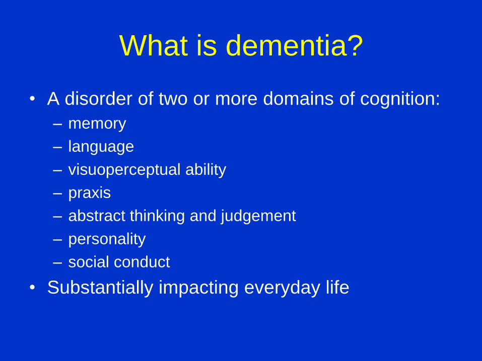
What is dementia?
• A disorder of two or more domains of cognition:
– memory
– language
– visuoperceptual ability
– praxis
– abstract thinking and judgement
– personality
– social conduct
• Substantially impacting everyday life
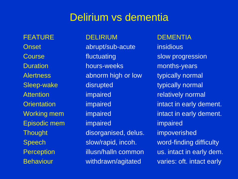
Delirium vs dementia
FEATURE DELIRIUM DEMENTIA
Onset abrupt/sub-acute insidious
Course fluctuating slow progression
Duration hours-weeks months-years
Alertness abnorm high or low typically normal
Sleep-wake disrupted typically normal
Attention impaired relatively normal
Orientation impaired intact in early dement.
Working mem impaired intact in early dement.
Episodic mem impaired impaired
Thought disorganised, delus. impoverished
Speech slow/rapid, incoh. word-finding difficulty
Perception illusn/halln common us. intact in early dem.
Behaviour withdrawn/agitated varies: oft. intact early

Cortical vs subcortical dementia
FUNCTION CORTICAL SUBCORTICAL
eg AD eg HD
Alertness normal ‘slowed up’
Attention normal early impaired
Executive ftn normal early impaired
Episodic mem amnesia forgetfulness
language aphasic reduced output
Praxis apraxia relatively normal
Perception + vis/sp impaired impaired
Personality preserved (unless apathetic, inert
frontal type)

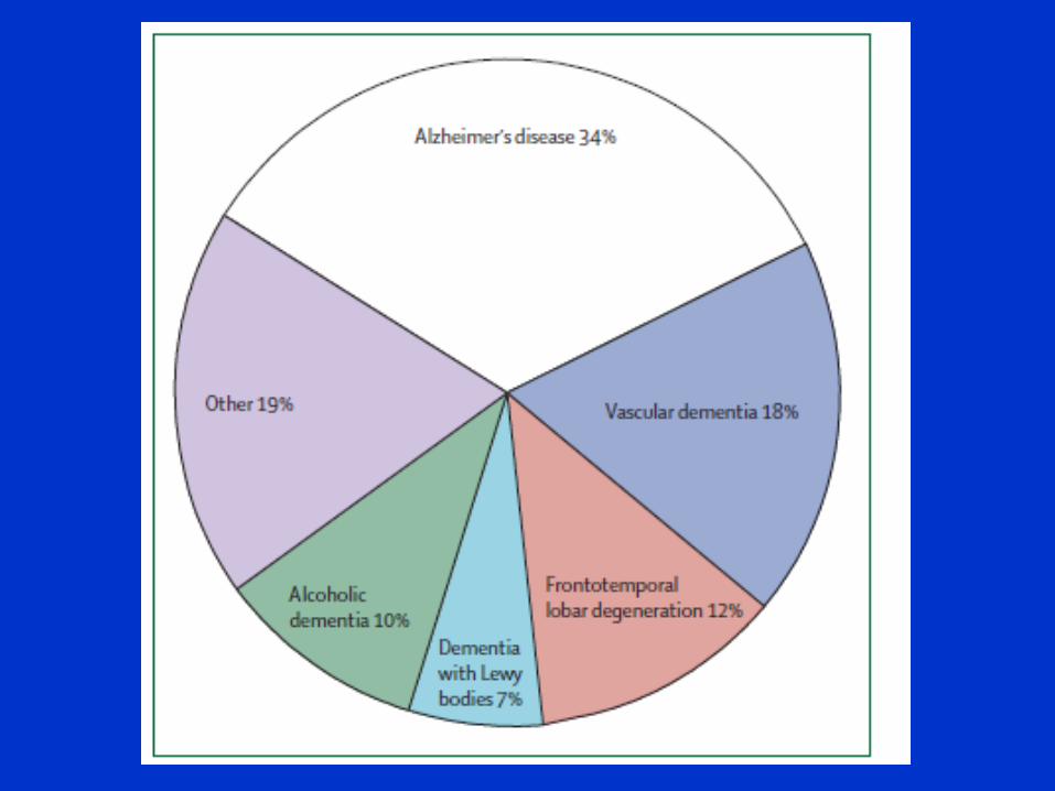
Causes of ‘dementia’
• Inherited HD, Wilson’s, leucodystrophies
• Primary degen Alzheimer’s, Cortical Lewy Body disease , Fronto-temporal dementia
• Vascular multi-infarct, subcortical, strategic infarction
• Infective HIV, TSE, HSE, Whipple’s, SSPE
• Inflammatory MS, vasculitis, Hashimoto’s, limbic encephalitis
• Neoplastic 1o/2
o CNS tumours, limbic encephalitis
• Traumatic Post head injury
• Structural hydrocephalus, chronic subdurals
• Physiological Epileptic amnesia (EAS, TEA)
• Metabol/endoc hypothyroidism
• Deficiency B12/folate
• Sleep-related OSA
• Substances/drugs alcohol, anticholinergics, hypnotics etc
• Psychiatric depression (pseudo-dementia)

History
General
examination
Systemic
Function
Cognitive
Function
Cognitive
examination
Neurological
examination
Neurological
Function
Thought
Mood
Personality
Behaviour
Neuropsychiatric
examination

Alzheimer’s disease
• Episodic memory impairment -> widespread cognitive decline
• apathy, disinhibition, agitation; psychosis; mood disturbance
• slowly progressive: circa 3 point MMSE decline/year
• pyramidal, extrapyramidal signs; primitive reflexes; epilepsy
• neuritic plaques: Abeta amyloid derived from APP
• neurofibrillary tangles: hyperphosphorylated tau
• Cholinergic deficit
• <5% autosomal dominant: presenilin 1(14), 2(1), APP (21)
• Apolipoprotein E alleles 2, 3 and 4; Down’s syndrome; vascular risk
factors
• CT, MRI, SPECT, PET, amyloid imaging
• CSF biomarkers
• central Achase inhibitors; memantine


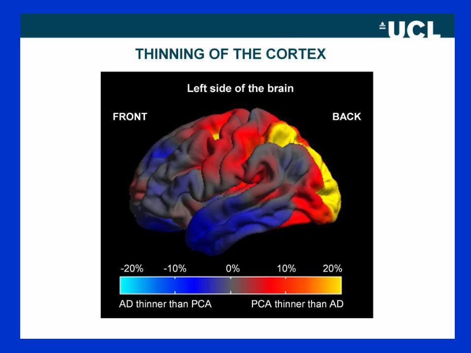
Atypical presentations of AD
• <10% AD
• Posterior cortical atrophy (PCA)
– Almost always due to AD
• Slowly progressive aphasia
– More varied pathology
– Usually non-fluent, occasionally fluent in AD
• Slowly progressive apraxia
• Dysexecutive or ‘behavioural’ presentation
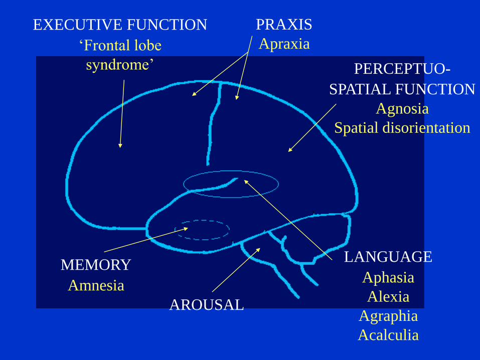
MEMORY
Amnesia
EXECUTIVE FUNCTION
‘Frontal lobe
syndrome’
PRAXIS
Apraxia
PERCEPTUO-
SPATIAL FUNCTION
Agnosia
Spatial disorientation
LANGUAGE
Aphasia
Alexia
Agraphia
Acalculia
AROUSAL

Posterior cortical atrophy - PCA
• Circa 5% AD cases have ‘visual presentation’
• Early onset, typically mid 50s-early 60s
• Mild female predominance
• Most common features are
– Alexia and agraphia
– Simultanagnosia
– Optic ataxia
• ‘dorsal stream’ symptoms and signs predominate but
both ventral and dorsal streams affected
• Relative preservation of memory, insight, language,
executive function


Vascular dementia
• subcortical ischaemic dementia: cognitive slowing, attentional and
executive impairment, gait disturbance
• multi-infarct dementia: mixed cortical/subcortical features, fluctuating
cognition, pseudobulbar palsy/affect, gait/bladder disturbance
• strategic infarction eg thalamus, basal forebrain, left angular gyrus
• vascular risk factors including thrombophilia, vasculitis, CADASIL
• Imaging - MRI
• manage vascular risk factors

CADASIL


Dementia with Lewy bodies
• Cognitive decline + Parkinsonism, visual hallucinations, fluctuations
(two features -> probable DLB, one feature possible DLB)
• Neuropsychology: impaired psychomotor speed, attention, executive
function, visuospatial ability
• REM sleep behaviour disorder
• more rapidly progressive than pure AD
• Lewy body: eosinophilic intraneuronal inclusion bodies, mainly
aggregated filaments of alpha-synuclein
• DLB and Lewy body variant of AD
• Neurochemical pathology
• The therapeutic dilemma
• Achase inhibitors


Atypical dementia
• Features atypical of a common cause !
– Age
– family history
– systemic features (apart from vascular disease)
– rapidity
– syndromes eg:
• cognitive decline, epilepsy, chorea, oral self-mutilation
• frontal lobe dementia with bulbar and pseudo-bulbar palsy
• apraxia, limb myoclonus, alien limb
• falls, axial rigidity, supranuclear gaze palsy, subcortical dementia


Frontotemporal dementia
• 10-15% dementia < 65 years, 25-50% familial
• Frontal lobe (behavioural) variant
– personality and behavioural change with loss of insight
• Temporal lobe variant
– L: semantic dementia
– R: recognition + knowledge of people
• Progressive non-fluent aphasia
• FTD with MND
• NB relative preservation of episodic memory
• Pathologies – see Appendix!
• Imaging: focal atrophy

Frontotemporal dementia
• FH in 25-50% cases
• Autosomal dominant FTD
• Microtubule associated protein gene (MAPT)
• Progranulin
• Valosin-containing protein
• Charged multi-vesicular body protein 2B
• C9ORF72 – ~35% familial ALS, 25% familial FTD
– NB association with psychosis

Recommended nomeclature for FTLD (Neumann et al, 2009)
Previous
terminology
Recommended
terminology
Major
pathological
subtypes
Associated
gene
Tau +ve FTLD PiD
CBD
PSP
AgD
MSTD
FTDP-17
MAPT
Tau –ve FTLD FTLD-U, TDP-43 +ve
FTLD-U, TDP-43 –ve
NIFID
DLDH
Other (BIBD)
FTLD-TDP
FTLD-UPS
FTLD-IF
FTLD-ni
BIBD
Type 1 (SD)
Type 2 (FTD +/- MND)
Type 3 (FTD/PNFA)
Type 4 (IBMPFD)
aFTLD-U
FTD-3
C9ORF72
GRN
VCP
CHMP2B

PSP + CBD
• PSP • supranuclear gaze palsy
• truncal rigidity, instability,
akinesia, falls
• bulbar features
• subcortical dementia
• mood, personality, behaviour
• neurofibrillary tangles (tau) in
basal ganglia and brain stem
• MRI: midbrain atrophy
• CBD • asymmetric limb apraxia
• alien limb phenomena
• limb myoclonus
• Parkinsonism
• cognitive impairment
• neurofibrillary tangles (tau) in
frontal and parietal cortex and
basal ganglia
• MRI: frontoparietal atrophy

PSP

PSP


Huntington’s disease
• autosomal dominant, variable age of onset, anticipation
• chorea -> other extrapyramidal + features; Westphal variant;
epilepsy; subcortical dementia; depression, apathy, agressivity
common, psychosis, obsessional behaviour, suicide in a minority:
progression to immobility and dementia over 15-20 years
• loss of small striatal neurons + cortical neurons
• CAG repeat expansion in Huntingtin (IT-15) gene chromosome 4:10-
35 normal, 36-39 +/- pathogenic, >39 symptomatic, 27-35 unstable
• anticipation especially with paternal transmission

Prion dementia
• spCJD - rapid dementia, ataxia, visual syndromes, multifocal signs,
myoclonus: usually 55-70 yrs, median duration circa 4 months
• vCJD - psychiatric prodrome, dysaesthesiae, cognitive impairment,
multifocal signs, myoclonus: 2nd - 4th decades, med durn 14 m.
• iatrogenic and familial CJD; GSS; FFI; kuru
• Pathology
• The prion gene (chromosome 20) and the prion hypothesis
• Investigations: EEG in spCJD; CSF 14-3-3 in spCJD, tau in vCJD;
MRI; tonsillar and brain biopsy


Sporadic CJD

HIV-1 associated dementia
• 20-30% AIDS patients present with or develop
• insidious, subcortical: poor concentration, slowing, forgetfulness,
apathy, social withdrawal; pyramidal and cerebellar signs
• tends to occur with CD4 count < 200 x 106
• Early entry of HIV-1 to CNS -> persistence in macrophages and
microglia -> multinucleated giant cells; white matter affected focally
(MGCs) or diffusely; cortical neuronal loss
• MRI, CSF
• Differential diagnosis: depression, drugs, substance abuse, systemic
illness, CNS tumours, opportunistic infection (toxoplasmosis, TB,
cryptococcus, CMV, syphilis, PML).


Hydrocephalus
• large head
• headache
• hydrocephalic attacks + death
• visual failure
• gait disturbance
• incontinence
• subcortical dementia
• communicating, obstructive, compensated, normal pressure
• Imaging, lumbar puncture, CSF pressure studies
• Shunting


Wilson’s disease
• rare, treatable, autosomal recessive
• usually presents in childhood or adolescence but up to 5th decade
• psychiatry: personality change, psychosis, dementia
• neurology: extrapyramidal features (tremor, dysarthria, drooling,
rigidity, bradykinesia, dystonia)
• KF rings
• liver failure
• copper transporting ATP-ase on chromosome 13 -> copper
deposition especially in GP and putamen
• copper chelation: penicillamine, trientine, zinc: controversial


Obstructive Sleep Apnoea
• Common in middle age
• Clues
– snoring history
– Daytime sleepiness – see ESS
– Morning headache
– Befuddlement
• Sleep study
• CPAP

Daytime sleepiness: Epworth Sleepiness Scale
– Sitting and reading
– Watching TV
– Sitting inactive in a public place eg theatre, meeting
– Passenger in a car for an hour
– Lying down to rest in the afternoon
– Sitting and talking to someone
– Sitting quietly after lunch
– In a car while stopped in traffic
• 0 = would never dose
• 1 = slight chance of dosing
• 2 = moderate chance
• 3 = high chance
Transient Epileptic Amnesia (Butler et al Ann Neurol 2007)
• Onset in later life
• Male predominance
• Attacks 30-60 minutes
• Attacks one/month
• Attacks on waking common
• Amnesia can be sole feature
• +/- olfactory hallucinations, automatisms, brief unresponsiveness
• Partial recall common
• Excellent treatment response
• Diagnosis usually delayed
• Interictal memory complaints usual – 2/3 Autobiographical amnesia
– 1/2 Accelerated forgetting
– 1/3 Topographical amnesia

TEA: Neuroimaging Butler C, Zeman A Nat Clin Pract Neurol 2008;4:517-521
Peri-ictal MRI Peri-ictal FDG-PET
One month later
L
L L
L
L R

Young and atypical dementia
• Wide differential diagnosis
• Broad assessment required
• Degenerative dementia as a network disease
• Some treatable conditions present as dementia
• Rossor et al Lancet Neurol 2010; 9: 793–806
