case report late onset atypical pantothenate-kinase...
TRANSCRIPT

Hindawi Publishing CorporationCase Reports in Neurological MedicineVolume 2013, Article ID 860201, 5 pageshttp://dx.doi.org/10.1155/2013/860201
Case ReportLate Onset Atypical Pantothenate-Kinase-AssociatedNeurodegeneration
Natalie Diaz
Los Angeles Biomedical Institute, Harbor-UCLA Medical Center, Box No. 492, 1000 W. Carson Street, Los Angeles,Torrance, CA 90509, USA
Correspondence should be addressed to Natalie Diaz; [email protected]
Received 11 February 2013; Accepted 1 March 2013
Academic Editors: R. Hashimoto, J. C. Kattah, and M. Toft
Copyright © 2013 Natalie Diaz. This is an open access article distributed under the Creative Commons Attribution License, whichpermits unrestricted use, distribution, and reproduction in any medium, provided the original work is properly cited.
Introduction. Pantothenate-kinase-associated neurodegeneration (PKAN) is a rare genetic disease and a form of neurodegenerationwith brain iron accumulation (NBIA). It most commonly begins in the first two decades of life but should be considered in thedifferential diagnosis of patients at any age with an atypical progressive extrapyramidal disorder and cognitive impairment. Fewlate-adult cases have been reported. Case Report. A 50-year-old woman presented with a history of progressive dysarthria anddysphagia secondary to orolingual dystonia. Initial work-up was normal. There was no family history. Her initial symptoms werefollowed by the onset of blepharospasm, cervical dystonia, Parkinsonism, and cognitive impairment. Follow-upMRI four years afterpresentation revealed the diagnostic “eye-of-the-tiger” sign. Genetic testing confirmed a homozygousmissensemutation consistentwith the diagnosis of PKAN. Conclusion. Although PKAN is a rare genetic disorder most commonly seen in childhood, it shouldbe considered in adult patients with a history of progressive focal dystonia or atypical Parkinsonism. As the radiographic findingsare quite characteristic, genetic testing should be performed if the MRI shows evidence of iron accumulation. Optimal treatmentstrategies are not known, and at the current time therapies should be directed at the specific manifestations of the disease.
1. Introduction
Pantothenate-kinase-associated neurodegeneration (PKAN)is a rare, autosomal recessive disorder that most commonlybegins in the first two decades of life with progressiveextrapyramidal manifestations. It is part of a group of disor-ders under the umbrella term neurodegeneration with brainiron accumulation (NBIA). Originally called Hallervorden-Spatz Syndrome (HSS) after the two German pathologistswho first published descriptions of a progressive extrapyra-midal disorder associated with pathological iron depositionin the globus pallidus and substantia nigra pars reticulata [1],it was later renamed NBIA in 2003 to dissociate the disorderfrom atrocities committed during the second World War.With the discovery of the first genetic mutation found inthe pantothenate kinase 2 (PANK2 gene) in 2001 [2], NBIApatients with this mutation were subclassified under thenew term pantothenate kinase-associated neurodegeneration(PKAN). NBIA now includes (a) PKAN (also called NBIA1)
caused by mutations in the PANK2 gene, (b) PLA2G6-associated neurodegeneration (PLAN, NBIA2) due to phos-pholipaseA2mutations, (c) neuroferritinopathy due tomuta-tions in the ferritin light chain (FTL) gene, (d) aceruloplas-minemia due to amutation in the ceruloplasmin gene, and (e)sporadic cases of NBIA in which the genetic background hasnot been identified. PKAN is thought to be themost commonform of NBIA and accounts for more than 50% of cases ofNBIA [3]. PKANandPLAN typically have onset in childhoodwhile aceruloplasminemia and neuroferritinopathy typicallypresent at an older age in the fifth and sixth decade. Here wedescribe a patient who presented in her late fifth decade withsymptoms of a childhood onset NBIA disorder.
2. Case Report
A 50-year-old woman was referred for neurological eval-uation with a two-year history of progressive dysarthriaand dysphagia. Two years after the onset of symptoms

2 Case Reports in Neurological Medicine
she was no longer able to sing in church, her speech wasdifficult to understand, she had difficulty eating, and shecomplained of throat pain. There was no history of headtrauma, no family history of movement disorders, and nohistory of consanguinity. On examination the patient hadinvoluntary twisting movements of the tongue consistentwith orolingual dystonia. Her laboratory workup includednormal serum copper, ceruloplasmin, and ferritin levels andno acanthocytes were found on a blood smear. Initial neu-roimaging showed no abnormalities. The patient was triedon trihexyphenidyl, diazepam, and carbidopa/levodopa all ofwhich offered minimal relief. Botulinum toxin A injectionswere effective in treating the orolingual dystonia to a pointthat allowed her to eat and sing with minimal difficulty fortwo-month periods. Three years after the initial symptomsshe developed blepharospasm and cervical dystonia followeda year later by the appearance of progressive and symmetricalParkinsonism. Examination also revealed mild cognitiveimpairment. Due to her progressive symptoms, a follow-up brain MRI was performed and found to show bilateraland symmetrical T2 weighted hypodensities in the globuspallidus with a medial area of hyperintensity consistent withthe “eye of the tiger” sign (see Figure 1). This new imagingfinding prompted a referral for genetic testing at the age of 54which revealed a known homozygous pathogenic mutations(881A>T/p.N294I) confirming the diagnosis of PKAN.
3. Discussion
Neurodegeneration with brain iron accumulation (NBIA)is an umbrella term for a group of disorders that presentwith a progressive extrapyramidal syndrome associated withabnormal iron accumulation in the brain, especially the basalganglia. The main syndrome among the NBIA disorders isPKAN which accounts for more than half the cases of NBIA[3].
Based on clinical features, PKAN can be classified(Table 1) into (a) classic PKAN, with onset in the first decadeand a fairly rapid and progressive course leading to lossof independence ten to fifteen years after onset, or (b)atypical PKAN with onset in the second or third decadeand a slower disease course that may last 40 years [3].In both forms, focal dystonias are a common presentingand prominent feature. Children with the classic form oftenpresent with a clumsy gait due to limb dystonia. About twothirds of childrenwith classic PKANalso develop pigmentaryretinopathy early in the disease which may cause blindness[3]. As the disease progresses, corticospinal tract signs suchas spasticity, hyper-reflexia, and extensor plantar toes becomesupportive in the diagnosis. Orolingual dystonia, leading todysarthria and dysphagia, can be seen in both forms but is acommon presenting feature in atypical patients. Psychiatricsymptoms are common in the atypical form and can oftenpredate any motor features and may present as depression,anxiety, emotional lability, obsessive compulsive disorder, orpsychosis [4–6]. Progressive cognitive impairment occursconcurrently in both forms. A limited number of reports ofPANK2 mutation positive late adult-onset cases (see Table 2)
Table 1: Clinical presentation of PKAN.
Features Typical PKAN Atypical PKANOnset First decade Second or third decade
Features
Gait impairment, focaldystonia, pyramidaldysfunction, pigmentaryretinopathy, andcognitive impairment
Psychiatric symptoms,focal dystonia, ±parkinsonism or chorea,cognitive impairment,late gait dysfunction
Progression
Rapid progressionPeriods of stabilityinterspersed withperiods of rapidprogression.Loss of ambulationoccurs 10 to 15 years afteronset
Slower progressionLoss of ambulation after15 to 40 years after onset
Imaging Eye-of-the-tiger Eye-of-the-tiger
have been published [5, 7–10]. As far as we are aware, ourpatient is the oldest onset casewith themutation that has beenreported.
The majority of cases of PKAN have a characteristicMR imaging finding known as the eye-of-the-tiger sign (seeFigure 1). Abnormal iron accumulation is seen as diffuseand bilateral low T2 weighted signal intensity in the globuspallidus (internal and external segments) surrounding acentral area of high T2 weighted signal intensity in theanteromedial globus pallidus corresponding to neuronal lossand gliosis and producing the image of an eye-of-the-tiger[11]. MRI abnormalities may be found early in mutationpositive patients [12] but sometimes can lag behind clinicalsymptoms and, as in our described case, not be seen on initialimaging [13]. The central T2 hyperintensity may be transientand fade with time while evidence of iron accumulation seenas diffuse low T2 signal in the globus pallidus appears laterand does not fade [12]. The eye-of-the-tiger was previouslyconsidered to have a one-to-one correlationwith the presenceof a positive PANK2 mutation but more recently severalPANK2 negative eye-of-the-tiger cases have been reported[14–17] most of which have been late onset or atypical cases.Therefore, evidence of iron accumulation in the brain may beseen as an indication for genetic testing.
PKAN is an autosomal recessive disorder caused bymutations in the pantothenate kinase 2 (PANK2) gene onchromosome 20 [18]. Numerous mutations in the PANK2gene have been identified [3]. Homozygous null mutations(resulting in protein truncation) result in classic early onsetdisease with rapid progression and missense mutations thatlikely result in partial enzyme function have been associ-ated with atypical late onset disease and slower progression[3, 14]. The mechanism by which PANK2 gene mutationscause abnormal iron accumulation and neurodegenerationis unclear. PANK2 is mainly targeted to the mitochondria[19, 20] and its protein product catalyzes the phosphorylationof pantothenate (vitamin B5) to phosphopantothenate, thefirst and rate limiting step of coenzyme A biosynthesis.Coenzyme A is an essential cofactor in several metabolic

Case Reports in Neurological Medicine 3
(a) (b)
(c) (d)
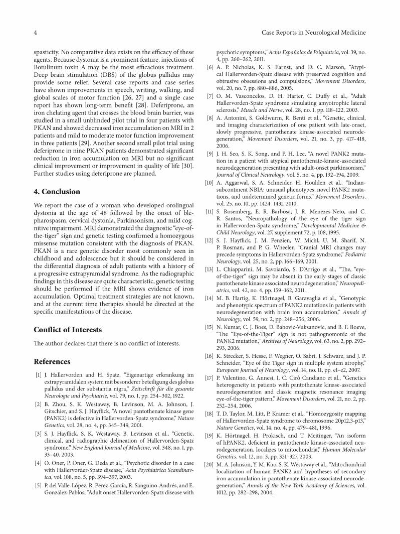
Figure 1: (a) Axial T1 weighted, (b) coronal T2 weighted, and (c) axial fluid attenuation inversion recovery MRI showing low signal intensityin the bilateral globus pallidus with a medial area of signal intensity, presenting the classic “eye-of-the-tiger” sign. (d) Axial fluid attenuationinversion recovery MRI shows bilateral low signal intensity in the substantia nigra pars reticulata.
Table 2: Published reports of late adult-onset atypical PKAN.
Reference Age of onset Clinical Eye-of-the-Tiger
PANK2mutation
Vasconcelos et al. 2003 [7] 36 Dysarthria, tongue atrophy Yes YesAntonini et al. 2006 [8] 30 Choreoathetosis, postural tremor, personality changes, and paranoia Yes YesSeo et al. 2009 [9] 35 Parkinsonism Yes YesAggarwal et al. 2010 [10] 37 Postural/action tremor Yes Yesdel Valle-Lopez et al. 2011 [5] 30 Acute psychosis, clumsiness, and frequent falls Yes Yes
pathways including the citric acid cycle, steroid and hemebiosynthesis, amino acid metabolism, and beta-oxidation offatty acids. Metabolic profiling of patients with PKAN showsmitochondrial dysfunction with an elevated lactate/pyruvateratio and a deficiency of fatty acids necessary for cellularmembrane synthesis [21]. Elevated levels of cysteine, whichnormally conjugates with phosphopantothenate, have alsobeen observed in PKAN patients [22] and may chelateiron and result in free radical production. Animal modelsof PANK2 deficiency, PANK2 knock-out mice [23], and adrosophila fruit fly model [24] have failed to reproduce theclinical syndrome seen in humans. In contrast, both Pank2
mutant and wild type mice fed pantothenate (vitamin B5)deficient diets resulted in a progressive movement disorderin wild type mice and an early death in PANK2 mutant miceindicating the importance of pantothenate metabolism inPKAN [25].
Current treatment for all NBIA disorders consists ofsymptomatic relief. Anticholinergic medications, such astrihexyphenidyl and benztropine, help reduce rigidity, dys-tonia, and tremor. Baclofen, both orally and intrathecally, ishelpful in treating spasticity. Dopaminergic medications mayhelp if there is coexistent Parkinsonism. Benzodiazepinesmay be useful in the treatment of chorea, tremor, and
4 Case Reports in Neurological Medicine
spasticity. No comparative data exists on the efficacy of theseagents. Because dystonia is a prominent feature, injections ofBotulinum toxin A may be the most efficacious treatment.Deep brain stimulation (DBS) of the globus pallidus mayprovide some relief. Several case reports and case serieshave shown improvements in speech, writing, walking, andglobal scales of motor function [26, 27] and a single casereport has shown long-term benefit [28]. Deferiprone, aniron chelating agent that crosses the blood brain barrier, wasstudied in a small unblinded pilot trial in four patients withPKAN and showed decreased iron accumulation onMRI in 2patients and mild to moderate motor function improvementin three patients [29]. Another second small pilot trial usingdeferiprone in nine PKAN patients demonstrated significantreduction in iron accumulation on MRI but no significantclinical improvement or improvement in quality of life [30].Further studies using deferiprone are planned.
4. Conclusion
We report the case of a woman who developed orolingualdystonia at the age of 48 followed by the onset of ble-pharospasm, cervical dystonia, Parkinsonism, and mild cog-nitive impairment.MRI demonstrated the diagnostic “eye-of-the-tiger” sign and genetic testing confirmed a homozygousmissense mutation consistent with the diagnosis of PKAN.PKAN is a rare genetic disorder most commonly seen inchildhood and adolescence but it should be considered inthe differential diagnosis of adult patients with a history ofa progressive extrapyramidal syndrome. As the radiographicfindings in this disease are quite characteristic, genetic testingshould be performed if the MRI shows evidence of ironaccumulation. Optimal treatment strategies are not known,and at the current time therapies should be directed at thespecific manifestations of the disease.
Conflict of Interests
The author declares that there is no conflict of interests.
References
[1] J. Hallervorden and H. Spatz, “Eigenartige erkrankung imextrapyramidalen systemmit besonderer beteiligung des globuspallidus und der substantia nigra,” Zeitschrift fur die gesamteNeurologie und Psychiatrie, vol. 79, no. 1, pp. 254–302, 1922.
[2] B. Zhou, S. K. Westaway, B. Levinson, M. A. Johnson, J.Gitschier, and S. J. Hayflick, “A novel pantothenate kinase gene(PANK2) is defective in Hallervorden-Spatz syndrome,”NatureGenetics, vol. 28, no. 4, pp. 345–349, 2001.
[3] S. J. Hayflick, S. K. Westaway, B. Levinson et al., “Genetic,clinical, and radiographic delineation of Hallervorden-Spatzsyndrome,”New England Journal of Medicine, vol. 348, no. 1, pp.33–40, 2003.
[4] O. Oner, P. Oner, G. Deda et al., “Psychotic disorder in a casewith Hallervorder-Spatz disease,” Acta Psychiatrica Scandinav-ica, vol. 108, no. 5, pp. 394–397, 2003.
[5] P. del Valle-Lopez, R. Perez-Garcıa, R. Sanguino-Andres, and E.Gonzalez-Pablos, “Adult onset Hallervorden-Spatz disease with
psychotic symptoms,”Actas Espanolas de Psiquiatrıa, vol. 39, no.4, pp. 260–262, 2011.
[6] A. P. Nicholas, K. S. Earnst, and D. C. Marson, “Atypi-cal Hallervorden-Spatz disease with preserved cognition andobtrusive obsessions and compulsions,” Movement Disorders,vol. 20, no. 7, pp. 880–886, 2005.
[7] O. M. Vasconcelos, D. H. Harter, C. Duffy et al., “AdultHallervorden-Spatz syndrome simulating amyotrophic lateralsclerosis,”Muscle and Nerve, vol. 28, no. 1, pp. 118–122, 2003.
[8] A. Antonini, S. Goldwurm, R. Benti et al., “Genetic, clinical,and imaging characterization of one patient with late-onset,slowly progressive, pantothenate kinase-associated neurode-generation,” Movement Disorders, vol. 21, no. 3, pp. 417–418,2006.
[9] J. H. Seo, S. K. Song, and P. H. Lee, “A novel PANK2 muta-tion in a patient with atypical pantothenate-kinase-associatedneurodegeneration presenting with adult-onset parkinsonism,”Journal of Clinical Neurology, vol. 5, no. 4, pp. 192–194, 2009.
[10] A. Aggarwal, S. A. Schneider, H. Houlden et al., “Indian-subcontinent NBIA: unusual phenotypes, novel PANK2 muta-tions, and undetermined genetic forms,” Movement Disorders,vol. 25, no. 10, pp. 1424–1431, 2010.
[11] S. Rosemberg, E. R. Barbosa, J. R. Menezes-Neto, and C.R. Santos, “Neuropathology of the eye of the tiger signin Hallervorden-Spatz syndrome,” Developmental Medicine &Child Neurology, vol. 27, supplement 72, p. 108, 1995.
[12] S. J. Hayflick, J. M. Penzien, W. Michl, U. M. Sharif, N.P. Rosman, and P. G. Wheeler, “Cranial MRI changes mayprecede symptoms in Hallervorden-Spatz syndrome,” PediatricNeurology, vol. 25, no. 2, pp. 166–169, 2001.
[13] L. Chiapparini, M. Savoiardo, S. D’Arrigo et al., “The, “eye-of-the-tiger” sign may be absent in the early stages of classicpantothenate kinase associated neurodegeneration,”Neuropedi-atrics, vol. 42, no. 4, pp. 159–162, 2011.
[14] M. B. Hartig, K. Hortnagel, B. Garavaglia et al., “Genotypicand phenotypic spectrum of PANK2mutations in patients withneurodegeneration with brain iron accumulation,” Annals ofNeurology, vol. 59, no. 2, pp. 248–256, 2006.
[15] N. Kumar, C. J. Boes, D. Babovic-Vuksanovic, and B. F. Boeve,“The “Eye-of-the-Tiger” sign is not pathognomonic of thePANK2mutation,”Archives of Neurology, vol. 63, no. 2, pp. 292–293, 2006.
[16] K. Strecker, S. Hesse, F. Wegner, O. Sabri, J. Schwarz, and J. P.Schneider, “Eye of the Tiger sign in multiple system atrophy,”European Journal of Neurology, vol. 14, no. 11, pp. e1–e2, 2007.
[17] P. Valentino, G. Annesi, I. C. Ciro Candiano et al., “Geneticsheterogeneity in patients with pantothenate kinase-associatedneurodegeneration and classic magnetic resonance imagingeye-of-the-tiger pattern,”Movement Disorders, vol. 21, no. 2, pp.252–254, 2006.
[18] T. D. Taylor, M. Litt, P. Kramer et al., “Homozygosity mappingof Hallervorden-Spatz syndrome to chromosome 20p12.3-p13,”Nature Genetics, vol. 14, no. 4, pp. 479–481, 1996.
[19] K. Hortnagel, H. Prokisch, and T. Meitinger, “An isoformof hPANK2, deficient in pantothenate kinase-associated neu-rodegeneration, localizes to mitochondria,” Human MolecularGenetics, vol. 12, no. 3, pp. 321–327, 2003.
[20] M. A. Johnson, Y.M. Kuo, S. K.Westaway et al., “Mitochondriallocalization of human PANK2 and hypotheses of secondaryiron accumulation in pantothenate kinase-associated neurode-generation,” Annals of the New York Academy of Sciences, vol.1012, pp. 282–298, 2004.

Case Reports in Neurological Medicine 5
[21] V. Leoni, L. Strittmatter, G. Zorzi et al., “Metabolic conse-quences of mitochondrial coenzyme A deficiency in patientswith PANK2 mutations,” Molecular Genetics and Metabolism,vol. 105, no. 3, pp. 463–471, 2012.
[22] T. L. Perry, M. G. Norman, and V. W. Yong, “Hallervorden-Spatz disease: cysteine accumulation and cysteine dioxygenasedeficiency in the globus pallidus,” Annals of Neurology, vol. 18,no. 4, pp. 482–489, 1985.
[23] Y. M. Kuo, J. L. Duncan, S. K. Westaway et al., “Deficiencyof pantothenate kinase 2 (Pank2) in mice leads to retinaldegeneration and azoospermia,” Human Molecular Genetics,vol. 14, no. 1, pp. 49–57, 2005.
[24] Y. Yang, Z. Wu, Y. M. Kuo, and B. Zhou, “Dietary rescue offumble-aDrosophilamodel for pantothenate-kinase-associatedneurodegeneration,” Journal of Inherited Metabolic Disease, vol.28, no. 6, pp. 1055–1064, 2005.
[25] Y. M. Kuo, S. J. Hayflick, and J. Gitschier, “Deprivation ofpantothenic acid elicits a movement disorder and azoospermiain a mouse model of pantothenate kinase-associated neurode-generation,” Journal of Inherited Metabolic Disease, vol. 30, no.3, pp. 310–317, 2007.
[26] P. Castelnau, L. Cif, E. M. Valente et al., “Pallidal stimulationimproves pantothenate kinase-associated neurodegeneration,”Annals of Neurology, vol. 57, no. 5, pp. 738–741, 2005.
[27] C. Isaac, I. Wright, D. Bhattacharyya, P. Baxter, and J. Rowe,“Pallidal stimulation for pantothenate kinase-associated neu-rodegeneration dystonia,” Archives of Disease in Childhood, vol.93, no. 3, pp. 239–240, 2008.
[28] M. Krause, W. Fogel, V. Tronnier et al., “Long-term benefit topallidal deep brain stimulation in a case of dystonia secondaryto pantothenate kinase-associated neurodegeneration,” Move-ment Disorders, vol. 21, no. 12, pp. 2255–2257, 2006.
[29] G. Abbruzzese, G. Cossu, M. Balocco et al., “A pilot trial ofdeferiprone for neurodegeneration with brain iron accumula-tion,” Haematologica, vol. 96, no. 11, pp. 1708–1711, 2011.
[30] G. Zorzi, F. Zibordi, L. Chiapparini et al., “Iron-related MRIimages in patients with pantothenate kinase-associated neu-rodegeneration (PKAN) treated with deferiprone: results of aphase II pilot trial,”Movement Disorders, vol. 26, no. 9, pp. 1755–1759, 2011.

Submit your manuscripts athttp://www.hindawi.com
Stem CellsInternational
Hindawi Publishing Corporationhttp://www.hindawi.com Volume 2014
Hindawi Publishing Corporationhttp://www.hindawi.com Volume 2014
MEDIATORSINFLAMMATION
of
Hindawi Publishing Corporationhttp://www.hindawi.com Volume 2014
Behavioural Neurology
EndocrinologyInternational Journal of
Hindawi Publishing Corporationhttp://www.hindawi.com Volume 2014
Hindawi Publishing Corporationhttp://www.hindawi.com Volume 2014
Disease Markers
Hindawi Publishing Corporationhttp://www.hindawi.com Volume 2014
BioMed Research International
OncologyJournal of
Hindawi Publishing Corporationhttp://www.hindawi.com Volume 2014
Hindawi Publishing Corporationhttp://www.hindawi.com Volume 2014
Oxidative Medicine and Cellular Longevity
Hindawi Publishing Corporationhttp://www.hindawi.com Volume 2014
PPAR Research
The Scientific World JournalHindawi Publishing Corporation http://www.hindawi.com Volume 2014
Immunology ResearchHindawi Publishing Corporationhttp://www.hindawi.com Volume 2014
Journal of
ObesityJournal of
Hindawi Publishing Corporationhttp://www.hindawi.com Volume 2014
Hindawi Publishing Corporationhttp://www.hindawi.com Volume 2014
Computational and Mathematical Methods in Medicine
OphthalmologyJournal of
Hindawi Publishing Corporationhttp://www.hindawi.com Volume 2014
Diabetes ResearchJournal of
Hindawi Publishing Corporationhttp://www.hindawi.com Volume 2014
Hindawi Publishing Corporationhttp://www.hindawi.com Volume 2014
Research and TreatmentAIDS
Hindawi Publishing Corporationhttp://www.hindawi.com Volume 2014
Gastroenterology Research and Practice
Hindawi Publishing Corporationhttp://www.hindawi.com Volume 2014
Parkinson’s Disease
Evidence-Based Complementary and Alternative Medicine
Volume 2014Hindawi Publishing Corporationhttp://www.hindawi.com