yige guo - academic commons
TRANSCRIPT

Molecular mechanisms of mitotic spindle assembly and accurate chromosome segregation
Yige Guo
Submitted in partial fulfillment of the
requirements for the degree of
Doctor of Philosophy
in the Graduate School of Arts and Sciences
COLUMBIA UNIVERSITY
2013

© 2013
Yige Guo
All rights reserved

Abstract
Molecular mechanisms of mitotic spindle assembly and accurate chromosome segregation
Yige Guo
During the cell cycle, duplicated DNA in S phase is segregated, in the form of
chromatids, into two daughter cells in mitosis. The accuracy of chromosome segregation is
essential as two daughter cells have the same genetic contents as the mother cell. Two major
mechanisms are utilized by the cell to ensure accurate chromosome segregation. First,
interactions between the dynamic microtubules and kinetochores, the proteinaceous structures
built on centromeres of mitotic chromosomes that act as the attachment site for microtubules,
serve as major forces to position each pair of chromosomes to the metaphase plate. Secondly, a
surveillance system, known as the mitotic checkpoint, put the anaphase onset on hold until each
pair of sister chromosomes are aligned at the metaphase plate and appropriately attached with
microtubule plus ends by kinetochores.
In the first part (Chapter 2) of this thesis, I illustrate the role of the auto-phosphorylation
of BubR1, a mitotic checkpoint protein, in kinetochore-microtubule attachment and the mitotic
checkpoint. Using a phospho-specific antibody against the auto-phosphorylation site identified
by mass spectrometry, I demonstrate that kinetochore-associated BubR1 phosphorylates itself in
human cells in vivo and that this phosphorylation is dependent on its binding partner, the
kinetochore-associated kinesin motor CENP-E. Studies using cells expressing a non-
phosphorylatable BubR1 mutant revealed that the CENP-E–dependent BubR1 phosphorylation
at unattached kinetochores is important for a full-strength mitotic checkpoint to prevent single
chromosome loss. Furthermore, replacing endogenous BubR1 with the non-phosphorylatable

BubR1 mutant or depletion of CENP-E, the BubR1 kinase activator, results in metaphase
chromosome misalignment and increased incidents of syntelic attachments. Using indirect
immunofluorescence, I have discovered a decreased level of Aurora B–mediated Ndc80
phosphorylation at the kinetochore of cells expressing the non-phosphorylatable BubR1 mutant,
which might contribute to the alignment defect. Moreover, expressing a phosphomimetic BubR1
mutant substantially reduces the incidence of polar chromosomes in CENP-E–depleted cells,
further supporting a signaling cascade function of CENP-E and BubR1 on the kinetochore. Thus,
the state of CENP-E–dependent BubR1 auto-phosphorylation in response to spindle microtubule
capture by CENP-E is important for kinetochore functions in achieving accurate chromosome
segregation.
In the second part (Chapter 3), my colleague and I demonstrate a novel mechanism of
mitotic spindle assembly in Xenopus egg extracts and mammalian cells. I show that the MRN
(Mre11, Rad50, and Nbs1) complex is required for metaphase chromosome alignment.
Consistent with the result of my colleague using Xenopus egg extracts, disruption of MRN
function by depleting Mre11 using an inducible shRNA system, or Mre11 inhibitor mirin,
triggers a metaphase delay and disrupts the RCC1-dependent Ran-GTP gradient. Addition of
mirin to mammalian cells reduces RCC1 association with mitotic chromosomes and changes the
confirmation of RCC1. Thus, the MRN-CtIP pathway contributes to Ran-dependent mitotic
spindle assembly by modulating RCC1 chromosome association.
In summary, my novel findings have revealed a pair of molecular mechanisms not known
previously, which are important to the mitosis field.

i
Contents
Chapter 1. Introduction: Regulation of Chromosome Alignment and the Mitotic Checkpoint in Mitosis . 1
Chromosome alignment and Error correction ....................................................................................... 2
Spindle formation ................................................................................................................................ 3
Kinetochore-microtubule attachment error correction ........................................................................... 5
The Mitotic checkpoint ...................................................................................................................... 12
Aneuploidy ........................................................................................................................................ 14
BubR1 ............................................................................................................................................... 16
CENP-E ............................................................................................................................................ 20
The MRN complex ............................................................................................................................ 21
Chapter 2. CENP-E–dependent BubR1 auto-phosphorylation enhances chromosome alignment and the
mitotic checkpoint ................................................................................................................................. 22
Introduction ....................................................................................................................................... 23
Methods ............................................................................................................................................ 26
Results .............................................................................................................................................. 28
Discussion ......................................................................................................................................... 52
Chapter 3. The MRN-CtIP pathway is required for metaphase chromosome alignment ......................... 60
Introduction ....................................................................................................................................... 61
Methods ............................................................................................................................................ 63
Results .............................................................................................................................................. 67
Discussions ....................................................................................................................................... 82
Chapter 4. Discussions and Future Directions ....................................................................................... 84
The BubR1’s kinase activity requires a co-activator CENP-E ............................................................. 85
CENP-E is a microtubule sensor independent of tension to regulate Aurora B-mediated
phosphorylation prior to end-on attachment ....................................................................................... 96
CENP-E – BubR1 regulates phosphatase recruitment ....................................................................... 100
Reference ............................................................................................................................................ 103

ii
List of Figures
Figure 1.1. Initial interaction between kinetochores and microtubules. ..................................................... 4
Figure 1.2. Correct and incorrect microtubule kinetochore attachment. .................................................... 6
Figure 1.3. A model for establishing proper stable kinetochore-microtubule attachment. ........................ 10
Figure 1.4. Illustration of different domains shows similarity between BubR1 and Bub1. ....................... 16
Figure 2.1. BubR1 phosphorylates itself. ............................................................................................... 31
Figure 2.2. BubR1 auto-phosphorylation at the kinetochore is sensitive to spindle microtubule attachment.
.............................................................................................................................................................. 32
Figure 2.3. BubR1 auto-phosphorylation at the kinetochore is CENP-E dependent. ................................ 33
Figure 2.4. BubR1 auto-phosphorylation is essential for accurate chromosome segregation. ................... 36
Figure 2.5. BubR1 kinase has other substrates that are important for kinetochore function. .................... 37
Figure 2.6. BubR1 auto-phosphorylation is required for efficient kinetochore targeting of Mad2 and a
prolonged mitosis induced by nocodazole. ............................................................................................. 39
Figure 2.7. The nonphosphorylated form of BubR1 reduces the levels of Mad1 association with
unattached kinetochores induced by nocodazole treatment. .................................................................... 40
Figure 2.8. BubR1 auto-phosphorylation is necessary for metaphase chromosome alignment. ................ 43
Figure 2.9. The nonphosphorylated form of BubR1 reduces Aurora B–mediated Ndc80 phosphorylation
at kinetochores. ..................................................................................................................................... 46
Figure 2.10. CENP-E depletion, but not BubR1 depletion, in human cells causes a decrease of Aurora B–
mediated Ndc80 phosphorylation at the kinetochore. ............................................................................. 47
Figure 2.11. The polar chromosome phenotype in CENP-E–depleted cells can be rescued by expression of
a phosphomimetic BubR1 mutant. ......................................................................................................... 51
Figure 2.12. A model for the role of CENP-E–dependent BubR1 auto-phosphorylation at the kinetochore.
.............................................................................................................................................................. 59
Figure 3.1. The MRN Complex Is Essential for Metaphase Chromosome Alignment in Xenopus Egg
Extracts (These experiements were performed by Dr. Lorene Rozier) .................................................... 69
Figure 3.2. Inhibition of MRN Results in Prolonged Metaphase in Mammalian Cells (These experiements
were performed by Dr. Lorene Rozier) .................................................................................................. 72
Figure 3.3. Reducing the expression level of MRE11 causes a metaphase delay in mammalian cultured
cells. ...................................................................................................................................................... 74
Figure 3.4. MRN inhibition disrupts the Ran-GTP gradient during metaphase. ....................................... 76
Figure 3.5. MRN inhibition results in an RCC1 conformational change ................................................. 78
Figure 3.6. MRN inhibition results in a reduction of RCC1 binding to chromatin. .................................. 79
Figure 3.7. MRN functions to stabilize RCC1 interaction with chromosomes during mitosis. ................. 81

iii
.............................................................................................................................................................. 89
Figure 4.1. Sequence analysis reveals the four key amino acids on BubR1 kinase domain crucial for the
catalytic activity. ................................................................................................................................... 89
Figure 4.2. The “GFSGS” motif may substitute the degenerated P-loop and facilitate ATP binding upon
CENP-E binding to BubR1 and the induction of confirmation change. ................................................... 91
Figure 4.3. Zebrafish BubR1 does not possess the crucial Asp in the “HRD” motif in the catalytic loop. 92
Figure 4.4. Phylogenetic analysis of BubR1, Bub1 and CENP-E among different species. ..................... 94
Figure 4.5. Phosphorylation of T608 is not dependent on Aurora B, Mps1, Cdk and Plk1. ..................... 95
Figure 4.5. A model for the role of CENP-E–dependent BubR1 auto-phosphorylation to facilitate initial
microtubule capture. .............................................................................................................................. 97
Figure 4.6. The CENP-E-dependent BubR1 auto-phosphorylation at the kinetochore is sensitive to lateral
microtubule binding prior to end-on attachment. .................................................................................... 99
Figure 4.7. CENP-E-dependent BubR1 auto-phosphorylation at T608 residue controls the access of Cdk1
to T620, which is essential for Plk1 binding and phosphorylation of the KARD domain. ...................... 101

iv
Abbreviation list
APC/C : Anaphase promoting complex/Cyclosome
Bub: Budding Uninhibited by Benzimidazole
BubR1: Bub-Related 1
CENP-E: Centromere protein-E
CPC: Chromosomal passenger complex
GEF: Guanine Nucleotide exchange factor
CtIP: C-terminal binding protein interacting protein
Hec1: Highly Expressed in Cancer protein 1
KD: Kinase dead
KT: Kinetochore
Mad: Mitotic-Arrest Deficient
MCC: Mitotic checkpoint complex
MEF: Mouse embryonic fibroblast
MRN complex: Mre11, Rad50, and Nbs1
MT: Microtubule
RCC1: Regulator of chromosome condensation 1

v
Acknowledgements
First and foremost I want to thank my advisor Dr. Yinghui Mao. It has been an honor to
be his first Ph.D. student. He has always been very supportive to my study during these five
years and I have learnt a lot both inside and outside of the academia from him. He always tried to
help whenever I encountered any problems in my study, and his expertise in this field usually
offered great solutions to address those problems. He is a live example of an excellent scientific
researcher and a mentor, and acted as my role model during my study and training.
The members of the Mao lab have also made great contributions to my study. All of the
current and past members are excellent colleague, who are really nice to work with.
Collaborations among lab members made my project to progress much more efficiently. I
specifically want to acknowledge some lab members who have directly contributed to my thesis
work. Sana Ahmad used to be our lab technician when I joined the lab. She offered so much help
to me and I actually learnt most of my bench techniques from her. She was always very patient
whenever I have any questions regarding to the experiment methods, and helped me a lot with
troubleshooting. Her excellent skills of lab management made my experiment in the lab going on
smoothly. Dr. Jiayin Zhang, one of our former post-docs also contributed to the BubR1 auto-
phosphorylation study. She was actually the one who discovered the auto-phosphorylation site
through Mass Spectrometry and I was lucky enough to take over this project from her. I also
want to thank Dr. Lorene Rozier, the other post-doc when I joined the lab, who I later
collaborated with on the MRN project. She discovered some very interesting phenotypes in
Xenopus egg extracts and I followed up later with mammalian cells. Lorene is a very interesting
person to work with and she has extensive interests like camping, photographing and skiing.

vi
I also appreciate the time and ideas my thesis committee members have contributed to my
Ph.D. work. The committee, consists of great scientists in the cell biology field, Dr. Richard
Vallee, Dr. Gregg Gundersen, Dr. Ronald Liem and Dr. Geri Kreitzer together with my advisor
have provided many pieces of valuable advice to my research. Besides, they also generously
shared some resource in their lab. Dr. Susumu Antoku from the Gundersen lab taught me virus
transfection, which was a key experiment in my later project.
In my later study on the MRN project, I want to thank Dr. Theresa Swayne and Dr. Adam
White in the Shared Resource of Columbia University for their help with the FRET experiment.
No one in the Mao lab has any previous experience with FRET and this experiment is known to
be hard to perform. Adam and Theresa are definitely experts in the imaging field and I was able
to acquire some really nice images and analyzed the data without much difficulty.
I also want to thank Ms. Zaia Sivo, the program coordinator of my graduate program,
who provided a lot of help and advice to me when I was trying to find a lab to rotate in the first
year of my Ph.D. study. She did a great job to keep everyone in the graduate program on track
and made my Ph.D. study trouble-free and so efficient.
Five years was quite a period of time, and Ph.D. study can sometimes be very stressful,
especially when experiments did not work. But I feel my time at Columbia and New York City
enjoyable in large part, and I have made a lot of friends here. I was also an active member of
several student organizations, and I thank them for providing such a good platform for me to
build up my social network.
Lastly, I want to thank my family for all their support and love. As the only child of my
family, I especially want to show gratitude to my parents in China. Without their support, it

vii
would be impossible for me to focus on and complete my study. And I really appreciate their
understanding that I could not visit them very often as children in other families do and again I
want to thank them for their support.
Yige Guo
Columbia University
June 2013

1
Chapter 1*
Introduction: Regulation of Chromosome Alignment and the Mitotic
Checkpoint in Mitosis
* Some contents of this chapter are from the article “New insights into the mechanism for chromosome alignment in metaphase”, Int Rev Cell Mol Biol. 2013;303:237-62, in which the first author is the author of this thesis.

2
Chromosome alignment and Error correction
To ensure equal segregation of chromatids that duplicated in S phase into two daughter
cells during mitosis, the sister chromosome pairs are aligned at the metaphase plate prior to
anaphase onset, with one sister kinetochore attached with microtubules from one pole and the
other sister kinetochore capturing microtubules from the other pole. This step is essential to
silence the mitotic checkpoint, which triggers anaphase onset followed by the separation of sister
chromosomes. The interaction between kinetochores and microtubules, as well as the force
generated at the kinetochore, contribute to chromosome congression and alignment at the
metaphase plate. Kinetochores are initially captured by the spindle microtubule through several
mechanisms. Bi-oriented kinetochore-microtubule attachments are stabilized, whereas incorrect
attachments are destabilized. Here, I will briefly introduce and review currently known models
and mechanisms involved in these processes.

3
Spindle formation
The classic “search-and-capture” model
In metazoan cells, chromosomes are initially captured by microtubules in a “search and
capture” manner. According to this model (Kirschner and Mitchison, 1986), centrosome
nucleated microtubules undergo repeated growth and shrinkage until they are captured and
stabilized by kinetochores (Figure 1.1A). In this process, one kinetochore of a sister pair is
initially captured and attached with microtubules, where it binds to the microtubule lattice in a
“lateral” manner (Rieder and Alexander, 1990). The monotelic-attached chromosome then
moves rapidly to the pole, which is believed to be mediated by cytoplasmic dynein, the minus
end-directed motor. This process can be countered by CENP-E, the plus end-directed motor,
which has been suggested to transport polar chromosomes back to the metaphase plate (Kapoor
et al., 2006), where the unattached kinetochore has a larger chance to be captured by
microtubules from the opposite pole.
A Ran-GTP gradient-dependent process promoting microtubule nucleation around
chromosomes
The “search-and-capture” model is a major pathway of mitotic spindle assembly.
However, computer simulation and mathematical analysis have shown that this mechanism alone
is not efficient enough to align all chromosomes at the metaphase plate within the normal mitotic
timescale (Wollman et al., 2005). A “self-assembly” mechanism (Figure 1.1B) is proposed based
on the fact that microtubules can be nucleated around chromosomes even without centrosomes
(McKim and Hawley, 1995; Schmit et al., 1994). This model posits that microtubules are
nucleated around chromosomes and then sorted into antiparallel arrays to generate the bipolar

4
spindle (Heald et al., 1996). This centrosome-independent microtubule nucleation activity relies
on a Ran-GTP gradient around chromosomes (Carazo-Salas et al., 1999; Ohba et al., 1999;
Wilde and Zheng, 1999), which is established by the association of guanine nucleotide exchange
factor (GEF), RCC1, with chromosomes (Li et al., 2003b). The conversion of Ran-GDP to Ran-
GTP by catalysis of RCC1 around chromosomes releases spindle assembly factors, such as
TPX2 and NuMA, from importins, which facilitates the mitotic spindle formation.
Figure 1.1. Initial interaction between kinetochores and microtubules.
(A) “Search-and-capture” model. Centrosome-nucleate microtubules undergo repeated growth and shrinkage in
various directions until they are captured and stabilized by kinetochores. (B) A Ran-GTP gradient dependent “self-
assembly” model. The chromatin association of the guanine nucleotide exchange factor (GEF) RCC1 produces a
Ran-GTP gradient around mitotic chromosomes to simulate centrosome-independent microtubule nucleation.
The two models described above are not mutually exclusive and work together to
orchestrate the microtubule capture by kinetochores. However, the relative contributions of each
mechanism vary in different systems. For instance, microtubule self-assembly based on the Ran-

5
GTP gradient is the primary mechanism for spindle assembly in Xenopus egg extracts, but not in
mammalian cells. Abolishing the Ran-GTP gradient in egg extracts causes severe defects of
chromosome-microtubule attachment (Caudron et al., 2005) and metaphase chromosome
alignment, but shows only minor phenotypes in mammalian cells (Kalab et al., 2006).
Kinetochore-microtubule attachment error correction
When chromosomes are aligned at the metaphase plate, one sister kinetochore is attached
with microtubules from one pole and the other sister kinetochore is capturing microtubules from
the other pole. This end-on, bi-oriented attachment (Amphitelic attachment, Figure 1.2A) is
important for the generation of tension between sister kinetochores, which plays an important
role to align chromosomes at the metaphase plate, and silence the mitotic checkpoint (discussed
later). However, due to the nature of the microtubule capture by kinetochores, incorrect
attachments occur. Syntelic attachments (Figure 1.2B), for instance, occur when both sister-
kinetochores are captured by microtubules from the same spindle pole. In this case, tension is not
generated, and thus this attachment configuration cannot be stabilized. The cell utilizes an
Aurora B-based error correction mechanism to correct the syntelic attachment.

6
Figure 1.2. Correct and incorrect microtubule kinetochore attachment.
(A) Amphitelic attachment: microtubules are attached to kinetochores in a bi-oriented manner. One sister
kinetochore is attached with microtubules from one pole and the other sister kinetochore is capturing microtubules
from the other pole. Tension generated along the microtubule can be balanced (B) Syntelic attachment: both sister-
kinetochores are captured by microtubules from the same spindle pole.
Attachment error correction mechanisms centered on Aurora B
The chromosomal passenger complex (CPC) comprises of four components: Aurora B
kinase, INCENP, Survivin and Borealin. Theis complex translocates from centromeres in early
mitosis to the spindle midzone during anaphase, and finally accumulates at the midbody in
telophase. Aurora B, the only enzymatic member of the complex, depends on other components
to target at the centromere during metaphase, and achieve a full kinase activity. Aurora B kinase
activity is activated by its binding partner INCENP in a two-step manner. Interaction with
INCENP partially activates Aurora B, which subsequently undergoes auto-phosphorylation on
the T-loop. A highly conserved motif Thr-Ser-Ser (TSS) on INCENP is also phosphorylated, and
this further stimulates Aurora B’s kinase activity. The interaction of INCENP with Aurora B not

7
only stimulates Aurora B kinase activity, but also targets Aurora B to the centromere during
metaphase through the binding of its N terminus to Survivin. Survivin interacts with Sgo, which
recognizes histone H2 phosphorylated by Bub1 kinase. Furthermore, there are reports showing
that the binding of Survivin to phosphorylated histone H3 (at Thr 3 by Haspin kinase) is also
implicated in CPC centromere targeting. This H3 phosphorylation also contributes to TD-60
(Telophase disk 60 kDa)-dependent Aurora B activation.
The geometry of a pair of sister kinetochores favors proper bi-oriented kinetochore-
microtubule attachment, termed amphitelic, in which one sister kinetochore captures
microtubules from one spindle pole and the other one is attached to microtubules from the
opposite pole (Loncarek et al., 2007). However, improper attachments, such as syntelic
attachments (both sister kinetochores attach to the same pole) and merotelic attachments (a
single kinetochore captures microtubules from both spindle poles), frequently occur in early
prometaphase, producing polar chromosomes in metaphase (Hauf et al., 2003). Current studies
clearly demonstrate that the Aurora B kinase is a central component actively involved in the
error correction process (Lampson and Cheeseman, 2011; Walczak and Heald, 2008). Aurora B
is a family member of serine/threonine protein kinases (Kimura et al., 1997) and has the
preferred phosphorylation consensus sequence as [RK]x[TS][ILV] (Cheeseman et al., 2002). In
budding yeast, Ipl1, the yeast homolog of Aurora B, facilitates bi-orientation by promoting
turnover of kinetochore microtubules until tension is generated when sister kinetochores are
attached to opposite spindle poles (Tanaka et al., 2002). In vertebrates, inhibiting Aurora B
kinase activity with small molecules or depleting Aurora B with siRNA results in an increased
number of mono-oriented chromosomes with syntelic attachments (Ditchfield et al., 2003; Hauf

8
et al., 2003). Aurora B is enriched at merotelic attachment sites (Knowlton et al., 2006) and
promotes turnover of kinetochore microtubules to reduce segregation errors (Cimini et al., 2006).
There are two major substrates Aurora B could act through as the attachment error
correction mechanisms. MCAK is the first substrate of the Aurora B kinase that has been argued
to be involved in attachment error correction. MCAK is enriched at merotelic attachments
(Knowlton et al., 2006). Depletion of the centromeric MCAK in mammalian cultured cells
results in kinetochore-microtubule attachment defects, including merotelic and syntelic
attachments (Kline-Smith et al., 2004). These results would make MCAK an attractive candidate
to depolymerize improperly attached microtubules upon Aurora B activation were it not that
Aurora B phosphorylation of MCAK actually inhibits its microtubule disassembly activity
(Andrews et al., 2004; Lan et al., 2004; Ohi et al., 2004).
A group of kinetochore-associated microtubule binding proteins have also been shown to
be the substrates of the Aurora B kinase, including the Dam1 complex (Cheeseman et al., 2002),
the KMN (KNL1-Mis12-Ndc80) network (Cheeseman et al., 2006; DeLuca et al., 2006; Welburn
et al., 2010), and the formin mDia3 (Cheng et al., 2011). The Aurora B – mediated
phosphorylation reduces the microtubule-binding activity of these proteins (Cheeseman et al.,
2006; Cheng et al., 2011; Welburn et al., 2010) that is consistent with a role in destabilizing
improper attached kinetochore microtubules. Furthermore, the Aurora B phosphorylation can
also inhibit the cooperation between the Ndc80 complex with either the Dam1 complex (Lampert
et al., 2010) or the Ska complex (Chan et al., 2012) to control kinetochore-microtubule
attachments.

9
The spatial separation model
One of the important unresolved questions for the error correction mechanism is how to
differentiate proper and improper attachments. A “spatial separation” model has been proposed
that the physical distance between the Aurora B kinase and its kinetochore-associated substrates
determines whether the kinetochore-microtubule attachment will be stabilized (Lampson and
Cheeseman, 2011). Bi-oriented proper attachments exert tension across the sister kinetochores
(Akiyoshi et al., 2010; Nicklas, 1997), which separates Aurora B, that localizes at the inner
centromere, from its outer kinetochore substrates (Keating et al., 2009; Liu et al., 2009). The
dephosphorylated form of these Aurora B substrates are able to bind to spindle microtubules and
stabilize correct attachments (Cheeseman et al., 2006; Cheng et al., 2011; DeLuca et al., 2006;
Welburn et al., 2009) (Figure 1.3C). Conversely, with the improper attachment when there is
little or no tension, the Aurora B kinase is physically close to and phosphorylates its substrates,
resulting in reduced microtubule binding affinity and leading to destabilization (Figure 1.3A, B).
This model assumes that tension can physically separate the inner centromere –
associated Aurora B from its substrates localized at the outer kinetochore. However, some
results from recent studies have provided alternative possibilities. Firstly, a population of active
Aurora B kinase has been shown to be enriched at the outer kinetochore in both HeLa and PtK1
cells throughout mitosis (DeLuca et al., 2011). And second, centromere localization of Ipl1
(yeast homologue of Aurora B) is shown to be dispensable for its role of tension sensing
(Campbell and Desai, 2013).
Furthermore, in order to produce tension, the phosphorylated form of these outer
kinetochore components has to be reversed to initiate spindle microtubule capture in the vicinity

10
of Aurora B kinase (Figure 1.3A, C). However, the mechanisms of dephosphorylation of these
outer kinetochore proteins upon initial microtubule capture still remain to be largely unknown.
My studies show that BubR1 and CENP-E, two outer kinetochore proteins are involved in this
process, and I will discuss it in the Chapter 3 of this thesis.
Figure 1.3. A model for establishing proper stable kinetochore-microtubule attachment.
(A and B) Unattached (A) or mis-attached (B) kinetochores have Aurora B-mediated phosphorylation of the KMN
network (represented by Ndc80 complex in the cartoon), which causes destabilization of improperly attached
kinetochore microtubules. (C) After converting into end-on attachment to produce tension, the inter kinetochore
stretch separates the inner centromeric Aurora B from outer kinetochore substrates, resulting in stable kinetochore-
microtubule attachment. Aurora B-mediated phosphorylation on kinetochore-associated substrates is then reduced,
further stabilizing kinetochore-microtubule attachment.

11
Kinetochore-associated protein phosphatase activity
Activity of kinases is usually restricted by protein phosphatases. PP1 is the likely
phosphatase for opposing the Aurora B kinase at kinetochores. Studies with a PP1 mutant (glc7-
10) of Saccharomyces cerevisiae have revealed that the phosphatase activity is important for the
microtubule binding activity of the kinetochore in vitro and in vivo (Sassoon et al., 1999). The
budding yeast PP1 is recruited to the kinetochore by the Fin1 protein (Akiyoshi et al., 2009). In
human cells, time-lapse imaging reveals that the fluorescent fused PP1 protein localizes to
kinetochores and exchanges rapidly with the diffuse cytoplasmic pool (Trinkle-Mulcahy et al.,
2003). Two kinetochore-associated proteins, KNL1 (Liu et al., 2010) and CENP-E (Kim et al.,
2010), have been shown to directly interact with PP1 through a conserved docking motif. KNL1
mediated kinetochore recruitment of PP1 opposes Aurora B kinase activity and is important for
the formation of cold-stable kinetochore-associated microtubule fibers (Liu et al., 2010).
Injecting an antibody, which inhibits PP1-mediated dephosphorylation of CENP-E, in human
cells produces polar chromosomes that cannot form stable microtubule attachment (Kim et al.,
2010). Recently, a conserved and highly phosphorylated domain (KARD) on BubR1 has been
identified to recruit PP2A-B56α to antagonize Aurora B’s kinase activity (Suijkerbuijk et al.,
2012b).

12
The Mitotic checkpoint
The mitotic checkpoint, which is conserved across eukaryotes, includes diffusible signals
composed of Mad2 (Mitotic-Arrest Deficient 2), the human homologue of yeast Mad3, BubR1
(Bub related-1) and Bub3 (Hoyt et al., 1991; Li and Murray, 1991) as well as components on the
kinetochores: Mad1, Bub1 (Budding Uninhibited by Benzimidazole 1). Other checkpoint related
proteins are essential for the localization of checkpoint components onto unattached
kinetochores. Mps1 has been shown to facilitate the recruitment of Mad1, Mad2, BubR1, Bub1
and CENP-E to the kinetochore (Abrieu et al., 2001; Liu et al., 2003; Vigneron et al., 2004;
Weiss and Winey, 1996) and CENP-E is important for checkpoint activation in the presence of
one or a few unattached kinetochores, by recruiting BubR1 to polar kinetochores (Weaver et al.,
2003). Besides, recent reports suggest that Aurora B also contributes to the mitotic checkpoint
signaling independent of its error correction functions (Santaguida et al., 2011).
During mitosis, the unattached kinetochore produces checkpoint signaling and delays
chromosome segregation through the inactivation of Cdc20 (Hwang et al., 1998; Kim et al.,
1998), a cofactor of the E3 ubiquitin ligase APC/C (Anaphase promoting complex/Cyclosome)
(Fang et al., 1998). The APC/C ubiquitylates and leads to subsequent proteasome-dependent
degradation of Securin (Yamamoto et al., 1996) and Cyclin B (Glotzer et al., 1991), which
triggers the cleavage of Cohesin by Separase followed by sister chromatid segregation and the
onset of anaphase. Recent structural analyses have revealed more molecular details of the
mechanism of how the APC/C targets Cyclin B and Securin for ubiquitylation. APC/C cofactors,
including Cdc20 and Cdh1 (Cdc20 homologue 1) (Schwab et al., 1997; Visintin et al., 1997),
form, together with the APC/C subunit Apc10, a complex that recognizes a destruction box (D-
box) sequence presenting in Cyclin B and Securin (Chao et al., 2012; da Fonseca et al., 2011).

13
The diffusible signal that inactivates Cdc20 and delays anaphase onset is referred to as the
mitotic checkpoint complex (MCC), which is a heterotetramer composed of Cdc20, Mad2,
BubR1 and Bub3 (Sudakin et al., 2001). A model proposed the sequential production of mitotic
checkpoint inhibitors by unattached kinetochores: cytosolic Mad2 in one conformation is
recruited to unattached kinetochores and converted into a catalytically active confirmation by
Mad1-Mad2 heterodimer on the kinetochore. It then captures Cdc20 and promotes its binding to
BubR1, converting the complex to a final mitotic checkpoint inhibitor (Kulukian et al., 2009).
Checkpoint is silenced upon microtubule capture by the kinetochore. Several mechanisms
are involved in this process. In metazoans, dynein strips off Mad1-Mad2 from attached
kinetochores, through its minus-ended motility (Howell et al., 2001; Wojcik et al., 2001).
Inhibition of the dynein/dynactin activity at metaphase kinetochores by micro-injection leads to
the return of Mad2 to about 25% of the level at unattached kinetochores without a loss in
kinetochore microtubule numbers, about 20-fold higher than that of normal metaphase
kinetochores (Howell et al., 2001). CENP-E, the kinetochore associated motor protein, also
contributes to silence the checkpoint when it captures microtubules and subsequently silences
BubR1 kinase activity in a ternary complex of BubR1-CENP-E-microtubule (Mao et al., 2005).
Another mechanism of inactivation is centered on p31comet
. This protein selectively binds to
Mad2 in the closed conformation (C-Mad2) when all kinetochores are attached, and thus
competes with O-Mad2 (Mad2 in the open conformation) to bind C-Mad2-Mad1 complex or C-
Mad2-Cdc20 (Mapelli et al., 2006; Xia et al., 2004). This prevents the dimerization of O-Mad2
with C-Mad2-Mad1 or C-Mad2-Cdc20 and inhibits the checkpoint signal cascade. Besides
terminating the signaling cascade, silencing the checkpoint also requires dissociation of the MCC
complex and liberalization of Cdc20. It has been recently shown that Cdc20 autouniquitination

14
by the APC/C subunit Apc15 promotes the turnover of the MCC-Cdc20 complex and mitotic
checkpoint deactivation (Foster and Morgan, 2012; Mansfeld et al., 2011; Uzunova et al., 2012).
Checkpoint silencing is a downstream event of intrakinetochore stretch, a result of both
microtubule attachment and force generated by microtubule dynamics (Maresca and Salmon,
2009; McEwen and Dong, 2009; Uchida et al., 2009). Intrakinetochore stretch might lead to re-
arrangement of kinetochore structures and trigger checkpoint extinguish. The upstream signaling
pathways still remain elusive.

15
Aneuploidy
Errors in chromosome segregation results in one daughter cell gaining extra
chromosomes while the other one losing the corresponding chromosomes, a phenotype called
aneuploidy. As early as 100 years ago, Theodor Boveri has proposed the hypothesis that
aneuploidy is the cause of human cancers due to loss of chromosomes that contains tumor
suppressor genes. However, the test of the causal-relationship between aneuploidy and
tumorigenesis remains to be a challenge, partly due to late-stage cancer cell’s nature of genetic
alterations to promote chromosomal instability, and partly due to the lack of a good model
system. Recently, several pieces of evidence have confirmed the relationship between
aneuploidy and tumorigenesis. First, as much as 71.8% of solid tumors and 69.6% of
haematopoietic tumors are near diploid (Weaver and Cleveland, 2006). Second, injection of
MEFs with reduced level of the mitosis-specific, centromere-linked motor protein CENP-E into
nude mice has showed the ability to develop aneuploidy and increased incidents of spontaneous
lymphomas and lung tumors (Weaver et al., 2007). And third, loss of function or haplo-
insufficiency of other checkpoint related proteins, such as Mad1 (Iwanaga et al., 2007), Cdc20
(Schliekelman et al., 2009) and Bub1 (Schliekelman et al., 2009), also shows increased
tumorigenesis events in animal models. The molecular and cellular mechanisms of tumorigenesis
caused by aneuploidy remain elusive. However, some research showe that aneuploidy caused by
Bub1 hypormorphism, promotes loss of APC heterozygosity to potentiate tumorigenesis (Baker
and van Deursen, 2010). Latest research using a mouse model has shown that increased level of
BubR1 protects against aneuploidy and cancer, and extends lifespan (Baker et al., 2013),
suggesting an intriguing relationship between aneuploidy and ageing.

16
BubR1
BubR1 (Bub1-related 1) is originally characterized as a human Mad3/Bub1-related
protein kinase in mammalian cells (Taylor et al., 1998) . As the name suggested, Bub1 and
BubR1, two proteins playing important roles during mitosis, share several homology domains,
such as the Mad3-Bub1 homologous domain (TPR domain) on the N terminus, a central motif
(GLEBS) that binds to Bub3, and a C-terminal kinase domain (Figure 1.4). However, unlike
Bub1, which contains two KEN boxes adjacent to each other, BubR1 has two KEN boxes
separated by the TPR domain. While both KEN boxes in Bub1 contribute to the binding and
subsequent phosphorylation of Cdc20 by Bub1 (Tang et al., 2004), only the N-terminal KEN box
in BubR1 is reported to be crucial for its checkpoint function (Rahmani et al., 2009). Despite
their distinct functions in checkpoint activation during mitosis, structural analyses have revealed
high structural similarity between the two proteins on several domains, such as the TPR domain
that binds to KNL1 (Bolanos-Garcia et al., 2012), and the kinase domain.
Figure 1.4. Illustration of different domains shows similarity between BubR1 and Bub1.
Microinjection of BubR1 antibodies into HeLa cells abrogates the mitotic arrest induced
by nocodazole treatment to disassemble microtubules (Chan et al., 1999; Chan et al., 1998). A

17
similar result is also observed in Xenopus egg extracts when BubR1 is immunodepleted (Chen,
2002b). BubR1 associates and phosphorylates Cdc20, an activator of Anaphase Promoting
Complex/Cyclosome (APC/C), which triggers anaphase onset by ubiquitylation of Cyclin B and
Securin. The binding of Cdc20 by BubR1 inhibits APC/C activation, indicating that BubR1
correlates with spindle checkpoint activation (Fang, 2002; Sudakin et al., 2001; Tang et al.,
2001).
BubR1 has a kinase domain at its C terminus and its kinase activity is active in mitotic
cells (Chan et al., 1999). Furthermore, although BubR1 kinase activity is below detectable level
with purified components in vitro (Mao et al., 2003; Wong and Fang, 2007); its auto-
phosphorylation is significantly increased upon either pre-phosphorylation by Cdk1 and Plx1
(Wong and Fang, 2007) or addition of CENP-E (Mao et al., 2003), a BubR1 binding partner
(Chan et al., 1998; Yao et al., 2000). Furthermore, microtubule capture by CENP-E can silence
BubR1 kinase activity in a ternary complex of BubR1-CENP-E-microtubules (Mao et al., 2005).
BubR1’s kinase activity has been shown to be important for its checkpoint function. Expressing
a kinase-null mutant abolished the mitotic arrest induced by microtubule disassembly in cells
(Chan et al., 1999) and in Xenopus egg extracts (Mao et al., 2003).
Recent studies have revealed that BubR1 also plays an important role in kinetochore-
microtubule attachment and metaphase chromosome alignment in both mammalian cells
(Ditchfield et al., 2003; Lampson and Kapoor, 2005; Zhang et al., 2007) and Xenopus egg
extracts (Zhang et al., 2007). Depletion of BubR1 in human cells leads to unstable kinetochore-
microtubule attachment and chromosome misalignment, which can be partially restored by
inhibiting Aurora B kinase activity. The kinase activity is also important for BubR1’s role in

18
kinetochore-microtubule attachment. Replacing endogenous BubR1 with a kinase-inactive (KD)
BubR1 in Xenopus egg extracts (Zhang et al., 2007), Drosophila melanogaster neuroblasts
(Rahmani et al., 2009), or human cells (Matsumura et al., 2007) all results in metaphase spindles
with misaligned chromosomes.
Besides auto-phosphorylation, BubR1 is phosphorylated by several other mitotic kinases
and these phosphorylations are important for BubR1 functions in kinetochore-microtubule
attachment and the mitotic checkpoint. Three sites on BubR1 (S676, T792, T1008) have been
identified to be phosphorylated by Plk1. Phosphorylation of both T792 and T1008 has been
shown to stimulate BubR1’s autokinase activity in vitro and rescue the chromosome alignment
defect caused by BubR1 depletion in cells (Matsumura et al., 2007). The wild-type BubR1, but
not the kinase-dead mutant containing the two phosphomimetic T792 and T1008 mutations is
able to rescue the chromosome misalignment phenotype, indicating an intra-BubR1 regulation
between BubR1 auto-phosphorylation and Plk1-meidated phosphorylation. T620 was identified
to be phosphorylated by Cdk1, which is essential for direct interaction between Plk1 and BubR1,
and the subsequent Plk1-mediated phosphorylation of BubR1. These results have been
confirmed using the Xenopus egg extracts (Wong and Fang, 2007).
BubR1 is related to several disease phenotypes. Mutations in BuBR1 have been
associated with Mosaic Variegated Aneuploidy (MVA), a rare human syndrome characterized by
aneuploidization, tumor predisposition and several progeroid traits, including short lifespan,
growth and mental retardation, cataracts and facial dysmorphisms. 37% of patients develop
cancers including rhabdomyosarcoma, Wilm’s tumor, and leukemia (Suijkerbuijk et al., 2010b).
Moreover, mice carrying BubR1 hypomorphic alleles that produce low amounts of proteins are

19
prone to aneuploidy and develop various progeroid and age-related phenotypes, including short
lifespan, growth retardation, cataracts, sarcopenia, subdermal fat loss, impaired wound healing
and reduced dermal thickness (Baker et al., 2004). BubR1 overabundance exerts its protective
effect by correcting mitotic checkpoint impairment and microtubule-kinetochore attachment
defects. Sustained high-level expression of BubR1 extends lifespan and delays age-related
deterioration and aneuploidy in several tissues (Baker et al., 2013).

20
CENP-E
CENP-E, the activator of BubR1 kinase, is a kinetochore-associated kinesin motor
protein. The motor domain captures microtubules in metaphase and its tail domain bind to
kinetochores through direct interaction with BubR1 kinase domain (Chan et al., 1998; Mao et al.,
2003). The unique feature to this motor is a very long (~230 nm) and highly flexible coiled-coil
domain compared to other kinesin motors (Kim et al., 2008). Studies have shown that CENP-E is
involved in chromosome alignment at metaphase, mitotic checkpoint and congression of
chromosomes prior to bi-orientation. Interference with CENP-E function using anti-CENP-E
antibody injection (McEwen et al., 2001) or CENP-E-depletion by antisense oligonucleotides
(Yao et al., 2000) or small interfering RNAs (Martin-Lluesma et al., 2002) results in an obvious
but incomplete metaphase plate with variable numbers of polar chromosomes. Individual cells
from cultured CENP-E null embryos also show one or more misaligned chromosomes (Putkey et
al., 2002). Furthermore, the mitotic checkpoint cannot be activated or maintained in Xenopus
egg extracts depleted of CENP-E, probably due to the loss of Mad1/Mad2 from unattached
kinetochores (Abrieu et al., 2000). Cells without CENP-E in vitro and in vivo also have reduced
levels of Mad1/Mad2 associated with unattached kinetochores and produce premature anaphase
onset with one or a few polar chromosomes, resulting in an increase of aneuploidy (Putkey et al.,
2002; Weaver et al., 2003; Weaver et al., 2007). These results indicate that the mitotic
checkpoint cannot be maintained in the absence of CENP-E when there are only one or a few
unattached kinetochores. Moreover, CENP-E’s plus end-directed motility has been shown to
transport polar chromosomes back to the metaphase plate along k-fibers attached with already
aligned chromosomes (Kapoor et al., 2006).

21
The MRN complex
The MRN complex composed of Mre11, Rad50, and Nbs1 is an evolutionarily conserved
protein complex important for the DNA repair pathway (Symington, 2002). Genetic studies have
indicated that the MRN complex is required for genomic stability. The complex participates in
many aspects of the DNA repair process, including initial recognition of DSBs (Double Strand
Breaks), timely activation of the DNA damage checkpoint, and DNA repair through either
homologous recombination repair or nonhomologous end joining of DNA DSBs (Assenmacher
and Hopfner, 2004; D'Amours and Jackson, 2002; de Jager et al., 2001; Symington and Gautier,
2011). Functional MRN is required for the ATM protein kinase activation and consequently for
timely activation of ATM-mediated pathways in response to DSB (Uziel et al., 2003). Disruption
of mRAD50 causes embryonic stem cell lethality, abnormal embryonic development, and
sensitivity to ionizing radiation (Luo et al., 1999). Targeted disruption of NBS1 in mice results in
hypersensitive to ionizing radiation. The mice exhibit multiple lymphoid developmental defects
and rapidly develop thymic lymphoma (Kang et al., 2002). Hypomorphic mutations in patients in
the mre11 and rad50 give rise to the autosomal-recessive diseases Nijmegen breakage syndrome
(NBS) and ataxia-telangiectasia-like disorder (ATLD), respectively. A recent report has shown
that a patient with a RAD50 deficiency has a clinical phenotype that can be classified as an NBS-
like disorder (NBSLD) (Waltes et al., 2009). All three disorders are characterized by
microcephaly, a distinct facial appearance, short stature, immunodeficiency, radiation sensitivity
and a strong predisposition to lymphoid malignancy. MRN can associate with the
BRCA1/BARD1 heterodimer to regulate the G2 DNA damage checkpoint (Greenberg et al.,
2006; Wang et al., 2000; Yu and Chen, 2004). Nonetheless, BRCA1 also regulates mitotic
spindle assembly downstream of the Ran GTPase (Joukov et al., 2006).

22
Chapter 2*
CENP-E–dependent BubR1 auto-phosphorylation enhances chromosome
alignment and the mitotic checkpoint
* This chapter is from the article “CENP-E-dependent BubR1 autophosphorylation enhances chromosome alignment and the mitotic checkpoint”, JCB (2012) 198: 205-217, in which the first author is the author of this thesis.

23
Introduction
During mitosis, the kinetochore, the protein complex assembled at each centromere on
each chromosome, serves as the attachment site for spindle microtubules and powers
chromosome movement along the mitotic spindle (Cleveland et al., 2003; Joglekar et al., 2010;
Santaguida and Musacchio, 2009). Unattached kinetochores generate the “waiting signal” for
the mitotic checkpoint (also known as the spindle assembly checkpoint), which delays anaphase
onset prior to successful attachment of every chromosome to microtubules of the spindle
(Cleveland et al., 2003; Musacchio, 2011). Errors in this process cause aneuploidy, which early
in development leads to lethal development defects and later is the hallmark of human tumor
progression (Hartwell and Kastan, 1994).
BubR1, an essential mitotic checkpoint kinase (Chan et al., 1999; Chen, 2002a), also
plays an important role in kinetochore-microtubule attachment and metaphase chromosome
alignment (Ditchfield et al., 2003; Lampson and Kapoor, 2005; Zhang et al., 2007). BubR1 has
been shown to be phosphorylated by several other mitotic kinases and these phosphorylations are
important for BubR1 functions in kinetochore-microtubule attachment, as well as the mitotic
checkpoint (Elowe et al., 2010; Elowe et al., 2007; Huang et al., 2008; Matsumura et al., 2007).
However, how BubR1’s own kinase activity is involved in its kinetochore functions are largely
unknown. Although BubR1 kinase activity is below detectable level in vitro with purified
components (Mao et al., 2003; Wong and Fang, 2007), its auto-phosphorylation activity is
significantly increased upon either pre-phosphorylation by Cdk1 and Plx1 (Wong and Fang,
2007) or addition of CENP-E (Mao et al., 2003), a kinetochore-associated microtubule motor
protein (Yen et al., 1992) and a BubR1 binding partner (Chan et al., 1998; Yao et al., 2000).
Furthermore, microtubule capture by CENP-E can silence BubR1 kinase activity in a ternary

24
complex of BubR1-CENP-E-microtubules (Mao et al., 2005). The BubR1 kinase activity has
been shown to be important for the mitotic checkpoint in Xenopus egg extracts (Mao et al., 2003)
and human cells (Kops et al., 2004). Replacing endogenous BubR1 with a kinase-inactive (KD)
BubR1 in Xenopus egg extracts (Zhang et al., 2007), Drosophila melanogaster (Rahmani et al.,
2009), and human cells (Matsumura et al., 2007) all results in metaphase spindles with
misaligned chromosomes, indicating that BubR1 kinase activity also directly modulates
microtubule capture at the kinetochore.
CENP-E, the activator of BubR1 kinase, is a kinetochore-associated kinesin motor
protein. Interference with CENP-E function using anti-CENP-E antibody injection (McEwen et
al., 2001) or CENP-E-depletion by antisense oligonucleotides (Yao et al., 2000) or small
interfering RNAs (Martin-Lluesma et al., 2002) results in an obvious but incomplete metaphase
plate with variable numbers of polar chromosomes. Individual cells from cultured CENP-E null
embryos also show one or more misaligned chromosomes (Putkey et al., 2002). Furthermore,
the mitotic checkpoint cannot be activated or maintained in Xenopus egg extracts depleted of
CENP-E, probably due to the loss of Mad1/Mad2 from unattached kinetochores (Abrieu et al.,
2000). Cells without CENP-E in vitro and in vivo also have reduced levels of Mad1/Mad2
associated with unattached kinetochores and produce premature anaphase onset with one or a
few polar chromosomes, resulting in an increase of aneuploidy (Putkey et al., 2002; Weaver et
al., 2003; Weaver et al., 2007). These results indicate that the mitotic checkpoint cannot be
maintained in the absence of CENP-E when there are only one or a few unattached kinetochores.
Upon identifying a CENP-E-dependent BubR1 auto-phosphorylation site using purified
components, we now show that BubR1 kinase activity and its auto-phosphorylation are

25
important for kinetochore function in achieving accurate chromosome segregation to prevent
single chromosome loss in human cells.

26
Methods
In vitro kinase assay and mass spectrometry
Recombinant full-length Xenopus BubR1 was expressed in E. coli with a His tag at the
NH2 terminus and purified over a Ni-NTA column. Recombinant Xenopus CENP-E protein was
produced as previously described (Abrieu et al., 2000). In summary, the full-length CENP-E
was expressed in Hi5 insect cells using the Bac-to-Bac expression system (GIBCO/Life
Technologies). The recombinant bacmids were produced as directed by the manufacturer. The
CENP-E protein was then purified from whole cell extracts, upon frozen in liquid nitrogen and
thaw, through a two-step ion exchange chromatography, a HiTrap SP Sepharose ion-exchange
column and a Source 15Q column. The protein was eluted using a linear gradient from 100 mM
to 1M KCl in IEX buffer (10 mM PIPES, pH 6.8, 0.5 mM EGTA, 2 mM MgCl2
ATP).
Purified BubR1 were incubated with or without CENP-E at room temperature for 30 min
with 25 mM Hepes (pH 7.5), 10 mM MgCl2, 200 µM ATP. BubR1 from both reactions were
recovered from the SDS-PAGE. The gel bands were subjected to LC-MS/MS analysis.
Tissue culture, transfection, and drug treatment
T98G cells were cultured in DMEM with 10% FBS at 37 C in 5% CO2. BubR1 siRNA
were purchased from Qiagen. Transfection was carried using HiPerFect (Qiagen) according to
the manufacturer’s instruction. In all chromosome alignment analysis, 48 hrs post transfection,
cells were treated with monastrol to accumulate mitotic cells for 4 hrs and released into MG132
medium for 1hr. Cells were then fixed and subjected to immunofluorescence analysis.

27
Nocodazole, monastrol, and MG132 (Sigma) were added to a final concentration of 100 ng/ml,
Immunofluorescence microscopy and live cell imaging
Synthesized phosphorylated peptide, KPNPED-pT-CDFARAAR-amide, was used to
raise BubR1 pT608 antibodies in rabbits and sera were affinity purified (YenZym Antibodies,
LLC). Other antibodies were from Abcam.
For indirect immunofluorescence, cells grown on poly-L-lysine-coated coverslips were
washed once with microtubule stabilizing buffer (MTSB: 100 mM Pipes, 1 mM EGTA, 1 mM
MgSO4, and 30% of glycerol), extracted with 0.5% Triton X-100 in MTSB for 1 min, fixed with
4% formaldehyde in MTSB or methanol for 10 min, and blocked in TBS containing 0.1%
Tween-20 and 4% BSA (Sigma) for 1 hr. Coverslips were subjected to primary antibodies
diluted in blocking buffer for 1 h and to FITC or Rhodamine secondary antibodies (Jackson
ImmunoResearch Laboratories). Cover-glasses were mounted with ProLong Gold antifade
reagent with DAPI (Molecular Probes). Image acquisition and data analysis were performed at
room temperature using an inverted microscope (IX81; Olympus) with a 60× NA 1.42 plan Apo
oil immersion objective lens (Olympus), a monochrome CCD camera (Sensicam QE; Cooke),
and the Slidebook software package (Olympus). All images in each experiment were collected
on the same day using identical exposure time, and were deconvolved and presented as stacked
images. Quantitative analysis of the immunofluorescence was carried with the Slidebook
software. For quantification of kinetochore intensities, a mask was created with a circular region

28
covering each kinetochore and the mean pixel intensity were obtained within the mask. For live-
cell imaging, HeLa cells stably expressing H2B-EYFP were plated onto 35 mm glass bottom
dishes (MatTek). Images were acquired using a 40× NA dry objective lens every 3 or 5 min with
a 37 C, 5% CO2 chamber and processed using Slidebook software. All statistical significance of
quantification experiments was verified by Student’s t-test using Microsoft Excel software.

29
Results
BubR1 is a kinase and phosphorylates itself in human cells in a CENP-E-dependent
manner
As my lab showed before (Mao et al., 2003; Mao et al., 2005), purified recombinant
Xenopus BubR1 was able to phosphorylate itself in vitro, but only in the presence of its binding
partner, CENP-E (Figure 2.1A, lane 2). By mass spectrometry, we subsequently identified a
single CENP-E-dependent BubR1 auto-phosphorylation site, Thr593 (Thr608 in human BubR1),
which lies just adjacent to the kinase domain and is conserved through frog to human (Figure
2.1B).
I generated a rabbit polyclonal antibody specific for the phosphorylated Thr608 (pAb-
T608) against human BubR1. Immunoblotting of cell lysates with this phospho-antibody
revealed a clear increase in phosphorylation of this site during mitosis with a band corresponding
to the size of human BubR1 (Figure 2.2A, compare lanes 1 and 2). Immunoreactivity with the
pAb-T608 was eliminated either by treating the lysates with λ protein phosphatase (Figure 2.2A,
compare lanes 3 and 4) or by adsorption of the antibody with the phospho-peptide but not with
the non-phospho-peptide (Figure 2.2B). More importantly, indirect immunofluorescence
analysis in human T98G cells revealed that the kinetochore staining of pAb-T608 was
substantially reduced upon depletion of BubR1 in cells transfected with BubR1 siRNA (Figure
2.1, compare C and D). Expressing a siRNA resistant mCherry-tagged wild-type BubR1, but not
a non-phosphorylatable BubR1T608A mutant, restored pAb-T608 signals at the kinetochore
(Figure 2.1, compare E and F). Furthermore, replacing endogenous BubR1 with a kinase-
inactive (KD) point mutant (K795A) did not display kinetochore-associated pAb-T608 signals
even with an over-exposed time compared to cells with wild-type BubR1 (Figure 2.1G).

30
Collectively, these results strongly demonstrate that BubR1 is a kinase in human cells and
phosphorylates itself at the kinetochore.
I further evaluated the BubR1 auto-phosphorylation at kinetochores during mitotic
progression using the BubR1 pAb-T608 phospho-antibody. Cells in prometaphase showed
kinetochore signals of BubR1 auto-phosphorylation; whereas metaphase cells displayed low
BubR1 auto-phosphorylation at all aligned kinetochores (Figure 2.2C). Thus, BubR1 auto-
phosphorylation is sensitive to kinetochore-microtubule attachment. Treatment of nocodazole to
depolymerize microtubules indeed resulted in a relatively uniform high level of BubR1 auto-
phosphorylation on all unattached kinetochores (Figure 2.2C).
BubR1 kinase activity and its auto-phosphorylation were substantially increased in the
presence of its binding partner, the kinetochore motor CENP-E, in vitro (Figure 2.1A, lane 2).
Decreased levels of CENP-E after siRNA to CENP-E also reduced levels of BubR1 auto-
phosphorylation on unattached kinetochores (Figure 2.3, A – D). In contrast, the antibody that
detects BubR1 regardless of its auto-phosphorylation state did not display substantial change in
signal levels at kinetochores in CENP-E-depleted cells (Figure 2.3, E – H). These results suggest
that BubR1 kinase activity and its auto-phosphorylation at the kinetochore in human cells are
dependent on its interaction with CENP-E.

31
Figure 2.1. BubR1 phosphorylates itself.
(A) BubR1 kinase activity, as well as BubR1 auto-phosphorylation, is directly stimulated by CENP-E in vitro. Equal
amounts of purified recombinant Xenopus BubR1 with (lanes 2 and 4) or without (lanes 1 and 3) purified
recombinant Xenopus CENP-E were analyzed for BubR1 autokinase activity. (left) Autoradiography of SDS-PAGE
gel of purified recombinant BubR1 auto-phosphorylated, as well as purified recombinant CENP-E phosphorylated,
in the presence of γ-[32P]ATP. (right) Coomassie blue staining of purified recombinant BubR1 and CENP-E
proteins. (B) BubR1 protein structure showing the relative positions of the KEN box, the N-terminal Mad3-like
domain, the Bub3-binding domain, the kinase domain, and the auto-phosphorylation site identified by LC-MS/MS,
which is conserved in frog (xBubR1), human (hBubR1), and mouse (mBubR1). The auto-phosphorylation site is shown in red in the DNA sequences. P, phosphorylated. (C–G) Immunofluorescence images acquired using the
indicated antibodies against BubR1, the auto-phosphorylation site (pT608), and anticentromere antigen (ACA; a
centromere/kinetochore marker) in nocodazole-treated human T98G cells with endogenous BubR1 (C), with
endogenous BubR1 depleted (D), replaced with siRNA-resistant mCherry-tagged wild-type (WT) BubR1 (E), the
non-phosphorylatable BubR1T608A mutant (F), or kinase-inactive (KD) BubR1 (G). Bar, 10 μm.

32
Figure 2.2. BubR1 auto-phosphorylation at the kinetochore is sensitive to spindle microtubule attachment.
(A and B) Characterization of the BubR1 pAb-T608 against phosphorylated BubR1 at its auto-phosphorylation site.
Asynchronous or mitotic (upon nocodazole treatment) cell lysates were probed with an anti-BubR1 antibody (Ab; top) or the pAb-T608 (bottom). In A, the cell lysates were treated or untreated with γ protein phosphatase (PPase) as
indicated. In B, for the blot with the pAb-T608, the antibodies were pretreated or not treated with the
phosphopeptide or the nonphosphopeptide as indicated. A single membrane loaded with the same nonsynchronized
and mitotic lysates (indicated by the nocodazole treatment) in triplicate was cut into three pieces, and each was
probed with nontreated or treated pAb-T608 separately and then developed on a single film. (C)
Immunofluorescence images were acquired using the indicated antibodies in untreated T98G cells or in cells treated
with nocodazole for 1 h as indicated. The pAb-T608 showing spindle pole background staining was not affected in
prometaphase and metaphase cells. Numbers indicate the average percentage of quantitated fluorescence (± SEM)
relative to prometaphase cells. Bars: (main images) 10 μm; (insets) 1 μm.

33
Figure 2.3. BubR1 auto-phosphorylation at the kinetochore is CENP-E dependent.
(A–D) Immunofluorescence analysis of BubR1 auto-phosphorylation using antibodies against ACA (A), CENP-E
(B), and pT608 (C) in cells transfected with CENP-E siRNA. A neighbor cell in the same microscopic field, which
obviously still has endogenous CENP-E, serves as a control. Cells were treated with nocodazole for 1 h before they
were subjected to indirect immunofluorescence analysis. (E–H) BubR1 kinetochore localization is not affected upon
CENP-E depletion. Immunolocalization of ACA (E), CENP-E (F), and BubR1 (G) in CENP-E siRNA–transfected
cells treated with nocodazole. Again, a neighbor cell in the same microscopic field, which obviously still has
endogenous CENP-E, serves a control. Bar, 10 μm.

34
BubR1 kinase activity and its auto-phosphorylation are essential for accurate chromosome
segregation
In order to determine whether the auto-phosphorylation is important for BubR1
functional roles in mitosis, I used site-directed mutagenesis to convert the auto-phosphorylatable
residue to an amino acid mimicking constitutive phosphorylation (glutamic acid, E) and to a non-
phosphorylatable amino acid (alanine, A). The endogenous BubR1 expression level in T98G
cells can be reduced by a small interfering RNA (siRNA) duplex targeted to a region of human
BubR1, and the BubR1 expression can be restored with a co-transfected BubR1 gene mutated in
3 silent base positions within the site of action of the siRNA (Huang et al., 2008). Using this
approach, I replaced endogenous BubR1 with mCherry-tagged siRNA-resistant wild-type BubR1
or BubR1 phosphorylation mutants to a similar expression level in human cells (Figure 2.4A).
Cell with BubR1 replaced by either type of mutants at the auto-phosphorylation site were
assessed by live cell imaging in unperturbed mitosis; each yielded overall time spent in mitosis
comparable to that of cells expressing wild-type BubR1 (Figure 2.4, B and C). While a majority
of the cells expressing wild-type BubR1 (84%) progressed through normal mitosis with accurate
chromosome segregation (Figure 2.4, B and C), more than 75% of the cells expressing the non-
phosphorylatable BubR1T608A mutant had misaligned (polar) chromosomes which
subsequently missegregated during anaphase (Figure 2.4, B – D). Notably, cells have many
misaligned chromosome spent much longer time in mitosis. The majority of the cells expressing
the phosphomimetic BubR1T608E mutant (86%) had full congression without obvious
misaligned chromosomes; however, 40% of them exhibited unequal chromosome segregation
with chromosome bridges during anaphase (Figure 2.4, B, C, and E). These results demonstrate
that CENP-E-dependent BubR1 auto-phosphorylation is essential for both metaphase

35
chromosome alignment and a full-strength mitotic checkpoint to prevent single chromosome
loss.
To better understand the role for BubR1 kinase activity in chromosome segregation, I
replaced endogenous BubR1 with a BubR1T608E-K795A double mutant, which is constitutively
phosphorylated at the auto-phosphorylation site but does not have a kinase activity. Live cell
imaging analysis revealed that cells expressing this BubR1 double mutant exited mitosis with
unaligned chromosomes more frequently than those expressing the BubR1T608E single mutant
do (Figure 2.5). Furthermore, in contrast to cells expressing the non-phosphorylatable or
phosphomimetic BubR1 single mutant, cells expressing the BubR1T608E-K795A double mutant
spent less time in mitosis in comparison to cells expressing wild-type BubR1 (Figure 2.5B).
These results indicate that BubR1 kinase has other substrates that are important for achieving
accurate chromosome segregation, further supporting a role of BubR1 kinase in mitosis.

36
Figure 2.4. BubR1 auto-phosphorylation is essential for accurate chromosome segregation.
(A) Immunoblotting of mitotic lysates of T98G cells 48 h after transfection with the indicated siRNAs and siRNA-
resistant mCherry-BubR1 expression vectors as indicated. endo., endogenous; TA, T608A; TE, T608E. (B) Time
spent in prometaphase and metaphase (or pseudometaphase) of transiently transfected HeLa cells (stably transfected
with EYFP-H2B) with BubR1 siRNA and indicated plasmids encoding siRNA-resistant mCherry-tagged wild-type
(WT) BubR1 or the BubR1 phosphorylation mutants. Each vertical bar represents a single cell. The transfected cells
were identified by mCherry before live-cell imaging. Pseudometaphase is designated as when the majority of
chromosomes are aligned with an obvious metaphase plate, but a few chromosomes are at the poles. (C) A summary of median time spent in prometaphase and metaphase (or pseudometaphase) as well as the percentage of cells with
polar chromosomes (pseudometaphase) and chromosome missegregation during anaphase onset from data presented
in B. (D and E) Stills of live-cell imaging showing a cell in which endogenous BubR1 was replaced with the non-
phosphorylatable BubR1T608A mutant (D) or the phosphomimetic BubR1T608E mutant (E) during unperturbed
mitoses. Bar, 10 μm.

37
Figure 2.5. BubR1 kinase has other substrates that are important for kinetochore function.
(A) Time spent in prometaphase and metaphase (or pseudometaphase) of transiently transfected T98G cells (stably
transfected with EGFP-H2B) with BubR1 siRNA and plasmids encoding the siRNA-resistant mCherry-tagged
BubR1T608E-K795A double mutant. Each vertical bar represents a single cell. The transfected cells were identified
by mCherry before live-cell imaging. Pseudometaphase is designated when the majority of chromosomes are aligned
with an obvious metaphase plate, but a few chromosomes are at the poles. (B) A summary of median time spent in
prometaphase and metaphase (or pseudometaphase) as well as the percentage of cells with polar chromosomes (pseudometaphase) and chromosome missegregation during anaphase onset. (C) Stills of live-cell imaging showing
a cell expressing the BubR1T608E-K795A double mutant during unperturbed mitoses. Bar, 10 μm. WT, wild type.

38
The phosphorylated form of BubR1 is required for full levels of Mad1 and Mad2
association with unattached kinetochores
The basic plan of the mitotic checkpoint signaling cascade is established. Prior to spindle
microtubule capture, the Mad1/Mad2 complex is targeted to unattached kinetochores (Chen et
al., 1998), where additional Mad2 is recruited (De Antoni et al., 2005; Luo et al., 2004) and
converted into a rapidly released, inhibitory form (Shah et al., 2004; Vink et al., 2006),
including possible assembly of complexes with other checkpoint proteins (Fang, 2002; Kulukian
et al., 2009; Sudakin et al., 2001). To test if BubR1 auto-phosphorylation at unattached
kinetochores affects recruitment of Mad1 and Mad2, quantitative immunofluorescence was used
to determine the levels of kinetochore-bound Mad1 and Mad2 upon treatment with nocodazole,
which induces spindle microtubule disassembly. Relative to cells expressing wild-type BubR1
or the phosphomimetic BubR1 mutant, cells expressing the non-phosphorylatable BubR1T608A
mutant had diminished levels of Mad1 on unattached kinetochores (58.4%) (Figure 2.7).
Furthermore, cells expressing the non-phosphorylatable BubR1 mutant recruited even lower
levels of Mad2 (17.3%) on unattached kinetochores compared to cells expressing wild-type
BubR1 (Figure 2.6, A and B). Thus, the non-phosphorylated form of BubR1 reduces
Mad1/Mad2 recruitment at the kinetochore.
Kinetochore levels of Mad2 were also determined in the absence of the microtubule
inhibitor, nocodazole. Mad2 signals at metaphase kinetochores were largely undetectable in
cells expressing wild-type BubR1, as well as the non-phosphorylatable BubR1T608A and the
phosphomimetic BubR1T608E mutants (Figure 2.6C). However, kinetochores of unaligned
chromosomes in cells expressing wild-type BubR1 and the phosphomimetic BubR1T608E
mutant had strong Mad2 signals whereas the levels of kinetochore-associated Mad2 signals on

39
polar chromosomes were substantially diminished in cells expressing the non-phosphorylatable
BubR1T608A mutant (Figure 2.6C).
Figure 2.6. BubR1 auto-phosphorylation is required for efficient kinetochore targeting of Mad2 and a
prolonged mitosis induced by nocodazole.
(A and C) Kinetochore localization of mCherry-BubR1 (mCherry; by an anti-mCherry antibody), ACA, and Mad2
in cells expressing BubR1 siRNA and siRNA-resistant mCherry-tagged wild-type (WT) BubR1 or BubR1
phosphorylation mutants as indicated and treated with 3.3 μM nocodazole (A) or not treated (C). In C, a pair of
sister kinetochores on unaligned chromosomes indicated by arrows is shown in the insets. Bars: (main images) 10
μm; (insets) 1 μm. (B) Quantitation of the normalized integrated intensity of Mad2 signals against ACA signals at kinetochores in cells treated with 3.3 μM nocodazole. The experiments were repeated twice, and the numbers of
kinetochores from ≥10 different cells quantified for each condition were shown. Error bars represent standard error
(*, P < 0.05). (D) Time spent in mitosis (measured as the time of a cell’s rounding up by time-lapse imaging) at the
indicated nocodazole concentrations for H2B-EYFP cells transfected with BubR1 siRNA along with or without an
mCherry expression vector fused with wild-type BubR1 (WT) or the non-phosphorylatable BubR1 mutant (TA) as
indicated. Each dot represents a single cell, and the numbers of cells filmed are presented. We should note that a
majority of the cells expressing wild-type BubR1 were still in mitosis when the filming was ended; therefore, the
time in mitosis for those cells showed here is underrepresented.

40
Figure 2.7. The nonphosphorylated form of BubR1 reduces the levels of Mad1 association with unattached
kinetochores induced by nocodazole treatment.
(A) Kinetochore localization of mCherry-BubR1 (mCherry), ACA, and Mad1 in cells expressing BubR1 siRNA and
siRNA-resistant wild-type (WT) BubR1 or the BubR1 phosphorylation mutants as indicated and treated with
nocodazole. Bar, 10 μm. (B) Quantitation of the normalized integrated intensity of Mad1 signals against ACA
signals at kinetochores in cells treated with nocodazole. The number of kinetochores from two repeated experiments
quantified for each bar is shown. Error bars represent standard error (*, P < 0.05).

41
BubR1 auto-phosphorylation inhibition weakens the mitotic checkpoint response in the
absence of microtubules
Cells expressing the non-phosphorylatable BubR1T608A mutant enter anaphase with a
few unaligned chromosomes and have reduced levels of Mad1/Mad2 on unattached
kinetochores, suggesting a weakened mitotic checkpoint. I therefore compared the duration of
the mitotic arrest in cells expressing either wild-type BubR1 or the BubR1T608A mutant by
treating them with 0.33 and 3.3 µM nocodazole. As expected, I observed a prolonged mitotic
arrest (> 10 hrs) in cells expressing wild-type BubR1 at both concentrations (Figure 2.6D). In
contrast, the duration of the mitotic arrest (359 ± 46 min) for cells expressing the non-
phosphorylatable BubR1T608A mutant in the presence of high concentrations of nocodazole (3.3
µM) decreased significantly in comparison to cells expressing wild-type BubR1 (Figure 2.6D).
Furthermore, cells expressing the BubR1T608A mutant left mitosis more rapidly (125 ± 36 min)
at low concentrations of nocodazole (0.33 µM) than at high nocodazole concentrations (3.3 µM),
though the duration of mitosis in those cells was still longer than cells without BubR1 (41 ± 10
min) (Figure 2.6D). Collectively, these results support the notion that inhibition of BubR1 auto-
phosphorylation results in a weakened mitotic checkpoint.

42
BubR1 auto-phosphorylation is essential for metaphase chromosome alignment
Though it is clear that BubR1 plays an important role in metaphase chromosome
alignment (Lampson and Kapoor, 2005; Zhang et al., 2007), the molecular mechanism remains
largely unknown. To specifically test for effects of BubR1 auto-phosphorylation on
chromosome alignment, I treated cells with monastrol and MG132 for 4 h to accumulate mitotic
cells and then released them into MG132 alone for 1 h (to prevent premature anaphase onset)
(Figure 2.8A). Consistent with an earlier report (Lampson and Kapoor, 2005), BubR1 siRNA
resulted in only 30% of cells with fully aligned chromosomes, a decrease from 67% for cells
with mismatch siRNA (Figure 2.8, B and C). The siRNA resistant wild-type BubR1 was able to
effectively rescue the metaphase chromosome alignment defect (Figure 2.8, B and C). While
64% cells expressing the phosphomimetic BubR1T608E mutant had all chromosomes fully
aligned, only 25% of cells with the non-phosphorylatable BubR1T608A mutant achieved
metaphase chromosome alignment (Figure 2.8, B and C). Further, replacing endogenous BubR1
with a kinase inactive (KD) BubR1 mutant phenocopied the non-phosphorylatable BubR1
mutant, which produced the majority of bipolar spindles with misaligned chromosomes (Figure
2.8C). These results are consistent with a role of BubR1 kinase activity and its auto-
phosphorylation in metaphase chromosome alignment.
In comparison to wild-type BubR1, the relative levels of the non-phosphorylatable or
phosphomimetic mutants on unaligned (polar) kinetochores and aligned kinetochores were not
significantly altered, though, as expected, the levels of all three BubR1 proteins on aligned
kinetochores were reduced (Figure 2.8D). Thus, it is consistent with a role for the auto-
phosphorylation state of BubR1 at kinetochores in regulating metaphase chromosome alignment.

43
Figure 2.8. BubR1 auto-phosphorylation is necessary for metaphase chromosome alignment.
(A) Schematic representation of BubR1 siRNA, rescue transfection, and chemical inhibitor treatment protocol. IF,
immunofluorescence. (B) Replacing endogenous BubR1 with a non-phosphorylatable mutant (T608A) or a kinase-
inactive (KD) mutant results in severe chromosome misalignment. Cells were imaged for mCherry-BubR1
(mCherry), ACA, microtubules (MT; by an antitubulin antibody), and DNA (visualized by DAPI). Bar, 10 μm. (C)
The percentage of mitotic cells with fully aligned chromosomes was analyzed in cells transfected with BubR1
siRNA and the siRNA-resistant mCherry-BubR1 constructs as indicated. For each condition, >200 mitotic cells
from three repeated experiments were counted to quantify the percentage of cells. Error bars represent standard error; n = 3. (D) Quantitation of the normalized integrated intensity of mCherry-BubR1 signals against ACA signals
at unattached and attached kinetochores from ≥10 mitotic cells each from two experimental repeats. Error bars
represent standard error. WT, wild type; TA, T608A; TE, T608E.

44
The non-phosphorylated form of BubR1 reduces levels of Aurora B-mediated Ndc80
phosphorylation at kinetochores
The high frequency of syntelic kinetochore-microtubule attachments even after cold-
treatment in cells expressing the non-phosphorylatable BubR1T608A mutant (Figure 2.10A)
prompted me to investigate the Aurora B-mediated Ndc80 phosphorylation in these cells.
Aurora B has been shown to function in destabilizing improperly attached kinetochore
microtubules by phosphorylating kinetochore-associated microtubule interactors, such as the
KMN network (e.g. Ndc80) (Cheeseman et al., 2006; DeLuca et al., 2006; Welburn et al., 2009)
and the formin mDia3 (Cheng et al., 2011), to reduce their affinities to microtubules in the
absence of tension (Lampson and Cheeseman, 2010; Mao, 2011). I first examined the levels of
phosphorylated Hec1 (pS55-Hec1), a component of the Ndc80 complex and a major Aurora B
substrate in attachment error correction (DeLuca et al., 2006), in nocodazole treated cells. This
revealed low levels of kinetochore-associated pS55-Hec1 signals in cells in which endogenous
BubR1 was replaced by the non-phosphorylatable BubR1T608A mutant or the kinase inactive
(KD) mutant, but not by the wild-type BubR1 or the phosphomimetic BubR1 T608E mutant
(Figure 2.9, A and B). The kinetochore localization of Hec1 itself was obviously not affected in
those cells (Figure 2.9C). Next, I examined the levels of phosphorylated pS55-Hec1 signals in
untreated cells with both aligned and unaligned chromosomes. In agreement with the results
obtained in cells treated with nocodazole, the levels of pS55-Hec1 signals on unattached
kinetochores in cells expressing the non-phosphorylatable BubR1T608A mutant were also
diminished in comparison to those in cells expressing wild-type BubR1 or the phosphomimetic
mutant (Figure 2.10B). In addition, the levels of Aurora B phosphorylation on Hec1 at S55 site
on aligned kinetochores were diminished in all cells expressing any of the three BubR1 proteins

45
(Figure 2.10B). Thus, my results support a hypothesis that the non-phosphorylated form of
BubR1 is able to reduce Aurora B-mediated Ndc80 phosphorylation at kinetochores.
The effects of the non-phosphorylatable BubR1T608A mutant on the Hec1
phosphorylation by Aurora B at the kinetochore is surprising, given that BubR1 depletion has
been shown to increase Aurora B-mediated CENP-A phosphorylation at kinetochores (Lampson
and Kapoor, 2005). Thus, I tested the effects on Hec1 phosphorylation at the S55 site by Aurora
B upon BubR1 depletion. In contrast to the reduced kinetochore signals of pS55-Hec1 in cells
expressing the non-phosphorylatable BubR1 mutant, the levels of pS55-Hec1 signals on
kinetochores in BubR1-depleted cells were increased, or at least similar (Figure 2.10C).
Therefore, the state of BubR1 auto-phosphorylation is important for the regulation of Aurora B-
mediated Ndc80 phosphorylation at the kinetochore.
BubR1 auto-phosphorylation is dependent on CENP-E, the kinetochore-associated motor
and the activator of BubR1 kinase, in vitro (Figure 2.1A) and at the kinetochore in human cells
(Figure 2.3). Depletion of CENP-E resulted in metaphase chromosome misalignment with polar
chromosomes (Martin-Lluesma et al., 2002; Putkey et al., 2002; Weaver et al., 2003), that
phenocopies the expression of the BubR1 non-phosphorylatable mutant, as well as the kinase
inactive (KD) BubR1 mutant (Figure 2.8). Therefore, we next tested whether a decreased level
of CENP-E expression upon treatment of CENP-E siRNA can also cause reduced levels of
Ndc80 phosphorylation by Aurora B at kinetochores in human cells. I indeed observed
substantially decreased levels of kinetochore-associated pS55-Hec1 signals in CENP-E-depleted
cells (Figure 2.10D), which is consistent with our hypothesis that the Aurora B-mediated Ndc80
phosphorylation is reduced at kinetochores with a non-phosphorylated form of BubR1.

46
Figure 2.9. The nonphosphorylated form of BubR1 reduces Aurora B–mediated Ndc80 phosphorylation at
kinetochores.
(A) Immunolocalization of mCherry-BubR1, ACA, and pS55-Hec1 in cells expressing wild-type (WT) BubR1 or
BubR1 mutants as indicated. Bar, 10 μm. (B) Quantification of the normalized integrated intensities of the kinetochore signals of pS55-Hec1/ACA (±SD; the number of kinetochores was analyzed from ≥10 different cells as
shown with three repeated experiments; *, P < 0.05). (C) Hec1 kinetochore localization is not affected in cells
expressing BubR1 phosphorylation mutants. Immunolocalization of mCherry-BubR1 and Hec1 as indicated.

47
Figure 2.10. CENP-E depletion, but not BubR1 depletion, in human cells causes a decrease of Aurora B–
mediated Ndc80 phosphorylation at the kinetochore.
(A) Immunofluorescence analysis of ACA and microtubules (MT) in cold-treated cells (15 min) in which endogenous BubR1 was replaced by the mCherry-BubR1T608A mutant. (insets) Syntelic attachment of a pair of
sister kinetochores. (B) Immunolocalization of mCherry-BubR1, ACA, and pS55-Hec1 in untreated cells with both
unaligned and aligned chromosomes. A pair of sister kinetochores on unaligned chromosomes indicated by arrows is
shown in the insets. WT, wild type. (C) BubR1 depletion does not affect Hec1 phosphorylation at the Ser55 site in
nocodazole-treated cells. Immunofluorescence analysis of ACA, BubR1, and pS55-Hec1 as indicated in BubR1-
depleted cells. A neighbor cell in the same microscopic field, which obviously still has BubR1, serves as a control.
(D) CENP-E depletion in human cells results in reduced levels of pS55-Hec1 signals at unattached kinetochores

48
induced by nocodazole treatment. Immunolocalization of ACA, CENP-E, and pS55-Hec1 as indicated. Again, a
neighbor cell in the same microscopic field, which obviously still has endogenous CENP-E, serves as a control. Bars
(A–D, main images) 10 μm; (A and B, insets) 1 μm.

49
The expression of the BubR1 phosphomimetic mutant rescues the polar chromosome
phenotype in cells lacking CENP-E
Interference with CENP-E expression or function produces a striking and consistent
phenotype: an obvious metaphase plate with a few polar chromosomes (Martin-Lluesma et al.,
2002; Putkey et al., 2002; Weaver et al., 2003). This phenotype has been attributed to the motor
activity of CENP-E to power congression of polar chromosomes to the metaphase plate (Kapoor
et al., 2006). However, if our hypothesis that CENP-E-dependent BubR1 auto-phosphorylation
is important for Aurora B-mediated Ndc80 phosphorylation at the kinetochore is true, the polar
chromosomes in cells without CENP-E function could also result from difficulties in correcting
improper microtubule attachments (e.g. syntelic attachments) due to the persistent existence of
the non-phosphorylated form of Ndc80 at those kinetochores. To test this possibility, I examined
whether expressing the constitutively phosphorylated BubR1T608E mutant can rescue the polar
chromosome phenotype in CENP-E-depleted cells. Cells expressing H2B-EYFP were treated
with monastrol (and MG132 to prevent premature anaphase onset) to produce large amounts of
syntelic and monotelic attachments. Upon releasing into MG132 medium alone, cells were
followed by time-lapse microscopic analysis (Figure 2.11A). Control cells with wild-type
CENP-E were able to position all chromosomes at the metaphase plate (Figure 2.11B), whereas
CENP-E-depleted cells (11 out of 11), as expected, had persistent polar chromosomes (Figure
2.11C). In contrast, albeit it took a relatively long time, a majority of the CENP-E-depleted cells
(9 out of 10) expressing the phosphomimetic BubR1T608E mutant were able to transport polar
chromosomes to the metaphase plate in the absence of CENP-E (Figure 2.11D). Thus, the polar
chromosome phenotype in cells without CENP-E motor function could be rescued, at least in a
large part, by expression of a constitutively phosphorylated BubR1.

50
To further confirm that the sustained presence of polar chromosomes in CENP-E-
depleted cells is attributed to CENP-E regulation of BubR1 kinase activity instead of its role in
transport, I quantified our results in fixed cells upon indirect immunofluorescence. Compared to
control cells, depletion of CENP-E produced polar chromosomes in 82% of cells. In contrast,
70% of CENP-E-depleted cells expressing the BubR1T608E mutant did not have any polar
chromosomes with a complete metaphase plate (Figure 2.11, E – I). These results clearly
suggest that the polar chromosome phenotype induced upon CENP-E inhibition is, at least partly,
due to inability in attachment error correction.

51
Figure 2.11. The polar chromosome phenotype in CENP-E–depleted cells can be rescued by expression of a
phosphomimetic BubR1 mutant.
(A) Schematic representation of transfection, chemical inhibitor treatment, and live-cell imaging protocol. (B–D)
Representative still frames of live-cell microscopy of H2B-EYFP cells transfected without (B) or with CENP-E siRNA and mCherry (C) or mCherry-BubR1T608E (D) expression vectors. Arrows point to polar chromosome
outside the metaphase plate. The transfected cells were identified by mCherry before live-cell imaging. In C, all 11
filmed cells had polar chromosomes by the end of the filming (300 min). In D, 9 out of 10 filmed cells had all
chromosomes aligned at the metaphase plate. Bar, 10 μm. (E–H) Expression of the phosphomimetic BubR1T608E
mutant rescues polar chromosome phenotype in CENP-E–depleted cells. Indirect immunofluorescence analysis of
CENP-E (E), mCherry-BubR1T608E (F), ACA (G), and DNA (H). Arrows point to polar chromosomes outside the
metaphase plate. (I) The percentage of mitotic cells with or without polar chromosomes analyzed in cells transfected
with CENP-E siRNA and with or without the mCherry-BubR1T608 construct as indicated (±SD; ≥50 cells each
from two repeated experiments were counted). Bars, 10 μm.

52
Discussion
BubR1 is a kinase, and its kinase activity, as well as its auto-phosphorylation, is essential
for a full-strength mitotic checkpoint to prevent single chromosome loss
Although the yeast homolog of BubR1, Mad3, does not harbor a kinase domain, several
lines of evidence in Drosophila melanogaster (Rahmani et al., 2009), Xenopus egg extracts (Mao
et al., 2003; Zhang et al., 2007), and human cells (Kops et al., 2004; Matsumura et al., 2007)
support a role of BubR1 kinase activity in metaphase chromosome alignment and/or the mitotic
checkpoint. However, this role is challenged by reports that replacing endogenous BubR1 with a
kinase-inactive BubR1 mutant (Elowe et al., 2007) or a BubR1 mutant without the kinase
domain (Suijkerbuijk et al., 2010a) is still able to achieve accurate chromosome segregation in
unperturbed mitoses. Furthermore, even a cytosolic murine BubR1 fragment (amino acids 1-
386) without the Bub3-binding domain (essential for BubR1 kinetochore localization) and the
kinase domain has also been shown to be able to support metaphase chromosome alignment, the
mitotic checkpoint, and cell survival (Malureanu et al., 2009). Adding to these is the
undetectable kinase activity for purified recombinant BubR1 alone in vitro (Mao et al., 2003;
Wong and Fang, 2007), as wells as the lack of substrates. The disparity of those results could be
attributed to inefficient knockdown or depletion of endogenous BubR1 (Chen, 2002a) or
different experimental approaches, as well as different systems, to examine subtle phenotypic
differences.
In this study, I have identified a CENP-E-dependent BubR1 auto-phosphorylation site
using purified components and mass spectrometry. This site has also been identified in human
cells using systematic phosphorylation analysis of mitotic protein complexes (Hegemann et al.,
2011). Using a specific phospho-antibody against this site, I show that kinetochore-associated

53
signals are eliminated by BubR1 depletion (siRNA) in mitotic cells. More importantly, the
kinetochore-associated signals are restored only by expressing the siRNA-resistant wild-type
BubR1, but not a BubR1 kinase inactive (KD) point mutant (Figure 2.1). To our knowledge, this
is the first direct evidence to support the kinase activity of BubR1 in human cells. Further, the
kinetochore-associated BubR1 auto-phosphorylation signals in cells are also dependent on
CENP-E, which is consistent with previous results that CENP-E can stimulate BubR1 kinase
activity in vitro (Mao et al., 2003; Weaver et al., 2003) and in Xenopus egg extracts (Mao et al.,
2003).
Live cell imaging analysis have revealed that BubR1 auto-phosphorylation is not only
important for metaphase chromosome alignment (details discussed below), but also a full-
strength mitotic checkpoint. Cells harboring the non-phosphorylatable BubR1 mutant spend
similar time in mitosis to that of cells having wild-type BubR1, suggesting that BubR1 auto-
phosphorylation is not required for mitotic timing and the basic mitotic checkpoint strength when
there are many unattached kinetochores in a short time frame. However, a majority of those cells
expressing the non-phosphorylatable BubR1 mutant initiate anaphase onset with polar
chromosomes (Figure 2.4), phenocopying what has been shown in cells without CENP-E
function (Putkey et al., 2002; Weaver et al., 2003; Weaver et al., 2007). When treated with
either a low concentration (0.33 µM) or a high concentration (3.3 µM) of nocodazole, cells
expressing the non-phosphorylatable BubR1 mutant show an active checkpoint response,
compared with BubR1 knock down cells (Figure 2.6D). However, the duration of mitotic arrest
is significantly shorter in comparison to cells expressing wild-type BubR1, especially when
treated with a low concentration of nocodazole (0.33 µM), where residual microtubules further
reduce duration of mitotic arrest by satisfying the spindle checkpoint (Santaguida et al., 2011). In

54
the presence of residue microtubules when cells are treatment with a low concentration of
nocodazole (0.33 µM), checkpoint defect becomes more obvious, since satisfying the checkpoint
is easier. This is in consistence with what have been found in other model system (Malureanu et
al., 2009; Rahmani et al., 2009), in which BubR1 kinase activity has been shown to be essential
for a prolong checkpoint activation. Furthermore, similar to what has been shown in the absence
of CENP-E (Weaver et al., 2003), unattached kinetochores in cells expressing the non-
phosphorylatable BubR1 mutant have reduced levels of checkpoint components Mad1 and Mad2
(Figure 2.6, A-C, and Figure 2.7). Therefore, the CENP-E-dependent BubR1 kinase activity and
BubR1 auto-phosphorylation are essential for a full-strength mitotic checkpoint to prevent
chromosome missegregation when there are only one or a few unattached kinetochores (Figure
2.12). Studies using BubR1-conditional knockout cells and BubR1 domain mutants have
suggested that a prolonged mitotic checkpoint signaling requires BubR1 kinase activity at
kinetochores (Malureanu et al., 2009). The CENP-E-dependent BubR1 kinase activity is also
important for the mitotic checkpoint in Xenopus egg extracts (Mao et al., 2003), a system known
to produce a weak mitotic checkpoint signal to the extent that more than 9000 sperm head nuclei
(haploid without sister kinetochores)/ml are needed to activate the checkpoint (Murray, 1991).
Although the crystal structure of BubR1 kinase has not been resolved, superposition of a
3D model structure of BubR1 kinase generated by comparative modeling using the known Bub1
kinase as a template has suggested that these two kinases have very similar structures (Bolanos-
Garcia and Blundell, 2011). One of the particularly intriguing features is the interaction of the
N-terminal extension with the kinase domain, similar to what has been shown for the activation
of CDKs by cyclins (Kang et al., 2008). Similarly, the interaction between BubR1 and CENP-E

55
and subsequent BubR1 auto-phosphorylation could cause a structural change of BubR1 and,
therefore, influence the functional roles of BubR1 at the kinetochore.

56
The non-phosphorylated form of BubR1 can reduce levels of Aurora B-mediated Ndc80
phosphorylation at attached kinetochores upon spindle microtubule capture by CENP-E
The current “spatial separation” model suggests that centromere tension produces spatial
separation of Aurora B from the kinetochore substrates, which reduces their phosphorylation,
therefore, stabilizes kinetochore-microtubule interactions (Lampson and Cheeseman, 2011).
KNL1- and CENP-E-dependent PP1 recruitment at the outer kinetochore has also been suggested
to reverse Aurora B-mediated phosphorylation of the core microtubule-binding proteins (e.g., the
Ndc80 complex) for stable microtubule capture by chromosomes (Kim et al., 2010; Liu et al.,
2010; Rosenberg et al., 2011). Here, my findings support another mechanism involving in
reducing Aurora B-mediated Ndc80 phosphorylation at attached kinetochores to stabilize
microtubule attachment (Figure 2.12). CENP-E-dependent activation of BubR1 is sensitive to
the kinetochore attachment state, occurring only on unattached kinetochores (Mao et al., 2003;
Mao et al., 2005). The capture of spindle microtubules by kinetochore-associated motor CENP-
E silences BubR1 kinase activity (Mao et al., 2005). Therefore, the non-phosphorylated form of
BubR1 residing on aligned kinetochores is able to reduce Aurora B-mediated Ndc80
phosphorylation through an unknown mechanism, leading to subsequent stable attachment
between kinetochores and dynamic spindle microtubule ends.
In cells expressing BubR1 T608A non-phosphorylatable mutant, majority of
chromosomes can still be aligned to the metaphase plate, after released from monastrol into
MG132. This is probably attributed to some alternative kinetochore-microtubule error correction
mechanism currently unknown, or other redundant chromosomes alignment mechanism. For
instance, previous studies have demonstrated that chromosomes in human cells co-depleted for
HSET (kinesin-14) and hNuf2 (a component of the Ndc80/Hec1 complex) can congress to the

57
metaphase plate in the absence of K-fibres (Dupre et al., 2006). RanGTP-dependent spindle
assembly might also facilitate chromosome alignment. It would be interesting to perform the
chromosome alignment assay while inhibiting the RanGTP gradient pathway and see if more
chromosomes fail to be aligned.
The hypothesis that the non-phosphorylated form of BubR1 is able to reduce Aurora B-
mediated Ndc80 phosphorylation at attached kinetochores offers another mechanistic insight for
CENP-E function at the kinetochore. Cells without CENP-E, the BubR1 kinase activator, cannot
correct attachment errors upon release from monastrol with large amounts of syntelic
attachments caused by the reduced levels of Aurora B–mediated Ndc80 phosphorylation in the
absence of CENP-E–dependent BubR1 auto-phosphorylation, whereas expression of a
phosphomimetic BubR1 mutant is able to largely rescue the phenotypes (Figure 2.11 and Figure
2.12B). However, it takes a much longer time for CENP-E–depleted cells expressing the
phosphomimetic BubR1 mutant to align all chromosomes at the metaphase plate, and a quarter
of the cells remain to have polar chromosomes. This could be attributed to two possibilities.
First, the levels of Aurora B–mediated Ndc80 phosphorylation at the kinetochores are influenced
by the balance of the phosphomimetic BubR1 mutant and the endogenous residual
nonphosphorylated form of BubR1 in the absence of CENP-E to achieve correct kinetochore–
microtubule attachment. And second, CENP-E could also play other important roles in achieving
metaphase chromosome alignment. CENP-E has been proposed to stabilize microtubule capture
at kinetochores because most CENP-E–free aligned kinetochores bound only half the normal
number of microtubules, and polar chromosomes have no obvious attached microtubules in
primary mouse fibroblasts (Putkey et al., 2002). It has also been proposed that congression of
polar chromosomes to the metaphase plate can be powered by the processive, plus end–directed

58
kinetochore motor CENP-E (Kim et al., 2008; Yardimci et al., 2008) alongside kinetochore
fibers attached to other already bi-oriented chromosomes (Kapoor et al., 2006).

59
Figure 2.12. A model for the role of CENP-E–dependent BubR1 auto-phosphorylation at the kinetochore.
(A) Incorrect kinetochore–microtubule attachments (purple chromosomes) trigger Aurora B–mediated
phosphorylation of the KMN network (represented by the Ndc80 complex in the cartoon) and CENP-E, resulting in
destabilization of kinetochore microtubules. At unattached kinetochores, CENP-E stimulates BubR1 kinase activity
and its auto-phosphorylation. Mad1 and Mad2 are also recruited onto unattached kinetochore to generate mitotic
checkpoint signaling. At attached bi-oriented kinetochores (red chromosomes), spindle microtubule capture by
CENP-E silences BubR1 kinase activity and recruits Phosphatase 1 (PP1) to outer kinetochores. The nonphosphorylated form of BubR1 reduces Aurora B–mediated Ndc80 phosphorylation and Mad1–Mad2
association at kinetochores. These coordinated events result in dephosphorylation of key components at the
kinetochore–microtubule interface (e.g., Ndc80), leading to stable attachment upon bi-orientation and separation of
the inner centromeric Aurora B from outer kinetochore substrates, as well as silencing the mitotic checkpoint
signaling. (B) During the formation of the bipolar spindle upon release from monastrol treatment, a large number of
attachment errors needs to be corrected. In the absence of CENP-E, the nonphosphorylated form of endogenous
BubR1 resides on incorrectly attached kinetochores, which reduces Aurora B–mediated Ndc80 phosphorylation,
leading to the persistent existence of polar chromosomes. Expressing a phosphomimetic BubR1 mutant to replace
some of the nonphosphorylated endogenous BubR1 at kinetochores helps to correct misattachments, although it
takes a much longer time for those cells to achieve metaphase chromosome alignment. MT, microtubule; P,
phosphorylated.

60
Chapter 3*
The MRN-CtIP pathway is required for metaphase chromosome
alignment
* This chapter is from part of the article “The MRN-CtIP Pathway Is Required for Metaphase Chromosome Alignment”, Mol Cell. 2013 Mar 28;49(6):1097-107, in which the second author is the author of this thesis.

61
Introduction
Proper mitotic spindle assembly is essential for accurate chromosome segregation. There
are two major pathways of spindle assembly: search-and-capture and self-assembly (Gadde and
Heald, 2004; Walczak and Heald, 2008). During search-and-capture, microtubules are nucleated
from centrosomes, captured by kinetochores, and thereby stabilized to form the mitotic spindle
(Kirschner and Mitchison, 1986). In contrast, the self-assembly model posits that microtubules
are nucleated around chromosomes and sorted into an antiparallel array to generate the bipolar
spindle (Heald et al., 1996). The relative contribution of these two pathways varies in different
systems (See Chapter 1). Xenopus egg extracts have the advantage of allowing independent
examination of both pathways during mitosis. On one hand, the addition of sperm chromosomes
to these extracts can induce functional centrosome formation, kinetochore-mediated spindle
assembly, and chromosome alignment (Murray, 1991). On the other hand, adding plasmid DNA-
coated beads to Xenopus egg extracts can drive bipolar microtubule structure formation in the
absence of centrosomes and kinetochores (Heald et al., 1996). This chromatin-dependent spindle
formation relies primarily upon the establishment of a RanGTP gradient (Carazo-Salas et al.,
1999; Ohba et al., 1999; Wilde and Zheng, 1999), which is generated by regulator of
chromosome condensation 1 (RCCI), the chromatin-bound guanine nucleotide exchange factor
(GEF) (Li et al., 2003).
The MRN complex is an evolutionarily conserved protein complex composed of Mre11,
Rad50, and Nbs1 (Xrs2 in budding yeast) (Symington, 2002). Mutations in the genes encoding
the yeast MRX proteins result in genomic instability, increased sensitivity to ionizing radiation,
telomere shortening, and meiosis defects, indicating that the MRX complex is involved in

62
multiple aspects of DNA end metabolism, including double-strand break (DSB) sensing, end
processing, and repair (Assenmacher and Hopfner, 2004; D’Amours and Jackson, 2002; Stracker
et al., 2004; Symington and Gautier, 2011) (See Chapter 1 for more details). The C-terminal
binding protein interacting protein (CtIP) cooperates with MRN to perform a subset of its
functions, especially the resection of DSBs to yield a 30 ssDNA overhang required for
homologydirected repair (HDR) (Chen et al., 2008; Sartori et al., 2007). Both MRN and CtIP can
associate with the BRCA1/BARD1 heterodimer to regulate the G2 DNA damage checkpoint
(Greenberg et al., 2006; Wang et al., 2000; Yu and Chen, 2004). The tumor suppression function
of BRCA1 is thought to reflect its role in homology-dependent repair of double-strand breaks.
Nonetheless, BRCA1 also regulates mitotic spindle assembly downstream of the Ran GTPase
(Joukov et al., 2006).
Here in this thesis, my colleague and I have used Xenopus egg extracts and mammalian
cells to support a model in which the MRN-CtIP pathway contributes to Ran-dependent mitotic
spindle assembly and metaphase chromosome alignment by recruiting or stabilizing RCC1
chromosome association.

63
Methods
Xenopus Egg Extracts
CSF-arrested extracts were prepared from unfertilized Xenopus eggs as previously
described (Murray, 1991). For immunodepletion, 100 mg of affinity-purified MRE11, CtIP,
BRCA1 antibodies, or nonimmune rabbit IgG (immunoglobulin G) was bound to 100 ml
Dynabeads Protein A. CSF egg extracts (100 ml) were added for 1 hr at 4 °C. For antibody
addition experiments, affinity-purified antibodies or nonimmune IgG was added to CSF egg
extracts at 100 mg/ml. Purified recombinant MRN complex used in reconstitution experiments
was a generous gift from Dr. Tanya Paull (University of Texas at Austin). The RanL43E
construct was kindly provided by Dr. Yixian Zheng (Carnegie Institution of Washington). All
experiments using Xenopus egg extracts were performed according to procedures approved by
Columbia University Institutional Animal Care and Use Committee (IACUC).
Demembranated sperm were added to a portion of each extract, rhodamine- labeled
bovine brain tubulin was added at 1 ml/300 ml of extract, and exit from metaphase arrest was
induced by addition of Ca2+
. Cell cycle progress of egg extracts was followed by fluorescence
microscopic examination of 1 ml aliquots squashed under a coverslip. At 80 min after exiting
from metaphase, half the volume of the appropriate egg extracts was added and incubated for an
additional 60–120 min. M phase structures accumulating in egg extracts were scored in squashed
samples. Bipolar spindles with all chromosomes aligned at the spindle equator were scored as
bipolar aligned, while bipolar spindles with scattered chromosomes were counted as bipolar
misaligned. Monopolar and other mitotic structures were scored as other. Image acquisition and
data analysis were performed at room temperature using an Olympus IX81 inverted microscope
64
with a 603 Plan Apo oil immersion objective lens (NA = 1.42), a Cooke Sensicam QE
monochrome CCD, and the SlideBook software package.
Immunoprecipitation and Immunoblotting Analysis
Affinity-purified antibodies were coupled to Dynabeads, and the beads were washed
twice. The beads were then incubated at 4_C for 1 hr with egg extracts that had been incubated at
room temperature for 30 min with or without sperm nuclei. After mixing, the beads were washed
twice with PBS buffer, twice with PBS plus 0.5 M NaCl. The immunoprecipitates were then
solubilized in SDS sample buffer and subjected to immunoblot analysis.
Immunoblots were blocked with Tris-buffered saline with tween (TBST) (20 mM
Tris_HCl (pH 7.6), 150 mM NaCl, 0.1% Tween) containing nonfat dry milk and then probed
with affinity-purified primary antibodies in TBST. Primary antibodies were visualized using
horseradish peroxidase (HRP)- labeled goat anti-rabbit IgG secondary antibodies and enhanced
chemiluminescence (ECL).
Tissue Culture and Drug Treatment
HeLa cells stably expressing H2B-EYFP were cultured in Dulbecco’s modified Eagle’s
medium (DMEM) with 10%FBS and 1% penicillin-streptavidin at 37 °C in 5% CO2. Mirin was
added to a final concentration of 25 or 50 mM.
To generate cell lines expressing inducible MRE11 shRNA, shRNA vectors were
coexpressed with helper viruses to obtain MRE11 lenti-shRNA viruses. HeLa cells stably
expressing H2B-EYFP were supplemented with 10 mg/ml polybrene and spin infected (1000

65
rpm) with 1 ml of viruses at 30%–50% confluency. At 48 hr postinfection, cells were passed into
medium with puromycin (2 mg/ml) to select stable cell lines. The shRNA expression, as well as
RFP coexpression, was induced by the addition of doxycycline (1 mg/ml). Cells were then either
lysed to assess the MRE11 protein level with immunoblotting analysis after 24 hr or subjected to
live-cell imaging to follow the first mitotic progression.
Immunofluorescence Microscopy and Live-Cell Imaging
For indirect immunofluorescence, cells grown on poly-L-lysine-coated coverslips were
washed once with microtubule stabilizing buffer (100 mM PIPES, 1 mM EGTA, 1 mM MgSO4,
and 30% of glycerol), extracted with 0.5% Triton X-100 in MTSB for 5 min, fixed with 4%
formaldehyde in MTSB or methanol for 10 min, and blocked in Tris-buffered saline (TBS)
containing 0.5% Tween-20 and 1% BSA (Sigma-Aldrich) for 1 hr. Coverslips were subjected to
primary antibodies diluted in blocking buffer for 1 hr and to secondary antibodies (Jackson
ImmunoResearch Laboratories). Image acquisition and data analysis were performed at room
temperature using an inverted microscope (IX81; Olympus) with a 603 Plan Apo oil immersion
objective lens (NA = 1.42; Olympus), a monochrome CCD camera (Sensicam QE; Cooke), and
the SlideBook software package (Olympus). All images in each experiment were collected on the
same day using identical exposure time.
For live-cell imaging, HeLa cells stably expressing H2B-EYFP were plated onto 35 mm
glass bottom dishes (MatTek). Images were acquired using a 403 NA dry objective lens every 4
min with a 37_C chamber and processed using SlideBook software. All statistical significance of
quantification experiments were verified by Student’s t test using Microsoft Excel software.

66
FRET
The Rango and CFP-RCC1-YFP sensors were created and kindly provided by the
laboratories of K. Weis and R. Heald (University of California at Berkeley) and I. Macara
(University of Virginia), respectively. The distribution of the RanGTP gradient in live cells was
visualized with the FRET biosensor Rango (Kala´b et al., 2006). The conformation change of
chromatin-associated RCC1 in live cells was visualized with CFP-RCC1-YFP (Hao and Macara,
2008). At 48 hr after plasmid transfection, cells were treated with mirin or not. Images were
immediately obtained in the CFP, YFP, and FRET channels (CFP excitation/YFP emission)
using an inverted Nikon AIR MP confocal microscope with a 60x NA 1.49 lens. Images were
collected under identical conditions and analyzed using NIS-Elements Software (Nikon).

67
Results
The MRN Complex Is Essential for Metaphase Chromosome Alignment in CSF-Arrested
Xenopus Egg Extracts (These experiments were performed by Dr. Lorene Rozier)
To assess whether the MRN complex regulates mitotic spindle functions, my colleague
Lorene examined the consequences of quantitative MRN depletion on mitotic spindle assembly
and metaphase chromosome alignment using a specific anti-Mre11 antibody (Costanzo et al.,
2001; Dupre et al., 2006) in Xenopus egg extracts. She confirmed by immunoblotting that Mre11
was efficiently removed from CSF (cytostatic factor)-arrested (M phase) extracts following
immunodepletion (Figure 3.1B). Following addition of sperm nuclei to M phase extracts,
condensed mitotic chromosomes are formed. As anticipated, Mre11 was associated with mitotic
chromosomes in control and mock-depleted extracts, but not in MRN-depleted extracts (Figure
3.1C). Depletion of MRN did not yield obvious effects on mitotic chromosome morphology or
kinetochore assembly in Xenopus egg extracts (Figure 3.1C).
Mre11 depletion from interphase extracts prior to DNA replication results in the
accumulation of low levels of DSBs (Costanzo et al., 2001), which could adversely affect spindle
assembly. To circumvent this potential problem, Lorene first assessed the consequences of MRN
depletion from CSF-arrested noncycled M phase egg extracts, which support spindle assembly
and metaphase chromosome alignment independently of DNA replication. Undepleted, mock-
depleted, and Mre11-depleted CSF extracts were supplemented with demembranated sperm
nuclei and spindle formation was monitored in the presence of rhodamine-labeled tubulin. Mock-
depleted and undepleted extracts yielded predominantly bipolar spindles with chromosomes
properly aligned at the metaphase plate. Depletion of the MRN complex did not affect
centrosome-dependent microtubule nucleation or spindle assembly; however, it did induce

68
misaligned chromosomes in a majority of bipolar spindles (Figures 3.1D and 3.1E).
Significantly, the latter defect could be partially rescued by supplementing Mre11-depleted
extracts with purified recombinant MRN complexes (Figures 3.1D and 3.1E). Longer incubation
times did not alter the proportion of bipolar spindles with properly aligned chromosomes. These
results suggest that the MRN complex is essential for proper chromosome alignment during
mitotic spindle assembly, but not for centrosome-dependent microtubule nucleation or bipolar-
spindle formation, in Xenopus egg extracts.

69
Figure 3.1. The MRN Complex Is Essential for Metaphase Chromosome Alignment in Xenopus Egg Extracts
(These experiments were performed by Dr. Lorene Rozier)
(A) Mechanisms of mitotic spindle assembly and metaphase chromosome alignment. Through the search-and-
capture mechanism, centrosomally nucleated microtubules form bipolar spindles and align all chromosomes at the
metaphase plate. Alternatively, microtubules are nucleated in the vicinity of chromatin due to a surrounded RanGTP
gradient (pink oval), microtubule self-organization, that assists metaphase chromosome alignment. Compared to
mammalian cells, the RanGTP gradient covers a larger area around the metaphase plate in Xenopus egg extracts
(Kaláb et al., 2006). (B) Immunoblot of immunodepletion of CSF egg extracts. CSF-arrested Xenopus egg extracts
were immunodepleted of MRE11 using a specific antibody. Mock-depleted and MRE11-depleted extracts (1 μl )
were analyzed for MRE11. (C) MRE11 associates with mitotic chromosomes in Xenopus egg extracts.
Immunofluorescence detection of BubR1 (a kinetochore marker) and Mre11 in mock- or Mre11-depleted CSF-
arrested egg extracts upon addition of sperm nuclei. DNA was visualized with DAPI. (D) Metaphase spindles
assembled in uncycled egg extracts. Extracts were mock depleted or MRE11 depleted and supplemented with buffer
or purified recombinant MRN complexes (2.5 ng/μl final concentration). DAPI = green and microtubule = magenta.
(E) Quantification of structures formed from sperm nuclei in uncycled (D) egg extracts as indicated. At least 50
mitotic structures were scored for each extract. Data are presented from one representative experiment. Three
independent depletion experiments revealed similar results. Mitotic structures were scored as bipolar spindles with

70
chromosomes aligned at the metaphase plate, bipolar spindles with misaligned chromosomes, and others including
monopolar spindles, half spindles, and chromosomes associated with irregular microtubule organizations.

71
The MRN Complex Is Essential for Normal Mitotic Progression in Mammalian Cells
(These experiments were performed by Dr. Lorene Rozier)
Lorene next assessed whether the mitotic defects seen in MRN-deficient cell-free extracts
were also observed in mammalian cells, a setting in which metaphase chromosome alignment is
less reliant on the RanGTP gradient compared to Xenopus egg extracts (O’Connell et al., 2009)
(Figure 3.1A). Previous studies indicate that Mre11 associates with M phase chromatin (Peterson
et al., 2011). Here Lorene confirm by indirect immunofluorescence analysis that Mre11 is
associated with mitotic chromosomes in the absence of DNA damage (Figure 3.2A).
MRN is essential for cell growth, and sustained downregulation of Mre11 yields complex
pleiotropic phenotypes (Yamaguchi-Iwai et al., 1999). Therefore, Lorene initially used mirin to
achieve acute Mre11 inhibition. HeLa cells expressing histone H2B-EYFP (enhanced yellow
fluorescent protein) were treated with mirin (25 or 50 μM), and mitotic progression was
immediately monitored using live-cell imaging. Although mirin-treated cells were able to
congress chromosomes to the metaphase plate with similar kinetics as untreated cells, a
significant fraction of mirin-treated cells paused for extended periods of time in a metaphase-like
stage without anaphase onset (Figures 3.2B). A delay in anaphase onset could reflect defects in
stable kinetochore-microtubule attachment. To test this possibility, Lorene used indirect
immunofluorescence to monitor BubR1, a mitotic checkpoint protein whose kinetochore
localization is substantially reduced once microtubules are attached and tension is generated
between sister kinetochores (Chan et al., 1998; Cheng et al., 2011; Guo et al., 2012). In control
cells, BubR1 levels became reduced at kinetochores once chromosomes were aligned at the
metaphase plate (Figures 3.2C). In contrast, BubR1 was present on at least two kinetochores per
metaphase plate in mirin-treated cells (Figures 3.2C), indicating a defect in stable kinetochore-
microtubule attachment. These results suggest that inhibition of MRN function in mammalian

72
cells results in kinetochore-microtubule attachment defects, leading to mitotic checkpoint
activation and anaphase onset delay.
Figure 3.2. Inhibition of MRN Results in Prolonged Metaphase in Mammalian Cells (These experiments were
performed by Dr. Lorene Rozier)
(A) The MRN complex is associated with mitotic chromosomes in mammalian cells. Indirect immunofluorescence
images of metaphase cells acquired using antibodies against CENP-A (a centromere/kinetochore marker), MRE11,
and γ-H2AX, as indicated. (B) Cells treated with mirin have a prolonged metaphase-like stage. Representative still
frames of live-cell microscopy of H2B-EYFP HeLa cells treated with or without 25 μM mirin. (C) HeLa cells
transfected with H2B-EYFP were treated with or without mirin (25 and 50 mM) as indicated. Each dot represents a
single cell. The numbers of cells filmed, the mean time ± SD, and the p value are presented. See also Movies S1 and
S2. (D) BubR1 and ACA were observed by indirect immunofluorescence with specific antibodies. In merge: BubR1,
green; ACA, red; chromatin visualized with DAPI, blue.(E) Indirect immunofluorescence showing the number of
kinetochores with high-level BubR1 per metaphase cell (more than 30 cells counted for each condition).

73
The MRN complex is essential for normal mitotic progression in mammalian cells
As described before, my colleague has shown that both the MRN complex and CtIP are
essential for metaphase chromosome alignment in both CSF-arrested and cycled Xenopus egg
extracts. To better understand the function of MRN complex in mitosis, I constructed a cell line
transfected with inducible shRNAs to conditionally deplete Mre11 levels in mammalian cells
(Figure 3.3A). Western blot analysis of stable cell lines expressing Mre11-specific shRNAs
identified two shRNAs that significantly down regulate Mre11 protein levels within 24 hrs after
doxycycline treatment (Figure 3.3B). Importantly, shRNA-mediated Mre11 depletion following
S-phase elicited a substantial delay in metaphase, but not in prometaphase, similar to mirin
treatment (Figure 3.3, C and D). This result supports a critical role for the MRN complex in
normal mitotic progression of mammalian cells.

74
Figure 3.3. Reducing the expression level of MRE11 causes a metaphase delay in mammalian cultured cells.
(A) MRE11 shRNA was induced in cells transfected with lenti-shRNA viruses. (B) Cell lysates were probed with
MRE11 and tubulin antibodies 24 h after control or MRE11 shRNA induction by addition of doxycycline as
indicated. (C) Time spent in prometaphase and metaphase in H2B-EYFP cells stably transfected with control or
MRE11 lenti-shRNA viruses following addition of doxycycline. Each dot represents a single cell. The mean time ±
SD and the p value are presented. We should note that some cells entering mitosis early may still have high levels of
MRE11; therefore, the MRE11-depletion effect on the time in mitosis for those cells showed here is probably
underrepresented. (D) Stills of live-cell imaging showing a cell expressing MRE11 shRNA as determined by
coexpression of RFP.

75
The MRN complex is essential for establishing the Ran-GTP gradient around mitotic
chromosomes
As noted above in Chapter 1, the mechanisms of chromosome-microtubule interaction
and metaphase chromosome alignment differ between Xenopus egg extracts and mammalian
cells. Whereas Ran-GTP-dependent “self-assembly” plays a major role in achieving
chromosome alignment in Xenopus egg extracts, mammalian cells mainly use a “search and
capture” mechanism to align chromosomes at the metaphase plate that still relies, to a significant
but lesser extent, on chromosomal Ran-GTP (Gadde and Heald, 2004; Walczak and Heald,
2008).
Our lab has investigated the chromosome misalignment phenotype observed in Xenopus
egg extracts with DNA coated beads. Inhibition of Mre11 with immunodepletion, neutralizing
antibodies, or mirin, the small molecule inhibitor, all resulted in abnormal phenotypes of spindle
assembly and mitotic structures. Since centrosomes and kinetochores are absent from this
“artificial chromosomes” system, and the Ran-GTP gradient serves as the primary mechanism of
spindle assembly, these results suggests MRN complex is essential for the Ran-GTP pathway in
Xenopus egg extracts.
To test whether MRN inhibition also affects the Ran-GTP gradient in mammalian cells, I
employed a fluorescence resonance energy transfer (FRET) biosensor, Rango (Ran-regulated
importin-β cargo) that contains the importin-β-binding domain flanked by EYFP and CFP (Kalab
et al., 2006). HeLa cells were treated with 50 μM mirin following transfection of Rango. As
anticipated, FRET was highest across the metaphase plate in control untreated cells, and
gradually decreased toward the spindle poles (Figure 3.4, A and B), confirming the presence of a
Ran-GTP gradient (Kalab et al., 2006). In contrast, FRET was more uniform when plotted across

76
the spindle equator in mirin treated cells (Figure 3.4, E and F). These data show that MRN
inhibition results in a loss of the Ran-GTP gradient during mitosis.
Figure 3.4. MRN inhibition disrupts the Ran-GTP gradient during metaphase.
(A) DIC, CFP, and FRET/CFP of Rango intensities in a control metaphase cell. (B) Linear intensity plot of the cell
in (A) from one spindle pole to the other with the metaphase plate designated as 0 μm (blue line). (E) DIC, CFP, and
FRET/CFP of Rango intensities in a metaphase cell treated with 50 μM mirin. (F) Linear intensity plots of the cell in
(E) from one spindle pole to the other with the metaphase plate designated as 0 μm (blue line). All imaged cells in
(B) and (F) (n = 5), including the cell plotted as the gray line, showed similar phenotypes, though the IFRET/ICFP ratio
differs among individual cells

77
MRN inhibition results in RCC1 conformation change and dissociation from mitotic
chromosomes
Chromatin-associated RCC1, a guanine nucleotide exchange factor for Ran GTPase, is
critical for the generation of the Ran-GTP gradient that regulates mitotic spindle assembly (Li et
al., 2003a). RCC1 binds to chromatin through interactions with both histone and DNA (Makde et
al., 2010) that entail a conformational switch within RCC1 (Hao and Macara, 2008). Since MRN
associates with mitotic chromosomes, we tested whether MRN modulates RCC1 conformation.
We used a FRET based RCC1 biosensor (CFP-RCC1-YFP) (Hao and Macara, 2008) to monitor
RCC1 conformational changes upon MRN inhibition by mirin. In HeLa cells transfected with
CFP-RCC1-YFP, I immediately visualize the RCC1 conformation change upon treatment of
mirin. Notably, FRET was significantly reduced (p < 0.05) (Figures 3.5, A - E), indicating that
RCC1 conformation was affected.

78
Figure 3.5. MRN inhibition results in an RCC1 conformational change
(A and C) CFP and FRET of the CFP-RCC1-YFP biosensor bound to chromatin in control mitotic cell (A) and a
mitotic cell treated with 50 µM mirin (C). (B and D) Linear intensity plots of the cells along the mitotic spindles as
shown in (A) and (C), respectively. (E) Quantification of FRET/CFP of the CFP-RCC1-YFP biosensor bound to
chromatin in control cells and cells treated with 50 µM mirin. Error bars represent ±SD (n=5).
To test whether MRN inhibition directly affects RCC1 association with mitotic
chromosomes, I next followed chromosome-associated CFP-RCC1-YFP by live cell imaging
(Figures 3.6, A and C). Quantification of fluorescence intensity revealed that there was a
significant reduction in chromosome associated RCC1 in mirin-treated, but not control, mitotic
cells (Figure 3.6B). In contrast to mitotic cells, mirin treatment did not affect the intensity of
DNA-bound RCC1 in interphase cells (Figure 3.6D).

79
Figure 3.6. MRN inhibition results in a reduction of RCC1 binding to chromatin.
(A) Stills of live-cell imaging showing metaphase cells expressing RCC1-YFP upon treatment of mirin, or not, as
indicated. Cells were arrested in metaphase upon treatment of MG132. (B) Quantitation of the normalized integrated
intensity of chromatin-associated RCC1-YFP signals. At least ten cells quantified for each bar were shown. Error
bars represent SD (p < 0.05). (C) RCC1-YFP imaging at 0 min and 70 min of an interphase (I) and a neighbor
mitotic (M) cell in the same microscope field upon treatment of DMSO or mirin (50 µM) as indicated, as well as
MG132 (to arrest mitotic cells in metaphase). (D) Stills of live cell imaging showing interphase cells expressing
RCC1-YFP upon treatment of mirin or not as indicated.

80
I reasoned that if MRN regulates RCC1 chromatin association, which in turn establishes a
Ran-GTP gradient, addition of active Ran (Ran-GTP form) should bypass the requirement for
MRN in spindle microtubule assembly. I examined the stimulation of microtubule formation and
spindle assembly by the constitutively active GTPase Ran. An allele of Ran with a mutation in
the effector domain, RanL43E, was able to stimulate the formation of bipolar microtubule
structures that resembled mitotic spindles (Figure 3.7A), as previous reported (Wilde and Zheng,
1999). Moreover, inhibition of MRN by Mre11 depletion or mirin treatment did not affect the
formation of bipolar microtubule structures induced by RanL43E (Figure 3.7, B - C), confirming
that MRN plays a role in mitotic spindle assembly upstream of Ran, consistent with its
interaction with RCC1.

81
Figure 3.7. MRN functions to stabilize RCC1 interaction with chromosomes during mitosis.
(A–C) Bipolar microtubule structures were induced in control egg extracts (A), MRE11-depleted egg extracts (B),
and egg extracts supplemented with 50 μM mirin (C). All egg extracts were supplemented with 25 μM GST-RanL43E, a constitutively active form of Ran. A typical bipolar microtubule structure is shown in the inset of each
panel. (D) A model for the regulation of spindle assembly and metaphase chromosome alignment by the MRN-CtIP
pathway. MRN (shown as a hypothetical ring) is present on undamaged chromosomes, interacts with RCC1, and
stabilizes its interaction with chromatin. Stable chromatin association of RCC1 results in the generation of a Ran-
GTP gradient yielding stable kinetochore-microtubule attachment (left). Upon inhibition of the MRN-CtIP pathway,
RCC1 association to chromatin is reduced, and a Ran-GTP gradient fails to develop. Consistent with previous
reports (Kalab et al., 2006), disruption of the Ran-GTP gradient impairs metaphase chromosome alignment
in Xenopus egg extracts but has less severe consequences (metaphase-like arrest) in mammalian cells.

82
Discussions
Increasing evidence has suggested that proteins involved in DNA transactions in
interphase can play additional roles in mitosis. For instance, it has been reported that a subunit of
the origin recognition complex, Orc1, that marks the sites of replication origins in interphase,
also regulates centrioles, centrosomes, and sister chromatid cohesion independent of its role in
DNA replication (Hemerly et al., 2009; Shimada and Gasser, 2007). Conversely, mitotic kinases
Plk1 can modulate genomic stability during interphase as an important modulator of the DNA
damage checkpoint (Takaki et al., 2008). Previous studies have indicated potential function of
the MRN complex outside interphase. The MRN complex is present not only on undamaged
chromosomes in interphase (Maser et al., 2001; Peterson et al., 2011) but also during mitosis
(Giunta and Jackson, 2011; Peterson et al., 2011). MRN is expressed at significant levels in
G2/M (Yu and Chen, 2004), and Rad50 is structurally related to SMC (structural maintenance of
chromosomes) proteins, a family of proteins that comprises cohesions and condensins, which
are critical regulators of chromosome structure (D'Amours and Jackson, 2002; Stracker et al.,
2004). Moreover, mutations in the MRN pathway in flies result in telomere defects that activate
both the DNA damage response and the mitotic checkpoint (Musaro et al., 2008). In budding
yeast, Mre11 interacts physically with the Spc24 kinetochore components (Cho et al., 1998) and
also shows genetic interactions with the mitotic checkpoint components Bub1, Bud3, Mad2, and
Mad3 (Myung et al., 2004). Research of my colleague and I reveal a critical role for the MRN-
CtIP pathway in regulating proper chromosome alignment and kinetochore-microtubule
attachment in mitosis in two different systems: Xenopus egg extracts and mammalian cells. The
requirement for MRN and CtIP in proper alignment of mitotic chromosomes at the metaphase
83
plate may account, at least in part, for the chromosome and genomic instability observed in cells
lacking these factors.
My data strongly suggest that the MRN complex regulates mitotic spindle assembly and
metaphase chromosome alignment by regulating RCC1 conformation, its chromatin association
and the subsequent establishment of a Ran-GTP gradient (Figure 3.2). Chromatin-bound RCC1
is essential to establishing a spherical Ran-GTP gradient that emanates from chromatin, thereby
facilitating stable kinetochore-microtubule attachment in mammalian cells. While RCC1 binds
nucleosomes (England et al., 2010; Makde et al., 2010), additional factors are likely to affect
RCC1 chromatin association. Firstly, MRN physically associates with RCC1 and assists RCC1
to achieve a preferred conformation for chromatin binding. Second, the MRN pathway is able to
regulate chromatin structure in at least two ways: by recruiting ATM, which phosphorylates
multiple chromatin proteins (Matsuoka et al., 2007), and by generating single-stranded DNA at
sites of DNA damage (Mimitou and Symington, 2011; Williams et al., 2007). It is therefore
conceivable that loss of chromatin-bound RCC1 upon MRN downregulation results from defects
in chromatin organization. In summary, the study of my colleague and I unravels a surprising,
yet important function for the MRN-CtIP DNA repair complex in mitotic spindle assembly
which could account for the essential role of MRN and CtIP in genome stability maintenance.

84
Chapter 4
Discussions and Future Directions

85
The BubR1’s kinase activity requires a co-activator CENP-E
Although the yeast homolog of BubR1, Mad3, does not harbor a kinase domain, several
lines of evidence in Drosophila melanogaster (Rahmani et al., 2009), Xenopus egg extracts (Mao
et al., 2003; Zhang et al., 2007), and human cells (Kops et al., 2004; Matsumura et al., 2007)
support a role of BubR1 kinase activity in metaphase chromosome alignment and/or the mitotic
checkpoint. However, this role is challenged by reports showing that replacing endogenous
BubR1 with a kinase-inactive BubR1 mutant (Elowe et al., 2007) or a BubR1 mutant without the
kinase domain (Suijkerbuijk et al., 2010a) does not affect accurate chromosome segregations in
unperturbed mitosis. Furthermore, even a cytosolic murine BubR1 fragment (amino acids 1-386)
without the Bub3-binding domain (essential for BubR1 kinetochore localization) and the kinase
domain has been shown to be able to support the metaphase chromosome alignment, the mitotic
checkpoint, and cell survival (Malureanu et al., 2009). Adding to these is the undetectable
kinase activity for purified recombinant BubR1 alone in vitro (Mao et al., 2003; Wong and Fang,
2007), as wells as the lack of substrates. A recent study shows that residues that interact with
ATP in conventional kinases are essential for conformational stability in BUBR1, and several
BubR1 kinase-dead mutants are not stable (Suijkerbuijk et al., 2012a). However, in our study,
the K795A BubR1 mutant is stably expressed and targeted to the kinetochore. This is consistent
with several previous studies (Mao et al., 2003; Matsumura et al., 2007; Rahmani et al., 2009;
Zhang et al., 2007), all of which have successfully expressed BubR1 kinase-dead mutant or
kinase domain-depleted BubR1 in different systems, without observing any reduction in protein
stability and kinetochore association.
Here I summarized publications that have shown BubR1, its kinase domain or any
phosphorylation site is essential or dispensable for the checkpoint or chromosome alignment

86
function (Chart 4.1). The disparity of these results could be attributed to inefficient knockdown
or depletion of endogenous BubR1 (Chen, 2002b) or different experimental approaches, as well
as different systems, to examine subtle phenotypic differences.
Chart 4.1. Summary of Studies on BubR1’s function in different systems
BubR1 essential for the
Checkpoint/Alignment?
BubR1 kinase activity essential for
the Checkpoint/Alignment?
BubR1 phosphorylation essential for
the Checkpoint/Alignment?
Chec
kpoin
t
Yes. HeLa cells. Chan et al, 1999. Yes. HeLa cells. Chan et al, 1999.
Yes. Xenopus egg extracts. Chen et al, 2002.
No. Xenopus egg extracts. Chen et al, 2002.
*Might be due to inefficient depletion.
Yes. Xenopus egg extracts. Mao et al, 2003, 2005.
Yes. Xenopus egg extracts. Mao et al, 2003, 2005.
Yes. Xenopus egg extracts. Wong et al, 2007.
Yes. Xenopus egg extracts. Wong et al, 2007.
*Plx dependent BubR1 phosphorylation stimulate BubR1 kinase activity.
Yes. Xenopus egg extracts. Wong et al, 2007.
Yes. HeLa cells. Huang et al, 2008. Yes. HeLa cells. Huang et al, 2008.
Yes. MEF cells. Malureanu et al,
2009.
Yes. MEF cells. Malureanu et al,
2009.
*BubR1 kinase activity is not essential to activate the checkpoint, but is required to sustain checkpoint for an excessive period of time.
Yes. Drosophila neuroblast. Rahmani et al, 2009.
*However, checkpoint proteins like BubR1 or Mad2 are not essential for cell survival.
Yes. Drosophila neuroblast. Rahmani et al, 2009.
*BubR1 kinase activity is not essential to activate the checkpoint, but is required to sustain checkpoint for an excessive period of time.
Yes. HeLa cells. Elowe et al, 2010. Yes. HeLa cells. Elowe et al, 2010.
Yes. Brain tumor initiating cells (BITCs). Ding et al, 2013.
*BITC has high level of checkpoint
proteins and a robust checkpoint.
Yes. Neural stem cells. Ding et al, 2013.
*However, checkpoint proteins like
BubR1 or Mad2 are not essential for cell survival.
Chro
mo
som
e
Ali
gnm
ent
BubR1 BubR1 kinase activity BubR1 phosphorylation
Yes. DLD-1 cells. Ditchfield et al, 2003.
Yes. HeLa cells. Lampson et al, 2004.

87
Yes. Xenopus egg extracts. Zhang et al, 2007.
Yes. Xenopus egg extracts. Zhang et al, 2007.
Yes. HeLa cells. Matsumura et al,
2007.
Yes. HeLa cells. Matsumura et al,
2007.
*BubR1 2E mutant combined with the kinase dead mutant cannot fully restore alignment defect.
Yes. HeLa cells. Matsumura et al,
2007.
Yes. HeLa cells. Elowe et al, 2007. No HeLa cells. Elowe et al, 2007.
*Replacing the endogenous BubR1 with a KD mutant doesn’t affect chromosome alignment.
Yes. HeLa cells. Elowe et al, 2007.
Yes. HeLa cells. Suijkerbuijk et al, 2012.
Yes. HeLa cells. Suijkerbuijk et al, 2012.
Yes. HeLa cells. Huang et al, 2008. Yes. HeLa cells. Huang et al, 2008.
Yes. MEF cells. Malureanu et al, 2009.
No. MEF cells. Malureanu et al, 2009.
*BubR1 is essential for chromosome
alignment. However, the N-terminal soluble truncation is enough to achieve alignment function.
Yes. Drosophila neuroblast. Rahmani et al, 2009.
Yes. Drosophila neuroblast. Rahmani et al, 2009.
Yes. HeLa cells. Elowe et al, 2010. Yes. HeLa cells. Elowe et al, 2010.
Ding et al, 2013. Yes. Brain tumor initiating cells (BTICs).
*Cells with a normal interkinetochore distance (IKD) is BubR1 resistant.
No. Brain tumor initiating cells (BTICs). Ding et al, 2013.
*Replacing the endogenous BubR1 with a KD2 mutant doesn’t affect chromosome alignment. However, BubR1’s localization on the
kinetochore is required for chromosome alignment and cell survival.
No. Neural stem cells, RPE-1 cells and MEF. Ding et al, 2013.
*Cells with a shortened interkinetochore distance (IKD) is BubR1 sensitive.
Consistent with the results listed above, a recent study suggests that the requirement of
BubR1 for cell survival is cancer-specific (Ding et al., 2013). In this study, kinetochore
functions of glioblastoma tumors and other genetically transformed cells have been shown to be
altered, and consequently have additive requirement for BubR1 to stable microtubule-
kinetochore attachment. However, in non-transformed cells (e.g. MEFs, Neural stem cells),

88
kinetochores function robustly to capture microtubules even without BubR1. This, to some
extent, shed light on previous result that a cytosolic murine BubR1 fragment (amino acids 1-386)
without the Bub3-binding domain (essential for BubR1 kinetochore localization) and the kinase
domain has been shown to be able to support the metaphase chromosome alignment, the mitotic
checkpoint, and cell survival (Malureanu et al., 2009). Moreover, checkpoint proteins (e.g.
BubR1, Mad2) are not essential for cell survival in Drosophila neuroblast (Rahmani et al., 2009)
and neural stem cells (Ding et al., 2013). It is likely that the kinetochores of these cells are robust
enough to efficiently capture microtubules and never activate the checkpoint. This probably
explains previous observation that BubR1 kinase domain is not essential for cell survival in
MEF, but is required for prolonged mitotic arrest upon nocodazole treatment (Malureanu et al.,
2009). In conclusion, BubR1 and its kinase activity might be dispensable for the survival of
normal, non-transformed cell lines, but is required for transformed cells to suppress lethal
consequences of altered kinetochore function. Future studies comparing kinetochore functions in
normal and transformed cells will shed light on this hypothesis.
BubR1 C terminus has a typical kinase structure. Although the crystal structure remains
unsolved, the superposition of a 3D model structure of BubR1 generated by comparative
modeling using the crystal structure of Bub1 kinase as a template suggests that two kinases adopt
a very similar structure (Bolanos-Garcia and Blundell, 2011). The 3D model of BubR1 C
terminus shows canonical features of a protein kinase: an N-terminal lobe that consists of a series
of antiparallel β-sheet and a conserved α-helix, and a C-terminal lobe that is predominantly
helical. A cavity located between the two lobes defines the ATP binding site (Bolanos-Garcia
and Blundell, 2011).

89
Sequence analysis of the kinome identified three consensus motifs on a canonical
catalytic loop in the BubR1 kinase domain, which are crucial for the catalytic activity: “VAIK”
to form an ion pair with the glutamate (E) nearby, which interacts with the α and β phosphates of
ATP, and anchors and orients the ATP; “HRD” to act as a base acceptor and “DFG” to chelate
Mg2+
ions of ATP, which coordinates the β and γ phosphates of ATP (Boudeau et al., 2006;
Endicott et al., 2012; Manning et al., 2002). The Lys in “VAIK”, the Glu pairing with the Lys,
the Asp in “HRD” and in “DFG” are essential for the catalytic activity, since point mutations of
these amino acids have been shown to interfere with the kinase activity. Sequence alignment
shows all four key amino acids in the consensus motifs in BubR1 kinase domain, suggesting a
potent kinase activity of BubR1 (Figure 4.1).
Figure 4.1. Sequence analysis reveals the four key amino acids on BubR1 kinase domain crucial for the
catalytic activity.

90
Similar as PKA and Bub1, BubR1 also possesses the four key amino acids (highlighted in red) in the catalytic loop,
crucial for catalyzing the transfer of phosphor groups. The lysine (K) in the “VAIK” motif forms an ion pair with the
glutamate (E) nearby, which interacts with the α and β phosphates of ATP, and anchors and orients the ATP; the
aspartic acid (D) in the “HRD” motif acts as a base acceptor; the aspartic acid (D) in the “DFG” motif chelates Mg2+
ions of ATP, which coordinates the β and γ phosphates of ATP.
Surprisingly, a recent study suggested BubR1 to be an “unusual pseudokinase”, based on
evidence including a degenerated Gly-rich loop (Suijkerbuijk et al., 2012a). BubR1 does not
have a typical phosphate binding loop (P-loop) with the consensus “GxGxxGxV” motif
signature, in the position where Bub1 and PKA does. However, given the structural and
functional diversity of the kinome, it is possible that kinases might utilize some other
mechanisms to achieve nucleotide binding. Examples of kinases lacking the typical
“GxGxxGxV” motif, but are nevertheless active, includes Reverse kinase (RevK), Capsule
kinase (CapK) and Phosphoinositide 3-kinase (PI3K) (Kannan et al., 2007). BubR1’s kinase
activity is stimulated by the motor protein CENP-E, which binds to the C-terminal 641 amino
acids of BubR1’s kinase domain (Chan et al., 1998). Based on the evidence above, I propose that
the binding of CENP-E to BubR1 might result in a change of structure and confirmation shift,
which possibly enables motifs in other sub-domains to facilitate the ATP anchoring and
orientation, in addition to the “TVIK” and “DFS” motifs already present. Search on BubR1
sequence indeed revealed a “GFSGS” motif nearby. Interestingly, “GFSGS” is conserved in
many higher species, but not in zebrafish BubR1, which contains a substitution of the Asp in the
“HRD” motif, making it more like a pseudokinase. It’s also absent in the yeasts homologue of
BubR1, Mad3, which doesn’t possess a kinase domain (Figure 4.2). Future studies, including
defining the CENP-E binding motif on BubR1 at a high resolution, will justify the hypothesis
above. Moreover, this study could provide important insights to construct a small fragment of
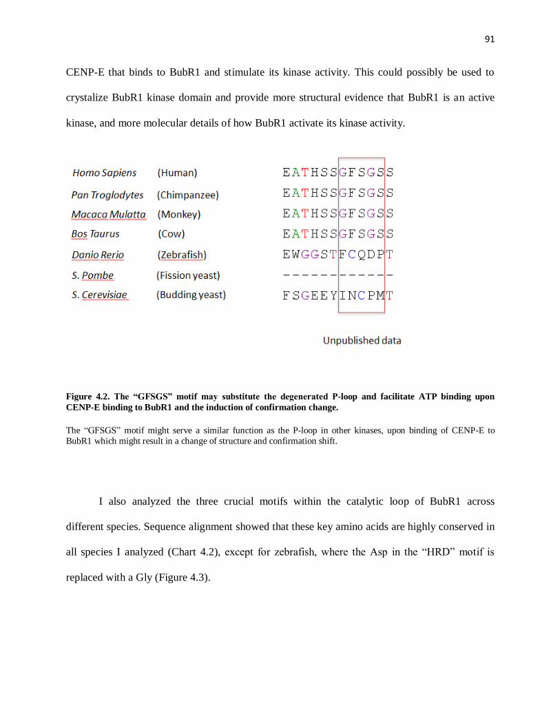
91
CENP-E that binds to BubR1 and stimulate its kinase activity. This could possibly be used to
crystalize BubR1 kinase domain and provide more structural evidence that BubR1 is an active
kinase, and more molecular details of how BubR1 activate its kinase activity.
Figure 4.2. The “GFSGS” motif may substitute the degenerated P-loop and facilitate ATP binding upon
CENP-E binding to BubR1 and the induction of confirmation change.
The “GFSGS” motif might serve a similar function as the P-loop in other kinases, upon binding of CENP-E to
BubR1 which might result in a change of structure and confirmation shift.
I also analyzed the three crucial motifs within the catalytic loop of BubR1 across
different species. Sequence alignment showed that these key amino acids are highly conserved in
all species I analyzed (Chart 4.2), except for zebrafish, where the Asp in the “HRD” motif is
replaced with a Gly (Figure 4.3).

92
Figure 4.3. Zebrafish BubR1 does not possess the crucial Asp in the “HRD” motif in the catalytic loop.
Whereas BubR1 of all other species analyzed possess the three key amino acids in the catalytic loop crucial for an
active kinase, Zebrafish BubR1 contains a substitution of the Asp in the “HRD” motif. Multi-sequence alignment is
performed using software ClustalX.
Chart 4.2. Version numbers of sequences used in the phylogenetic analysis
Species NCBI Version number
BubR1 or Mad3 CENP-E Bub1
Bos taurus Cattle gi|223556025 gi|296486796 gi|151556314
Caenorhabditis
elegans
Worm gi|392885899 N/A N/A
Danio rerio Zebra fish gi|189533436 ENSDARP0000009
9552*
gi|326676926
Drosophila
melanogaster
Fruit fly gi|320544558 gi|442627453
gi|442627461
gi|3421375
Gallus gallus Chicken gi|45383352 gi|363733943 gi|61098015

93
Homo sapiens Human gi|119612800 gi|71061468 gi|2981233
Macaca mulatta Monkey gi|383421155 gi|109075207
gi|109075209
gi|383413657
Mus musculus Mouse gi|40388490 gi|20810104
Pan troglodytes Chimpanzee gi|410225748 gi|114595517 gi|114579502
gi|114579504
gi|332814098
Rattus norvegicus Rat gi|109470615 gi|392339037 gi|157817678
Saccharomyces
cerevisiae
Budding yeast gi|1006729
N/A gi|190406801
Schizosaccharomyces pombe
Fission yeast gi|157310504
N/A gi|4176552
Xenopus laevis Frog gi|148229150 gi|2586071 gi|13122446
* protein ID of e Ensembl
BubR1’s kinase activity is dependent on its binding partner CENP-E, and I thus propose
a hypothesis that CENP-E and BubR1 undergo parallel evolution. Since zebrafish BubR1 might
be a pseudokinase, due to the key amino acid substitution in the “HRD” motif, zebrafish CENP-
E should be more evolutionarily divergent from the CENP-E of other species, which serves as a
bona fide BubR1 activator. This is supported by my phylogenetic analysis (Figure 4.4).
Moreover, the phylogenetic analysis shows that in species that lack CENP-E (Caenorhabditis
elegans, Saccharomyces cerevisiae, Schizosaccharomyces pombe), BubR1 are evolutionarily
more divergent from other species. This could serve as another piece of evidence that BubR1 and
CENP-E indeed undergo parallel evolution. In the future, I plan to analyze more sequences of
different species, and further confirm the parallel evolution of BubR1 and CENP-E. This will
provide us information on how protein divergence during evolution gives rise to new functions.

94
Figure 4.4. Phylogenetic analysis of BubR1, Bub1 and CENP-E among different species.
Phylogenetic analysis shows that compared with Bub1, BubR1 and CENP-E of zebrafish are evolutionarily more
divergent from other species. BubR1 of Caenorhabditis elegans, Saccharomyces cerevisiae, Schizosaccharomyces pombe,
which lack CENP-E, are evolutionarily more divergent from other species. This supports our hypothesis that CENP-E
and BubR1 undergo parallel evolution.
If BubR1 were a pseudokinase, although I feel it very unlikely, I will identify the kinase
that is responsible for the CENP-E-dependent phosphorylation of the T608 site. BubR1 is hyper-
phosphorylated during mitosis and confirmed phosphorylation sites include S676, T792, T1008
by Plk1 and T620 by Cdk1 which facilitates Plk1 binding to BubR1 and primes Plk1
phosphorylation (Elowe et al., 2007). Moreover, several other phosphorylation sites have been
shown to be Mps1-dependent (Huang et al., 2008) , although there is no evidence showing direct
phosphorylation. My unpublished results show that T608 phosphorylation in mitosis is not
sensitive to BI 2536 (Plk1 inhibitor), Purvalanol A (Cdk inhibitor), AZ 3146 (Mps1 inhibitor) or

95
ZM 447439 (Aurora B inhibitor) (Figure 4.5). In the future, I will confirm these results and look
for other possible candidates that phosphorylate this site, should it not be an auto-
phosphorylation site. In particular, I will examine Bub1, given its association with BubR1 on
kinetochores and similar phenotypes resulting from BubR1 and Bub1 depletion.
Figure 4.5. Phosphorylation of T608 is not dependent on Aurora B, Mps1, Cdk and Plk1.
(A) T98G cells arrested in mitosis with nocodazole are treated with specific inhibitors of Mps1, Plk1, CDK and
Aurora B as indicated. Indirect immunofluorescence is performed to visualize BubR1 on the kinetochore and
phosphorylation of site T608,using monoclonal BubR1 antibody and phospho-specific antibody against pT608. (B)
Quantification of the normalized integrated fluorescence intensities of the signals of T608 phosphorylation against
BubR1.

96
CENP-E is a microtubule sensor independent of tension to regulate Aurora B-
mediated phosphorylation prior to end-on attachment
The currently prevailing “spatial separation” model suggests that the physical separation
of several outer kinetochore proteins (e.g. The Ndc80 complex and mDia3) from Aurora B
kinase localized at the inner centromere due to tension exerted, and their subsequent
dephosphorylation, serve as the main contributing factor to stabilize microtubule-kinetochore
attachment. However, this model does not successfully explain how the attachment process
initiates in the vicinity of Aurora B kinase activity, where the outer kinetochore proteins are still
phosphorylated by Aurora B and have low microtubule binding affinity. In our study, I observed
that Aurora B-mediated Ndc80 phosphorylation was reduced in CENP-E–depleted cells and in
cells expressing a non-phosphorylatable BubR1 mutant at its CENP-E-dependent auto-
phosphorylation site. This has led to a hypothesis that CENP-E is a microtubule sensor in
response to initial microtubule capture, before the generation of end-on attachment with tension
(Figure 4.5). Specifically, in the lateral microtubule binding stage, microtubule binding by
CENP-E, and possibly its motor activity, switches off BubR1 kinase activity and its auto-
phosphorylation, leading to a reduced level of Aurora B-mediated phosphorylation of outer
kinetochore proteins. This initial step promotes the kinetochores to bind to microtubules from
opposite poles and the subsequent formation of amphitelic attachment.

97
Figure 4.5. A model for the role of CENP-E–dependent BubR1 auto-phosphorylation to facilitate initial
microtubule capture.
(A) Microtubule (MT) captured by CENP-E silences BubR1 kinase. Dephosphorylated BubR1 at its auto-
phosphorylation site reduces Aurora B-mediated phosphorylation of outer kinetochore components (e.g. Ndc80), possibly by recruiting PP2A. This transient reduction of Aurora B kinase activity initiates the stabilization of
kinetochore-microtubule attachment. (B) Upon bi-orientation, outer kinetochore substrates (e.g. Ndc80) become
separated from the inner centromeric Aurora B. CENP-E and others also recruit PP1 at kinetochores. These events
coordinate to further stabilize correct microtubule attachments.
To test this hypothesis, I decided to take advantage of cells depleted of Hec1, a
component of the Ndc80 complex. The Hec1-depleted cells do not form a complete metaphase
plate alignment because end-on attachments are severely disrupted; however, kinetochores are
capable of moving through lateral interactions with spindle microtubules (DeLuca et al., 2005),
presuming through kinetochore-associated motors including CENP-E and cytoplasmic dynein. I
evaluated the CENP-E-dependent BubR1 auto-phosphorylation at kinetochores using the BubR1
pAb-T608 phospho-antibody. Cells treated with nocodazole to depolymerize microtubules
showed high levels of BubR1 auto-phopshorylation signals, while cells in metaphase displayed

98
low levels of phosphorylation signal at all aligned kinetochores. This is consistent with my
previous observation that BubR1 auto-phosphorylation at kinetochores is sensitive to
microtubule attachment (Figure 4.6, A and B). In cells depleted of Hec1, however, the auto-
phosphorylation level of BubR1 was substantially diminished, suggesting the lateral binding to
microtubules is sufficient to switch off BubR1 kinase activity and reduce its auto-
phosphorylation level (Figure 4.6D). Treatment of nocodazole to Hec1 siRNA cells led to an
increased level of BubR1 auto-phosphorylation, to a comparable level of Mock siRNA cells
(Figure 4.6C). In the future, I plan to test levels of Aurora B-mediated phosphorylation of outer
kinetochore proteins (e.g. mDia3) to further prove the hypothesis.

99
Figure 4.6. The CENP-E-dependent BubR1 auto-phosphorylation at the kinetochore is sensitive to lateral
microtubule binding prior to end-on attachment.
Immunolocalization of ACA, Hec1 and pT608 of cells transfected with mock siRNA (A and B) or Hec1 siRNA (C
and D). In either cases, cells were treated with nocodazole to disassembly microtubules (A and C) or not (B and D)
before immnofluorescence.

100
CENP-E – BubR1 regulates phosphatase recruitment
It is of great interest to identify the mechanism how the CENP-E-BubR1 pathway
regulates Aurora-mediated phosphorylation. It is possible that the BubR1 kinase activity and the
auto-phosphorylation could directly regulate the catalytic activity of Aurora B. Full strength
Aurora B kinase activity is controlled by several regulatory components, including its auto-
phosphorylation, and the interaction with its binding partner, INCENP. By indirect immune-
fluorescence using antibodies against Aurora B itself or the Aurora B auto-phosphorylation site
T232, I have shown that neither Aurora B nor Aurora B auto-phosphorylation at centromeres has
changed in cells expressing BubR1 WT or phospho-mutants (data not shown). This indicates that
the CENP-E-BubR1 pathway does not seem to directly regulate Aurora B’s kinase activity.
The phosphorylation event is a balanced force between kinases and phosphatases which
counteract the kinase activity by dephosphorylating the phospho-residues. The CENP-E-BubR1
pathway, alternatively, could regulate the recruitment of phosphatases. Two phosphatases have
been reported to directly/indirectly interact with BubR1, making them attractive candidates.
Previous study has shown a direct interaction between BubR1’s TPR domain and the N-terminal
KNL1 (Kiyomitsu et al., 2007). The N terminus of KNL1 directly interacts with and targets PP1-
γ to outer kinetochores, which is essential to dephosphorylate Aurora B substrates and stabilize
microtubule attachments (Liu et al., 2010). Given the proximity of the PP1-γ binding site in
KNL1 to that of the BubR1 binding region, it is possible that the auto-phosphorylation of BubR1
could modulate its interaction with KNL1, inducing local conformational changes, and thus
regulate the KNL1-PP1-γ binding. A second potential phosphatase involved in the regulatory
mechanism of CENP-E-BubR1 is PP2A-B56α. It has been shown to directly bind to the BubR1
KARD (Kinetochore Attachment Regulatory Domain) domain, where three serine/threonine

101
residues are phosphorylated by Plk1 (Suijkerbuijk et al., 2012b). Binding of Plk1 to BubR1 and
subsequent phosphorylation of all three residues requires the prime phosphorylation of T620 by
Cdk1. The BubR1 auto-phosphorylation site, only 11 amino acid apart from this site, could
effectively control its phosphorylation by Cdk1, and thus regulate PP2A-B56 binding to the
KARD domain. I thus hypothesize that the CENP-E-dependent BubR1 auto-phosphorylation at
T608 residue controls the access of Cdk1 to T620, which is essential for Plk1 binding and
phosphorylation of the KARD domain. Only when BubR1 kinase activity is silenced by CENP-
E-mediated microtubule capture, Cdk1 phosphorylates the non-autophosphorylated form of
BubR1 and allows Plk1 binding and phosphorylating the KARD domain. Future analysis using
site-specific phospho-antibody and biochemistry assay will reveal more mechanistic details
about this potential regulatory pathway.
Figure 4.7. CENP-E-dependent BubR1 auto-phosphorylation at T608 residue controls the access of Cdk1 to
T620, which is essential for Plk1 binding and phosphorylation of the KARD domain.

102
BubR1 auto-phosphorylation site (T608) is only 11 amino acid apart from the Cdk1 dependent phosphorylation site
(T620). Phosphorylation of T620 residue is essential for the binding of Plk1 to BubR1 and subsequent
phosphorylation on several residues, including those on the KARD domain. I hypothesize that the CENP-E-
dependent BubR1 auto-phosphorylation at T608 residue controls the access of Cdk1 to T620.

103
Reference
Abrieu, A., Kahana, J.A., Wood, K.W., Cleveland, D.W., 2000. CENP-E as an essential component of the mitotic checkpoint in vitro. Cell 102, 817-826. Abrieu, A., Magnaghi-Jaulin, L., Kahana, J.A., Peter, M., Castro, A., Vigneron, S., Lorca, T., Cleveland, D.W., Labbe, J.C., 2001. Mps1 is a kinetochore-associated kinase essential for the vertebrate mitotic checkpoint. Cell 106, 83-93. Akiyoshi, B., Nelson, C.R., Ranish, J.A., Biggins, S., 2009. Quantitative proteomic analysis of purified yeast kinetochores identifies a PP1 regulatory subunit. Genes & development 23, 2887-2899. Akiyoshi, B., Sarangapani, K.K., Powers, A.F., Nelson, C.R., Reichow, S.L., Arellano-Santoyo, H., Gonen, T., Ranish, J.A., Asbury, C.L., Biggins, S., 2010. Tension directly stabilizes reconstituted kinetochore-microtubule attachments. Nature 468, 576-579. Andrews, P.D., Ovechkina, Y., Morrice, N., Wagenbach, M., Duncan, K., Wordeman, L., Swedlow, J.R., 2004. Aurora B regulates MCAK at the mitotic centromere. Developmental cell 6, 253-268. Assenmacher, N., Hopfner, K.P., 2004. MRE11/RAD50/NBS1: complex activities. Chromosoma 113, 157-166. Baker, D.J., Dawlaty, M.M., Wijshake, T., Jeganathan, K.B., Malureanu, L., van Ree, J.H., Crespo-Diaz, R., Reyes, S., Seaburg, L., Shapiro, V., Behfar, A., Terzic, A., van de Sluis, B., van Deursen, J.M., 2013. Increased expression of BubR1 protects against aneuploidy and cancer and extends healthy lifespan. Nature cell biology 15, 96-102. Baker, D.J., Jeganathan, K.B., Cameron, J.D., Thompson, M., Juneja, S., Kopecka, A., Kumar, R., Jenkins, R.B., de Groen, P.C., Roche, P., van Deursen, J.M., 2004. BubR1 insufficiency causes early onset of aging-associated phenotypes and infertility in mice. Nature genetics 36, 744-749. Baker, D.J., van Deursen, J.M., 2010. Chromosome missegregation causes colon cancer by APC loss of heterozygosity. Cell Cycle 9, 1711-1716. Bolanos-Garcia, V.M., Blundell, T.L., 2011. BUB1 and BUBR1: multifaceted kinases of the cell cycle. Trends Biochem Sci 36, 141-150. Bolanos-Garcia, V.M., Nilsson, J., Blundell, T.L., 2012. The architecture of the BubR1 tetratricopeptide tandem repeat defines a protein motif underlying mitotic checkpoint-kinetochore communication. Bioarchitecture 2, 23-27. Boudeau, J., Miranda-Saavedra, D., Barton, G.J., Alessi, D.R., 2006. Emerging roles of pseudokinases. Trends Cell Biol 16, 443-452. Campbell, C.S., Desai, A., 2013. Tension sensing by Aurora B kinase is independent of survivin-based centromere localization. Nature 497, 118-121. Carazo-Salas, R.E., Guarguaglini, G., Gruss, O.J., Segref, A., Karsenti, E., Mattaj, I.W., 1999. Generation of GTP-bound Ran by RCC1 is required for chromatin-induced mitotic spindle formation. Nature 400, 178-181. Caudron, M., Bunt, G., Bastiaens, P., Karsenti, E., 2005. Spatial coordination of spindle assembly by chromosome-mediated signaling gradients. Science 309, 1373-1376. Chan, G.K., Jablonski, S.A., Sudakin, V., Hittle, J.C., Yen, T.J., 1999. Human BUBR1 is a mitotic checkpoint kinase that monitors CENP-E functions at kinetochores and binds the cyclosome/APC. J Cell Biol 146, 941-954. Chan, G.K., Schaar, B.T., Yen, T.J., 1998. Characterization of the kinetochore binding domain of CENP-E reveals interactions with the kinetochore proteins CENP-F and hBUBR1. J Cell Biol 143, 49-63. Chan, Y.W., Jeyaprakash, A.A., Nigg, E.A., Santamaria, A., 2012. Aurora B controls kinetochore-microtubule attachments by inhibiting Ska complex-KMN network interaction. J Cell Biol 196, 563-571.

104
Chao, W.C., Kulkarni, K., Zhang, Z., Kong, E.H., Barford, D., 2012. Structure of the mitotic checkpoint complex. Nature 484, 208-213. Cheeseman, I.M., Anderson, S., Jwa, M., Green, E.M., Kang, J., Yates, J.R., 3rd, Chan, C.S., Drubin, D.G., Barnes, G., 2002. Phospho-regulation of kinetochore-microtubule attachments by the Aurora kinase Ipl1p. Cell 111, 163-172. Cheeseman, I.M., Chappie, J.S., Wilson-Kubalek, E.M., Desai, A., 2006. The conserved KMN network constitutes the core microtubule-binding site of the kinetochore. Cell 127, 983-997. Chen, R.-H., 2002a. BubR1 is essential for kinetochore localization of other spindle checkpoint proteins and its phosphorylation requires Mad1. J. Cell Biol. 158, 487-496. Chen, R.H., 2002b. BubR1 is essential for kinetochore localization of other spindle checkpoint proteins and its phosphorylation requires Mad1. Journal of Cell Biology 158, 487-496. Chen, R.H., Shevchenko, A., Mann, M., Murray, A.W., 1998. Spindle checkpoint protein Xmad1 recruits Xmad2 to unattached kinetochores. J Cell Biol 143, 283-295. Cheng, L., Zhang, J., Ahmad, S., Rozier, L., Yu, H., Deng, H., Mao, Y., 2011. Aurora B regulates formin mDia3 in achieving metaphase chromosome alignment. Developmental cell 20, 342-352. Cho, R.J., Fromont-Racine, M., Wodicka, L., Feierbach, B., Stearns, T., Legrain, P., Lockhart, D.J., Davis, R.W., 1998. Parallel analysis of genetic selections using whole genome oligonucleotide arrays. Proc Natl Acad Sci U S A 95, 3752-3757. Cimini, D., Wan, X., Hirel, C.B., Salmon, E.D., 2006. Aurora kinase promotes turnover of kinetochore microtubules to reduce chromosome segregation errors. Current biology : CB 16, 1711-1718. Cleveland, D.W., Mao, Y., Sullivan, K.F., 2003. Centromeres and kinetochores: from epigenetics to mitotic checkpoint signaling. Cell 112, 407-421. Costanzo, V., Robertson, K., Bibikova, M., Kim, E., Grieco, D., Gottesman, M., Carroll, D., Gautier, J., 2001. Mre11 protein complex prevents double-strand break accumulation during chromosomal DNA replication. Molecular cell 8, 137-147. D'Amours, D., Jackson, S.P., 2002. The Mre11 complex: at the crossroads of dna repair and checkpoint signalling. Nat Rev Mol Cell Biol 3, 317-327. da Fonseca, P.C., Kong, E.H., Zhang, Z., Schreiber, A., Williams, M.A., Morris, E.P., Barford, D., 2011. Structures of APC/C(Cdh1) with substrates identify Cdh1 and Apc10 as the D-box co-receptor. Nature 470, 274-278. De Antoni, A., Pearson, C.G., Cimini, D., Canman, J.C., Sala, V., Nezi, L., Mapelli, M., Sironi, L., Faretta, M., Salmon, E.D., Musacchio, A., 2005. The Mad1/Mad2 complex as a template for Mad2 activation in the spindle assembly checkpoint. Current biology : CB 15, 214-225. de Jager, M., van Noort, J., van Gent, D.C., Dekker, C., Kanaar, R., Wyman, C., 2001. Human Rad50/Mre11 is a flexible complex that can tether DNA ends. Molecular cell 8, 1129-1135. DeLuca, J.G., Dong, Y., Hergert, P., Strauss, J., Hickey, J.M., Salmon, E.D., McEwen, B.F., 2005. Hec1 and nuf2 are core components of the kinetochore outer plate essential for organizing microtubule attachment sites. Molecular biology of the cell 16, 519-531. DeLuca, J.G., Gall, W.E., Ciferri, C., Cimini, D., Musacchio, A., Salmon, E.D., 2006. Kinetochore microtubule dynamics and attachment stability are regulated by Hec1. Cell 127, 969-982. DeLuca, K.F., Lens, S.M., DeLuca, J.G., 2011. Temporal changes in Hec1 phosphorylation control kinetochore-microtubule attachment stability during mitosis. Journal of cell science 124, 622-634. Ding, Y., Hubert, C.G., Herman, J., Corrin, P., Toledo, C.M., Skutt-Kakaria, K., Vazquez, J., Basom, R., Zhang, B., Risler, J.K., Pollard, S.M., Nam, D.H., Delrow, J.J., Zhu, J., Lee, J., DeLuca, J., Olson, J.M., Paddison, P.J., 2013. Cancer-Specific requirement for BUB1B/BUBR1 in human brain tumor isolates and genetically transformed cells. Cancer discovery 3, 198-211.

105
Ditchfield, C., Johnson, V.L., Tighe, A., Ellston, R., Haworth, C., Johnson, T., Mortlock, A., Keen, N., Taylor, S.S., 2003. Aurora B couples chromosome alignment with anaphase by targeting BubR1, Mad2, and Cenp-E to kinetochores. J Cell Biol 161, 267-280. Dupre, A., Boyer-Chatenet, L., Gautier, J., 2006. Two-step activation of ATM by DNA and the Mre11-Rad50-Nbs1 complex. Nat Struct Mol Biol 13, 451-457. Elowe, S., Dulla, K., Uldschmid, A., Li, X., Dou, Z., Nigg, E.A., 2010. Uncoupling of the spindle-checkpoint and chromosome-congression functions of BubR1. Journal of cell science 123, 84-94. Elowe, S., Hummer, S., Uldschmid, A., Li, X., Nigg, E.A., 2007. Tension-sensitive Plk1 phosphorylation on BubR1 regulates the stability of kinetochore microtubule interactions. Genes & development 21, 2205-2219. Endicott, J.A., Noble, M.E., Johnson, L.N., 2012. The structural basis for control of eukaryotic protein kinases. Annual review of biochemistry 81, 587-613. England, J.R., Huang, J., Jennings, M.J., Makde, R.D., Tan, S., 2010. RCC1 uses a conformationally diverse loop region to interact with the nucleosome: a model for the RCC1-nucleosome complex. Journal of molecular biology 398, 518-529. Fang, G., 2002. Checkpoint protein BubR1 acts synergistically with Mad2 to inhibit anaphase-promoting complex. Molecular biology of the cell 13, 755-766. Fang, G., Yu, H., Kirschner, M.W., 1998. Direct binding of CDC20 protein family members activates the anaphase-promoting complex in mitosis and G1. Molecular cell 2, 163-171. Foster, S.A., Morgan, D.O., 2012. The APC/C subunit Mnd2/Apc15 promotes Cdc20 autoubiquitination and spindle assembly checkpoint inactivation. Molecular cell 47, 921-932. Gadde, S., Heald, R., 2004. Mechanisms and molecules of the mitotic spindle. Current biology : CB 14, R797-805. Giunta, S., Jackson, S.P., 2011. Give me a break, but not in mitosis: the mitotic DNA damage response marks DNA double-strand breaks with early signaling events. Cell Cycle 10, 1215-1221. Glotzer, M., Murray, A.W., Kirschner, M.W., 1991. Cyclin is degraded by the ubiquitin pathway. Nature 349, 132-138. Greenberg, R.A., Sobhian, B., Pathania, S., Cantor, S.B., Nakatani, Y., Livingston, D.M., 2006. Multifactorial contributions to an acute DNA damage response by BRCA1/BARD1-containing complexes. Genes & development 20, 34-46. Hao, Y., Macara, I.G., 2008. Regulation of chromatin binding by a conformational switch in the tail of the Ran exchange factor RCC1. J Cell Biol 182, 827-836. Hartwell, L.H., Kastan, M.B., 1994. Cell cycle control and cancer. Science 266, 1821-1828. Hauf, S., Cole, R.W., LaTerra, S., Zimmer, C., Schnapp, G., Walter, R., Heckel, A., van Meel, J., Rieder, C.L., Peters, J.M., 2003. The small molecule Hesperadin reveals a role for Aurora B in correcting kinetochore-microtubule attachment and in maintaining the spindle assembly checkpoint. J Cell Biol 161, 281-294. Heald, R., Tournebize, R., Blank, T., Sandaltzopoulos, R., Becker, P., Hyman, A., Karsenti, E., 1996. Self-organization of microtubules into bipolar spindles around artificial chromosomes in Xenopus egg extracts. Nature 382, 420-425. Hegemann, B., Hutchins, J.R., Hudecz, O., Novatchkova, M., Rameseder, J., Sykora, M.M., Liu, S., Mazanek, M., Lenart, P., Heriche, J.K., Poser, I., Kraut, N., Hyman, A.A., Yaffe, M.B., Mechtler, K., Peters, J.M., 2011. Systematic phosphorylation analysis of human mitotic protein complexes. Sci Signal 4, rs12. Hemerly, A.S., Prasanth, S.G., Siddiqui, K., Stillman, B., 2009. Orc1 controls centriole and centrosome copy number in human cells. Science 323, 789-793. Howell, B.J., McEwen, B.F., Canman, J.C., Hoffman, D.B., Farrar, E.M., Rieder, C.L., Salmon, E.D., 2001. Cytoplasmic dynein/dynactin drives kinetochore protein transport to the spindle poles and has a role in mitotic spindle checkpoint inactivation. J Cell Biol 155, 1159-1172.

106
Hoyt, M.A., Totis, L., Roberts, B.T., 1991. S. cerevisiae genes required for cell cycle arrest in response to loss of microtubule function. Cell 66, 507-517. Huang, H., Hittle, J., Zappacosta, F., Annan, R.S., Hershko, A., Yen, T.J., 2008. Phosphorylation sites in BubR1 that regulate kinetochore attachment, tension, and mitotic exit. The Journal of cell biology 183, 667-680. Hwang, L.H., Lau, L.F., Smith, D.L., Mistrot, C.A., Hardwick, K.G., Hwang, E.S., Amon, A., Murray, A.W., 1998. Budding yeast Cdc20: a target of the spindle checkpoint. Science 279, 1041-1044. Iwanaga, Y., Chi, Y.H., Miyazato, A., Sheleg, S., Haller, K., Peloponese, J.M., Jr., Li, Y., Ward, J.M., Benezra, R., Jeang, K.T., 2007. Heterozygous deletion of mitotic arrest-deficient protein 1 (MAD1) increases the incidence of tumors in mice. Cancer research 67, 160-166. Joglekar, A.P., Bloom, K.S., Salmon, E.D., 2010. Mechanisms of force generation by end-on kinetochore-microtubule attachments. Current opinion in cell biology 22, 57-67. Joukov, V., Groen, A.C., Prokhorova, T., Gerson, R., White, E., Rodriguez, A., Walter, J.C., Livingston, D.M., 2006. The BRCA1/BARD1 heterodimer modulates ran-dependent mitotic spindle assembly. Cell 127, 539-552. Kalab, P., Pralle, A., Isacoff, E.Y., Heald, R., Weis, K., 2006. Analysis of a RanGTP-regulated gradient in mitotic somatic cells. Nature 440, 697-701. Kang, J., Bronson, R.T., Xu, Y., 2002. Targeted disruption of NBS1 reveals its roles in mouse development and DNA repair. The EMBO journal 21, 1447-1455. Kang, J., Yang, M., Li, B., Qi, W., Zhang, C., Shokat, K.M., Tomchick, D.R., Machius, M., Yu, H., 2008. Structure and substrate recruitment of the human spindle checkpoint kinase Bub1. Molecular cell 32, 394-405. Kannan, N., Taylor, S.S., Zhai, Y., Venter, J.C., Manning, G., 2007. Structural and functional diversity of the microbial kinome. PLoS biology 5, e17. Kapoor, T.M., Lampson, M.A., Hergert, P., Cameron, L., Cimini, D., Salmon, E.D., McEwen, B.F., Khodjakov, A., 2006. Chromosomes can congress to the metaphase plate before biorientation. Science 311, 388-391. Keating, P., Rachidi, N., Tanaka, T.U., Stark, M.J., 2009. Ipl1-dependent phosphorylation of Dam1 is reduced by tension applied on kinetochores. Journal of cell science 122, 4375-4382. Kim, S.H., Lin, D.P., Matsumoto, S., Kitazono, A., Matsumoto, T., 1998. Fission yeast Slp1: an effector of the Mad2-dependent spindle checkpoint. Science 279, 1045-1047. Kim, Y., Heuser, J.E., Waterman, C.M., Cleveland, D.W., 2008. CENP-E combines a slow, processive motor and a flexible coiled coil to produce an essential motile kinetochore tether. J Cell Biol 181, 411-419. Kim, Y., Holland, A.J., Lan, W., Cleveland, D.W., 2010. Aurora kinases and protein phosphatase 1 mediate chromosome congression through regulation of CENP-E. Cell 142, 444-455. Kimura, M., Kotani, S., Hattori, T., Sumi, N., Yoshioka, T., Todokoro, K., Okano, Y., 1997. Cell cycle-dependent expression and spindle pole localization of a novel human protein kinase, Aik, related to Aurora of Drosophila and yeast Ipl1. J Biol Chem 272, 13766-13771. Kirschner, M.W., Mitchison, T., 1986. Microtubule dynamics. Nature 324, 621. Kiyomitsu, T., Obuse, C., Yanagida, M., 2007. Human Blinkin/AF15q14 is required for chromosome alignment and the mitotic checkpoint through direct interaction with Bub1 and BubR1. Developmental cell 13, 663-676. Kline-Smith, S.L., Khodjakov, A., Hergert, P., Walczak, C.E., 2004. Depletion of centromeric MCAK leads to chromosome congression and segregation defects due to improper kinetochore attachments. Molecular biology of the cell 15, 1146-1159. Knowlton, A.L., Lan, W., Stukenberg, P.T., 2006. Aurora B is enriched at merotelic attachment sites, where it regulates MCAK. Current biology : CB 16, 1705-1710.

107
Kops, G.J., Foltz, D.R., Cleveland, D.W., 2004. Lethality to human cancer cells through massive chromosome loss by inhibition of the mitotic checkpoint. Proc Natl Acad Sci U S A 101, 8699-8704. Kulukian, A., Han, J.S., Cleveland, D.W., 2009. Unattached kinetochores catalyze production of an anaphase inhibitor that requires a Mad2 template to prime Cdc20 for BubR1 binding. Developmental cell 16, 105-117. Lampert, F., Hornung, P., Westermann, S., 2010. The Dam1 complex confers microtubule plus end-tracking activity to the Ndc80 kinetochore complex. Journal of Cell Biology 189, 641-649. Lampson, M.A., Cheeseman, I.M., 2010. Sensing centromere tension: Aurora B and the regulation of kinetochore function. Trends Cell Biol. Lampson, M.A., Cheeseman, I.M., 2011. Sensing centromere tension: Aurora B and the regulation of kinetochore function. Trends Cell Biol 21, 133-140. Lampson, M.A., Kapoor, T.M., 2005. The human mitotic checkpoint protein BubR1 regulates chromosome-spindle attachments. Nature cell biology 7, 93-98. Lan, W., Zhang, X., Kline-Smith, S.L., Rosasco, S.E., Barrett-Wilt, G.A., Shabanowitz, J., Hunt, D.F., Walczak, C.E., Stukenberg, P.T., 2004. Aurora B phosphorylates centromeric MCAK and regulates its localization and microtubule depolymerization activity. Current biology : CB 14, 273-286. Li, H.Y., Wirtz, D., Zheng, Y., 2003a. A mechanism of coupling RCC1 mobility to RanGTP production on the chromatin in vivo. J Cell Biol 160, 635-644. Li, H.Y., Wirtz, D., Zheng, Y., 2003b. A mechanism of coupling RCC1 mobility to RanGTP production on the chromatin in vivo. The Journal of cell biology 160, 635-644. Li, R., Murray, A.W., 1991. Feedback control of mitosis in budding yeast. Cell 66, 519-531. Liu, D., Vader, G., Vromans, M.J., Lampson, M.A., Lens, S.M., 2009. Sensing chromosome bi-orientation by spatial separation of aurora B kinase from kinetochore substrates. Science 323, 1350-1353. Liu, D., Vleugel, M., Backer, C.B., Hori, T., Fukagawa, T., Cheeseman, I.M., Lampson, M.A., 2010. Regulated targeting of protein phosphatase 1 to the outer kinetochore by KNL1 opposes Aurora B kinase. J Cell Biol 188, 809-820. Liu, S.T., Chan, G.K., Hittle, J.C., Fujii, G., Lees, E., Yen, T.J., 2003. Human MPS1 kinase is required for mitotic arrest induced by the loss of CENP-E from kinetochores. Molecular biology of the cell 14, 1638-1651. Loncarek, J., Kisurina-Evgenieva, O., Vinogradova, T., Hergert, P., La Terra, S., Kapoor, T.M., Khodjakov, A., 2007. The centromere geometry essential for keeping mitosis error free is controlled by spindle forces. Nature 450, 745-749. Luo, G., Yao, M.S., Bender, C.F., Mills, M., Bladl, A.R., Bradley, A., Petrini, J.H., 1999. Disruption of mRad50 causes embryonic stem cell lethality, abnormal embryonic development, and sensitivity to ionizing radiation. Proc Natl Acad Sci U S A 96, 7376-7381. Luo, X., Tang, Z., Xia, G., Wassmann, K., Matsumoto, T., Rizo, J., Yu, H., 2004. The Mad2 spindle checkpoint protein has two distinct natively folded states. Nat Struct Mol Biol 11, 338-345. Makde, R.D., England, J.R., Yennawar, H.P., Tan, S., 2010. Structure of RCC1 chromatin factor bound to the nucleosome core particle. Nature 467, 562-566. Malureanu, L.A., Jeganathan, K.B., Hamada, M., Wasilewski, L., Davenport, J., van Deursen, J.M., 2009. BubR1 N terminus acts as a soluble inhibitor of cyclin B degradation by APC/C(Cdc20) in interphase. Developmental cell 16, 118-131. Manning, G., Whyte, D.B., Martinez, R., Hunter, T., Sudarsanam, S., 2002. The protein kinase complement of the human genome. Science 298, 1912-1934. Mansfeld, J., Collin, P., Collins, M.O., Choudhary, J.S., Pines, J., 2011. APC15 drives the turnover of MCC-CDC20 to make the spindle assembly checkpoint responsive to kinetochore attachment. Nature cell biology 13, 1234-1243. Mao, Y., 2011. FORMIN a link between kinetochores and microtubule ends. Trends Cell Biol 21, 625-629.

108
Mao, Y., Abrieu, A., Cleveland, D.W., 2003. Activating and silencing the mitotic checkpoint through CENP-E-dependent activation/inactivation of BubR1. Cell 114, 87-98. Mao, Y., Desai, A., Cleveland, D.W., 2005. Microtubule capture by CENP-E silences BubR1-dependent mitotic checkpoint signaling. J Cell Biol 170, 873-880. Mapelli, M., Filipp, F.V., Rancati, G., Massimiliano, L., Nezi, L., Stier, G., Hagan, R.S., Confalonieri, S., Piatti, S., Sattler, M., Musacchio, A., 2006. Determinants of conformational dimerization of Mad2 and its inhibition by p31comet. The EMBO journal 25, 1273-1284. Maresca, T.J., Salmon, E.D., 2009. Intrakinetochore stretch is associated with changes in kinetochore phosphorylation and spindle assembly checkpoint activity. Journal of Cell Biology 184, 373-381. Martin-Lluesma, S., Stucke, V.M., Nigg, E.A., 2002. Role of hec1 in spindle checkpoint signaling and kinetochore recruitment of mad1/mad2. Science 297, 2267-2270. Maser, R.S., Mirzoeva, O.K., Wells, J., Olivares, H., Williams, B.R., Zinkel, R.A., Farnham, P.J., Petrini, J.H., 2001. Mre11 complex and DNA replication: linkage to E2F and sites of DNA synthesis. Molecular and cellular biology 21, 6006-6016. Matsumura, S., Toyoshima, F., Nishida, E., 2007. Polo-like kinase 1 facilitates chromosome alignment during prometaphase through BubR1. J Biol Chem 282, 15217-15227. Matsuoka, S., Ballif, B.A., Smogorzewska, A., McDonald, E.R., 3rd, Hurov, K.E., Luo, J., Bakalarski, C.E., Zhao, Z., Solimini, N., Lerenthal, Y., Shiloh, Y., Gygi, S.P., Elledge, S.J., 2007. ATM and ATR substrate analysis reveals extensive protein networks responsive to DNA damage. Science 316, 1160-1166. McEwen, B.F., Chan, G.K., Zubrowski, B., Savoian, M.S., Sauer, M.T., Yen, T.J., 2001. CENP-E is essential for reliable bioriented spindle attachment, but chromosome alignment can be achieved via redundant mechanisms in mammalian cells. Molecular biology of the cell 12, 2776-2789. McEwen, B.F., Dong, Y.M., 2009. Releasing the spindle assembly checkpoint without tension. Journal of Cell Biology 184, 355-356. McKim, K.S., Hawley, R.S., 1995. Chromosomal control of meiotic cell division. Science 270, 1595-1601. Mimitou, E.P., Symington, L.S., 2011. DNA end resection--unraveling the tail. DNA repair 10, 344-348. Murray, A.W., 1991. Cell cycle extracts. Methods Cell Biol 36, 581-605. Musacchio, A., 2011. Spindle assembly checkpoint: the third decade. Philos Trans R Soc Lond B Biol Sci 366, 3595-3604. Musaro, M., Ciapponi, L., Fasulo, B., Gatti, M., Cenci, G., 2008. Unprotected Drosophila melanogaster telomeres activate the spindle assembly checkpoint. Nature genetics 40, 362-366. Myung, K., Smith, S., Kolodner, R.D., 2004. Mitotic checkpoint function in the formation of gross chromosomal rearrangements in Saccharomyces cerevisiae. Proc Natl Acad Sci U S A 101, 15980-15985. Nicklas, R.B., 1997. How cells get the right chromosomes. Science 275, 632-637. Ohba, T., Nakamura, M., Nishitani, H., Nishimoto, T., 1999. Self-organization of microtubule asters induced in Xenopus egg extracts by GTP-bound Ran. Science 284, 1356-1358. Ohi, R., Sapra, T., Howard, J., Mitchison, T.J., 2004. Differentiation of cytoplasmic and meiotic spindle assembly MCAK functions by Aurora B-dependent phosphorylation. Molecular biology of the cell 15, 2895-2906. Peterson, S.E., Li, Y., Chait, B.T., Gottesman, M.E., Baer, R., Gautier, J., 2011. Cdk1 uncouples CtIP-dependent resection and Rad51 filament formation during M-phase double-strand break repair. J Cell Biol 194, 705-720. Putkey, F.R., Cramer, T., Morphew, M.K., Silk, A.D., Johnson, R.S., McIntosh, J.R., Cleveland, D.W., 2002. Unstable kinetochore-microtubule capture and chromosomal instability following deletion of CENP-E. Developmental cell 3, 351-365. Rahmani, Z., Gagou, M.E., Lefebvre, C., Emre, D., Karess, R.E., 2009. Separating the spindle, checkpoint, and timer functions of BubR1. The Journal of cell biology 187, 597-605.

109
Rieder, C.L., Alexander, S.P., 1990. Kinetochores are transported poleward along a single astral microtubule during chromosome attachment to the spindle in newt lung cells. J Cell Biol 110, 81-95. Rosenberg, J.S., Cross, F.R., Funabiki, H., 2011. KNL1/Spc105 recruits PP1 to silence the spindle assembly checkpoint. Current biology : CB 21, 942-947. Santaguida, S., Musacchio, A., 2009. The life and miracles of kinetochores. Embo J 28, 2511-2531. Santaguida, S., Vernieri, C., Villa, F., Ciliberto, A., Musacchio, A., 2011. Evidence that Aurora B is implicated in spindle checkpoint signalling independently of error correction. Embo Journal 30, 1508-1519. Sassoon, I., Severin, F.F., Andrews, P.D., Taba, M.R., Kaplan, K.B., Ashford, A.J., Stark, M.J., Sorger, P.K., Hyman, A.A., 1999. Regulation of Saccharomyces cerevisiae kinetochores by the type 1 phosphatase Glc7p. Genes & development 13, 545-555. Schliekelman, M., Cowley, D.O., O'Quinn, R., Oliver, T.G., Lu, L., Salmon, E.D., Van Dyke, T., 2009. Impaired Bub1 function in vivo compromises tension-dependent checkpoint function leading to aneuploidy and tumorigenesis. Cancer research 69, 45-54. Schmit, A.C., Stoppin, V., Chevrier, V., Job, D., Lambert, A.M., 1994. Cell cycle dependent distribution of a centrosomal antigen at the perinuclear MTOC or at the kinetochores of higher plant cells. Chromosoma 103, 343-351. Schwab, M., Lutum, A.S., Seufert, W., 1997. Yeast Hct1 is a regulator of Clb2 cyclin proteolysis. Cell 90, 683-693. Shah, J.V., Botvinick, E., Bonday, Z., Furnari, F., Berns, M., Cleveland, D.W., 2004. Dynamics of centromere and kinetochore proteins; implications for checkpoint signaling and silencing. Current biology : CB 14, 942-952. Shimada, K., Gasser, S.M., 2007. The origin recognition complex functions in sister-chromatid cohesion in Saccharomyces cerevisiae. Cell 128, 85-99. Stracker, T.H., Theunissen, J.W., Morales, M., Petrini, J.H., 2004. The Mre11 complex and the metabolism of chromosome breaks: the importance of communicating and holding things together. DNA repair 3, 845-854. Sudakin, V., Chan, G.K., Yen, T.J., 2001. Checkpoint inhibition of the APC/C in HeLa cells is mediated by a complex of BUBR1, BUB3, CDC20, and MAD2. J Cell Biol 154, 925-936. Suijkerbuijk, S.J., van Dam, T.J., Karagoz, G.E., von Castelmur, E., Hubner, N.C., Duarte, A.M., Vleugel, M., Perrakis, A., Rudiger, S.G., Snel, B., Kops, G.J., 2012a. The vertebrate mitotic checkpoint protein BUBR1 is an unusual pseudokinase. Developmental cell 22, 1321-1329. Suijkerbuijk, S.J., van Osch, M.H., Bos, F.L., Hanks, S., Rahman, N., Kops, G.J., 2010a. Molecular causes for BUBR1 dysfunction in the human cancer predisposition syndrome mosaic variegated aneuploidy. Cancer research 70, 4891-4900. Suijkerbuijk, S.J., van Osch, M.H., Bos, F.L., Hanks, S., Rahman, N., Kops, G.J., 2010b. Molecular causes for BUBR1 dysfunction in the human cancer predisposition syndrome mosaic variegated aneuploidy. Cancer research 70, 4891-4900. Suijkerbuijk, S.J., Vleugel, M., Teixeira, A., Kops, G.J., 2012b. Integration of kinase and phosphatase activities by BUBR1 ensures formation of stable kinetochore-microtubule attachments. Developmental cell 23, 745-755. Symington, L.S., 2002. Role of RAD52 epistasis group genes in homologous recombination and double-strand break repair. Microbiology and molecular biology reviews : MMBR 66, 630-670, table of contents. Symington, L.S., Gautier, J., 2011. Double-strand break end resection and repair pathway choice. Annual review of genetics 45, 247-271. Takaki, T., Trenz, K., Costanzo, V., Petronczki, M., 2008. Polo-like kinase 1 reaches beyond mitosis--cytokinesis, DNA damage response, and development. Current opinion in cell biology 20, 650-660.

110
Tanaka, T.U., Rachidi, N., Janke, C., Pereira, G., Galova, M., Schiebel, E., Stark, M.J., Nasmyth, K., 2002. Evidence that the Ipl1-Sli15 (Aurora kinase-INCENP) complex promotes chromosome bi-orientation by altering kinetochore-spindle pole connections. Cell 108, 317-329. Tang, Z., Bharadwaj, R., Li, B., Yu, H., 2001. Mad2-Independent inhibition of APCCdc20 by the mitotic checkpoint protein BubR1. Developmental cell 1, 227-237. Tang, Z., Shu, H., Oncel, D., Chen, S., Yu, H., 2004. Phosphorylation of Cdc20 by Bub1 provides a catalytic mechanism for APC/C inhibition by the spindle checkpoint. Molecular cell 16, 387-397. Taylor, S.S., Ha, E., McKeon, F., 1998. The human homologue of Bub3 is required for kinetochore localization of Bub1 and a Mad3/Bub1-related protein kinase. J Cell Biol 142, 1-11. Trinkle-Mulcahy, L., Andrews, P.D., Wickramasinghe, S., Sleeman, J., Prescott, A., Lam, Y.W., Lyon, C., Swedlow, J.R., Lamond, A.I., 2003. Time-lapse imaging reveals dynamic relocalization of PP1gamma throughout the mammalian cell cycle. Molecular biology of the cell 14, 107-117. Uchida, K.S.K., Takagaki, K., Kumada, K., Hirayama, Y., Noda, T., Hirota, T., 2009. Kinetochore stretching inactivates the spindle assembly checkpoint. Journal of Cell Biology 184, 383-390. Uziel, T., Lerenthal, Y., Moyal, L., Andegeko, Y., Mittelman, L., Shiloh, Y., 2003. Requirement of the MRN complex for ATM activation by DNA damage. The EMBO journal 22, 5612-5621. Uzunova, K., Dye, B.T., Schutz, H., Ladurner, R., Petzold, G., Toyoda, Y., Jarvis, M.A., Brown, N.G., Poser, I., Novatchkova, M., Mechtler, K., Hyman, A.A., Stark, H., Schulman, B.A., Peters, J.M., 2012. APC15 mediates CDC20 autoubiquitylation by APC/C(MCC) and disassembly of the mitotic checkpoint complex. Nat Struct Mol Biol 19, 1116-1123. Vigneron, S., Prieto, S., Bernis, C., Labbe, J.C., Castro, A., Lorca, T., 2004. Kinetochore localization of spindle checkpoint proteins: who controls whom? Molecular biology of the cell 15, 4584-4596. Vink, M., Simonetta, M., Transidico, P., Ferrari, K., Mapelli, M., De Antoni, A., Massimiliano, L., Ciliberto, A., Faretta, M., Salmon, E.D., Musacchio, A., 2006. In vitro FRAP identifies the minimal requirements for Mad2 kinetochore dynamics. Current biology : CB 16, 755-766. Visintin, R., Prinz, S., Amon, A., 1997. CDC20 and CDH1: a family of substrate-specific activators of APC-dependent proteolysis. Science 278, 460-463. Walczak, C.E., Heald, R., 2008. Mechanisms of mitotic spindle assembly and function. Int Rev Cytol 265, 111-158. Waltes, R., Kalb, R., Gatei, M., Kijas, A.W., Stumm, M., Sobeck, A., Wieland, B., Varon, R., Lerenthal, Y., Lavin, M.F., Schindler, D., Dork, T., 2009. Human RAD50 deficiency in a Nijmegen breakage syndrome-like disorder. American journal of human genetics 84, 605-616. Wang, Y., Cortez, D., Yazdi, P., Neff, N., Elledge, S.J., Qin, J., 2000. BASC, a super complex of BRCA1-associated proteins involved in the recognition and repair of aberrant DNA structures. Genes & development 14, 927-939. Weaver, B.A., Bonday, Z.Q., Putkey, F.R., Kops, G.J., Silk, A.D., Cleveland, D.W., 2003. Centromere-associated protein-E is essential for the mammalian mitotic checkpoint to prevent aneuploidy due to single chromosome loss. J Cell Biol 162, 551-563. Weaver, B.A., Cleveland, D.W., 2006. Does aneuploidy cause cancer? Current opinion in cell biology 18, 658-667. Weaver, B.A., Silk, A.D., Montagna, C., Verdier-Pinard, P., Cleveland, D.W., 2007. Aneuploidy acts both oncogenically and as a tumor suppressor. Cancer cell 11, 25-36. Weiss, E., Winey, M., 1996. The Saccharomyces cerevisiae spindle pole body duplication gene MPS1 is part of a mitotic checkpoint. J Cell Biol 132, 111-123. Welburn, J.P., Grishchuk, E.L., Backer, C.B., Wilson-Kubalek, E.M., Yates, J.R., 3rd, Cheeseman, I.M., 2009. The human kinetochore Ska1 complex facilitates microtubule depolymerization-coupled motility. Developmental cell 16, 374-385.

111
Welburn, J.P., Vleugel, M., Liu, D., Yates, J.R., 3rd, Lampson, M.A., Fukagawa, T., Cheeseman, I.M., 2010. Aurora B phosphorylates spatially distinct targets to differentially regulate the kinetochore-microtubule interface. Molecular cell 38, 383-392. Wilde, A., Zheng, Y., 1999. Stimulation of microtubule aster formation and spindle assembly by the small GTPase Ran. Science 284, 1359-1362. Williams, R.S., Williams, J.S., Tainer, J.A., 2007. Mre11-Rad50-Nbs1 is a keystone complex connecting DNA repair machinery, double-strand break signaling, and the chromatin template. Biochemistry and cell biology = Biochimie et biologie cellulaire 85, 509-520. Wojcik, E., Basto, R., Serr, M., Scaerou, F., Karess, R., Hays, T., 2001. Kinetochore dynein: its dynamics and role in the transport of the Rough deal checkpoint protein. Nature cell biology 3, 1001-1007. Wollman, R., Cytrynbaum, E.N., Jones, J.T., Meyer, T., Scholey, J.M., Mogilner, A., 2005. Efficient chromosome capture requires a bias in the 'search-and-capture' process during mitotic-spindle assembly. Current biology : CB 15, 828-832. Wong, O.K., Fang, G., 2007. Cdk1 phosphorylation of BubR1 controls spindle checkpoint arrest and Plk1-mediated formation of the 3F3/2 epitope. J Cell Biol 179, 611-617. Xia, G., Luo, X., Habu, T., Rizo, J., Matsumoto, T., Yu, H., 2004. Conformation-specific binding of p31(comet) antagonizes the function of Mad2 in the spindle checkpoint. The EMBO journal 23, 3133-3143. Yamamoto, A., Guacci, V., Koshland, D., 1996. Pds1p, an inhibitor of anaphase in budding yeast, plays a critical role in the APC and checkpoint pathway(s). J Cell Biol 133, 99-110. Yao, X., Abrieu, A., Zheng, Y., Sullivan, K.F., Cleveland, D.W., 2000. CENP-E forms a link between attachment of spindle microtubules to kinetochores and the mitotic checkpoint. Nature cell biology 2, 484-491. Yardimci, H., van Duffelen, M., Mao, Y., Rosenfeld, S.S., Selvin, P.R., 2008. The mitotic kinesin CENP-E is a processive transport motor. Proc Natl Acad Sci U S A 105, 6016-6021. Yen, T.J., Li, G., Schaar, B.T., Szilak, I., Cleveland, D.W., 1992. CENP-E is a putative kinetochore motor that accumulates just before mitosis. Nature 359, 536-539. Yu, X., Chen, J., 2004. DNA damage-induced cell cycle checkpoint control requires CtIP, a phosphorylation-dependent binding partner of BRCA1 C-terminal domains. Molecular and cellular biology 24, 9478-9486. Zhang, J., Ahmad, S., Mao, Y., 2007. BubR1 and APC/EB1 cooperate to maintain metaphase chromosome alignment. J Cell Biol 178, 773-784.