x. pennec introduction to medical image acquisition … 1 introduction to medical image acquisition...
TRANSCRIPT

1
1
Introduction to medical image
acquisition & treatment
X. Pennec
Epione team
2004, route des Lucioles B.P. 93
06902 Sophia Antipolis Cedex
http://www-sop.inria.fr/epione
Medical Imaging
MVA 2018-2019
http://www-sop.inria.fr/teams/asclepios/cours/MVA/Module1
2
Medical Image Analysis – Module 1 - MVA 2017-2018
Wed. Oct. 3, 2018, 13:30-17:00 Introduction to Medical Image Acquisition [XP]
Wed. Oct. 10, 2018, 14:30-18:00 Segmentation, Filtering and Mathematical Morphology [HD]
Wed. Oct. 17, 2018, 13:30-17:00 Parametric Registration [XP]
Wed. Oct. 24, 2018, 13:30-17:00 Clustering and Gaussian Mixture Models [HD]
Wed. Nov. 7, 2018, 13:30-17:00 Markov Random Fields & Deformable Models [HD]
Wed. Nov. 14, 2018: 13:30-17:00 Deformable Registration [XP]
Wed. Nov. 21, 2018, 13:30-17:00 Deformable Models [HD]
Wed. Nov. 28, 2017, 13:30-17:00 Exam [HD, XP]
Time of courses : 14:15 -> 17:30 ?
3
Course overview
Introduction
Image acquisition
Tomography
Nuclear medicine
MRI
Image processing
2
4
1632…
Rembrandt: The anatomy lesson of Dr. Tulp
5
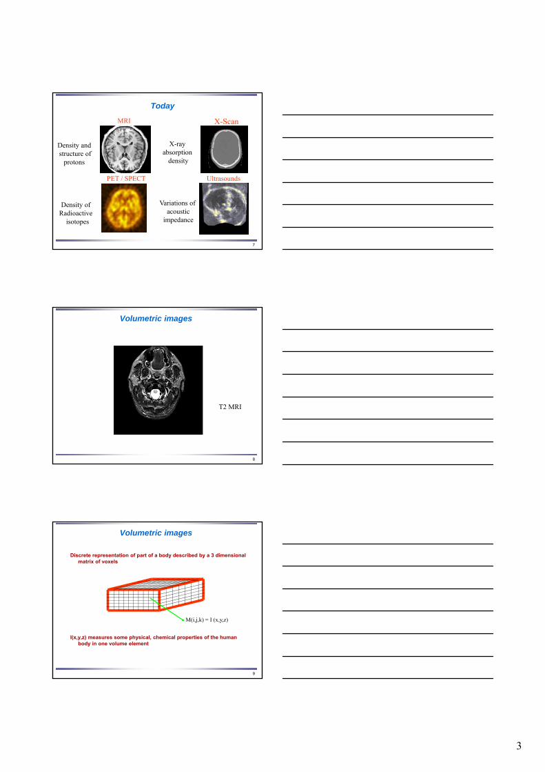
1895
First Nobel prize in Physics in 1901
Roentgen
6
Different imaging modalities
X-ray
Magnetic resonance imaging anatomic, functional, angiographic, diffusion,
spectroscopic, tagged
Transmission Tomography (X Scan)
Nuclear Medicine : Positron emission tomography (PET)
Single photon emission tomography (SPECT)
Ultrasonography
Histological Imaging, confocal in-vivo microscopy, molecular imaging,…
3
7
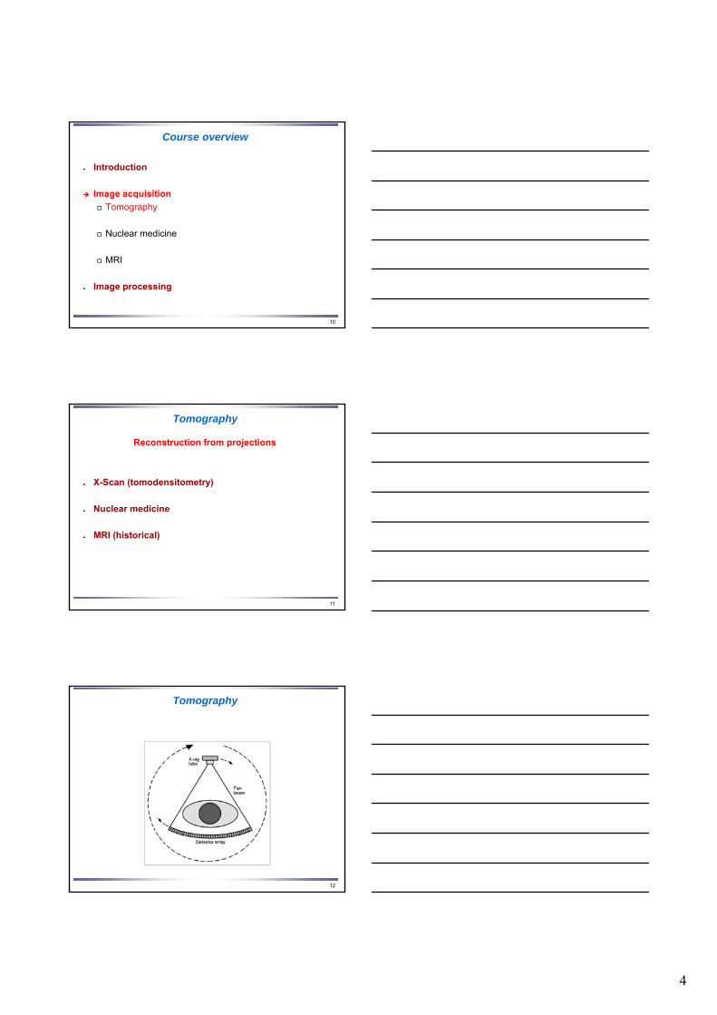
Today
MRI
UltrasoundsPET / SPECT
X-Scan
X-ray absorption
density
Variations of acoustic
impedance
Density of Radioactive
isotopes
Density and structure of
protons
8
Volumetric images
T2 MRI
9
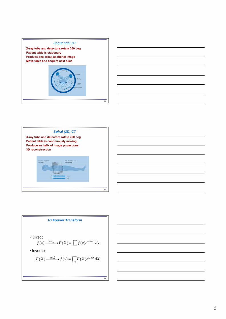
Volumetric images
Discrete representation of part of a body described by a 3 dimensional matrix of voxels
I(x,y,z) measures some physical, chemical properties of the human body in one volume element
M(i,j,k) = I (x,y,z)
4
10
Course overview
Introduction
Image acquisition
Tomography
Nuclear medicine
MRI
Image processing
11
Tomography
Reconstruction from projections
X-Scan (tomodensitometry)
Nuclear medicine
MRI (historical)
Tomography
12
5
Sequential CT
13
X-ray tube and detectors rotate 360 deg
Patient table is stationary
Produce one cross-sectional image
Move table and acquire next slice
Spiral (3D) CT
X-ray tube and detectors rotate 360 deg
Patient table is continuously moving
Produce an helix of image projections
3D reconstruction
14
15
1D Fourier Transform
dxexfXFxf xXiTF D 2)()()( 1
dXeXFxfXF xXiTF D 2)()()(1
1
• Direct
• Inverse

6
17
Fourier for tomography
The 1D Fourier transform of a 1D projection of the original 2D image is equal to the 1D slice in the same direction of the 2D Fourier transform of the original image.
18
Fourier for tomography
19
Tomotensitometrie (Scanner X)
7
20
Course overview
Introduction
Image acquisition
Tomography
Nuclear medicine
MRI
Image processing
21
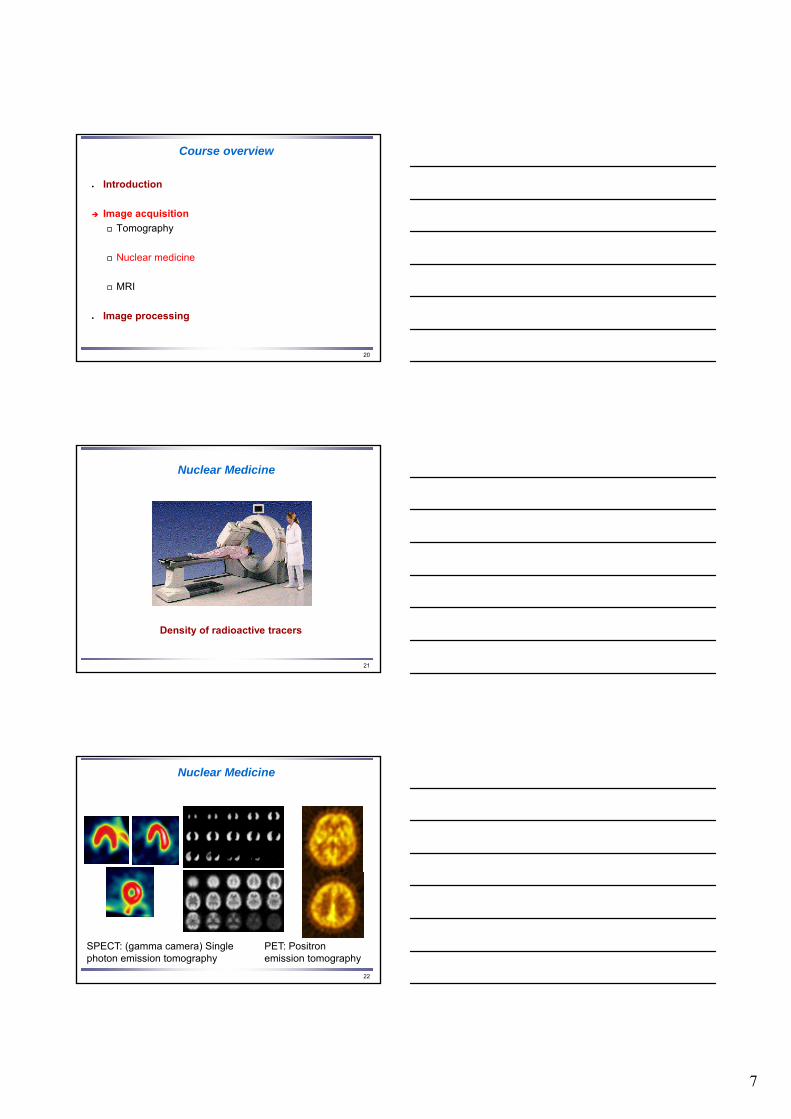
Nuclear Medicine
Density of radioactive tracers
22
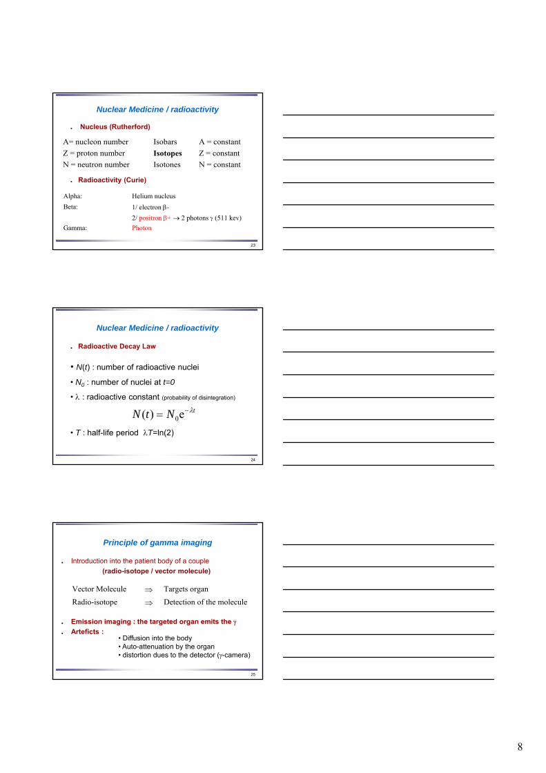
Nuclear Medicine
SPECT: (gamma camera) Single photon emission tomography
PET: Positron emission tomography
8
23
Nuclear Medicine / radioactivity
Nucleus (Rutherford)
Radioactivity (Curie)
A= nucleon number Isobars A = constant
Z = proton number Isotopes Z = constant
N = neutron number Isotones N = constant
Alpha: Helium nucleus
Beta: 1/ electron -
2/ positron + 2 photons (511 kev)
Gamma: Photon
24
Nuclear Medicine / radioactivity
Radioactive Decay Law
• N(t) : number of radioactive nuclei
• N0 : number of nuclei at t=0
• : radioactive constant (probability of disintegration)
• T : half-life period T=ln(2)
tNtN e)( 0
25
Principle of gamma imaging
Introduction into the patient body of a couple
(radio-isotope / vector molecule)
Emission imaging : the targeted organ emits the Arteficts :
Vector Molecule Targets organ
Radio-isotope Detection of the molecule
• Diffusion into the body• Auto-attenuation by the organ• distortion dues to the detector (-camera)
9
26
Principle of gamma imaging
Information given by the image
Vector molecules :
drug, protein, blood cells, ...
Reflect the metabolic function of the organ Metabolic or functional imaging Local relative concentration (relative) Concentration evolution during time Possible quantitative measures
27
Single photon gamma imaging
Radio-isotopes
Single photon emitters Technetium Tc 99m 6 h 140 kev Portative generator Iodine I 131 8 j 360 kev Reacteur (fission) Iodine I 123 13 h 159 kev Cyclotron (industry) Thallium Tl 201 73 h 80 kev Cyclotron (industry)
Krypton (Kr 81 m), Gallium (Ga 67), Indium (In 111), Xenon (Xe 133, gaz)
28
Single photon gamma imaging
Detection-camera: scintillation crystals (NaI), photomultiplicatorsCollimators: to measure the rays arriving in a known
direction (tomography assumptions)
Single Photon Emitting Computed Tomography (SPECT)
Images = projection of the volume on a plane
10
29
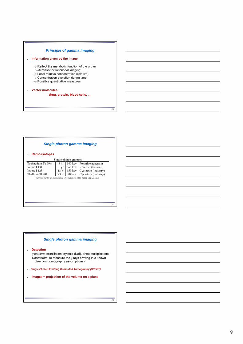
Single photon gamma imaging
BrainNecrosis: hypo fixation Tumor: hyper fixation
30
Single photon gamma imaging
Bronchopneumopathy: change of texture Pulmonary embolism: change of morphology
Lungs
31
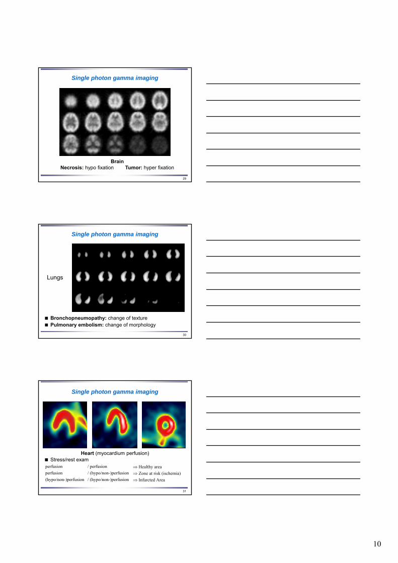
Single photon gamma imaging
Heart (myocardium perfusion) Stress/rest examperfusion / perfusion Healthy area perfusion / (hypo/non-)perfusion Zone at risk (ischemia) (hypo/non-)perfusion / (hypo/non-)perfusion Infarcted Area

11
32
Radio-isotopes
Positron emission tomography (PET)
Positron emitters Carbon 11C 20 mn cyclotron (medical) Nitrogen 13N 10 mn cyclotron (medical) Oxygen 15O 2 mn cyclotron (medical) Fluor 18F 112 mn cyclotron (medical)
Emission : positron (+) Annihilation 2 photons of 511 kev at 180º
33
Positron emission tomography (PET)
Physiological molecules
DetectionPositron camera: scintillation crystals (NaI) and
photomultiplicators diametrically opposed .Detection of coinciding of annihilation photons
Image(s) = projection of the radioactive volume on a plane
water H2O15
glucose fluoro-deoxyglucose (F18DG)
34
Positron emission tomography (PET)
12
35
Course overview
Introduction
Image acquisition
Tomography
Nuclear medicine
MRI
Image processing
36
Magnetic resonance imaging
Density and structure of protons
37
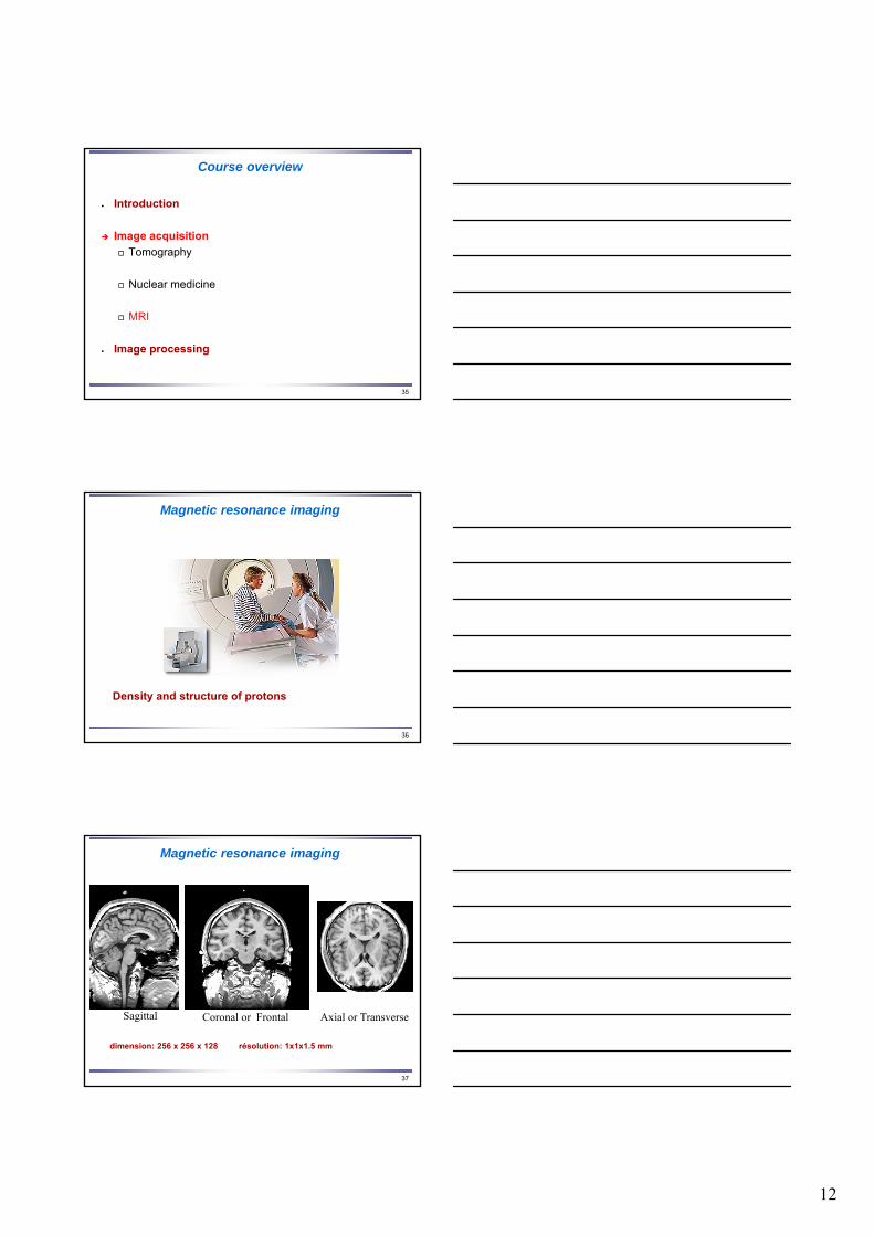
Magnetic resonance imaging
dimension: 256 x 256 x 128 résolution: 1x1x1.5 mm
Sagittal Coronal or Frontal Axial or Transverse

13
38
MRI: a few dates
• 1946: MR phenomenon - Bloch et Purcell
• 1952: Nobel prize - Bloch et Purcell
• 1950-1970: development but no imaging
• 1980: MRI feasibility
• 1986 - …: real development
39
MRI: One modality with multiple sequences
• Anatomic MRI: T1, T2, DP weighted images
• Angiographic MR
• Functional MR: cognitive studies
• Diffusion MR: brain connectivity
• MR Spectroscopy
No absolute quantification
40
Plan du Cours
Introduction
Tomography
MRI
Magnetic resonance The molecular level
The macroscopic level
resonance
Nuclear medicine
14
41
Magnetism at the molecular level
Electric charges in motion:
• magnetic momentum
42
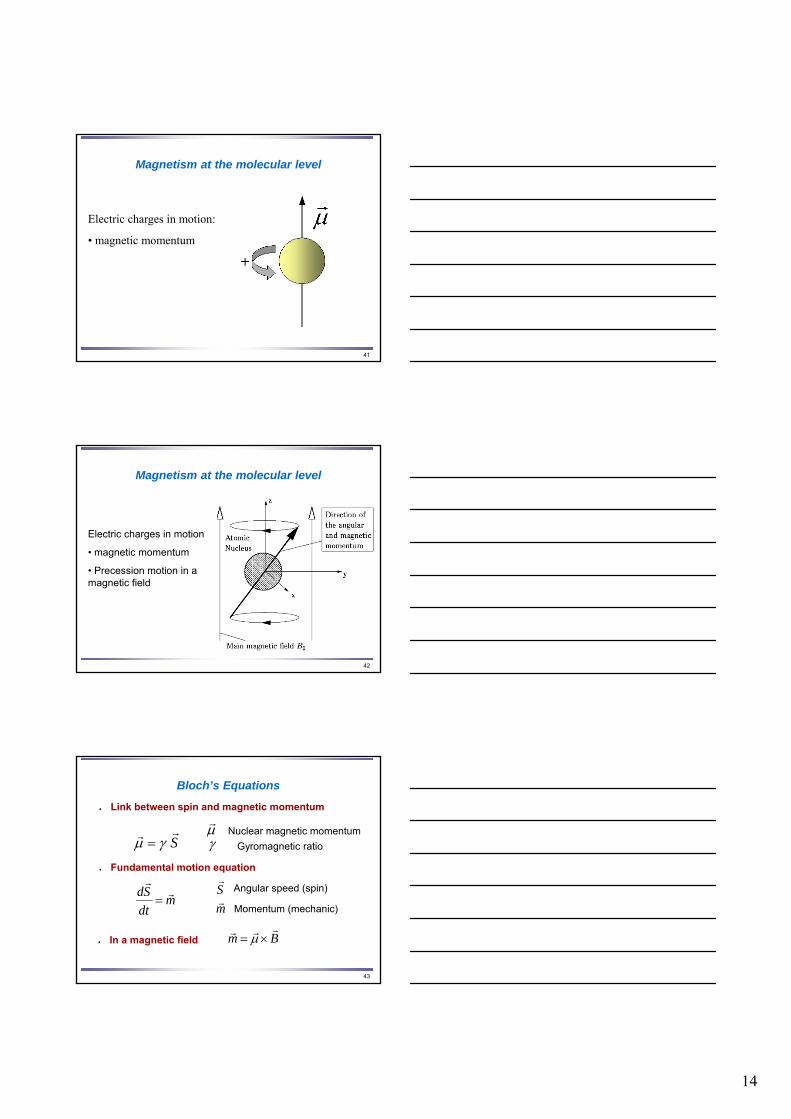
Magnetism at the molecular level
Electric charges in motion
• magnetic momentum
• Precession motion in a magnetic field
43
Bloch’s Equations
Fundamental motion equation
mdt
Sd rr
Link between spin and magnetic momentum
Sr
Angular speed (spin)
mr
Momentum (mechanic)
Srr
r Nuclear magnetic momentum
Gyromagnetic ratio
In a magnetic field Bmrrr
15
44
Bloch’s Equations
Bdt
d rrr
Thus
0
0 0
0
B
Br
0
)(sin
)( cos
z
Lt
Lt
t
t
r
0 BL Larmor’s frequency
45
Magnetism at the molecular level
Nucleus % γ (MHz/T) 1H 99.985 42.575 2H 0.015 6.53 13C 1.108 10.71 14N 99.63 3.078 15N 0.37 4.32 17O 0.037 5.77 19F 100 40.08
23Na 100 11.27 31P 100 17.25
46
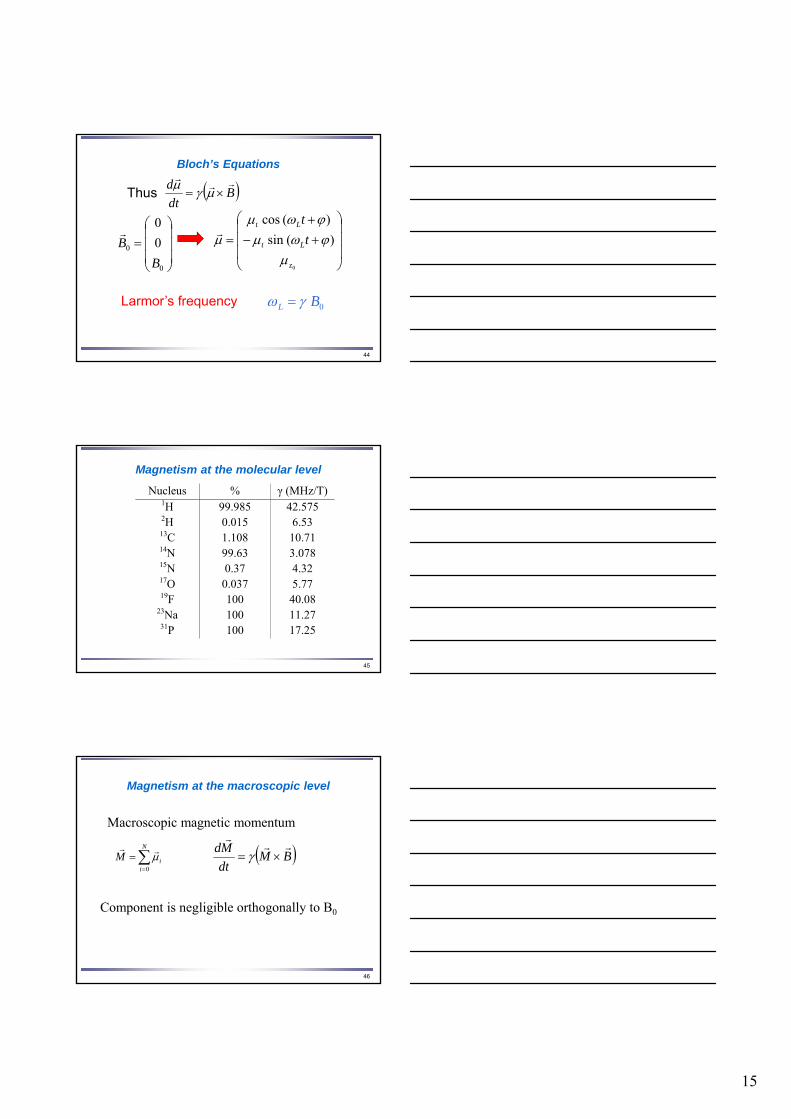
Magnetism at the macroscopic level
Macroscopic magnetic momentum
N
iiM
0
rr BM
dt
Md rrr
Component is negligible orthogonally to B0
16
47
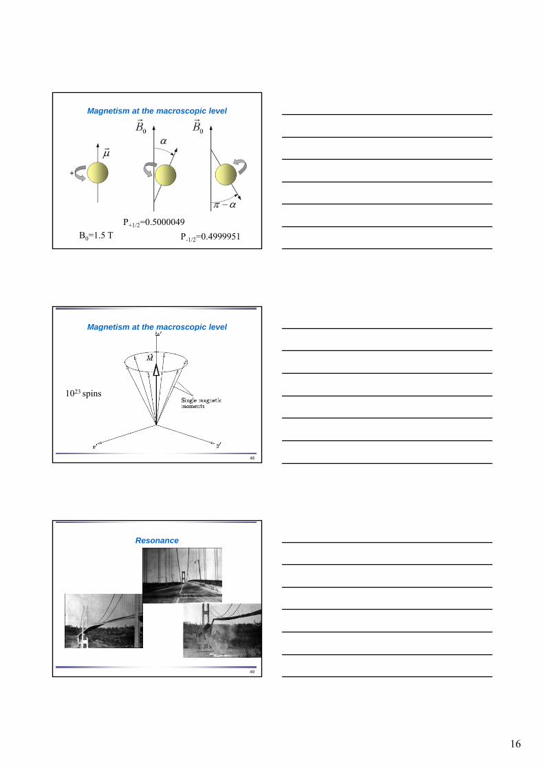
Magnetism at the macroscopic level
P+1/2=0.5000049
P-1/2=0.4999951B0=1.5 T
48
Magnetism at the macroscopic level
1023 spins
49
Resonance
17
50
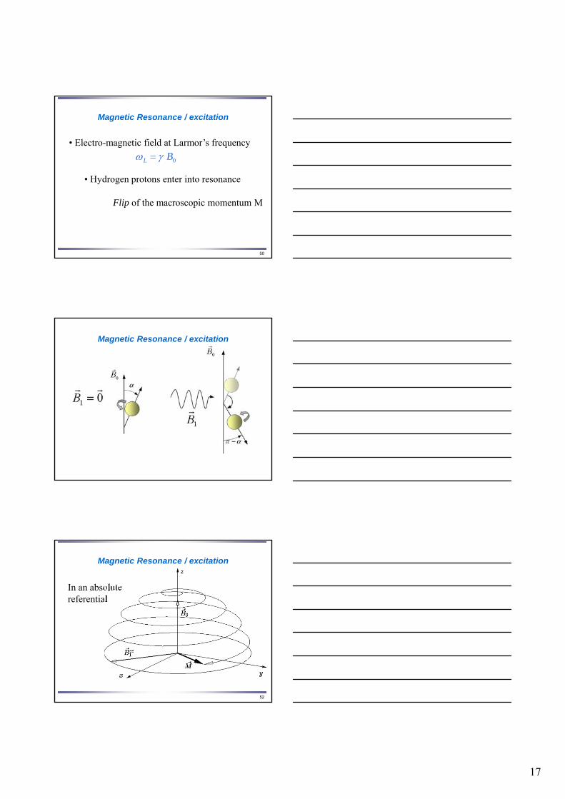
Magnetic Resonance / excitation
• Electro-magnetic field at Larmor’s frequency
0 BL
• Hydrogen protons enter into resonance
Flip of the macroscopic momentum M
51
Magnetic Resonance / excitation
52
Magnetic Resonance / excitation
In an absolute referential
18
53
Magnetic Resonance / relaxation
1T
MBM
dt
dM zz
z rr
• Return to equilibrium / B0: time constant T1
• Spin dephasing: Time constant T2
2
,,
,
T
MBM
dt
dM yxyx
yx rr
54
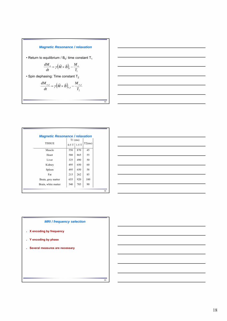
Magnetic Resonance / relaxationT1 (ms)
TISSUE 0.5 T 1.5 T
T2(ms)
Muscle 550 870 45
Heart 580 865 55
Liver 325 490 50
Kidney 495 650 60
Spleen 495 650 58
Fat 215 262 85
Brain, grey matter 655 920 100
Brain, white matter 540 785 90 540
63
MRI / frequency selection
X encoding by frequency
Y encoding by phase
Several measures are necessary
19
64
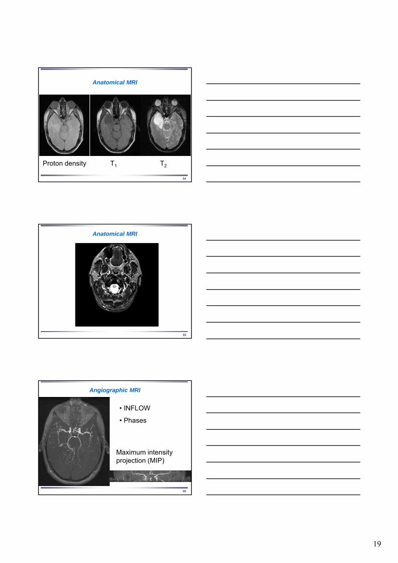
Anatomical MRI
Proton density T1 T2
65
Anatomical MRI
66
Angiographic MRI
Maximum intensity projection (MIP)
• INFLOW
• Phases
20
67
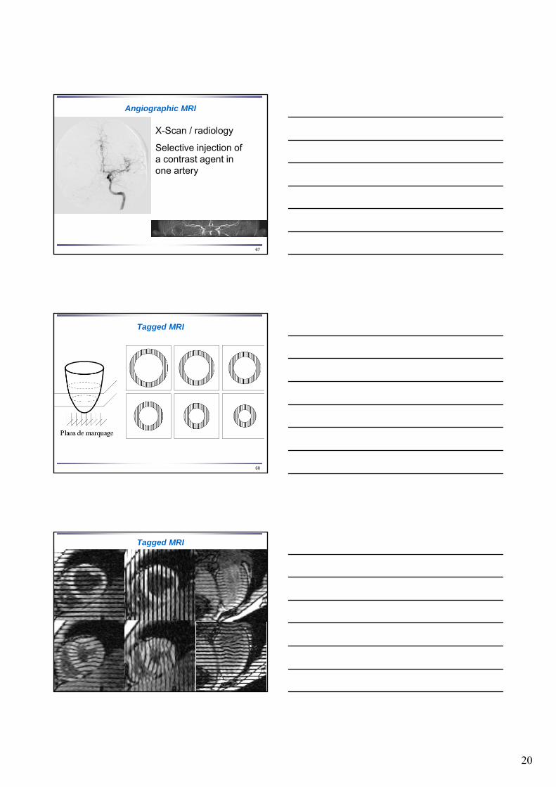
Angiographic MRI
X-Scan / radiology
Selective injection of a contrast agent in one artery
68
Tagged MRI
69
Tagged MRI
21
70
Functional MRI
• BOLD : blood oxygen level dependent
(oxy)-hemoglobin: diamagnetic
deoxy-hemoglobin: paramagnetic
Neuronal activation
Local oxygen consumption
Change the ratio of concentration
Variation of the measured signal
71
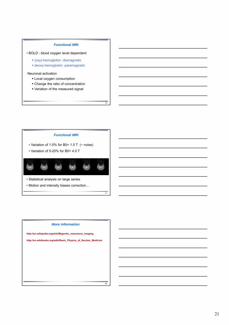
Functional MRI
• Statistical analysis on large series
• Motion and intensity biases correction…
• Variation of 1-5% for B0= 1.5 T (~ noise)
• Variation of 5-20% for B0= 4.0 T
80
More information
http://en.wikipedia.org/wiki/Magnetic_resonance_imaging
http://en.wikibooks.org/wiki/Basic_Physics_of_Nuclear_Medicine
22
81
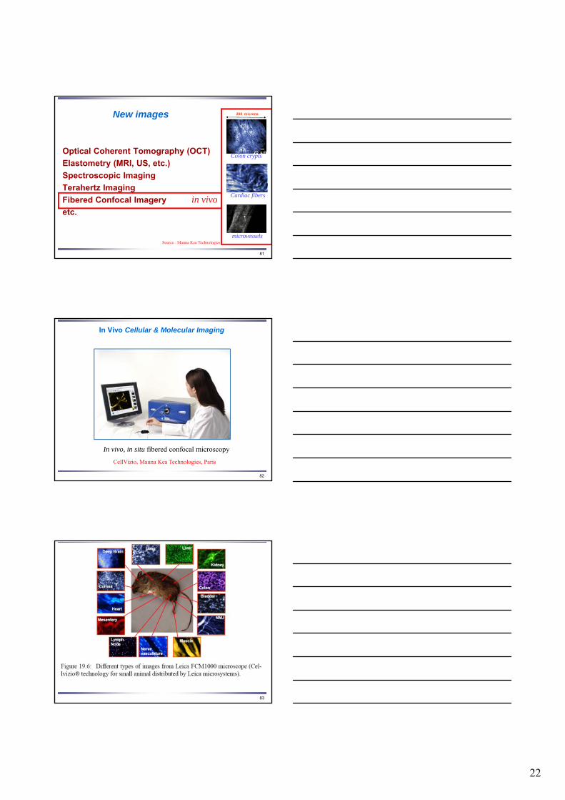
New images
Optical Coherent Tomography (OCT)
Elastometry (MRI, US, etc.)
Spectroscopic Imaging
Terahertz Imaging
Fibered Confocal Imagery
etc.
200 microns
Cardiac fibers
microvessels
Colon crypts
in vivo
Source : Mauna Kea Technologies
82
In Vivo Cellular & Molecular Imaging
CellVizio, Mauna Kea Technologies, Paris
In vivo, in situ fibered confocal microscopy
83

23
84
In vivo ClinicalMicroscopic Imagery
Cellvizio®, Mauna Kea Technologies (MKT) , Paris
300 microns
Pr A. Meining, Munich
gastro-esophagusMucosa
85
In Vivo MicroEndoscopy
Colon
Duodenum and
small intestineStomach
Rectum
Bile duct
EsophagusEsophagus
86
Advanced Image Processing
240 microns
Precision and field of view?
Small bowel
24
87
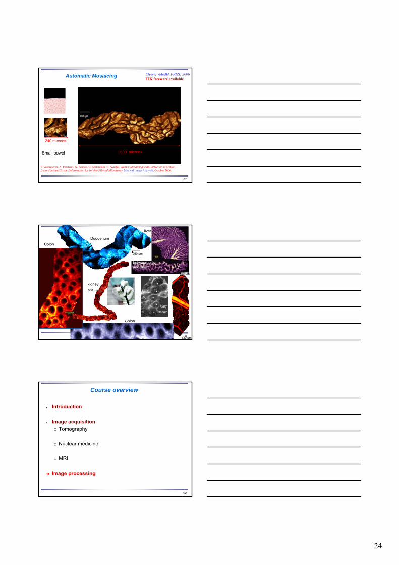
Automatic Mosaicing
T. Vercauteren, A. Perchant, X. Pennec, G. Malandain, N. Ayache, Robust Mosaicing with Correction of Motion Distortions and Tissue Deformation for In Vivo Fibered Microscopy. Medical Image Analysis, October 2006.
240 microns
Small bowel
Elsevier-MedIA PRIZE 2006ITK freeware available
3600 microns
88130 μm
Duodenum
250 μm
between colon and ileum
250 μm
50μm
mouth
Colon
liver
kidney
500 μm
Colon
92
Course overview
Introduction
Image acquisition
Tomography
Nuclear medicine
MRI
Image processing
25
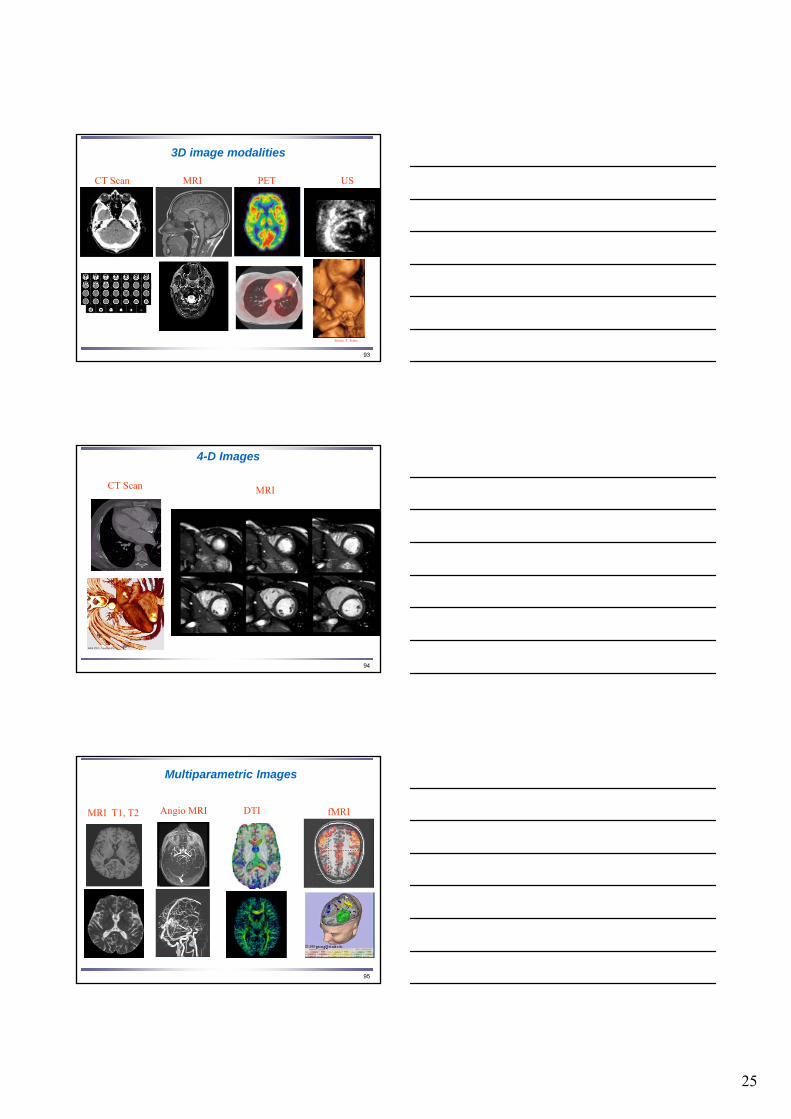
93
3D image modalities
USCT Scan MRI PET
Source :T. Peters
94
4-D Images
MRICT Scan
95
Multiparametric Images
Angio MRI fMRIDTIMRI T1, T2

26
96
Medical Imaging Today
Large Choice of in vivo modalities
High temporal and spatial resolution
Large parameter space
Large Databases
Image-guided Therapy
Quantity of information too high : cannot be processed without the help of computer science
Da VinciSurgical
Robot
97
Computational Medical Image Analysis (1980 - Today)
Assist Diagnosis
Objective quantitative measurements
fusion of multimodal, multidimensional, multiparameter images
Assist Therapy
Plan, simulate (before)
Control (during), follow-up (after)
J. Duncan & N. A, Medical Image Analysis, Progress over two decades and the challenges ahead, IEEE – Pami, 2000.
99
Classification of 3D image processing problems
Segmentation (organs, lesions, activations,…)
Registration (comparison, fusions)
Motion analysis (cardiac imaging)
Deformable models (Surgery simulation)
Medical Robotics (image guided surgery, telesurgery…)
27
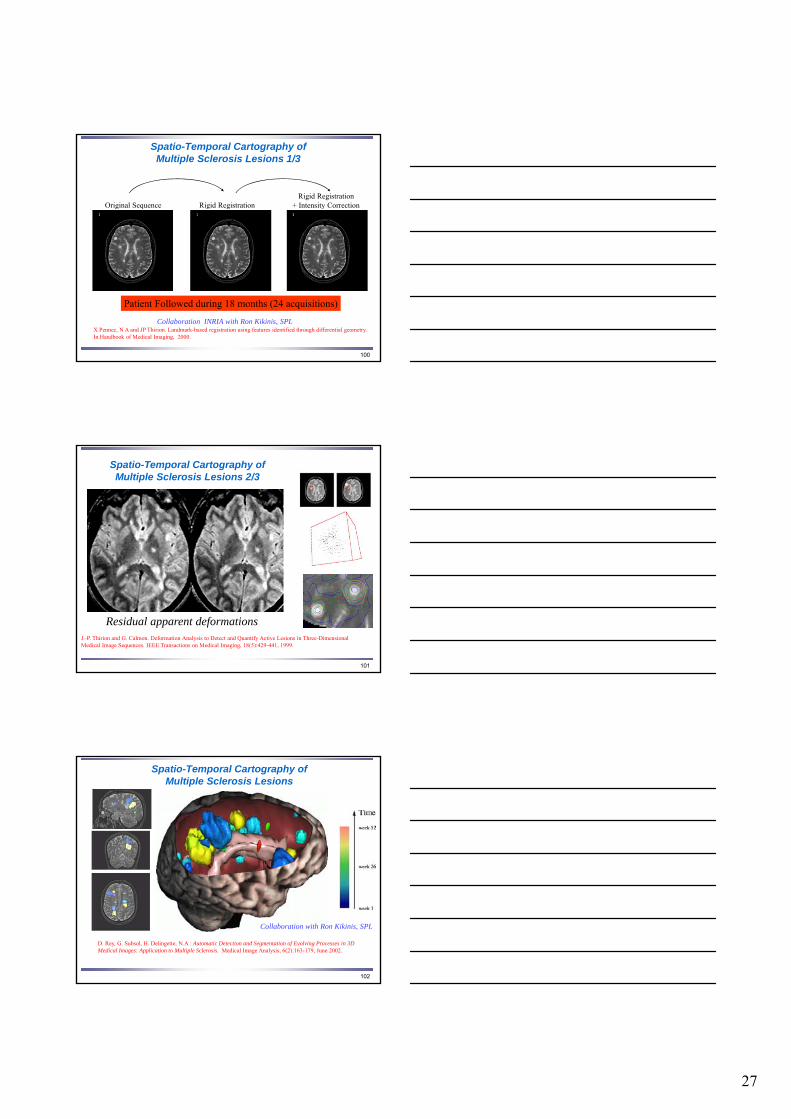
100
Spatio-Temporal Cartography of Multiple Sclerosis Lesions 1/3
Original Sequence Rigid RegistrationRigid Registration
+ Intensity Correction
Patient Followed during 18 months (24 acquisitions)
Collaboration INRIA with Ron Kikinis, SPLX Pennec, N A and JP Thirion. Landmark-based registration using features identified through differential geometry. In Handbook of Medical Imaging, 2000.
101
Spatio-Temporal Cartography of Multiple Sclerosis Lesions 2/3
Residual apparent deformationsJ.-P. Thirion and G. Calmon. Deformation Analysis to Detect and Quantify Active Lesions in Three-Dimensional Medical Image Sequences. IEEE Transactions on Medical Imaging, 18(5):429-441, 1999.
102
Spatio-Temporal Cartography of Multiple Sclerosis Lesions
D. Rey, G. Subsol, H. Delingette, N.A : Automatic Detection and Segmentation of Evolving Processes in 3D Medical Images: Application to Multiple Sclerosis. Medical Image Analysis, 6(2):163-179, June 2002.
Collaboration with Ron Kikinis, SPL
28
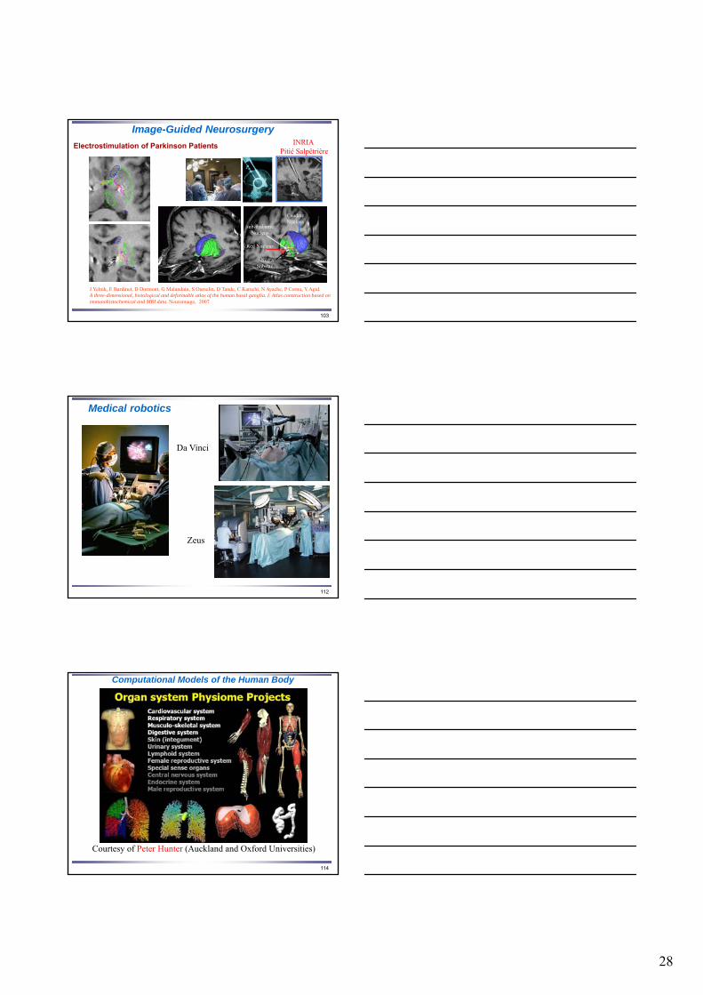
103
Image-Guided Neurosurgery
Electrostimulation of Parkinson Patients
Caudate Nucleus
Red Nucleus
sub-thalamic Nucleus
Negra Substance
J Yelnik, E Bardinet, D Dormont, G Malandain, S Ourselin, D Tande, C Karachi, N Ayache, P Cornu, Y Agid. A three-dimensional, histological and deformable atlas of the human basal ganglia. I. Atlas construction based on immunohistochemical and MRI data. Neuroimage, 2007
INRIA Pitié Salpêtrière
112
Medical robotics
Zeus
Da Vinci
114
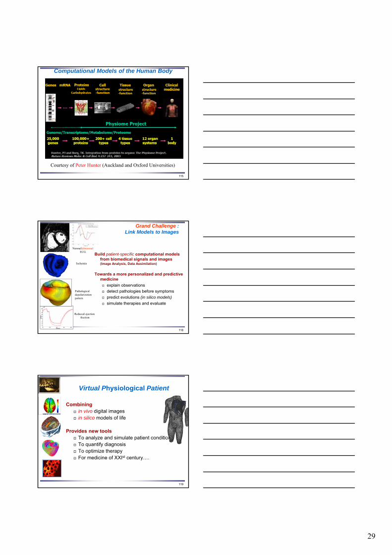
Computational Models of the Human Body
Courtesy of Peter Hunter (Auckland and Oxford Universities)
29
115
Courtesy of Peter Hunter (Auckland and Oxford Universities)
Computational Models of the Human Body
118
Grand Challenge : Link Models to Images
Build patient-specific computational models from biomedical signals and images (Image Analysis, Data Assimilation)
Towards a more personalized and predictive medicine
explain observations
detect pathologies before symptoms
predict evolutions (in silico models)
simulate therapies and evaluate
Reduced ejectionfraction
Pathologicaldepolarization pattern
Ischemia
Normal/abnormalECG
119
Virtual Physiological Patient
Combining in vivo digital images in silico models of life
Provides new tools To analyze and simulate patient condition To quantify diagnosis To optimize therapy For medicine of XXIst century….
30
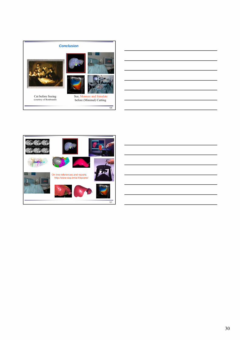
120
Conclusion
Cut before Seeing(courtesy of Rembrandt)
See, Measure and Simulatebefore (Minimal) Cutting
121
On line references and reportshttp://www-sop.inria.fr/epione/