x-pand.me body stretching system - pro course
DESCRIPTION
X-pand.Me Body Stretching System is based on a unique series of intense stretching exercises. Kaaberbol’s thoroughly developed program of advanced stretching exercises affect the core of the muscular system, thus optimizes athletes’ performances.TRANSCRIPT


X-pand.Me ® Body Stretching System - PRO course
2
WarningImportant health note: If you or your clients are suffering from any medical condition or have concerns about your health, please consult a doctor before proceeding. The author takes no responsibility for any injury or illness resulting from suggested stretches or poses.
All rights reserved - Copyright © X-pand Me LLC - 2013
X-pand.Me ® is a trademarked name and logo and can only be used with written approval from X-pand Me LLC. Author: Peter KaaberbolDesigner: www.squaws.dk Photography: www.henriksorensen.dkLayout: X-pand Me LLC
All rights reserved. No part of this publication may be reproduced, stored in a retrieval system, or transmitted in any or by any means electronic, mechanical, photocopied, recorded or otherwise, without the prior written permission of the publisher.

X-pand.Me ® Body Stretching System - PRO course
3
The X-pand.Me ® Body Stretching System
Enables you to treat athletes with a unique connected series of stretches
Including a unique body mechanic system that increases core strength, flexibility and balance f or the X-pand.Me PRO trainer
X-pand.Me ® Body Stretching System - PRO course
4
X-pand.Me ® Body Stretching System - PRO course
5
Who will benefit from the X-pand.Me Body Stretching System
X-pand.Me is developed to optimize performance for: • PRO athletes • High performance athletes • Daily athletes • Yoga practitioners • Recreational practitioners • People from all walks of life who desire optimum health X-pand.Me is to be performed by:
• Fitness instructors • Personal trainers • Body workers and massage therapists • Yoga teachers • Physical therapists
X-pand.Me is intended to be used by:
• Sport clubs and teams • Fitness studios • Wellness studios • Training facilities • Yoga studios
And everyone else…

X-pand.Me ® Body Stretching System - PRO course
6

X-pand.Me ® Body Stretching System - PRO course
7
Career Network - 2 Steps to become a fully certified instructor
X-pand.Me PRO 10 hours online/correspondance, 20 hour public/private course
• Full body stretching series covering major and minor muscles • Correct alignment for all exercises and stretches performed • Body mechanics for practitioner during exercises for maximum safety • Flowing transitions between stretching • In-depth muscle anatomy • 192 page manual which includes easy-to-use, step-by-step charts • Examination
After passing the final exam, the student becomes a certified X-pand.Me Body Stretching System, PRO Trainer.
X-pand.Me PRO is availible through distance/e-learning, correspondence or classroom courses.
X-pand.Me INSTRUCTOR
10 hour public/private course
• Evaluation of the X-pand.Me Body Stretching System PRO • Working with body communication as an X-pand.Me PRO Trainer • Stretches vs. sport types and injuries • Theoretical and practical teacher training in the classroom • X-pand.Me YOURSELF class room stretch series for groups
After passing the final exam, the student becomes an X-pand.Me Body Stretching System INSTRUTOR.

X-pand.Me ® Body Stretching System - PRO course
8
pp. 9 pp. 10pp. 11pp. 15pp. 18pp. 24
pp. 137pp. 160pp. 164pp. 186pp. 187pp. 188pp. 189pp. 190 pp. 191
Content 1 - Foreword ............................................................................. 2 - Introduction ......................................................................... 3 - Guidelines ........................................................................... 4 - Step-by-Step use of the manual .......................................... 5 - Charts of the complete series .............................................. 6 - The complete series ........................................................... 7 - Anatomy and Physiology ..................................................... 8 - Glossary .............................................................................. 9 - Muscle listing ....................................................................... 10 - Conclusion.......................................................................... 11 - Ethical Guidelines .............................................................. 12 - About the Author................................................................. 13 - Evaluation.......................................................................... 14 - Examination ...................................................................... 15 - Bibliography ......................................................................
X-pand.Me ® Body Stretching System - PRO course
9
Foreword
In my life, I have been an athlete. I have worked extensively as a teacher and therapist of massage, yoga
and other body treatment programs. I have felt and seen the importance of flexibility through combining
strength and stretching and how they support each other. Utilizing routines to stay supple and strong will
prevent injuries and allow us to lead active, productive lives into old age. Stretching the muscles of the
body regularly and methodically allows everyone to retain and increase their muscle elasticity and range of
movement. This applies to exercise as well as everyday life.
The X-pand.Me ® Body Stretching System came to me in a blissful moment a few years ago during the
soccer world championship. The players prepared themselves for the penalty kick competition during a
five minute break. The idea came to me in a split second as the personal trainers and physical therapists
worked on the players, stretching their legs on the field. The X-pand.Me concept took shape before my
eyes. I realized I had the knowledge and background to create a stretching system that would make a
huge difference for athletes and people from all walks of life. I would create a unique, assisted muscle-
stretching system performed methodically in order to optimize performance. This system would help
people reach a state of well-being and support athletes to achieve their goals.
The X-pand.Me Body Stretching System originated from systematic experience with my yoga teachings
using assisted stretches. Skills from my career as a PRO body worker also contributed to the final product.
I shared the blueprint of this system with body worker colleagues and personal trainers who saw its incred-
ible value and they encouraged me to promote this unique program.
Through dedication, hard work and the cooperation of many talented people, I have had the pleasure of
creating this revolutionary bodywork system. I have combined assisted stretches and flowing movements
to work all major and minor muscle groups to create suppleness and alignment throughout the body.
It’s in our human nature—we strive to achieve more. We all want to lead healthy, active and productive
lives, and through life we explore ways to enhance our performance. Top athletes, people in general,
young or old, who exercise regularly for wellbeing, all want to improve. We strive to improve our soccer
kick and score a goal. We would love to make a hole-in-one on the golf course. We would love to run
faster, jump higher or swim longer. The X-pand.Me Body Stretching System helps make all of this possible.
Founder, Peter Kaaberbol

X-pand.Me ® Body Stretching System - PRO course
10
Introduction
Welcome to the X-pand.Me Body Stretching System, a unique, powerful and esthetically-pleasing movement system that:
• Optimizes performances • Increases endurance • Improves range of motion
The uniqueness of the X-Pand.Me Body Stretching System lies in the athlete’s total relaxation (passive stretching) during the 53 stretching poses and 5 alignment exercises.
The X-pand.Me Body Stretching System is an interconnected series of advanced stretches covering all of the athlete’s major and minor muscle groups. This program is structured and explained systematically and simply so that every practitioner is capable of becoming an X-pand.Me PRO Trainer.
The X-pand.Me PRO Trainer guides the athlete’s body through all stretches in a continuous flow, so the athlete receives a series of beneficial stretches as well as an amazing body treatment.
Developed from a holistic viewpoint, the transitions are performed in a beautifully-coordinated movement that flows like choreography.
All stretches are in precise alignment with the athlete’s body as well as the trainer’s so the trainer will also benefit from performing the program.
Reasons to Use the Method
X-pand.Me is a powerful, advanced body stretching system that is very easy to learn and teach. The program is structured and explained systematically and simply so any practitioner can train to become an X-pand.Me PRO Trainer.
Athletes will add that extra magic ingredient, leading them to peak performance. The concept focuses on flexibility through stretching and strength combined. Regular exercise combined with this advanced system is the way to achieve athletic goals, prevent injuries and optimize performance, bringing those medals within reach.

X-pand.Me ® Body Stretching System - PRO course
11
Guidelines for Using the X-pand.Me Body Stretching System Manual
The X-pand.Me Body Stretching System consists of 53 stretching poses and 5 alignment exercis-es. In the program section we show each stretch, alignment and movement of the system individu-ally. Each interconnected stretch flows into and supports the next stretch. Understanding and mas-tering this movement pattern creates the foundation of the X-pand.Me Body Stretching System.
From this basic integrated structure, some parts of the stretches are also used to create shorter and more specific stretching sessions.
X-pand.Me students will learn to use this system’s special body mechanics to increase their own performance and awareness of their athlete’s range of motion (R.O.M.).
The various changing positions in the system will be illustrated clearly including all grips and locks used. It is important to repeat and practice these movements to fully incorporate the flowing pat-tern.
The performed stretches and alignments aim ultimately at reaching the athlete’s R.O.M. safely and therapeutically. An emphasis will be placed on sensing and allowing the athlete’s muscles to let go easily and naturally. As the X-pand.Me student’s skill and confidence grow, a more efficient, profound and safe result is created for the athlete. The more comfortable the X-pand.Me student becomes with the flowing positions, the more profound the effect on the athlete.
Advice for X-pand.Me PRO trainers will be given on various topics: communication; establishing trust; and post-treatment suggestions.

X-pand.Me ® Body Stretching System - PRO course
12
Specific Guidelines
Common sense and a few rules combine to create the foundation of these guidelines. The proce-dures guarantee that your athlete’s physical state improves safely. Important terms are introduced to facilitate memorization and practice of the techniques. These terms are repeated throughout the manual and describe all grips used as well as each physical position of the student. These simple terms help the student orient himself and easily understand which grip and applied stretch come next.
It is very important to understand the various techniques assuring the proper movement patterns and positions, “inner strength” and “body locks” used in the X-pand.Me system. These techniques are vital to both the comfort of the athlete and the focus of the practitioner while performing the stretches. To ease the understanding of this technique the manual contains a concise introduction of the grips, positions and transitions in Principles of Stretching (P.O.S.).
During X-pand.Me classes, the importance of staying aligned and connected with the core of the body will be continuously reinforced.
• Terminology of grips and positions
• “inner strength” & “body locks”
• Principles of Stretching (P.O.S)
Terminology of Grips and Positions
Often the X-pand.Me PRO Trainer will utilize a posture called the “knight’s position.” The trainer sits with one knee placed on the mat with the opposite knee lifted. This position allows the trainer to be close to the body and still move freely. It is safe and minimizes the risk of lifting with the lower back.
The trainer must repeatedly practice proper hand placement as shown in the grip and alignment section of the P.O.S. to ensure correct grip position.
“Inner Strength” and “Body Locks”
Staying connected to the core of the body will ensure a stable and firm balance as well as increase strength and accuracy in the routine. The trainer should engage the lower core muscles while ap-plying the techniques. X-pand.Me refers to this as “inner strength.”
The X-pand.Me PRO Trainer must activate the muscle groups surrounding each joint to prevent hyper-extending or over-stretching. X-pand.Me refers to this technique as “body locks,” which help ensure a safe performance of the X-pand.Me routine.
X-pand.Me ® Body Stretching System - PRO course
13
Principles of Stretching (P.O.S)
The Principles of Stretching used in X-pand.Me are based on creating and maintaining a secure foundation for both the trainer and athlete. The application and performance of the X-pand.Me series is divided into four stages, as shown below. These four sections describe the components of each stretch and correctly position the trainer for the next stretch.
By skillfully using balance and core awareness, the trainer creates a firm, steady movement pattern that the athlete yields to naturally.
1. Foundation—Primary Position
The X-pand.Me PRO Trainer and the athlete must be in Primary Position (as shown in the movie, study picture and directions) before the applied stretching begins. The performance of the primary position by the trainer calls for balance and “inner strength.” Attention to the proper alignment of the athlete ensures an ideal foundation for the stretch to be applied.
2. Alignment—Grip Preparation
This is a description of adjustments performed to keep the stretch balanced and of high quality. The body of the athlete is further stabilized and secured by the proper alignment preparation between the athlete and the X-pand.Me PRO Trainer. The trainer’s grip on the athlete’s body needs to be secure and firm. The trainer must allow for adjustments but never compromise safety by adopting an incorrect position.
3. Action—Performed Stretch
This section describes how the actual stretches are performed. The quality of the stretch is based on the proper balance and effort of the X-pand.Me PRO Trainer. The trainer should use a careful and attentive approach. Using gravity and acute shifts of balance, the applied stretches become effortless and the trainer’s awareness assures the safety of the athlete. When applying stretches, the trainer should take the time to listen to their athlete’s breathing. The trainer should follow the athlete’s exhale as they always stretch 1-2-3 times on the exhales for a minimum of 10 seconds for each stretch.
4. Flow—Transition Phase
This phase defines the precise flow of movements needed to move from the end of a stretch to the next Primary Position. The importance of the transition phase is based on the trainer’s ability to anticipate the subsequent Primary Position.
Note:The X-pand.Me PRO Trainer must be aware of the athlete’s body alignment at all times. Quickly check the athlete’s body now and then to ensure correct alignment.

X-pand.Me ® Body Stretching System - PRO course
14
Additional Tips
• Stay present and aware of athlete’s breath, sounds and possible signs of discomfort during the applied stretches.
• Support the athlete’s body as much as possible to create maximum trust, relaxation and comfort.
• Create length and space in every stretch applied.
• Do not add pressure onto any joint or body part.
• Guide every applied stretch within the limit of the athlete’s allowable range of motion. • Trainer should pay attention to his/her own body mechanics to protect and support proper technique within the connected series of stretches.
• Create a rhythm based on the breath and muscle sensitivity of the athlete to allow the stretch to happen and not force it.
• Always take into consideration the physical history of the athlete (e.g., old injuries).
• Allow for expression of personal experiences from the athlete before and after the session.
• Keep in mind the importance of a tidy environment and a clean and presentable appearance.
• Both athlete and trainer should drink a lot of water after an X-pand.Me session.
X-pand.Me ® Body Stretching System - PRO course
15
Step-by-Step use of the Manual, charts and movie
The X-pand.Me PRO course contains two components. • The Full Body Alignment • The Body Stretching Series
Obtain an overall view of the X-pand.Me system by studying the section of the full sequence sketch.
Then proceed through each section in numerical order.
Important: Thoroughly study one section at a time. Each section is interconnected and creates a foundation for the next section.
1. Begin each section by studying the pictures and getting to know the muscle groups shown in the manual. Read the Quick Notes for ease and safety in performing the stretch.
2. Read each section of the manual in depth including the muscle-group section. Use the pictures as a performance guide. Take notes on key issues.
3. Watch the section on the DVD. Repeat as needed.
4. Practice the section using a partner and allow for extensive feedback. 5. Repeat steps one through four until a level of proficiency has been reached.
6. Proceed to next section/sections.
We all learn differently, so please accept your own speed and do not move ahead until you are truly ready. You will know you are ready when you can perform the sections with ease of both body and mind.
We recommend that you study 3-5 sections at a time before proceeding to the next set of 3-5 sec-tions. When completed in this way, you will master the complete series of X-pand.Me.
The trainer should complete work on one entire leg and one entire arm before changing to the other side of the body. Work on the opposite leg and arm before moving on to the rest of the body..Note! In this section of the manual we will call the X-pand.Me PRO Trainer “X-trainer” and the Athlete simply “Athlete”

X-pand.Me ® Body Stretching System - PRO course
16
The Body Stretching Series
In each stretch we affect many muscles. On our muscle listing, we have chosen to write the larger muscles and/or most affected muscle groups first. We have included the most important muscles stretched though more may be affected.
For information origin and insertion see the chapter on muscle listings in which we have listed the precise actions, insertions, origins.
Overview
On every page of a given stretch we have included the following terms:
The body part name: in general language, in PRO language (latin name) Picture: of the position Hint: what to be aware of in this particular stretch
Primary position: see specific guidelines Alignment prep: see specific guidelines Applied stretch: see specific guidelines Transition phase: see specific guidelines
Anatomical picture: the outer/general muscle/body part Muscle: latin name General action: the action that the muscles perform (specifics in the following chapter) Interest: a fact about this body part and /or muscle

X-pand.Me ® Body Stretching System - PRO course
17
Full Body Alignment
The first thing we do before any X-pand.Me Body Stretching System session is the Full Body Align-ment.
When asking the athletes to lie back we must ensure that they are positioned correctly. We call this the correct alignment position. Very often athletes will lie down with the patterns that they have built up throughout life, or casually just lie down. These patterns will not likely be optimal for the X-pand.Me PRO session.
When the athlete places himself on the mat, he will often contract the muscles. As a result, the body will not be in its relaxed state or open body position. It is very important that we place the athlete in the correct position for an optimal session.
Before and after any X-pand.Me body stretching session, this alignment series should be per-formed as preparation for the athlete’s body. Even while the session is conducted, these simple alignments can be given to ensure the optimal conditions for each session.
Because of the importance of this full body alignment we have included it in the full series both at the beginning and at the end. This is not a part of the body stretching series itself, but is definitely seen as an important ingredient in making the best stretches possible.
Please note that if the body of the athlete is misaligned during the body stretching session, the trainer should stop and perform the full body alignment before continuing on with the X-pand.Me series.

X-pand.Me ® Body Stretching System - PRO course
18

X-pand.Me ® Body Stretching System - PRO course
19

X-pand.Me ® Body Stretching System - PRO course
20

X-pand.Me ® Body Stretching System - PRO course
21

X-pand.Me ® Body Stretching System - PRO course
22

X-pand.Me ® Body Stretching System - PRO course
23

X-pand.Me ® Body Stretching System - PRO course
24

X-pand.Me ® Body Stretching System - PRO course
25
Hint: Observe the athlete’s body position before performing Alignment to assess the body’s posture and overall condition.
Primary Position: The X-trainer stands or kneels above the athlete with one leg on either side at the level of the torso. The athlete lies on her back with arms down at her sides.
Grip and Alignment Preparation: The X-trainer’s left hand gently lifts the shoulder from above and then slides his right hand below the shoulder blade of the athlete from inside of the arm. The X-trainer places the left hand on top of the athlete’s shoulder.
Performed Alignment: The X-trainer’s right hand pulls the shoulder blade down the back as the left hand grounds the shoulder of the athlete.
Transition Phase: The X-trainer releases both hands, changing the foot/knee position to get closer to the left shoulder, preparing to lift the shoulder with the right hand.
Shoulder alignment - left shoulder

X-pand.Me ® Body Stretching System - PRO course
26
Shoulder alignment - right shoulder
Hint: Whenever “lifting” any part of the athlete’s body, shift your own weight so that you actually lift without using muscle, relying instead on gravity.
Primary Position: The X-trainer stands or kneels above the athlete’s body with one leg on either side at the level of the torso. The athlete lies on her back with arms down at her sides.
Grip and Alignment Preparation: The X-trainer’s right hand gently lifts the shoulder from above and then slides his left hand below the shoulder blade of the athlete from inside of the arm. The X-train-er is placing the right hand on top of the athlete’s shoulder.
Performed Alignment: The X-trainer’s left hand pulls the shoulder blade down the back as the right hand grounds the shoulder of the athlete.
Transition Phase: The X-trainer releases both hands and changes his foot /knee position to step a little closer to the athlete’s shoulders, then moves his hands forward to the neck of the athlete.

X-pand.Me ® Body Stretching System - PRO course
27
Neck alignment
Hint: Make sure that you have your “inner strength” activated.
Primary Position: The X-trainer is stands or kneels above the athlete’s body with one leg on either side at the level of the chest. The athlete lies on her back with arms down at her sides.
Grip and Alignment Preparation: The X-trainer places his hand below the neck of the athlete at the level where the cranium starts.
Performed Stretch: The X-trainer gently lifts the head of the athlete off of the mat and pulls the neck away from the athlete’s body so that the neck lengthens and the arch of the neck decreases.
Transition Phase: The X-trainer then steps back so he is standing or kneeling above the athlete’s body with one leg on either side at the level of the knees.

X-pand.Me ® Body Stretching System - PRO course
28
Hip and lower back alignment
Hint: You can do both sides simultaneously or one side at a time.
Primary Position: The X-trainer stands or kneels above the athlete’s body with one leg on either side at the level of the knees. The athlete lies on her back with arms down at her sides.
Grip and Alignment Preparation: The X-trainer places his hand/s below the upper hip bones of the athlete (at the level of the sacrum).
Applied Alignment: The X-trainer gently lifts the athlete’s hip off of the mat by leaning back and slid-ing the hip and buttocks of the athlete down toward the feet. If lifting the hips simultaneously feels too heavy, start with one side and follow with the other.
Transition Phase: The X-trainer steps back so he is standing or kneeling below the level of the ath-lete’s feet and places both hands on the athlete’s feet.

X-pand.Me ® Body Stretching System - PRO course
29
Feet alignment
Hint: Support the body of the athlete so you control how her body is placed on the mat.
Primary Position: The X-trainer stands or kneels below the level of the athlete’s feet. The athlete lies on her back with arms down at her sides.
Grip and Alignment Preparation: The X-trainer grips the instep of the feet and holds firmly.
Applied Alignment: The X-trainer lifts the feet off of the mat and separates them approximately 1½ feet apart and places the legs back down onto the mat. When placing the legs down, remember to lengthen the back of the legs by gently pulling the heels of the athlete towards you.
Transition Phase: The X-trainer releases the feet and takes one large step to the right side of the athlete’s body.

X-pand.Me ® Body Stretching System - PRO course
30
The Sole of the Foot - right foot
Hint: For optimal stretching and muscle release, apply each stretch three times for ten seconds each time. Primary Position: The X-trainer kneels in knight’s pose with his right foot placed by the foot of the athlete’s straight leg. The left knee is placed by athlete’s right hip. The athlete lies on her back with her right leg bent and lifted over the X-trainer’s thigh.
Grip Preparation: The X-trainer holds the athlete’s heel in his right hand and places his left hand on the ball of the foot and the toes of the athlete.
Performed Stretch: The X-trainer’s right hand pulls the heel of the athlete away from her body, as the left hand pushes the toes and ball of the foot toward athlete’s shin.
Transition Phase: The X-trainer stays in the same position and shifts his left hand to the heel and the right hand to the instep of the athlete’s foot.

X-pand.Me ® Body Stretching System - PRO course
31
The Sole of the Foot - muscles and their action
Muscle: Flexor digitorum brevis:action: Flexes lateral four toes
Muscle: Quadratis plantae:Action: Assists Flexor Digitorum Longus in flexion
Muscle: Flexor digiti minimi brevisAction: Extend and adduct the fifth toe
Muscle: Abductor hallicusAction: Abducts hallux (big toe)
Muscle: Abductor digiti minimiAction: Flex and abduct the fifth toe
Muscle: Adductor hallicusAction: Adducts hallux
Muscle: Flexor hallicus brevisAction: Flex hallux
Muscle: LumbricalesAction: Maintain extension of digits at interphalangeal joints

X-pand.Me ® Body Stretching System - PRO course
32
The Shin - right leg
Hint: Support whichever part of the body you are working on when applying stretches, as this al-lows the athlete to relax her muscles.
Primary Position: The X-trainer kneels in knight’s pose with his left knee placed down by the ath-lete’s right hip and his foot by the knee. The athlete lies on her back with her leg lifted and bent at a 90 degree angle.
Grip Preparation: The X-trainer’s left hand holds the athlete’s heel and the right hand is placed on the ball of the foot and the toes of the athlete.
Performed Stretch: The X-trainer’s right hand pushes the instep and toes of the athlete’s foot down toward the mat and simultaneously pulls the heel of the athlete toward her body.
Transition Phase: The X-trainer stays in the same position and changes the hand position so that the right hand holds the heel and the left hand holds the ball of the foot and stretches the athlete’s leg to be fully extended.

X-pand.Me ® Body Stretching System - PRO course
33
The Shin - muscles and their action
Muscle: Tibialis anteriorAction: Dorsiflex ankle and invert the foot
Muscle: Extensor hallucis longusAction: Extends the big toe and assists in dorsiflexion of the foot at the ankle
Muscle: Extensor digitorum brevisAction: Extends digits 2, 3, and 4
Muscle: Extensor hallucis brevisAction: Extension of hallux
Muscle: Peroneus tertiusAction: Works with the extensor digitorum longus to dorsiflex, evert and abduct the foot
Muscle: Dorsal interosseousAction: Abducts toes, flex proximal phalanges
X-pand.Me ® Body Stretching System - PRO course
34
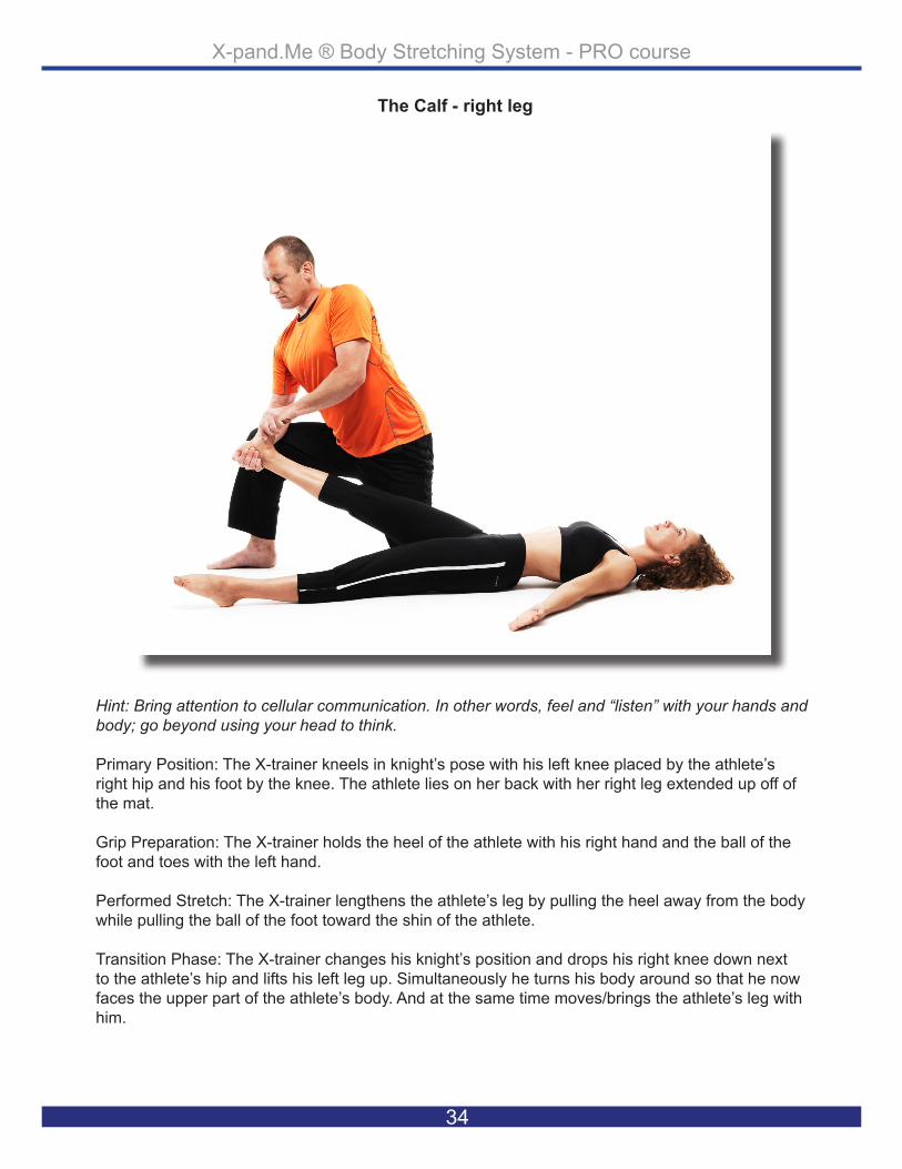
Hint: Bring attention to cellular communication. In other words, feel and “listen” with your hands and body; go beyond using your head to think.
Primary Position: The X-trainer kneels in knight’s pose with his left knee placed by the athlete’s right hip and his foot by the knee. The athlete lies on her back with her right leg extended up off of the mat.
Grip Preparation: The X-trainer holds the heel of the athlete with his right hand and the ball of the foot and toes with the left hand.
Performed Stretch: The X-trainer lengthens the athlete’s leg by pulling the heel away from the body while pulling the ball of the foot toward the shin of the athlete.
Transition Phase: The X-trainer changes his knight’s position and drops his right knee down next to the athlete’s hip and lifts his left leg up. Simultaneously he turns his body around so that he now faces the upper part of the athlete’s body. And at the same time moves/brings the athlete’s leg with him.
The Calf - right leg

X-pand.Me ® Body Stretching System - PRO course
35
The Calf - muscles and their action
Muscle: GastrocnemiusAction: Plantar flexion of ankle/ foot, flexes knee
Muscle: PlantarisAction: Plantar flexion of foot, flexes leg
Muscle: SoleusAction: Plantar flexion of foot/ankle
Muscle: Fibularis/peroneus LongusAction: Everts foot and plantar flexes ankle; also helps to support the transverse arch of the foot
Muscle: Fibularis/peroneus brevisAction: Plantar flexion, eversion of foot
Muscle: Flexor hallicus longusAction: Flexes great toe, helps to supinate ankle, and is a very weak plantar

X-pand.Me ® Body Stretching System - PRO course
36
The Hamstrings - right leg
Hint: Allow the athlete’s body to lead the subtle direction in any movement. In other words, don’t force the body in one direction if the body part wants to go a slightly different direction.
Primary Position: The X-Trainer is in knight’s position with his right knee placed on the mat by the athlete’s right knee. The athlete lies on her back with the right leg lifted, slightly bent, above her abdominal area.
Grip Preparation: The X-trainer places his left hand on the athlete’s sit bone and holds his right hand on the back of the athlete’s lifted heel. The athlete’s knee is centered over the body.
Performed Stretch: The X-trainer grounds the hip of the athlete by pressing down and away from the body on the sit bone. Simultaneously, the X-trainer leans forward, pushing the heel toward the head in the direction of the center of the body.
Transition Phase: The X-trainer’s left hand drops to the athlete’s right knee and bends the leg across the body, so that the shin runs horizontally across her body. The X-trainer drops his left knee to the mat and lifts his right foot up, placing it on the other side of the athlete’s body at thigh-level.

X-pand.Me ® Body Stretching System - PRO course
37
The Hamstrings - muscles and their action
Muscle: Biceps femoris, long headAction: Flexes the knee, and also rotates the tibia laterally; long head also extends the hip joint
Muscle: Biceps femoris, short headAction: Flexes the knee, and also rotates the tibia laterally; long head also extends the hip joint
Muscle: SemitendinosusAction: Extends the thigh and flexes the knee, and also rotates the tibia medially, espe-cially when the knee is flexed
Muscle: SemimembranosusAction: Extends the thigh, flexes the knee, and also rotates the tibia medially, especially when the knee is flexed
Muscle: GracilisAction: Flexes the knee, adducts the thigh, and helps to medially rotate the tibia on the femur
Muscle: Gluteus maximusAction: Major extensor of hip joint, assists in laterally rotating the thigh; upper and middle third section of the muscle are abductors

X-pand.Me ® Body Stretching System - PRO course
38
The Deep Buttock - right leg
Hint: Always move the body very slowly in order to feel any resistance that is present.
Primary Position: The X-trainer kneels in knight’s pose with the left knee placed on the mat, level with the athlete’s hip. The right foot is placed on the other side of the body, level with the abdominal area. The athlete lies on her back with the right lower leg bent placed across her body, the knee above the right side and heel above the left side of the body.
Grip Preparation: The X-trainer’s right hand holds the outside of the heel and left hand holds the inside of the knee, adjusting the lower leg of the athlete so it is parallel to the body.
Applied Stretch: The X-trainer pushes the lower leg of the athlete closer to her chest, balancing the lower leg to keep it parallel to the body. The X-trainer does this by gently pulling back on the knee while simultaneously pushing the lower leg forward. Transition Phase: The X-trainer changes to the opposite knight’s position and the right knee drops to the inside of the athlete’s left leg at the level of the knee. The left foot steps out to be placed to the right side of the body.

X-pand.Me ® Body Stretching System - PRO course
39
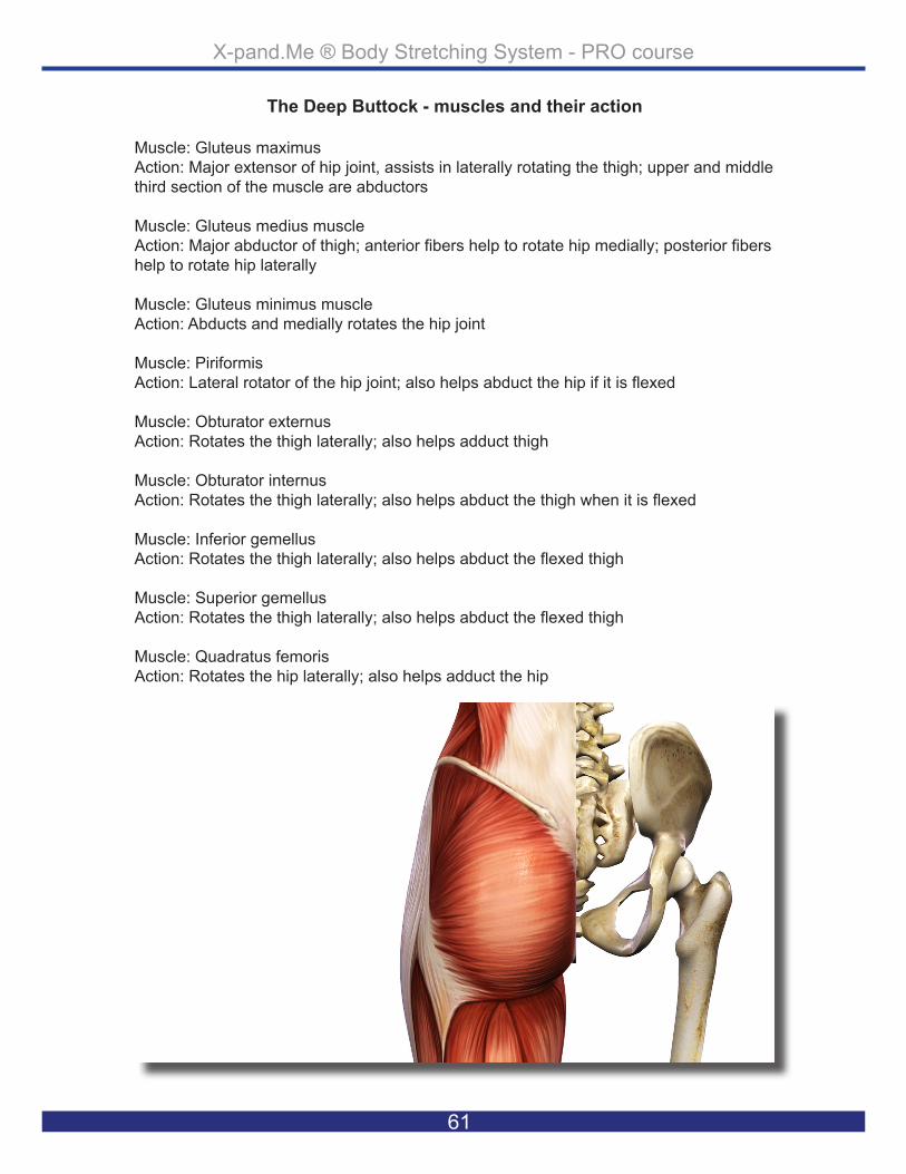
The Deep Buttock - muscles and their action
Muscle: Gluteus maximusAction: Major extensor of hip joint, assists in laterally rotating the thigh; upper and middle third section of the muscle are abductors
Muscle: Gluteus medius muscle Action: Major abductor of thigh; anterior fibers help to rotate hip medially; posterior fibers help to rotate hip laterally
Muscle: Gluteus minimus muscleAction: Abducts and medially rotates the hip joint
Muscle: PiriformisAction: Lateral rotator of the hip joint; also helps abduct the hip if it is flexed
Muscle: Obturator externusAction: Rotates the thigh laterally; also helps adduct thigh
Muscle: Obturator internusAction: Rotates the thigh laterally; also helps abduct the thigh when it is flexed
Muscle: Inferior gemellusAction: Rotates the thigh laterally; also helps abduct the flexed thigh
Muscle: Superior gemellusAction: Rotates the thigh laterally; also helps abduct the flexed thigh
Muscle: Quadratus femorisAction: Rotates the hip laterally; also helps adduct the hip

X-pand.Me ® Body Stretching System - PRO course
40
The Inner Thigh - right leg
Hint: Always be near to the body part stretching limit in order to keep full control over athlete’s body and to protect your own body too.
Primary Position: The X-trainer kneels in the knight’s position with the right knee placed on the inside of the athlete’s left leg. The trainer’s left foot is placed out to the left side. The athlete lies on her back with the right leg opened and extended to the right side.
Grip Preparation: The X-trainer’s right hand is placed on the athlete’s right hip as the left hand holds the heel. The trainer keeps the athlete’s left leg extended, lifted approximately 1-2 inches from the mat.
Performing Stretch: The X-trainer keeps the left hip of the athlete grounded to the mat as he push-es the open extended right leg out to the side and forward up.
Transition Phase: The X-trainer brings the left knee back and places it next to the right knee so he is seated on both knees.

X-pand.Me ® Body Stretching System - PRO course
41
The Inner Thigh - muscles and their action
Muscle: GracilisAction: Flexes the knee, adducts the thigh, and helps to medially rotate the tibia on the femur
Muscle: PectineusAction: Adducts the thigh and flexes the hip joint
Muscle: Adductor brevisAction: Adducts and flexes the thigh, and helps to laterally rotate the thigh
Muscle: Adductor longusAction: Adducts and flexes the thigh, and helps to laterally rotate the hip joint
Muscle: Adductor magnusAction: Powerful thigh adductor; superior horizontal fibers also help flex the thigh, while vertical fibers help extend the thigh

X-pand.Me ® Body Stretching System - PRO course
42
The Outer Thigh - right leg
Hint: Check the natural flexibility of the athlete before starting your stretches. In this case see how close the knee comes to the mat.
Primary Position: The X-trainer sits on both of his knees, at the level of the athlete’s right knee. The athlete lies on her back with her right foot bent out to the side by her hip and her knee facing down toward the thighs of the X-trainer.
Grip Preparation: The X-trainer catches the athlete’s tipped knee with his thigh. He then places the left hand on the athlete’s hip for grounding and the right hand on the outside top of the knee.
Performing Stretch: The X-trainer’s left hand grounds the hip while the right hand pushes the knee down toward the mat and back toward the feet of the athlete.
Transition Phase: The X-trainer steps into knight’s position with his right leg across and places it on the right side of the athlete’s left leg at knee level.

X-pand.Me ® Body Stretching System - PRO course
43
The Outer Thigh - muscles and their action
Muscle: Tensor fascie lateAction: Helps stabilize and steady the hip and knee joints by putting tension on the ilio-tibial band of fascia
Muscle: Rectus femorisAction: Extends the knee
Muscle: Vastus lateralisAction: Extends the knee
Muscle: Vastus intermediusAction: Extends the knee
Muscle: Vastus medialisAction: Extends the knee

X-pand.Me ® Body Stretching System - PRO course
44
The Buttock - right leg
Hint: Always be conscious about creating length between the parts of the body before, during and after stretching.
Primary Position: The X-trainer kneels in the knight’s position with the left knee on the mat just be-low the athlete’s right hip; the right foot is stepped out to the left side of athlete’s body. The athlete lies on her back with the extended right leg lifted over the left pointing away from the left side of her body. The right hip of the athlete lifts approximately 2-3 inches from the mat.
Grip Preparation: The X-trainer’s left thumb is placed alongside the athlete’s groin area as the rest of the left hand holds on to the outside of the hip. The right hand holds the back side of the right heel, extending it across and over to the left side of the athlete’s body.
Performing Stretch: The X-trainer lifts the right hip of the athlete gently about 2-3 inches off of the floor and holds the hip fixated with his left hand at that level. The right hand pulls the right extended leg in a 90 degree angle away from the hip and simultaneously pushes gently down toward the mat at hip level.
Transition Phase: The X-trainer stays in the same knight’s position but steps his left knee up and behind the athlete’s back. He then steps the right foot back and places it on the outside line of the athlete’s body at knee level.

X-pand.Me ® Body Stretching System - PRO course
45
The Buttock - muscles and their action
Muscle: Tensor fascie lateAction: Helps stabilize and steady the hip and knee joints by putting tension on the ilio-tibial band of fasci
Muscle: Gluteus maximusAction: Major extensor of hip joint, assists in laterally rotating the thigh; upper and middle third section of the muscle are abductors
Muscle: Gluteus medius Action: Major abductor of thigh; anterior fibers help to rotate hip medially; posterior fibers help to rotate hip laterally
Muscle: Gluteus minimus Action: Abducts and medially rotates the hip joint

X-pand.Me ® Body Stretching System - PRO course
46
The Thigh - right leg
Hint: Most often the athlete’s body balance depends on your body’s balance; always stay in proper position before starting the stretches.
Primary Position: The X-trainer kneels in knight’s pose with his left knee behind the lower back of the athlete and his right foot at the level of the athlete’s knee. The athlete’s body lies on the left side supported by the X-trainer’s thigh and has her right leg bent, with the right heel to the right buttock.
Grip Preparation: The X-trainer’s right hand holds the athlete’s right knee as the left hand holds the left ankle while the left thigh and the X-trainer support and hold the lower back of the athlete.
Performed Stretch: The X-trainer gently pushes his thigh into the lower back of the athlete, to make sure that the body is lying on the side and keeps the position steady. The X-trainer’s right hand pulls the knee down so that it is in direct alignment with the rest of the athlete’s body. The X-train-er’s left hand pushes the right heel of the athlete to the right buttock.
Transition Phase: The X-trainer extends the right leg of the athlete and step his right foot back to meet the level of his left foot.

X-pand.Me ® Body Stretching System - PRO course
47
The Thigh - muscles and their action
Muscle: Gectus femorisAction: Extends the knee
Muscle: Vastus lateralisAction: Extends the knee
Muscle: Vastus intermediusAction: Extends the knee
Muscle: Vastus medialisAction: Extends the knee
Muscle: SartoriusAction: Flexes and laterally rotates the hip joint and flexes the knee
Muscle: IliacusAction: Flex the torso and thigh with respect to each other
Muscle: Psoas majorAction: Flex the torso and thigh with respect to each other

X-pand.Me ® Body Stretching System - PRO course
48
The Psoas - right side
Hint: Use your inhalations when you need strength and exhalations when you need release.
Primary Position: The X-trainer kneels in the knight’s position with his left knee placed behind the lower back of the athlete, and his right foot placed in level with his left foot. The athlete’s body lies on the left side, supported by the X-trainer’s thigh as she extends her right leg.
Grip Preparation: The X-trainer’s thigh in placed in the lower back and the left hand holds the side of the athlete’s hip. The X-trainer’s right hand holds the ankle of the athlete’s right foot.
Performed Stretch: The X-trainer gently pushes his thigh into the lower back of the athlete as his left hand pushes the body forward, to make sure that the body is lying on the side and maintains its position while the X-trainer’s right hand pulls the extended leg’s ankle back in the direction the X-trainer.
Transition Phase: The X-trainer stays in the same knight’s position but swings his left lower leg down in the direction of the athlete’s left leg and places his other foot across the athlete’s left leg.

X-pand.Me ® Body Stretching System - PRO course
49
The Psoas - muscles and their action
Muscle: Psoas majorAction: Flex the torso and thigh with respect to each other
Muscle: IliacusAction: Flex the torso and thigh with respect to each other
Muscle: Rectus abdominusAction: Flexion of trunk, lumbar vertebrae, compresses abdomen
Muscle: Internal obliqueAction: Compresses abdomen and rotates vertebral column.
Muscle: External obliqueAction: Compresses abdomen, laterally flexes and rotates vertebral column
Muscle: Transversus abdominisAction: Compresse abdoman

X-pand.Me ® Body Stretching System - PRO course
50
The Lower Back - right side
Hint: Allow time for the athlete to feel what you are doing, perhaps stopping for a few seconds to let the athlete trust your moves.
Primary Position: The X-trainer kneels in the knight’s position with the left knee placed next to the right hip of the athlete and his right leg placed across the athlete’s leg at knee level. The athlete lies on her left side with left leg extended and the right leg bent, placed on the mat in front of her body at abdominal level.
Grip Preparation: The X-trainer holds his right hand on the athlete’s left shoulder and left hand where the athlete’s lower back meets the back of the hip (the sacrum). The X-trainer’s right shin is placed to hold the athlete’s right knee.
Performed Stretch: The X-trainer’s right hand grounds the shoulder of the athlete. Making sure that the shoulder stays grounded to the mat, the left hand pulls down and across the athlete’s body. The X-trainer’s right shin moves forward with the athlete’s knee to help pull the body over.
Transition Phase: The X-trainer gently retracts his left thigh and lets the athlete’s body slide down to lie flat on the back, simultaneously placing the leg flat on the mat. The X-trainer takes a few big but controlled steps to the left side of the athlete’s body and places his right knee on the mat next to the athlete’s hip, with his foot next to the knee.

X-pand.Me ® Body Stretching System - PRO course
51
The Lower Back - muscles and their action
Muscle: MultifidusAction: Extend and rotate vertebral column
Muscle: RotatorsAction: Extend and rotate vertebral column
Muscle: Quadratus lomborumAction: Alone, lateralflexion of vertebral column; Together,depression of thoracic rib cage
Muscle: Intertransversarii lateralis lumborumAction: Both sides used then erects the spine and laterally flexes lumbar spine if use in 1 side is used
Muscle: Intertransversarii medialis lumborumAction: Both sides used then erects the spine and laterally flexes lumbar spine if use in 1 side is used
Muscle: Gluteus maximusAction: Major extensor of hip joint, assists in laterally rotating the thigh; upper and middle third section of the muscle are abductors
Muscle: Tensor fascie lateAction: Helps stabilize and steady the hip and knee joints by putting tension on the ilio-tibial band of fasci
Extra muscle group: Erector spinae – see muscle info at: Muscle listing at chapter

X-pand.Me ® Body Stretching System - PRO course
52
Hint: Take time to reposition yourself if needed; if your position feels awkward, it will likely feel awk-ward for the athlete.
Primary Position: The X-trainer kneels in the knight’s pose with his left foot placed by the foot of the athlete’s straight leg. The trainer’s right knee is placed by athlete’s left hip. The athlete lies on her back with her left leg bent and lifted over the X-trainer’s thigh.
Grip Preparation: The X-trainer’s left hand holds the athlete’s heel and his right hand is placed on the ball of the foot and the toes of the athlete.
Performed stretch: The X-trainer’s left hand pulls the heel of the athlete away from her body as the right hand pushes the toes and ball of the foot toward athlete’s shin.
Transition Phase: The X-trainer stays in the same position and shifts his right hand to the heel and his left hand to the instep of the athlete’s foot.
The Sole of the Foot - left foot

X-pand.Me ® Body Stretching System - PRO course
53
The Sole of the Foot - muscles and their action
Muscle: Flexor digitorum brevis:action: flexes lateral four toes
Muscle: Quadratis plantae:Action: Assists Flexor Digitorum Longus in flexion
Muscle: Flexor digiti minimi brevisAction: extend and adduct the fifth toe
Muscle: Abductor hallicusAction: abducts hallux (big toe)
Muscle: Abductor digiti minimiAction: flex and abduct the fifth toe
Muscle: Adductor hallicusAction: adducts hallux
Muscle: Flexor hallicus brevisAction: flex hallux
Muscle: LumbricalesAction: maintain extension of digits at interphalangeal joints

X-pand.Me ® Body Stretching System - PRO course
54
Hint: Be firm but relaxed in your body; if you are all tensed up, the athletes can’t relax either, so trust will not be built between X-trainer and athlete.
Primary Position: The X-trainer kneels in the knight’s position with his right knee placed down by the athlete’s left hip, his foot by the knee. The athlete lies on her back with her leg lifted and bent at a 90 degree angle.
Grip Preparation: The X-trainer’s right hand holds the athlete’s heel and the left hand is placed on the ball of the foot and the toes.
Performed Stretch: The X-trainer’s left hand pushes the instep and toes of the athlete’s foot down toward the mat and simultaneously pulls the heel of the athlete toward her body.
Transition Phase: The X-trainer stays in the same position, changing the hand position so that the left hand holds the heel as the right holds the ball of the foot and opens the athlete’s leg to be fully extended.
The Shin - left leg

X-pand.Me ® Body Stretching System - PRO course
55
The Shin - muscles and their action
Muscle: tibialis anteriorAction: dorsiflex ankle and invert the foot
Muscle: extensor hallucis longusAction: extends the big toe and assists in dorsiflexion of the foot at the ankle
Muscle: extensor digitorum brevisAction: extends digits 2, 3, and 4
Muscle: extensor hallucis brevisAction: extension of hallux
Muscle: peroneus tertiusAction: Works with the extensor digitorum longus to dorsiflex, evert and abduct the foot
Muscle: dorsal interosseousAction: abducts toes, flex proximal phalanges

X-pand.Me ® Body Stretching System - PRO course
56
Hint: Remind the athlete to express herself while working with the X-pand.Me Body Stretching Series.
Primary Position: The X-trainer kneels in the knight’s position with his left knee placed by the ath-lete’s left hip, his foot by the knee. The athlete lies on her back with an extended left leg lifted off the mat.
Grip Preparation: The X-trainer holds the heel of the athlete with his left hand and the ball of the foot and the toes with the right hand.
Performed Stretch: The X-trainer lengthens the athlete’s leg by pulling the heel away from the body while pulling the ball of the foot toward the shin of the athlete.
Transition Phase: The X-trainer changes his knight’s position and drops his left knee down and places it next to athlete’s hip, lifting his right leg up. Simultaneously he turns his body around so that he now faces the upper part of athlete’s body. And at the same time moves/brings the athlete’s leg with him.
The Calf - left leg

X-pand.Me ® Body Stretching System - PRO course
57
The Calf - muscles and their action
Muscle: GastrocnemiusAction: Plantar flexion of ankle/ foot, flexes knee
Muscle: PlantarisAction: Plantar flexion of foot, flexes leg
Muscle: SoleusAction: Plantar flexion of foot/ankle
Muscle: Fibularis/peroneus LongusAction: Everts foot and plantar flexes ankle; also helps to support the transverse arch of the foot
Muscle: Fibularis/peroneus brevisAction: Plantar flexion, eversion of foot
Muscle: Flexor hallicus longusAction: Flexes great toe, helps to supinate ankle, and is a very weak plantar

X-pand.Me ® Body Stretching System - PRO course
58
Hint: Don’t lean ahead of your knee.
Primary Position: The X-trainer kneels in the knight’s position with the left knee placed by athlete’s left hip with his foot by side of the trunk. The athlete lies on her back with the left leg lifted, slightly bent, above her abdominal area.
Grip Preparation: The X-trainer places his right hand on the athlete’s sit bone and holds his left hand on the back of the athlete’s lifted heel. The athlete’s knee is centered over the body.
Performed Stretch: The X-trainer grounds the hip of the athlete by pressing down and away from the body on the sit bone and at the same time leaning forward and pushing the heel toward the head in the direction of the center of the body.
Transition Phase: The X-trainer’s right hand drops to the left knee of the athlete and bends the leg across the body, so that the shin runs horizontally across her body. The X-trainer’s right knee drops to the mat and the left foot lifts up to be placed on the other side of the athlete’s body at thigh level.
The Hamstrings - left leg

X-pand.Me ® Body Stretching System - PRO course
59
The Hamstrings - muscles and their action
Muscle: biceps femoris, long headAction: Flexes the knee, and also rotates the tibia laterally; long head also extends the hip joint
Muscle: biceps femoris, short headAction: Flexes the knee, and also rotates the tibia laterally; long head also extends the hip joint
Muscle: semitendinosusAction: Extends the thigh and flexes the knee, and also rotates the tibia medially, espe-cially when the knee is flexed
Muscle: semimembranosusAction: Extends the thigh, flexes the knee, and also rotates the tibia medially, especially when the knee is flexed
Muscle: gracilisAction: Flexes the knee, adducts the thigh, and helps to medially rotate the tibia on the femur
Muscle: Gluteus maximusAction: Major extensor of hip joint, assists in laterally rotating the thigh; upper and middle third section of the muscle are abductors

X-pand.Me ® Body Stretching System - PRO course
60
Hint: If you feel uncertain of the stretches performed while giving the series, communicate with the athlete.
Primary Position: The X-trainer kneels in the knight’s position with the right knee placed on the mat level with the athlete’s hip. The left foot is placed on the other side of the body, level with the abdominal area. The athlete lies on her back with her right lower leg placed across her body, the knee above the left side and heel above the right side of the body.
Grip Preparation: The X-trainer’s left hand holds the outside of the heel as the right hand holds the inside of the knee, adjusting the lower leg of the athlete to be parallel to the body.
Applied Stretch: The X-trainer pushes the lower leg of the athlete closer to her chest, balancing the lower leg so it remains parallel to the body by gently pulling back on the knee while simultaneously pushing the lower leg forward. Transition Phase: The X-trainer changes to the opposite knight’s position, dropping the left knee to the inside of the athlete’s right leg at knee level. The right foot opens out and is placed to the left side of the athlete’s body.
The Deep Buttock - left leg
X-pand.Me ® Body Stretching System - PRO course
61
The Deep Buttock - muscles and their action
Muscle: Gluteus maximusAction: Major extensor of hip joint, assists in laterally rotating the thigh; upper and middle third section of the muscle are abductors
Muscle: Gluteus medius muscle Action: Major abductor of thigh; anterior fibers help to rotate hip medially; posterior fibers help to rotate hip laterally
Muscle: Gluteus minimus muscleAction: Abducts and medially rotates the hip joint
Muscle: PiriformisAction: Lateral rotator of the hip joint; also helps abduct the hip if it is flexed
Muscle: Obturator externusAction: Rotates the thigh laterally; also helps adduct thigh
Muscle: Obturator internusAction: Rotates the thigh laterally; also helps abduct the thigh when it is flexed
Muscle: Inferior gemellusAction: Rotates the thigh laterally; also helps abduct the flexed thigh
Muscle: Superior gemellusAction: Rotates the thigh laterally; also helps abduct the flexed thigh
Muscle: Quadratus femorisAction: Rotates the hip laterally; also helps adduct the hip

X-pand.Me ® Body Stretching System - PRO course
62
Hint: Grounding one body part of the athlete helps to create length across limbs.
Primary Position: The X-trainer kneels in the knight’s position with the left knee placed on the inside of the athlete’s right leg and the right foot placed out to the right side. The athlete lies on her back with the left leg opened and extended to the left side.
Grip Preparation: The X-trainer’s left hand is placed on the athlete’s left hip. The right hand holds the heel, keeping the right extended leg lifted approximately 1-2 inches from the mat.
Performing Stretch: The X-trainer keeps the right hip of the athlete grounded to the mat as he pushes the open extended left leg out to the side and forward up.
Transition Phase: The X-trainer brings the right knee back and places it next to the left to be seated on both knees.
The Inner Thigh - left leg

X-pand.Me ® Body Stretching System - PRO course
63
The Inner Thigh - muscles and their action
Muscle: GracilisAction: Flexes the knee, adducts the thigh, and helps to medially rotate the tibia on the femur
Muscle: PectineusAction: Adducts the thigh and flexes the hip joint
Muscle: Adductor brevisAction: Adducts and flexes the thigh, and helps to laterally rotate the thigh
Muscle: Adductor longusAction: Adducts and flexes the thigh, and helps to laterally rotate the hip joint
Muscle: Adductor magnusAction: Powerful thigh adductor; superior horizontal fibers also help flex the thigh, while vertical fibers help extend the thigh

X-pand.Me ® Body Stretching System - PRO course
64
Hint: If the athlete shows signs of discomfort, stop and reposition her and yourself.
Primary Position: The X-trainer sits on both his knees, at the level of the athlete’s left knee. The athlete lies on her back with her left foot bent out to the side by her hip and her knee facing down toward the extended leg of the X-trainer.
Grip Preparation: The X-trainer catches the athlete’s tipped knee with his thigh; here the X-trainer places the right hand on the athlete’s hip for grounding and his left hand on the outside top of the knee.
Performing Stretch: The X-trainer’s right hand grounds the hip while the left hand pushes the knee down towards the mat and back toward the feet of the athlete.
Transition Phase: The X-trainer steps into a knight’s position with his left leg across and places it to the left side of the athlete’s right leg at knee level.
The Outer Thigh - left leg

X-pand.Me ® Body Stretching System - PRO course
65
The Outer Thigh - muscles and their action
Muscle: Tensor fascie lateAction: Helps stabilize and steady the hip and knee joints by putting tension on the ilio-tibial band of fascia
Muscle: Rectus femorisAction: Extends the knee
Muscle: Vastus lateralisAction: Extends the knee
Muscle: Vastus intermediusAction: Extends the knee
Muscle: Vastus medialisAction: Extends the knee

X-pand.Me ® Body Stretching System - PRO course
66
Hint: Be present with the stretch that you are performing, don’t think ahead while having the body in a stretch. Wait until you are done and then pause to think.
Primary Position: The X-trainer kneels in the knight’s position with the left knee on the mat just below the athlete’s right hip. The right foot is out to the left side of the athlete’s body. The athlete lies on her back with the extended right leg lifted over the left pointing away from the left side of her body. The athlete’s right hip of lifts approximately 2-3 inches from the mat.
Grip Preparation: The X-trainer’s left thumb is placed alongside the athlete’s groin as the rest of the left hand holds onto the outside of the hip. The right hand holds the back of the right heel extended across and over to the left side of the athlete’s body.
Performing Stretch: The X-trainer lifts the right hip of the athlete gently about 2-3 inches off of the floor and holds the hip steady with his left hand at that level. The right hand pulls the right extended leg into a 90 degree angle away from the hip and simultaneously pushes gently down toward the mat in level with the hip.
Transition Phase: The X-trainer stays in the same knight’s position but steps his left knee up and behind the athlete’s back and steps the right foot back, placing it on the outside line of the athlete’s body in level with the knee.
The Buttock - left leg

X-pand.Me ® Body Stretching System - PRO course
67
The Buttock - muscles and their action
Muscle: Tensor fascie lateAction: Helps stabilize and steady the hip and knee joints by putting tension on the ilio-tibial band of fasci
Muscle: Gluteus maximusAction: Major extensor of hip joint, assists in laterally rotating the thigh; upper and middle third section of the muscle are abductors
Muscle: Gluteus medius Action: Major abductor of thigh; anterior fibers help to rotate hip medially; posterior fibers help to rotate hip laterally
Muscle: Gluteus minimus Action: Abducts and medially rotates the hip joint

X-pand.Me ® Body Stretching System - PRO course
68
Hint: Remember which part of the body you are stretching; don’t just do the work without this in mind. You will optimize the stretches when knowing everything about them.
Primary Position: The X-trainer places his right knee behind the lower back of the athlete and his left foot at the level of the athlete’s knee. The athlete’s body lies on the right side, supported by the X-trainer’s thigh, with her left leg bent, left heel to left buttock.
Grip Preparation: The X-trainer’s left hand holds the left knee of the athlete as the right hand holds the right ankle. The right thigh of the X-trainer meanwhile supports and holds the lower back of the athlete.
Performed Stretch: The X-trainer gently pushes his thigh into the lower back of the athlete, to make sure that the body is lying on the side and maintains the position. The X-trainer’s left hand pulls the knee down so that it is in direct alignment with the rest of the athlete’s body and the X-trainer’s right hand pushes the left heel of the athlete to the left buttock.
Transition Phase: The X-trainer extends the left leg of the athlete and steps his left foot back to meet the level of his right foot.
The Thigh - left leg

X-pand.Me ® Body Stretching System - PRO course
69
The Thigh - muscles and their action
Muscle: Gectus femorisAction: Extends the knee
Muscle: Vastus lateralisAction: Extends the knee
Muscle: Vastus intermediusAction: Extends the knee
Muscle: Vastus medialisAction: Extends the knee
Muscle: SartoriusAction: Flexes and laterally rotates the hip joint and flexes the knee
Muscle: IliacusAction: Flex the torso and thigh with respect to each other
Muscle: Psoas majorAction: Flex the torso and thigh with respect to each other

X-pand.Me ® Body Stretching System - PRO course
70
Hint: As this stretch goes for the deep psoas muscle, allow extra time to “get through” more superficial muscle groups.
Primary Position: The X-trainer places his right knee behind the lower back of the athlete; his left foot is placed in level with his right foot. The athlete lies on the right side, supported by the X-train-er’s thigh and has her left leg extended.
Grip Preparation: The X-trainer’s thigh is placed against the lower back and the right hand holds the side of the athlete’s hip. The left hand of the X-trainer holds the athlete’s left foot.
Performed Stretch: The X-trainer gently pushes his thigh against the lower back of the athlete as his right hand pushes the body forward, to make sure that the body is lying on the side and main-tains its position. The X-trainer’s left hand pulls the extended leg’s ankle back in the direction of the X-trainer.
Transition Phase: The X-trainer swings his right lower leg down in the direction of the athlete’s right leg and places his other foot across the athlete’s right leg.
The Psoas - left side

X-pand.Me ® Body Stretching System - PRO course
71
The Psoas - muscles and their action
Muscle: Psoas majorAction: Flex the torso and thigh with respect to each other
Muscle: IliacusAction: Flex the torso and thigh with respect to each other
Muscle: Rectus abdominusAction: Flexion of trunk, lumbar vertebrae, compresses abdomen
Muscle: Internal obliqueAction: Compresses abdomen and rotates vertebral column.
Muscle: External obliqueAction: Compresses abdomen, laterally flexes and rotates vertebral column
Muscle: Transversus abdominisAction: Compresse abdoman

X-pand.Me ® Body Stretching System - PRO course
72
Hint: Keep either the shoulder or the knee of the athlete grounded. In some cases the athlete can have both body parts grounded.
Primary Position: The X-trainer’s right knee is placed next to the athlete’s left hip. His left leg is placed across the athlete’s leg at the level of the knee. The athlete lies on her right side with her right leg extended. The left leg is bent and placed on the mat in front of her body at abdominal level.
Grip Preparation: The X-trainer holds his left hand on the athlete’s right shoulder and places his right hand where the athlete’s lower back meets the back of the hip (sacrum). The X-trainer’s left shin holds the athlete’s left knee in place.
Performed Stretch: The X-trainer’s left hand grounds the shoulder of the athlete to make sure that the shoulder stays grounded to the mat, meanwhile the right hand pulls down and across the athlete’s body. The X-trainer’s left shin moves forward with the athlete’s knee to help pull the body over.
Transition Phase: The X-trainer gently moves his right thigh and lets the body of the athlete slide down to lie flat on the back, simultaneously placing the leg flat onto the mat. The X-trainer steps out with his feet to be standing on both sides of the athlete’s knees.
The Lower Back - left side

X-pand.Me ® Body Stretching System - PRO course
73
The Lower Back - muscles and their action
Muscle: MultifidusAction: Extend and rotate vertebral column
Muscle: RotatorsAction: Extend and rotate vertebral column
Muscle: Quadratus lomborumAction: Alone, lateralflexion of vertebral column; Together,depression of thoracic rib cage
Muscle: Intertransversarii lateralis lumborumAction: Both sides used then erects the spine and laterally flexes lumbar spine if use in 1 side is used
Muscle: Intertransversarii medialis lumborumAction: Both sides used then erects the spine and laterally flexes lumbar spine if use in 1 side is used
Muscle: Gluteus maximusAction: Major extensor of hip joint, assists in laterally rotating the thigh; upper and middle third section of the muscle are abductors
Muscle: Tensor fascie lateAction: Helps stabilize and steady the hip and knee joints by putting tension on the ilio-tibial band of fasci
Extra muscle group: Erector spinae – see muscle info at: Muscle listing at chapter

X-pand.Me ® Body Stretching System - PRO course
74
Hint: This is a very gentle stretch; we see it more as a misalignment /discomfort release.
Primary Position: The X-trainer stands with both feet close to the buttocks of the athlete. The ath-lete lies on her back with both knees bent to the chest.
Grip Preparation: The X-trainer holds both of the athlete’s knees, making sure he adds no pres-sure.
Performed Stretch: The X-trainer presses the knees gently toward the chest of the athlete and simultaneously presses the knees back in the direction of his own feet. At the same time he gently rocks both knees from side to side.
Transition Phase: The X-trainer places the athlete’s leg in cross-legged position, and places the shin of the athlete to his own shin, then grabs the arms and gently pulls the athlete’s body to a fully- seated position. The X-trainer steps his right foot out to the right side of the athlete’s body in level with athlete’s knee and places his left knee by the left hip of the athlete.
The Lower Back - both sides

X-pand.Me ® Body Stretching System - PRO course
75
The Lower Back - muscles and their action
Muscle:Iliocostalis lomborumAction: Extension of the vertebral column; assist with lateral flexion of vertebral column;;
Muscle: Gluteus maximusAction: Major extensor of hip joint, assists in laterally rotating the thigh; upper and middle third section of the muscle are abductors
Muscle:Longissimus thoraticsAction: Extend the vertebral column
Muscle:Spinalis thoraticsAction: Extension of the vertebral column

X-pand.Me ® Body Stretching System - PRO course
76
Hint: Know that some body-types will never get their heads to the floor.
Primary Position: The X-trainer’s right foot is placed out to the right side of the athlete’s body in level with the athlete’s knee. He then places his left knee by the left hip of the athlete. The athlete is seated in cross-legged position with her body bent forward (fully or partly, depending on body type.)
Grip Preparation: The X-trainer places his left hand on the lower back of the athlete and his right hand on the back of the athlete’s head.
Performed Stretch: The X-trainer’s left hand grounds the hip/sit bones of the athlete by pressing his hand toward the mat while his right hand lengthens the body forward and down towards the mat.
Transition Phase: The X-trainer places his right hand on the athlete’s neck and gently lifts her to an upright, seated position, and simultaneously places his right knee by the athlete’s right hip and left foot by the left side of the athlete.
The Back - both sides

X-pand.Me ® Body Stretching System - PRO course
77
The Back - muscles and their action
Muscle: Serratus anteriorAction: Draws scapula forward and upward; abducts scapula and rotates it; stabilizes vertebral border of scapula
Muscle:Lattisimus dorsiAction: Extends, adducts, and medially rotates humerus; raises body toward arms during climbing
Muscle: Teres majorAction: Adducts and medially rotates arm
Muscle: Iliocostalis lomborumAction: Extension of the vertebral column; assist with lateral flexion of vertebral column;; postural stabilization of vertebral column
Muscle: Gluteus maximusAction: Major extensor of hip joint, assists in laterally rotating the thigh; upper and middle third section of the muscle are abductors
Muscle:Longissimus thoraticsAction: Extend the vertebral column
Muscle:Spinalis thoraticsAction: Extension of the vertebral column

X-pand.Me ® Body Stretching System - PRO course
78
Hint: In this beautiful grip, please do not add pressure to the athlete’s neck.
Primary Position: The X-trainer’s right knee is placed by the athlete’s right hip and his left foot is placed next to the athlete’s left hip.
Grip Preparation: Before the X-trainer places his hand, he will lift the athlete’s left arm over his thigh and right arm to ceiling. The X-trainer then places his right hand on the athlete’s right hip and his left hand on the front of the athlete’s shoulder. (The arm of the athlete is lifted and the X-trainer places his arm between the lifted arm and head of athlete.)
Performed Stretch: The X-trainer grounds the right hip by pressing his right hand down as his left hand lifts the right shoulder up and to the left. The X-trainer’s left leg will open slightly to the left side, but not more than the athlete’s body does, so it continues supporting of the body.
Transition Phase: The X-trainer lifts the athlete’s body to a fully upright position and steps his left knee next to the athlete’s left hip. He places his right foot next to the athlete’s right hip.
The Side of the Body - right side

X-pand.Me ® Body Stretching System - PRO course
79
The Side of the Body - muscles and their action
Muscle: Quadratus lomborumAction: Fixes 12th rib to stabilize diaphragm attachments during inspiration; lateral flexes the vertebral column, extends lumbar vertebrae
Muscle:External obliqueAction: Help compresses the abdominal cavity; compress and depress the lower thoracic cavity to aid in expiration; rotates trunk to opposite side; weakly assists in flexion
Muscle: Intenal obliqueAction: Strong compressor of the abdominal cavity; rotates trunk to the same side; weakly assists in flexion of lumbar vertebrae
Muscle:Iliocostalis lomborumAction: Extension, lateral flexion of vertebral column, rotates ribs for forceful inspiration
Muscle: MultifidusAction: Extend and rotate vertebral column
Muscle: Teres majorAction: Adducts and medially rotates arm

X-pand.Me ® Body Stretching System - PRO course
80
Hint: Do not let the body of the athlete lean to one side from the hip, only through a lateral spinal bend.
Primary Position: The X-trainer places his right knee by the athlete’s right hip and his left foot next to the athlete’s left hip.
Grip Preparation: Before the X-trainer positions his hand, he will lift the athlete’s right arm over his thigh and left arm to the ceiling. The X-trainer then places his left hand on the athlete’s left hip and right hand is placed on the front of the shoulder of the athlete. (The arm of the athlete is lifted and the X-trainer places his arm between the lifted arm and head of athlete.)
Performed Stretch: The X-trainer grounds the left hip by pressing his left hand down. As his right hand lifts the athlete’s left shoulder up and to the right, the X-trainer’s right leg will raise slightly to the right side, but not more than the athlete’s body does, so it continues supporting the body.
Transition Phase: The X-trainer lifts the body of the athlete to become fully upright and then sit back on his buttock behind the athlete. He places his feet on either side of the thighs of the athlete and places his knees at the back.
The Side of the Body - left side

X-pand.Me ® Body Stretching System - PRO course
81
The Side of the Body - muscles and their action
Muscle: Quadratus lomborumAction: Fixes 12th rib to stabilize diaphragm attachments during inspiration; lateral flexes the vertebral column, extends lumbar vertebrae
Muscle:External obliqueAction: Help compresses the abdominal cavity; compress and depress the lower thoracic cavity to aid in expiration; rotates trunk to opposite side; weakly assists in flexion
Muscle: Intenal obliqueAction: Strong compressor of the abdominal cavity; rotates trunk to the same side; weakly assists in flexion of lumbar vertebrae
Muscle:Iliocostalis lomborumAction: Extension, lateral flexion of vertebral column, rotates ribs for forceful inspiration
Muscle: MultifidusAction: Extend and rotate vertebral column
Muscle: Teres majorAction: Adducts and medially rotates arm

X-pand.Me ® Body Stretching System - PRO course
82
Hint: In this stretch, move back with the client and get a deeper stretch, even for yourself.
Primary Position: The X-trainer sits back on his buttocks with his feet on either side of the athlete’s thighs and places his knees close together at the athlete’s back. The athlete then sits in a cross legged position, leaning back on the X-trainer’s knees.
Grip Preparation: The X-trainer adjusts his knees so that the shoulder blades of the athlete rest on the knees. (The X-trainer can lift or lower knees by moving his feet forward or back or coming to the balls of the feet). The X-trainer then places his hands on the front of the athlete’s shoulders.
Performed Stretch: The X-trainer stabilizes the knees against the shoulder blades of the athlete and gently lifts the athlete’s body back.
Transition Phase: The X-trainer brings the athlete to an upright, seated position and steps back, placing his knee and foot behind the athlete.
The Front of the Body - both sides

X-pand.Me ® Body Stretching System - PRO course
83
The Front of the Body - muscles and their action
Muscle: Rectus abdominusAction: Flexion of trunk, lumbar vertebrae, compresses abdomen
Muscle: External obliqueAction: Compresses abdomen, laterally flexes and rotates vertebral column
Muscle: Internal intercostalAction: Stiffen the chest wall to prevent paradoxical motion during descent of the dia-phragm; move the ribs
Muscle: Pectoralis minorAction: Draws scapula forward and downward and raises ribs
Muscle: Pectoralis majorAction: Adducts and medially rotates humerus; draws scapula anteriorly and inferiorly; Acting alone: clavicular head flexes humerus and sternocostal head extends it
Muscle: Transversus abdominalAction: Compresses the abdominal cavity

X-pand.Me ® Body Stretching System - PRO course
84
Hint: When we have the athlete in a seated position, be careful not to push them off balance.
Primary Position: The X-trainer sits with one knee and one foot on the mat. The athlete sits in a cross-legged, upright position.
Grip Preparation: The X-trainer places the pinky side of his hands on the inner shoulder of the ath-lete and places all his fingers along the jaw bone.
Performed Stretch: The X-trainer stabilizes athlete’s body with the part of the hand that is placed on the shoulder while he lifts the jaw/head back with his fingers; if the X-trainer needs more of a lift he will move his lower arms to the area of the athlete’s shoulders.
Transition Phase: Both the X-trainer and the athlete stay in the same positions and the X-trainer releases the neck of the athlete to neutral.
The Throat - both sides

X-pand.Me ® Body Stretching System - PRO course
85
The Throat - muscles and their action
Muscle: Sternocleido mastoideusAction: Unilaterally it extends (sternal fibers) or flexes (clavicular fibers) the head and rotates it toward the opposite shoulder; flexes and extends the head; raises rib cage
Muscle: PlatysmaAction: Depresses and draws lower lip laterally, draws up skin of chest, depresses man-diple
Muscle: OmohyoidAction: Depresses an elevated hyoid bone and assists in raising the cervical fascia dur-ing inspiration
Muscle: SternothyroidAction: Depresses the thyroid cartilage
Muscle: SternohyoidAction: Depresses the hyoid bone

X-pand.Me ® Body Stretching System - PRO course
86
Hint: Athletes might think that you will “crack” their neck in this stretch, please assure them that it is not the case in our work.
Primary Position: The X-trainer sits approximately one foot behind the athlete in low knight’s pose with the right foot up. The athlete sits in upright, cross-legged position.
Grip Preparation: The athlete’s hands are placed on either side of the athlete’s at ear level. The head of the athlete is then turned to the left side and the X-trainer rests his right elbow on his knee for stability while turning the athlete’s head to the right side.
Performed Stretch: The X-trainer lifts the pinky side of his hand up and leans the athlete’s head back; the X-trainer’s thumbs stabilize with a gentle lift for a better stretch.
Transition Phase: The X-trainer changes his knight position to the other side, (so that he now has his left knee up) and turns the athlete’s head back to the center/neutral Position.
The Side of the Throat - right side

X-pand.Me ® Body Stretching System - PRO course
87
The Side of the Throat - muscles and their action
Muscle: Sternocleido mastoideusAction: Unilaterally it extends (sternal fibers) or flexes (clavicular fibers) the head and rotates it toward the opposite shoulder; flexes and extends the head; raises rib cage
Muscle: Scalenus anteriorAction: Raises first rib (respiratory inspiration); acting together they flex neck; acting on one side, they laterally fie rotate neck
Muscle: Scalenus mediusAction: Raises first rib (respiratory inspiration); acting together, they flex neck; acting on one side, they laterally flex, rotate neck
Muscle: Scalenus posteriorAction: Lifts second rib, acting together, they flex neck; acting on one side, they laterally flex, rotate neck

X-pand.Me ® Body Stretching System - PRO course
88
Hint: Remember to look for alignment between yourself and the athlete. In this case, the lower the X-trainer sits down the deeper he will be able to stretch.
Primary Position: The X-trainer sits approximately one foot behind the athlete in low knight’s pose with his left foot up. The athlete sits in an upright, cross-legged position.
Grip Preparation: The athlete’s hands are placed on either side of the athlete’s at ear level. The head of the athlete is then turned to the right side and the X-trainer rests his left elbow on his knee for stability while turning the athlete’s head to the left side.
Performed Stretch: The X-trainer lifts the pinky side of his hand up and leans the athlete’s head back; the X-trainer’s thumbs stabilize with a gentle lift for a better stretch.
Transition Phase: The X-trainer places himself in knight’s pose with left leg up, his right knee cen-tered behind the athlete and his left leg back so that his body is facing to the side.
The Side of the Throat - left side

X-pand.Me ® Body Stretching System - PRO course
89
The Side of the Throat - muscles and their action
Muscle: Sternocleido mastoideusAction: Unilaterally it extends (sternal fibers) or flexes (clavicular fibers) the head and rotates it toward the opposite shoulder; flexes and extends the head; raises rib cage
Muscle: Scalenus anteriorAction: Raises first rib (respiratory inspiration); acting together they flex neck; acting on one side, they laterally fie rotate neck
Muscle: Scalenus mediusAction: Raises first rib (respiratory inspiration); acting together, they flex neck; acting on one side, they laterally flex, rotate neck
Muscle: Scalenus posteriorAction: Lifts second rib, acting together, they flex neck; acting on one side, they laterally flex, rotate neck

X-pand.Me ® Body Stretching System - PRO course
90
Hint: If you feel like starting with the other arm, feel free. There will be no difference; just don’t get confused. Stick to one side all the way through.
Primary Position: The X-trainer is in knight pose with left leg up, his body is pointing in the direc-tion of the left of the athlete’s body. His right knee is placed on the mat just behind the athlete and his left foot is placed approximately one arm length behind the athlete. The athlete is seated in a cross- legged position with the left arm reaching out to the left side with the inner forearm pointing towards the ceiling.
Grip Preparation: The X-trainer supports with his right hand below the athlete’s left upper arm/el-bow, and holds the palm/fingers of the athlete with his left hand.
Performed stretch: The X-trainer makes sure that the arm of the athlete is at full length (not allow-ing the arm to bend) with his right hand. With the left hand he gently pulls the arm away from the athlete’s body and pushes the athlete’s palm/fingers down toward the mat.
Transition Phase: The X-trainer releases the stretch and turns the arm of the athlete, staying in the same position.
The Inner Forearm - left arm

X-pand.Me ® Body Stretching System - PRO course
91
The Inner Forearm - muscles and their action
Muscle: Flexor carpi radialisAction: Flexes and abducts hand (at wrist)
Muscle: Palmaris longusAction: Flexes hand (at wrist) and tightens palmar aponeurosis
Muscle: BrachioradialisAction: Major flexor of forearm -- flexes forearm in all positions
Muscle: Flexor carpi radialisAction: Flexes and abducts hand (at wrist)
Muscle: Flexor digitorum profundusAction: Flexes distal phalanges at distal interphalangeal joints of medial four digits; as-sists with flexion of hand
Muscle: Flexor digitorum superficialisAction: Flexes middle phalanges at proximal interphalangeal joints of medial four digits; acting more strongly, it also flexes proximal phalanges at metacarpophalangeal joints and hand

X-pand.Me ® Body Stretching System - PRO course
92
Hint: Constantly be mindful that you are in a safe position and never compensate your position for short-cuts. Stop and have an inner look, correcting yourself if necessary and continue on.
Primary Position: The X-trainer’s body faces the left of the athlete’s body and he places his right knee on the mat just behind the athlete. The trainer’s left foot is placed approximately one arm’s length behind the athlete. The athlete is seated in a cross-legged position with the left arm reaching back.
Grip Preparation: The X-trainer supports with his right hand below the athlete’s left upper arm/el-bow and forms a clenched fist of the athlete and holds around it with his left hand.
Performed stretch: The X-trainer first makes sure the athlete’s arm is extended its full length (not allowing the arm to bend) with his right hand. With the left hand he gently pulls the arm away from the athlete’s body and pushes the athlete’s fist down toward the mat.
Transition Phase: The X-trainer releases the athlete’s arm and brings it to the back of her body and steps his left foot behind the athlete’s left buttock.
The The Outer Forearm - left arm

X-pand.Me ® Body Stretching System - PRO course
93
The Outer Forearm - muscles and their action
Muscle: Extensor carpi radiallis longusAction: Extend and abduct hand at wrist joint
Muscle: Extensor carpi radiallis brevisAction: Extend and abduct hand at wrist joint
Muscle: Extensor digitorumAction: Extends medial four digits at metacarpophalangeal joints; Extends hand at wrist joint
Muscle: Extensor indicisAction: Extends 2nd digit and helps to extend hand
Muscle: Extensor digiti minimiAction: Extends 5th digit at metacarpophalangeal and interphalangeal joints
Muscle: Extensor carpi ulnarisAction: Extends and adducts hand at wrist joint

X-pand.Me ® Body Stretching System - PRO course
94
Hint: Be aware that if the athlete turns her body in the same direction as the shoulder keeps them from facing forward and the stretch will be less effective.
Primary Position: The X-trainer’s places his right knee and left foot on the mat. He sits approxi-mately one foot behind the athlete. The athlete is seated in an upright, cross-legged position with the left arm placed diagonally across her back.
Grip Preparation: The X-trainer holds his left hand on the front of the athlete’s left shoulder. The right hand holds the wrist of the athlete’s right hand, which is placed on the right lower back of the athlete.
Performed stretch: The X-trainer’s right hand lengthens the athlete’s right hand toward the right side of the athlete’s body and simultaneously pulls the left shoulder of the athlete back toward himself.
Transition Phase: The X-trainer releases the arm of the athlete and brings it across the front of the athlete’s body, and changes his knight’s position to high knight with the right foot up.
The Front of the Shoulder - left shoulder
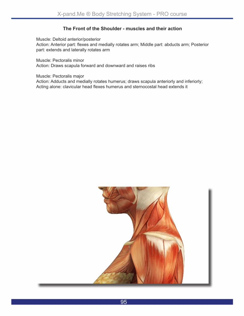
X-pand.Me ® Body Stretching System - PRO course
95
The Front of the Shoulder - muscles and their action
Muscle: Deltoid anterior/posterior Action: Anterior part: flexes and medially rotates arm; Middle part: abducts arm; Posterior part: extends and laterally rotates arm
Muscle: Pectoralis minorAction: Draws scapula forward and downward and raises ribs
Muscle: Pectoralis majorAction: Adducts and medially rotates humerus; draws scapula anteriorly and inferiorly; Acting alone: clavicular head flexes humerus and sternocostal head extends it

X-pand.Me ® Body Stretching System - PRO course
96
Hint: When moving around the athlete, keep a distance from her - in this case you don’t want to touch her back with your belly or other “non-working” body parts.
Primary Position: The X-trainer sits in high knight’s position with his right knee up with the athlete in an upright, cross-legged position with her left arm across her chest.
Grip Position: The X-trainer’s right hand holds the wrist of the athlete’s hand and his right hand against the front of her left shoulder.
Performed stretch: The X-trainer gently pulls the athlete’s right wrist to the right side and simultane-ously pushes the left shoulder of the athlete forward and over to the right side too.
Transition Phase: The X-trainer brings the arm of the athlete to point back and changes his position to the opposite knight’s position with right knee down and left leg up, seated approximately one foot from the back of the athlete.
The Back of the Shoulder - left shoulder

X-pand.Me ® Body Stretching System - PRO course
97
The Back of the Shoulder - muscles and their action
Muscle: Deltoid anterior/posterior Action: Anterior part: flexes and medially rotates arm; Middle part: abducts arm; Posterior part: extends and laterally rotates arm
Muscle: Rhomboid major/minorAction: Retract scapula and rotate it to depress glenoid cavity; fix scapula to thoracic wall
Muscle: TrapeziusAction: Elevates, retracts and rotates scapula; superior fibers elevate, middle fibers retract, and inferior fibers depress scapula; superior and inferior fibers act together in superior rotation of scapula

X-pand.Me ® Body Stretching System - PRO course
98
Hint: Be careful and work slow in general. For example, the shoulder has many muscles when it moves so take your time to move the arm back.
Primary Position: The X-trainer is in knight’s pose with his left foot up and facing out to the left side, seated approximately one foot behind the athlete’s body. The athlete sits in a cross-legged position with her left arm pointing back.
Grip Preparation: The X-trainer places his right hand across the athlete’s shoulder so that his thumb points down the back on the inside of the shoulder blade and the rest of the fingers point down the chest. His left hand holds around the shoulder has the athlete’s arm resting on his thigh, supported by his left forearm.
Performed stretch: The X-trainer stabilizes the athlete’s shoulder/body with his right hand as he pulls and brings the arm of the athlete back and towards himself.
Transition Phase: The X-trainer releases the stretch and stays in the same body position.
The Chest - left side

X-pand.Me ® Body Stretching System - PRO course
99
The Chest - muscles and their action
Muscle: Pectoralis minorAction: Draws scapula forward and downward and raises ribs
Muscle: Pectoralis majorAction: Adducts and medially rotates humerus; draws scapula anteriorly and inferiorly; Acting alone: clavicular head flexes humerus and sternocostal head extends it
Muscle: Deltoid anterior/posterior Action: Anterior part: flexes and medially rotates arm; Middle part: abducts arm; Posterior part: extends and laterally rotates arm
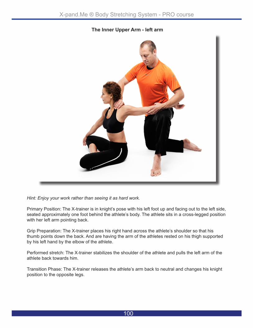
X-pand.Me ® Body Stretching System - PRO course
100
Hint: Enjoy your work rather than seeing it as hard work.
Primary Position: The X-trainer is in knight’s pose with his left foot up and facing out to the left side, seated approximately one foot behind the athlete’s body. The athlete sits in a cross-legged position with her left arm pointing back.
Grip Preparation: The X-trainer places his right hand across the athlete’s shoulder so that his thumb points down the back. And are having the arm of the athletes rested on his thigh supported by his left hand by the elbow of the athlete.
Performed stretch: The X-trainer stabilizes the shoulder of the athlete and pulls the left arm of the athlete back towards him.
Transition Phase: The X-trainer releases the athlete’s arm back to neutral and changes his knight position to the opposite legs.
The Inner Upper Arm - left arm

X-pand.Me ® Body Stretching System - PRO course
101
The Inner Upper Arm - muscles and their action
Muscle: Biceps brachiiAction: Supinates forearm and, when it is supine, flexes forearm
Muscle: BrachioradialisAction: Flexes forearm
Muscle. CoracobrachialisAction: Helps to flex and adduct arm

X-pand.Me ® Body Stretching System - PRO course
102
Hint: Create length by lifting the arm up and back rather than back and down.
Primary Position: The X-trainer is in knight’s pose with his right foot up and facing out to the left side, seated close to the athlete’s body. The athlete sits in a cross-legged position with her left arm lifted up toward the ceiling and forearm pointing back.
Grip Preparation: The X-trainer places his right elbow to stabilize the shoulder blade of the athlete. His right hand holds the inner forearm, close to the elbow and his left hand “says hello” to the ath-lete’s left hand as the athlete’s palm faces the mat.
Performed Stretch: The X-trainer keeps his right elbow at the level of the shoulder blade at all times, making sure that the athlete’s body stays upright. With his left and right hand he pulls the athlete’s arm back. (Avoid pulling down, only pull back.)
Transition Phase: The X-trainer stays in the same knight’s pose and faces his body to the opposite side/to the athlete’s right side.
The Outer Upper Arm - left arm

X-pand.Me ® Body Stretching System - PRO course
103
The Outer Upper Arm - muscles and their action
Muscle: Triceps brachiiAction: Chief extensor of forearm; long head steadies head of abducted humerus
Muscle: Teres minorAction: Laterally rotate arm; helps to hold humeral head in glenoid cavity of scapula
Muscle: Teres majorAction: Adducts and medially rotates arm
Muscle: Lattisimus dorsiAction: Extends, adducts, and medially rotates humerus; raises body toward arms during climbing

X-pand.Me ® Body Stretching System - PRO course
104
Hint: Know your positions and grips well and the work will become elegant like choreography.
Primary Position: The X-trainer kneels in knight’s pose with his right leg up, his body facing the right side of the athlete. His right knee is placed on the mat just behind the athlete and his right foot is placed approximately one arm’s length behind the athlete. The athlete is seated in a cross-legged position with the right arm reaching out to the right side with the inner forearm pointing towards the ceiling.
Grip Preparation: The X-trainer supports with his left hand below the athlete’s right upper arm/el-bow and holds the palm/fingers of the athlete with his right hand.
Performed stretch: The X-trainer is making sure the arm of the athlete is at full length (not allowing the arm to bend) with his left hand and with the right hand he gently pulls the arm away from the athlete’s body and pushes the palm/fingers of the athlete down toward the mat.
Transition Phase: The X-trainer releases the stretch and turns the arm of the athlete, staying in the same position.
The Inner Forearm - right side

X-pand.Me ® Body Stretching System - PRO course
105
The Inner Forearm - muscles and their action
Muscle: Flexor carpi radialisAction: Flexes and abducts hand (at wrist)
Muscle: Palmaris longusAction: Flexes hand (at wrist) and tightens palmar aponeurosis
Muscle: BrachioradialisAction: Major flexor of forearm -- flexes forearm in all positions
Muscle: Flexor carpi radialisAction: Flexes and abducts hand (at wrist)
Muscle: Flexor digitorum profundusAction: Flexes distal phalanges at distal interphalangeal joints of medial four digits; as-sists with flexion of hand
Muscle: Flexor digitorum superficialisAction: Flexes middle phalanges at proximal interphalangeal joints of medial four digits; acting more strongly, it also flexes proximal phalanges at metacarpophalangeal joints and hand

X-pand.Me ® Body Stretching System - PRO course
106
Hint: When working with the same athlete over and over, allow the stretches to deepen. The body will often become more and more flexible.
Primary Position: The X-trainer’s body faces the right side of the athlete’s body. His left knee is on the mat just behind the athlete and his right foot is approximately one arm‘s length behind the ath-lete. The athlete is seated in a cross-legged position with the right arm reaching back.
Grip Preparation: The X-trainer supports with his left hand below the athlete’s right upper arm/el-bow and forms a clenched fist of the athlete and holds around it with his right hand.
Performed stretch: The X-trainer is making sure the arm of the athlete is at full length (not allowing the arm to bend) with his left hand and with the right hand he gently pulls the arm away from the athlete’s body and pushes the fist of the athlete down toward the mat.
Transition Phase: The X-trainer releases the arm of the athlete and brings it to the back of her body and steps his right foot behind the athlete’s right buttock.
The Outer Forearm - right side

X-pand.Me ® Body Stretching System - PRO course
107
The Outer Forearm - muscles and their action
Muscle: Extensor carpi radiallis longusAction: Extend and abduct hand at wrist joint
Muscle: Extensor carpi radiallis brevisAction: Extend and abduct hand at wrist joint
Muscle: Extensor digitorumAction: Extends medial four digits at metacarpophalangeal joints; Extends hand at wrist joint
Muscle: Extensor indicisAction: Extends 2nd digit and helps to extend hand
Muscle: Extensor digiti minimiAction: Extends 5th digit at metacarpophalangeal and interphalangeal joints
Muscle: Extensor carpi ulnarisAction: Extends and adducts hand at wrist joint

X-pand.Me ® Body Stretching System - PRO course
108
Hint: If you don’t feel that the 3 * 10 seconds was enough time to fully stretch a muscle group, con-tinue until the desired effect has been reached.
Primary Position: The X-trainer’s left knee and right foot are placed on the mat and he is sitting ap-proximately one foot behind the athlete. The athlete is seated in an upright, cross-legged position with the right arm being placed diagonally across her back.
Grip Preparation: The X-trainer holds his right hand on the front of the athlete’s right shoulder and the left hand holds the wrist of the left hand. It is placed on the left lower back of the athlete.
Performed stretch: The X-trainer’s left hand lengthens the athlete’s left hand toward the left side of the athlete’s body while simultaneously pulling the athlete’s right shoulder back toward himself.
Transition Phase: The X-trainer releases the athlete’s arm and brings it across the front of the ath-lete’s body, and changes his knight’s position to high knight with his left foot up.
The Front of the Shoulder - right side

X-pand.Me ® Body Stretching System - PRO course
109
The Front of the Shoulder - muscles and their action
Muscle: Deltoid anterior/posterior Action: Anterior part: flexes and medially rotates arm; Middle part: abducts arm; Posterior part: extends and laterally rotates arm
Muscle: Pectoralis minorAction: Draws scapula forward and downward and raises ribs
Muscle: Pectoralis majorAction: Adducts and medially rotates humerus; draws scapula anteriorly and inferiorly; Acting alone: clavicular head flexes humerus and sternocostal head extends it

X-pand.Me ® Body Stretching System - PRO course
110
Hint: By now you might be warm and sweating, so be sure to keep your sweat from dripping on the athlete.
Primary Position: The X-trainer sits in high knight’s position with his left knee up as the athlete sits in an upright, cross-legged position with her right arm across her chest.
Grip Position: The X-trainer’s left hand holds the athlete’s left wrist and the front of her right shoul-der.
Performed Stretch: The X-trainer gently pulls the athlete’s left wrist to the right side, simultaneously pushing the right shoulder of the athlete forward and over to the left side.
Transition Phase: The X-trainer brings the arm of the athlete to point back and changes his position to the opposite knight’s position with the left knee down and right leg up, seated approximately one foot from the back of the athlete.
The Back of the Shoulder - righht shoulder

X-pand.Me ® Body Stretching System - PRO course
111
The Back of the Shoulder - muscles and their action
Muscle: Deltoid anterior/posterior Action: Anterior part: flexes and medially rotates arm; Middle part: abducts arm; Posterior part: extends and laterally rotates arm
Muscle: Rhomboid major/minorAction: Retract scapula and rotate it to depress glenoid cavity; fix scapula to thoracic wall
Muscle: TrapeziusAction: Elevates, retracts and rotates scapula; superior fibers elevate, middle fibers retract, and inferior fibers depress scapula; superior and inferior fibers act together in superior rotation of scapula
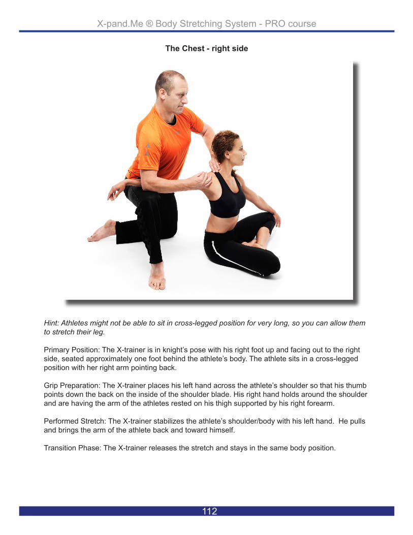
X-pand.Me ® Body Stretching System - PRO course
112
Hint: Athletes might not be able to sit in cross-legged position for very long, so you can allow them to stretch their leg.
Primary Position: The X-trainer is in knight’s pose with his right foot up and facing out to the right side, seated approximately one foot behind the athlete’s body. The athlete sits in a cross-legged position with her right arm pointing back.
Grip Preparation: The X-trainer places his left hand across the athlete’s shoulder so that his thumb points down the back on the inside of the shoulder blade. His right hand holds around the shoulder and are having the arm of the athletes rested on his thigh supported by his right forearm.
Performed Stretch: The X-trainer stabilizes the athlete’s shoulder/body with his left hand. He pulls and brings the arm of the athlete back and toward himself.
Transition Phase: The X-trainer releases the stretch and stays in the same body position.
The Chest - right side
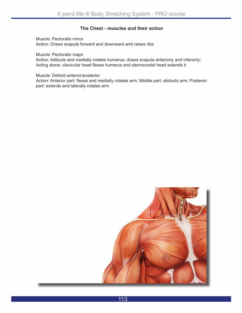
X-pand.Me ® Body Stretching System - PRO course
113
The Chest - muscles and their action
Muscle: Pectoralis minorAction: Draws scapula forward and downward and raises ribs
Muscle: Pectoralis majorAction: Adducts and medially rotates humerus; draws scapula anteriorly and inferiorly; Acting alone: clavicular head flexes humerus and sternocostal head extends it
Muscle: Deltoid anterior/posterior Action: Anterior part: flexes and medially rotates arm; Middle part: abducts arm; Posterior part: extends and laterally rotates arm

X-pand.Me ® Body Stretching System - PRO course
114
Hint: The biceps are a very strong muscle group, but that doesn’t mean that you should pull much harder; we allow the work to happen and will not force it to happen.
Primary Position: The X-trainer is in knight’s pose with his right foot up and facing out to the right side, seated approximately one foot behind the athlete’s body. The athlete sits in a cross-legged position with her right arm pointing back.
Grip Preparation: The X-trainer places his left hand across the athlete’s shoulder so that his thumb points down the back. And are having the arm of the athletes rested on his thigh supported by his right hand by the elbow of the athlete.
Performed Stretch: The X-trainer stabilizes the shoulder of the athlete and pulls the right arm of the athlete back towards him.
Transition Phase: The X-trainer releases the arm of the athlete back to neutral and changes his knight’s position to the opposite legs.
The Inner Upper Arm - right arm

X-pand.Me ® Body Stretching System - PRO course
115
The Inner Upper Arm - muscles and their action
Muscle: Biceps brachiiAction: Supinates forearm and, when it is supine, flexes forearm
Muscle: BrachioradialisAction: Flexes forearm
Muscle. CoracobrachialisAction: Helps to flex and adduct arm

X-pand.Me ® Body Stretching System - PRO course
116
Hint: Be “light” in your body and the stretches will feel better. In this case the X-trainer often tight-ens up, so please observe your own body.
Primary Position: The X-trainer is in knight’s pose with his left foot up and facing out to the right side, seated close to the athlete’s body. The athlete sits in a cross-legged position with her right arm lifted up toward the ceiling and forearm pointing back.
Grip Preparation: The X-trainer places his left elbow to stabilize the shoulder blade of the athlete and his left hand holds the inner forearm, close to the elbow. His right hand “says hello” to the ath-lete’s right hand and the athlete’s palm faces the mat.
Performed Stretch: The X-trainer keeps his left elbow at the level of the shoulder blade at all times making sure that the athlete’s body stays upright. With his right hand he pulls the athlete’s arm back.
Transition Phase: The X-trainer stays in the same knight’s pose and faces his body toward the op-posite side/to the athlete’s left side.
The Outer Upper Arm - right arm

X-pand.Me ® Body Stretching System - PRO course
117
The Outer Upper Arm - muscles and their action
Muscle: Triceps brachiiAction: Chief extensor of forearm; long head steadies head of abducted humerus
Muscle: Teres minorAction: Laterally rotate arm; helps to hold humeral head in glenoid cavity of scapula
Muscle: Teres majorAction: Adducts and medially rotates arm
Muscle: Lattisimus dorsiAction: Extends, adducts, and medially rotates humerus; raises body toward arms during climbing

X-pand.Me ® Body Stretching System - PRO course
118
Hint: If your grip feels slippery you can use training gloves or have a towel next to you or even place a towel upon the athlete’s body.
Primary Position: The X-trainer is in knight’s pose with his right leg up, approximately four inches from the athlete’s body. The athlete is seated in a cross-legged position with both arms lifted above her head.
Grip Preparation: The X-trainer holds the arms of the athlete straight up and above her head with his right hand squeezing both her palms together. The left hand of the X-trainer is placed on the side trunk of the athlete at the level where the lower ribs meets the abdominal area.
Performed Stretch: The X-trainer lifts the hands of the athletes over to her right side, while his thigh is “catching” the athlete’s right side. Keep lifting over to the side even after the side of the athlete is resting on the thigh. The X-trainer’s left hand is grounding the athlete’s body towards the mat.
Transition Phase: The X-trainer shifts his knight’s pose and hand position to the athlete’s other side.
The Side of the Body - left side

X-pand.Me ® Body Stretching System - PRO course
119
The Side of the Body - muscles and their action
Muscle: Lattisimus dorsiAction: Extends, adducts, and medially rotates humerus; raises body toward arms during climbing
Muscle: Serratus anteriorAction: Draws scapula forward and upward; abducts scapula and rotates it; stabilizes vertebral border of scapula
Muscle: Teres majorAction: Adducts and medially rotates arm
Muscle: External intercostalsAction: Stiffen the chest wall to prevent paradoxical motion during descent of the diaphragm; move the ribs
Muscle: Internal intercostalsAction: Stiffen the chest wall to prevent paradoxical motion during descent of the diaphragm; move the ribs

X-pand.Me ® Body Stretching System - PRO course
120
Hint: Lengthen your grip/their body on inhalations and stretch on exhalations.
Primary Position: The X-trainer is in knight’s pose with his left leg up, approximately four inches from the athlete’s body. The athlete is seated in a cross-legged position with both arms lifted above her head.
Grip Preparation: The X-trainer holds the arms of the athlete straight up and above her head with his left hand squeezing both of her palms together. The right hand of the X-trainer is placed on the side trunk of the athlete at the level where the lower ribs meet the abdominal area.
Performed Stretch: The X-trainer lifts the hands of the athletes over to her left side, while his thigh is “catching” the athlete’s left side. Keep lifting over to the side even after the side of the athlete is resting on the thigh. The X-trainer’s right hand is grounding the athlete’s body towards the mat.
Transition Phase: The X-trainer steps approximately one foot back and changes to his opposite knight’s pose.
The Side of the Body - right side

X-pand.Me ® Body Stretching System - PRO course
121
The Side of the Body - muscles and their action
Muscle: Lattisimus dorsiAction: Extends, adducts, and medially rotates humerus; raises body toward arms during climbing
Muscle: Serratus anteriorAction: Draws scapula forward and upward; abducts scapula and rotates it; stabilizes vertebral border of scapula
Muscle: Teres majorAction: Adducts and medially rotates arm Muscle: External intercostalsAction: Stiffen the chest wall to prevent paradoxical motion during descent of the diaphragm; move the ribs
Muscle: Internal intercostalsAction: Stiffen the chest wall to prevent paradoxical motion during descent of the diaphragm; move the ribs

X-pand.Me ® Body Stretching System - PRO course
122
Hint: Feel grounding through their body and start the stretching movements from down to up.
Primary Position: The X-trainer is in knight’s pose with his right leg up, approximately one foot behind the athlete’s body. The athlete is seated in a cross-legged position with both arms by her sides.
Grip Preparation: The X-trainer sits in knight’s pose with his right leg up, placed approximately one foot behind the athlete. The athlete is sitting in a cross-legged position.
Performed Stretch: The hands of the X-trainer are placed on both the shoulders, right to right, left to left of the athlete. The X-trainer’s left hand pushes the athlete forward and the right pulls her back. Both hands must use the same push and pull pressure to maintain a fully upright position for the athlete.
Transition Phase: Release the body of the athlete to a neutral position and change position to the opposite knight’s pose.
The Spinal Muscles - left side

X-pand.Me ® Body Stretching System - PRO course
123
The Spinal Muscles - muscles and their action
Muscle: MultifidusAction: Extend and rotate vertebral column
Muscle: RotatorsAction: Extend and rotate vertebral column
Muscle: Quadratus lomborumAction: Alone, lateralflexion of vertebral column; Together,depression of thoracic rib cage
Muscle: Iliocostalis lomborumAction: Extension, lateral flexion of vertebral column, rotates ribs for forceful inspiration
Muscle: InterspinalesAction: Extend vertebral column
Muscle: Intertransversarii anteriores Action: Lateral flexion of vertebral column
Muscle: Intertransversarii posteriorsAction: Lateral flexion of vertebral column
Lumbar region Muscle: Intertransversarii laterales Action: Lateral flexion of vertebral column
Muscle: Intertransversarii mediales Action: Lateral flexion of vertebral column

X-pand.Me ® Body Stretching System - PRO course
124
Hint: Remember to bring the athlete back to their starting position to correct your Alignment - if you have by chance pulled them out of Alignment.
Primary Position: The X-trainer is in knight’s pose with his left leg up to approximately one foot behind the athlete’s body and the athlete is seated in a cross-legged position with both arms by her sides.
Grip Preparation: The X-trainer sits in knight’s pose with his left leg up, placed approximately one foot behind the athlete. The athlete is sitting in a cross-legged position.
Performed Stretch: The hands of the X-trainer are placed on both the shoulders, right to right, left to left of the athlete. The X-trainer’s right hand pushes the athlete forward and the left pulls her back. Both hands must use the same push and pull pressure to maintain a fully upright position for the athlete.
Transition Phase: Release the hands from the athlete’s shoulders and stay in the same position but move closer to the athlete’s body.
The Spinal Muscles - right side

X-pand.Me ® Body Stretching System - PRO course
125
The Spinal Muscles - muscles and their action
Muscle: MultifidusAction: Extend and rotate vertebral column
Muscle: RotatorsAction: Extend and rotate vertebral column
Muscle: Quadratus lomborumAction: Alone, lateralflexion of vertebral column; Together,depression of thoracic rib cage
Muscle: Iliocostalis lomborumAction: Extension, lateral flexion of vertebral column, rotates ribs for forceful inspiration
Muscle: InterspinalesAction: Extend vertebral column
Muscle: Intertransversarii anteriores Action: Lateral flexion of vertebral column
Muscle: Intertransversarii posteriorsAction: Lateral flexion of vertebral column
Lumbar region Muscle: Intertransversarii laterales Action: Lateral flexion of vertebral column
Muscle: Intertransversarii mediales Action: Lateral flexion of vertebral column

X-pand.Me ® Body Stretching System - PRO course
126
Hint: Be patient with your stretching series; we often begin to speed up toward the end of the se-ries, but the muscles still need their time to “open up”.
Primary Position: The X-trainer kneels in knight’s pose with his left leg up, positioning himself about 1-2 inches from the athlete’s body. The athlete is seated in a cross-legged position with both of her arms down at her sides.
Grip Preparation: The X-trainer’s left hand is placed on the athlete’s left shoulder and places the right hand on her head at the side just above the ear.
Performed Stretch: The X-trainer grounds the left shoulder of the athlete with his left hand and lifts and pushes the athlete’s head to the right with his right hand.
Transition Phase: The X-trainer releases both hands and changes knight’s position to the opposite side.
The Side of the Neck - left side

X-pand.Me ® Body Stretching System - PRO course
127
The Side of the Neck - muscles and their action
Muscle: Levator scapulaAction: Elevates scapula and tilts its glenoid cavity inferiorly by rotating scapula
Muscle: Sternocleido mastoideusAction: Unilaterally it extends (sternal fibers) or flexes (clavicular fibers) the head and rotates it toward the opposite shoulder; flexes and extends the head; raises rib cage
Muscle: TrapeziusAction: Elevates, retracts and rotates scapula; superior fibers elevate, middle fibers retract, and inferior fibers depress scapula; superior and inferior fibers act together in superior rotation of scapula
Muscle: Scalenus mediusAction: Raises first rib (respiratory inspiration); acting together, they flex neck; acting on one side, they laterally flex, rotate neck
Muscle: Scalenus anteriorAction: Raises first rib (respiratory inspiration); acting together, they flex neck; acting on one side, they laterally fie rotate neck

X-pand.Me ® Body Stretching System - PRO course
128
Hint: Always create space and length, in this case, pulling your hands apart rather than pushing down and to the side.
Primary Position: The X-trainer kneels in knight’s pose with his right leg up, positioning himself about 1-2 inches from the athlete’s body. The athlete is seated in a cross-legged position with both of her arms at her sides.
Grip Preparation: The X-trainer places his right hand on the athlete’s right shoulder and places the left hand on her head at the side just above the ear.
Performed Stretch: The X-trainer grounds the right shoulder of the athlete with his right hand and lifts and pushes the athlete’s head to the left.with his left hand.
Transition Phase: The X-trainer releases both hands, stepping to the side of the athlete’s body and placing one hand on the back of the athlete at the level of the shoulders, placing the other hand in front of the chest by the collar bone. The X-trainer gently pushes the athlete back and supports her with his hand until the athlete lies on her back. The X-trainer sits back behind the athlete’s head.
The Side of the Neck - right side

X-pand.Me ® Body Stretching System - PRO course
129
The Side of the Neck - muscles and their action
Muscle: Levator scapulaAction: Elevates scapula and tilts its glenoid cavity inferiorly by rotating scapula
Muscle: Sternocleido mastoideusAction: Unilaterally it extends (sternal fibers) or flexes (clavicular fibers) the head and rotates it toward the opposite shoulder; flexes and extends the head; raises rib cage
Muscle: TrapeziusAction: Elevates, retracts and rotates scapula; superior fibers elevate, middle fibers retract, and inferior fibers depress scapula; superior and inferior fibers act together in superior rotation of scapula
Muscle: Scalenus mediusAction: Raises first rib (respiratory inspiration); acting together, they flex neck; acting on one side, they laterally flex, rotate neck
Muscle: Scalenus anteriorAction: Raises first rib (respiratory inspiration); acting together, they flex neck; acting on one side, they laterally fie rotate neck

X-pand.Me ® Body Stretching System - PRO course
130
Hint: Make sure that you don’t breathe right into the face of your client.
Primary Position: The X-trainer sits on his buttocks 4-5 inches behind the athlete’s head, with one of his legs bent so the shin bone is facing the head and other leg stretched out or slightly bent with the foot on the floor. The athlete lies flat on her back with both arms down at her sides.
Grip Preparation: The X-trainer places his right hand behind the neck of the athlete and allows her head to fall to the right, resting her forehead on his forearm. The X-trainer places his left hand on top of the athlete’s shoulder.
Performed stretch: The X-trainer’s left hand pushes the left shoulder of the athlete toward the mat in a downward direction or her hand and his right hand lift the athlete’s head to the side and up simultaneously.
Transition Phase: The X-trainer stays in the same position and releases both his hands.
The “Back Side” Side of the Neck - left side

X-pand.Me ® Body Stretching System - PRO course
131
The “Back Side” Side of the Neck - muscles and their action
Muscle: Scalenus anteriorAction: Raises first rib (respiratory inspiration); acting together they flex neck; acting on one side, they laterally fie rotate neck
Muscle: Levator scapulaAction: Elevates scapula and tilts its glenoid cavity inferiorly by rotating scapula
Muscle: TrazpeziusAction: Elevates, retracts and rotates scapula; superior fibers elevate, middle fibers retract, and inferior fibers depress scapula; superior and inferior fibers act together in superior rotation of scapula
Muscle: Splenius capitisAction: Bilaterally they extend the head and neck; unilaterally they laterally flex the head and neck and rotate the head to the same side.
Muscle: splenius cervicisAction: Bilaterally they extend the neck; unilaterally they laterally flex the neck
Muscle: Spinalis cervicisAction: Extends vertebral column

X-pand.Me ® Body Stretching System - PRO course
132
Hint: Make sure that you keep yourself upright to protect your lower back, using your inner strength.
Primary Position: The X-trainer sits on his buttocks 4-5 inches behind the head of the athlete, with one leg bent so the shin bone is facing the head and other leg is stretched out or slightly bent with the foot on the floor. The athlete lies flat on her back with both arms down at her sides.
Grip Preparation: The X-trainer places his left hand behind the neck of the athlete and allows her head to fall to the left, resting her forehead on his forearm. The right hand of the X-trainer is placed on top of the athlete’s shoulder.
Performed Stretch: The X-trainer’s right hand pushes the right shoulder of the athlete toward the mat in a downward direction or her hand and his left hand lift the athlete’s head to the side and up simultaneously.
Transition Phase: The X-trainer stays in the same position and releases both his hands.
The “Back Side” Side of the Neck - right side

X-pand.Me ® Body Stretching System - PRO course
133
The “Back Side” Side of the Neck - muscles and their action
Muscle: Scalenus anteriorAction: Raises first rib (respiratory inspiration); acting together they flex neck; acting on one side, they laterally fie rotate neck
Muscle: Levator scapulaAction: Elevates scapula and tilts its glenoid cavity inferiorly by rotating scapula
Muscle: TrazpeziusAction: Elevates, retracts and rotates scapula; superior fibers elevate, middle fibers retract, and inferior fibers depress scapula; superior and inferior fibers act together in superior rotation of scapula
Muscle: Splenius capitisAction: Bilaterally they extend the head and neck; unilaterally they laterally flex the head and neck and rotate the head to the same side.
Muscle: splenius cervicisAction: Bilaterally they extend the neck; unilaterally they laterally flex the neck
Muscle: Spinalis cervicisAction: Extends vertebral column

X-pand.Me ® Body Stretching System - PRO course
134
Hint: Take a moment to feel the difference in your own body too as this series can be an amazing workout. If you have any pain, look for misalignments next time you do the X-pand.Me Stretching Series.
Primary Position: The X-trainer sits on his buttocks 4-5 inches behind the head of the athlete, with one leg bent so the shin bone is facing the head and other leg is stretched out or slightly bent with the foot on the floor. The athlete lies flat on her back with both arms at her sides.
Grip Preparation: The X-trainer places his hands below the athlete’s head with thumb on the side of the head and the eight other fingers in line with where the skull and neck meet.
Performed Stretch: The X-trainer gently pulls the head of the athlete toward himself, simultane-ously lifting the head up.
Transition Phase: The X-trainer returns athlete’s head to neutral and rises to standing or kneeling above the athlete’s body with one leg on either side at the level of the trunk.
The Neck - both sides

X-pand.Me ® Body Stretching System - PRO course
135
The Neck - muscles and their action
Muscle: Levator scapulaAction: Elevates scapula and tilts its glenoid cavity inferiorly by rotating scapula
Muscle: TrazpeziusAction: Elevates, retracts and rotates scapula; superior fibers elevate, middle fibers retract, and inferior fibers depress scapula; superior and inferior fibers act together in superior rotation of scapula
Muscle: Rhomboid major/minorAction: Retract scapula and rotate it to depress glenoid cavity; fix scapula to thoracic wall
Muscle: Spinalis cervicisAction: Extends vertebral column
Muscle: Splenius capitisAction: Bilaterally they extend the head and neck; unilaterally they laterally flex the head and neck and rotate the head to the same side.
Muscle: splenius cervicisAction: Bilaterally they extend the neck; unilaterally they laterally flex the neck
Muscle: spinalis capitisAction: Extension of the vertebral column and head

X-pand.Me ® Body Stretching System - PRO course
136
Return to page 25 for the full body alignment...

X-pand.Me ® Body Stretching System - PRO course
137
Anatomy and Physiology of the Muscle System
Introduction;
Any class or activity regarding the performance of the body, such as we are doing with this stretch-ing and training program, deserves and benefits from a detailed examination of the anatomy and physiol-ogy (a.k.a. Form and Function) of the human musculoskeletal system. It is truly a remarkable system, complex in design, but simply elegant in execution, and the more we understand about it, the better we will be able to enhance the over-all results we are looking for.
At least as much as, if not more than, any other type of cell in the human body, muscle cells are a great example of the old adage, ‘Form Follows Function.’ To first explain in general terms, of which we will get into much further detail, there are three major types of muscle fibers that make up the muscle tissue in the human body; skeletal muscle fibers, also known as voluntary muscle fibers, smooth muscle fibers, sometimes referred to as visceral muscle fibers, and cardiac muscle fibers. The study of our entire amaz-ing muscular system, incidentally, is called myology (myo = muscle; logy = the study of).

X-pand.Me ® Body Stretching System - PRO course
138
Going from the macro to the micro view (or, from the broad, or over-all view, to the smallest, or micro-scopic, or for our purposes even the sub-micro-scopic view), in which we will need to learn a little bit about the actual molecules involved in active muscular movement, release, rest, respiration-life support-ing activity, and homeostasis-when all relevant systems are in a state of equilibrium, or balance), we know that typically, muscles are arranged in a group according to the job (or jobs) they may be asked to perform. Take for example the “simple” act of placing your hand from one location to another, as in performing a stretching movement.
As you may already know from any previous study of the muscular system you may have done, to actually engage “the shoulder,” which will in-turn move “the arm,” which will in-turn place the hand where we want it to go; it takes a coordinated effort, just in “the shoulder” region alone, (technically known as the pectoral girdle) of no less than seven different muscles, including the trapezius (superior and inferior), levator scapulae, rhomboideus major, rhomboideus minor, serratus anterior, and pectoralis minor, (whew! And we haven’t even mentioned…) the deltoid, and three additional muscles acting on the arm itself including the pectoralis major, latissimus dorsi, and the coracobrachialis, together with the four muscles of the “rotator cuff,” to say nothing of the muscles of the upper and lower arm itself (another eight), along with all the above-mentioned that actually go into “simply” moving the shoulder, to move the arm, to place the hand where we want it to go (and we didn‘t even mention any of the muscles in the wrist or hand itself, which would give us another fifteen!).
For anyone who is counting, that is a potential total of 38 (depending on exactly what motion or motions we might be doing with our arm and hand as we move it from one location in space to another), muscles that could potentially be involved in the one “simple” movement. Did we mention any of the mus-cles of the “core,” which we would need to keep us stabilized in the first place? Were we standing up or sitting down? (muscles acting on the pelvis/hips/legs/feet)? What about the muscles of our internal organs, allowing us to carry on the necessary life processes in the first place? I think you get the idea.
Now, where were we? Oh yes; just as muscle groups are arranged in a certain specific way and coordinate in groups to accomplish certain tasks, so are individual muscle fibers arranged in groups, to compose still larger groups of similar fascicle orientation (or, if you prefer, “bands,” or similar to “the grain” in a piece of wood, if you like), which get us to still larger groups, which eventually leads us to large enough, and coordinated-enough groupings of similarly-laid out muscle tissue that we begin to know them as individually-named muscles (ex. the deltoid).
It is important to know that these muscle fibers, from the smallest bundles of muscle fibers/cells, called fascicles, all the way up to all of the major muscle groupings, indeed most of the major systems in the entire body, are wrapped in, or at least come in contact with, a type of tough-yet-flexible (well, flex-ible most of the time!) membranous connective tissue called fascia. Some even say many of our so-called “muscular” ailments are really just the result of unaddressed issues relating to fascia, which can lead us in to unhealthy movement patterns, which can lead to more problems with the fascia, and on, and on the cycle goes, until, unaddressed (or improperly addressed) it can very well eventually turn in to a major mus-culoskeletal issue.
Getting back to the Function Follows Form adage, the skeletal muscle cells themselves, that are also called muscle fibers, that make up the muscle tissue, that make up the named/individual muscles themselves, that…(okay, I think you’ve got the idea now), these muscle cells themselves are actually called myofibers due to their typically elongated shape. Of course due to their very specialized and dy-namic nature, the myofibers themselves, as well as their contents, (the organelles, etc, which perform all the life-sustaining, as well as action-producing functions necessary) are all arranged in a very unique, tightly bound and neatly arranged pattern unlike any other cells in the body. They have to be to be able to do what they do for us.

X-pand.Me ® Body Stretching System - PRO course
139
For example, did you know a typical skeletal muscle cell is about 100 um (um stands for microm-eter. A micrometer is a very small unit of measurement equal to 1 x 10 to the negative 6th meters, or another way of saying it is that a micrometer is 0.000001 meters in length) in diameter and 3 cm, or 30,000 um, long. Some skeletal muscle cells, however, are actually up to 500 um thick and 30 cm long! Imagine a single cell 30 cm long?! Wow; muscles and the muscular system are truly amazing, indeed!
It will also be important for us to know the macro and micro make-up of the muscular system, as well as that of fascia, tendons, ligaments-their similarities and differences, in addition to how muscles oper-ates, obtains and uses energy, the waste products produced during these processes, the different types of blood cells and how they are used in the different instances, as well as the structure and functions of the major muscle groups, along with the joints, range of motion issues, and ultimately how our activity, inactiv-ity and targeted stretching routines impact all of the above, to better know how it all functions (there we go with the relationship with Function and Form again) together as a whole, so in-turn we can know better how to keep our muscles functioning at their peak, while preventing and defining muscular injury, and to engage in an effective recovery if and when there is any accident or injury to this marvelous creation we call The Muscular System.
Universal Characteristics of Muscle Tissue;
To perform all of the requisite functions that muscle tissue is required to perform for us, such as movement (both our whole-outer-body movement and, as touched on above, the movement of most of our life-sustaining body fluids and other substances such as food and waste), stability, and heat production, as well as control of body openings and passages, and communication (via the tongue/respiratory system and/or sign language and/or other gestures and/or facial expressions), there are certain characteristics that all muscle cells have in common. And while the three major types of muscle cells may be composed differently, have different functions, and thus ultimately look and act differently, they still share some basic traits. These traits shared by all three types of muscle tissue are:
• Responsiveness, or, Excitability. This is the ability of the muscle cell, when stimulated by neu rotransmitters (chemical signals), stretch, and other stimuli, to respond with electrical charges across cell membranes.
• Conductivity. Stimulation of a muscle fiber produces more than just a local effect (effect to the immediately surrounding area). The local change triggers a wave of excitation that travels at an extremely rapid pace along the fiber and initiates processes that lead to muscle contraction.
• Contractility. A unique characteristic of myofibers is their ability to become substantially shorter when stimulated. This is what allows them to pull on bones and other tissue, thus creating inner and/or outer movement.
• Extensibility. Extensibility is the muscle cells ability to stretch between contractions. Most other body cells rupture when stretched, but skeletal muscle fibers can stretch up to three times their contracted length.

X-pand.Me ® Body Stretching System - PRO course
140
• Elasticity. Commonly mistaken for the ability to stretch (which as we have just seen directly above, is the definition of Extensibility), elasticity is actually the term used to describe the muscle cell’s tendency and ability to recoil and return to its original resting length once the stretching phase has concluded.
Major Types of Muscles; Anatomy, Physiology.
As mentioned, there are three distinct major types of muscle in the human body; skeletal muscle fibers, also known as voluntary muscle fibers, smooth muscle fibers, sometimes referred to as visceral muscle fibers, and cardiac muscle fibers. Though for our purposes we will be focusing primarily with skel-etal muscle, the other two types, smooth and cardiac, are, of course, equally important to our lives over-all.
Skeletal Muscle
Skeletal muscle, as previously mentioned, is constructed of long, fiber-like cells called muscle fibers, or myofibers. Most skeletal tissue is attached to bone (hence its name Skeletal Muscle), but there are exceptions in the face, tongue, upper esophagus and some sphincter muscles (ring-like muscles that allow opening and closing of certain interior and/or exterior body passages). In other words, the very individual cells that make up the skeletal muscle fibers/tissue/etc, are actually multi-nucleated (more than one nucleus), elongated cells filled mostly by long protein bundles called myofibrils (not to be confused with the muscle cells themselves, the myofibers). Incidentally, the reason skeletal muscle cells have more than one nucleus per cell, is because of the fact that when they are in their embryonic developmental state, several stem cells (also known as myoblasts) fuse to form the single muscle cell/fiber, with each stem cell/myoblast contributing its own nucleus to the finished product we call a muscle cell, or again, muscle fiber.
Some of these myoblasts remain and can be called upon later to multiply and produce new muscle fibers to aid in muscle repair, but it is important to realize that (unfortunately for us, at least in terms of function and flexibility) most muscle repair is by fibrosis rather than regeneration of functional muscle tissue.
This means that oftentimes, instead of torn muscle tissue being replaced by new and fully function-al muscle tissue, it is instead replaced by fibrous tissue that is more of a “patch,” or what is more common-ly referred to as “fibrotic scar tissue.” This is a major reason why flexibility can be so limited after a muscle injury, why we don’t want to “over do it” too soon, and why strategies such as “breaking up” the scar tissue with Deep Transverse Friction Massage (to get the fibrous scar tissue to at least align with the “grain” of the surrounding muscle), among other treatment techniques, such as Contrast Therapy (alternating, timed applications of heat and cold to the affected area), as well as a disciplined approach to an effective target-ed stretching routine, initiated at the first safe period in the healing process, are so important to regaining as much of our original strength and range of motion as possible.
There is no reason why, in many cases, we can’t recover fully from most injuries, and in some cas-es, with this new emphasis on the affected area, become even stronger and more flexible than we were before the onset of the injury that got us paying so much attention to the affected area in the first place!
Moving on, in summation, a skeletal muscle cell is also known as a muscle fiber, which is also known as a myofiber, and there are two types; fast-twitch or white, and slow–twitch, or red. As another old adage goes; A Picture Is Worth A Thousand Words. To that end, please see Figure #1 to get a better understanding of the typical make-up of the typical skeletal muscle.

X-pand.Me ® Body Stretching System - PRO course
141

X-pand.Me ® Body Stretching System - PRO course
142
Skeletal muscle tissue is distinguished from other muscle tissue in that it is voluntary (under our control-most of the time!), striated, and connected to bone (thus the name “skeletal muscle”). The striated appearance is a result of the alternating light and dark bands of the protein filaments within the myofibers that are able to cause muscle contraction. The voluntary part means, of course, that we usually have con-trol over when these myofibers contract, and equally as important, especially for our studies, relax. Inci-dentally, don’t get the “striations” at this cellular level confused with the “striations” that are often referred to in the world of bodybuilding. The striations we are referring to are much too microscopic to be seen coming through the cutaneous (skin) layer, no matter how “ripped” and “cut” (and usually, dehydrated) the one who is posing might be. The bodybuilders striations, however, are simply more-easily seen muscle tis-sue groupings, in fact due to low levels of sub-cutaneous fluid and fat covering up the previously-referred to layers of connective tissue, which compartmentalize the muscle groups from each other and from the deeper organs and tissues, within the various named muscle groupings. Take a look at our Fig. #1 and you get a better idea of the various organizational methods used by the body to group the myofibers, muscles tissue, and muscle groups themselves.
There are two types of skeletal muscle fibers: slow-twitch (or red, or type I, or slow oxidative, or SO) fibers, and fast-twitch (or white, or type II, or fast glycolic, or FG) fibers. The slow-twitch have many mitochondria, myoglobin and blood capillaries, and therefor a relatively deep red color. They are best adapted for aerobic respiration, which does not produce lactic acid, and so are therefor resistant to fatigue. Fast-twitch fibers are rich in enzymes of the phosphagen and glycogen – lactic acid systems. They have SR, (sarcoplasmic reticulum) that releases and reabsorbs Ca quickly, which accounts for their ability to produce forceful, quick actions. Most muscles are a combination of both, but with a heavy emphasis on one or the other type, and the genetically-inherited ratio of each can play a large role in determining what type of sports or other activities a person will be best suited for.
Of course skeletal muscle is more than just muscle tissue, as it contains connective tissue, nervous tissue, and blood vessels. As many times our bodies are models of efficiency in terms of both form and function, skeletal muscle is no different. In terms of general function, the skeletal muscle system is respon-sible for movement of our bodies, of course, but these movements, in addition to getting us and/or our limbs from point A to point B, themselves have an additional purpose. Whenever we contract and release a skeletal muscle, we are also almost always affecting some other system and/or substance within the body.
One prime example is how the all-important lymph system, with its essential functions of fluid recovery, immunity, and lipid absorption, depends in part on the contractions of the skeletal system con-gruently with the increased blood flow created by exercise, made possible, again, by the skeletal muscle system. In addition to other obvious factors relating to overall health and immunity, can you see how this fact might make a “couch potato” more susceptible to disease and infection than someone who spends more time in movement, or especially in strategic and targeted movements such as those from an effective stretching routine? This instrumental role in lymph function in addition to the fact that our muscular con-tractions are responsible for producing up to 85% of one’s body heat, which is crucial to our survival on a cold day, as well as to our life-sustaining metabolism in general (ever wonder why you “shiver” when cold?

X-pand.Me ® Body Stretching System - PRO course
143
Your body is using those rapid, if minute, muscle contractions and releases to produce heat through friction produced by...here it comes; movement!). Even when we are not moving, you can thank your friendly neighborhood skeletal system for that, too. As in, our muscles help us maintain stability and an erect posture while many muscles also stabilize our joints by maintaining tension on tendons and bones.
In order to do what they do, (create and allow stability and movement) our skeletal muscles at-tach to bones through the extension of their connective tissue. In what is known as an indirect attachment, the muscle ends short of its apparent destination. The space is bridged and secured by a band of fibrous tissue we know as a Tendon (Ligaments, which will be discussed later, attach bone-to-bone. Think of the ligaments of the knee, et al). The two ends of our biceps brachii are a prime example of this type of muscle attachment. In other instances the tendon may fan out, and be known as an aponuerosis, as in the case of our lower lumbar area, where the muscles from the latissimus dorsi attach to, ultimately, our lower spine and the P.S.I.S., or Posterior Superior Illiac Spine, or, if you prefer, the top of our rear pelvic bone. In a reti-naculum, or band of connective tissue, tendons from distinct muscles pass under this band, as in our wrist. In what is known as a direct attachment, such as in the spaces of the brachialis and lateral head of the triceps brachii, there is so little space, it would appear that there is, in fact, direct muscle-to-bone attach-ment. However under a microscope we see that in fact, there is a slight space between muscle and bone that is spanned by collagen fibers. The types of attachments we are dealing with, and their cellular and tis-sue make-up becomes increasingly important as we begin formulating an effective strategy for stretching and lengthening our muscles and developing effective routines for each; what works for one muscle group may or may not work for another, and the type of attachment(s) we are dealing with can play a crucial role in determining an effective strategy to get the most out of our efforts.
Talk about organization! Skeletal muscles are “bundled” in one form of connective tissue to another, from deep to superficial, as follows; the endomysium is a thin sheath of connective tissue that surrounds each myofiber. It is relatively loose to allow for blood capillaries and nerves to reach every muscle fiber. This insures that each muscle cell has access to stimulation and nourishment/waste exchange. The endo-mysium also provides for the extra-cellular (outside the cell itself) chemical environment of the muscle fiber and its nerve ending. Stimulation of a myofiber occurs as the result of an exchange of calcium, sodium, and potassium ions between the endomysial tissue fluid and the nerve and muscle fibers (now, maybe you can start to picture why and how these minerals are so important to us, especially as active individuals, and how their balance, or lack thereof in our bodies can lead to peak performance, and/or performance-hindering issues).
The perimysium is a thicker sheath of connective tissue that wraps myofibers together in the bundles called fascicles. These fascicles are visible to the naked eye as parallel strands, or “the grain” in meat. In fact, meat is easier pulled apart along these grains than against. The perimysium carries larger nerves, blood vessels, as well as the all-important stretch receptors called muscle spindles, which we will definitely get in to more detail with later. The epimysium is the fibrous sheath that encompasses the entire muscle. On its outside, it fuses into the fascia, on its inside, it projects between the fascicles to form the perimysium. The fascia is a sheet of connective tissue that distinguishes neighboring muscles and muscle groups from each other, and from the subcutaneous tissue.

X-pand.Me ® Body Stretching System - PRO course
144
One last concept we need to be aware of with respect to skeletal muscles is that of origin and in-sertion. Though not always a perfectly accurate way to describe the relationship between all muscles and bones in all situations, for the most part this concept deals with the knowledge that most skeletal muscle, under most conditions and movements, has a more stationary point of bony attachment known as the origin, and the attachment location that does most of the moving, at the “other end,” if you will, called the point of insertion. As mentioned, this model does not always work due to the fact that with different move-ments, the same muscle could be considered to have a different and opposite site of origin and insertion. Consider how, if you kick a soccer ball, the tibia moves more than the femur, so the tibia would be the insertion of the quadriceps and the femur the origin. However, if you are in the act of sitting down, as in a chair, the tibia remains stationary and it is the femur that moves.
Each of the above examples, by way of origin/insertion, would give us the opposite definition for each of the corresponding attachment sites, respectively. Because of this fact, many anatomists are no longer using the origin/insertion models and are instead tending towards the proximal/distal model of refer-ence. Proximal = closer (think “closer to the proximity of the core,”), and Distal = further, “more distant from the center or core.” Lastly, however defined in any one example, between the point of origin and insertion lies the muscle belly, or the thickest part of the muscle. A great example of a muscle with (usually) a clearly identifiable belly would be the bicep brachii. We’ve known this ever since we were kids, right? It’s what we flexed whenever someone would say, “show me your muscle.” Too bad we weren’t smart enough then to respond with something like; “Show you my muscle? Certainly! But of course I have many of them; ap-proximately 640 to be exact, depending on your source and definition. However you must be requesting that I initiate the flexor movement of my upper arm, which will in-turn produce a noticeable protrusion of the easily identifiable muscle belly of my biceps brachii?” Then again...
Smooth Muscle
Moving on…as we have mentioned, voluntary, striated skeletal muscle is one kind of muscle, and cer-tainly for the purpose of our training sessions, it is the one we will be most concerned with. However it is definitely not the only type of muscle tissue in the body.
There is also smooth muscle, sometimes referred to as visceral, muscle tissue (which is actually only one of two kinds of smooth muscle tissue), and also known as involuntary muscle. Smooth muscle tissue is referred to as smooth, because, though it does contain both thick and thin filaments, similar to skeletal muscle tissue, these filaments are not lined up with each other and so do not produce any regular, (microscopically) visable striations. Neither are there sarcomeres (contractile units between the Z discs in skeletal muscle, but that is language that is, admittedly, getting us ahead of ourselves, and of that which we will explain in more detail shortly). Also, different than skeletal muscle, smooth muscle tissue has fusi-form shaped (“skinny-diamond-shaped,” as-in, take a square, turn it one eighth turn, and now stretch the opposing top and bottom corners; now just add some water, and…okay, okay…) cells with one nucleus that is usually close to the center of the cell. To explain in a bit more detail; the two types of smooth muscle tissue are called multi-unit and single unit types. Multiunit smooth muscle exists in some of the larger arter-ies and air passages, in the tiny piloerector muscles of the follicles of our hair, and in the iris of the eye.

X-pand.Me ® Body Stretching System - PRO course
145
It is served by nerves in much the same way as skeletal muscle in that there is one nerve ending for each individual cell (though again, in skeletal muscle the innervation is a function of the somatic, or for our purposes, the more voluntary-based nervous system, as opposed to when as in smooth muscle tissue, the nerves are a part of the autonomic, or for our purposes, more automatic and/or involuntary nervous system).
Each motor unit of multiunit smooth muscle fiber is able to contract independently of the others, which is why it is known as multi-unit. In single unit smooth muscle tissue, also known as visceral muscle because this is the type of muscle tissue that is so widespread throughout our internal, or visceral organs (such as the digestive, reproductive, and urinary, systems as well as the respiratory tract and in most blood vessels), single-unit is in reference to the fact that the cells in this type of muscular tissue are elec-trically tied together through what are known as electrical gap junctions. In this way, the cells can directly stimulate each other, enabling multiple units over large regions to act as one, in effect contracting and releasing as one single cell, even when, unlike skeletal muscle, smooth muscle cells each have only one single nucleus per cell. This type of nerve-to-cell set up and ratio are what make it possible for our intes-tines to push substance through in waves, or for our blood vessels to act as more-or-less one unit through-out our bodies, and in the case of blood vessels specifically, maintain a relatively constant system-wide blood pressure.
When a smooth muscle cell contracts, (as opposed to a skeletal muscle that generally “shortens” in a more-or-less straight line from point a to point b), it puckers and twists, sort of like wringing out a wet sponge. Also different than skeletal muscle, smooth muscle is not attached to bone, and though is slower to contract or relax than voluntary muscle, is also much more resistant to fatigue. You can see how impor-tant this is when we talk, again, about blood pressure and the bodies ability to maintain a constant, healthy blood pressure (because the muscles involved in this necessary and life-sustaining-function don’t just “get tired” and stop working at some arbitrary point in the middle of the day), or equally importantly, to keep our food moving along it’s merry way through our digestive system. Constant, or near constant tonicity, while sometimes impossible and/or detrimental in regards to the skeletal muscular system, is often a functional goal and necessity of certain smooth muscle systems. We will get in to some of the explanations and reasons, at a biochemical level, for many of these characteristics when we look in to the hows and whys of muscle function in more detail a little on down the road.
Another important functional difference of smooth muscle from skeletal, is that smooth muscle is able to contract even when significantly stretched out (good thing, when you again think about how your stomach takes care of moving a big meal on it’s way…mmmmm, a big meal. Is anybody else hungry?), as opposed to skeletal muscle, which must be relatively flexed before being able to fully contract. For example, skeletal muscle needs to be within around 30% of optimum length in order to be able to contract forcefully when stimulated by the nervous system. However, smooth muscle can be anywhere from 50% all the way up to 100% of its homeostatic, or normal-resting-state, length, and still contract forcefully! Again, good thing, when it comes to the structure and function of our esophagus, stomach, or our bladder, for some obvious examples. Lastly, smooth muscle has an incredible degree of placticity, (a feature that I happen to think is really super cool, to use a not-so-well-known scientific phrase…) or the ability to stretch significantly, but then contract again, and not become loose, flabby, or worse, ineffective once returning to the smaller size.

X-pand.Me ® Body Stretching System - PRO course
146
Another handy feature when we’re talking about the regular and repeated filling up of, and then the subsequent regular and repeated emptying out of, the various hollow cavities and internal organs of the amazing human body!
Cardiac Muscle
The last of the three major and distinct types of muscle tissue in the human body is cardiac muscle. You might assume that the name cardiac indicates that this muscle is found in, and is unique in structure and function to, the heart (and you would be correct in that assumption). When studying cardiac muscle anatomy and physiology, we discover that it shares some characteristics with both skeletal and smooth muscle, while also having some unique and stunning traits all of it’s own. First, similar to skeletal muscle tissue, cardiac muscle is striated, but its cells, or myocytes, or more accurately its cardiocytes, are shaped like a short, thick log or roll, with ends that are uneven and notched. These cardiocytes (cardio = of the heart, cytes = cells) are joined at the ends through a linking system known as intercalated discs. In stained tissue sample observation, these areas will appear as thick, dark lines. Similar to smooth muscle, these discs have electrical gap junctions that allow each cardiocyte to directly stimulate its neighboring cells. There are also mechanical junctions, in addition to the electrical ones, that keep the cells from pulling away from each other when the heart contracts (something it does quite a bit of, as you might already be aware). The sarcoplasmic make-up that we previously touched on, and will go over in more detail in a mo-ment, is different than the other two types of muscle, to allow for the near endless use of the tissue, while keeping it both powerful and virtually fatigue-free.
Along these same lines of thinking, different than skeletal muscle, cardiac muscle can contract without any outer, direct nervous stimulation. It can perform in this way due to its own built-in pacemaker that sets off waves of electrical excitation in a very regular, in fact rhythmic, pattern. Each wave travels through the muscle and triggers the necessary and systematic chambers in proper order to trigger the con-traction of each of the four heart chambers, each at the exactly appropriate (well, at least in most people the majority of the time) moment, to propel blood through the heart itself, and then through the rest of the body, or, if you like, through the body and then into the heart itself. I guess the viewpoint would depend on where you want to call the beginning and where you want to call the end of the endless (well, endless until the end, that is…) cycle and circuit we know as blood flow. It is precisely because of the fact that heart muscle has this ability to contract rhythmically and independently that cardiac muscle is described as auto-rhythmic.
Even though this is so, the cardiocytes do still receive messages from the autonomic nervous system via nerve fibers. These messages, or if you prefer, impulses, can increase and/or decrease both the rate at which the heart beats (the frequency per unit of time measurement), as well as the strength, or force, of each contraction. This comes in handy when we are, say, running towards prey, or maybe away from a predator, or maybe even if we are simply getting into a challenging stretching routine, right? And of course once each particular episode concludes, be it for the hunter, the hunted, or the yogi or yogini, it’s equally important to be able to “calm the jets” as it were, back down to a homeostatic state again, too (at least until the next challenge, anyway).

X-pand.Me ® Body Stretching System - PRO course
147
Heart muscle has about 25% of it’s cell volume filled with the “energy manufacturing plants” of the cell called the mitochondria, as opposed to skeletal muscle, which has only about 2% of their cells taken up with mitochondria (obviously, and again, lucky for us the body’s innate wisdom says that it’s probably a good thing that the heart places so much emphasis on being able to have enough energy to constantly continue pumping!). As such is the case with such vast amounts of real estate in the cell devoted to en-ergy production, the heart is quite adaptable when it comes to being able to use various types of fuel, but because cardiac muscle uses aerobic respiration almost exclusively, its performance can be susceptible to interruptions in oxygen supply. Overall, however, as you might also be able to imagine, cardiac muscle is highly resistant to fatigue. “Use it or lose it” seems to fit the bill here, with the adjunct, “and the more you use it, the better it gets.” Of course like any muscle or system; we don’t want to do too much too soon, and depending on our age, fitness level, and other variables, too much for too long. In other words, we do want to give the heart and ourselves a good workout, while still always allowing for a chance at rest and recov-ery, (eventually, anyway, my fellow dopamine and adrenaline junkies…).
Muscle Anatomy and Physiology: The Micro-View and Molecular Composition
Okay, we have just covered the three distinct types of muscle tissue in the body in more-or-less of a detailed, mostly macro-overview. Now let’s get into the micro-anatomy (the details of the actual physical make-up of the cells and cell structures that comprise the myocytes, at a microscopic level), and life at the molecular level, and the biochemical physiology (the combination of the chemical and biological processes that allow each myocyte to do what it does) of the different muscle cells, along with their internal parts, and the “outer” corresponding muscle tissue.
Though the thought of getting into such detail may seem a little daunting at this point to some of you, I’m pretty sure it will actually help us all in the long run in terms of making more sense of the similari-ties, the differences, and the overall picture of the various structures and functions that are relevant to our daily activities as well as our training sessions, and how these impact each other. We will be going through a number of terms and definitions you may have never heard of before (depending, of course, on how detailed your knowledge of anatomy and physiology are), but don’t get nervous. First, you can always refer back to the glossary during and after your reading of this section. And second, there is another illustration, Figure #2, to help you visualize the structures and functions we are speaking of. It’s important to remem-ber that none of the things we have been or will be covering exists in a vacuum, but rather, as the holistic model of life and wellness tells us, nothing exists completely on it’s own, but rather everything exists as an individual working part of an integrated whole.
The relationship(s) between the individual parts and the whole, and vise verse, is important to keep in mind, even as we dive into the seemingly separate details to get a better understanding of both the de-tails and the functioning whole. Our training sessions, indeed our studying itself, along with things like fuel and fluid intake, as well as rest and recovery time, etc, are all simply real-life scenarios and experiential examples of the macrocosmic extension and physical endpoints of the microscopic world we are studying. Or is that; the microcosmic world we are studying is simply the detailed explanations and descriptions of the universe that the macrocosmic world we are always readily perceiving through our 5, or some might say 6, or more, senses, is made up of and based on? I guess the best answer to those two questions might be “yes” and “yes,” respectively, (depending on your outlook, of course…).

X-pand.Me ® Body Stretching System - PRO course
148
As we have touched on previously, all of the three different muscle types share some common functions, but all of these functions might be able to be boiled down to, or are at least merely offshoots and/or the peripheral results of, movement. So what, exactly, makes these particular cells we call muscle cells so special when it comes to movement? Or more specifically, how and/or why are these cells capable of creating this movement in the first place, when other cells in the body (for example brain tissue cells, or lung tissue cells) are not? Notice I make the distinction of creating movement; because while brain tissue should not be moving (per se), or creating movement at all, lung tissue cells, of course, have to move to allow us to breathe, BUT the lung tissue is only moving because of the muscle tissue that is surround-ing and acting on it (principally the intercostal muscles that are located between the ribs, and to an even greater extent, the diaphragm muscle that is located between our lungs/thoracic cavity, and our intestines, or abdominal cavity). Again, think back to the relationship of function and form. Quite simply, muscle cells can move and thus create movement on a larger scale, because of the way they are put together. And this means we need to talk about the ingenious way in which the muscle cells themselves are put together.
First of all, as mentioned earlier as well, skeletal muscle cells are long (sometimes very long, relatively-speaking), multinucleated cells crammed full of long protein bundles called myofibrils. There are three different kinds of these; thick filaments, thin filaments, and elastic filaments. Thick filaments are made of myosin, thin are made of fibrous (F) actin, and as in a bead necklace, have a string of subunits called globular (G) actin. Each G actin has a site where it can bind to the head of a myosin molecule. A thin filament also has 40 – 60 molecules of still one more type of protein called tropomyosin. Tropomysin keeps a muscle in a relaxed state by blocking the active sites of six or seven G actins and it also prevents myosin from binding to them. Each tropomyosin molecule has a smaller, calcium-binding protein called troponin bound to it. Lastly, elastic filaments are made of relatively very large protein molecules called titin, or connectin.
These are on either side of each thick filament and anchor it to a very important structure called a Z disc. All this helps to create stability for the thick filament and center it between the thin filaments, to pre-vent overstretching. (Stay with me…) Myosin and actin are contractile proteins, so named because they are responsible for the work of shortening the muscle fiber. Tropomyosin and troponin are known as regu-latory proteins because they act like a guard, of sorts, and determine when a myofiber can contract and when it cannot. Most of the organelles of the cell, including the mitochondria, as well as the actual bands and discs that are largely responsible for the myocyte’s ability to move, and thus move us, are crammed in long-ways, along-side and along-with these myofibrils.
The smooth endoplasmic reticulum (a series of interconnected tubules within the cell’s cytoplasm) in muscle cells is actually called sarcoplasmic reticulum (SR), forms its network around each myofibril. It periodically has dilated end sacs called the terminal cisternae, which cross the muscle fiber/elongated cell, from one side of the cell to the other, horizontally. The sarcolemma (the plasma membrane of a muscle fiber) has tubular folds called transverse (T) tubules, which penetrate all the way through the cell and emerge on the other side. Each T tubule has two terminal cisternae running along each side.

X-pand.Me ® Body Stretching System - PRO course
149

X-pand.Me ® Body Stretching System - PRO course
150
Now then, striated skeletal fiber, as we touched on previously, is in fact striated because of the dark bands (A bands, or anisotropic bands), and lighter (I, or isotropic bands). Each A band contains thick fila-ments laying side by side and a part of the A band where thick and thin filaments overlap. In the middle of the A band there is a region, lighter in color, because and where the lighter thin filaments do not reach. Finally, each light I band is bisected by a narrow Z disc, or Z line, which provides anchorage for the thin fila-ments and the elastic filaments. The distance from one Z disc to the next is called the sarcomere. Finally, this is the contractile unit of the muscle cell/fiber. A muscle shortens because of the fact that its individual sarcomeres shorten and pull the Z discs closer together, and the linking proteins pull on the extracellular (outside of the cell) proteins of the muscle. The Z discs are pulled closer together in a muscle contraction, and this in turn pulls on the sarcolemma, which results in the overall shortening of the muscle cell.Myofiber Process of Energy Production, Usage, and Action
So what (?!), you might be saying to yourself. What does all this have to do with the price of tea in China? Well, for starters, it is this microscopic act that begins as merely a thought, or intention, and contin-ues through the electrical, then the molecular levels, that makes the foundation for all muscular, and thus all human, movement. Additionally, as we are only halfway there, we only have heard half of the story. The rest of the story is about energy. Specifically the energy that it takes, to produce the movement that we want, and what it takes to turn a nerve impulse into a muscular contraction, thereby producing the movement(s) that we seek.
So, similar to movement in an automobile, both “fuel,” which in our case would probably go by the name of acetylcholine (Ach), (though some could argue calcium (Ca) is the fuel, my feeling is that the Ca is responsible for “telling” the fuel (Ach) when to be released (maybe like Ca is service attendant who is un-locking the pump to allow the gas (Ach) to flow?), and “fire,” or for our purposes, an electrical signal/nerve signal, are required to produce said movement.
What happens is, on average about 200 regionally grouped, but not necessarily clustered right next to each other (this is to distribute the “load” or “action” over a wider field, which in-turn helps to avoid fatigue of regional areas of muscle all at the same time) muscle fibers are innervated (accessed and serviced by) a motor neuron (no pun intended). Each motor neuron/nerve fiber has “branches.” When the nerve sig-nal arrives at the point of contact with the muscular fiber (known as the synapse, or more specifically, the neuromuscular junction, which is also known as the motor end plate – again, no pun intended regarding our “fuel and fire” example, stored calcium (Ca) diffuses up into the synaptic knob, which stimulates this synap-tic vesicle (simply the end of the nerve fiber closest to our muscle) to release acetylcholine (Ach).
This is because the electrical signal from the nerve impulse cannot jump across the synapse itself (like, say, a spark plug might do), but relies on this chemical, or neurochemical messenger (Ach) to diffuse across the gap. To respond to this chemical, the myofiber has about 50 million Ach receptors, in it’s plasma membrane, and nearly all of them directly across from these synaptic knobs, to maximize sensitivity and thus response-ability. The Ach diffuses across the NMJ, and binds to receptor proteins in the sarcolema (the plasma membrane of a myofiber). These receptors are ligand-regulated ion gates, (or, gates that swing both ways, depending on what is acting upon them) and two Ach molecules must bind to each receptor. Once this happens, Sodium (NA+) diffuses quickly into the cell, while Potassium (K+) diffuses out. All of this movement of Na+ and K+ back and forth, in and out of the cell, literally causes minute electrical changes in the cell and creates what is called action potential, as the myofiber is now excited.

X-pand.Me ® Body Stretching System - PRO course
151
These changes cause a wave of action potentials to spread from the end of the motor end plate, which spreads out in all directions, like ripples in a pond, which, once this wave of excitation reaches the T tubules we spoke of earlier, continue down the sarcoplasm. Action potentials open voltage-regulated ion gates in the T tubules. These are directly, physically linked to Ca channels in the terminal cisternae of the SR (sarcoplasmic reticulum) which causes the gates of the SR to open while CA diffuses out of the SR, down the concentration gradient and into the cytoplasm of our affected cell. CA binds to the troponin of the thin filaments. The troponin-tropomyosin complex’s structural integrity is altered and it sinks deeper into the groove of the thin filament. This exposes the active sites on the actin filaments and makes them avail-able for binding to myosin heads.
Now that we have gotten to the point, through the aforementioned biochemical changes, initiated by the electrical impulses, created by something as simple-yet-powerful-and-profound as a single thought or intent, let’s see what happens physically in the muscle cell. Next, the myosin head must have ATP, or Adenosine Triphosphate. This is the body’s most important energy-producing molecule, which briefly stores energy gained from processes related to glucose oxidation. Glucose oxidation is simply a fancy way of describing the process of how the body, after converting the many foods we eat to the simple sugar molecule known as glucose, burns that fuel.
In our bodies, many, if not most cells, can use fats, or fatty acids, as well as glucose, for energy, but it is important to remember that some cells, such as neurons, cannot use alternate energy sources, and so must rely on glucose exclusively. Again, taking the myosin head, with an ATP molecule attached to it, the body can initiate the muscular contraction process. Myosin ATP-ase (a specialized enzyme in the myosin head) hydrolizes, (breaks the bond) of this ATP. This releases energy, which activates the head (in this case by “cocking” it into an extended position). The head then binds to an exposed active site on the thin filament, forming a cross-bridge between the myosin and actin. The myosin now releases the ADP and phosphate and flexes into a low-energy position, which now tugs the thin filament, and brings it “along with.” This is what is known as The Power Stroke. The head remains bound to actin until it binds a new ATP.
Upon binding more ATP, myosin releases the actin and is now prepared to repeat the whole thing over again; it will hydrolyze the ATP, re-cock, (this, incidentally, is known as The Recovery Stroke), at-tach to a new active site farther down on the thin filament, and produce another power stroke. There are many heads on the thick filament, that are binding to the thin filament, that prevent the whole mechanism from simply “sliding back” during this recovery stage. Or, another way to look at it is that in any one given moment, half the heads are bound to the thin filament, and half are extending to grab the filament farther down. Similar to how one pulls a rope, hand-over-hand, the filaments do not get shorter, but simply slide over each other, making the total length of the fiber functionally shorter, even if all the filaments are still the same length.
A single power and recovery stroke uses about one molecule of ATP, and a single cycle of power and recovery will shorten a fiber by only about 1%, but all-told, the fiber is capable of shortening by up to 40% of its resting length, so as you can see, the cycle needs to be repeated many times by each myosin head, which, though herky-jerky in movement taken singularly, produces a smooth stroke indeed when hundreds of them are all acting together creating a steady pull on the thin filament.

X-pand.Me ® Body Stretching System - PRO course
152
When a muscle fiber’s work is done, it returns to its resting length. This occurs when the electrical/nerve impulses stop arriving at the NMJ, so the synaptic knob stops releasing Ach. As the Ach is broken down and no longer stimulates the muscle fibers, the synaptic knob reabsorbs these fragments for recy-cling and later use. Active Transport Pumps in the SR begin to pump CA+ from the cytosol back into the cisternae. Here it binds with a protein until it is needed again (when the myofiber is again stimulated). Since active transport requires ATP, this is why ATP is needed for both muscle excitation and relaxation. Calcium ions break off from the troponin, are pumped into the SR and are not replaced, and tropomyosin moves back into the position where it blocks the active sites of the actin filament. Myosin can no longer bind to actin, and the muscle fiber ceases to be able to produce or maintain tension.Hard to believe that so many different, complex, individual steps take place, allowing us to carry on with everything from shopping for groceries, to engaging in challenging stretching and training, in such a seam-less flow, back and forth, from tension to release and back again, and in such a short period of time, (prac-tically instantaneously, really) isn’t it? Well, that is the incredible nature of our incredible bodies…
Some of the main concepts and understanding I hope you will take away from the detailed defini-tions and descriptions of life-processes specifically related to muscle function, and energy production and use are how it is therefor important we maintain an adequate, and steady supply of glucose, as well as a properly balanced spectrum of minerals and electrolytes including Sodium, Calcium, Magnesium, and Po-tassium in our diet and why. Simply put; for optimum energy availability and for optimum muscle use and recovery, as well as the fact that ATP is necessary for both muscle excitation and relaxation. So hopefully you can now also see why an inadequate supply of food/glucose source, and/or a shortage or imbalance in minerals can cause so many different, serious problems, from diminished muscular performance, to more serious and often debilitating muscular cramps, to various nervous system disorders, all the way to disorientation and even death. Consider…
To give you another idea of the physiology of muscles at the molecular and cellular level, consider when things are going less-than smooth and seamless as a result of say, how some substances, poisons for one example, interfere with synaptic function and can paralyze muscles by preventing Ach from being degraded. The muscle therefore can go into a type of permanent contraction, resulting in a state of ten-sion, or spastic paralysis. Forget inconvenient, if this type of situation happens to your airways, or respira-tory system as a whole, suffocation is a very real possibility. Can you think of what might be a more-mild example of this type of paralysis? How about the aforementioned, and always annoying (at minimum) muscle cramp? Experienced often by many, not necessarily by something added (ie poison), but more by something missing; minerals, particularly Calcium, Sodium, Potassium, and in some cases Magnesium, (which can have a directly relaxing affect on myofibers, which is therefor a good explanation of how an Ep-som Salt a.k.a. Magnesium Sulfate bath can feel so good, and be so good at relaxing tight sore muscles). These minerals are needed, and in the proper balance, for our muscles to be able to carry on their neces-sary processes, which allow for movement, as a result of energy usage (and waste-product removal).

X-pand.Me ® Body Stretching System - PRO course
153
Then again, going “way too far” in the other direction, there is also Flaccid Paralysis, which is of course when the muscles are much too relaxed and cannot contract at all. Botulism is a good example of a cause for this type of paralysis. Technically-speaking, botulism is a specific type of food poisoning, caused by a neuromuscular toxin secreted by the bacterium Clostridium botulinum. Botulinum blocks the initial release of Ach, so muscles cannot begin the process of contracting, and thereby causes flaccid paralysis. Of course now that the FDA has approved injecting the bacterium (“purified,” of course) into the skin to, essentially “relax,” a.k.a. paralyze, wrinkles away, many have indulged in this somewhat costly procedure who’s effects last around four months, or so. “Never say never,” as the saying goes, but for now I think I’ll take a pass on that one…
Now that you have learned some of the details of the different types of major muscles, the macro and micro inner-workings of how a muscle is built and actually fires, and relaxes, and what biochemical substances are involved, let’s pull back a little and take a more general view of what is going on with some of our body systems, and why.
Muscle Soreness, Muscle Injury, and Muscle Energy Metabolism; the Relationship.
It is important to note that all muscle contraction depends on ATP; when it comes to muscles and their functioning, there is no other energy source that can take its place. As we have touched on, the sup-ply of ATP depends on the availability of oxygen and organic energy sources such as glucose and fatty acids. Also as mentioned, ATP briefly stores energy gained from reactions like glucose oxidation, and releases it within seconds for work, depending on the body’s needs, such as, among other things, muscle contractions. Glycolysis is the fist stage of the glucose oxidation pathway. The literal meaning of glycoly-sis is “sugar splitting.” The major effect of this process is to split the molecule of six carbon atoms into two three-carbon molecules of pyruvic acid. A small amount of ATP is produced at this stage of the reaction chain, but most of the energy of the original glucose molecule remains in the pyruvic acid.
This is important to us as athletes and people who train athletes because of its effect on the body, and what happens next to the pyruvic acid, or pyruvate, is dependent upon whether or not there is oxygen available. If there is no oxygen available, the pyruvic acid is converted to lactic acid, or lactate by a path-way called anaerobic fermentation. If oxygen is available, then the pathway of aerobic respiration occurs. The major differences are important to know as; anaerobic fermentation does not extract any additional energy from the pyruvic acid than that which was made available from the original “sugar-splitting,” or gly-colysis.
Second, this anaerobic process produces toxic lactic acid. Because of this, most of our cells can only use this process as a temporary measure. The only reason the body uses this pathway at all is simply due to the fact that it allows for the continuation of ATP production, albeit not as efficiently as aerobic respiration. In aerobic respiration, the reactions are conducted within the cell’s mitochondria, known as the cell’s “energy factories,” or “ATP factories,” (again, showing how interchangeable the terms energy, and ATP are when we are speaking in terms of muscle cells). During this pathway, the pyruvic acid breaks down into carbon dioxide and water, and produces up to around 36 more molecules of ATP per each origi-nal glucose molecule.

X-pand.Me ® Body Stretching System - PRO course
154
So as we can see, the body would much-prefer we have plenty of oxygen to perform the more ef-ficient of the two processes, and as-well, avoid the toxic lactate build-up. But, in instances when we call on more energy production than the body can readily keep up with in the preferred aerobic pathway process; simply put, the body does what it can until it can do more. It is important to note here that, contrary to the popular and long-held belief by many that it is lactic acid buildup in the muscles that causes muscle tight-ness and soreness the day and/or two after a strenuous workout, this is not necessarily the case.
What researchers now believe is going on in the muscles is that, while it is the intense buildup of lactate during extreme exertion that causes the “muscle burn” that many know of during a heavy workout, (which is actually an ingenious natural defense mechanism of the body which causes us to slow down, and allows the body to rid the muscles of excess lactate and other metabolites), about 80% of the lactate produced by muscle enters the bloodstream and is reconverted to pyruvic acid in the kidneys, the cardiac muscle, and especially the liver, where most is turned back into glucose, to be used to replenish glycogen stores of the muscles. So then it is probably more actual cellular damage, and the resultant metabolite release and buildup around the area of said damage, at the cellular and related tissue levels, that causes the tightness, soreness, and accompanying limited range of motion, in the day(s) following the extreme exertion. You may already know that this condition goes by the name of Delayed Onset Muscle Soreness, or DOMS.
Though some of the exact reasons and mechanisms of DOMS remain unclear, it is believed that the specific type of muscular effort/contractions that were being performed during the exertion period has an important role in the degree of DOMS that a person might experience. Specifically, if the athlete was involved in activities that required maximum effort, but included limited range of motion (also known as eccentric muscle contraction), the incidence of DOMS seemed to produce more intense pain, due to more muscle cell damage, compared to if the activity required a similar amount of effort, but the muscle was able to contract in a more-fully-complete range of motion, (for the same amount of effort; also known as concentric muscle contraction). This is why an activity such as downhill running, for an example, where there can be much exertion with little muscle shortening, can result in such severe DOMS, even though when we were doing it, there was hardly any soreness/lactate buildup whatsoever. In either case, all of the above are just more examples of the body doing what it thinks is best for us, as it attempts to slow us down, both during and after extreme exertion, so that we might give it time to rest and repair the damage that has resulted from our minds deciding that we ought to go out and push ourselves to the edge and beyond (or maybe just to try and do too much, too soon?).
ATP and creatine phosphate, together are known as the phosphagen system, and provide nearly all the energy required for short bursts of intense activity. After this initial burst of energy is used up, the glycogen-lactic acid system produces enough ATP for about 30 – 40 seconds of maximum effort. After around 40 seconds or so of strenuous activity and anaerobic respiration, the body usually has had enough time for the respiratory and cardiovascular systems to “pay back” the oxygen debt and begin delivering enough oxygen to the muscles, fast enough, for aerobic respiration to kick in and meet the demands of our hard-working cells. Even though only small amounts of lactate are built up once aerobic respiration is in full swing, the body still has limits on the amount and duration of activity it can perform.

X-pand.Me ® Body Stretching System - PRO course
155
The depletion of glycogen and blood glucose, along with the loss of fluids and electrolytes through sweating, will automatically set limits on our capabilities, regardless of the role of lactic acid. This then becomes what is known as fatigue.
Technically speaking, muscle fatigue is the progressive weakness and loss of contractility that results from prolonged use of the muscles. And though the characteristic symptoms of fatigue are pretty simple, easy to spot, and vary little, they are the result of several different causes, namely:
• ATP synthesis declines as oxygen is consumed • The ATP shortage slows down the sodium-potassium pumps and this contributes to a compro mised ability to maintain the resting membrane potential and excitability of the myofibers. • Lactate lowers the pH of the sarcoplasm, which inhibits the enzymes involved in contraction, ATP synthesis, and other important aspects of muscle function. • Each action potential releases potassium from the sarcoplasm to the extracellular fluid. The accu mulation of extracellular potassium lowers the membrane potential and makes the muscle cell less excitable. • Motor nerve fibers use up their Ach, which leaves them less capable of stimulating muscle fibers (also known as functional fatigue). • For reasons still not yet fully understood, the CNS (central nervous system), where all motor com mands begin, has less signal output to the skeletal muscles after prolonged activity.
Of course, even though the body apparently thinks it best to take such measures as lactic acid buildup and DOMS, (again, during the time of said physically demanding activity, and for the next day or so), this doesn’t mean we aren’t going to “feel the pain,” one way or another. On the contrary; it is the pain that helps to immobilize us, to give the body the time and space to heal (and nobody told you what was best for you would be what was most painless). Then again, we are talking here about what the body is trying to do to protect us, while still producing the energy we are demanding of it. But what if we could per-form activities that would prevent such pain from occurring in the first place? “Ah ha!” As you may already know, that is exactly what proper warm-up, together with a systematic approach to targeted stretching and training can often do for us.
The Different Blood Cells
One last note of fatigue in the body; Not all fatigue is caused by this series of points listed above, having mostly to do with ATP and lactate. There is also the importance of adequate oxygen supply to our hard working cells and tissue. This supply is delivered by the red blood cells, (erythrocytes) which are of course one of the two different types of blood cells in the body. The other type of blood cells in our body are known, of course, as white blood cells, of which there are five different kinds, (neutrophils, eosinophils, basophils, lymphocytes, and monocytes), all playing important roles in our immune system response to help keep us from succumbing to disease and dysfunction in it’s many and various forms. Blood platlets are a third type of blood material, but they are not considered true cells, as they have no nucleus contain-ing DNA.
Still, they are important in that it is these clear, irregularly shaped ‘blood cell fragments,’ if you will, that rush to the scene of injury, to help form clots to control bleeding and inflammation. But it is the red blood cells specifically that most relate to our studies, as they help to deliver the oxygen that is so essen-tial, as we have just seen, in the aerobic respiration processes that keeps our muscles operating at peak efficiency, with minimal soreness and injury.

X-pand.Me ® Body Stretching System - PRO course
156
The feedback mechanism the body uses to maintain the proper amount of oxygen-carrying red blood cells is another example of the pure orchestrated genius of the human body. The body needs a way to assess the concentration of erythrocytes in the blood such that erythrocytes are produced at a rate equal to that at which they are being destroyed (usually by simple wearing out, or sometimes by disease), which incidentally is at a rate of about 2-3 million per second, as the average life span of a red blood cell is about 120 days. The relatively short life span is due to the fact that the RBC loses its nucleus to make more room to carry oxygen, which binds to the protein hemoglobin, within the oxygen-carrying cell.
Moving on…The mechanism by which the body maintains adequate supply of erythrocytes is called erythropoiesis, and it works like this; The kidney monitors the level of oxygen in the blood. If oxygen levels are low then the kidney secretes a hormone called erythropoetin. Erythropoetin enters the blood stream and travels throughout the body. All of our cells throughout our body are exposed to the erythropoetin, but only red bone marrow cells, which have erythropoetin receptors, respond to the hormone. Erythropoetin stimulates the production of erythrocytes in the bone marrow. These erythrocytes leave the bone marrow and move into the blood stream. As the erythrocyte population increases, the oxygen carrying capacity of the blood increases. When the kidney senses that oxygen levels are adequate, it responds by slowing the secretion of erythropoetin. This negative feedback loop ensures that the size of the erythrocyte popula-tion remains relatively constant and that the oxygen carrying capacity of the blood is always sufficient to meet the needs of the body. One last term of importance in this system is hematocrit, and it is defined as the percentage of whole blood that has the oxygen-carrying red blood cells. Any activity or condition that consistently lowers oxygen levels in the blood, such as, as we have seen, intense and prolonged exercise, will cause an increase in erythropoesis and a subsequent rise in the hematocrit. Factors that will raise the hematocrit include:
• Exercise. As mentioned previously, during aerobic exercise blood oxygen levels are lowered due to rapid consumption of oxygen by active skeletal muscle. This stimulates an increase in erythro poesis, which increases hematocrit, which increases the oxygen carrying capacity of the blood. This is yet another example of the benefits of regular exercise, as it raises the oxygen-carrying- capacity of our blood. • Living at high altitude. The air is thinner at higher altitudes, therefore fewer molecules of oxygen enter the lungs with each breath. Oxygen levels in the blood are lower when breathing such thin air, and so the body tries to compensate by producing more hematocrit. This accounts for why so many world-class marathon runners come from training at areas of high altitude. • Injection of recombinant erythropoetin. Even though it is illegal, some endurance athletes use erythropoetin to increase their hematocrit as a way to increase their stamina for competitive purposes.
So once again, three cheers for the amazing human body, and its incredible use of various, and complimentary, systems and processes to keep us operating at peak efficiency. The more you know, the more you realize just how spectacular our bodies are, and the more you realize that it really pays benefits to move, move, move on a regular basis! Good information for us. Good information for our clients. But equally important as the why to move, are the details of when and how best to move , as these are also important pieces to the holistic puzzle, as ‘well’ (get it? Holistic well-ness) Moving on…

X-pand.Me ® Body Stretching System - PRO course
157
The Benefits and Hazards of Activity and Stretching.
What exactly do we mean by ‘when and how to move’? Well, just as important as knowing that we need to move our bodies, is the knowledge that not all movement in appropriate for every body, some movement is not appropriate for any body, and all movement needs to be done in a properly thought out, systematic sequence, otherwise we run the risk of injury, possibly doing more harm than good. Performing only proper types of movements, mindfully, and after adequate warming up of the tissues is vital to obtain-ing and maintaining optimum health and performance.
For if we never engage in activity, or engage in training but never stretching, we are setting our-selves up for contracted muscle tissue. And if we do not learn how to become ‘body aware,’ and thus fit and flexible, our muscles, fascia, and other connective tissue becomes constricted and our movements limited. This stiffness can lead to aches and pains, resulting in loss of coordination and premature fatigue, which can cause even more aches and pains, possible tissue damage, with even more restricted move-ment, and on and on the downward spiral can go, until still even more particularly life-threatening patho-logical (of or relating to dis-ease) conditions may develop. Fortunately, as we have already learned, there is much we can do to affect our body (and our body-mind) in positive ways, and that produce positive, verifiable, and long-term results! As we have already touched on, our musculoskeletal system includes the major components of muscle tissue such as muscles, tendons, ligaments, and connective tissue. The specific name of the con-nective tissue is, at least in terms of naming, almost more a function of its location within the muscle itself than its composition, (though relative thickness can also vary), but just remember that connective tissue of some name or other (“A rose by any other name…”) wraps itself around muscle, all the way from the smallest individual muscle fiber, (where we call it the endomysium), to muscle fiber bundles (the perimy-sium), to individual muscles (the epimysium), to keeping the largest compartmentalized groups of muscles separated from each other and our skin, (where we call it fascia). In addition, as you may be aware, the name we have given the collagenous cord or band of tissue that connects these muscles to our skeletal system is tendon.
The ligaments are then, tough collagenous cords and bands that bind bone to bone, as in the cruci-ate ligaments of the knee (as well as, though less crucial for our studies, helping to bind organs together, such as in the case of the falciform ligament of the liver). Lastly we come to the muscles themselves (I believe we have previously gone in to enough anatomical and physiological detail regarding the muscles). What is important at this point is to understand how best to facilitate these groups of tissue to work in harmony, and how we can use our knowledge to maintain and improve the health and functioning of these vital elements of our well-being.
We already know that lack of movement, or training without stretching can lead to various patholo-gies. Now, understand that lack of proper stretching, or more accurately muscle and nerve lengthening, can contribute to injury, which can also lead to the above-mentioned cascade of health challenges. So just what is the best way (or the how) to stretch, and when, if we are to fully understand and experience the answer to the question of why stretch at all? In terms of specific stretching movements, we will be mostly concerning ourselves with mindfully performed active (done when you assume and hold a stretch with no assistance other than your agonist, or engaged, muscle), and passive (or relaxed-passive stretching, where you assume a part of your body in a position and it is held there, either by your hand, gravity, or some other apparatus) stretching.

X-pand.Me ® Body Stretching System - PRO course
158
The specific types of movement involved, and their sequential completion are what the ma-jority of the rest of this program will be covering. Indeed, these movements are probably what you signed up for in the first place. But in more general, and scientifically-explained terms, let’s look at the type of stretching activities that will best serve our needs and why.
First off, it is important to remember from our previous study that muscles are designed to respond to electrical stimuli from our nervous system (which may be transmitting a signal due to a response from outside our own bodies, or from within our own mind/intentions). Keeping this in mind (no pun intended), it would make sense that when stretching a muscle, and its associ-ated nerve(s), we will want to go very slowly, and very deliberately, or mindfully, because what we definitely don’t want to do is trigger the muscle to contract at the very same time we are trying to get it to relax and lengthen! In fact, the amount of tension generated by a muscle depends on how stretched or contracted it was before it was stimulated (among other reasons). This concept goes by the name the length-tension relationship.
Consider that, if a myofiber is overly contracted in its resting state, its thick filaments are relatively close to the Z discs. The stimulated muscle may contract, but soon the thick filaments come up against the Z discs and cannot go any farther. This makes the contraction a weak one. Conversely, if the fiber is too stretched before it is stimulated, there isn’t enough overlap of the thick and thin filaments, and so when stimulated, the myosin heads cannot “get a good grip” on the thin filaments, and again, the contraction is week. This is why we say, for example, one shouldn’t bend at the waist to pick up a heavy object. The muscles of the spine cannot contract enough to effectively contract to straighten your spine, as they are overly stretched and the myosin heads cannot get that “good grip.” But this is not just important in muscle contraction, but it also plays into what we are concerned about in regards to optimum results from our stretching program. Lastly, it doesn’t matter of which type of muscular contractions we are speaking (isometric or isotonic), the concept is the same; “slow and steady (stretching) wins the race.” Between the above-mentioned extremes, indeed lies the “sweet spot.” This is the place where the muscle has its optimum resting length for being able to respond with the greatest force. In this, or any scenario, the nervous system is constantly monitoring and adjusting the resting length of the muscles, and this state and condition of the muscle is what we come to know as muscle tone (the partially-contracted, regular resting state and length of a muscle). But if we, through systematic and regularly repeated mindfully-based stretching, can, over time, increase this optimum resting length of the muscle belly and its accompanying nerve(s), than we are setting our-selves up for a cascade of positive events in the good direction of things; regular, effective stretch-ing leads to a longer optimum resting length of the muscle, with less undesirable over-contractility, which results in less injury due to over-stretching, which allows for still further stretching and an increased resting length, and on and on we go in the right direction of things!
Additionally, another benefit of an increased optimum resting state, with less contraction, is the better ability of the blood vessels and nerve impulses to “do their thing;” More oxygen and nutrients in, with less obstruction for incoming nerve impulses in – more carbon dioxide and waste products, and unobstructed outgoing nerve impulses, out; leads to more healthy muscle tissue, and on and on it goes…

X-pand.Me ® Body Stretching System - PRO course
159
So, as mentioned, with our slow, mindful, sequential series of stretches (as opposed to quick, or, god forbid, bouncing-type stretching!), what we are attempting to avoid is having the muscle we are trying to lengthen to begin firing, and thereby contract, at the very same time we are trying to stretch it, which could lead to injury such as micro-tears in the muscle or connective tissue, or worse. Along these lines, it will be important to know that we want to always stretch to the point of tension, but not pain! We should always go to our edge, but not beyond. “No pain no gain” may (or may not) work well in the weight room, but it most certainly is a false statement when it comes to stretching! So go to your edge, and then, at the first sign of actual pain, back off and hold. If it is results you seek, this is where you will find them. The point of, say, a forward bend, is not to reach the ground, per se, but to elongate the hamstrings. Similar to the weight room, form trumps “performance.” Please remember this concept as you embark on this or any stretching program.
Don’t worry about what your neighbor is doing, just do your own best, every time, and before you know it, you may be the one everyone is asking for advice! At the least, you will be getting the maximum benefit for you, each and every time you stretch. In conclusion on this point, one would do well to keep in mind that in general there are two types of over/incorrect use injuries that we are trying to avoid with our systematic approach; acute injuries, from doing too much of the wrong thing at once, and chronic injuries, or doing too much of the wrong thing over time. Correct implementation of a proper stretching routine can help us and our clients avoid both of the above, while instead obtaining optimum results. At the same time it is important to notice that we don’t speak much of stretching tendons or liga-ments. First, these tissues are quite avascular, or very little blood flows through them, making them more difficult to morph, or remodel (as the highly-vascular and easier restructured muscle tissue, or as the rela-tively-more elastic in-nature neurons) into anything other than the connectors and stabilizers they were de-signed to be. When it comes to the tendons and ligaments and joints, we are actually better off increasing our range of motion and flexibility by focusing on the muscular system and staying away from attempts to remodel this category of musculoskeletal components. As for fascia, this connective tissue slowly follows the lead of the muscle fibers as they grow and lengthen. But these stretching systems need to be pursued with intelligence, discipline, patience, and for prolonged periods of time. Additionally, with this knowledge and approach, stretching can also be done to allow for the maximum range of motion necessary in many mild rehabilitory stretching situations.
Finally, any physically-based routine, be it weights, ballet, or stretching, needs to be performed at the right time. By this we do not mean to say morning, noon, or evening, but rather the when is the best time to practice is in regards to, as already alluded to previously, when the muscles are sufficiently warmed up! This, as you might imagine by the way we are addressing the issue, most certainly means that stretch-ing should not be considered a warm up activity; An excellent cool-down activity, absolutely! A good mid-workout routine, depending on your workout, maybe. A workout in itself, of course, but, again, stretching is not a good warm-up activity. In fact, in some sports where the fast-twitch muscles are predominantly used, such as sprinting, some advise no pre-event stretching at all. However, a proper warm up is never out of line. Again, we are trying to avoid any micro tearing of the target tissue(s), and so the best way to do that would be to make sure the tissues are warmed up (blood-filled and oxygenated), by something as simple as say, at least an easy 5 minutes or so of walking, or even walking in place. Please take the time to do proper warm up before engaging in this, or any physically demanding practice.
If you don’t properly warm up the tissues before calling on them to perform your intended move-ment, you can very easily do more harm than good, either immediately and/or over the long term. If you do take the time to warm up, and then engage in a properly sequenced and properly performed training/stretching routine, your body should reward you and your clients with a lifetime of optimum performance.

X-pand.Me ® Body Stretching System - PRO course
160
Glossary
A bands, or anisotropic bands – The dark band of a myofiber formed by parallel thick filaments that par-tially overlap the this filaments.Acetylcholine – A.K.A. ACh, It is a neurotransmitter released by muscle fibers.Action potential – A rapid voltage change when a plasma membrane briefly reverses electrical polarity. Produces a travelling wave of excitation in nerve and muscle cells.Active stretching – Stretching done when you assume and hold a stretch with no assistance other than your engaged muscle.Acute injuries – Injuries recently acquired, in a specific area, usually marked by sharp pain and visible inflammation and/or trauma.Adenosine Triphosphate - A.K.A. ATP, A universal energy transfer molecule.Aerobic respiration – The oxidation of organic compounds in a reaction series that requires oxygen and produces ATP.Agonist – The muscle that is engaged in a particular stretch or contraction.Anaerobic fermentation – The reduction reaction independent of oxygen that converts pyruvate to lactate and allows glycolysis to continue under anaerobic conditions.Aponuerosis – Broad sheet of tendonous tissue, as in lumbar area.ATP - The body’s most important energy-producing molecule, which briefly stores energy gained from pro-cesses related to glucose oxidation.ATP-ase - a specialized enzyme in the myosin head which hydrolizes, (breaks the bond) of ATP. This releases energy, which activates/cocks the head.Autonomic – The more automatic and/or involuntary nervous system.Autorhythmic - Has this ability to contract rhythmically and independently, as in heart muscle tissue.Blood platlets – Irregularly-shaped, non-nucleated, clear particles of the blood tissue that form clots neces-sary to control bleeding within bodies inflammation response system.Cardiac muscle – Characterized by usually single-nucleated, short, slightly branched cells used in the heart. Highly resistant to fatigue.Cardiocytes – Heart muscle tissue cells.Chronic injuries – Injuries usually marked by a dull or throbbing pain, often-times registered by the body as a painful over-all region or area which may or may not include tissue degradation and/or swelling.Concentric – A muscle contraction that includes effort and maximum range –of-motion.Conductivity - Stimulation of a muscle fiber produces more than just a local effect (effect to the immediately surrounding area). The local change triggers a wave of excitation that travels at an extremely rapid pace along the fiber and initiates processes that lead to muscle contraction.Connectin –Relatively very large protein molecules, a.k.a. titin, that make up the elastic filaments.Contractile Proteins – The proteins responsible for the actual work of contracting the muscle fibers (Myosin and Actin are Contractile Proteins).

X-pand.Me ® Body Stretching System - PRO course
161
Contractility - A unique characteristic of myofibers is their ability to become substantially shorter when stimulated. This is what allows them to pull on bones and other tissue, thus creating inner and/or outer movement.Direct attachment – areas of muscle-to-bone attachment appearing as though there is direct muscle-to-bone attachment, due to the extremely short bands of tendon existing in these attachment spaces.DOMS – Delayed Onset Muscle Soreness. The often-times somewhat debilitating, and temporary, condi-tion experienced by athletes 24 – 48 hours after extreme exertion, which is mostly due to micro trauma to the actual muscle tissue.Eccentric – Muscle contractions that include effort, but limited range of motion.elastic filaments – A thin myofilament in the myofiber that flanks a thick filament, and anchors it to a Z-disc.Elasticity - Commonly mistaken for the ability to stretch (which as we have just seen directly above, is the definition of Extensibility), elasticity is actually the term used to describe the muscle cell’s tendency and ability to recoil and return to its original resting length once the stretching phase has concluded.Endomysium – A thin sheath of connective tissue that surrounds each myofiber. Epimysium - The fibrous sheath that encompasses the entire muscle. On its outside, it fuses into the fas-cia, on its inside, it projects between the fascicles to form the perimysium.Erythropoiesis - The mechanism by which the body maintains adequate supply of erythrocytes is called.Erythropoietin – A hormone secreted by the kidneys to stimulate red blood cell production when blood oxygen levels are low.Excitability - This is the ability of the muscle cell, when stimulated by neurotransmitters (chemical signals), stretch, and other stimuli, to respond with electrical charges across cell membranes.Extensibility - The muscle cells ability to stretch between contractions. Most other body cells rupture when stretched, but skeletal muscle fibers can stretch up to three times their contracted length.Fascia - Tough-yet-flexible sheet of connective tissue that distinguishes neighboring muscles and muscle groups from each other, and from the subcutaneous tissue.Fascicles – Bundles of parallel strands of myofibers.Fatigue - The progressive weakness and loss of contractility that results from prolonged use of the mus-cles.Fibrous (F) actin – The two intertwined, like a bead necklace, strands of protein on a thin myofilament. Globular (G) actin – A string of subunits with active sites that can bind to the head of a myosin molecule in myofilament contraction.Glucose oxidation - The process of how the body, after converting the many foods we eat to the simple sugar molecule known as glucose, burns that fuel.Glycogen-lactic acid system – System of energy production used after the initial burst of energy from phosphagen system is used up; produces enough ATP for about 30 – 40 seconds of maximum effort.Glycolysis - The fist stage of the glucose oxidation pathway. The literal meaning of glycolysis is “sugar splitting.” The major effect of this process is to split the molecule of six carbon atoms into two three-carbon molecules of pyruvic acid. Hematocrit - The percentage of whole blood that has the oxygen-carrying red blood cells.I, or isotropic bands – A light band of thin filaments within the myofiber.Insertion - The boney attachment location that does most of the moving in muscle attachment. Isometric – An exercise performed where there may be increased effort but little or no movement of the limb being exercised.Isotonic – An exercise performed where there is movement of the limb being exercised, but the amount of force remains nearly the same throughout the movement.

X-pand.Me ® Body Stretching System - PRO course
162
Lactate – A.K.A. Lactic Acid; produced as an end product of anaerobic fermentation of pyruvic acid, also a contributing factor in muscle fatigue.Lactic acid - A.K.A. Lactic Acid; produced as an end product of anaerobic fermentation of pyruvic acid, also a contributing factor in muscle fatigue.Length-tension relationship - The amount of tension generated by a muscle depends on how stretched or contracted it was before it was stimulated.Ligaments - Tough collagenous connective tissue connecting muscle to boneMitochondria - The energy/ATP manufacturing organelles of the cell.Motor end plate - A synapse between a nerve fiber and a muscle fiber. Also called a neuromuscular junc-tion.motor neuron – A nerve cell that transmits signals from the CNS to the muscle.Multiunit smooth muscle fiber - Able to contract independently of the other muscle fibers.Muscle belly - Between the point of origin and insertion, or the thickest part of the muscle.Muscle spindles - Stretch receptors in the muscles.Muscle tone - The partially-contracted, regular resting state and length of a muscleMyocytes – Muscle tissue cells.Myofibers – Individual muscle cellsMyosin – A motor protein that makes up the thick myofilaments of muscle, and has globular, mobile heads of ATPase that bind to actin molecules.Neuromuscular junction – A synapse between a nerve fiber and a muscle fiber. Also called a Motor end Plate.Organelles – Sub-units within each cell that perform all the life-sustaining functions for that cell.Origin – The more stationary point of bony attachment for a muscle.Pacemaker – A.K.A. the Sinoatrial (S.A.) Node, which is a patch of modified cardiocytes in the right atrium that initiates each heartbeat and determines the heart rate.Passive stretching - Relaxed stretching, where you assume a part of your body in a position and it is held there, either by your hand, gravity, or some other apparatus.Pathological – Of or relating to disease. Perimysium - Thicker sheath of connective tissue that wraps myofibers together in the bundles called fas-cicles.Phosphagen system - Using ATP and creatine phosphate together; provides nearly all the energy required for short bursts of intense activity.Placticity - The ability to stretch significantly, but then contract again, and not become loose, flabby, or inef-fective once returning to the smaller size.Pyruvate – A.K.A. Pyruvic Acid; the output of the anaerobic metabolism of glycolysis. One molecule of glu-cose breaks down into two molecules of pyruvate, which are then used to provide further energy.Pyruvic acid – A.K.A. Pyruvate; the output of the anaerobic metabolism of glycolysis. One molecule of glu-cose breaks down into two molecules of pyruvate, which are then used to provide further energy.Red blood cells – A.K.A. Eurythrocytes; the oxygen-carrying cells of the body.Regulatory proteins – Act like a switch to allow the fiber to contract or not (Tropomyosin and Troponin are Regulatory Proteins).Responsiveness - This is the ability of the muscle cell, when stimulated by neurotransmitters (chemical signals), stretch, and other stimuli, to respond with electrical charges across cell membranes.

X-pand.Me ® Body Stretching System - PRO course
163
Retinaculum – A band of connective tissue, such as encircles the wrist.Sarcolemma - The plasma membrane of a muscle fiber.Sarcomeres – Each segment of a myofibril from one Z-disc to the next.Sarcoplasmic reticulum (SR)- a series of interconnected tubules within the cell’s cytoplasm) in muscle cells is, forms its network around each myofibril.Single unit smooth muscle tissue - The cells in this type of muscular tissue are electrically tied together through what are known as electrical gap junctions. In this way, the cells can directly stimulate each other, enabling multiple units over large regions to act as one.Skeletal muscle - Also known as multi-nucleated, voluntary, striated muscle tissue, connects muscle to bone.Smooth endoplasmic reticulum - A series of interconnected tubules within the cell’s cytoplasm in muscle cells.Smooth muscle – Also known as single-nucleated, involuntary tissue, not attatched to bone, lining internal organs and used throughout circulatory systemSomatic - The more voluntary-based nervous system.Tendons – Tough collagenous connective tissue connecting muscle to bone.Terminal cisternae - Dilated end sacs which cross the muscle fiber/elongated cell, from one side of the cell to the other, horizontally.The Power Stroke – In the process of muscle contraction, when the myosin releases ADP and phosphate and flexes into a low-energy position, which now tugs the thin filament, and brings it “along with.”The Recovery Stroke – In the process of muscle contraction, when the myosin head releases actin, hydro-lyzes ATP, and “re-cocks,” attaching to a new active site farther down the thin filament.Thick filaments – Bundles of several hundred myosin protein bundles within the myofilaments, around 15nm in diameter.Thin filaments –Composed mostly of two intertwined bands of F actin and G actin , tropomyosin, and tro-ponin and includes the active site that can bind to the head of a myosin molecule. Titin - Made of relatively very large protein molecules., a.k.a. connectin, they make up the elastic filaments in the myofilaments.Transverse (T) tubules- Tubular folds in the sarcolemma which penetrate all the way through the cell and emerge on the other side.Tropomyosin – This keeps a muscle in a relaxed state by blocking the active sites of six or seven G actins and it also prevents myosin from binding to them.Troponin - A smaller, calcium-binding protein located on tropomyosin.White blood cells – The immune-defense-response-system cells of the body.Z disc – Protein disc/anchor site for the thin filaments and the elastic filaments in the myofibrils. Appears as a dark, narrow line under the microscope.

X-pand.Me ® Body Stretching System - PRO course
164
Muscle series - In order of stretching series
The Sole of the foot
Muscle: Flexor digitorum brevis:Action: Flexes lateral four toesOrigin: CalcaneusInsertion: Phalanges of toe 2-5,
Muscle: Quadratis plantaeAction: Assists Flexor Digitorum Longus in flexion Origin: CalcaneusInsertion: Tendons of Flexor Digitorum Longus
Muscle: Flexor digiti minimi brevisAction: Extend and adduct the fifth toeOrigin: Fifth metatarsal boneInsertion: Phalanx of the fifth toe
Muscle: Abductor hallicusAction: Abducts hallux (big toe)Origin: Medial process of calcaneus,Insertion: Medial side of base of proximal phalanx of first digiti
Muscle: Abductor digiti minimiAction: Flex and abduct the fifth toeOrigin: Plantar aponeurosis (fascia of heel bone)Insertion: Fifth toe or Phalanges
Muscle: Adductor hallicusAction: Adducts halluxOrigin: Oblique Head: proximal ends of middle 3 metatarsal bones; Transverse Head: metatarsophalan-geal ligaments of middle 3 toesInsertion: Lateral side of base of first phalanx of the 1st toe
Muscle: Flexor hallicus brevisAction: Flex halluxOrigin: Plantar surface of cuneiform bonesInsertion: Medial and lateral sesamoid bones of first metatarsal
Muscle: LumbricalesAction: Maintain extension of digits at interphalangeal jointsOrigin: Tendons of flexor digitorum longusInsertion: Medial aspect of extensor expansion of proximal phalanges of lateral four digits
Muscle: Tibialis anteriorAction: Dorsiflex ankle and invert the footOrigin: Lateral condyle of tibia, proximal 1/2 - 2/3 or lateral surface of tibial shaft, interosseous membrane, and the deep surface of the fascia crurisInsertion: Medial and plantar surfaces of 1st cuneiform and on base of first metatarsal

X-pand.Me ® Body Stretching System - PRO course
165
Muscle: Extensor hallucis longusAction: Extends the big toe and assists in dorsiflexion of the foot at the ankleOrigin: Anterior surface of the fibula and the adjacent interosseous membraneInsertion: Base and dorsal center of distal phalanx of great toe
Muscle: Extensor digitorum brevisAction: Extends digits 2, 3, and 4Origin: Anterior and lateral surface of calcaneusInsertion: Toes
Muscle: Extensor hallucis brevisAction: Extension of halluxOrigin: CalcaneusInsertion: Base of proximalphalanx of hallux
Muscle: Peroneus tertiusAction: Works with the extensor digitorum longus to dorsiflex, evert and abduct the foot Origin: Arises with the extensor digitorum longus from the medial fibular shaft surface and the anterior intermuscular septum (between the extensor digitorum longus and the tibialis anterior)Insertion: Dorsal surface of the base of the fifth metatarsal
Muscle: Dorsal interosseousAction: Abducts toes, flex proximal phalangesOrigin: MetatarsalsInsertion: Proximal phalanges
The Calf
Muscle: GastrocnemiusAction: Plantar flexion of ankle/ foot, flexes kneeOrigin: Medial head from posterior nonarticular surface of medial femoral condyle; Lateral head from lat-eral surface of femoral lateral condyleInsertion: The two heads unite into a broad aponeurosis which eventually unites with the deep tendon of the soleus to form the Achilles tendon, inserting on the middle 1/3 of the posterior calcaneal surface
Muscle: PlantarisAction: Plantar flexion of foot, flexes legOrigin: Inferior aspect of lateral supracondylar line of distal femurInsertion: Middle 1/3 of the posterior calcaneal surface, just medial to Achilles tendon
Muscle: SoleusAction: Plantar flexion of foot/ankleOrigin: Posterior aspect of fibular head, upper 1/4 - 1/3 of posterior surface of fibula, middle 1/3 of medial border of tibial shaft, and from posterior surface of a tendinous arch spanning the two sites of bone originInsertion: Eventually unites with the gastrocnemius aponeurosis to form the Achilles tendon, inserting on the middle 1/3 of the posterior calcaneal surface
Muscle: Fibularis/peroneus LongusAction: Everts foot and plantar flexes ankle; also helps to support the transverse arch of the footOrigin: Head of fibula, upper 1/2 - 2/3 of lateral fibular shaft surface; also anterior and posterior intermus-cular septa of leg

X-pand.Me ® Body Stretching System - PRO course
166
Insertion: Plantar posterolateral aspect of medial cuneiform and lateral side of 1st metatarsal base
Muscle: Fibularis/peroneus brevisAction: Plantar flexion, eversion of footOrigin: Inferior 2/3 of lateral fibular surface; also anterior and posterior intermuscular septa of legInsertion: Lateral surface of styloid process of 5th metatarsal base
Muscle: Flexor hallicus longusAction: Flexes great toe, helps to supinate ankle, and is a very weak plantar flexor of ankleOrigin: Inferior 2/3 of posterior surface of fibula, lower part of interosseous membraneInsertion Plantar surface of base of distal phalanx of great toe
The Hamstrings
Muscle: Biceps femoris, long headAction: Flexes the knee, and also rotates the tibia laterally; long head also extends the hip joint Origin: Common tendon with semitendinosus from superior medial quadrant of the posterior portion of the ischial tuberosityInsertion: Primarily on fibular head; also on lateral collateral ligament and lateral tibial condyle
Muscle: Biceps femoris, short headAction: Flexes the knee, and also rotates the tibia laterally; long head also extends the hip joint Origin: Lateral lip of linea aspera, lateral supracondylar ridge of femur, and lateral intermuscular septum of thighInsertion: Primarily on fibular head; also on lateral collateral ligament and lateral tibial condyle
Muscle: SemitendinosusAction: Extends the thigh and flexes the knee, and also rotates the tibia medially, especially when the knee is flexedOrigin: From common tendon with long head of biceps femoris from superior medial quadrantInsertion: Superior aspect of medial portion of tibial shaft
Muscle: SemimembranosusAction: Extends the thigh, flexes the knee, and also rotates the tibia medially, especially when the knee is flexedOrigin: Superior lateral quadrant of the ischial tuberosityInsertion: Posterior surface of the medial tibial condyle
Muscle: GracilisAction: Flexes the knee, adducts the thigh, and helps to medially rotate the tibia on the femurOrigin: Inferior margin of pubic symphysis, inferior ramus of pubis, and adjacent ramus of ischiumInsertion: Medial surface of tibial shaft, just posterior to sartorius
Muscle: Gluteus maximusAction: Major extensor of hip joint, assists in laterally rotating the thigh; upper and middle third section of the muscle are abductors Origin: Posterior aspect of dorsal ilium posterior to posterior gluteal line, posterior superior iliac crest, pos-terior inferior aspect of sacrum and coccyx, and sacrotuberous ligamentInsertion: Primarily in fascia lata at the iliotibial band; also into the gluteal tuberosity on posterior femoral surface

X-pand.Me ® Body Stretching System - PRO course
167
The Deep Buttock
Muscle: Gluteus maximusAction: Major extensor of hip joint, assists in laterally rotating the thigh; upper and middle third section of the muscle are abductors Origin: Posterior aspect of dorsal ilium posterior to posterior gluteal line, posterior superior iliac crest, pos-terior inferior aspect of sacrum and coccyx, and sacrotuberous ligamentInsertion: Primarily in fascia lata at the iliotibial band; also into the gluteal tuberosity on posterior femoral surface
Muscle: Gluteus medius muscle Action: Major abductor of thigh; anterior fibers help to rotate hip medially; posterior fibers help to rotate hip laterallyOrigin: Dorsal ilium inferior to iliac crestInsertion: Lateral and superior surfaces of greater trochanter
Muscle: Gluteus minimus muscleAction: Abducts and medially rotates the hip jointOrigin: Dorsal ilium between inferior and anterior gluteal lines; also from edge of greater sciatic notchInsertion: Anterior surface of greater trochanter
Muscle: PiriformisAction: Lateral rotator of the hip joint; also helps abduct the hip if it is flexedOrigin: Anterior surface of lateral process of sacrum and gluteal surface of ilium at the margin ofthe greater sciatic notchInsertion: Superior border of greater trochanter
Muscle: Obturator externusAction: Rotates the thigh laterally; also helps adduct thighOrigin: External surface of obturator membrane and anterior bony margins of obturator foramenInsertion: Posteromedial surface of greater trochanter of femur
Muscle: Obturator internusAction: Rotates the thigh laterally; also helps abduct the thigh when it is flexedOrigin: Internal surface of obturator membrane and posterior bony margins of obturator foramenInsertion: Medial surface of greater trochanter of femur, in common with superior and inferior gemelli
Muscle: Inferior gemellusAction: Rotates the thigh laterally; also helps abduct the flexed thighOrigin: Posterior portions of ischial tuberosity and lateral obturator ringInsertion: Medial surface of greater trochanter of femur, in common with obturator internus
Muscle: Superior gemellusAction: Rotates the thigh laterally; also helps abduct the flexed thighOrigin: Ischial spineInsertion: Medial surface of greater trochanter of femur, in common with obturator internus

X-pand.Me ® Body Stretching System - PRO course
168
Muscle: Quadratus femorisAction: Rotates the hip laterally; also helps adduct the hipOrigin: Lateral margin of obturator ring above ischial tuberosityInsertion: Quadrate tubercle and adjacent bone of intertrochanteric crest of proximal posterior femur
The Inner Thigh
Muscle: GracilisAction: Flexes the knee, adducts the thigh, and helps to medially rotate the tibia on the femurOrigin: Inferior margin of pubic symphysis, inferior ramus of pubis, and adjacent ramus of ischiumInsertion: Medial surface of tibial shaft, just posterior to sartorius
Muscle: PectineusAction: Adducts the thigh and flexes the hip jointOrigin: Pecten pubis and pectineal surface of the pubisInsertion: Pectineal line of femur
Muscle: Adductor brevisAction: Adducts and flexes the thigh, and helps to laterally rotate the thighOrigin: Anterior surface of inferior pubic ramus, inferior to origin of adductor longusInsertion: Pectineal line and superior part of medial lip of linea aspera
Muscle: Adductor longusAction: Adducts and flexes the thigh, and helps to laterally rotate the hip jointOrigin: Anterior surface of body of pubis, just lateral to pubic symphysisInsertion: Middle third of linea aspera, between the more medial adductor magnus and brevis insertions and the more lateral origin of the vastus medialis
Muscle: Adductor magnusAction: Powerful thigh adductor; superior horizontal fibers also help flex the thigh, while vertical fibers help extend the thighOrigin: Inferior pubic ramus, ischial ramus, and inferolateral area of ischial tuberosityInsertion: Gluteal tuberosity of femur, medial lip of linea aspera, medial supracondylar ridge, and adductor tubercle
The Outer Thigh
Muscle: Tensor fascie lateAction: Helps stabilize and steady the hip and knee joints by putting tension on the iliotibial band of fasciaOrigin: Anterior superior iliac spine, outer lip of anterior iliac crest and fascia lataInsertion: Iliotibial band
Muscle: Rectus femorisAction: Extends the kneeOrigin: Straight head from anterior inferior iliac spine; reflected head from groove just above acetabulumInsertion: Base of patella to form the more central portion of the quadriceps femoris tendon
Muscle: Vastus lateralisAction: Extends the kneeOrigin: Superior portion of intertrochanteric line, anterior and inferior borders of greater trochanter, superior portion of lateral lip of linea aspera, and lateral portion of gluteal tuberosity of femu

X-pand.Me ® Body Stretching System - PRO course
169
Insertion: Lateral base and border of patella; also forms the lateral patellar retinaculum and lateral side of quadriceps femoris tendon
Muscle: Vastus intermediusAction: Extends the kneeOrigin: Superior 2/3 of anterior and lateral surfaces of femur; also from lateral intermuscular septum of thighInsertion: Lateral border of patella; also forms the deep portion of the quadriceps tendon
Muscle: Vastus medialisAction: Extends the kneeOrigin: Inferior portion of intertrochanteric line, spiral line, medial lip of linea aspera, superior part of medial supracondylar ridge of femur, and medial intermuscular septum Insertion: Medial base and border of patella; also forms the medial patellar retinaculum and medial side of quadriceps femoris tendon
The Buttock
Muscle: Tensor fascie lateAction: Helps stabilize and steady the hip and knee joints by putting tension on the iliotibial band of fascieOrigin: Anterior superior iliac spine, outer lip of anterior iliac crest and fascia lataInsertion: Iliotibial band
Muscle: Gluteus maximusAction: Major extensor of hip joint, assists in laterally rotating the thigh; upper and middle third section of the muscle are abductors Origin: Posterior aspect of dorsal ilium posterior to posterior gluteal line, posterior superior iliac crest, pos-terior inferior aspect of sacrum and coccyx, and sacrotuberous ligamentInsertion: Primarily in fascia lata at the iliotibial band; also into the gluteal tuberosity on posterior femoral surface
Muscle: Gluteus medius Action: Major abductor of thigh; anterior fibers help to rotate hip medially; posterior fibers help to rotate hip laterallyOrigin: Dorsal ilium inferior to iliac crestInsertion: Lateral and superior surfaces of greater trochanter
Muscle: Gluteus minimus Action: Abducts and medially rotates the hip jointOrigin: Dorsal ilium between inferior and anterior gluteal lines; also from edge of greater sciatic notchInsertion: Anterior surface of greater trochanter
The Thigh
Muscle: Gectus femorisAction: Extends the kneeOrigin: Straight head from anterior inferior iliac spine; reflected head from groove just above acetabulumInsertion: Base of patella to form the more central portion of the quadriceps femoris tendon

X-pand.Me ® Body Stretching System - PRO course
170
Muscle: Vastus lateralisAction: Extends the kneeOrigin: Superior portion of intertrochanteric line, anterior and inferior borders of greater trochanter, superior portion of lateral lip of linea aspera, and lateral portion of gluteal tuberosity of femuInsertion: Lateral base and border of patella; also forms the lateral patellar retinaculum and lateral side of quadriceps femoris tendon
Muscle: Vastus intermediusAction: Extends the kneeOrigin: Superior 2/3 of anterior and lateral surfaces of femur; also from lateral intermuscular septum of thighInsertion: Lateral border of patella; also forms the deep portion of the quadriceps tendon
Muscle: Vastus medialisAction: Extends the kneeOrigin: Inferior portion of intertrochanteric line, spiral line, medial lip of linea aspera, superior part of medial supracondylar ridge of femur, and medial intermuscular septum Insertion: Medial base and border of patella; also forms the medial patellar retinaculum and medial side of quadriceps femoris tendon
Muscle: SartoriusAction: Flexes and laterally rotates the hip joint and flexes the kneeOrigin: Anterior superior iliac spineInsertion: Superior aspect of the medial surface of the tibial shaft near the tibial tuberosity
Muscle: IliacusAction: Flex the torso and thigh with respect to each otherOrigin: Upper 2/3 of iliac fossa of ilium, internal lip of iliac crest, lateral aspect of sacrum, ventral sacroiliac ligament, and lower portion of iliolumbar ligamentInsertion: Lesser trochanter
Muscle: Psoas majorAction: Flex the torso and thigh with respect to each otherOrigin: Anterior surfaces and lower borders of transverse processes of L1 - L5 and bodies and discs of T12 - L5Insertion: Lesser trochanter
The Psoas
Muscle: IliacusAction: Flex the torso and thigh with respect to each otherOrigin: Upper 2/3 of iliac fossa of ilium, internal lip of iliac crest, lateral aspect of sacrum, ventral sacroiliac ligament, and lower portion of iliolumbar ligamentInsertion: Lesser trochanter
Muscle: Psoas majorAction: Flex the torso and thigh with respect to each otherOrigin: Anterior surfaces and lower borders of transverse processes of L1 - L5 and bodies and discs of T12 - L5Insertion: Lesser trochanter

X-pand.Me ® Body Stretching System - PRO course
171
Muscle: Rectus abdominusAction: Flexion of trunk, lumbar vertebrae, compresses abdomenOrigin: Crest of pubis, pubis symphysisInsertion: Costal cartilages of ribs 5-7, xiphoid process of sternum
Muscle: Internal obliqueAction: Compresses abdomen and rotates vertebral column.Origin: Ilia crest, lateral half of inguinal ligament, thoraclumbar fasciaInsertion: Cartilage of bottom 3-4 ribs, abdominal aponeurosis to Linea alba
Muscle: External obliqueAction: Compresses abdomen, laterally flexes and rotates vertebral columnOrigin: Lower 8 costaeInsertion: Anterior part of Ilia crest, abdominal aponeurosis to Linea alba
Muscle: Transversus abdominisAction: Compresses abdomanOrigin: Lateral påart of inguinal ligament, iliac ccrest, thoracolumbar fascia and the cartilage of the 6 lower ribsInsertion: Abdominal aponeurosis to linea alba
The Lower Back
Muscle: MultifidusAction: Extend and rotate vertebral columnOrigin: Sacral region-along sacral foramina up to posterior superior iliac spine, Lumbar region-mammillary processes’ of vertebrae, Thoracic region-transverse processes, Cervical region-articular processes of lower four vertebrae (C4-C7) Insertion: Spinous process two to four vertebrae superior to origin
Muscle: RotatorsAction: Extend and rotate vertebral columnOrigin: Tranverse process of each vertebraInsertion: Base of spinous process og next vertebra above
Muscle: Quadratus lomborumAction: Alone, lateralflexion of vertebral column; Together,depression of thoracic rib cageOrigin: iliac crestandiliolumbar ligamentInsertion: Last rib andtransverse processes of lumbar vertebrae
Muscle: Erector spinae – group of the 9 following musclesMuscle: Iliocostalis cervicisAction: Extension of the vertebral column; assist with lateral flexion of vertebral column;; postural stabiliza-tion of vertebral columnOrigin: Superior margin of the angles of ribs 3 to 6 just medial to attachment of the iliocostalis thoracisInsertion: Posterior tubercles of transverse processes of C4 to C6
Muscle: Iliocostalis thoraticAction: Extension, lateral flexion of vertebral column, rotates ribs for forcefull inspirationOrigin: Angles of lower 6 ribs medial to iliocostalis lomborum

X-pand.Me ® Body Stretching System - PRO course
172
Insertion: Angles of upper six ribs and transverse process of seventh cervical vertebra
Muscle:Iliocostalis lomborumAction: Extension, lateral flexion of vertebral column, rotates ribs for forceful inspiration Origin: Medial and lateral sacral crests and medial part of iliac crests Insertion: Angles of lower six ribs
Muscle: Longissimus capatisAction: Extends and rotates head Origin: Posterior part of mastoid process of temporal bone Insertion: Posterior part of mastoid process of temporal bone
Muscle: Longissimus cervicisAction: Extension, lateral flexion of vertebral column Origin: Transverse processes of upper four or five thoracic vertebrae (T1-T5) Insertion: Transverse processes of second through sixth cervical vertebrae (C2-(6)
Muscle: Longissimus thoracisAction: Extension, lateral flexion of vertebral column, rotates ribs for forceful inspiration Origin: Medial and lateral sacral crests, spinous processes and supraspinal ligament of lumbar and eleventh and twelfth thoracic vertebrae, and medial part of iliac crests Insertion: Transverse processes of all thoracic vertebrae, between tubercles and angles of lower nine or ten ribs
Muscle: Spinalis capatisAction: Extend the headOrigin: Transversal process of lower cervical and higher thoracal columnaInsertion: Area between superior and inferior nuchal line
Muscle: Spinalis cervicisAction: Extends vertebral column Origin: Ligamentum nuchae, spinous process of seventh cervical VertebraInsertion: Spinous process of axis
Muscle: Spinalis thoracisAction: Extends vertebral column Origin: Spinous processes of lower two thoracic (T11, T12) and upper two lumbar (L 1, L2) vertebrae Insertion: Spinous processes of upper thoracic vertebrae (T1-T8)
Muscle: Intertransversarii lateralis lumborumAction: Both sides used then erects the spine and laterally flexes lumbar spine if use in 1 side is usedOrigin: Inferior border of the transverse process of each of the lumbar vertebraeInsertion: Superior border of the transverse process of each of the lumbar vertebrae

X-pand.Me ® Body Stretching System - PRO course
173
Action: Both sides used then erects the spine and laterally flexes lumbar spine if use in 1 side is usedOrigin: Inferior border of the transverse process of each of the lumbar vertebraeInsertion: Superior border of the transverse process of each of the lumbar vertebrae
Muscle: Gluteus maximusAction: Major extensor of hip joint, assists in laterally rotating the thigh; upper and middle third section of the muscle are abductors Origin: Posterior aspect of dorsal ilium posterior to posterior gluteal line, posterior superior iliac crest, pos-terior inferior aspect of sacrum and coccyx, and sacrotuberous ligamentInsertion: Primarily in fascia lata at the iliotibial band; also into the gluteal tuberosity on posterior femoral surface
Muscle: Tensor fascie lateAction: Helps stabilize and steady the hip and knee joints by putting tension on the iliotibial band of fasciOrigin: Anterior superior iliac spine, outer lip of anterior iliac crest and fascia lataInsertion: Iliotibial band
The Lower back
Muscle:Iliocostalis lomborumaction: Extension of the vertebral column; assist with lateral flexion of vertebral column;; postural stabiliza-tion of vertebral column.Origin: Posterior iliac crest and adjoining costal part of sacrumInsertion: Tip of transverse processes of L1 to L5 and lower borders of ribs 6 to 12 near the angle
Muscle: Gluteus maximusAction: Major extensor of hip joint, assists in laterally rotating the thigh; upper and middle third section of the muscle are abductors Origin: Posterior aspect of dorsal ilium posterior to posterior gluteal line, posterior superior iliac crest, pos-terior inferior aspect of sacrum and coccyx, and sacrotuberous ligamentInsertion: Primarily in fascia lata at the iliotibial band; also into the gluteal tuberosity on posterior femoral surface
Muscle:Longissimus thoraticsAction: Extend the vertebral columnOrigin: Medial part of posterior iliac crest and spinous processes from L3 to posterior sacrum between median crest and lateral crestInsertion: Base of transverse process and adjacent accessory process of L1 to L5 and two tendinous slips to the tips of each transverse process and adjacent rib from T1 to T12
Muscle:Spinalis thoraticsAction: Extension of the vertebral columnOrigin: Sides of the spinous processes of T11 to L2Inserrtion: Sides of the spinous processes of T1 to T8
The Back
Muscle: Serratus anteriorAction: Draws scapula forward and upward; abducts scapula and rotates it; stabilizes vertebral border of scapulaOrigin: Superolateral surfaces of upper 8 or 9 ribs at the side of chest

X-pand.Me ® Body Stretching System - PRO course
174
Insertion: Vertebral border of scapula
Muscle:Lattisimus dorsiAction: Extends, adducts, and medially rotates humerus; raises body toward arms during climbingOrigin: Spinous processes of inferior 6 thoracic vertebrae, thoracolumbar fascia, iliac crest, and inferior 3 or 4 ribsInsertion: Floor of intertubercular groove of humerus
Muscle: Teres majorAction: Adducts and medially rotates armOrigin: Dorsal surface of inferior angle of scapulaInsertion: Medial lip of intertubercular groove of humerus
Muscle: Iliocostalis lomborumAction: Extension of the vertebral column; assist with lateral flexion of vertebral column;; postural stabiliza-tion of vertebral columnOrigin: Posterior iliac crest and adjoining costal part of sacrumInsertion: Tip of transverse processes of L1 to L5 and lower borders of ribs 6 to 12 near the angle
Muscle: Gluteus maximusAction: Major extensor of hip joint, assists in laterally rotating the thigh; upper and middle third section of the muscle are abductors Origin: Posterior aspect of dorsal ilium posterior to posterior gluteal line, posterior superior iliac crest, pos-terior inferior aspect of sacrum and coccyx, and sacrotuberous ligamentInsertion: Primarily in fascia lata at the iliotibial band; also into the gluteal tuberosity on posterior femoral surface
Muscle:Longissimus thoraticsAction: Extend the vertebral columnOrigin: Medial part of posterior iliac crest and spinous processes from L3 to posterior sacrum between median crest and lateral crestInsertion: Base of transverse process and adjacent accessory process of L1 to L5 and two tendinous slips to the tips of each transverse process and adjacent rib from T1 to T12
Muscle:Spinalis thoraticsAction: Extension of the vertebral columnOrigin: Sides of the spinous processes of T11 to L2Inserrtion: Sides of the spinous processes of T1 to T8
The Side of the Body
Muscle: Quadratus lomborumAction: Fixes 12th rib to stabilize diaphragm attachments during inspiration; lateral flexes the vertebral column, extends lumbar vertebraeOrigin: Posterior iliac crestInsertion: Medial half of 12th rib and transverse processes of lumbar vertebrae
Muscle:External obliqueAction: Help compresses the abdominal cavity; compress and depress the lower thoracic cavity to aid in expiration; rotates trunk to opposite side; weakly assists in flexionOrigin: External surfaces and inferior borders of lower 8 ribs

X-pand.Me ® Body Stretching System - PRO course
175
Insertion: Linea albaLinea alba
Muscle: Intenal obliqueAction: Strong compressor of the abdominal cavity; rotates trunk to the same side; weakly assists in flex-ion of lumbar vertebraeOrigin: Lateral half of inguinal ligament, anterior portion of iliac crest, and thoracolumbar fasciaInsertion: Costal cartilages of lowest four ribs and linea alba
Muscle:Iliocostalis lomborumAction: Extension, lateral flexion of vertebral column, rotates ribs for forceful inspiration Origin: Medial and lateral sacral crests and medial part of iliac crests Insertion: Angles of lower six ribs
Muscle: MultifidusAction: Extend and rotate vertebral columnOrigin: Sacral region-along sacral foramina up to posterior superior iliac spine, Lumbar region-mammillary processes’ of vertebrae, Thoracic region-transverse processes, Cervical region-articular processes of lower four vertebrae (C4-C7) Insertion: Spinous process two to four vertebrae superior to origin
Muscle: Teres majorAction: Adducts and medially rotates armOrigin: Dorsal surface of inferior angle of scapulaInsertion: Medial lip of intertubercular groove of humerus
The front of the Body
Muscle: Rectus abdominusAction: Flexion of trunk, lumbar vertebrae, compresses abdomenOrigin: Crest of pubis, pubis symphysisInsertion: Costal cartilages of ribs 5-7, xiphoid process of sternum

X-pand.Me ® Body Stretching System - PRO course
176
Muscle: External obliqueAction: Compresses abdomen, laterally flexes and rotates vertebral columnOrigin: Lower 8 costaeInsertion: Anterior part of Ilia crest, abdominal aponeurosis to Linea albaMuscle: internal oblique
Muscle: Internal intercostalAction: Stiffen the chest wall to prevent paradoxical motion during descent of the diaphragm; move the ribsOrigin: Inferior border of superior ribInsertion: Superior border of adjacent inferior rib
Muscle: Pectoralis minorAction: Draws scapula forward and downward and raises ribsOrigin: External surfaces of the third, fourth, and fith ribsInsertion: Coracoid process of the scapula
Muscle: Pectoralis majorAction: Adducts and medially rotates humerus; draws scapula anteriorly and inferiorly; Acting alone: cla-vicular head flexes humerus and sternocostal head extends itOrigin: Clavicular head: anterior surface of medial half of clavicle; Sternocostal head: anterior surface of sternum, superior six costal cartilages, and aponeurosis of external oblique muscleInsetion: Lateral lip of intertubercular groove of humerus
Muscle: Transversus abdominalAction: Compresses the abdominal cavityOrigin: Lateral third of inguinal ligament, anterior iliac crest, thoracolumbar fascia, and cartilages of lowest 6 ribsInsertion: Linea alba
The Throat
Muscle: Sternocleido mastoideusAction: Unilaterally it extends (sternal fibers) or flexes (clavicular fibers) the head and rotates it toward the opposite shoulder; flexes and extends the head; raises rib cageOrigin: Anterior upper margin of the manubrium of the sternum; superior aspect of the medial third of the clavicleInsertion: Lateral side of the mastoid process and lateral portion of the superior nuchal line
Muscle: PlatysmaAction: Depresses and draws lower lip laterally, draws up skin of chest, depresses mandipleOrigin: Subcotaneous fascia of upper one-fourth of chest below the clavicleInsertion: Subcotaneous fascia and muscles of chin and jaw, mandible
Muscle: OmohyoidAction: Depresses an elevated hyoid bone and assists in raising the cervical fascia during inspirationOrigin: Inferior belly arises from the superior border of the scapula at the scapular notch and the superior transverse scapular ligamentInsertion: Passes via an intermediate tendon to the superior belly, which inserts on the inferior border of the hyoid body laterally

X-pand.Me ® Body Stretching System - PRO course
177
Muscle: SternothyroidAction: Depresses the thyroid cartilageOrigin: Upper posterior surface of the manubrium of the sternum and the posterior side of the first costal cartilageInsertion: Oblique line on the lamina of the thyroid cartilage
Muscle: SternohyoidAction: Depresses the hyoid boneOrigin: Posterior aspect of the sternal end of the clavicle and the superior and posterior surface of the manubrium of the sternumInsertion: Inferior margin of the hyoid bone
The side of the Throat
Muscle: Sternocleido mastoideusAction: Unilaterally it extends (sternal fibers) or flexes (clavicular fibers) the head and rotates it toward the opposite shoulder; flexes and extends the head; raises rib cageOrigin: Anterior upper margin of the manubrium of the sternum; superior aspect of the medial third of the clavicleInsertion: Lateral side of the mastoid process and lateral portion of the superior nuchal line
Muscle: Scalenus anteriorAction: Raises first rib (respiratory inspiration); acting together they flex neck; acting on one side, they laterally fie rotate neckOrigin: Transverse processes of third through sixth cervical vertebrae Insertion: Inner border of first rib (scalene tubercle)
Muscle: Scalenus mediusAction: Raises first rib (respiratory inspiration); acting together, they flex neck; acting on one side, they laterally flex, rotate neck Origin: Transverse processes of lower six cervical vertebrae (C2-(7)Insertion: Upper surface of first rib
Muscle: Scalenus posteriorAction: Lifts second rib, acting together, they flex neck; acting on one side, they laterally flex, rotate neckOrigin: Transverse processes of lower three cervical vertebrae C5-C7Insertion: Outer surface of second rib
The Inner Forearm
Muscle: Flexor carpi radialisAction: Flexes and abducts hand (at wrist)Origin: Medial epicondyle of humerusInserrtion: Base of 2nd metacarpal
Muscle: Palmaris longusAction: Flexes hand (at wrist) and tightens palmar aponeurosisOrigin: Medial epicondyle of humerusInsertion: Distal half of flexor retinaculum and palmar aponeurosis

X-pand.Me ® Body Stretching System - PRO course
178
Muscle: BrachioradialisAction: Major flexor of forearm -- flexes forearm in all positionsOrigin: Distal half of anterior surface of humerusInsertion: Coronoid process and tuberosity of ulna
Muscle: Flexor carpi radialisAction: Flexes and abducts hand (at wrist)Origin: Medial epicondyle of humerusInsertion: Base of 2nd metacarpal
Muscle: Flexor digitorum profundusAction: Flexes distal phalanges at distal interphalangeal joints of medial four digits; assists with flexion of handOrigin: Proximal 3/4 of medial and anterior surfaces of ulna and interosseous membraneInsertion: Base of the distal phalanx of digits 2 - 5
Muscle: Flexor digitorum superficialisAction: Flexes middle phalanges at proximal interphalangeal joints of medial four digits; acting more strongly, it also flexes proximal phalanges at metacarpophalangeal joints and handOrigin: Humeroulnar head: medial epicondyle of humerus, ulnar collateral ligament, and coronoid process of ulna; Radial head: superior half of anterior border of radiusInsertion: Bodies of middle phalanges of digits 2 - 5
The outer forearm
Muscle: Extensor carpi radiallis longusAction: Extend and abduct hand at wrist jointOrigin: Lateral supracondyle ridge of humerusInsertion: Base of 2nd metacarpal
Muscle: Extensor carpi radiallis brevisAction: Extend and abduct hand at wrist jointOrigin: Lateral epicondyle of humerusInsertion: Base of 3rd metacarpal
Muscle: Extensor digitorumAction: Extends medial four digits at metacarpophalangeal joints; Extends hand at wrist jointOrigin: Lateral epicondyle of humerusInsertion: Extensor expansions of medial four digits
Muscle: Extensor indicisAction: Extends 2nd digit and helps to extend hand Origin: Posterior surface of ulna and interosseous membraneInsertion: Extensor expansion of 2nd digit
Muscle: Extensor digiti minimiAction: Extends 5th digit at metacarpophalangeal and interphalangeal jointsOrigin: Lateral epicondyle of humerusInsertion: Extensor expansion of 5th digit

X-pand.Me ® Body Stretching System - PRO course
179
Muscle: Extensor carpi ulnarisAction: Extends and adducts hand at wrist jointOrigin: Lateral epicondyle of humerus and posterior border of ulnaInsertion: Base of 5th metacarpal
The front of the Shoulder
Muscle: Deltoid anterior/posterior Action: Anterior part: flexes and medially rotates arm; Middle part: abducts arm; Posterior part: extends and laterally rotates armOrigin: Lateral third of clavicle, acromion, and spine of scapulaInsertion: Deltoid tuberosity of humerus
Muscle: Pectoralis minorAction: Draws scapula forward and downward and raises ribsOrigin: External surfaces of the third, fourth, and fith ribsInsertion: Coracoid process of the scapula
Muscle: Pectoralis majorAction: Adducts and medially rotates humerus; draws scapula anteriorly and inferiorly; Acting alone: cla-vicular head flexes humerus and sternocostal head extends itOrigin: Clavicular head: anterior surface of medial half of clavicle; Sternocostal head: anterior surface of sternum, superior six costal cartilages, and aponeurosis of external oblique muscleInsetion: Lateral lip of intertubercular groove of humerus
The back of the Shoulder
Muscle: Deltoid anterior/posterior Action: Anterior part: flexes and medially rotates arm; Middle part: abducts arm; Posterior part: extends and laterally rotates arm
Origin: Lateral third of clavicle, acromion, and spine of scapulaInsertion: Deltoid tuberosity of humerus
Muscle: Rhomboid major/minorAction: Retract scapula and rotate it to depress glenoid cavity; fix scapula to thoracic wallOrigin: Minor: nuchal ligament and spinous processes of C7 and T1 vertebrae; Major: spinous processes of T2 - T5 vertebrae
Muscle: TrapeziusAction: Elevates, retracts and rotates scapula; superior fibers elevate, middle fibers retract, and inferior fibers depress scapula; superior and inferior fibers act together in superior rotation of scapulaOrigin: Medial third of superior nuchal line; external occipital protruberance, nuchal ligament, and spinous processes of C7 - T12 vertebraeInsertion: Lateral third of clavicle, acromion, and spine of scapula
X-pand.Me ® Body Stretching System - PRO course
180
The Chest
Muscle: Pectoralis minorAction: Draws scapula forward and downward and raises ribsOrigin: External surfaces of the third, fourth, and fith ribsInsertion: Coracoid process of the scapula
Muscle: Pectoralis majorAction: Adducts and medially rotates humerus; draws scapula anteriorly and inferiorly; Acting alone: cla-vicular head flexes humerus and sternocostal head extends itOrigin: Clavicular head: anterior surface of medial half of clavicle; Sternocostal head: anterior surface of sternum, superior six costal cartilages, and aponeurosis of external oblique muscleInsetion: Lateral lip of intertubercular groove of humerus
Muscle: Deltoid anterior/posterior Action: Anterior part: flexes and medially rotates arm; Middle part: abducts arm; Posterior part: extends and laterally rotates armOrigin: Lateral third of clavicle, acromion, and spine of scapulaInsertion: Deltoid tuberosity of humerus
The Inner Upper Arm
Muscle: Biceps brachiiAction: Supinates forearm and, when it is supine, flexes forearmOrigin: Short head: tip of coracoid process of scapula; Long head: supraglenoid tubercle of scapulaInsertion: Tuberosity of radius and fascia of forearm via bicipital aponeurosis
Muscle: BrachioradialisAction: Flexes forearmOrigin: Proximal 2/3 of lateral supracondyle ridge of humerusInsertion. Lateral surface of distal end of radius
Muscle. CoracobrachialisAction: Helps to flex and adduct armOrigin: Tip of coracoid process of scapulaInsertion: Middle third of medial surface of humerus
The Outer Upper arm
Muscle: Triceps brachiiAction: Chief extensor of forearm; long head steadies head of abducted humerus Origin: Long head: infraglenoid tubercle of scapula; Lateral head: posterior surface of humerus, superior to radial groove; Medial head: posterior surface of humerus, inferior to radial grooveInsertion: Proximal end of olecranon process of ulna and fascia of forearm
Muscle: Teres minorAction: Laterally rotate arm; helps to hold humeral head in glenoid cavity of scapula Origin: Superior part of lateral border of scapulaInsertion: Inferior facet on greater tuberosity of humerus

X-pand.Me ® Body Stretching System - PRO course
181
Muscle: Teres majorAction: Adducts and medially rotates armOrigin: Dorsal surface of inferior angle of scapulaInsertion: Medial lip of intertubercular groove of humerus
Muscle: Lattisimus dorsiAction: Extends, adducts, and medially rotates humerus; raises body toward arms during climbingOrigin: Spinous processes of inferior 6 thoracic vertebrae, thoracolumbar fascia, iliac crest, and inferior 3 or 4 ribsInsertion: Floor of intertubercular groove of humerus
The “Wing”
Muscle: Lattisimus dorsiAction: Extends, adducts, and medially rotates humerus; raises body toward arms during climbingOrigin: Spinous processes of inferior 6 thoracic vertebrae, thoracolumbar fascia, iliac crest, and inferior 3 or 4 ribsInsertion: Floor of intertubercular groove of humerus
Muscle: Serratus anteriorAction: Draws scapula forward and upward; abducts scapula and rotates it; stabilizes vertebral border of scapulaOrigin: Superolateral surfaces of upper 8 or 9 ribs at the side of chest Insertion: Vertebral border of scapula
Muscle: Teres majorAction: Adducts and medially rotates armOrigin: Dorsal surface of inferior angle of scapulaInsertion: Medial lip of intertubercular groove of humerus
Muscle: External intercostalsAction: Stiffen the chest wall to prevent paradoxical motion during descent of the diaphragm; move the ribsOrigin: Inferior border of superior ribInsertion: Superior border of adjacent inferior rib
Muscle: Internal intercostalsAction: Stiffen the chest wall to prevent paradoxical motion during descent of the diaphragm; move the ribsOrigin: Inferior border of superior ribInsertion: Superior border of adjacent inferior rib
The Spinal MusclesMuscle: MultifidusAction: Extend and rotate vertebral columnOrigin: Sacral region-along sacral foramina up to posterior superior iliac spine, Lumbar region-mammillary processes’ of vertebrae, Thoracic region-transverse processes, Cervical region-articular processes of lower four vertebrae (C4-C7) Insertion: Spinous process two to four vertebrae superior to origin

X-pand.Me ® Body Stretching System - PRO course
182
Muscle: RotatorsAction: Extend and rotate vertebral columnOrigin: Tranverse process of each vertebraInsertion: Base of spinous process og next vertebra above
Muscle: Quadratus lomborumAction: Alone, lateralflexion of vertebral column; Together,depression of thoracic rib cageOrigin: iliac crestandiliolumbar ligamentInsertion: Last rib andtransverse processes of lumbar vertebrae
Muscle: Iliocostalis lomborumAction: Extension, lateral flexion of vertebral column, rotates ribs for forceful inspiration Origin: Medial and lateral sacral crests and medial part of iliac crests Insertion: Angles of lower six ribs
Muscle: InterspinalesAction: Extend vertebral columnOrigin: Cervical region-spinous processes of third to seventh cervical vertebrae. Thoracic region-spinous processes of first to third and eleventh and twelfth thoracic vertebrae. Lumbar region-spinous processes of second to fifth lumbar vertebraeInsertion: Spinous process of next vertebra superior to origin Muscle: Intertransversarii anteriores Action: Lateral flexion of vertebral column Origin: Anterior tubercle of transverse processes of vertebrae from first thoracic to axis Insertion: Anterior tubercle of next superior vertebra Muscle: Intertransversarii posteriors
Action: Lateral flexion of vertebral columnOrigin: Posterior tubercle of transverse processes of vertebrae from first thoracic to axis Insertion: Posterior tubercle of next superior vertebra
Thoracic region Origin: Transverse processes of first lumbar to eleventh thoracic vertebrae Insertion: Transverse processes of next superior vertebra
Lumbar region Muscle: Intertransversarii laterales Action: Lateral flexion of vertebral column Origin Transverse processes of lumbar vertebrae Insertion: Transverse process of next superior vertebra
Muscle: Intertransversarii mediales Action: Lateral flexion of vertebral column Origin: Mammillary process of each lumbar vertebra Insertion: Accessory process of the next superior lumbar vertebra
The side of the Neck
Muscle: Levator scapulaAction: Elevates scapula and tilts its glenoid cavity inferiorly by rotating scapula

X-pand.Me ® Body Stretching System - PRO course
183
Origin: Posterior tubercles of transverse processes of C1 - C4 vertebraeInsertion: Superior part of medial border of scapula
Muscle: Sternocleido mastoideusAction: Unilaterally it extends (sternal fibers) or flexes (clavicular fibers) the head and rotates it toward the opposite shoulder; flexes and extends the head; raises rib cageOrigin: Anterior upper margin of the manubrium of the sternum; superior aspect of the medial third of the clavicleInsertion: Lateral side of the mastoid process and lateral portion of the superior nuchal line
Muscle: TrapeziusAction: Elevates, retracts and rotates scapula; superior fibers elevate, middle fibers retract, and inferior fibers depress scapula; superior and inferior fibers act together in superior rotation of scapulaOrigin: Medial third of superior nuchal line; external occipital protruberance, nuchal ligament, andspinous processes of C7 - T12 vertebraeInsertion: Lateral third of clavicle, acromion, and spine of scapula
Muscle: Scalenus mediusAction: Raises first rib (respiratory inspiration); acting together, they flex neck; acting on one side, they laterally flex, rotate neck Origin: Transverse processes of lower six cervical vertebrae (C2-(7)Insertion: Upper surface of first rib
Muscle: Scalenus anteriorAction: Raises first rib (respiratory inspiration); acting together, they flex neck; acting on one side, they laterally fie rotate neckOrigin: Transverse processes of third through sixth cervical vertebrae Insertion: Inner border of first rib (scalene tubercle)
The Backside side of the Neck
Muscle: Scalenus anteriorAction: Raises first rib (respiratory inspiration); acting together they flex neck; acting on one side, they laterally fie rotate neckOrigin: Transverse processes of third through sixth cervical vertebrae Insertion: Inner border of first rib (scalene tubercle)
Muscle: Levator scapulaAction: Elevates scapula and tilts its glenoid cavity inferiorly by rotating scapulaOrigin: Posterior tubercles of transverse processes of C1 - C4 vertebraeInsertion: Superior part of medial border of scapula
Muscle: TrazpeziusAction: Elevates, retracts and rotates scapula; superior fibers elevate, middle fibers retract, and inferior fibers depress scapula; superior and inferior fibers act together in superior rotation of scapulaOrigin: Medial third of superior nuchal line; external occipital protruberance, nuchal ligament, and spinous processes of C7 - T12 vertebraeInsertion: Lateral third of clavicle, acromion, and spine of scapula
Muscle: Splenius capitisAction: Bilaterally they extend the head and neck; unilaterally they laterally flex the head and neck and

X-pand.Me ® Body Stretching System - PRO course
184
rotate the head to the same side.
Origin: Lower half of ligamentum nuchae (C4 to C6) and spinous processes of C7 to T4Insertion: Mastoid process and rough surface adjoining occipital bone
Muscle: splenius cervicisAction: Bilaterally they extend the neck; unilaterally they laterally flex the neckOrigin: Spinous processes of T3 to T6Insertion: Posterior tubercles of transverse processes of C1 to C3
Muscle: Spinalis cervicisAction: Extends vertebral column Origin: Ligamentum nuchae, spinous process of seventh cervical VertebraInsertion: Spinous process of axis
The Neck
Muscle: Levator scapulaAction: Elevates scapula and tilts its glenoid cavity inferiorly by rotating scapulaOrigin: Posterior tubercles of transverse processes of C1 - C4 vertebraeInsertion: Superior part of medial border of scapula
Muscle: TrazpeziusAction: Elevates, retracts and rotates scapula; superior fibers elevate, middle fibers retract, and inferior fibers depress scapula; superior and inferior fibers act together in superior rotation of scapulaOrigin: Medial third of superior nuchal line; external occipital protruberance, nuchal ligament, and spinous processes of C7 - T12 vertebraeInsertion: Lateral third of clavicle, acromion, and spine of scapula
Muscle: Rhomboid major/minorAction: Retract scapula and rotate it to depress glenoid cavity; fix scapula to thoracic wallOrigin: Minor: nuchal ligament and spinous processes of C7 and T1 vertebrae; Major: spinous processes of T2 - T5 vertebrae
Muscle: Spinalis cervicisAction: Extends vertebral column Origin: Ligamentum nuchae, spinous process of seventh cervical VertebraInsertion: Spinous process of axis
Muscle: Splenius capitisAction: Bilaterally they extend the head and neck; unilaterally they laterally flex the head and neck and rotate the head to the same side.Origin: Lower half of ligamentum nuchae (C4 to C6) and spinous processes of C7 to T4Insertion: Mastoid process and rough surface adjoining occipital bone
Muscle: splenius cervicisAction: Bilaterally they extend the neck; unilaterally they laterally flex the neckOrigin: Spinous processes of T3 to T6Insertion: Posterior tubercles of transverse processes of C1 to C3

X-pand.Me ® Body Stretching System - PRO course
185
Muscle: spinalis capitisAction: Extension of the vertebral column and head.Origin: Side of spinous process of C7Insertion: Near the midline between superior and inferior nuchal lines of occipital bone

X-pand.Me ® Body Stretching System - PRO course
186
Conclusion
Congratulations!
You have now completed the X-pand.Me Body Stretching System PRO course. This is the begin-ning of a new way of seeing and knowing the body.
A new world has opened up. With the tools and techniques you have learned throughout the train-ing, you are now able to branch out on your own, an athlete or even a team.
With steady practice you will discover that X-pand.Me will help you work through many levels of self-discovery, both mental and physical. With improved focus and clearer consciousness you may begin elegantly navigating life even in situations that you previously found challenging. The quality of your work life will improve and your potential will flourish.
If you wish to take a step further into the world of X-pand.Me and deepen your practice, there is plenty of opportunity to do so. We wish you the best of luck on your journey forward. If you should have any questions regarding your own practice or in connection with the program, you are always welcome to contact us for guidance.
This is just the beginning of your journey with the X-pand.Me Body Stretching System. After further practice, you will be ready to join our INSTRUCTOR course for deeper work and inspiration.

X-pand.Me ® Body Stretching System - PRO course
187
Ethical Guidelines
X-pand.Me would like to suggest the following guidelines for our program.
Trainers should: • Dedicate themselves to studying, teaching, disseminating and promoting the art, science, and philosophy of the X-pand.Me program.
• Commit to maintaining high standards of professional competence and integrity.
• Study and stay current with the teaching and practice of X-pand.Me in all its forms and be in a constant practice of self-study and exploration.
• Accurately represent their education, training, and experience.
• Not publicly criticize any other teacher’s character or denigrate other systems in the fields of sport and fitness.
• Treat their students with complete respect and honor the student’s courage and commit ment to the practice.
• Welcome all students regardless of race, gender, religion, national origin, sexual prefer ence, or physical disability (skill level of teacher permitting).
• Avoid intimate relationships with their students.
• Avoid abuse of drugs and alcohol.
• Maintain a clean and well-groomed appearance and dress in a modest manner while teaching.
Lack of familiarity or misinterpretation of these ethical guidelines does not justify unethical conduct.
Any X-pand.Me practitioner who is uncertain of how these ethical guidelines should be applied in a particu-lar situation should contact the X-pand.Me ethics committee for guidance before acting.

X-pand.Me ® Body Stretching System - PRO course
188
About the Author
Peter Kaaberbol, founder and instructor of X-pand.Me
The fiery soul behind the praised stretching concept, X-pand.Me, is Peter Kaaberbol. Peter has traveled through life as a top-performing athlete based in Denmark. Early on he traveled abroad to expand his athletic playground and to develop his skills. During this time he began to explore and study the amazing structure of the body.
Peter Kaaberbol is a trained yoga instructor and massage therapist. Since 1995 he has worked in many parts of the world, growing and developing his craft. Peter owns and operates a yoga studio in Copenha-gen, taking on the roles of both manager and yoga teacher-trainer/instructor. He holds regular courses and teaches anatomy in these courses at home and abroad.
His mission has been to crack the code to top performance, and to help people lead healthy, active and productive lives through exercise. The past 20 years have been dedicated to accomplishing this mission—and now X-pand.Me is here. The body stretching system is the fruit of Peter’s amazing journey into the core of the body through deep study of anatomy and hands-on practice through a great variety of martial arts, yoga, massage and other modalities.
Kaaberbol is a highly sought-after bodyworker and therapist from his recognized teaching and practices across the globe, from Denmark to Nepal, USA to Brazil, Guatemala to India and beyond. He is now also praised for sharing his advanced body stretching system, X-pand.Me. International colleagues, fitness and health PROs and athletes who have had the pleasure of Kaaberbol´s deep insight, training and exercise techniques, call his X-pand.Me concept a stroke of genius. It’s a gift to those who exercise regularly, who strive for long, healthy lives, as well as for those aiming to win gold medals.
X-pand.Me ® Body Stretching System - PRO course
189
Evaluation
We are always striving to improve the course to better meet the needs of the students. Your feedback and suggestions are important to the development of future trainings.
Please send us an email with scanned paper or a written answer.
What did you find helpful?
What do you feel could have been improved?
What are your suggestions?

X-pand.Me ® Body Stretching System - PRO course
190
Examination
In order to become certified, you must complete all steps of the X-pand.Me PRO Training and pass the examination. When you have passed the examination you will be able to officially practice as an X-pand.Me PRO Trainer.
For online training, there will be a one-month period allotted to complete the training, and take the exam of the performed X-pand.Me Body Stretching System.
Online students, go to www.x-pandme.com, login and click on examination
For classroom training, the practical examination will be held during the very last class. For the written section, there will be a one-month period to mail in projects or complete the examination online. Material will be handed out.
For correspondence training there will be a one-month period allotted to complete the training and send in written or online test. Examination materials is included in the material allready required.

X-pand.Me ® Body Stretching System - PRO course
191
Bibliography
Anatomy and Physiology, The Unity of Form and Function, Fifth Edition, Saladin. McGraw – Hill, 2010.
The Anatomy Coloring Book, Third Edition, Wynn Kapit/Lawrence M. Elson. Benjamin Cummings Publishing, 2002 Scientific American, Jan 23rd, 2006, Stephen Roth, Why does lactic acid build up in muscles? And why does it cause soreness?
Mayo Clinic Web Site Article, Feb. 23, 2001, Mayo Clinic Staff: Stretching: Focus on Flexibility.
Web Site, biobcc.net, SBCC Biomedical Sciences 108, Human Physiology, James Doohan, 2000.
Web Site, Medical-Dictionary. The Free Dictionary.com/blood+platlets

X-pand.Me ® Body Stretching System - PRO course
192