x-linked mental retardation syndrome: three brothers with the brooks-wisniewski-brown syndrome
TRANSCRIPT

American Journal of Medical Genetics 64:5%62 (1996)
~~ ~
X-Linked Mental Retardation Syndrome: Three Brothers With the Brooks-Wisniewski-Brown Syndrome
Eva Morava, Judit Storcz, and Gyorgy Kosztolanyi Department of Pediatrics, University Medical School, Pkcs, Hungary
We report on 3 brothers with growth and mental retardation, bifrontal narrowness, short palpebral fissures, deeply set eyes with entropion, wide bulbous nose, small mouth, myopia, and spastic diplegia. The patients were born to normal and non- consanguineous parents. The similarity of our cases with those recently reported by Brooks et al. [Am J Med Genet 51:586-590, 19941 supports their suggestion that these patients are representative of a distinct entity. 0 1996 Wiley-Liss, Inc.
KEY WORDS: X-linked mental retardation, new syndrome, deeply set eyes, short palpebral fis- sures, entropion, wide nose tip, small mouth
INTRODUCTION Recently, Brooks et al. [1994] described a family with
an apparent X-linked recessive form of mental retarda- tion (XLMR) syndrome characterized by a distinct fa- cial appearance, growth retardation, severe mental re- tardation, spastic diplegia, ocular findings, and atrophic hydrocephalus. They postulated that their pa- tients may represent a new XLMR syndrome. Here we report on a non-consanguineous couple whose 3 sons had an almost identical clinical picture with particu- larly similar facial appearance. This syndrome seems to be a distinct form of XLMR out of the more than 50 different forms described so far [Neri et al., 19941.
CLINICAL REPORTS Patient 1
The family was investigated through the 2 sibs (pa- tients 2 and 3) still alive in 1995. Pedigree analysis showed a deceased older brother (patient 1) who was the first child of healthy, non-consanguineous parents
Received for publication September 29, 1995; revision received March 4, 1996.
Address reprint requests to G. Kosztolanyi, M.D., Department of Pediatrics, University Medical School, H-7623 Pecs, Hungary.
0 1996 Wiley-Liss, Inc.
after an induced abortion. He was born at term with a birthweight of 2,800 g after an uneventful pregnancy. No perinatal problems were noted. His growth was de- layed. At the age of 4 years, his weight was 6,900 g (-5 SD), length 85 cm (-4 SD), head circumference 44 cm (-5 SD). On a picture taken by the parents a t the age of 6 months (Fig. l), bifrontal narrowness, deeply set eyes, short palpebral fissures, wide nose tip, small mouth, and low hairline can be seen. According to the medical records, he also had limited movement a t the knees and elbows, mild optic atrophy, and myopia. He was followed up since the age of 6 months because of seizures. The ultrasound examination of the head demonstrated a moderate internal hydrocephalus. On EEG, generalized paroxysmal discharges were found. He had spastic diplegia, decreased muscle strength, tone, and bulk. His psychomotor development was pro- foundly retarded. He could not sit and was non-verbal. He died a t the age of 4 years during an episode of re- spiratory tract infection.
Patient 2 Patient 2 was born from the fifth uneventful preg-
nancy after two spontaneous abortions when the mother was 24 and the father 25 years old. He was born a t 36 gestational weeks with a birthweight of 2,720 g by induced delivery because of rupture of the membranes associated with maternal fever. He had hyperbilirubi- naemia. Neurological examination at the age of 1 year showed profound psychomotor retardation, generalized hypotonia, and he could only turn over. He became pro- gressively more dystrophic.
When examined a t 3 years (Fig. 2), he could not sit, his psychomotor development was that of an 8.5- month-old, length 77 cm (-4.5 SD), weight 7,300 g (-4.5 SD), head circumference 44 cm (-4.5 SD). Deeply set eyes and almond shaped palpebral fissures were noted. The inner canthal distance was 2.4 cm (- 1 SD), the outer canthal distance 7.1 cm (-4 SD). He had epi- canthus and a remarkable entropion of the lower lids. Myopia was of 2.5 D on both eyes. Philtrum length was 0.9 cm (3rd-25th centile). He had a triangular face, bifrontal narrowness, low hairline, full medial eye- brows, wide nose tip, low-set ears, small mouth with thin upper lip, and malar flatness. There was limited extension at the left elbow, mild pectus excavatum, and clinodactyly of the 5 fingers. He had hypotonic cerebral

60 Morava et al.
Fig. 1. Patient 1 on a family picture a t the age of 6 months. Note bifrontal narrowness, deeply set eyes, wide nose, and small mouth.
palsy with decreased muscle strength and muscle wast- ing. There was also a tendency to hypertonicity with deLange sign (crossing of lower limbs).
Cranial ultrasound and MRI examinations showed ventricular enlargement and corpus callosum dysgene- sis. EEG was normal. Results of chromosome examina- tion including fragile X analysis were normal.
Patient 3 Patient 3, the youngest brother, was born at term 2
years later with a birthweight of 2,600 g. He could turn over by the age of 6 months. Since the age of 10 months, he has been on anticonvulsive medication because of episodes of tonic-clonic jerks and abnormal EEG. When examined at the age of 1 year (Fig. 3), he could not sit. His length was 74 cm (mean), weight 7,100 g (-3 SD) and head circumference 42 cm ( -3 SD). Except for a rounded shape, his face and facial anomalies were strikingly similar to those of his brothers (Table I). In- ner canthal distance was 2.1 cm (-2 SD) and outer can- thal distance 7.2 cm ( -6 SD). The philtrum length was
Fig. 2. Facial appearance of patient 2.
Fig. 3. Facial appearance of patient 3.
0.95 cm (P25). Myopia (2.0 D, 1.75 D) was also found. No joint contractures were found, and pectus excava- tum was mild. Neurological examination at the age of 1 year showed generalized hypotonia with spasticity in the lower limbs. He did not react to noises and sounds and did not try to reach for objects. He gave no sounds, only cried.
Ultrasound examination of the head did not show any abnormalities. Laboratory tests including kary- otype analysis and fragile X test were normal. On BERA examination, reactions for signals could be de- tected only to 60 DB and 80 DB on the right and left side, respectively.
DISCUSSION The constellation and the remarkable similarity of
the signs and symptoms of the 3 reported sibs and the lack of abnormalities of the parents are suggestive of a distinct syndrome with recessive inheritance. The fact that all 3 sibs were boys suggests the possibility of an XLMR syndrome.
In a recent review, Neri et al. [1994] provided a com- prehensive list of all known forms of X-linked mental retardation, altogether 127 entries, including 57 clini- cally recognisable syndromes. Although some of these conditions overlap with the syndrome of our patients, none but one fits in acceptably. Hamel et al. [1994] re- ported on four male relatives with mental retardation who share with our patients the short stature, narrow face, malar flatness, and bulbous nose. However, they additionally had congenital heart defect and cleft palate. The patients of Prieto et al. [1987] and those of Stoll et al. [1991] had hypertelorism, a finding absent in our cases. Patients of Zollino et al. [ 19921 had a large mouth and short anteverted nose, in addition to a char-
X-Linked Mental Retardation Syndrome 61
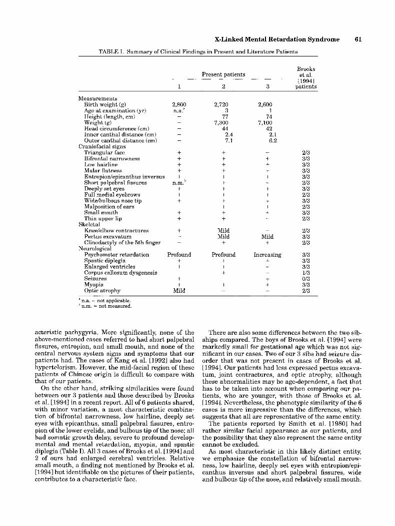
TABLE I. Summary of Clinical Findings in Present and Literature Patients
Present patients
1 2 3
Measurements Birth weight (g) Age at examination (yr) Height (length, cm) Weight (g) Head circumference (cm) Inner canthal distance (cm) Outer canthal distance (cm)
Craniofacial signs Triangular face Bifrontal narrowness Low hairline Malar flatness Entropionlepicanthus inversus Short palpebral fissures Deeply set eyes Full medial eyebrows Widebulbous nose tip Malposition of ears Small mouth Thin upper lip
Kneelelbow contractures Pectus excavatum Clinodactyly of the 5th finger
Psychomotor retardation Spastic diplegia Enlarged ventricles Corpus callosum dysgenesis Seizures Myopia Optic atrophy
Skeletal
Neurological
2,800 n.a.a -
- - -
-
+ + + + + b n.m. + + + + + +
-
-
-
Profound + + + +
Mild
-
2,720 3
77 7,300
44 2.4 7.1
+ + + + + + + + + + + +
Mild Mild
+ Profound +
+ + + -
-
2,600 1
74 7,100
42 2.1 6.2
- + + + + + + + + + + -
-
Mild + Increasing +
- -
+ + -
Brooks et al.
[19941 patients
213 313 313 313 313 213 313 213 313 213 313 213
213 313 213
313 313 313 113 013 313 213
a n.a. = not applicable. n.m. = not measured.
acteristic pachygyria. More significantly, none of the above-mentioned cases referred to had short palpebral fissures, entropion, and small mouth, and none of the central nervous system signs and symptoms that our patients had. The cases of Kang et al. [1992] also had hypertelorism. However, the mid-facial region of these patients of Chinese origin is difficult to compare with that of our patients.
On the other hand, striking similarities were found between our 3 patients and those described by Brooks et al. [19941 in a recent report. All of 6 patients shared, with minor variation, a most characteristic combina- tion of bifrontal narrowness, low hairline, deeply set eyes with epicanthus, small palpebral fissures, entro- pion of the lower eyelids, and bulbous tip of the nose; all had somatic growth delay, severe to profound develop- mental and mental retardation, myopia, and spastic diplegia (Table I). All 3 cases of Brooks et al. [ 19941 and 2 of ours had enlarged cerebral ventricles. Relative small mouth, a finding not mentioned by Brooks et al. [ 19941 but identifiable on the pictures of their patients, contributes to a characteristic face.
There are also some differences between the two sib- ships compared. The boys of Brooks et al. [1994] were markedly small for gestational age which was not sig- nificant in our cases. Two of our 3 sibs had seizure dis- order that was not present in cases of Brooks et al. [ 19941. Our patients had less expressed pectus excava- tum, joint contractures, and optic atrophy, although these abnormalities may be age-dependent, a fact that has to be taken into account when comparing our pa- tients, who are younger, with those of Brooks et al. [1994]. Nevertheless, the phenotypic similarity of the 6 cases is more impressive than the differences, which suggests that all are representative of the same entity.
The patients reported by Smith et al. [19801 had rather similar facial appearance as our patients, and the possibility that they also represent the same entity cannot be excluded.
As most characteristic in this likely distinct entity, we emphasize the constellation of bifrontal narrow- ness, low hairline, deeply set eyes with entropiodepi- canthus inversus and short palpebral fissures, wide and bulbous tip of the nose, and relatively small mouth.

62 Morava et al.
When these anomalies can be found in a boy with oth- erwise unidentified mental retardation, severe develop- mental delay and/or cerebral palsy, this XLMR syn- drome should be considered.
ACKNOWLEDGMENTS This study was supported by a grant (ETT 661/93)
from the Hungarian National Research Fund.
REFERENCES Brooks SS, Wisniewski K, Brown WB (1994): New X-linked mental re-
tardation (XLMR) syndrome with distinct facial appearance and growth retardation. Am J Med Genet 51:586-590.
Hamel BCJ, Mariman CM, vanBeersum SEC, Schoonbrood-Lenssen AMJ, Ropers HH (1994): Mental retardation, congenital heart defect, cleft palate, short stature, and facial anomalies: A new
X-linked multiple congenital anomaliedmental retardation syn- drome. Am J Med Genet 51591-597.
Kang WM, Huang CC, Lin SJ (1992): X-linked recessive inheritance of dysgenesis of corpus callosum in a Chinese family. Am J Med Genet
Neri G, Chiurazzi P, Arena JF, Lubs HA (1994): XLMR genes: Update 1994. Am J Med Genet 51542-549.
Prieto F, Badia L, Mulas F, Mora F (1987): X-linked dysmorphic syn- drome with mental retardation. Clin Genet 32:326-334.
Smith RD, Fineman RM, Myers GG (1980): Short stature, psychomo- tor retardation, and unusual facial appearance in two brothers. Am J Med Genet 7:5-9.
Stall C, Gerauldel A, Chauvin A (1991): New X-linked syndrome of mental retardation, short stature and hypertelorism. Am J Med Genet 39:474-478.
Zollino M, Mastroiacovo P, Zampino G, Neri G (1992): A new XLMR syndrome with characteristic face, hypogenitalism, congenital hy- potonia and dysgenesis of the corpus callosum. Am J Med Genet 43:452-457.
44:619-623.