world journal of - microsoft · ibrahim m salman, new south wales david alan vesey, ... bak-leong...
TRANSCRIPT

World Journal of NephrologyWorld J Nephrol 2017 March 6; 6(2): 59-85
ISSN 2220-6124 (online)
Published by Baishideng Publishing Group Inc
World Journal of NephrologyW J N
EDITORS-IN-CHIEFJosep M Campistol, BarcelonaAnil K Mandal, St. Augustine
ASSOCIATE EDITORSLi Zuo, BeijingHiroyuki Kobori, KagawaHiromichi Suzuki, SaitamaJuan F Navarro-Gonzalez, Santa Cruz de Tenerife
GUEST EDITORIAL BOARD MEMBERS Wen-Chi Chen, TaipeiYi-Wen Chiu, KaohsiungRu-Lan Hsieh, TaipeiYH Hsu, TaipeiPo-Chang Lee, TainanChing-Yuang Lin, TaichungChi-Feng Liu, TaipeiKuo-Cheng Lu, New TaipeiPei-Jung Lu, TainanYee-Yung Ng, TaipeiJunne-Ming Sung, TainanJiunn-Jong Wu, TainanTzung-Hai Yen, Taipei
MEMBERS OF THE EDITORIAL BOARD
AlgeriaKhedidja Mekki, Oran
ArgentinaCarlos Guido Musso, TemperleyHernan Trimarchi, Buenos Aires
L Trumper, RosarioPatricia G Valles, Mendoza
AustraliaNeil C Boudville, WaRobert Gordon Fassett, QueenslandHelen Grania Healy, BrisbaneMohamed Saleem, AdelaideIbrahim M Salman, New South WalesDavid Alan Vesey, BrisbaneHuiling Wu, SydneyGuoping Zheng, Sydney
Belgium
Maarten Naesens, LeuvenBenjamin A Vervaet, Antwerp
Bosnia and Herzegovina
Halima Resic, Sarajevo
Brazil
Liborio Braga Alexandre, FortalezaNiels OS Camara, Cidade UniversitáriaJozélio Freire de Carvalho, Salvador-BahiaSilvia Maria De Oliveira Titan, Sao PauloJose Mario F de Oliveira, Rio de JaneiroMaria Franco, Sao PauloJosé AR Gontijo, CampinasSonia M Oliani, CEP Leonardo O Reis, Unicamp
Nestor Schor, Sao Paulo
Canada
Shao-Bin Duan, ChangshaPaul A Keown, VancouverMarcel Lebel, QuebecOzgur Mete, Ontario
Chile
Guillermo E Lema, Santiago
China
Chia-Chu Chang, ChanghuaFeng Ding, ShanghaiHua-Feng Liu, ZhanjiangFei-Zhang Shou, HangzhouYan-Qing Tong, ChangchunAngela Yee-Moon Wang, Hong KongDan-Xia Zheng, Beijing
Croatia
Dean Markic, RijekaDrasko Pavlovic, ZagrebVladimir Trkulja, Zagreb
Czech Republic
Sylvie Opatrná, Pilsen
I
Editorial Board2016-2019
The World Journal of Nephrology Editorial Board consists of 282 members representing a team of worldwide experts in nephrology. They are from 49 countries, Afghanistan (1), Algeria (1), Argentina (4), Australia (1), Belgium (1), Bosnia and Herzegovina (1), Brazil (10), Canada (4), Chile (1), China (21), Croatia (3), Czech Repoublic (2), Denmark (2), Egypt (6), Finland (1), France (1), Germany (4), Greece (14), Hungary (4), India (12), Iran (9), Ireland (1), Israel (2), Italy (21), Japan (15), Jordan (1), Malaysia (1), Mexico (2), Netherlands (4), Morocco (1), Nigeria (2), Oman (1), Pakistan (2), Palestine (1), Poland (6), Portugal (4), Qatar (1), Romania (1), Serbia (2), Singapore (3), South Africa (1), South Korea (3), Spain (10), Sweden (3), Thailand (5), Turkey (9), United Arab Emirates (1), United Kingdom (11), and United States (59).
March 2, 2016WJN|www.wjgnet.com

Vladimír Tesar, Prague
Denmark
Robert A Fenton, AarhusErling B Pedersen, Holstebro
Egypt
Ahmed I Akl, MansouraMohammad Al-Haggar, MansouraAmgad El Agroudy, BahrainOsama Gheith, MansouraHussein A Sheashaa, MansouraNeveen A Soliman, Cairo
Finland
Sanna Helena Lehtonen, Helsinki
France
Dominique Guerrot, Rouen
Germany
Wolfgang E Jelkmann, LuebeckNadezda Koleganova, HeidelbergJan Menne, HannoverPeter Schemmer, Heidelberg
Greece
Dimitra Bacharaki, RiminiGrapsa Eirini, AthensTheodoros Eleftheriadis, LarissaMoses S Elisaf, IoanninaDimitrios Karakitsos, AthensDimitrios A Kirmizis, ThessalonikiAikaterini Papagianni, ThessalonikiKosmas I Paraskevas, AthensPloumis Stavros Passadakis, AlexandroupolisGiorgos K Sakkas, ThessalyPantelis A Sarafidis, ThessalonikiAristeidis Stavroulopoulos, KallitheaGeorgios Tsaνgalis, Maroussi-attikisParaskevi Tseke, Athens
Hungary
Miklos Zsolt Molnar, BudapestJános Nemcsik, BudapestTaha EL Hadj Othmane, BudapestLaszlo Rosivall, Budapest
India
Sanjay K Agarwal, New DelhiAnish Bhattacharya, ChandigarhSanjay D’Cruz, ChandigarhAmit K Dinda, New Delhi
Vivekanand Jha, ChandigarhMadhu Khullar, Chandigarh Chitra Madiwale, MumbaiShivanand Karopadi Nayak, HyderabadMayoor V Prabhu, MangaloreSidharth Kumar Sethi, HaryanaRajiv Sinha, KolkataKushaljit S Sodhi, Chandigarh
Iran
Mohammadreza Ardalan, TabrizBehzad Einollahi, TehranSeyed-Mohammad Fereshtehnejad, TehranPatricia Khashayar, TehranHamid Tayebi Khosroshahi, TabrizFarzaneh Montazerifar, ZahedanHasan Otukesh, TehranAmir Keshvari Persian, TehranSaeed Taheri, Tehran
Ireland
Harry Holthofer, Dublin
Israel
Farid M Nakhoul, Lower GalileeOded Olsha, Jer United Stateslem
Italy
Gianni Bellomo, FolignoCristina Costa, TorinoPaolo Cravedi, Ranica BergamoBiagio Raffaele Di Iorio, SolofraLuciana Ghio, MilanoAndrea Giusti, GenovaAntonio Granata, AgrigentoFrancesco Locatelli, LeccoLorenzo S Malatino, CataniaGiancarlo Marenzi, MilanPiergiorgio Messa, MilanNicola Perrotti, CatanzaroFrancesco Perticone, CatanzaroGiorgina B Piccoli, TorinoPierangela Presta, CatanzaroClaudio Ronco, VicenzaDomenico Santoro, MessinaRoberto Scarpioni, PiacenzaVincenzo Sepe, PaviaGiovanni Luigi Tripepi, Reggio CalabriaLuca V Valenti, Milano
Japan
Yoshihide Fujigaki, HamamatsuKeiju Hiromura, GunmaKazunari Kaneko, OsakaSatoshi Morimoto, HirakataToshio Nishikimi, KyotoNaro Ohashi, HamamatsuTakashi Oite, NiigataGeorge Seki, Shizuoka prefecture
Akira Shimizu, TokyoKouichi Tamura, YokohamaH Tanaka, HirosakiToru Watanabe, NiigataNoriaki Yorioka, Hiroshima
Jordan
Mohammad Khassawneh, Irbid
Malaysia
Bak-Leong Goh, Kajang
Mexico
Gustavo Martínez-Mier, MéxicoAlejandro Trevino-Becerra, Mexico
Morocco
Faissal Tarrass, Casablanca
Netherlands
Sebastian Dolff, EssenPeter JH Smak Gregoor, DordrechtJoris Hubertus Robben, NijmegenJoris JTH Roelofs, Amsterdam
Nigeria
Martin A Nzegwu, EnuguWasiu Adekunle Olowu, Ile-Ife
Pakistan
Ali AA Lanewala, KarachiMuhammed Mubarak, Karachi
Palestine
Mahmoud M Othman, Nablus
Poland
Alicja E Grzegorzewska, PoznańAndrzej Jaroszynski, KielceJerzy Konstantynowicz, BialystokMariusz Kusztal, WroclawJacek Wiktor Manitiu, BydgoszczMarcin Tkaczyk, Lodz
Portugal
Márcia Carvalho, PortoElísio Costa, Porto
II March 2, 2016WJN|www.wjgnet.com

III March 2, 2016WJN|www.wjgnet.com
La Salete S Martins, OportoManuel Pestana Vasconcelos, Porto
Qatar
Khaled Mohamed Mahmoud, Doha
Romania
Gheorghe Nechifor, Bucharest
Serbia
Amira Peco-Antic, BelgradeRadojica V Stolic, Kragujevac
Singapore
Tan Ban Hock, SingaporeAnselm Mak, SingaporeWoo Keng Thye, Singapore
South Africa
Rajendra Bhimma, Congella
South Korea
Byoung Soo Cho, SeoulChan Kyo Kim, SeoulJae IL Shin, Seoul
Spain
Miguel A Arrabal-Polo, GranadaRicardo J Bosch, Alcala de HenaresJavier Fernandez de Canete, MalagaVictor M Garcia-Nieto, Santa Cruz de TenerifeFrancisco J Lopez-Hernandez, SalamancaAlberto Ortiz, MadridKatia Lopez Revuelta, MadridFernando Santos, Oviedo
Sweden
Peter Bárány, Stockholm
Per Magnusson, Linkoping
Thailand
Pornanong Aramwit, BangkokSinee Disthabanchong, BangkokSomchai Eiam-Ong, BangkokPrasit Futrakul, BangkokViroj Wiwanitkit, Bangkok
Turkey
Turgay Akgul, OsmaniyeFiliz Akyuz, IstanbulMustafa Arici, AnkaraOzgu Aydogdu, NigdeEsra Güzeldemir-Akcakanat, YuvacikMehmet Kanbay, IstanbulSalih Kavukcu, Balcova IzmirAhmet Kiykim, MersinHabibe Sahin Sahin, Kayseri
United Arab Emirates
Bassam Bernieh, Arabe Emirates
United Kingdom
Jacob A Akoh, PlymouthRodney D Gilbert, SouthamptonColin Andrew Hutchison, BirminghamJonathon Olsburgh, LondonDipen S Parikh, DurhamAdrian W Philbey, GlasgowBhusana Premande, LondonBadri M Shrestha, SheffieldNestor Velasco, KilmarnockAlexander Woywodt, PrestonQihe Xu, London
United States
Horacio J Adrogué, HoustonAnil K Agarwal, ColumbusPatrick D Brophy, Hawkins Drive IowaYiqiang Cai, New HavenDaniel J Canter, Atlanta
Oscar A Carretero, DetroitJames CM Chan, PortlandBrian S Decker, IndianapolisJames V Donadio, RochesterAmy C Dwyer, LouisvilleEwa Elenberg, HoustonKevin Finkel, HoustonEA Friedman, BrooklynCA Gadegbeku, Ann ArborClaudia Gragnoli, HersheyParta Hatamizadeh, Ann ArborAdriana M Hung, NashvillePedro A Jose, WashingtonTheodoros Kelesidis, Los AngelesBruce C Kone, HoustonRajesh Kumar, TempleDaniel L Landry, SpringfieldKrista Lentine, St LouisYan Chun Li, ChicagoYouhua Liu, PittsburghMaria-Luisa SS Lopez, Charlottesville John K Maesaka, Mineola Joseph Keith Melancon, WashingtonTibor Nadasdy, ColumbusMacaulay Onuigbo, Eau ClaireIsak Prohovnik, New YorkAmanda C Raff, New YorkArmin Rashidi, NorfolkAnjay Rastogi, Los AngelesMohammed S Razzaque, BostonJeff M Sands, AtlantaMartin J Schreiber, ClevelandGerald Schulman, TennesseeJames Alan Shayman, MichiganAndrey Staruschenko, MilwaukeeAlison Leah Steiber, ClevelandTheodore I Steinman, BostonJames D Stockand, San AntonioMingming Su, KannapolisYunxia Tao, AmarilloGeorge Christos Tsokos, BostonJaime Uribarri, New YorkUlka Vaishampayan, DetroitVolker Vallon, San DiegoPaul A Voziyan, NashvilleBradford Lee West, IllinoisMark E Williams, BostonAnna Woodbury, AtlantaRobert Peter Woroniecki, BronxJ Ruth Wu-Wong, ChicagoDu Yong, HoustonRubin Zhang, LouisianaXin-Jin Zhou, Dallas

W JContents
IWJN|www.wjgnet.com March 6, 2017|Volume 6|Issue 2|
World Journal of NephrologyN
Bimonthly Volume 6 Number 2 March 6, 2017
EDITORIAL59 Applicationofestablishedpathophysiologicprocessesbringsgreaterclaritytodiagnosisandtreatmentof
hyponatremia
Maesaka JK, Imbriano LJ, Miyawaki N
MINIREVIEWS72 Monoclonalgammopathyofrenalsignificance:Diagnosticworkup
Correia SO, Santos S, Malheiro J, Cabrita A, Martins LS, Santos J
ORIGINAL ARTICLE
Retrospective Study
79 Acutekidneyinjuryfollowingspinalinstrumentationsurgeryinchildren
Jöbsis JJ, Alabbas A, Milner R, Reilly C, Mulpuri K, Mammen C

ContentsWorld Journal of Nephrology
Volume 6 Number 2 March 6, 2017
IIWJN|www.wjgnet.com
ABOUT COVER
AIM AND SCOPE
FLYLEAF
March 6, 2017|Volume 6|Issue 2|
EditorialBoardMemberofWorldJournalofNephrology ,RodneyDGilbert,MD,Doctor,DepartmentofNephrology,SouthamptonGeneralHospital,SouthamptonS166YD,UnitedKingdom
World Journal of Nephrology (World J Nephrol, WJN, online ISSN 2220-6124, DOI: 10.5527) is a peer-reviewed open access academic journal that aims to guide clinical practice and improve diagnostic and therapeutic skills of clinicians.
WJN covers topics concerning kidney development, renal regeneration, kidney tumors, therapy of renal disease, hemodialysis, peritoneal dialysis, kidney transplanta-tion, diagnostic imaging, evidence-based medicine, epidemiology and nursing. Priority publication will be given to articles concerning diagnosis and treatment of nephrology diseases. The following aspects are covered: Clinical diagnosis, laboratory diagnosis, dif-ferential diagnosis, imaging tests, pathological diagnosis, molecular biological diagnosis, immunological diagnosis, genetic diagnosis, functional diagnostics, and physical diagno-sis; and comprehensive therapy, drug therapy, surgical therapy, interventional treatment, minimally invasive therapy, and robot-assisted therapy.
We encourage authors to submit their manuscripts to WJN. We will give priority to manuscripts that are supported by major national and international foundations and those that are of great basic and clinical significance.
World Journal of Nephrology is now indexed in PubMed, PubMed Central.
I-III EditorialBoard
INDExING/ABSTRACTING
EDITORS FOR THIS ISSUE
Responsible Assistant Editor: Xiang Li Responsible Science Editor: Jin-Xin KongResponsible Electronic Editor: Huan-Liang Wu Proofing Editorial Office Director: Xiu-Xia SongProofing Editor-in-Chief: Lian-Sheng Ma
www.wjgnet.com/2220-6124/editorialboard.htm
EDITORIALOFFICEXiu-Xia Song, DirectorWorld Journal of NephrologyBaishideng Publishing Group Inc8226 Regency Drive, Pleasanton, CA 94588, USATelephone: +1-925-2238242Fax: +1-925-2238243E-mail: [email protected] Desk: http://www.wjgnet.com/esps/helpdesk.aspxhttp://www.wjgnet.com
PUBLISHERBaishideng Publishing Group Inc8226 Regency Drive, Pleasanton, CA 94588, USATelephone: +1-925-2238242Fax: +1-925-2238243E-mail: [email protected] Desk: http://www.wjgnet.com/esps/helpdesk.aspxhttp://www.wjgnet.com
PUBLICATIONDATEMarch 6, 2017
COPYRIGHT© 2017 Baishideng Publishing Group Inc. Articles pub-lished by this Open-Access journal are distributed under the terms of the Creative Commons Attribution Non-commercial License, which permits use, distribution, and reproduction in any medium, provided the original work is properly cited, the use is non commercial and is otherwise in compliance with the license.
SPECIALSTATEMENTAll articles published in journals owned by the Baishideng Publishing Group (BPG) represent the views and opin-ions of their authors, and not the views, opinions or policies of the BPG, except where otherwise explicitly indicated.
INSTRUCTIONSTOAUTHORShttp://www.wjgnet.com/bpg/gerinfo/204
ONLINESUBMISSIONhttp://www.wjgnet.com/esps/
NAMEOFJOURNALWorld Journal of Nephrology
ISSNISSN 2220-6124 (online)
LAUNCHDATEFebruary 6, 2012
FREQUENCYBimonthly
EDITORS-IN-CHIEFJosep M Campistol, Professor, ICNU Director, Hos-pital Clínic, Universitat de Barcelona, c/Villarroel, 170 ESC 12-5, 08036 Barcelona, Spain
Anil K Mandal, MB, BS, Professor, Department of Medicine, University of Florida, Gainesville, Florida; Man-dal Diabetes Research Foundation, 105 Southpark Blvd., Suite B-202, Saint Augustine, FL 32086, United States
EDITORIALBOARDMEMBERSAll editorial board members resources online at http://
John K Maesaka, Louis J Imbriano, Nobuyuki Miyawaki
EDITORIAL
59 March 6, 2017|Volume 6|Issue 2|WJN|www.wjgnet.com
Application of established pathophysiologic processes brings greater clarity to diagnosis and treatment of hyponatremia
John K Maesaka, Louis J Imbriano, Nobuyuki Miyawaki, Department of Medicine and Division of Nephrology and Hypertension, Winthrop-University Hospital, Mineola, NY 11501, United States
Author contributions: All the authors contributed to this manuscript.
Conflict-of-interest statement: No conflict of interest.
Open-Access: This article is an open-access article which was selected by an in-house editor and fully peer-reviewed by external reviewers. It is distributed in accordance with the Creative Commons Attribution Non Commercial (CC BY-NC 4.0) license, which permits others to distribute, remix, adapt, build upon this work non-commercially, and license their derivative works on different terms, provided the original work is properly cited and the use is non-commercial. See: http://creativecommons.org/licenses/by-nc/4.0/
Manuscript source: Invited manuscript
Correspondence to: John K Maesaka, MD, Department of Medicine and Division of Nephrology and Hypertension, Winthrop-University Hospital, 200 Old Country Road, Suite 135, Mineola, NY 11501, United States. [email protected]: +1-516-6632169Fax: +1-516-6632179
Received: September 20, 2016Peer-review started: September 24, 2016First decision: October 20, 2016Revised: December 6, 2016Accepted: December 27, 2016Article in press: December 28, 2016Published online: March 6, 2017
AbstractHyponatremia, serum sodium < 135 mEq/L, is the most common electrolyte abnormality and is in a
state of flux. Hyponatremic patients are symptomatic and should be treated but our inability to consistently determine the causes of hyponatremia has hampered the delivery of appropriate therapy. This is especially applicable to differentiating syndrome of inappropriate antidiuresis (SIAD) from cerebral salt wasting (CSW) or more appropriately, renal salt wasting (RSW), because of divergent therapeutic goals, to water-restrict in SIAD and administer salt and water in RSW. Differentiating SIAD from RSW is extremely difficult because of identical clinical parameters that define both syndromes and the mindset that CSW occurs rarely. It is thus insufficient to make the diagnosis of SIAD simply because it meets the defined characteristics. We review the pathophysiology of SIAD and RSW, the evolution of an algorithm that is based on determinations of fractional excretion of urate and distinctive responses to saline infusions to differentiate SIAD from RSW. This algorithm also simplifies the diagnosis of hyponatremic patients due to Addison’s disease, reset osmostat and prerenal states. It is a common perception that we cannot accurately assess the volume status of a patient by clinical criteria. Our algorithm eliminates the need to determine the volume status with the realization that too many factors affect plasma renin, aldosterone, atrial/brain natriuretic peptide or urine sodium concentration to be useful. Reports and increasing recognition of RSW occurring in patients without evidence of cerebral disease should thus elicit the need to consider RSW in a broader group of patients and to question any diagnosis of SIAD. Based on the accumulation of supporting data, we make the clinically important proposal to change CSW to RSW, to eliminate reset osmostat as type C SIAD and stress the need for a new definition of SIAD.
Key words: Hyponatremia; Cerebral-renal salt wasting; Fractional excretion of urate
© The Author(s) 2017. Published by Baishideng Publishing
World Journal of NephrologyW J N
Submit a Manuscript: http://www.wjgnet.com/esps/
DOI: 10.5527/wjn.v6.i2.59
World J Nephrol 2017 March 6; 6(2): 59-71
ISSN 2220-6124 (online)

60 March 6, 2017|Volume 6|Issue 2|WJN|www.wjgnet.com
Maesaka JK et al . Fractional excretion of urate in hyponatremia
Group Inc. All rights reserved.
Core tip: When dealing with normo-volemic, non-edematous hyponatremic patients the initial treatment should be i.v. normal saline, combined with measuring the fractional excretion of urate. As serum sodium is corrected, the patients with syndrome of inappropriate antidiuresis (SIAD) will normalize the fractional excretion of urate, while patients with cerebral-renal salt wasting will have a persistently elevated fractional excretion of urate. It appears that patients with SIAD will have a slow or no increase in serum sodium with saline, while patients with renal salt wasting will have a more rapid increase in serum sodium.
Maesaka JK, Imbriano LJ, Miyawaki N. Application of established pathophysiologic processes brings greater clarity to diagnosis and treatment of hyponatremia. World J Nephrol 2017; 6(2): 59-71 Available from: URL: http://www.wjgnet.com/2220-6124/full/v6/i2/59.htm DOI: http://dx.doi.org/10.5527/wjn.v6.i2.59
INTRODUCTIONHyponatremia, defined as a serum sodium < 135 mEq/L, is the most common electrolyte abnormality that is undergoing changes in methodology with the potential of bringing greater clarity to diagnosis and improved therapeutic outcomes. The present approach initiates the work up of a hyponatremic patient by assessing the status of their extracellular volume despite a unanimous agreement that we cannot accurately estimate the volume status of patients by usual clinical criteria[1-3]. Nevertheless, we continue to attempt to determine whether the patient is euvolemic, hypovolemic or hypervolemic while considering other nuances such as urine sodium concentration (UNa), urine osmolality (Uosm), serum osmolality and hormones such as renin, aldosterone and atrial/brain natriuretic peptide. We decided to abandon the very tenuous volume approach to hyponatremia and constructed a new algorithm that will hopefully bring greater clarity to the diagnosis and treatment of hyponatremia in addition to encouraging others to explore other parameters that might resolve many of the controversies that exist today.
In this review, we will discuss the evolution of a new approach that will hopefully create changes that are based on credible and reproducible data. We will stress the complexity and importance of differentiating syndrome of inappropriate antidiuresis (SIAD) from cerebral salt wasting (CSW) or more appropriately renal salt wasting (RSW). The importance of this differentiation can be appreciated by our realization that virtually all patients with hyponatremia are symptomatic and should, therefore, be treated[4-9]. Hyponatremia has created a perfect storm for complications by increasing osteoporosis, especially in an elderly population that is
already undergoing bone demineralization, and inducing a fourfold increase in falls and fractures[6,7,10]. Textbooks and review articles in medicine consider CSW to be a rare clinical entity as compared to neurosurgeons and critical care physicians who consider CSW/RSW to be common. There is thus an urgency to differentiate CSW/RSW from SIAD because of differences in therapeutic goals, to water-restrict in SIAD and administer salt and water in CSW/RSW. Differentiating CSW/RSW from SIAD has been further complicated by having identical key parameters that identify both syndromes (Table 1). Both syndromes present with hyponatremia, hypouricemia, increased fractional excretion of urate (FEurate), concentrated urine, urine sodium usually > 20 mEq/L with normal renal, adrenal and thyroid function (Table 1). The diagnosis of SIAD cannot, therefore, be made merely because it fulfills the criteria used to define SIAD[11]. A major difference between both syndromes is the volume status (euvolemic/hypervolemic in SIAD and hypovolemic in CSW/RSW), the assessment of which is universally agreed to be inaccurate and not very useful; yet we continue to initiate the evaluation of hyponatremic patients by first addressing their volume status. This diagnostic and therapeutic dilemma has been further complicated by our reports of unequivocal cases of RSW occurring without clinical evidence of cerebral disease, which led to our proposal to change CSW to RSW[12-14]. This important change in nomenclature has important clinical implications because RSW would not be con-sidered in the absence of cerebral disease. The true prevalence of RSW is, therefore, not known and cannot be considered a rare entity until a study of a broader population of patients has been conducted.
EvOlUTION Of a NEw algORIThmThe report of hypouricemia, serum urate < 4 mg/dL, with increased FEurate of > 10%, coexisting with hyponatremia in SIAD by Beck in 1979 concluded that the coexistence of hypouricemia and hyponatremia differentiated SIAD from most other causes of hypona-
Findings common to both SIADH and RSW
Association with intracranial disease Hyponatremia Concentrated urine Urine sodium [Na] usually > 20 mEq/L Non-edematous Hypouricemia, with increased fractional excretion urate [FEurate] Only difference between SIADH and RSW Volume state: Normal/high in SIADH, low in RSW
Table 1 Listing the clinical features that are found in syndrome of inappropriate anti-diuretic hormone and cerebral salt wasting/renal salt wasting
Note: How the overlapping features are the most commonly encountered clinically in both syndromes and how the presence of edema can be found in CSW/RSW. SIADH: Syndrome of inappropriate anti-diuretic hormone; RSW: Renal salt wasting; FE: Fractional excretion; CSW: Cerebral salt wasting.

61 March 6, 2017|Volume 6|Issue 2|WJN|www.wjgnet.com
tremia[15]. Interestingly, the increased FEurate in SIAD returned to normal when the hyponatremia was corrected by water restriction[15]. Others not only demonstrated this unique relationship between hypouri-cemia and hyponatremia in SIAD but also noted normali-zation of a previously increased FEurate with correction of hyponatremia by water restriction[15-20]. We encountered a patient with bronchogenic carcinoma and hypouricemia and increased FEurate, concentrated urine, UNa of 42 mEq/L and normal renal, adrenal and thyroid function, who presented with postural hypotension and reflex tachycardia that was consistent with volume depletion. He responded well to saline infusions with a rapid rise in serum sodium but in view of the coexistence of hyponatremia, hypouricemia and increased FEurate a diagnosis of SIAD was made. Despite the absence of cerebral disease and negative CT scan of brain a diagnosis of salt wasting was made because of postural hypotension with reflex tachycardia and a rapid increase in serum sodium. We corrected the hyponatremia by water restriction to determine whether FEurate would normalize or remain increased as postulated. Correcting the hyponatremia by water restriction and salt supplementation resulted in symptoms of hypo-volemia, including return of postural hypotension with reflex tachycardia, postural dizziness, slurred speech, somnolence and staggered gait. The serum sodium finally increased to 138 mEq/L while FEurate remained increased at 14.7%. The clinical course of the patient was highly consistent with volume depletion due to RSW as the persistently increased FEurate after correction of hyponatremia was pathophysiologically different from SIAD. We instead postulated it might be a common feature of RSW[21]. We proceeded to report FEurate to be persistently increased after correction of hyponatremia by water restriction in a patient with meta-static pancreatic carcinoma with ascites, edema and serum albumin of 1.1 mg/dL, bronchogenic carcinoma metastatic to brain, disseminated cryptococcosus with meningitis and uncomplicated Hodgkin’s disease[21]. Absence of cerebral disease in 3 of the 5 patients sug-gested at this time that RSW can occur without clinical evidence of cerebral disease but we waited until we had stronger evidence for RSW to make such a proposal. We
reported hypouricemia, hyponatremia, increased FEurate (many with normonatremia and some after correction of hyponatremia) and cerebral atrophy in patients with various intracranial diseases and in AIDS, 10 of whom had postural hypotension and CVP of 0 cmH2O that were consistent with RSW[22-24]. As a result of these studies, we felt we had enough data to propose differentiating SIAD from RSW by correcting the hyponatremia and observing whether there was normalization of a previously increa-sed FEurate as in SIAD or was persistently increased as in RSW (Figures 1 and 2)[14,25-28]. This unique relationship between FEurate and serum sodium appears to have identified two pathophysiologically different groups of patients.
Reset osmostatWhile attempting to sort out the relationship between FEurate and serum sodium, we encountered hypona-tremic patients who met the criteria for SIAD and RSW but had normal FEurates[29]. Of 14 consecutive hyponatremic patients with normal FEurate, 6 had spon-taneously excreted dilute urines which was diagnostic of a reset osmostat (RO). The remaining 8 patients had a normal water-loading test to prove the diagnosis of RO, 6 of whom had undetectable plasma antidiuretic hormone (ADH) levels at a time when the urine was dilute to prove further the diagnosis of RO (Figure 3)[29]. As noted in Figure 3, type C SIAD represents patients with RO, which was found to make up about 30% of the patients studied[29,30]. Our experience is that many hyponatremic patients admitted to the hospital have intercurrent illnesses that reset their osmostat and normalizes after resolution of their intercurrent illness. This was exem-plified by a patient with a renal transplant who presented with mild hyponatremia of 133 mEq/L on a routine outpatient visit. He later developed a fever for 10 d and was admitted to the hospital with a pneumocystis pneumoniae infection after falling and being confused with a serum sodium of 119 mEq/L. A diagnosis of RO was made by noting spontaneously excreted dilute urine of only 92 mosm/kg and normal FEurate of 7% and 8%. His serum sodium returned to normal one month after successful treatment of his pneumonia. This case illustrates how a slowly evolving pneumonia had
Hyponatremia
FEurate
< 4%Volume depletionAddison’s diseaseEdematous states CHF Cirrhosis Nephrosis
4%-11%Psychogenic polydipsiaReset osmostat
Normonatremia
> 11%
Normonatremia
FEurate < 11% SIADH HCTZ
FEurate > 11%RSW
Figure 1 Proposed algorithm based on determinations of FEurate to evaluate hyponatremic patients without the need to assess the volume status of the patient or determinations of UNa, plasma renin or aldosterone levels. The dotted line connecting normonatremia and RSW with FEurate > 11% needs further verification. Modified from Ref. [28]. SIADH: Syndrome of inappropriate anti-diuretic hormone; RSW: Renal salt wasting; HCTZ: Hydrochlorothiazide; CHF: Congestive heart failure.
Maesaka JK et al . Fractional excretion of urate in hyponatremia

62 March 6, 2017|Volume 6|Issue 2|WJN|www.wjgnet.com
gradually lowered his osmostat and serum sodium[31]. RO, however, can also exist chronically for over 10 years and the persistently normal FEurate is not consistent with the proposal that chronic hyponatremia is responsible for the high FEurate in SIAD[29,32]. While a normal FEurate has effectively identified patients with RO, patients with psychogenic polydipsia also have a normal FEurate, but differentiating RO from psychogenic polydipsia can be readily made by the large volumes of water ingested and polyuria with dilute urines in psychogenic polydipsia (Figure 1)[33]. Not only did this study simplify the dia-gnosis of RO but it provided additional data to support our proposal that FEurate was superior to hypouricemia when evaluating patients with hyponatremia. We have noted normal or increased FEurate with hypouricemia and increased FEurate with normonatremia[22,24,29]. It also provided important pathophysiologic data to pro-pose eliminating RO as a subtype of SIAD by virtue of a normal FEurate and predictable inhibition of ADH secretion by water loading[30].
aDDITIONal CONTRIBUTIONS Of DETERmININg fEURaTEDeterminations of FEurate have also been extremely useful in identifying the cause of hyponatremia in other clinical conditions. In a recent publication, we report a hyponatremic patient with a bronchogenic carcinoma who met the criteria for SIAD and RSW (Table 1). His unresponsiveness to liberal amounts of saline was most
consistent with SIAD so to prove the diagnosis of SIAD we administered 1.5% hypertonic saline to increase serum sodium to 138 mEq/L while observing a gradual decrease in FEurate from 26.2% at baseline, to 11% and 8% (Figure 4)[31]. This maneuver not only provided a potentially convenient way to differentiate SIAD from RSW by correcting the hyponatremia to see if FEurate normalizes as in SIAD or remains persistently increased as in RSW (Figures 1 and 2). It also confirmed our long-held contention that contrary to popular opinion, saline has only a meager effect on FEurate (Table 2)[26,34-36].
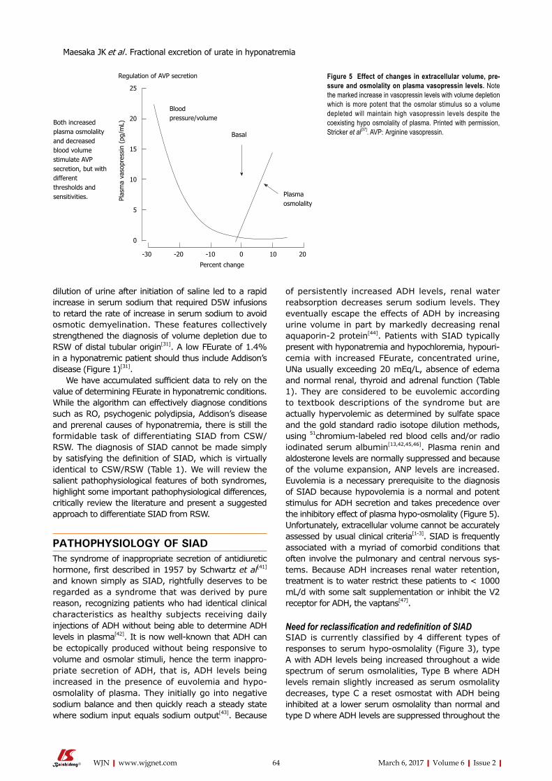
RSW with edemaDetermining FEurate was pivotal in a patient with advanced Hodgkin’s disease who presented with a serum sodium of 127 mEq/L, Uosm of 308 mosm/kg, a 20 pound weight gain over one month with increasing edema of both lower extremities, postural hypotension with reflex tachycardia, ascites, pleural effusion, urine sodium of only 10 mEq/L, decreased cardiac output and normal renal, thyroid and adrenal function[31]. The nephrology attending made a diagnosis of RSW based on the combination of a high FEurate of 17.2% and postural hypotension and reflex tachycardia that was consistent with a volume depleted state. The lower extremity edema was postulated to be due to obstruction of the inferior vena, a finding that was later confirmed by CT scan of the abdomen. Isotonic saline was administered and within 10 h after initiation of saline therapy, the Uosm decreased to 140 mosm/kg when a plasma ADH was undetectable. The dilution of urine and undetectable plasma ADH level illustrated how saline eliminated the more potent volume stimulus for ADH secretion and allowed the coexisting hypo-osmolality to inhibit ADH secretion (Figure 5)[37]. Because of the increase in free water excretion his serum sodium increased by 6 mEq/L within 12 h so D5W was administered to prevent serum sodium from increasing beyond 6 mEq/L per day to prevent osmotic demyelination[38]. On the second day, the medical team, using the unreliable volume approach, made an alternate diagnosis of congestive heart failure that was based on the edema, ascites, pleural effusion, UNa of 10 mEq/L, concentrated urine and decreased cardiac output (actually due to volume depletion) and administered
SIADH RSW
FEUA
SNa
Before After Before After
Correction of hyponatremia
Figure 2 Figure depicting the relationship between serum sodium and FEurate in syndrome of inappropriate anti-diuretic hormone and renal salt wasting. Dotted areas denote normal values. Note FEurate to be increased when patients with SIADH and RSW are hyponatremic but normalize in SIADH and remain increased in RSW. Modified from Ref. [28]. SIADH: Syndrome of inappropriate anti-diuretic hormone; RSW: Renal salt wasting; FEUA: Feurate; SNa: Serum sodium.
FENa (%) FEurate (%) Ref.
Control Exp Control Exp Isotonic 1.04 4.43 7.98 9.76 [36]
1.6 8.2 5.0 5.8 [35] Hypertonic 2.9 18.6 5.4 12.1 [35]
1.4 14.5 12.5 18.7 [34] Hypotonic 1.1 6.1 4.0 7.3 [35]
Table 2 Summary of extracellular volume expansion with isotonic, hypotonic and hypertonic saline on fractional excretion of sodium [FEsodium] and urate [FEurate] at control and experimental periods after saline administration. Note the meager changes in FEurate despite very high FENa
Printed with permission Ref. [26]. Exp: Experimental.
Maesaka JK et al . Fractional excretion of urate in hyponatremia

63 March 6, 2017|Volume 6|Issue 2|WJN|www.wjgnet.com
furosemide. Because the proximal tubule is the main site of natriuretic activity in RSW, see below, the large sodium load that would ordinarily be transported by the distal nephron was inhibited by furosemide resulting in a markedly increased urine output and hemodynamic instability that required large volumes of saline to attain hemodynamic stability. The initial finding of a high FEurate was pivotal in arriving at the diagnosis of RSW because a patient in heart failure and normal kidneys would decrease FEurate to < 4% (Figure 1)[31]. The UNa of only 10 mEq/L was consistent with a prerenal state in which proximal tubule solute reabsorption increases to give rise to low UNa, decreased FEurate of < 4% and the hallmark increase in BUN to creatinine ratio[39]. The low UNa was actually indicative of a decreased appetite and low salt intake, which can occur in RSW and SIAD.
Addison’s diseaseAddison’s disease is a well-known cause of RSW, but in contrast to other causes of RSW, mineralocorticoid deficiency induces a defect in sodium transport in the distal nephron to create a volume depleted state with an intact proximal tubule where urate is exclusively transported[40]. The volume depletion in Addison’s disease due to a defect in sodium transport in the distal tubule leads to the classic prerenal state in which an intact proximal tubule increases solute transport to give rise to the oft-used increase in the BUN to creatinine ratio to characterize a prerenal state[39]. So the low FEurate of 1.4% was a simple means of identifying Addison’s disease as the cause of hyponatremia[31]. The hyperkalemia, high UNa of 140 mEq/L, high ACTH and low cortisol levels accompanied the low FEurate and the
120 130 140 150
12
8
4
0
Plasma sodium (mEq/L)
A B C D
Plas
ma
AVP
(pg/
mL)
Figure 3 Types of the syndrome of inappropriate anti-diuretic hormone. Printed with permission, Ref. [66]. AVP: Arginine vasopressin.
140
135
130
125
120
115
Seru
m s
odiu
m m
mol
/L
4 5 6 7 8 9 10
Hospital day
0.9% NaCl1.5% NaCl
0.9% NaCl
30
25
20
15
10
5
0
Fractional excretion of urate (%)
Serumsodium
Fractionalexcretionof urate
Figure 4 Illustrating how FEurate progressively decreases from an increased rate of 26.2% to a normal of 8% as serum sodium increases to a normal of 138 mEq/L in syndrome of inappropriate anti-diuretic hormone. Normalization of serum sodium did not occur after receiving large volumes of isotonic saline and was possible by using 1.5% hypertonic saline after receiving volumes of isotonic saline. The increase in serum sodium from 131 to 135 mEq/L occurred when he was on very large salt supplementation. This figure also confirms saline does not increase FEurate. Printed with permission, Ref. [31].
Maesaka JK et al . Fractional excretion of urate in hyponatremia
64 March 6, 2017|Volume 6|Issue 2|WJN|www.wjgnet.com
dilution of urine after initiation of saline led to a rapid increase in serum sodium that required D5W infusions to retard the rate of increase in serum sodium to avoid osmotic demyelination. These features collectively strengthened the diagnosis of volume depletion due to RSW of distal tubular origin[31]. A low FEurate of 1.4% in a hyponatremic patient should thus include Addison’s disease (Figure 1)[31].
We have accumulated sufficient data to rely on the value of determining FEurate in hyponatremic conditions. While the algorithm can effectively diagnose conditions such as RO, psychogenic polydipsia, Addison’s disease and prerenal causes of hyponatremia, there is still the formidable task of differentiating SIAD from CSW/RSW. The diagnosis of SIAD cannot be made simply by satisfying the definition of SIAD, which is virtually identical to CSW/RSW (Table 1). We will review the salient pathophysiological features of both syndromes, highlight some important pathophysiological differences, critically review the literature and present a suggested approach to differentiate SIAD from RSW.
PaThOPhySIOlOgy Of SIaDThe syndrome of inappropriate secretion of antidiuretic hormone, first described in 1957 by Schwartz et al[41] and known simply as SIAD, rightfully deserves to be regarded as a syndrome that was derived by pure reason, recognizing patients who had identical clinical characteristics as healthy subjects receiving daily injections of ADH without being able to determine ADH levels in plasma[42]. It is now well-known that ADH can be ectopically produced without being responsive to volume and osmolar stimuli, hence the term inappro-priate secretion of ADH, that is, ADH levels being increased in the presence of euvolemia and hypo-osmolality of plasma. They initially go into negative sodium balance and then quickly reach a steady state where sodium input equals sodium output[43]. Because
of persistently increased ADH levels, renal water reabsorption decreases serum sodium levels. They eventually escape the effects of ADH by increasing urine volume in part by markedly decreasing renal aquaporin-2 protein[44]. Patients with SIAD typically present with hyponatremia and hypochloremia, hypouri-cemia with increased FEurate, concentrated urine, UNa usually exceeding 20 mEq/L, absence of edema and normal renal, thyroid and adrenal function (Table 1). They are considered to be euvolemic according to textbook descriptions of the syndrome but are actually hypervolemic as determined by sulfate space and the gold standard radio isotope dilution methods, using 51chromium-labeled red blood cells and/or radio iodinated serum albumin[13,42,45,46]. Plasma renin and aldosterone levels are normally suppressed and because of the volume expansion, ANP levels are increased. Euvolemia is a necessary prerequisite to the diagnosis of SIAD because hypovolemia is a normal and potent stimulus for ADH secretion and takes precedence over the inhibitory effect of plasma hypo-osmolality (Figure 5). Unfortunately, extracellular volume cannot be accurately assessed by usual clinical criteria[1-3]. SIAD is frequently associated with a myriad of comorbid conditions that often involve the pulmonary and central nervous sys-tems. Because ADH increases renal water retention, treatment is to water restrict these patients to < 1000 mL/d with some salt supplementation or inhibit the V2 receptor for ADH, the vaptans[47].
Need for reclassification and redefinition of SIADSIAD is currently classified by 4 different types of responses to serum hypo-osmolality (Figure 3), type A with ADH levels being increased throughout a wide spectrum of serum osmolalities, Type B where ADH levels remain slightly increased as serum osmolality decreases, type C a reset osmostat with ADH being inhibited at a lower serum osmolality than normal and type D where ADH levels are suppressed throughout the
Both increasedplasma osmolalityand decreasedblood volumestimulate AVPsecretion, but withdifferentthresholds andsensitivities.
Regulation of AVP secretion
Bloodpressure/volume
Basal
Plasmaosmolality
Percent change
-30 -20 -10 0 10 20
Plas
ma
vaso
pres
sin
(pg/
mL)
25
20
15
10
5
0
Figure 5 Effect of changes in extracellular volume, pre-ssure and osmolality on plasma vasopressin levels. Note the marked increase in vasopressin levels with volume depletion which is more potent that the osmolar stimulus so a volume depleted will maintain high vasopressin levels despite the coexisting hypo osmolality of plasma. Printed with permission, Stricker et al[37]. AVP: Arginine vasopressin.
Maesaka JK et al . Fractional excretion of urate in hyponatremia

65 March 6, 2017|Volume 6|Issue 2|WJN|www.wjgnet.com
spectrum of serum osmolalities due to a gain in function of the V2 receptor that is referred to as nephrogenic syndrome of inappropriate antidiuresis[30]. Because the patients selected for this study were considered to be euvolemic by unreliable clinical methods, it is probable that type B SIAD may be renal salt wasters because the patients in this study were given hypertonic saline to increase serum osmolality to normal and then given a water load to determine ADH levels as serum osmolality decreased. The assumption is that these patients had RSW but remained volume depleted to maintain increased levels of ADH despite the hypertonic saline[30]. The classification of type C SIAD has also come under scrutiny because patients with RO have pathophysiologic differences from SIAD by a normal FEurate as compared to being increased in SIAD and noting how lowering serum osmolality appropriately and predictably inhibits ADH secretion[29]. RO should thus be considered a separate clinical syndrome and eliminated as type C SIAD. Elimination of RO as a subtype of SIAD should thus lead to a redefinition of SIAD by eliminating the Uosm of < 100 mosm/kg, since excretion of dilute urines, Uosm < Posm, is extremely unusual in SIAD, vide infra[11,30].
CEREBRal/RENal SalT waSTINgCSW, as first described by Peters et al[48] in 1950, had fallen into disrepute for many years in part because, as previously reviewed, they did not prove RSW in their report. SIAD became the preferred and dominant syndrome. RSW is now accepted as a legitimate synd-rome that exists and is being recognized with grea-ter frequency. It appears that a natriuretic factor, as discussed below, initiates RSW by increasing renal salt and water excretion to a point where the patient becomes volume depleted, the extent being determined by the combination of the severity of the inhibition on sodium transport and salt and water intake. The defect in sodium transport appears to reside in the proximal tubule because urate, which is transported exclusively in the proximal tubule by reabsorbing and secreting transporters, is also reduced[40]. An increase in BUN to creatinine ratio as previously proposed in RSW has not been confirmed by recent reports addressing this problem[12,13]. In RSW, similarities in the BUN to creatinine ratio in SIAD and RSW can be explained by the reduced sodium and water reabsorption in the proximal tubule that cannot establish a urea concentration gradient that is necessary for passive urea reabsorption to occur[49]. The patient initially goes into a state of negative sodium balance, but like SIAD eventually reaches a new steady state where input of salt matches output by activation of humoral, hemodynamic and neuronal factors, but reaches a steady state at a lower extracellular volume. UNa can thus be low if sodium intake is low as can be noted in both SIAD and RSW and is, thus, not a consistently reliable marker of both syndromes. It is extremely unlikely that atrial or brain
natriuretic peptides are responsible for RSW as proposed since their main meager action is in the distal tubule and have been found to be elevated in disparate conditions such as SIAD, subarachnoid hemorrhage (SAH) and salt retaining states such as congestive heart failure and low normal in an unequivocal case of RSW[12].
In contrast to SIAD, ADH levels in RSW are appro-priately increased because the more potent volume stimulus perpetuates the hyponatremia despite the increasing plasma hypo osmolality (Figure 5). As we demonstrated in 4 previous unequivocal cases of RSW, saline eliminates the volume stimulus for ADH secretion to reduce ADH to undetectable levels and allowing the coexistent hypo-osmolality to inhibit ADH secretion, which induces free water excretion by diluting the urine and promptly increasing serum sodium (Figure 6)[12,13,31]. In contrast, saline did not dilute the urine or significantly increase serum sodium in two documented cases of SIAD in whom the diagnosis of SIAD was made by an increased FEurate and hypervolemia as determined by gold standard radioisotope dilution methods by 51chromium-labeled red blood cells and radio iodinated serum albumin (Figure 7)[13]. This same maneuver has been used by others to propose a volume-depleted hyponatremic state if isotonic saline increases serum sodium increases by more than 5 mEq/L and at the same time commenting on our inability to asses accurately the volume status of patients by clinical criteria[1].
Our case of a hyponatremic patient with a hip fracture and no clinical evidence of cerebral disease illustrates the essential features of RSW and proves unequivocally the existence of RSW[12]. When seen in consultation, she was hyponatremic and hyperchloremic with normal renal, adrenal and thyroid function, concentrated urine, Uosm 321 mosm/kg and a UNa of only 6 mEq/L. Her hyponatremia was initially thought to be a due to a volume-depleted state with normal renal function that would give rise to a prerenal state where proximal tubule solute reabsorption would increase to give rise to a low FEurate of usually < 4%[39]. A low serum urate of 3.4 mg/dL led to a determination of FEurate which was elevated at 29.6% and was thus included in our IRB-approved protocol. Her baseline plasma renin, aldosterone and ADH levels were increased, ANP was low normal at 35 pg/mL and blood volume as determined by 51chromium-labeled red blood cells and radio-iodinated serum albumin was reduced by 7.1%[12]. She received isotonic saline and as can be seen in Figure 6, Uosm progressively decreased to dilute levels 13 h after initiation of isotonic saline infusion when plasma ADH was undetectable as would be expected in a hypovolemic, hyponatremic patient with RSW. The excretion of free water promptly increased serum sodium to normal within 48 h when FEurate remained persistently increased (Figure 6)[12]. The patient reported feeling much better and was hungry upon arising 18 h after initiation of isotonic saline infusion, which reflected an improvement of
Maesaka JK et al . Fractional excretion of urate in hyponatremia

66 March 6, 2017|Volume 6|Issue 2|WJN|www.wjgnet.com
her volume depleted state by an erroneous diagnosis of SIAD and fluid restricted for 10 d prior to our intervention. This patient satisfies all of the important features of RSW by the persistence of an elevated FEurate after correction of hyponatremia (Figures 1 and 2), and illustrated the appropriate increase in plasma ADH, elimination of the volume stimulus by saline to permit the coexistent hypo-osmolality to inhibit ADH secretion, excretion of dilute urines and prompt correction of hyponatremia. It also illustrates how RSW can occur without clinical evidence of cerebral disease, how the low ANP is a very unlikely cause of RSW, how fluid restriction of a patient with RSW for an erroneous diagnosis of SIAD can increase morbidity and how a low salt intake can result in low UNa even in RSW (and in SIAD as well). A similar case of RSW associated with an uncomplicated pneumonia and no evidence of cerebral disease responded to isotonic saline infusions by diluting the urine and correcting the hyponatremia within 48 h after initiation of isotonic saline infusions[13]. This case provided us with enough credible data to activate our earlier instincts by proposing to change CSW to RSW[14].
COmmENTS ON NaTRIURESIS IN RSw aND RElaTIONShIP BETwEEN NaTREmIa aND URaTE TRaNSPORTNatriuresis in RSW. The syndrome of RSW has been clearly established from a clinical perspective but the mechanism by which this occurs has not been resolved. The consistency with which we were encountering urate transport abnormalities in RSW suggested that a circulating natriuretic factor, if present, would have its major effect on proximal tubule sodium transport. But because of the ability of the distal tubule to increase sodium transport, we reasoned that sodium excretion induced by a factor affecting proximal sodium transport might be minimized or nullified. To this end we elected to study lithium transport in rats exposed to plasma
from neurosurgical patients who have been reported to have RSW and high FEurates[24,45,46,50]. Without entering into the controversy whether or not lithium is exclusively transported in the proximal tubule, lithium is known to be transported on a 1:1 basis with sodium almost exclusively in the proximal tubule under ambient conditions[51]. We injected the plasma from 21 neurosurgical patients, 14 of whom had FEurates exceeding 10%, and 14 age and gender-matched controls into rats and demonstrated a significant increase in FENa from 0.29% to 0.59% and FElithium from 24% to 36.6%, in controls and neurosurgical patients, respectively[52]).
We extended our interest in patients with increased FEurate with normonatremia by investigating patients with Alzheimer’s disease (AD) who have been reported to be hypouricemic[53]. We studied 18 patients with advanced AD, mini-mental examination scores of < 10, and compared them to 6 patients with multi-infarct or vascular dementia (MID) and 11 normal age and gender-matched controls. All patients were normonatremic except for one hyponatremic patient with AD, FEurate being significantly higher and serum urate lower in AD as compared to the other groups. Infusion of the sera from all groups of patients resulted in FENa of 0.33%, 0.38% and 0.63% and FElithium 27.2%, 31.2% and 41.7% in control, MID and AD, respectively. FENa and FElithium were significantly increased in AD as compared to control and MID. In both the neurosurgical and AD rat studies, the distal sodium load of 36.6% and 41.7% as determined by the FElithium, had a significant sodium uptake by the distal nephron to account for the FENa of only 0.59% and 0.63% in the final urine. The RSW patient with B cell lymphoma obstructing the inferior vena cava had a similarly high distal sodium load that was inhibited by furosemide to generate an exaggerated diuresis that resulted in profound hemodynamic instability[31]. Interestingly, there were no differences in blood pressure or inulin clearances throughout both studies.
Urine Osm and Serum Na
Urine Osm Serum Na
Time (h)
0 10 20 30 40
700
600
500
400
300
200
100
Urin
e O
sm (
mos
m/k
g)
Serum N
a (mm
Eq/L)
140
138
136
134
132
130
128
Figure 6 Changes in urine osmolality and serum sodium concentration during isotonic infusion in a hyponatremic patient with a hip fracture without clinical evidence of cerebral disease. The low Uosm at baseline reflected a low sodium intake when UNa was only 6 mEq/L and a weakened medullary solute concentration, which increased rapidly with a marked increase in Uosm after 4 h of saline infusion. Plasma antidiuretic hormone (ADH) was increased with increased plasma renin and aldosterone levels at baseline and ADH level was undetectable when the urine was dilute 13 h with increase in serum sodium to 138 mEq/L within 48 h after initiation of isotonic saline infusion. Printed with permission, Ref. [12].
Maesaka JK et al . Fractional excretion of urate in hyponatremia

67 March 6, 2017|Volume 6|Issue 2|WJN|www.wjgnet.com
In a third group of studies, proteins were purified from ammonium sulfate precipitates of urine proteins and placed in transwells to determine transport of radioactive sodium, 22Na, across cultured porcine proximal tubule cells, LLC-PK1 cells, grown to confluency on a semipermeable membrane. Urine proteins from normonatremic neurosurgical patients with increased FEurate inhibited 22Na transport across cultured LC-PK1 cells as compared to those with normal FEurate[54].
These studies demonstrate the presence of a natriuretic factor in the plasma and urine of normona-tremic neurosurgical patients with increased FEurate and in sera of normonatremic AD patients with increased FEurate. The natriuretic factor has its major effect on proximal tubule sodium transport to support our pro-posal that a high FEurate in the presence of normona-tremia might be a marker of RSW without going through a phase of hyponatremia (Figure 1). Future studies must address this interesting possibility. Atrial or brain natriuretic peptides are extremely unlikely as the natriuretic factor in RSW because their main site of action is in the inner medullary collecting duct[55].
Relationship between natremia and FEurateThe intriguing relationship between serum sodium and FEurate has been uniquely coupled in many hypona-tremic and non hyponatremic conditions such as RSW. Because the natriuretic factor present in the plasma of patients with RSW affect sodium transport mainly in the proximal tubule, it would be interesting to speculate that the natriuretic factor might also affect reabsorbing and/or secretory transporters or anion exchangers for urate in the proximal tubule[40]. This circulating factor can have effects on the sodium and urate transporters regardless of whether the patient is hyponatremic or normonatremic.
The relationship between sodium and urate in SIAD continues to elude any rational explanations. It can be readily understood why FEurate remains persistently increased in the presence of normonatremia in RSW, but in SIAD, normalization of FEurate after correction
of hyponatremia has not been fully explained. Some have implicated the V1 receptor activity of pitressin to explain the increase in FEurate in SIAD but others were able to induce SIAD with increased FEurate in healthy volunteers by DDAVP which lacks any V1 activity. In addition, the V1 activity of pitressin is an unlikely cause of the increase in FEurate in SIAD because pitressin levels are still increased when FEurate normalizes after correction of hyponatremia[56]. The same group has made commendable efforts to explain the increase in FEurate in SIAD by implicating chronic hyponatremia, but the normal FEurate reported in psychogenic polydipsia and reset osmostat where hyponatremia has been documented for up to 10 years do not support such an hypothesis[32]. At the present time, the relation-ship between serum sodium and FEurate remains unexplained in SIAD.
Prevalence of RSWThe prevalence of RSW in hyponatremic patients is presently unknown because of multiple factors: (1) the term CSW confined RSW to patients with cerebral disease; (2) textbooks and review articles in internal medicine consistently consider CSW to be a rare entity or is not included as a clinical entity of hyponatremia; (3) utilizing the ineffective volume approach to hypona-tremia; and (4) lack of application of the pathophy-siologic characteristics in the evaluations of SIAD and RSW. As noted in Table 1, overlapping of key clinical parameters that characterize SIAD and RSW makes it extremely difficult to differentiate one syndrome from the other. One key difference is to determine the volume status of the patient by credible means at a time when they are hyponatremic. There are 3 studies in hyponatremic and normonatremic neurosurgical patients that determined intravascular volume by gold standard radio isotope dilution methods, using 51chrome-labeled red blood cells and/or radio iodinated serum albumin. As can be seen in Table 3, 83% and 94% of patient with UNa exceeding 40 mEq/L and a variety of neurosurgical diseases had decreased blood volumes that were
Uos
m (
mos
m/k
g)
SNa (m
mol/L)
600
500
400
300
200
100
0
140
138
136
134
132
130
128
126
124
122
1200 12 24 36 48 60 72 84 96 108 120
Time (h)
Uosm SNa
Figure 7 Effect of isotonic infusion in patient with syndrome of inappropriate anti-diuretic hormone in whom blood volume determination by 51chromium-labeled red blood cells and radio iodinated serum albumin was increased. Note persistently increased urine osmolality or failure of urine to become dilute or correction of hyponatremia which are common features of SIAD response to isotonic saline infusion. Printed with permission, Ref. [13]. SIADH: Syndrome of inappropriate anti-diuretic hormone; SNa: Serum sodium; Uosm: Urine osmolality.
Maesaka JK et al . Fractional excretion of urate in hyponatremia

68 March 6, 2017|Volume 6|Issue 2|WJN|www.wjgnet.com
consistent with CSW/RSW. By comparison only 3 of the 39 patients studied had increased blood volumes that were consistent with SIAD[45,46,50]. Interestingly, 67% of normonatremic patients with SAH had decreased blood volumes that were consistent with RSW while only 33% had increased volume consistent with SIAD. RSW occurring in normonatremic patients is consistent with the popular view of CSW/RSW but we would like further clarification of whether normonatremia in the presence of a high FEurate is indicative of RSW (Figure 1). Fluid restricting patients with SAH was harmful by increasing morbidity and mortality so it is customary to administer saline to patients with SAH because of the prevalence of RSW in patients with SAH[57].
These volume studies in neurosurgical patients over-came the most important deficiency when trying to differentiate SIAD from RSW, but are rarely considered when discussing CSW. Instead, a retrospective study of patients with SAH where CSW was found in only 6.5% of patients with hyponatremia, the majority being SIAD, is cited to prove the rarity of CSW/RSW[58,59]. In two retrospective studies they determined without defining their method of analysis that 4.8% and 2.7% of patients had combined CSW and SIAD, a highly incongruous combination that might occur, but would be extremely difficult to prove, especially in a retrospective study[59,60]. We critically reviewed many flawed articles on CSW/RSW in a previous publication and feel it is important to comment on a more recent prospective study of hyponatremia in patients with SAH by the same group which concluded that the hyponatremia was again due to SIAD[28,61]. The diagnosis of SIAD was made because they fulfilled the definition of SIAD without commenting on the same definition being applicable to RSW. The accompanying editorial to this manuscript agreed with the findings in this large study and alluded to the rarity of CSW[62]. A critical review of this manuscript reveals that the authors followed the recommendation to administer saline rather than fluid-restrict hyponatremic patients with SAH[57]. Virtually every patient received isotonic saline from the time of admission to the hospital, none were fluid restricted and none received hypertonic saline or a vaptan class of drugs at any time.
It is intriguing to note that 36 of 49 patients developed hyponatremia within 3 d after SAH. This unaddressed outcome raises questions as to whether the patients received free water or desalinated when UNa exceeded 150 mEq/L while receiving isotonic saline[63]. Every patient corrected their hyponatremia during the hospitalization with the median time of correction being 3 d. Because of the correction of hyponatremia, they concluded that the “syndrome” was short lived. It is well known that patients with SIAD do not respond to saline infusions to correct their hyponatremia; otherwise there would be no need for fluid restriction or treatment with the vaptan class of drugs. Instead, a more likely diagnosis is RSW, which is consistent with the volume studies performed in patients with SAH (Table 3)[46]. Isotonic saline eliminated the volume stimulus for ADH secretion and permitted the coexisting hypo-osmolality to inhibit ADH secretion, excrete dilute urines and predictably correct the hyponatremia, which does not happen in SIAD[12,13,31]. ADH levels would have been undetectable at the time of excretion of dilute urines as we demonstrated in cases of RSW within 24-48 h after initiation of isotonic saline therapy[12,31]. We contend that these patients would have had high FEurates exceeding 11% and would have remained increased when serum sodium returned to normal (Figures 1 and 2). Correction of hyponatremia would not mean there was resolution of the underlying RSW. The reader is encouraged to read a more thorough critical review of controversial papers in the literature[28].
Differentiating SIAD from RSWAs noted in Table 1 and Figures 1 and 2, SIAD and RSW share common clinical parameters including an increased FEurate > 11%. An increased FEurate most often involves RSW and SIAD with hydrochlorothiazide (HCTZ) and possibly selective serotonin reuptake inhibitors (SSRIs) drugs coming into the picture. Discontinuation of HCTZ and SSRIs should result in correction of the hyponatremia but persistence of the hyponatremia and increased FEurate would indicate that SIAD must be differentiated from RSW in order to arrive at the proper therapeutic strategy of water-restricting or administering V2 receptor blockers for SIAD or administer saline for RSW. We have now accumulated enough data to utilize two strategies to differentiate SIAD from RSW. The first is to correct the hyponatremia by either water-restriction or use of hypertonic saline to determine whether FEurate normalizes as in SIAD or remains increased as in RSW (Figures 1, 2 and 4). It is important to note that contrary to common perceptions, saline has a very meager effect on FEurate as noted in Figure 4, Table 2. This is illustrated by Figure 4 when large volumes of saline, including hypertonic saline, gradually decreased FEurate from 26.2% to 8% as serum sodium increased to normal to confirm the diagnosis of SIAD[31].
The other alternative is to administer saline soon after FEurate has been shown to be increased, > 11%. As discussed above, the urine will usually become dilute
Ref. n of patients
Low blood volumeRSW
Increased blood
volumeSIADH
Urine Na mEq/L
Nelson et al[45] HN 12 10 (83%) 2 41-203 Wijdicks et al[46] HN 9 8 (89%) 1 -- NN 12 8 (67%) 4 Sivakumar et al[50] HN 18 17 (94%) 43-210
Table 3 Summary of volume studies by gold standard radio-isotope dilution methods in hyponatremic and normonatremic neurosurgical patients[45,46,50]
RSW much more common than SIAD. Printed with permission, Ref. [26]. SIADH: Syndrome of inappropriate anti-diuretic hormone; RSW: Renal salt wasting; HN: Hyponatremic; NN: Normonatremic.
Maesaka JK et al . Fractional excretion of urate in hyponatremia

69 March 6, 2017|Volume 6|Issue 2|WJN|www.wjgnet.com
within the first 24-48 h after initiation of saline infusion in RSW with a rapid increase in serum sodium which should not increase by more than 4-6 mEq/L per 24 h to prevent osmotic demyelination[38]. The rapid increase can be slowed by infusing D5W or administering DDAVP intranasally realizing that the recommended correction to prevent osmotic demyelination is based on 24 h and not less[38]. In a thorough review of the literature, we have found dilution of urine in SIAD only under two conditions, infusing isotonic saline at a rate of 16 mL/min for 2 h in the first case of SIAD reported in 1957 and extreme salt restriction in experimentally induced SIAD in normal human subjects[42,43]. It would appear therefore that the differentiation of SIAD from RSW can be accomplished by noting normalization of a previously increased FEurate as in SIAD or persistence of increased FEurate in RSW after correction of hyponatremia and response to saline infusions (Figures 1, 2, 6 and 7).
By utilizing both strategies and the algorithm in Table 2 and Figure 3 in a completed study of 52 patients with hyponatremia outside of the neurosurgical intensive care unit, we found about an equal number of patients with SIAD, RO and RSW with the majority of patients with RSW to be free of clinical evidence of cerebral disease. The surprising large number of patients with RSW and absence of cerebral disease in more than 80% of these patients supports our previous proposal to change CSW to RSW[14]. This change has enormous clinical relevance because RSW would not be considered in the absence cerebral disease[64].
FEphoshateWe have previously stated that an increased FEpho-sphate > 20% at baseline is consistent with RSW[21]. We have encountered only one patient with an increased FEphosphate with RSW, but while it might be potentially useful in differentiating SIAD from RSW, there are pitfalls that can alter its value. We have demonstrated in a previous renal micropuncture study that phosphate transport is very sensitive to saline infusions, because parathyroid hormone (PTH) increases rapidly as calcium and possibly magnesium decrease in serum to stimulate PTH release[65]. Since saline is frequently administered to patients with hyponatremia, determinations of FEphosphate must be performed at baseline or analyzed according to whether or not the patient was receiving saline at the time the test was performed.
CONClUSIONHyponatremia, the most common electrolyte, is under-going fundamental changes that would benefit from a new mindset of abandoning the volume approach. We present an algorithm based on supportive data where the determination of FEurate has been helpful in arriving at a more accurate diagnosis of the causes of hyponatremia. The realization that hyponatre-mic patients are symptomatic has led to the recom-mendation to treat virtually all hyponatremics, thus
creating an urgency to differentiate SIAD from RSW because of divergent therapeutic goals. The clinical utility of applying two distinctive pathophysiologic characteristics to distinguish SIAD from RSW, such as: (1) demonstrating normalization of a previously increased FEurate in SIAD and persistent increase in FEurate as in RSW after correction of hyponatremia by water restriction or hypertonic saline or; (2) to note whether infusion of isotonic saline induces excretion of dilute urines with a prompt increase in serum sodium as in RSW or continued excretion of concentrated urines without correction of hyponatremia as in SIAD. Utilization of this pathophysiologic approach has uncovered many with RSW without cerebral disease. So contrary to popular perceptions of the rarity of CSW/RSW, it is probable that the increase in morbidity and mortality associated with hyponatremia may in part be iatrogenic because patients with RSW are being fluid-restricted for an erroneous diagnosis of SIAD. Finally, we feel it is time to abandon the volume approach to patients with hyponatremia and to appreciate the unre-liability of determining plasma renin and aldosterone levels because of so many circumstances such as the use of ACE inhibitor, angiotensin II receptor blockers or saline that affect their blood levels, and to be aware of the following comments to which we have supporting data: (1) have less reliance on UNa in RSW and SIAD as sodium excretion is dependent on sodium intake; (2) the misconceptions of having an increased BUN to creatinine ratio in RSW when there is no difference; (3) appreciate the remarkable overlapping of common clinical characteristics between SIAD and RSW (Table 1); and (4) because of the expanding prevalence of RSW occurring in all hyponatremic patients, we must be vigilant in insisting on clearly distinguishing SIAD from RSW in the future and to question previous reports on SIAD, realizing that RSW could be included in significant numbers. We feel we have accumulated enough data to support these final comments and to encourage others to expand our efforts to derive the true prevalence of RSW in a broad population of hyponatremic and normonatremic patients.
REfERENCES1 Chung HM, Kluge R, Schrier RW, Anderson RJ. Clinical assessment
of extracellular fluid volume in hyponatremia. Am J Med 1987; 83: 905-908 [PMID: 3674097 DOI: 10.1016/0002-9343(87)90649-8]
2 Singh S, Bohn D, Carlotti AP, Cusimano M, Rutka JT, Halperin ML. Cerebral salt wasting: truths, fallacies, theories, and challenges. Crit Care Med 2002; 30: 2575-2579 [PMID: 12441772 DOI: 10.1097/00003246-200211000-00028]
3 Oh MS, Carroll HJ. Cerebral salt-wasting syndrome. We need better proof of its existence. Nephron 1999; 82: 110-114 [PMID: 10364701 DOI: 10.1159/000045385]
4 Decaux G. Is asymptomatic hyponatremia really asymptomatic? Am J Med 2006; 119: S79-S82 [PMID: 16843090 DOI: 10.1016/j.amjmed.2006.05.013]
5 Gankam Kengne F, Andres C, Sattar L, Melot C, Decaux G. Mild hyponatremia and risk of fracture in the ambulatory elderly. QJM 2008; 101: 583-588 [PMID: 18477645 DOI: 10.1093/qjmed/
Maesaka JK et al . Fractional excretion of urate in hyponatremia
70 March 6, 2017|Volume 6|Issue 2|WJN|www.wjgnet.com
hcn061]6 Hoorn EJ, van der Lubbe N, Zietse R. SIADH and hyponatraemia:
why does it matter? NDT Plus 2009; 2: iii5-iii11 [PMID: 19881934]7 Renneboog B, Musch W, Vandemergel X, Manto MU, Decaux G.
Mild chronic hyponatremia is associated with falls, unsteadiness, and attention deficits. Am J Med 2006; 119: 71.e1-71.e8 [PMID: 16431193 DOI: 10.1016/j.amjmed.2005.09.026]
8 Schrier RW. Does ‘asymptomatic hyponatremia’ exist? Nat Rev Nephrol 2010; 6: 185 [PMID: 20348927 DOI: 10.1038/nrne-ph.2010.21]
9 Arieff AI, Llach F, Massry SG. Neurological manifestations and morbidity of hyponatremia: correlation with brain water and electrolytes. Medicine (Baltimore) 1976; 55: 121-129 [PMID: 1256311 DOI: 10.1097/00005792-197603000-00002]
10 Verbalis JG, Barsony J, Sugimura Y, Tian Y, Adams DJ, Carter EA, Resnick HE. Hyponatremia-induced osteoporosis. J Bone Miner Res 2010; 25: 554-563 [PMID: 19751154 DOI: 10.1359/jbmr.090827]
11 Janicic N, Verbalis JG. Evaluation and management of hypo-osmolality in hospitalized patients. Endocrinol Metab Clin North Am 2003; 32: 459-481, vii [PMID: 12800541 DOI: 10.1016/S0889-8529(03)00004-5]
12 Maesaka JK, Miyawaki N, Palaia T, Fishbane S, Durham JH. Renal salt wasting without cerebral disease: diagnostic value of urate determinations in hyponatremia. Kidney Int 2007; 71: 822-826 [PMID: 17311074 DOI: 10.1038/sj.ki.5002093]
13 Bitew S, Imbriano L, Miyawaki N, Fishbane S, Maesaka JK. More on renal salt wasting without cerebral disease: response to saline infusion. Clin J Am Soc Nephrol 2009; 4: 309-315 [PMID: 19201917 DOI: 10.2215/CJN.02740608]
14 Maesaka JK, Imbriano LJ, Ali NM, Ilamathi E. Is it cerebral or renal salt wasting? Kidney Int 2009; 76: 934-938 [PMID: 19641485 DOI: 10.1038/ki.2009.263]
15 Beck LH. Hypouricemia in the syndrome of inappropriate secretion of antidiuretic hormone. N Engl J Med 1979; 301: 528-530 [PMID: 460306 DOI: 10.1056/NEJM197909063011005]
16 Assadi FK, John EG. Hypouricemia in neonates with syndrome of inappropriate secretion of antidiuretic hormone. Pediatr Res 1985; 19: 424-427 [PMID: 4000768 DOI: 10.1203/00006450-198505000-00003]
17 Dorhout Mees EJ, Blom van Assendelft P, Nieuwenhuis MG. Elevation of uric aicd clearance caused by inappropriate antidiuretic hormone secretion. Acta Med Scand 1971; 189: 69-72 [PMID: 5121533 DOI: 10.1111/j.0954-6820.1971.tb04340.x]
18 Sonnenblick M, Rosin A. Increased uric acid clearance in the syndrome of inappropriate secretion of antidiuretic hormone. Isr J Med Sci 1988; 24: 20-23 [PMID: 3346144]
19 Weinberger A, Santo M, Solomon F, Shalit M, Pinkhas J, Sperling O. Abnormality in renal urate handling in the syndrome of inappropriate secretion of antidiuretic hormone. Isr J Med Sci 1982; 18: 711-713 [PMID: 7107209]
20 Sørensen JB, Osterlind K, Kristjansen PE, Hammer M, Hansen M. Hypouricemia and urate excretion in small cell lung carcinoma patients with syndrome of inappropriate antidiuresis. Acta Oncol 1988; 27: 351-355 [PMID: 2849462 DOI: 10.3109/02841868809093553]
21 Maesaka JK, Batuman V, Yudd M, Salem M, Sved AF, Venkatesan J. Hyponatremia and hypouricemia: differentiation from SIADH. Clin Nephrol 1990; 33: 174-178 [PMID: 2350904]
22 Maesaka JK, Cusano AJ, Thies HL, Siegal FP, Dreisbach AW. Hypouricemia in acquired immunodeficiency syndrome. Am J Kidney Dis 1990; 15: 252-257 [PMID: 2305765 DOI: 10.1016/S0272-6386(12)80770-0]
23 Cusano AJ, Thies HL, Siegal FP, Dreisbach AW, Maesaka JK. Hyponatremia in patients with acquired immune deficiency syndrome. J Acquir Immune Defic Syndr 1990; 3: 949-953 [PMID: 2398458]
24 Maesaka JK, Venkatesan J, Piccione JM, Decker R, Dreisbach AW, Wetherington JD. Abnormal urate transport in patients with intracranial disease. Am J Kidney Dis 1992; 19: 10-15 [PMID: 1739076 DOI: 10.1016/S0272-6386(12)70196-8]
25 Maesaka JK. An expanded view of SIADH, hyponatremia and hypouricemia. Clin Nephrol 1996; 46: 79-83 [PMID: 8869783]
26 Maesaka JK, Fishbane S. Regulation of renal urate excretion: a critical review. Am J Kidney Dis 1998; 32: 917-933 [PMID: 9856507 DOI: 10.1016/S0272-6386(98)70067-8]
27 Maesaka JK, Gupta S, Fishbane S. Cerebral salt-wasting syndrome: does it exist? Nephron 1999; 82: 100-109 [PMID: 10364700 DOI: 10.1159/000045384]
28 Maesaka JK, Imbriano L, Shirazian S, Miyawaki N. Complexity of differentiating cerebral-renal salt wasting from SIAD, emerging importance of determining fractional urate excretion. In: Vijayakumar S, editor. Novel Insights on chronic kidney disease, acute kidney injury and polycystic kidney disease. Croatia: InTech, 2012: 41-66
29 Imbriano LJ, Ilamathi E, Ali NM, Miyawaki N, Maesaka JK. Normal fractional urate excretion identifies hyponatremic patients with reset osmostat. J Nephrol 2012; 25: 833-838 [PMID: 22307440 DOI: 10.5301/jn.5000074]
30 Zerbe R, Stropes L, Robertson G. Vasopressin function in the syndrome of inappropriate antidiuresis. Annu Rev Med 1980; 31: 315-327 [PMID: 6772090 DOI: 10.1146/annurev.me.31.020180.001531]
31 Imbriano LJ, Mattana J, Drakakis J, Maesaka JK. Identifying Different Causes of Hyponatremia With Fractional Excretion of Uric Acid. Am J Med Sci 2016; 352: 385-390 [PMID: 27776720 DOI: 10.1016/j.amjms.2016.05.035]
32 Decaux G, Prospert F, Soupart A, Musch W. Evidence that chronicity of hyponatremia contributes to the high urate clearance observed in the syndrome of inappropriate antidiuretic hormone secretion. Am J Kidney Dis 2000; 36: 745-751 [PMID: 11007676 DOI: 10.1053/ajkd.2000.17623]
33 Ali N, Imbriano LJ, Maesaka JK. The Case | A 66-year-old male with hyponatremia. Psychogenic polydipsia. Kidney Int 2009; 76: 233-234 [PMID: 19564861 DOI: 10.1038/ki.2009.150]
34 Cannon PJ, Svahn DS, Demartini FE. The influence of hypertonic saline infusions upon the fractional reabsorption of urate and other ions in normal and hypertensive man. Circulation 1970; 41: 97-108 [PMID: 5420637 DOI: 10.1161/01.CIR.41.1.97]
35 Diamond H, Meisel A. Influence of volume expansion, serum sodium, and fractional excretion of sodium on urate excretion. Pflugers Arch 1975; 356: 47-57 [PMID: 1238979 DOI: 10.1007/BF00583520]
36 Steele TH. Evidence for altered renal urate reabsorption during changes in volume of the extracellular fluid. J Lab Clin Med 1969; 74: 288-299 [PMID: 5799512]
37 Stricker EM, Verbalis J. Water intake and body fluids. In: Squire LE, Roberts JL, Spitzer NC, Zigmond MJ, McConnell SK, Bloom FE, editors. Fundamental Neuroscience. 2nd ed. California: Elsevier, 2003: 1011-1029
38 Sterns RH. Disorders of plasma sodium--causes, consequences, and correction. N Engl J Med 2015; 372: 55-65 [PMID: 25551526 DOI: 10.1056/NEJMra1404489]
39 Abuelo JG. Normotensive ischemic acute renal failure. N Engl J Med 2007; 357: 797-805 [PMID: 17715412 DOI: 10.1056/NEJMra064398]
40 Lipkowitz MS. Regulation of uric acid excretion by the kidney. Curr Rheumatol Rep 2012; 14: 179-188 [PMID: 22359229 DOI: 10.1007/s11926-012-0240-z]
41 Schwartz WB, Bennett W, Curelop S, Bartter FC. A syndrome of renal sodium loss and hyponatremia probably resulting from inappropriate secretion of antidiuretic hormone. Am J Med 1957; 23: 529-542 [PMID: 13469824 DOI: 10.1016/0002-9343(57)90224-3]
42 Leaf A, Bartter FC, Santos RF, Wrong O. Evidence in man that urinary electrolyte loss induced by pitressin is a function of water retention. J Clin Invest 1953; 32: 868-878 [PMID: 13084753 DOI: 10.1172/JCI102805]
43 Jaenike JR, Waterhouse C. The renal response to sustained administration of vasopressin and water in man. J Clin Endocrinol Metab 1961; 21: 231-242 [PMID: 13789146 DOI: 10.1210/jcem-21-3-231]
44 Verbalis JG, Murase T, Ecelbarger CA, Nielsen S, Knepper MA.
Maesaka JK et al . Fractional excretion of urate in hyponatremia

71 March 6, 2017|Volume 6|Issue 2|WJN|www.wjgnet.com
Studies of renal aquaporin-2 expression during renal escape from vasopressin-induced antidiuresis. Adv Exp Med Biol 1998; 449: 395-406 [PMID: 10026831 DOI: 10.1007/978-1-4615-4871-3_51]
45 Nelson PB, Seif SM, Maroon JC, Robinson AG. Hyponatremia in intracranial disease: perhaps not the syndrome of inappropriate secretion of antidiuretic hormone (SIADH). J Neurosurg 1981; 55: 938-941 [PMID: 7299468 DOI: 10.3171/jns.1981.55.6.0938]
46 Wijdicks EF, Vermeulen M, ten Haaf JA, Hijdra A, Bakker WH, van Gijn J. Volume depletion and natriuresis in patients with a ruptured intracranial aneurysm. Ann Neurol 1985; 18: 211-216 [PMID: 4037761 DOI: 10.1002/ana.410180208]
47 Berl T, Quittnat-Pelletier F, Verbalis JG, Schrier RW, Bichet DG, Ouyang J, Czerwiec FS. Oral tolvaptan is safe and effective in chronic hyponatremia. J Am Soc Nephrol 2010; 21: 705-712 [PMID: 20185637 DOI: 10.1681/ASN.2009080857]
48 Peters JP, Welt LG, Sims EA, Orloff J, Needham J. A salt-wasting syndrome associated with cerebral disease. Trans Assoc Am Physicians 1950; 63: 57-64 [PMID: 14855556]
49 Palmer BF. Hyponatremia in patients with central nervous system disease: SIADH versus CSW. Trends Endocrinol Metab 2003; 14: 182-187 [PMID: 12714279 DOI: 10.1016/S1043-2760(03)00048-1]
50 Sivakumar V, Rajshekhar V, Chandy MJ. Management of neuro-surgical patients with hyponatremia and natriuresis. Neurosurgery 1994; 34: 269-274; discussion 274 [PMID: 8177388 DOI: 10.1227/00006123-199402000-00010]
51 Hayslett JP, Kashgarian M. A micropuncture study of the renal handling of lithium. Pflugers Arch 1979; 380: 159-163 [PMID: 573443 DOI: 10.1007/BF00582152]
52 Maesaka JK, Venkatesan J, Piccione JM, Decker R, Dreisbach AW, Wetherington J. Plasma natriuretic factor(s) in patients with intracranial disease, renal salt wasting and hyperuricosuria. Life Sci 1993; 52: 1875-1882 [PMID: 8502124 DOI: 10.1016/0024-3205(93)90008-Q]
53 Maesaka JK, Wolf-Klein G, Piccione JM, Ma CM. Hypouricemia, abnormal renal tubular urate transport, and plasma natriuretic factor(s) in patients with Alzheimer’s disease. J Am Geriatr Soc 1993; 41: 501-506 [PMID: 8486882 DOI: 10.1111/j.1532-5415.1993.tb01885.x]
54 Youmans SJ, Fein MR, Wirkowski E, Maesaka JK. Demonstration of natriuretic activity in urine of neurosurgical patients with renal salt wasting. F1000Res 2013; 2: 126 [PMID: 24358843 DOI: 10.12688/f1000research.2-126.v2]
55 Bełtowski J, Wójcicka G. Regulation of renal tubular sodium transport by cardiac natriuretic peptides: two decades of research. Med Sci Monit 2002; 8: RA39-RA52 [PMID: 11859295]
56 Decaux G, Namias B, Gulbis B, Soupart A. Evidence in hyponatremia related to inappropriate secretion of ADH that V1 receptor stimulation
contributes to the increase in renal uric acid clearance. J Am Soc Nephrol 1996; 7: 805-810 [PMID: 8738818]
57 Wijdicks EF, Vermeulen M, Hijdra A, van Gijn J. Hyponatremia and cerebral infarction in patients with ruptured intracranial aneurysms: is fluid restriction harmful? Ann Neurol 1985; 17: 137-140 [PMID: 3977297 DOI: 10.1002/ana.410170206]
58 Ellison DH, Berl T. Clinical practice. The syndrome of inappropriate antidiuresis. N Engl J Med 2007; 356: 2064-2072 [PMID: 17507705 DOI: 10.1056/NEJMcp066837]
59 Sherlock M, O’Sullivan E, Agha A, Behan LA, Rawluk D, Brennan P, Tormey W, Thompson CJ. The incidence and pathophysiology of hyponatraemia after subarachnoid haemorrhage. Clin Endocrinol (Oxf) 2006; 64: 250-254 [PMID: 16487432 DOI: 10.1111/j.1365-2265.2006.02432.x]
60 Sherlock M, O’Sullivan E, Agha A, Behan LA, Owens D, Finucane F, Rawluk D, Tormey W, Thompson CJ. Incidence and pathophysiology of severe hyponatraemia in neurosurgical patients. Postgrad Med J 2009; 85: 171-175 [PMID: 19417163 DOI: 10.1136/pgmj.2008.072819]
61 Hannon MJ, Behan LA, O’Brien MM, Tormey W, Ball SG, Javadpour M, Sherlock M, Thompson CJ. Hyponatremia following mild/moderate subarachnoid hemorrhage is due to SIAD and glucocorticoid deficiency and not cerebral salt wasting. J Clin Endocrinol Metab 2014; 99: 291-298 [PMID: 24248182 DOI: 10.1210/jc.2013-3032]
62 Verbalis JG. Hyponatremia with intracranial disease: not often cerebral salt wasting. J Clin Endocrinol Metab 2014; 99: 59-62 [PMID: 24384014 DOI: 10.1210/jc.2013-4289]
63 Steele A, Gowrishankar M, Abrahamson S, Mazer CD, Feldman RD, Halperin ML. Postoperative hyponatremia despite near-isotonic saline infusion: a phenomenon of desalination. Ann Intern Med 1997; 126: 20-25 [PMID: 8992919 DOI: 10.7326/0003-4819-126-1-199701010-00003]
64 Maesaka JK, Gallagher D, Mattana J, Imbriano L. Unexpectedly High Frequency of Reset Osmostat and Cerebral-renal Salt Wasting in Non-edematous Hyponatremia; Value of Determining total Extracellular and Intracellular Water Volumes and Fractional Excretion of Urate. Proceedings of the American Society of Nephrology, Annual Kidney Week; 2013 Nov 5-10; Atlanta, Georgia. PUB 258, p 932
65 Maesaka JK, Levitt MF, Abramson RG. Effect of saline infusion on phosphate transport in intact and thyroparathyroidectomized rats. Am J Physiol 1973; 225: 1421-1429 [PMID: 4760455]
66 Robertson GL. Regulation of arginine vasopressin in the syndrome of inappropriate antidiuresis. Am J Med 2006; 119: S36-S42 [PMID: 16843083 DOI: 10.1016/j.amjmed.2006.05.006]
P- Reviewer: Bertocchio JP, Esposito P, Ganguli A, Marsenic O S- Editor: Kong JX L- Editor: A E- Editor: Wu HL
Maesaka JK et al . Fractional excretion of urate in hyponatremia

Sofia O Correia, Sofia Santos, Jorge Malheiro, António Cabrita, La Salete Martins, Josefina Santos
MINIREVIEWS
72 March 6, 2017|Volume 6|Issue 2|WJN|www.wjgnet.com
Monoclonal gammopathy of renal significance: Diagnostic workup
Sofia O Correia, Sofia Santos, Jorge Malheiro, António Cabrita, La Salete Martins, Josefina Santos, Nephrology and Transplant Department, Centro Hospitalar do Porto, 4099-001 Porto, Portugal
Author contributions: All authors equally contributed to this paper with literature review, critical revision and editing, and final approval of the final version.
Conflict-of-interest statement: None of the authors has any potential conflicts of interest related to this study.
Open-Access: This article is an open-access article which was selected by an in-house editor and fully peer-reviewed by external reviewers. It is distributed in accordance with the Creative Commons Attribution Non Commercial (CC BY-NC 4.0) license, which permits others to distribute, remix, adapt, build upon this work non-commercially, and license their derivative works on different terms, provided the original work is properly cited and the use is non-commercial. See: http://creativecommons.org/licenses/by-nc/4.0/
Manuscript source: Invited manuscript
Correspondence to: Sofia O Correia, MD, Nephrology and Transplant Department, Centro Hospitalar do Porto, Largo Prof.Abel Salazar, 4099-001 Porto, Portugal. [email protected]: +351-222-077500 Fax: +351-222-033189
Received: August 25, 2016Peer-review started: August 27, 2016First decision: September 27, 2016Revised: December 28, 2016Accepted: January 11, 2017Article in press: January 14, 2017Published online: March 6, 2017
AbstractThe clinical spectrum of diseases associated with monoclonal gammopathies is wide and they are most
commonly the consequence of renal deposition of monoclonal immunoglobulin or its components. The differential diagnosis is difficult and renal biopsy is essential. To distinguish many of these pathologies is necessary to use techniques that are not always avai-lable, even in tertiary central hospitals. This review will discuss the clinical presentation, pathologic features, treatment, prognosis and common diagnostic difficulties of these entities.
Key words: Algorithm; Immunoglobulin; Monoclonal gammopathy of renal significance; M protein; Mono-clonal gammopathy of undetermined significance
© The Author(s) 2017. Published by Baishideng Publishing Group Inc. All rights reserved.
Core tip: Monoclonal gammopathy of renal significance is a wide group of kidney diseases. We discuss the most common diagnostic difficulties and suggest an algorithm for clinical approach. Screening for monoclonal immuno-globulin and an appropriate hematologic workup are fundamental and, sometimes a difficult challenge. Kidney biopsy is required to determine the exact nature of the lesion and to evaluate the severity of renal dis-ease. Therefore, clinical and pathologic features are also discussed.
Correia SO, Santos S, Malheiro J, Cabrita A, Martins LS, Santos J. Monoclonal gammopathy of renal significance: Diagnostic workup. World J Nephrol 2017; 6(2): 72-78 Available from: URL: http://www.wjgnet.com/2220-6124/full/v6/i2/72.htm DOI: http://dx.doi.org/10.5527/wjn.v6.i2.72
INTRODUCTIONThe term monoclonal gammopathy of renal significance (MGRS) is a recent concept, introduced in 2012, to
World Journal of NephrologyW J N
Submit a Manuscript: http://www.wjgnet.com/esps/
DOI: 10.5527/wjn.v6.i2.72
World J Nephrol 2017 March 6; 6(2): 72-78
ISSN 2220-6124 (online)

73 March 6, 2017|Volume 6|Issue 2|WJN|www.wjgnet.com
Correia SO et al . MGRS: Diagnostic workup
distinguish the nephropathic nature of these diseases from the truly benign monoclonal gammopathy of undetermined significance[1-3].
Renal damage is caused by the deposition of secreted monoclonal immunoglobulin (MIg) or its frag-ments, produced by a B-cell or plasma cell clone. They are heterogeneous in nature and are not always related to the presence of a detectable M component in serum and/or urine.
The immunoglobulin (Ig) deposits associated to MGRS can be classified into two categories (Table 1). The first is characterized by organized deposits: Immunoglobulin related amyloidosis, fibrillar glomeru-lonephritis, immunotactoid and type I cryoglobulinemic glomerulonephritis. The second disease category is characterized by non-organized electro-dense granular deposits in clinical pathological entities as MIg deposition disease, proliferative glomerulonephritis with monoclonal IgG deposits and C3 glomerulopathy with monoclonal gammopathy. We decided to address only diseases with glomerular involvement.
Several mechanisms that induce injury have been described, such as deposition and precipitation of MIg in the different components of the kidney (glomeruli, vessels, interstitium)[4], dysregulation of the comple-ment pathway by the MIg[5], and the MIg itself acting like autoantibodies against complement factor or phospholipase A2 receptor[6]. Other mechanisms are still unknown.
Diagnostic workup Screening for MIg and an appropriate hematologic workup are essential. It should include serum (SPEP), urine electrophoresis, immunofixation studies and free light-chain assays (FLC) if conventional electrophoresis studies are negative. FLC is more sensitive for the detection of light chains than urine immunofixation. Results may be affected by the presence of renal failure, but a Kappa(κ)/lambda(λ) light-chain ratio > 3.0 is unlikely to be due to renal insufficiency alone.
MIg may be undetectable by these methods reflecting the “small” size of the underlying clone.
It is mandatory to characterize the clone by bone
marrow aspirate and biopsy, to establish the therapeutic strategy.
The M protein in light chain deposition disease can be identified by FLC, however only 25%-76% of the cases can be identified by SPEP or immunofixation studies[2,4]. In light chain amyloidosis, SPEP and immuno-fixation can identify the M protein in 66%-80% of the cases and FLC in 76%-88%[2].
A review of systems especially renal, cardiac, skin and nervous system should be performed when evaluating patients with monoclonal gammopathy.
Kidney biopsy is therefore required to determine the exact nature of the lesion and severity of renal disease, and in most situations, detailed immunofluorescence (IF) and electron microscopic (EM) studies are need to allow the identification of deposits composition and pattern of organization (Figure 1).
Hence, with such difficulties diagnosing and classi-fying these diseases, it is easy to understand that misdiagnosis and delayed treatment can occur, with an adverse impact on renal and patient prognosis. In this review we discuss the diagnostic approach, clinical and pathologic features of MGRS lesions related to Ig deposits and the diagnostic difficulties posed in the clinical practice.
ORGANIZED FIBRILLAR IG DEPOSITSImmunoglobulin related amyloidosisFree Ig subunits secreted by a single clone of B cells, mostly light chains (λ or κ isotype), are the cause of the most common and severe amyloidosis affecting the kidney. The fibrils in Ig light chain (AL) amyloidosis are derived from the variable region of λ light chains in approximately seventy-five percent of cases, and κ in the remaining[7,8]. The involvement of an Ig heavy chain in amyloidosis (heavy chain only - AH; and heavy chains and light chains - AHL amyloidosis) remains extremely rare.
By light microscopy, amyloid deposits are amorphous and acellular pale eosinophilic material. The glomeruli may show massive amyloid deposits, typically without increase in cellularity. Amyloid deposits may also involve arterioles, arteries, interstitium and tubules. Definitive diagnosis is made by Congo red stain detecting apple-green birefringence under polarized light[8,9]. By EM, amyloid appears as nonbranching fibrils with a diameter of 8 to 10 nm[9].
On IF microscopy, the staining for a single AL with negativity for Ig heavy chain, is diagnostic of AL. Deposition of the variable region explains why IF micro-scopy with anti-λ and anti-κ light chain antibodies is often weakly positive[10]. It is important to be aware that the absence of reactivity for either heavy or light chain does not rule out AL/AH/AHL disease[9].
Problematic amyloid cases, such as those with equivocal IF (which is more frequent with heavy chains than with light chains) can be accurately typed by laser
Organized immunoglobulin deposits Fibrillar deposits Immunoglobulin related amyloidosis
Fibrillar glomerulonephritis Microtubular deposits Immunotactoid glomerulopathy
Type I cryoglobulinemic glomerulo-nephritis
Non-organized immunoglobulin deposits Monoclonal Light chain deposition disease Immunoglobulin Light and heavy chain deposition disease Deposition disease Heavy deposition disease Proliferative glomerulonephritis with monoclonal IgG deposits C3 glomerulopathy with monoclonal gammopathy
Table 1 Monoclonal gammopathy of renal significance associated renal lesions

74 March 6, 2017|Volume 6|Issue 2|WJN|www.wjgnet.com
microdissection and mass spectrometry. This methods can identify the type of renal amyloidosis in more than 97% of cases, and can distinguish it from non-amyloid fibrillar glomerulonephritis[8,11,12].
The majority of patients will have a detectable serum and/or urinary M protein. All patients require immunofixation of serum and urine and FLC ratio.
AL/AH/AHL are associated with a higher degree of proteinuria and a higher frequency of nephrotic syndrome compared with the other types of amyloido-sis[12]. On presentation renal impairment may be present.
The goal of current treatment approaches for AL amyloidosis is to eradicate the clonal plasma cells that produce the amyloidogenic light chain. The prognosis of AL amyloidosis has improved substantially during the past decade with the increasing use of aggressive anti-plasma cell treatment[9].
Fibrillary glomerulonephritisFibrillary glomerulonephritis (FGN) is a rare primary glomerular disease. The fibrillar deposits have larger thickness than amyloid and are Congo red negative[13,14]. However, size alone, is an insufficient criterion for the diagnosis[13]. Light microscopy typically shows mesangial proliferation and a membranoproliferative glomerulonephritis (MPGN) pattern. The fibrils are deposited in the mesangium, glomerular basement membranes, or both. Tubular or interstitial deposits are rare. On IF, polyclonal glomerular Ig deposits (typically IgG, and light chains) are more common than monotypic glomerular deposits[15]. Occasionally staining for IgG may occur in a membranous pattern and IgG4 is the dominant subclass. The mesangial staining suggests the specific diagnosis, confirmed by negative Congo red stain and by EM. The EM findings show the presence of randomly aligned fibrils that resemble amyloid fibrils but
Renal disease
Renal biopsy
Extra renal disease?
AL? MIDD? Type 1 cryoglobulinemic
GN?ITG?FGN?
Consider other diagnostic approaches, e.g ., abdominal fat biopsy for AL
Serum EP/immunofixation urine EP/immunofixationFLC
Bone marrow biopsy Flow cytometry Immunohisthological studies
Acellular, mesangial/lobular deposits;Congo red positive [or Amytracker™ 5451 positive]
Nodular glomerulosclerosis,Thickened tubular basement membrane and vascular wall
Mesangial proliferation, MPGN pattern,Congo red negative
Mesangial GN, Membranous GP, MPGN pattern
MPGN pattern,Endocapillary GN
MPGN pattern,Endocapillary, mesangial GN,Menbranous GP
MPGN pattern,Endocapillary proliferative GN,Mesangial GN
LC and/or HC deposits
Ig deposits in tubular basement membrane and vascular wall
IgG polyclonalIgG often monotypic;C3, C4, C1q deposits
Monoclonal IgG, rarely: IgM or IgA (restricted to the glomerulus)
Monoclonal IgG or IgM; C3, C4, C1q deposits
Granular C3 deposits
AL/AH/AHL MIDD FGN ITG PGNMIType 1
cryoglobulinemic GNC3 glomerulopathy
with MG
If MPGN pattern, EM is needed
ITG is diagnosed by EMType 1 cryoglobulinemic: Clinical features + EM
Organized, random nonbranching fibrils, 8-10 nm
Non organized, punctate dense deposits on glomerular, vascular and tubular membrane
Organized, random fibrils, 12-24 nm (mostly 18-20 nm)
Organized, parallel, microtubular (> 30 nm)
Non organized deposits inmesangium, subendothelial and intramembranous zone
Organized, microtubular or vague, short fibrillary
DDD: Intramembranous deposits;C3GN: Mesangial, subendothelial and /or subepithelial deposits
Laser microdissectionMass spectrometry
EMIF
LM
Figure 1 Diagnostic work up. 1New sensitive method for early detection of amyloidosis in humans[36]. AH: Immunoglobulin heavy chain; AHL: Immunoglobulin heavy and light chain; AL: Immunoglobulin light chain; C3GN: C3 glomerulonephritis; DDD: Dense deposits disease; EM: Electronic microscopy; EP: Electrophoresis; FGN: Fibrillary glomerulonephritis; FLC: Free light chain assay; GN: Glomerulonephritis; GP: Glomerulopathy; IF: Immunofluorescence; Ig: Immunoglobulin; ITG: Immunotactoid glomerulonephritis; LC: Light chain; LM: Light microscopy; MG: Monoclonal gammopathy; MIDD: Monoclonal immunoglobulin deposition disease; MPGN: Membranoproliferative glomerulonephritis; PGNMID: Proliferative glomerulonephritis with monoclonal immunoglobulin deposits.
Correia SO et al . MGRS: Diagnostic workup

75 March 6, 2017|Volume 6|Issue 2|WJN|www.wjgnet.com
are larger.In a case series report, one third of the cases occur-
red in patients with history of malignancy (most com-monly carcinoma) or autoimmune diseases (most com-monly Crohn’s disease, systemic lupus, Graves’ disease, and idiopathic thrombocytopenic purpura)[16]. These cases should not be considered MGRS. In the same case series[16], 11% stained for IgG and light chains, which can lead to believe that the FGN is also a type of MGRS. M-spike was detected by SPEP/immunofixation in only 16% of 61 patients with fibrillary glomerulonephritis from a case series of a single medical center[16].
Clinically, FGN most often presents in middle aged to older patients. Patients typically present with pro-teinuria, 50% within nephrotic range, with or without renal insufficiency, hematuria or hypertension[15-17]. The outcome is frequently poor, progression to end-stage renal disease occurs in approximately half of the patients within years[15-17].
The differential diagnosis between other MPGN can be difficult without EM, which can delay the treatment targeted to the B cell or plasma cell clone. However an optimal treatment are yet to be demonstrated and prospective and controlled studies are needed to determine the appropriated therapeutic regimen. There is an ongoing phase 2 clinical trial to evaluate Rituximab as a treatment option[18]. Recurrence (20%) in transplant allograft has been reported[15].
ORGANIZED MICROTUBULAR IG DEPOSITSImmunotactoid glomerulopathyImmunotactoid (microtubular) glomerulopathy (ITG) is a glomerular disease characterized by the presence of Congo red negative organized glomerular deposits generally limited to the glomerulus, stain by IF for IgG (in most cases monoclonal) and complement. Renal biopsy shows lobular MPGN or membranous pattern[13,15,19]. The microtubular structure often measure > 30 nm in diameter by EM and are often organized in parallel arrays[17]. ITG occurs in an older population and is typically presented as a nephrotic syndrome. Hypocom-plementemia is common[15,17].
Underlying hematologic malignancy is frequent, and the most common is chronic lymphocytic leukemia (in contrast to AL amyloidosis and monoclonal immuno-globulin deposition disease in which the most common is myeloma)[13,19]. Lymphoplasmacytic lymphoma and MGRS are also common[13,19].
FGN and ITG can be overlooked when EM is not performed. Even with EM, the diagnosis can be difficult in a variety of circumstances: When fibrils are subepithelial; when they have an atypical ultrastructural appearance; when deposits of cryoglobulins are microtubular and indistinguishable from these; and when fibrils or micro-tubules are of a smaller admeasured size[17].
Type I cryoglobulinemic glomerulonephritisCryoglobulinemia is defined as the presence of circulating immunoglobulins that precipitate with cold temperature and dissolve with rewarming. Type I cryoglobulinemia consist of a single monoclonal immunoglobulin (usually of IgG or IgM class), while types II and III are mixed cryoglobulinemias, with a monoclonal component in type II and only polyclonal immunoglobulins in type III[20,21]. By light microscopy typical features are mem-branoproliferative or endocapillary proliferative glomeru-lonephritis with intraluminal periodic acid-Schiff positive (hyaline-like) deposits. IF microscopy demonstrates the presence of IgM and IgG as well as complement com-ponents. On EM, deposits are predominantly subendo-thelial and intracapillary. They may have a vague short fibrillar substructure, and sometimes a tubular configuration.
Type I cryoglobulin is associate with plasma cell dyscrasias or B-cell lymphoproliferative disorders (multiple myeloma, Waldenstrom macroglobulinemia, chronic lymphocytic leukemia, B-cell non-Hodgkin lymphoma, MGRS, and hairy cell leukemia)[20]. Occur-rence of cutaneous involvement (palpable purpura) is frequent and neurologic manifestations can vary from pure sensory axonopathy to mononeuritis multiplex[20]. Hypocomplementemia is not as frequent as in type II cryoglobulinemia[21].
The treatment of this entity is primarily directed to the underlying hematologic malignancy[20].
NON-ORGANIZED IG DEPOSITSMonoclonal immunoglobulin deposition diseaseIn clinical and pathologic terms, light-chain, light and heavy chain, and heavy chain deposition disease (LCDD, LHCDD, HCDD, respectively) are similar and may therefore be referred as monoclonal immunoglobulin deposition disease (MIDD)[4,22]. The majority of kidney diseases in MIDD are secondary to deposition of light chains (κ in most cases) instead of heavy chains or intact Ig[23]. These forms differ from amyloidosis in that the deposits lack affinity for Congo red and do not have a fibrillar organization.
Usually they show nodular sclerosing lesions and thickening of tubular basement membranes on light microscopy; a membranoproliferative pattern has also been described. Diffuse linear staining of monoclonal light/heavy chains along the glomerular and tubular basement membranes is shown on IF and punctate dense deposits along the glomerular and tubular basement membranes on EM[4,24].
The deposits in HCDD are composed of the Ig heavy chain, which typically lacks the first constant domain (CH1). IgD deposition disease was recently described based on laser microdissection and mass spectrometry in which the IF studies were negative for Ig deposits[25].
MIDD is typically diagnosed in the sixth decade, in the presence of renal insufficiency and proteinuria, often
Correia SO et al . MGRS: Diagnostic workup

76 March 6, 2017|Volume 6|Issue 2|WJN|www.wjgnet.com
accompanied by nephrotic syndrome or hypertension. It can occur in the absence of a detectable malignant process, even after prolonged follow-up[4]. In some case series clinical evidence of dysproteinemia was frequent, with myeloma and MGRS being described[4,24]. Treatment of the underling dysproteinemia should be considered, and studies have shown that chemotherapy and stem cell transplantation are an effective therapy for renal dysfunction in MIDD[24,26,27]. Recurrence in transplant allograft has been reported[24].
Proliferative glomerulonephritis with monoclonal IgG depositsMonoclonal gammopathy is an important cause of mem-branoproliferative glomerulonephritis pattern, which is an immune complex-mediated glomerulonephritis characterized by subendothelial and mesangial immune complexes deposition. Nars and collegues, described this entity of proliferative glomerulonephritis associated with monoclonal IgG deposition[28]. A similar entity with deposition of monoclonal IgM or IgA has been described[29,30].
IF demonstrates deposits restricted to the glomeru-lus that stained for a single light-chain isotype and a single heavy-chain subtype, most commonly IgG3[31]. EM reveals mesangial, subendothelial and intrame-mbranous granular non-organized deposits.
In cases of endocapillary proliferative or mem-branoproliferative glomerulonephritis in which the deposits stain for IgG and a single light chain, differen-tial diagnoses should be made with type 1 cryoglo-bulinemic glomerulonephritis, and ITG[28]. The diagnosis of ITG is established by EM and type 1 cryoglobulinemic glomerulonephritis should be excluded by clinical features. A specific clone was identified in 5% to 25% in some case series[28,31].
Proliferative glomerulonephritis with monoclonal IgG deposits is typically presented with proteinuria, variable degrees of hematuria, renal insufficiency and hypertension. Hypocomplementemia (mostly of the C3 component) is frequent.
Treatment recommendations are based on clinical experience with small numbers of patients. Immuno-suppressive therapy have been used with variable outcomes[32].
C3 glomerulopathy with monoclonal gammopathyC3 glomerulopathy is characterized by the accumulation of complement component C3 in glomeruli caused by abnormal control of complement activation, degradation or deposition.
On light microscopy it could show a variety of appear-ances: Mesangial proliferation, membranoproliferative pattern, endocapillary proliferation or crescent formation. C3 glomerulonephritis (C3GN) and dense deposit disease (DDD) are its subtypes and can be distinguished by EM. DDD is characterized by replacement of the basement membrane by highly electron dense deposits. C3GN is characterized by mesangial, subendothelial and/
or subepithelial granular deposits that are less electron dense[33,34].
C3 glomerulopathy could be an unusual complication of plasma cell dyscrasia[35]. Monoclonal protein (which in this case, does not deposit in the glomeruli) can interfere with complement regulating proteins such as factor H, and act as a C3 nephritic factor resulting in a pathological activation of the alternative pathway of complement.
The clinical presentation is usually with hematuria, proteinuria with or without renal insufficiency. Serum C3 levels can be low.
The optimal treatment remains undefined. There have been contradictory reports in published literature, about the efficacy of treatment based on glucocorticoid, mycophenolate mofetil, and rituximab[5,33]. There are many ongoing innovative approaches using eculi-zumab[33]. Studies have been reported in which the use of eculizumab in dense deposit disease and C3 glomerulonephritis resulted in proteinuria reduction and/or serum albumin normalization and/or creatinine decrease[33].
The risk for recurrence of C3 glomerulopathy is high, but one must take into account that all these results are based on small data sets[33].
In order to facilitate and summarize the clinical approach of the different entities mentioned above, we decided to build up a diagnostic work up algorithm, which we here propose (Figure 1).
CONCLUSIONMonoclonal immunoglobulin can cause a variety of renal diseases resulting from the direct renal deposition and precipitation or, from an indirect mechanism, for example, via dysregulation of the complement pathway.
In this group of renal disorders the differential dia-gnosis can be a clinical challenge and that’s why we considered that an algorithm for the approach has to be developed and improved.
A common clinical challenge begins with the identifi-cation of the underlying clone. Standardized diagnostic evaluations need to be carried out as summarize above. Diagnosis requires a detailed hematologic evaluation and kidney biopsy. Morphologic alterations on light microscopy and immunofluorescence often need to be integrated with the changes on electron microscopy.
The lack of experience in dealing with these dis-eases can delay treatment. Increased cognizance and appreciation of this clinical-pathological entity and associated treatment options may improve patient outcomes.
Successful treatment is based on chemotherapy that should be adapted to the underlying clone and renal function. A multidisciplinary team consisting of nephrologists and hematologists should take respon-sibility for an individualized therapeutic approach as no standardized treatments based on prospective studies exist.
Correia SO et al . MGRS: Diagnostic workup

77 March 6, 2017|Volume 6|Issue 2|WJN|www.wjgnet.com
REFERENCES1 Leung N, Bridoux F, Hutchison CA, Nasr SH, Cockwell P, Fermand
JP, Dispenzieri A, Song KW, Kyle RA. Monoclonal gammopathy of renal significance: when MGUS is no longer undetermined or insignificant. Blood 2012; 120: 4292-4295 [PMID: 23047823 DOI: 10.1182/blood-2012-07-445304]
2 Bridoux F, Leung N, Hutchison CA, Touchard G, Sethi S, Fermand JP, Picken MM, Herrera GA, Kastritis E, Merlini G, Roussel M, Fervenza FC, Dispenzieri A, Kyle RA, Nasr SH. Diagnosis of monoclonal gammopathy of renal significance. Kidney Int 2015; 87: 698-711 [PMID: 25607108 DOI: 10.1038/ki.2014.408]
3 van de Donk NW, Palumbo A, Johnsen HE, Engelhardt M, Gay F, Gregersen H, Hajek R, Kleber M, Ludwig H, Morgan G, Musto P, Plesner T, Sezer O, Terpos E, Waage A, Zweegman S, Einsele H, Sonneveld P, Lokhorst HM. The clinical relevance and management of monoclonal gammopathy of undetermined significance and related disorders: recommendations from the European Myeloma Network. Haematologica 2014; 99: 984-996 [PMID: 24658815 DOI: 10.3324/haematol.2013.100552]
4 Lin J, Markowitz GS, Valeri AM, Kambham N, Sherman WH, Appel GB, D’Agati VD. Renal monoclonal immunoglobulin deposition disease: the disease spectrum. J Am Soc Nephrol 2001; 12: 1482-1492 [PMID: 11423577]
5 Zand L, Kattah A, Fervenza FC, Smith RJ, Nasr SH, Zhang Y, Vrana JA, Leung N, Cornell LD, Sethi S. C3 glomerulonephritis associated with monoclonal gammopathy: a case series. Am J Kidney Dis 2013; 62: 506-514 [PMID: 23623956 DOI: 10.1053/j.ajkd.2013.02.370]
6 Debiec H, Hanoy M, Francois A, Guerrot D, Ferlicot S, Johanet C, Aucouturier P, Godin M, Ronco P. Recurrent membranous nephropathy in an allograft caused by IgG3κ targeting the PLA2 receptor. J Am Soc Nephrol 2012; 23: 1949-1954 [PMID: 23123401 DOI: 10.1681/ASN.2012060577]
7 Bellotti V, Merlini G, Bucciarelli E, Perfetti V, Quaglini S, Ascari E. Relevance of class, molecular weight and isoelectric point in predicting human light chain amyloidogenicity. Br J Haematol 1990; 74: 65-69 [PMID: 2106912]
8 Nasr SH, Said SM, Valeri AM, Sethi S, Fidler ME, Cornell LD, Gertz MA, Dispenzieri A, Buadi FK, Vrana JA, Theis JD, Dogan A, Leung N. The diagnosis and characteristics of renal heavy-chain and heavy/light-chain amyloidosis and their comparison with renal light-chain amyloidosis. Kidney Int 2013; 83: 463-470 [PMID: 23302715 DOI: 10.1038/ki.2012.414]
9 Dember LM. Amyloidosis-associated kidney disease. J Am Soc Nephrol 2006; 17: 3458-3471 [PMID: 17093068 DOI: 10.1681/ASN.2006050460]
10 Buxbaum JN, Chuba JV, Hellman GC, Solomon A, Gallo GR. Monoclonal immunoglobulin deposition disease: light chain and light and heavy chain deposition diseases and their relation to light chain amyloidosis. Clinical features, immunopathology, and molecular analysis. Ann Intern Med 1990; 112: 455-464 [PMID: 2106817]
11 Sethi S, Theis JD, Leung N, Dispenzieri A, Nasr SH, Fidler ME, Cornell LD, Gamez JD, Vrana JA, Dogan A. Mass spectrometry-based proteomic diagnosis of renal immunoglobulin heavy chain amyloidosis. Clin J Am Soc Nephrol 2010; 5: 2180-2187 [PMID: 20876678 DOI: 10.2215/CJN.02890310]
12 Said SM, Sethi S, Valeri AM, Leung N, Cornell LD, Fidler ME, Herrera Hernandez L, Vrana JA, Theis JD, Quint PS, Dogan A, Nasr SH. Renal amyloidosis: origin and clinicopathologic correlations of 474 recent cases. Clin J Am Soc Nephrol 2013; 8: 1515-1523 [PMID: 23704299 DOI: 10.2215/CJN.10491012]
13 Bridoux F, Hugue V, Coldefy O, Goujon JM, Bauwens M, Sechet A, Preud’Homme JL, Touchard G. Fibrillary glomerulonephritis and immunotactoid (microtubular) glomerulopathy are associated with distinct immunologic features. Kidney Int 2002; 62: 1764-1775 [PMID: 12371978 DOI: 10.1046/j.1523-1755.2002.00628.x]
14 Joh K. Pathology of glomerular deposition diseases and fibrillary glomerulopathies associated with paraproteinemia and haematopoietic disorder. Nephrology (Carlton) 2007; 12 Suppl 3: S21-S24 [PMID:
17995523 DOI: 10.1111/j.1440-1797.2007.00877.x]15 Rosenstock JL, Markowitz GS, Valeri AM, Sacchi G, Appel GB,
D’Agati VD. Fibrillary and immunotactoid glomerulonephritis: Distinct entities with different clinical and pathologic features. Kidney Int 2003; 63: 1450-1461 [PMID: 12631361 DOI: 10.1046/j.1523-1755.2003.00853.x]
16 Nasr SH, Valeri AM, Cornell LD, Fidler ME, Sethi S, Leung N, Fervenza FC. Fibrillary glomerulonephritis: a report of 66 cases from a single institution. Clin J Am Soc Nephrol 2011; 6: 775-784 [PMID: 21441134 DOI: 10.2215/CJN.08300910]
17 Alpers CE, Kowalewska J. Fibrillary glomerulonephritis and immunotactoid glomerulopathy. J Am Soc Nephrol 2008; 19: 34-37 [PMID: 18045849 DOI: 10.1681/ASN.2007070757]
18 Erickson SB. Mayo Clinic. A Single Center Pilot Trial of Rituximab in the Treatment of Fibrillary Glomerulonephritis. Mayo Clinic. Bethesda (MD): National Library of Medicine. Published 2014. [accessed 2016 Aug 10]. Available from: URL: http://clinicaltrials.gov/show/NCT02197767 NLM Identifier: NCT02197767
19 Nasr SH, Fidler ME, Cornell LD, Leung N, Cosio FG, Sheikh SS, Amir AA, Vrana JA, Theis JD, Dogan A, Sethi S. Immunotactoid glomerulopathy: clinicopathologic and proteomic study. Nephrol Dial Transplant 2012; 27: 4137-4146 [PMID: 22872726 DOI: 10.1093/ndt/gfs348]
20 Terrier B, Karras A, Kahn JE, Le Guenno G, Marie I, Benarous L, Lacraz A, Diot E, Hermine O, de Saint-Martin L, Cathébras P, Leblond V, Modiano P, Léger JM, Mariette X, Senet P, Plaisier E, Saadoun D, Cacoub P. The spectrum of type I cryoglobulinemia vasculitis: new insights based on 64 cases. Medicine (Baltimore) 2013; 92: 61-68 [PMID: 23429354 DOI: 10.1097/MD.0b013e318288925c]
21 Nasr SH, Markowitz GS, Reddy BS, Maesaka J, Swidler MA, D’Aga-ti VD. Dysproteinemia, proteinuria, and glomerulonephritis. Kidney Int 2006; 69: 772-775 [PMID: 16395248 DOI: 10.1038/sj.ki.5000123]
22 Kapoulas S, Raptis V, Papaioannou M. New aspects on the pathogenesis of renal disorders related to monoclonal gammopathies. Nephrol Ther 2015; 11: 135-143 [PMID: 25861714 DOI: 10.1016/j.nephro.2014.12.005]
23 Sanders PW, Herrera GA. Monoclonal immunoglobulin light chain-related renal diseases. Semin Nephrol 1993; 13: 324-341 [PMID: 8321932]
24 Nasr SH, Valeri AM, Cornell LD, Fidler ME, Sethi S, D’Agati VD, Leung N. Renal monoclonal immunoglobulin deposition disease: a report of 64 patients from a single institution. Clin J Am Soc Nephrol 2012; 7: 231-239 [PMID: 22156754 DOI: 10.2215/CJN.08640811]
25 Royal V, Quint P, Leblanc M, LeBlanc R, Duncanson GF, Perrizo RL, Fervenza FC, Kurtin P, Sethi S. IgD heavy-chain deposition disease: detection by laser microdissection and mass spectrometry. J Am Soc Nephrol 2015; 26: 784-790 [PMID: 25194005 DOI: 10.1681/ASN.2014050481]
26 Tovar N, Cibeira MT, Rosiñol L, Solé M, de Larrea CF, Escoda L, Rovira M, Bladé J. Bortezomib/dexamethasone followed by autologous stem cell transplantation as front line treatment for light-chain deposition disease. Eur J Haematol 2012; 89: 340-344 [PMID: 22690902 DOI: 10.1111/j.1600-0609.2012.01821.x]
27 Cohen C, Royer B, Javaugue V, Szalat R, El Karoui K, Caulier A, Knebelmann B, Jaccard A, Chevret S, Touchard G, Fermand JP, Arnulf B, Bridoux F. Bortezomib produces high hematological response rates with prolonged renal survival in monoclonal immunoglobulin deposition disease. Kidney Int 2015; 88: 1135-1143 [PMID: 26176826 DOI: 10.1038/ki.2015.201]
28 Nasr SH, Satoskar A, Markowitz GS, Valeri AM, Appel GB, Stokes MB, Nadasdy T, D’Agati VD. Proliferative glomerulonephritis with monoclonal IgG deposits. J Am Soc Nephrol 2009; 20: 2055-2064 [PMID: 19470674 DOI: 10.1681/ASN.2009010110]
29 Soares SM, Lager DJ, Leung N, Haugen EN, Fervenza FC. A proliferative glomerulonephritis secondary to a monoclonal IgA. Am J Kidney Dis 2006; 47: 342-349 [PMID: 16431264 DOI: 10.1053/j.ajkd.2005.10.023]
30 Yahata M, Nakaya I, Takahashi S, Sakuma T, Sato H, Soma J. Proliferative glomerulonephritis with monoclonal IgM deposits without Waldenström’s macroglobulinemia: case report and review of
Correia SO et al . MGRS: Diagnostic workup
78 March 6, 2017|Volume 6|Issue 2|WJN|www.wjgnet.com
the literature. Clin Nephrol 2012; 77: 254-260 [PMID: 22377259]31 Bhutani G, Nasr SH, Said SM, Sethi S, Fervenza FC, Morice
WG, Kurtin PJ, Buadi FK, Dingli D, Dispenzieri A, Gertz MA, Lacy MQ, Kapoor P, Kumar S, Kyle RA, Rajkumar SV, Leung N. Hematologic characteristics of proliferative glomerulonephritides with nonorganized monoclonal immunoglobulin deposits. Mayo Clin Proc 2015; 90: 587-596 [PMID: 25939936 DOI: 10.1016/j.mayocp.2015.01.024]
32 Hogan JJ, Weiss BM. Bridging the Divide: An Onco-Nephrologic Approach to the Monoclonal Gammopathies of Renal Significance. Clin J Am Soc Nephrol 2016; 11: 1681-1691 [PMID: 27416775 DOI: 10.2215/CJN.03160316]
33 Pickering MC, D’Agati VD, Nester CM, Smith RJ, Haas M, Appel GB, Alpers CE, Bajema IM, Bedrosian C, Braun M, Doyle M, Fakhouri F, Fervenza FC, Fogo AB, Frémeaux-Bacchi V, Gale DP, Goicoechea de Jorge E, Griffin G, Harris CL, Holers VM, Johnson S, Lavin PJ, Medjeral-Thomas N, Paul Morgan B, Nast
CC, Noel LH, Peters DK, Rodríguez de Córdoba S, Servais A, Sethi S, Song WC, Tamburini P, Thurman JM, Zavros M, Cook HT. C3 glomerulopathy: consensus report. Kidney Int 2013; 84: 1079-1089 [PMID: 24172683 DOI: 10.1038/ki.2013.377]
34 Barbour TD, Ruseva MM, Pickering MC. Update on C3 glomerulopathy. Nephrol Dial Transplant 2016; 31: 717-725 [PMID: 25326473 DOI: 10.1093/ndt/gfu317]
35 Sethi S, Sukov WR, Zhang Y, Fervenza FC, Lager DJ, Miller DV, Cornell LD, Krishnan SG, Smith RJ. Dense deposit disease associated with monoclonal gammopathy of undetermined significance. Am J Kidney Dis 2010; 56: 977-982 [PMID: 20832153 DOI: 10.1053/j.ajkd.2010.06.021]
36 Sjölander D, Röcken C, Westermark P, Westermark GT, Nilsson KP, Hammarström P. Establishing the fluorescent amyloid ligand h-FTAA for studying human tissues with systemic and localized amyloid. Amyloid 2016; 23: 98-108 [PMID: 26987044 DOI: 10.3109/13506129.2016.1158159]
P- Reviewer: Trumper L, Watanabe T S- Editor: Kong JX L- Editor: A E- Editor: Wu HL
Correia SO et al . MGRS: Diagnostic workup
Jasper J Jöbsis, Abdullah Alabbas, Ruth Milner, Christopher Reilly, Kishore Mulpuri, Cherry Mammen
ORIGINAL ARTICLE
79 March 6, 2017|Volume 6|Issue 1|WJN|www.wjgnet.com
Acute kidney injury following spinal instrumentation surgery in children
Jasper J Jöbsis, Department of Paediatrics, Tergooi Hospital, 1261 AN Blaricum, The Netherlands
Abdullah Alabbas, Stollery Children’s Hospital, Edmonton, AB T6G 2L9, Canada
Ruth Milner, Christopher Reilly, Kishore Mulpuri, Cherry Mammen, BC Children’s Hospital, Vancouver, BC V6H 3V4, Canada
Author contributions: Jöbsis JJ and Allabas A contributed equally in data collection and drafting the manuscript; Mammen C designed and supervised the study; Reilly C and Mulpuri K revised the manuscript for important intellectual content; Milner R offered statistical support; all authors have read and approved the final version to be published.
Institutional review board statement: This study is retrospective, involving anonymous clinical data without affecting the patient’s rights and welfare. The study was reviewed and approved by the BC Children’s Hospital/University of British Columbia Institutional Review Board.
Informed consent statement: As per the BC Children’s Hospital Institutional Review Board, informed consent was not required for this retrospective study. Subjects are not identifiable from any data presented in this manuscript.
Conflict-of-interest statement: None of the authors have any conflicts of interest related to this research topic.
Data sharing statement: Technical appendix, statistical code and dataset available from the corresponding author at [email protected]. Informed consent was not obtained, but the presented data are anonymized and risk of identification is low.
Open-Access: This article is an openaccess article which was selected by an inhouse editor and fully peerreviewed by external reviewers. It is distributed in accordance with the Creative Commons Attribution Non Commercial (CC BY-NC 4.0) license, which permits others to distribute, remix, adapt, build upon this work noncommercially, and license their derivative works on different terms, provided the original work is properly cited and the use is non-commercial. See: http://creativecommons.org/
licenses/by-nc/4.0/
Manuscript source: Invited manuscript
Correspondence to: Jasper J Jöbsis, MD, Department of Paediatrics, Tergooi Hospital, Rijksstraatweg 1, 1261 AN Blaricum, The Netherlands. [email protected]: +31887531753Fax: +31887531168
Received: June 5, 2016Peer-review started: June 9, 2016First decision: July 5, 2016Revised: December 23, 2016Accepted: January 11, 2017Article in press: January 14, 2017Published online: March 6, 2017
AbstractAIMTo determine acute kidney in jury (AKI) incidence and potential risk factors of AKI in children undergoing spinal instrumentation surgery.
METHODSAKI incidence in children undergoing spinal instru-mentation surgery at British Columbia Children’s Hospital between January 2006 and December 2008 was determined by the Acute Kidney Injury Networ classification using serum creatinine and urine output criteria. During this specific time period, all patients following spinal surgery were monitored in the pediatric intensive care unit and had an indwelling Foley catheter permitting hourly urine output recording. Cases of AKI were identified from our database. From the remaining cohort, we selected group-matched controls that did not satisfy criteria for AKI. The controls were matched for sex, age and underlying diagnosis (idiopathic vs non-idiopathic scoliosis).
World Journal of NephrologyW J N
Submit a Manuscript: http://www.wjgnet.com/esps/
DOI: 10.5527/wjn.v6.i2.79
World J Nephrol 2017 March 6; 6(2): 79-85
ISSN 2220-6124 (online)
Retrospective Study
80 March 6, 2017|Volume 6|Issue 1|WJN|www.wjgnet.com
Jöbsis JJ et al . AKI following pediatric spinal surgery
RESULTSThirty five of 208 patients met criteria for AKI with an incidence of 17% (95%CI: 12%-23%). Of all children who developed AKI, 17 (49%) developed mild AKI (AKI Stage 1), 17 (49%) developed moderate AKI (Stage 2) and 1 patient (3%) met criteria for severe AKI (Stage 3). An inverse relationship was observed with AKI incidence and the amount of fluids received intra-operatively. An inverse relationship was observed with AKI incidence and the amount of fluids received intra-operatively classified by fluid tertiles: 70% incidence in those that received the least amount of fluids vs 29% that received the most fluids (> 7.9, P = 0.02). Patients who developed AKI were more frequently exposed to nephrotoxins (non steroidal anti inflammatory drugs or aminoglycosides) than control patients during their peri-operative course (60% vs 22%, P < 0.001).
CONCLUSIONWe observed a high incidence of AKI following spinal instrumentation surgery in children that is potentially related to the frequent use of nephrotoxins and the amount of fluid administered peri-operatively.
Key words: Acute kidney injury; Epidemiology; Acute Kidney Injury Network; Spinal surgery; Children
© The Author(s) 2017. Published by Baishideng Publishing Group Inc. All rights reserved.
Core tip: We are the first to report a high incidence of acute kidney injury (AKI) of 17% in children undergoing orthopaedic spinal instrumentation surgery utilizing the Acute Kidney Injury Network definition. A relationship was observed between the development of AKI and the use of nephrotoxins including non-steroidal anti-inflammatory drugs and lower amounts of intravenous fluid administered peri-operatively. These results suggest that there are modifiable AKI risk factors with the potential of reducing AKI incidence in this understudied population. Further prospective studies with the use of novel AKI biomarkers are needed to validate our novel results.
Jöbsis JJ, Alabbas A, Milner R, Reilly C, Mulpuri K, Mammen C. Acute kidney injury following spinal instrumentation surgery in children. World J Nephrol 2017; 6(2): 79-85 Available from: URL: http://www.wjgnet.com/2220-6124/full/v6/i2/79.htm DOI: http://dx.doi.org/10.5527/wjn.v6.i2.79
INTRODUCTIONOver the past decade, there has been a major epidemiological shift in paediatric acute kidney injury (AKI) etiology. Previously, AKI was most often caused by primary renal diseases. Currently, the majority of AKI cases are related to secondary insults including sepsis and nephrotoxins. A high incidence of AKI has been
reported in various paediatric populations including 5% of all paediatrics hospitalizations, 10%17.9% of paediatric intensive care unit (PICU) admissions, 82% of the most severelyill children admitted to the PICU and up to 62% of infants following cardiac surgery[17]. Several studies have also revealed an increased morbidity and mortality associated with pediatric AKI in criticallyill children and an increased risk of chronic kidney disease in survivors of AKI[18].
Outside of cardiac surgery patients, the epidemiology of AKI has not been well studied in other paediatric surgical populations including those undergoing major orthopaedic surgeries. The aim of this retrospective study was to define the incidence of AKI in children following spinal instrumentation surgery and to identify potential modifiable risk factors that may direct potential change in future practice.
MATERIALS AND METHODSWe performed a nested casecontrol study of children undergoing orthopaedic spinal instrumentation surgery for scoliosis at BC Children’s Hospital (Vancouver, Canada). From a local database, we identified a cohort of 208 patients who had undergone spinal instrumentation surgery at our centre between January 2006 and December 2008. During this specific time period, all patients following spinal surgery were monitored in the PICU for at least 24 h postoperatively. All patients had an indwelling Foley catheter during their stay in the PICU with hourly urine output recording. Cases of AKI were identified from our database using the following criteria as per the Acute Kidney Injury Network (AKIN) definition: A 50% rise in serum creatinine (SCr) and/or urine output (U/O) < 0.5 mL/kg per hour for at least 6 h. AKIN staging criteria (stages 13), are shown in Table 1. From the remaining cohort, we selected groupmatched controls that did not satisfy criteria for AKI. The controls were matched for sex, age and underlying diagnosis (idiopathic vs nonidiopathic scoliosis) (Figure 1). Abstracted data from our database included demographics (age at surgery, gender), preoperative data (weight and height, hemoglobin, SCr, serum sodium, baseline blood pressure), intraoperative data (duration of surgery, intravenous fluid intake, urine output, blood pressure monitoring, use of nephrotoxins including nonsteroidal antiinflammatory drugs (NSAIDs) and aminoglycosides as well as postoperative data from the PICU and hospital admissions (serial SCr measurements, serum sodium, hourly urine output until Foley catheter removed, fluid intake, nephrotoxin use, ICU/hospital length of stay, and mortality). Data were supplemented, if required, with further chart review. Study data were collected and managed using REDCap electronic data capture tools hosted at BC Children’s Hospital.
All statistical analyses were performed with SPSS software (v 18.0; IBM SPSS Software for Predictive Analytics). Continuous variables following a normal
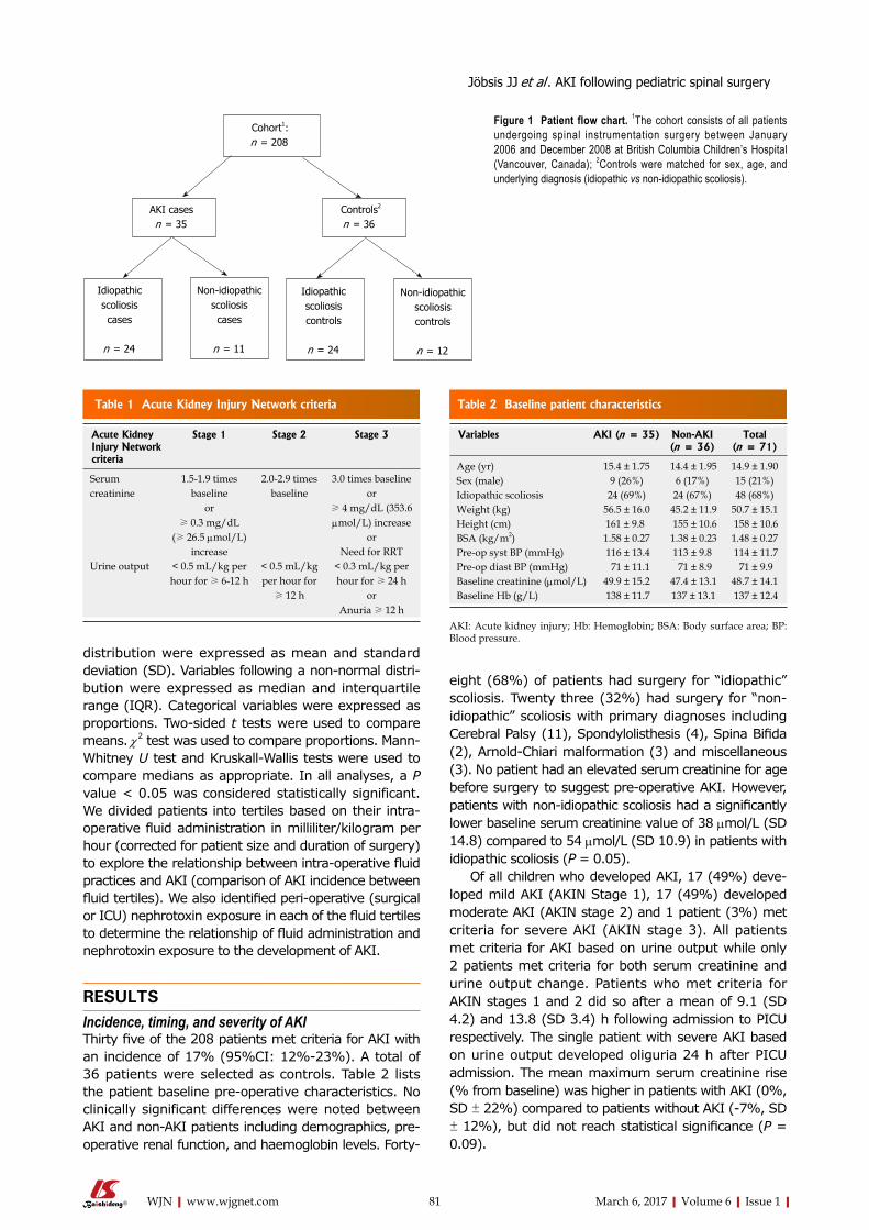
81 March 6, 2017|Volume 6|Issue 1|WJN|www.wjgnet.com
distribution were expressed as mean and standard deviation (SD). Variables following a nonnormal distribution were expressed as median and interquartile range (IQR). Categorical variables were expressed as proportions. Twosided t tests were used to compare means. χ2 test was used to compare proportions. MannWhitney U test and KruskallWallis tests were used to compare medians as appropriate. In all analyses, a P value < 0.05 was considered statistically significant. We divided patients into tertiles based on their intraoperative fluid administration in milliliter/kilogram per hour (corrected for patient size and duration of surgery) to explore the relationship between intra-operative fluid practices and AKI (comparison of AKI incidence between fluid tertiles). We also identified peri-operative (surgical or ICU) nephrotoxin exposure in each of the fluid tertiles to determine the relationship of fluid administration and nephrotoxin exposure to the development of AKI.
RESULTSIncidence, timing, and severity of AKIThirty five of the 208 patients met criteria for AKI with an incidence of 17% (95%CI: 12%-23%). A total of 36 patients were selected as controls. Table 2 lists the patient baseline preoperative characteristics. No clinically significant differences were noted between AKI and nonAKI patients including demographics, preoperative renal function, and haemoglobin levels. Forty
eight (68%) of patients had surgery for “idiopathic” scoliosis. Twenty three (32%) had surgery for “nonidiopathic” scoliosis with primary diagnoses including Cerebral Palsy (11), Spondylolisthesis (4), Spina Bifida (2), ArnoldChiari malformation (3) and miscellaneous (3). No patient had an elevated serum creatinine for age before surgery to suggest preoperative AKI. However, patients with non-idiopathic scoliosis had a significantly lower baseline serum creatinine value of 38 µmol/L (SD 14.8) compared to 54 µmol/L (SD 10.9) in patients with idiopathic scoliosis (P = 0.05).
Of all children who developed AKI, 17 (49%) developed mild AKI (AKIN Stage 1), 17 (49%) developed moderate AKI (AKIN stage 2) and 1 patient (3%) met criteria for severe AKI (AKIN stage 3). All patients met criteria for AKI based on urine output while only 2 patients met criteria for both serum creatinine and urine output change. Patients who met criteria for AKIN stages 1 and 2 did so after a mean of 9.1 (SD 4.2) and 13.8 (SD 3.4) h following admission to PICU respectively. The single patient with severe AKI based on urine output developed oliguria 24 h after PICU admission. The mean maximum serum creatinine rise (% from baseline) was higher in patients with AKI (0%, SD ± 22%) compared to patients without AKI (7%, SD ± 12%), but did not reach statistical significance (P = 0.09).
Cohort1:n = 208
AKI casesn = 35
Controls2
n = 36
Idiopathic scoliosis cases
n = 24
Non-idiopathicscoliosiscases
n = 11
Idiopathic scoliosiscontrols
n = 24
Non-idiopathic scoliosiscontrols
n = 12
Figure 1 Patient flow chart. 1The cohort consists of all patients undergoing spinal instrumentation surgery between January 2006 and December 2008 at British Columbia Children’s Hospital (Vancouver, Canada); 2Controls were matched for sex, age, and underlying diagnosis (idiopathic vs non-idiopathic scoliosis).
Acute Kidney Injury Network criteria
Stage 1 Stage 2 Stage 3
Serum creatinine
1.5-1.9 times baseline
or≥ 0.3 mg/dL
(≥ 26.5 µmol/L) increase
2.0-2.9 times baseline
3.0 times baselineor
≥ 4 mg/dL (353.6 µmol/L) increase
orNeed for RRT
Urine output < 0.5 mL/kg per hour for ≥ 6-12 h
< 0.5 mL/kg per hour for
≥ 12 h
< 0.3 mL/kg per hour for ≥ 24 h
orAnuria ≥ 12 h
Table 1 Acute Kidney Injury Network criteria
Variables AKI (n = 35) Non-AKI (n = 36)
Total (n = 71)
Age (yr) 15.4 ± 1.75 14.4 ± 1.95 14.9 ± 1.90 Sex (male) 9 (26%) 6 (17%) 15 (21%) Idiopathic scoliosis 24 (69%) 24 (67%) 48 (68%) Weight (kg) 56.5 ± 16.0 45.2 ± 11.9 50.7 ± 15.1 Height (cm) 161 ± 9.8 155 ± 10.6 158 ± 10.6 BSA (kg/m2) 1.58 ± 0.27 1.38 ± 0.23 1.48 ± 0.27 Pre-op syst BP (mmHg) 116 ± 13.4 113 ± 9.8 114 ± 11.7 Pre-op diast BP (mmHg) 71 ± 11.1 71 ± 8.9 71 ± 9.9 Baseline creatinine (µmol/L) 49.9 ± 15.2 47.4 ± 13.1 48.7 ± 14.1 Baseline Hb (g/L) 138 ± 11.7 137 ± 13.1 137 ± 12.4
Table 2 Baseline patient characteristics
Jöbsis JJ et al . AKI following pediatric spinal surgery
AKI: Acute kidney injury; Hb: Hemoglobin; BSA: Body surface area; BP: Blood pressure.

82 March 6, 2017|Volume 6|Issue 1|WJN|www.wjgnet.com
Intra-operative courseTable 3 compares several intraoperative and postoperative (PICU admission) variables between AKI and nonAKI patients. Overall, mean duration of surgery was 8.4 h (SD 2.3 h). Duration of surgery, decreases in blood pressure during surgery, percentage requiring blood transfusions, and decreases in haemoglobin levels were similar in AKI and nonAKI patients (all P > 0.05). There was wide practice variation in intra-operative fluid volume administration between patients. All intravenous fluid administered was isotonic. The mean recorded urine output during surgery was 2.1 (SD 1.4) mL/kg per hour and was not statistically different between AKI and non-AKI patients [1.9 mL/kg per hour (SD 1.1) vs 2.4 mL/kg per hour (SD 1.5) respectively (P = 0.17)]. We classified all children into tertiles according to intra-operative fluids received, corrected for patient weight and surgery duration: < 5.4 mL/kg per hour, 5.4-7.9 mL/kg per hour, and > 7.9 mL/kg per hour. An inverse relationship was observed with the incidence of AKI and the amount of fluids received intra-operatively: 70%, 50%, 29% AKI incidence in the 1st, 2nd and 3rd fluid tertiles respectively (P = 0.02) (Figure 2A). The
lowest recorded mean intraoperative serum sodium was 138 mmol/L (SD 2.4) while 4 patients (2%) developed hyponatremia during surgery (serum sodium < 135 mmol/L). During surgery, 14 out of 71 patients (19.7%) were exposed to a nephrotoxic medication (either aminoglycosides or NSAIDs). The majority of these patients (78.6%) received a dose of intravenous Ketorolac (NSAID) while 21.4% were exposed to intravenous Gentamycin (aminoglycoside). Overall, there was no difference in intraoperative nephrotoxin exposure between AKI and control groups (23% vs 17%) (P = 0.5).
Post-operative courseA detailed summary of postoperative variables in AKI and nonAKI patients is shown in Table 2. Mean urine output was reduced at 0.9 (SD 0.4) mL/kg per hour during the PICU admission. Patients who developed AKI post-operatively had significantly lower urine output (0.7 mL/kg per hour vs 1.1 mL/kg per hour) and a higher cumulative fluid balance (positive 1.3 L vs positive 0.8 L) compared to control patients during their PICU admission (both P < 0.05). The lowest recorded mean
Fluid tertile 1: < 5.4 mL/kg per hourFluid tertile 2: 5.4-7.9 mL/kg per hourFluid tertile 3: > 7.9 mL/kg per hour
Fluid tertile 1: < 5.4 mL/kg per hourFluid tertile 2: 5.4-7.9 mL/kg per hourFluid tertile 3: > 7.9 mL/kg per hour
AKI
inci
denc
e (%
)AK
I in
cide
nce
(%)
Fluid tertile
Fluid tertile
12
3
12
3
70
60
50
40
30
20
10
0
100
80
60
40
20
0 Nephrotoxin
No nephrotoxin
Figure 2 Acute kidney injury incidence according to intra-operative fluid management and nephrotoxin exposure. A: Acute kidney injury (AKI) incidence according to fluid tertile: Fluid tertile 1 (70%) vs fluid tertile 2 (50%) vs fluid tertile 3 (29%) (P = 0.02); B: AKI incidence according to fluid tertile and nephrotoxin exposure: Fluid tertile 1 with nephrotoxin exposure (75%) vs fluid tertile 3 with no nephrotoxin exposure (10%) (P = 0.04).
A
B
Jöbsis JJ et al . AKI following pediatric spinal surgery

83 March 6, 2017|Volume 6|Issue 1|WJN|www.wjgnet.com
post-operative serum sodium was 137 mmol/L (SD 2.8). Overall, 10 patients (14%) developed hyponatremia either during surgery or postoperatively. In patients who developed hyponatremia, the mean value of lowest sodium recorded was 133 mmol/L (SD 1.5). Patients who developed AKI were exposed to nephrotoxins (either aminoglycosides or NSAIDs) more frequently than control patients during their PICU admission (51% vs 11%, P < 0.001). Ninety-one percent of the patients exposed to NSAIDs received intravenous Ketorolac while the remainder received oral Ibuprofen. All patients with aminoglycoside exposure received intravenous Gentamycin. Overall, 21/35 (60%) patients who developed AKI were exposed to a nephrotoxic medication either during surgery or ICU admission compared to 8/36 (22%) of controls (P = 0.001). Figure 2B shows the relationship of intra-operative fluid admini-stration and perioperative nephrotoxin exposure on the development of AKI. The highest incidence of AKI was seen in patients exposed to less amounts of fluids intra-operatively (1st and 2nd fluid tertiles) while also being exposed to a nephrotoxin (75% and 80% AKI incidence respectively) while the lowest AKI incidence was observed in patients exposed to the highest amounts of fluids (3rd fluid tertile) with no exposure to a nephrotoxin (10% AKI incidence) (P = 0.04). Thirteen patients (18%) required mechanical ventilation in PICU with a mean duration of 1.8 (SD 1.2) d. Of those 13 children, 7 (20%) were in the AKI group vs 6 (17%) in the nonAKI group. Children with AKI were ventilated longer (mean 2.2 d vs 1.4 d in the control group; however this difference was not statistically significant (P = 0.7).
Mortality and hospital/PICU length of stayThere were no cases of mortality seen in any of the patients following surgery. Median hospital length of
stay in all patients was 6 d (IQR 3, range 449 d). Median ICU length of stay in all patients was 1 d (IQR 0, range 018 d). ICU length of stay was similar in AKI (median 1 d, IQR 0 d, range 17 d) and nonAKI patients (median 1 d, IQR 0 d, range 6 d) (P = 0.95). Similarly, hospital length of stay was similar in patients with AKI (median 6 d, IQR 3 d, range 25) and without AKI (median 6 d, IQR 2 d, range 45 d, P = 0.77). However, the single patient with severe AKI (AKIN stage 3) had a more prolonged PICU and hospital length of stay at 4 and 12 d respectively.
DISCUSSIONWe report a high estimated AKI incidence of 17% in children undergoing orthopaedic spinal instrumentation surgery utilizing a standardized AKI definition. A number of recent adult studies have reported AKI incidences from 3.9%16% following major orthopaedic surgeries[911]. There have been no prior epidemiological AKI studies in any paediatric orthopaedic populations.
In our cohort, no patient had preexisting kidney dysfunction prior to surgery, which suggests that our high incidence of AKI may be primarily related to perioperative risk factors. We identified 2 potential risk factors that may be related to the development of AKI in this population: (1) lower intravenous fluid exposure in the intraoperative period; and (2) high perioperative nephrotoxin exposure with 60% of the AKI population being exposed to NSAIDs or aminoglycosides. The exact interaction between perioperative fluid and nephrotoxin exposure towards the development of AKI cannot be determined in our retrospective analysis, but our results suggest that patients who are exposed to the highest amounts of fluids intraoperatively without nephrotoxin exposure may be more protected against
AKI (n = 35) Non-AKI (n = 36) P value
Intra-operative course Duration of surgery (h) 8.5 ± 2.4 8.2 ± 2.1 0.54 Cumulative intra-operative fluid administration (mL) 2555 ± 973 2732 ± 1264 0.51 Cumulative intra-operative fluid administration (mL/kg per hour ) 6.2 ± 2.9 7.5 ± 3.3 0.08 Intra-operative urine output (mL/kg per hour ) 1.9 ± 1.1 2.4 ± 1.5 0.17 Intra-operative fluid balance (L) 1.2 ± 1.0 1.3 ± 1.1 0.70 Max BP decline during surgery Systolic (% from baseline) 27 ± 12 33 ± 14 0.36 Diastolic (% from baseline) 30 ± 9 34 ± 13 0.93 Patients requiring blood transfusion [n (%)] 7 (20%) 12 (33%) 0.21 Nephrotoxin exposure [n (%)] 8 (23%) 6 (17%) 0.51 Post-operative course Duration of PICU stay (d) 1 (IQR 0, range 17) 1 (IQR 0, range 6) 0.77 Cumulative PICU fluid administration (mL) 2069 (IQR 886) 1918 (IQR 1048) 0.15 Cumulative PICU fluid administration (mL/kg per hour) 2.1 ± 0.8 2.3 ± 0.7 0.31 PICU urine output (mL/kg per hour) 0.7 ± 0.4 1.1 ± 0.4 < 0.001 PICU fluid balance (L) 1.3 ± 1.0 0.8 ± 0.8 0.02 Max Hb drop (% from baseline) 26 ± 9 26 ± 10 0.94 Nephrotoxin exposure in PICU 18 (51%) 4 (11%) < 0.001
Table 3 Intra-operative/post-operative course
AKI: Acute kidney injury; PICU: Paediatric intensive care unit.
Jöbsis JJ et al . AKI following pediatric spinal surgery

84 March 6, 2017|Volume 6|Issue 1|WJN|www.wjgnet.com
AKI. Interestingly, a recent adult orthopaedic study showed that perioperative dehydration, administration of NSAIDs, and use of nephrotoxic antibiotics were all independently associated with the development of postoperative kidney dysfunction[11]. According to a large epidemiological study analyzing the most common AKI etiologies in children, AKI related to nephrotoxin exposure accounts for up to 16% of inhospital paediatric AKI[12]. Outside of fluid intake and use of nephrotoxins, there may be other complex intraoperative factors related to AKI that we were not able to study based on the retrospective nature of our study. For example, a recent local observational study involving 30 children undergoing scoliosis surgery found that a significantly reduced cardiac output (18.5% reduction)was associated with the prolonged prone positioning required for these surgeries[13]. Reduced cardiac output may lead to a state of poor renal perfusion during these lengthy procedures, which may put these children at a higher risk of AKI compared to other general surgical populations.
Even though the rise of creatinine trended higher in AKI patients compared to controls, it is interesting to note that the majority of AKI patients in our cohort were “oliguric” without significant rises in serum creatinine that often did not meet the creatinine criteria of the AKIN definition (at least 50% rise in creatinine). This may be partly related to a reduction of serum creatinine concentration associated with hemodilution effects and relative fluid overload as the majority of our patients demonstrated a cumulative positive fluid balance intraoperatively and in the ICU postoperatively. In addition, approximately 1/3 of our surgical cohort had an underlying condition including spina bifida and cerebral palsy (nonidiopathic scoliosis population). The significantly lower baseline serum creatinine values observed in our nonidiopathic scoliosis patients implies that a significantly low muscle mass state may limit an appreciable rise in serum creatinine during an episode of AKI. This is also a population where the use of recently validated AKI biomarkers representing early tubular damage [e.g., neutrophil gelatinase associated lipocalin (NGAL), interleukin-18 (IL-18), and kidney-injury molecule (KIM1)] may be warranted in future orthopaedic AKI studies due to the potential issues of serum creatinine in this highly prevalent group of patients[1416]. Lastly, serum creatinine, was not moni-tored frequently past 24 h postoperatively in the majority of patients with the potential of higher rises in creatinine being missed in many patients.
Another potential reason for oliguria in this population is the syndrome of inappropriate antidiuretic hormone (SIADH), which has been reported in adult and some paediatric spinal orthopedic literature[1719]. The diagnosis of SIADH requires the presence of hyponatremia (serum sodium < 135 mmol/L). In our cohort, only 10 patients (14%) overall were recorded with hyponatremia either during surgery or postoperatively, and only 5 of the AKI patients (14%) had documented hyponatremia. Mean serum sodium levels were the
same in AKI and nonAKI patients both intra (138) and postoperatively (137). Even though we were not able to completely exclude SIADH with serum ADH levels and urine osmolality values, it is unlikely that SIADH was the primary cause of oliguria based on several reasons. As shown in a paediatric study of SIADH in children following spinal fusion surgery, serum ADH levels peaked immediately postop and declined by 6 h after surgery[20]. Our stage 1 AKI patients (n = 17) met criteria for oliguria (< 0.5 mL/kg per hour for at least 6 h) at a mean of 9.1 h after surgery and stage 2 AKI patients (n = 17) met criteria (< 0.5 cc/kg per hour for at least 12 h) at a mean of 13.8 h postoperatively, well after the peak of serum ADH levels was observed in prior studies[20]. In addition, the AKIN criteria for oliguria is strict (Stage 1: < 0.5 mL/kg per hour for at least 6 h) and is often more severe than what is observed in patients with SIADH. Therefore, SIADH as a primary reason for fulfilling AKIN urine output criteria is unlikely, but will need to be further explored in future prospective studies.
Unlike recent studies involving paediatric cardiac surgery patients, perioperative AKI was not associated with morbidity (increased hospital/ICU length of stay) or mortality in children undergoing spinal instrumentation surgery. This needs to be studied prospectively in larger orthopaedic populations before any further conclusions are made due to the potential concerns of our study being underpowered with a relatively small sample size. Regardless, the relatively high incidence of AKI in our cohort cannot be ignored, especially in the context of several recent adult and paediatric publications revealing an increased risk of chronic kidney disease following a single episode of AKI including those of milder severity[38].
In conclusion, we observed a high incidence of AKI following spinal instrumentation surgery in children that is potentially related to the frequent use of nephrotoxins and the amount of fluid administered peri-operatively.
COMMENTSBackgroundOutside of cardiac surgery patients, the epidemiology of acute kidney injury (AKI) has not been well studied in other paediatric surgical populations including those undergoing major orthopaedic surgeries.
Research frontiers Several studies have revealed an increased morbidity and mortality associated with pediatric AKI and an increased risk of chronic kidney disease in survivors of AKI.
Innovations and breakthroughsThe authors report a high incidence of AKI following spinal instrumentation surgery in children that is potentially related to the frequent use of nephrotoxins and the amount of fluid administered peri-operatively.
ApplicationsThis study suggests that there are modifiable AKI risk factors with the potential of reducing AKI incidence in this understudied population.
COMMENTS
Jöbsis JJ et al . AKI following pediatric spinal surgery

85 March 6, 2017|Volume 6|Issue 1|WJN|www.wjgnet.com
TerminologyAcute kidney injury network (AKIN)-criteria: A set of criteria based on urine output or rise in serum creatinine to define AKI, according to the AKIN.
Peer-review This is an interesting study which is well done and with new findings. It will be interesting to get more data about renal function after 3-5 years to have an idea about the impact of AKI on the long term renal prognosis.
REFERENCES1 Alkandari O, Eddington KA, Hyder A, Gauvin F, Ducruet T,
Gottesman R, Phan V, Zappitelli M. Acute kidney injury is an independent risk factor for pediatric intensive care unit mortality, longer length of stay and prolonged mechanical ventilation in critically ill children: a two-center retrospective cohort study. Crit Care 2011; 15: R146 [PMID: 21663616 DOI: 10.1186/cc10269]
2 Akcan-Arikan A, Zappitelli M, Loftis LL, Washburn KK, Jefferson LS, Goldstein SL. Modified RIFLE criteria in critically ill children with acute kidney injury. Kidney Int 2007; 71: 1028-1035 [PMID: 17396113 DOI: 10.1038/sj.ki.5002231]
3 Mammen C, Al Abbas A, Skippen P, Nadel H, Levine D, Collet JP, Matsell DG. Long-term risk of CKD in children surviving episodes of acute kidney injury in the intensive care unit: a prospective cohort study. Am J Kidney Dis 2012; 59: 523-530 [PMID: 22206744 DOI: 10.1053/j.ajkd.2011.10.048]
4 Menon S, Kirkendall ES, Nguyen H, Goldstein SL. Acute kidney injury associated with high nephrotoxic medication exposure leads to chronic kidney disease after 6 months. J Pediatr 2014; 165: 522-527.e2 [PMID: 24928698 DOI: 10.1016/j.jpeds.2014.04.058]
5 Askenazi DJ, Feig DI, Graham NM, Hui-Stickle S, Goldstein SL. 35 year longitudinal followup of pediatric patients after acute renal failure. Kidney Int 2006; 69: 184-189 [PMID: 16374442 DOI: 10.1038/sj.ki.5000032]
6 Viaud M, Llanas B, Harambat J. Renal outcome in long-term survivors from severe acute kidney injury in childhood. Pediatr Nephrol 2012; 27: 151-152; author reply 153 [PMID: 21947273 DOI: 10.1007/s00467-011-2016-5]
7 Slack R, Hawkins KC, Gilhooley L, Addison GM, Lewis MA, Webb NJ. Long-term outcome of meningococcal sepsis-associated acute renal failure. Pediatr Crit Care Med 2005; 6: 477-479 [PMID: 15982443 DOI: 10.1097/01.PCC.0000163285.74452.23]
8 Coca SG, Singanamala S, Parikh CR. Chronic kidney disease after acute kidney injury: a systematic review and meta-analysis. Kidney Int 2012; 81: 442-448 [PMID: 22113526 DOI: 10.1038/ki.2011.379]
9 White SM, Rashid N, Chakladar A. An analysis of renal dysfunction in 1511 patients with fractured neck of femur: the implications for peri-operative analgesia. Anaesthesia 2009; 64: 1061-1065 [PMID: 19735395 DOI: 10.1111/j.1365-2044.2009.06012.x]
10 Bennet SJ, Berry OM, Goddard J, Keating JF. Acute renal dysfunction following hip fracture. Injury 2010; 41: 335-338 [PMID: 19729159 DOI: 10.1016/j.injury.2009.07.009]
11 Kateros K, Doulgerakis C, Galanakos SP, Sakellariou VI, Papadakis SA, Macheras GA. Analysis of kidney dysfunction in orthopaedic patients. BMC Nephrol 2012; 13: 101 [PMID: 22943390 DOI: 10.1186/1471-2369-13-101]
12 Hui-Stickle S, Brewer ED, Goldstein SL. Pediatric ARF epidemiology at a tertiary care center from 1999 to 2001. Am J Kidney Dis 2005; 45: 96-101 [PMID: 15696448 DOI: 10.1053/j.ajkd.2004.09.028]
13 Brown ZE, Görges M, Cooke E, Malherbe S, Dumont GA, Ansermino JM. Changes in cardiac index and blood pressure on positioning children prone for scoliosis surgery. Anaesthesia 2013; 68: 742-746 [PMID: 23710730 DOI: 10.1111/anae.12310]
14 Devarajan P. Biomarkers for the early detection of acute kidney injury. Curr Opin Pediatr 2011; 23: 194-200 [PMID: 21252674 DOI: 10.1097/MOP.0b013e328343f4dd]
15 Al-Ismaili Z, Palijan A, Zappitelli M. Biomarkers of acute kidney injury in children: discovery, evaluation, and clinical application. Pediatr Nephrol 2011; 26: 29-40 [PMID: 20623143 DOI: 10.1007/s00467-010-1576-0]
16 Nguyen MT, Devarajan P. Biomarkers for the early detection of acute kidney injury. Pediatr Nephrol 2008; 23: 2151-2157 [PMID: 17394022 DOI: 10.1007/s00467-007-0470-x]
17 Brazel PW, McPhee IB. Inappropriate secretion of antidiuretic hormone in postoperative scoliosis patients: the role of fluid management. Spine (Phila Pa 1976) 1996; 21: 724-727 [PMID: 8882695 DOI: 10.1097/00007632-199603150-00013]
18 Callewart CC, Minchew JT, Kanim LE, Tsai YC, Salehmoghaddam S, Dawson EG, Delamarter RB. Hyponatremia and syndrome of inappropriate antidiuretic hormone secretion in adult spinal surgery. Spine (Phila Pa 1976) 1994; 19: 1674-1679 [PMID: 7973959 DOI: 10.1097/00007632-199408000-00004]
19 Mason RJ, Betz RR, Orlowski JP, Bell GR. The syndrome of inappropriate antidiuretic hormone secretion and its effect on blood indices following spinal fusion. Spine (Phila Pa 1976) 1989; 14: 722-726 [PMID: 2772722 DOI: 10.1097/00007632-198907000-00013]
20 Lieh-Lai MW, Stanitski DF, Sarnaik AP, Uy HG, Rossi NF, Simpson PM, Stanitski CL. Syndrome of inappropriate antidiuretic hormone secretion in children following spinal fusion. Crit Care Med 1999; 27: 622-627 [PMID: 10199545 DOI: 10.1097/00003246-199903000-00046]
P- Reviewer: Bhimma R, Friedman EA, Mahmoud KM, Olowu WA S- Editor: Kong JX L- Editor: A E- Editor: Wu HL
Jöbsis JJ et al . AKI following pediatric spinal surgery

© 2017 Baishideng Publishing Group Inc. All rights reserved.
Published by Baishideng Publishing Group Inc8226 Regency Drive, Pleasanton, CA 94588, USA
Telephone: +1-925-223-8242Fax: +1-925-223-8243
E-mail: [email protected] Desk: http://www.wjgnet.com/esps/helpdesk.aspx
http://www.wjgnet.com