wnt7a signaling promotes dendritic spine growth … · wnt7a signaling promotes dendritic spine...
TRANSCRIPT
Wnt7a signaling promotes dendritic spine growth andsynaptic strength through Ca2+/Calmodulin-dependentprotein kinase IILorenza Ciania,1, Kieran A. Boylea,1, Ellen Dickinsa, Macarena Sahoresa, Derek Ananea, Douglas M. Lopesa,Alasdair J. Gibbb, and Patricia C. Salinasa,2
Departments of aCell and Developmental Biology and bNeuroscience, Physiology, and Pharmacology, University College London, London WC1E 6BT, UnitedKingdom
Edited* by Ricardo Miledi, University of California, Irvine, CA, and approved May 10, 2011 (received for review December 7, 2010)
The balance between excitatory and inhibitory synapses is crucial fornormal brain function. Wnt proteins stimulate synapse formationby increasing synaptic assembly. However, it is unclear whetherWnt signaling differentially regulates the formation of excitatoryand inhibitory synapses. Here, we demonstrate that Wnt7a prefer-entially stimulates excitatory synapse formation and function. Inhippocampal neurons, Wnt7a increases the number of excitatorysynapses, whereas inhibitory synapses are unaffected. Wnt7a orpostsynaptic expression of Dishevelled-1 (Dvl1), a core Wnt signal-ing component, increases the frequency and amplitude of miniatureexcitatory postsynaptic currents (mEPSCs), but not miniature in-hibitory postsynaptic currents (mIPSCs). Wnt7a increases the densityand maturity of dendritic spines, whereas Wnt7a-Dvl1–deficientmice exhibit defects in spine morphogenesis and mossy fiber-CA3synaptic transmission in the hippocampus. Using a postsynapticreporter for Ca2+/Calmodulin-dependent protein kinase II (CaMKII)activity, we demonstrate that Wnt7a rapidly activates CaMKII inspines. Importantly, CaMKII inhibition abolishes the effects ofWnt7aon spine growth and excitatory synaptic strength. These data indi-cate that Wnt7a signaling is critical to regulate spine growth andsynaptic strength through the local activation of CaMKII at dendriticspines. Therefore, aberrant Wnt7a signaling may contribute to neu-rological disorders in which excitatory signaling is disrupted.
Wnt7a | Dvl1 mutant | plasticity
The formation of functional neuronal circuits requires the as-sembly of different types of synapses with great specificity. The
development of an appropriate balance of glutamatergic excitatoryand GABAergic inhibitory synapses (E/I ratio) is essential forproper circuit function (1, 2) because an imbalance in the E/I ratiocan result in neurological disorders (3–5). Some synaptogenic fac-tors regulate both excitatory and inhibitory synapses (6, 7), whereasother synaptic organizers are more specific (8–10). However, theprecise mechanisms by which synaptic organizing signals regulateexcitatory and inhibitory synapses remain poorly understood.In the central nervous system, most excitatory inputs are locat-
ed on dendritic spines, postsynaptic protrusions that function asdomains where synaptic activity is regulated in a compartmental-ized manner (11–13). Several intracellular molecules have beenimplicated in the formation and maturation of dendritic spines(14–16), but the mechanisms by which extracellular factors signalthrough intracellular regulators to promote spine developmentand maturation remain poorly characterized.Wnt secreted proteins are synaptic organizers that stimulate
the formation of central and peripheral synapses (17–19) bypromoting presynaptic assembly (20) and the clustering of post-synaptic components (19, 21–24). In cultured neurons, Wnt5aregulates postsynaptic development of both GABAergic andglutamatergic synapses (24, 25). However, it is unclear whetherother Wnts play a more selective role in promoting the formationof specific synapses and, therefore, regulating the balance be-tween excitatory and inhibitory inputs.
Here, we demonstrate that Wnt7a signaling specifically regu-lates the formation and function of excitatory synapses withoutaffecting inhibitory synapses in the hippocampus. Wnt7a signalsdirectly to the postsynaptic dendrite to stimulate spine growthand synaptic strength. Dishevelled-1 (Dvl1), a cytoplasmic coreWnt signaling component that promotes the activation of theWnt cascade at specific cellular locale (26), is present at thepostsynapse and is required for Wnt7a function. Analyses of Wntsignaling-deficient mutant mice demonstrate that Wnt7a-Dvl1signaling is required for spine morphogenesis and glutamatergictransmission in the hippocampus. Expression of PSD-95-Vim-CFP,a synaptic read-out of Ca2+/Calmodulin-dependent protein kinaseII (CaMKII) activity, demonstrates that Wnt7a activates CaMKIIat dendritic spines. Importantly, CaMKII is required for Wnt-mediated spine growth and increased synaptic strength. Thesefindings reveal a unique role for Wnt7a signaling in promoting theformation and function of excitatory synapses through CaMKII, akey molecule that regulates the functional and morphologicalplasticity of synapses.
ResultsWnt7a Specifically Increases the Number and Function of ExcitatorySynapses. To address the role of Wnts in the formation of specificsynapse types, we focused on the hippocampus, because Wnt7a/bprotein is present in neurons of the dentate gyrus, CA1, and CA3at the peak of synaptogenesis (Fig. S1). We therefore examinedwhether Wnt7a stimulates excitatory and inhibitory synapse for-mation in cultured hippocampal neurons at this developmentalstage. Wnt7a increases the density of vGlut and PSD-95 puncta(excitatory pre- and postsynaptic markers, respectively) within1 h, with further increases after 3 and 16 h treatment (Fig. 1 A andC and Fig. S2A). In contrast, the density of vGAT and Gephyrinpuncta (inhibitory pre- and postsynaptic markers, respectively)are unchanged at all time-points studied (Fig. 1 B and D and Fig.S2B). Crucially, the density of vGlut puncta apposed to PSD-95(putative excitatory synapses) displays a robust time-dependentincrease after Wnt7a treatment, whereas the density of putativeinhibitory synapses (vGAT puncta apposed to Gephyrin) is un-changed (Fig. 1C andD and Fig. S2A and Fig. S2B). Consistently,whole-cell patch-clamp recordings show that 3-h Wnt7a treat-ment increases the frequency and amplitude of mEPSCs (Fig. 1 Eand G), whereas mIPSCs are unaffected (Fig. 1 F and H). To-gether our results show that Wnt7a specifically increases the
Author contributions: L.C., K.A.B., M.S., A.J.G., and P.C.S. designed research; L.C., K.A.B.,E.D., M.S., D.A., and D.M.L. performed research; L.C., K.A.B., E.D., M.S., D.A., D.M.L., A.J.G.,and P.C.S. analyzed data; and L.C., K.A.B., M.S., A.J.G., and P.C.S. wrote the paper.
The authors declare no conflict of interest.
*This Direct Submission article had a prearranged editor.
Freely available online through the PNAS open access option.1L.C. and K.A.B. contributed equally to this work.2To whom correspondence should be addressed. E-mail: [email protected].
This article contains supporting information online at www.pnas.org/lookup/suppl/doi:10.1073/pnas.1018132108/-/DCSupplemental.
10732–10737 | PNAS | June 28, 2011 | vol. 108 | no. 26 www.pnas.org/cgi/doi/10.1073/pnas.1018132108

number and strength of excitatory synapses, while leaving in-hibitory synapses unaffected.
Wnt Signaling Through Dvl1 Regulates Dendritic SpineMorphogenesis.Given the specific effect of Wnt7a on excitatory synapses, we ex-amined the potential role of Wnt7a signaling on the formation ofdendritic spines, which receive the majority of central excitatoryinputs (11). Sixteen-hour Wnt7a treatment increases spine num-ber and spine head size by 65 ± 5.7% and 66 ± 3%, respectively(Fig. 2 A–D, Movie S1, and Movie S2). Three-hour Wnt7a expo-sure also increases spine number and spine head size by 48± 6.1%and 27± 3.3%, respectively (Fig. 2C andD). Increased spine headsize is an indication of spine maturity (11, 27), indicating thatWnt7a increases the formation andmaturation of dendritic spines.AlthoughWnt proteins can signal throughmultiple intracellular
cascades, recipient cells require the cytoplasmic protein Dish-evelled (Dvl), a core Wnt pathway component that functions asa hub to promote Wnt signaling within specific cellular compart-ments (26). Three mouse Dvl genes (Dvl1–Dvl3) are expressedbroadly in the postnatal and adult brain (Allen Brain Atlas).However, Dvl1 has been the most studied because Dvl1 mutantmice exhibit defects in presynaptic assembly (20) and in socialbehavior (28). Importantly, Wnt7a requires Dvl1 to regulate axo-nal remodelling and presynaptic differentiation (20). To determinethe downstream requirements for Wnt7a signaling in spines, thelocalization of Dvl1 was examined. Endogenous Dvl1 is present inthe postsynaptic density (PSD) fraction (Fig. 2E) and at dendritic
spines (Fig. 2F), suggesting that Wnt7a could regulate spine de-velopment by signaling directly via Dvl1 within dendritic spines.We next investigated whether Dvl1 is required for Wnt7a
function. Cultured hippocampal neurons from Dvl1 mutant miceexhibit a mild defect in spine morphogenesis compared withwild-type neurons (Fig. 2 G and H). Importantly, Dvl1 mutantneurons do not respond to Wnt7a, because the number and sizeof spines and the frequency and amplitude of mEPSCs remainunchanged after Wnt7a exposure (Fig. 2 G–J and Fig. S3). Thus,Dvl1 is required downstream of Wnt7a to regulate spine mor-phogenesis and excitatory synaptic function.
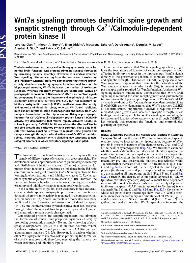
Wnt Signaling Is Required in Vivo for Dendritic Spine Morphogenesisand Excitatory Synaptic Function. We next examined possibledefects in spine morphogenesis inWnt signaling-deficient mice. Weanalyzed the double Wnt7a; Dvl1 mutant mouse because previousstudieshave shown that thismutant exhibits stronger synapticdefectsthan single mutants in the cerebellum (20). Analyses of organotypicbrain slices transfectedwithEGFP-actin reveal a significantdecreasein spine density (30%) and spine head width (15%) in the doublemutant mice compared with wild-type animals (Fig. S4).The in vivo role of Wnt7a signaling was investigated by Golgi
staining. DoubleWnt7a; Dvl1mutant mice exhibit a 20% decreasein the number of spines on CA3 cells, although no significantdifferences were detected in CA1 cells (Fig. 3 A and B), consistentwith the strongerWnt7a/b expression observed in the CA3 of wild-typemice (Fig. S1). In contrast, spine size is significantly reduced inboth CA1 and CA3 (Fig. 3 A and B).Wnt7a single knockout mice
Fig. 1. Wnt7a specifically stimulates the formation and function of excitatory synapses. (A and B) Fourteen days in vitro (DIV) hippocampal culture weretreated for 3 h with Wnt7a. Excitatory presynaptic (vGlut1) and postsynaptic (PSD-95) sites (A) and inhibitory presynaptic (vGAT) and postsynaptic (Gephyrin)sites along processes (MAP2) of neurons (B). (Scale bars: 5 μm.) (C and D) Quantification of excitatory and inhibitory puncta density normalized to neuritevolume, as assessed by MAP2 staining. *P < 0.05, **P < 0.01 by Student’s t test. (E and F) Representative 10-s traces of mEPSCs (E) and mIPSCs (F) from 14 DIVhippocampal cells treated with control or Wnt7a. (G and H) Quantification of mEPSC (G) and mIPSC (H) frequency and amplitude. *P < 0.05 by Mann–Whitneytest for frequency and Student’s t test for amplitude. All experiments were performed by using at least three independent cultures.
Ciani et al. PNAS | June 28, 2011 | vol. 108 | no. 26 | 10733
NEU
ROSC
IENCE

display a milder phenotype to double knockout mice, becausespine number is not significantly affected in the CA3 (Fig. S5A),suggesting other Wnts may partially compensate for the loss ofWnt7a in vivo. Collectively, these results demonstrate that Wnt7a-Dvl1 signaling is required in vivo for the proper formation andgrowth of dendritic spines in the hippocampus.Consistent with the observed spine phenotype, recordings of
mEPSCs in CA3 cells of acute hippocampal slices reveal a significantreduction in mEPSC frequency and amplitude in the double mutantcompared with wild-type animals (Fig. 3 C and D and Fig. S5B).Evoked recordings at the mossy fiber-CA3 synapse also reveal a de-crease inEPSCamplitude (Fig. 3EandF).These results demonstratethat Wnt7a-Dvl1 signaling is required for normal spine morphogen-esis and excitatory synaptic transmission in the hippocampus.
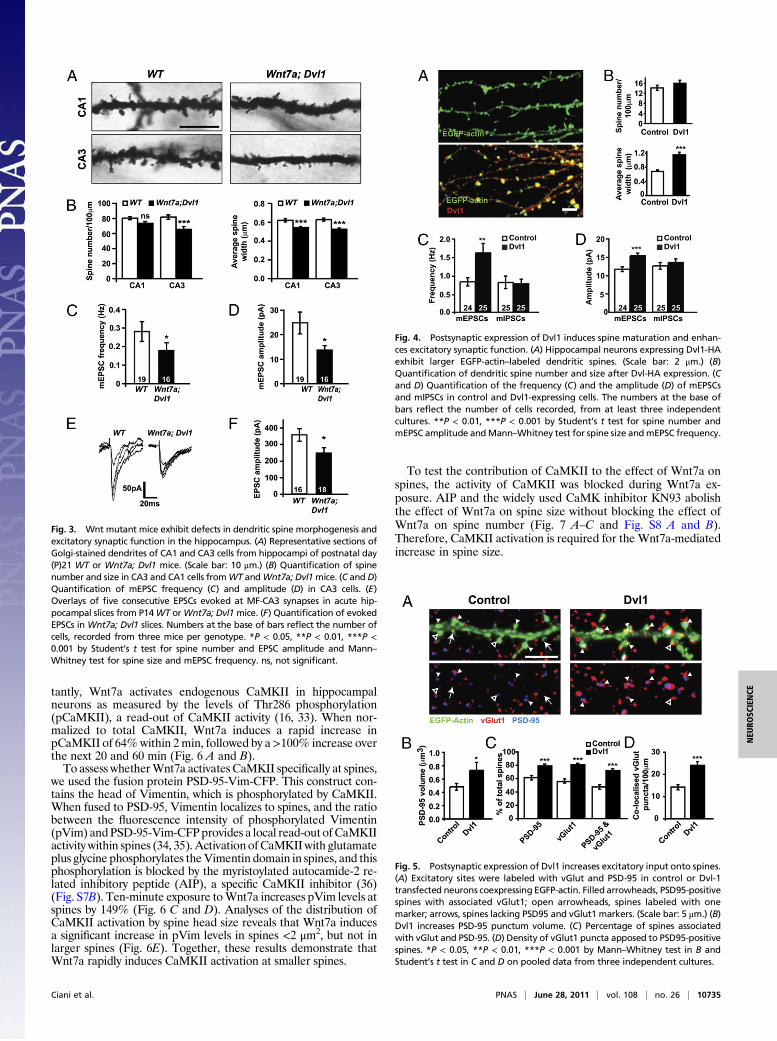
Postsynaptic Activation of Wnt Signaling Is Sufficient to StimulateSpine Growth. Wnt signaling is required in axons to regulate synap-tic differentiation (20). Therefore, changes in spine morphogenesisinduced by Wnt7a could be an indirect effect due to increasedpresynaptic assembly. Alternatively, Wnt7a may signal directly todendrites as suggested by the presence of endogenous Dvl1 atpostsynaptic sites (Fig. 2 E and F). To determine whether post-synaptic Wnt signaling is required for spine regulation, we usedsparse Dvl1 transfection to produce Dvl1-expressing neurons thatwere contacted mainly by nontransfected axons. Postsynaptic ex-pression of Dvl1 increases the width of spine heads by almosttwofold compared with control cells (Fig. 4 A and B). Interestinglyspine density does not change (Fig. 4 A and B), suggesting thatpostsynaptic activation of Wnt signaling stimulates spine growthwithout affecting spine formation.
Postsynaptic Dvl Expression Increases Excitatory Inputs and SynapticStrength. To determine the functional effect of postsynaptic Wnt
signaling, mEPSCs and mIPSCs were recorded from control orDvl1-transfected neurons. Dvl1 increases both the frequency andamplitude of mEPSCs, whereas mIPSCs are unaffected (Fig. 4 Cand D and Fig. S6A). Thus, gain of function of Dvl1 specificallyregulates excitatory connections.The finding that postsynaptic Dvl1 expression increases mEPSC
frequency was intriguing because Dvl1 does not affect dendriticspine number (Fig. 4A andB). Therefore, we investigated whetherpostsynaptic activation of Wnt signaling increases presynaptic in-put onto preexisting spines. Indeed, Dvl increases the percentageof spines receiving presynaptic inputs (vGlut1 puncta; Fig. 5 A andC). In addition, Dvl1 increases the percentage of spines containingPSD-95, and spines contain larger PSD-95 puncta (Fig. 5 A–C).Crucially, the percentage of spines containing PSD-95 in directapposition with vGlut1, and the density of vGlut1 puncta associ-ated with PSD-95-positive spines, also increases (Fig. 5 A, C, andD). Interestingly, Dvl does not change the density or size of vGatpuncta (Fig. S6 B–D). These results demonstrate that postsynapticDvl1 expression promotes the assembly of excitatory pre- andpostsynaptic structures at preexisting spines, thereby specificallystimulating the formation of excitatory synapses.
Wnt Signaling Activates CaMKII to Regulate Spine Morphogenesis. Toexamine the mechanism by which Wnt signaling regulates spinemorphogenesis, we focused our attention on CaMKII. This ser-ine/threonine kinase is enriched at the postsynaptic density andplays a central role in spine morphogenesis and synaptic strength(14, 29). Importantly, Wnt signaling can activate CaMKII duringearly embryonic patterning (30, 31).CaMKII is activated by increases in intracellular calcium levels
(16, 32). Consistent with this notion, the Ca2+ blockers 2-APBand SKF 96365 and the intracellular Ca2+ chelator BAPTA-AMsuppress the effect of Wnt7a on spine growth (Fig. S7A). Impor-
Fig. 2. Wnt7a acts through Dvl1 to regulate spine morphogenesis. (A) Representative EGFP-actin–transfected hippocampal neurons exposed to control orWnt7a for 16 h. Arrow heads indicate spines. (B) 3D reconstructions of typical examples of dendritic spines from control or Wnt7a-treated neurons. (C and D)Quantification of spine number (C) and size (D) in control and Wnt7a-treated neurons. (E) Synaptosome preparation from brain lysates: S, synaptosomalfraction; SMF, synaptosomal membrane fraction; PSD, postsynaptic density fraction; H, brain homogenate; Syp, synaptophysin. (F) Endogenous Dvl1 locali-zation in dendritic spines visualized by EGFP-actin. (G and H) Quantification of spine number (G) and size (H) in Dvl1mutant neurons exposed to Wnt7a. (I andJ) Quantification of mEPSC (I) frequency and amplitude (J) in Dvl1 mutant neurons exposed to Wnt7a. The numbers at the base of bars reflect the number ofcells, recorded from at least three independent cultures. *P < 0.05, **P < 0.01, ***P < 0.001 by Student’s t test or ANOVA for spine number and mPSCamplitude, and Kruskal-Wallis test or Mann–Whitney test for spine size and mPSC frequency.
10734 | www.pnas.org/cgi/doi/10.1073/pnas.1018132108 Ciani et al.
tantly, Wnt7a activates endogenous CaMKII in hippocampalneurons as measured by the levels of Thr286 phosphorylation(pCaMKII), a read-out of CaMKII activity (16, 33). When nor-malized to total CaMKII, Wnt7a induces a rapid increase inpCaMKII of 64%within 2min, followed by a>100% increase overthe next 20 and 60 min (Fig. 6 A and B).To assess whetherWnt7a activates CaMKII specifically at spines,
we used the fusion protein PSD-95-Vim-CFP. This construct con-tains the head of Vimentin, which is phosphorylated by CaMKII.When fused to PSD-95, Vimentin localizes to spines, and the ratiobetween the fluorescence intensity of phosphorylated Vimentin(pVim) andPSD-95-Vim-CFPprovides a local read-out of CaMKIIactivity within spines (34, 35). Activation ofCaMKIIwith glutamateplus glycine phosphorylates theVimentin domain in spines, and thisphosphorylation is blocked by the myristoylated autocamide-2 re-lated inhibitory peptide (AIP), a specific CaMKII inhibitor (36)(Fig. S7B). Ten-minute exposure toWnt7a increases pVim levels atspines by 149% (Fig. 6 C and D). Analyses of the distribution ofCaMKII activation by spine head size reveals that Wnt7a inducesa significant increase in pVim levels in spines <2 μm2, but not inlarger spines (Fig. 6E). Together, these results demonstrate thatWnt7a rapidly induces CaMKII activation at smaller spines.
To test the contribution of CaMKII to the effect of Wnt7a onspines, the activity of CaMKII was blocked during Wnt7a ex-posure. AIP and the widely used CaMK inhibitor KN93 abolishthe effect of Wnt7a on spine size without blocking the effect ofWnt7a on spine number (Fig. 7 A–C and Fig. S8 A and B).Therefore, CaMKII activation is required for the Wnt7a-mediatedincrease in spine size.
Fig. 3. Wnt mutant mice exhibit defects in dendritic spine morphogenesis andexcitatory synaptic function in the hippocampus. (A) Representative sections ofGolgi-stained dendrites of CA1 and CA3 cells from hippocampi of postnatal day(P)21 WT or Wnt7a; Dvl1 mice. (Scale bar: 10 μm.) (B) Quantification of spinenumber and size in CA3 and CA1 cells fromWT andWnt7a; Dvl1mice. (C and D)Quantification of mEPSC frequency (C) and amplitude (D) in CA3 cells. (E)Overlays of five consecutive EPSCs evoked at MF-CA3 synapses in acute hip-pocampal slices from P14WT orWnt7a; Dvl1mice. (F) Quantification of evokedEPSCs in Wnt7a; Dvl1 slices. Numbers at the base of bars reflect the number ofcells, recorded from three mice per genotype. *P < 0.05, **P < 0.01, ***P <0.001 by Student’s t test for spine number and EPSC amplitude and Mann–Whitney test for spine size and mEPSC frequency. ns, not significant.
Fig. 4. Postsynaptic expression of Dvl1 induces spine maturation and enhan-ces excitatory synaptic function. (A) Hippocampal neurons expressing Dvl1-HAexhibit larger EGFP-actin–labeled dendritic spines. (Scale bar: 2 μm.) (B)Quantification of dendritic spine number and size after Dvl-HA expression. (Cand D) Quantification of the frequency (C) and the amplitude (D) of mEPSCsand mIPSCs in control and Dvl1-expressing cells. The numbers at the base ofbars reflect the number of cells recorded, from at least three independentcultures. **P < 0.01, ***P < 0.001 by Student’s t test for spine number andmEPSC amplitude andMann–Whitney test for spine size andmEPSC frequency.
Fig. 5. Postsynaptic expression of Dvl1 increases excitatory input onto spines.(A) Excitatory sites were labeled with vGlut and PSD-95 in control or Dvl-1transfected neurons coexpressing EGFP-actin. Filled arrowheads, PSD95-positivespines with associated vGlut1; open arrowheads, spines labeled with onemarker; arrows, spines lacking PSD95 and vGlut1 markers. (Scale bar: 5 μm.) (B)Dvl1 increases PSD-95 punctum volume. (C) Percentage of spines associatedwith vGlut and PSD-95. (D) Density of vGlut1 puncta apposed to PSD95-positivespines. *P < 0.05, **P < 0.01, ***P < 0.001 by Mann–Whitney test in B andStudent’s t test in C and D on pooled data from three independent cultures.
Ciani et al. PNAS | June 28, 2011 | vol. 108 | no. 26 | 10735
NEU
ROSC
IENCE

Because synaptic strength is correlated with spine size, we rea-soned that CaMKII inhibitors might also block the effect of Wnt7aon synaptic strength. Indeed, the Wnt7a-mediated increase inmEPSC amplitude, but not frequency, is abolished by AIP (Fig. 7Dand E and Fig. S8C). These results suggest that Wnt7a signalsthrough CaMKII to promote spine enlargement and increase ex-citatory synaptic strength, whereas the changes in spine number andmEPSC frequency are CaMKII independent. Furthermore, in thepresenceofAIPorKN93,Dvl1expression isunable to increase spineheadwidth (Fig. 7F andG andFig. S8D). Together, our data suggestthat Wnt7a signals postsynaptically through Dvl1 and CaMKII topromote spine growth and increase excitatory synaptic strength.
DiscussionHere, we demonstrate a unique role for Wnt7a signaling in spe-cifically promoting the formation and function of excitatory syn-apses in the hippocampus through the regulation of spinemorpho-genesis and synaptic strength.Although a role for Wnts in the formation of both central and
peripheral synapses is well established (37, 38), a crucial unan-swered question is whether certain Wnt isoforms promote the as-sembly of specific types of synapses, thereby affecting the ratio ofexcitatory/inhibitory inputs. In sharp contrast to other Wnts, likeWnt5a (23, 25), Wnt7a specifically promotes the formation andfunction of excitatory synapses, whereas inhibitory synapses areunaffected. Thus, although certain Wnts may function as broadsynaptogenic factors, others, like Wnt7a, may promote the for-mation of specific types of synapse. Aberrant Wnt signaling couldtherefore result in the altered balance between excitatory and in-hibitory signaling that is characteristic of neurological disorderssuch as epilepsy.Wnt7a signaling promotes excitatory synapses through the for-
mation and maturation of dendritic spines, with a concomitant in-
crease in excitatory synaptic transmission. The scaffold protein Dvl1is required postsynaptically, because Wnt7a is unable to promotespine morphogenesis or changes in mEPSCs in neurons from Dvl1mutant mice. In vivo, the double mutant Wnt7a; Dvl1 mice exhibita stronger phenotype than Wnt7a single mutant mice. Interestingly,defects in spine morphogenesis are stronger in the CA3 than in theCA1 region, consistentwith thepatternofWnt7a/bproteinobserved.Importantly, evoked recordings at the mossy fiber-CA3 synapse re-veal defects in amplitude in theWnt7a; Dvl1mutant. Together, thesefindings demonstrate a unique function of Wnt7a signaling in spinematuration and synaptic strength in the hippocampus.Previous studies have shown that Wnt7a signaling stimulates
presynaptic differentiation at early stages of central synapto-genesis (20, 39). Wnt7a increases the number of presynaptic re-lease sites through a mechanism that requires Dvl1 and activationof the canonical/Gsk3β Wnt pathway in axons (20, 39). Electro-physiological recordings suggest that Wnt7a also regulates gluta-mate release probability (20, 40). Here, we demonstrate thatWnt7aalso directly regulates postsynaptic assembly. Together these stud-ies demonstrate that Wnt7a signals bidirectionally to regulate theassembly and function of excitatory synapses.How does Wnt7a signaling regulate spine morphogenesis?
Although Wnt7a increases spine number and size, activation ofthe Wnt pathway on the postsynaptic side promotes only spinegrowth. This result suggests that Wnt7a regulates spine numberindirectly by increasing the number of presynaptic release sites,which could then stimulate spine formation.
Fig. 6. Wnt7a induces CaMKII phosphorylation and promotes CaMKII activityat spines. (A) CaMKII phosphorylation of Thr286 by Wnt7a. (B) CaMKII phos-phorylationatThreonine286over aperiodof 2–60minafterWnt7a treatment,normalized to total CaMKII. (C) CaMKII activity at spines assessed by thephosphorylation of Vimentin (pVim) in PSD-95-Vim-CFP (Upper). Lower isa pseudocolor representation indicating the intensity of pVim. Arrowheads,spines containing pVim; arrows, spines without pVim. (Scale bar, 5 μm.) (D)Intensity ratio between pVim and PSD-Vim-CFP. (E) Levels of CaMKII activity inrelation to spine size. *P<0.05, **P<0.01by Student’s t test. ns, not significant.
Fig. 7. Wnt7a regulates spine size through CaMKII. (A) EGFP-transfected hip-pocampal neurons treated with Wnt7a and the peptide CaMKII inhibitor AIP.(Scale bar: 10 μm.) (B–E) Quantification of spine number (B), size (C), mEPSC fre-quency (D), and amplitude (E) in neurons treatedwith the CaMKII inhibitor AIP. (Fand G) Quantification of spine number (F) and size (G) in Dvl1-expressing cellstreatedwith the CaMKII inhibitors. *P< 0.05, **P< 0.01, ***P< 0.001 by ANOVAfor spine number and mEPSC amplitude, Kruskal-Wallis test for spine size, andmEPSC frequencyonpooleddata fromat least three independent cultures. ns, notsignificant.
10736 | www.pnas.org/cgi/doi/10.1073/pnas.1018132108 Ciani et al.

Previous studies have reported that Wnt7a does not affect thepostsynaptic side (23, 40). This apparent discrepancy with ourresults could be due to the different sources of Wnt7a used (23,40). Importantly, here we show that postsynaptic activation ofWnt signaling through Dvl1 expression promotes spine growthand excitatory synaptic transmission. Consistently, Dvl1 mutantdendrites do not respond to exogenous Wnt7a. Because Dvl1functions as a molecular hub to activate Wnt cascades at specificcellular locales (26), the presence of Dvl1 at spines suggests thatWnt signaling can be activated within this synaptic compartment.Indeed, Wnt7a rapidly induces the local activation of CaMKII atdendritic spines, whereas blockade of CaMKII suppresses theability of Wnt7a to stimulate spine growth and to increase syn-aptic strength. Our results therefore reveal a unique mechanismby which secreted Wnt factors can modulate synaptic growth andstrength through the activation of CaMKII in dendritic spines.Wnt signaling has been implicated in synaptic and morphological
plasticity (32, 41–44).Forexample,Wnt7a/bprotein levels increase inthe CA3 region of the hippocampus after environmental enrichment(41). Neuronal activity also regulates the expression or secretion ofWnts (32, 43) and the synaptic localization of the Wnt receptor Fz5(45). Importantly, synapse formation induced by high-frequencystimulation is mediated by Wnt-Fz5 signaling (45). Until now, themolecular mechanisms by which Wnt signaling regulates morpho-logical plasticity have remained poorly characterized. CaMKII plays
a critical role in postsynaptic morphological and functional plasticity(14, 29, 46, 47). The results reported here provide a possible mech-anism by which neuronal activity, through Wnt-CaMKII signaling,could modulate the morphological and functional plasticity ofneuronal circuits.
Material and MethodsNeuronal Cultures. Primary hippocampal cultures were prepared from em-bryonic day 18 Sprague–Dawley rat embryos. For more details, see SIMaterials and Methods.
Electrophysiology. Cells were patched in whole cell voltage-clamp configura-tion. EPSCs were recorded in the presence of 10 μM bicuculine. IPSCs wererecorded in the presence of 10 μM DNQX and 50 μM AP-5 (100 nM TTX was in-cluded when recording mPSCs). For more details, see SI Materials andMethods.
For further details of materials and methods used, see SI Materials andMethods.
ACKNOWLEDGMENTS. We thank Drs. Daniel Sussman, Tony Wynshaw-Boris,and Andy McMahon for mutant mice; Jeremy Nathans, Yukiko Goda, RobertMalenka, and Ann Marie Craig for constructs; Drs. Rob Malenka and TomSoderling for suggestions on CaMKII experiments; Eleanna Stamatakou forbreeding and genotyping of mice; and Dr. Alex Kolodkin and members ofour laboratory for useful discussion and comments on the manuscript. TheWellcome Trust, Biotechnology and Biological Sciences Research Council,and Medical Research Council supported this work.
1. Kenet T, Froemke RC, Schreiner CE, Pessah IN, Merzenich MM (2007) Perinatal
exposure to a noncoplanar polychlorinated biphenyl alters tonotopy, receptive fields,
and plasticity in rat primary auditory cortex. Proc Natl Acad Sci USA 104:7646–7651.2. Maffei A, Nataraj K, Nelson SB, Turrigiano GG (2006) Potentiation of cortical
inhibition by visual deprivation. Nature 443:81–84.3. Rubenstein JL, Merzenich MM (2003) Model of autism: Increased ratio of excitation/
inhibition in key neural systems. Genes Brain Behav 2:255–267.4. Leite JP, et al. (2005) Plasticity, synaptic strength, and epilepsy: What can we learn
from ultrastructural data? Epilepsia 46(Suppl 5):134–141.5. Kehrer C, Maziashvili N, Dugladze T, Gloveli T (2008) Altered excitatory-Inhibitory
balance in the NMDA-hypofunction model of schizophrenia. Front Mol Neurosci 1:6.6. Vicario-Abejón C, Collin C, McKay RD, Segal M (1998) Neurotrophins induce forma-
tion of functional excitatory and inhibitory synapses between cultured hippocampal
neurons. J Neurosci 18:7256–7271.7. Li AJ, Suzuki S, Suzuki M, Mizukoshi E, Imamura T (2002) Fibroblast growth factor-2
increases functional excitatory synapses on hippocampal neurons. Eur J Neurosci 16:
1313–1324.8. Biederer T, et al. (2002) SynCAM, a synaptic adhesion molecule that drives synapse
assembly. Science 297:1525–1531.9. Chih B, Engelman H, Scheiffele P (2005) Control of excitatory and inhibitory synapse
formation by neuroligins. Science 307:1324–1328.10. Craig AM, Kang Y (2007) Neurexin-neuroligin signaling in synapse development. Curr
Opin Neurobiol 17:43–52.11. Bourne JN, Harris KM (2008) Balancing structure and function at hippocampal
dendritic spines. Annu Rev Neurosci 31:47–67.12. Bloodgood BL, Sabatini BL (2007) Ca(2+) signaling in dendritic spines. Curr Opin
Neurobiol 17:345–351.13. Segal M (2005) Dendritic spines and long-term plasticity. Nat Rev Neurosci 6:277–284.14. Matsuzaki M, Honkura N, Ellis-Davies GC, Kasai H (2004) Structural basis of long-term
potentiation in single dendritic spines. Nature 429:761–766.15. Saneyoshi T, et al. (2008) Activity-dependent synaptogenesis: regulation by a CaM-
kinase kinase/CaM-kinase I/betaPIX signaling complex. Neuron 57:94–107.16. Lisman J, Schulman H, Cline H (2002) The molecular basis of CaMKII function in
synaptic and behavioural memory. Nat Rev Neurosci 3:175–190.17. Salinas PC, Zou Y (2008) Wnt signaling in neural circuit assembly. Annu Rev Neurosci
31:339–358.18. Speese SD, Budnik V (2007) Wnts: up-and-coming at the synapse. Trends Neurosci
30:268–275.19. Henriquez JP, et al. (2008) Wnt signaling promotes AChR aggregation at the neuromus-
cular synapse in collaboration with agrin. Proc Natl Acad Sci USA 105:18812–18817.20. Ahmad-Annuar A, et al. (2006) Signaling across the synapse: a role for Wnt and Dishev-
elled in presynaptic assembly and neurotransmitter release. J Cell Biol 174:127–139.21. Cerpa W, Dinamarca MC, Inestrosa NC (2008) Structure-function implications in
Alzheimer’s disease: Effect of Abeta oligomers at central synapses. Curr Alzheimer Res
5:233–243.22. Packard M, et al. (2002) The Drosophila Wnt, wingless, provides an essential signal for
pre- and postsynaptic differentiation. Cell 111:319–330.23. Farías GG, et al. (2009) Wnt-5a/JNK signaling promotes the clustering of PSD-95 in
hippocampal neurons. J Biol Chem 284:15857–15866.24. Cuitino L, et al. (2010) Wnt-5a modulates recycling of functional GABAA receptors on
hippocampal neurons. J Neurosci 30:8411–8420.
25. Varela-Nallar L, Alfaro IE, Serrano FG, Parodi J, Inestrosa NC (2010) Wingless-typefamily member 5A (Wnt-5a) stimulates synaptic differentiation and function ofglutamatergic synapses. Proc Natl Acad Sci USA 107:21164–21169.
26. Gao C, Chen YG (2010) Dishevelled: The hub of Wnt signaling. Cell Signal 22:717–727.27. Alvarez VA, Sabatini BL (2007) Anatomical and physiological plasticity of dendritic
spines. Annu Rev Neurosci 30:79–97.28. Lijam N, et al. (1997) Social interaction and sensorimotor gating abnormalities in mice
lacking Dvl1. Cell 90:895–905.29. Jourdain P, Fukunaga K, Muller D (2003) Calcium/calmodulin-dependent protein
kinase II contributes to activity-dependent filopodia growth and spine formation.J Neurosci 23:10645–10649.
30. Gordon MD, Nusse R (2006) Wnt signaling: Multiple pathways, multiple receptors,and multiple transcription factors. J Biol Chem 281:22429–22433.
31. Kohn AD, Moon RT (2005) Wnt and calcium signaling: beta-catenin-independentpathways. Cell Calcium 38:439–446.
32. Wayman GA, et al. (2006) Activity-dependent dendritic arborization mediated byCaM-kinase I activation and enhanced CREB-dependent transcription of Wnt-2.Neuron 50:897–909.
33. Griffith LC (2004) Calcium/calmodulin-dependent protein kinase II: an unforgettablekinase. J Neurosci 24:8391–8393.
34. Rose J, Jin SX, Craig AM (2009) Heterosynaptic molecular dynamics: locally inducedpropagating synaptic accumulation of CaM kinase II. Neuron 61:351–358.
35. Inagaki N, et al. (2000) Activation of Ca2+/calmodulin-dependent protein kinase IIwithin post-synaptic dendritic spines of cultured hippocampal neurons. J Biol Chem275:27165–27171.
36. Ishida A, Kameshita I, Okuno S, Kitani T, Fujisawa H (1995) A novel highly specific andpotent inhibitor of calmodulin-dependent protein kinase II. Biochem Biophys ResCommun 212:806–812.
37. Budnik V, Salinas PC (2011) Wnt signaling during synaptic development and plasticity.Curr Opin Neurobiol 21:151–159.
38. Inestrosa NC, Arenas E (2010) Emerging roles of Wnts in the adult nervous system. NatRev Neurosci 11:77–86.
39. Hall AC, et al. (2002) Valproate regulates GSK-3-mediated axonal remodeling andsynapsin I clustering in developing neurons. Mol Cell Neurosci 20:257–270.
40. Cerpa W, et al. (2008) Wnt-7a modulates the synaptic vesicle cycle and synaptictransmission in hippocampal neurons. J Biol Chem 283:5918–5927.
41. Gogolla N, Galimberti I, Deguchi Y, Caroni P (2009) Wnt signaling mediates experience-related regulation of synapse numbers and mossy fiber connectivities in the adulthippocampus. Neuron 62:510–525.
42. Chiang A, Priya R, Ramaswami M, Vijayraghavan K, Rodrigues V (2009) Neuronalactivity and Wnt signaling act through Gsk3-beta to regulate axonal integrity inmature Drosophila olfactory sensory neurons. Development 136:1273–1282.
43. Ataman B, et al. (2008) Rapid activity-dependent modifications in synaptic structureand function require bidirectional Wnt signaling. Neuron 57:705–718.
44. Chen J, Park CS, Tang SJ (2006) Activity-dependent synaptic Wnt release regulateshippocampal long term potentiation. J Biol Chem 281:11910–11916.
45. Sahores M, Gibb A, Salinas PC (2010) Frizzled-5, a receptor for the synaptic organizerWnt7a, regulates activity-mediated synaptogenesis. Development 137:2215–2225.
46. Asrican B, Lisman J, Otmakhov N (2007) Synaptic strength of individual spines correlateswith bound Ca2+-calmodulin-dependent kinase II. J Neurosci 27:14007–14011.
47. Okamoto K, Narayanan R, Lee SH, Murata K, Hayashi Y (2007) The role of CaMKII asan F-actin-bundling protein crucial for maintenance of dendritic spine structure. ProcNatl Acad Sci USA 104:6418–6423.
Ciani et al. PNAS | June 28, 2011 | vol. 108 | no. 26 | 10737
NEU
ROSC
IENCE