wnt antagonist, secreted frizzled-related protein 1, is
TRANSCRIPT

Yang et al. BMC Molecular Biology (2015) 16:4 DOI 10.1186/s12867-015-0035-7
RESEARCH ARTICLE Open Access
Wnt antagonist, secreted frizzled-related protein 1,is involved in prenatal skeletal muscle developmentand is a target of miRNA-1/206 in pigsYalan Yang1,2†, Wei Sun1,3†, Ruiqi Wang1, Chuzhao Lei3, Rong Zhou1, Zhonglin Tang1,2* and Kui Li1,2
Abstract
Background: The Wnt signaling pathway is involved in the control of cell proliferation and differentiation duringskeletal muscle development. Secreted frizzled-related proteins (SFRPs), such as SFRP1, function as inhibitors of Wntsignaling. MicroRNA-1/206(miRNA-1/206) is specifically expressed in skeletal muscle and play a critical role inmyogenesis. The miRNA-mRNA profiles and bioinformatics study suggested that the SFRP1 gene was potentiallyregulated by miRNA-1/206 during porcine skeletal muscle development.
Methods: To understand the function of SFRP1 and miRNA-1/206 in swine myogenesis, we first predicted thetargets of miRNA-1/206 with the TargetScan and PicTar programs, and analyzed the molecular characterizationof the porcine SFRP1 gene. We performed a temporal-spatial expression analysis of SFRP1 mRNA and miRNA-206 inTongcheng pigs (a Chinese indigenous breed) by quantitative real-time polymerase chain reaction, and conducted theco-expression analyses of SFRP1 and miRNA-1/206. Subsequently, the interaction between SFRP1 and miRNA-1/206 wasvalidated via dual luciferase and Western blot assays.
Results: The bioinformatics analysis predicted SFRP1 to be a target of miRNA-1/206. The expression level of the SFRP1was highly varied across numerous pig tissues and it was down-regulated during porcine skeletal muscle development.The expression level of the SFRP1 was significantly higher in the embryonic skeletal compared with postnatal skeletalmuscle, whereas miR-206 showed the inverse pattern of expression. A significant negative correlation wasobserved between the expression of miR-1/206 and SFRP1 during porcine skeletal muscle development (p <0.05).Dual luciferase assay and Western-blot results demonstrated that SFRP1 was a target of miR-1/206 in porcine iliacendothelial cells.
Conclusions: Our results indicate that the SFRP1 gene is regulated by miR-1/206 and potentially affects skeletal muscledevelopment. These findings increase understanding of the biological functions and the regulation of the SFRP1 genein mammals.
Keywords: SFRP1, miRNA-206, miRNA-1, Skeletal muscle, Development, Pig
BackgroundThe Wnt signaling pathway plays an essential role duringembryonic and postnatal muscle development [1,2] becauseit regulates the expression of myogenic regulatory factors,
* Correspondence: [email protected]†Equal contributors1Key Laboratory of Farm Animal Genetic Resources and GermplasmInnovation of Ministry of Agriculture, Institute of Animal Science, ChineseAcademy of Agricultural Sciences, Beijing 100193, P.R. China2Agricultural Genome Institute at Shenzhen, Chinese Academy of AgriculturalSciences, Shenzhen 518124, P.R. ChinaFull list of author information is available at the end of the article
© 2015 Yang et al.; licensee BioMed Central. TCommons Attribution License (http://creativecreproduction in any medium, provided the orDedication waiver (http://creativecommons.orunless otherwise stated.
which are essential for myogenic lineage progression andthe formation of functional multinucleated myotubes [3,4].The Wnt signaling pathway also simultaneously promotesmyogenic and inhibits adipogenic differentiation within pri-mary adult myoblasts [5]. During adult skeletal muscle re-generation, the Wnt signaling pathway is involved insatellite cell proliferation and differentiation as well as inself-renewal [6]. Secreted frizzled-related protein 1 (SFRP1)is a member of the SFRP family that inhibits Wnt signaling[7]. The SFRPs inhibit Wnt receptor binding to down-regulate pathway signaling during development [8]. The
his is an Open Access article distributed under the terms of the Creativeommons.org/licenses/by/4.0), which permits unrestricted use, distribution, andiginal work is properly credited. The Creative Commons Public Domaing/publicdomain/zero/1.0/) applies to the data made available in this article,

Yang et al. BMC Molecular Biology (2015) 16:4 Page 2 of 11
SFRP gene family has five members (SFRP1-5) in the mouseand human genomes [9], which play important roles in de-velopmental and oncogenic processes [10,11]. The additionof recombinant SFRP1 and SFRP2 to C2C12 or primary sat-ellite cells may inhibit myotube formation; therefore, SFRP1and SFRP2 act to prevent myoblasts from entering the ter-minal differentiation process [12]. Additionally, SFRP1 alsocontrols vascular cell proliferation in vitro and in vivo [13].MicroRNAs are a class of small, single-stranded, noncod-
ing RNA (~21-24 nt in length) that occur in the genomesof plants and animals. They function post-transcriptionallyby interacting directly with 3′-UTRs of mRNAs to represstheir expression by translational inhibition, mRNA degrad-ation, or both [14,15]. miRNAs are involved in multiple bio-logical processes, including development [16], cancer[17,18], cell differentiation [19], apoptosis [20], and metab-olism [21]. Moreover, miRNAs play a modulatory role inthe development and growth of skeletal muscles [22]. ThreemiRNAs, miRNA-1, −133 and −206, are specificallyexpressed in muscle and are considered to be myomiRs[23,24]. miRNA-1 and miRNA-133 are expressed in bothcardiac and skeletal muscles [25] and miRNA-206 is onlyexpressed in skeletal muscle [26]. miRNA-1 and miRNA-206 regulate skeletal muscle satellite cell proliferation anddifferentiation by repressing the paired box 7 (Pax7) gene[27]. These two miRNAs also promote myogenesis by tar-geting the histone deacetylase 4 (HDAC4) and the largestsubunit of DNA polymerase α (Pola1), whereas miRNA-133 may inhibit myoblast differentiation and increase prolif-eration by repressing the serum response factor (SRF)[28,29]. Our recent study documented that miRNA-1 andmiRNA-206 were abundantly and specifically expressed inporcine skeletal muscle [30].In our previous study, we conducted mRNA and
miRNA transcriptome profiling on prenatal skeletalmuscle of Tongcheng pigs at 33, 65 and 90 days post-coitus (dpc) using the microarray analysis. Integratedanalysis of miRNA and mRNA suggested that SFRP1and miRNA-1/206 exhibited opposite expression pat-terns and potentially interacted during prenatal skeletalmuscle development. To further explore the biologicalfunctions and regulatory mechanisms of SFRP1 gene andmiRNA-1/206 in porcine muscle development, we ana-lyzed the temporal and spatial expression patterns ofmiRNA-206 and SFRP1 in prenatal and postnatal skel-etal muscle at 20 developmental stages. Subsequently,the interaction between SFRP1 and miRNA-1/206 wasvalidated using dual luciferase and Western-blot assays.
MethodsBioinformatics analysisThe public TargetScan (http://www.targetscan.org/) andPicTar (http://pictar.mdc-berlin.de/cgi-bin/PicTar_verte-brate.cgi) programs were used to predict the targets and
binding sites of miRNA-1/206. A DAVID functional an-notation analysis (http://david.abcc.ncifcrf.gov/) was per-formed to investigate the potential biological functionand KEGG pathways of miRNA-1/206 targets [31]. ThemRNAs and protein sequences of the SFRP1 from differ-ent species were retrieved from the GenBank database.The isoelectric point and molecular weight of porcineSFRP1 were predicted using the ExPASy website (http://web.expasy.org/compute_pi/). The alignment of SFRP1se-quences and generation of the phylogenetic tree were per-formed by MEGA5.05 [32]. The protein localization sitesin cells and the porcine SFRP1 protein domains were pre-dicted by the PSORT program using the K-NN method(http://psort.nibb.ac.jp/) and SMART software (http://smart.embl-heidelberg.de/), respectively.
Animal sample collectionThe Biological Studies Animal Care and Use Committeeof Hubei Province, P.R. China approved the animal pro-cedures. In this study, all animals were sacrificed at acommercial slaughterhouse according to approved pro-cedures. Seven tissue samples, including heart, liver,spleen, lung, kidney, small intestine and longissimusdorsi muscle, were collected from three adult Tongchengpigs (postnatal days 240) for the spatial expression ana-lysis. Longissimus dorsi muscle samples were collectedfrom Tongcheng pigs for dynamic expression profileanalysis and were sampled at 20 developmental stages,including embryonic days 33, 40, 45, 55, 60, 70, 75, 80,85, 90, 95, 100, and 105 (abbreviated as E33, E40, E45,E55, E60, E70, E75, E80, E85, E90, E95, E100, and E105)and postnatal days 0, 20, 40, 60, 100, 120 and 160 (ab-breviated as D0, D20, D40, D60, D100, D120 and D160).At each time point, samples from three pigs were har-vested as biological replicates. All samples were storedimmediately in liquid nitrogen until further use.
Isolation of RNA and reverse transcriptionTotal RNA was extracted according to the manufacturer’sprotocol using Trizol Reagent (Invitrogen, Carlsbad, CA,USA). The total RNA concentration was determined byspectrophotometry, and sample integrity and quality wereestimated by agarose gel electrophoresis and the OD260/OD280 ratio (high quality being between 1.8 and 2.0). Gen-omic DNA was removed using DNase I enzyme. Onemicrogram of total RNA was reverse-transcribed intocDNA in a final volume of 20 μl using a RevertAid FirstStrand cDNA Synthesis Kit (MBI Fermentas, Vilnius,Lithuania) according to the manufacturer’s protocols. ThecDNA was stored at −20°C.
Real-time quantitative PCRThe expression of SFRP1 mRNA and miRNA-206 wasdetected by real-time quantitative polymerase chain

Yang et al. BMC Molecular Biology (2015) 16:4 Page 3 of 11
reaction (qPCR). The sequence of porcine miRNA-206was obtained from the miRBase database (Accession ID:MI0013084) (http://www.mirbase.org/) [33]. Specificstem-looped primers were designed according to a previ-ous study [34]. The gene-specific primers used for quan-titative PCR are listed in Table 1. Each real-time PCRreaction was performed in a final volume of 20 μL con-taining 10 μL SYBR Premix Ex Taq (2×), 0.4 μL Rox Ref-erence DyeII (50×)(TaKaRa, Dalian, China), 0.4 μLforward primer(10 μM), 0.4 μL reverse primer(10 μM),2.0 μL template cDNA and 6.8 μL dH2O. PCR amplifica-tion was performed on a 7500 FAST Real-Time PCRSystem (Applied Biosystems, Foster City, CA, USA)under the following cycling conditions: 30 s at 95°C,followed by 40 cycles at 95°C for 5 s, 60°C for 34 s. Por-cine GAPDH and the U6 genes were amplified as refer-ence controls for SFRP1 and miRNA-206, respectively.Each reaction was performed in triplicate, and the datawere analyzed by the 2-△△Ct method using 7500 SystemSDS software V 1.4.0.
Plasmid constructA 230 bp fragment encompassing a partial SFRP1 3′-UTR containing miRNA-1/206 binding sites was clonedfrom a Tongcheng pig using gene-specific primers(Table 1). This fragment was inserted downstream of theRenilla luciferase open reading frame in the psiCheck-2vector (Promega, USA) using NotI and XhoI restrictionsites. The mutant SFRP1 3′-UTR sequence, which had a7 bp deletion in the binding site, was cloned by bridgePCR and inserted into the final destination vectors toconstruct the mutated vector. To construct the SFRP1
Table 1 Primer information
Gene Primer sequence (5′-3′)
SFRP1-CDS F: ACCCAGGTCTTCCTCTGCTC
R: TTGGAGGCTTCGGTGGCAT
SFRP1-3′UTR F: CTCGAGTTCTTCTAGTTCCT
R: GCGGCCGCCGAGTGAATA
GAPDH F: ATGGTGAAGGTCGGAGTGA
R: CTCGCTCCTGGAAGATGGT
SFRP1 (mut-1) F: CTCGAGTTCTTCTAGTTCCT
R: ACAACAACACACCAATGA
SFRP1 (mut-2) F: AATACTGTGAAAACGTTTTA
R: GCGGCCGCCGAGTGAATA
miR-206 F: GGGTGGAATGTAAGGAA
R: CTCAACTGGTGTCGTGGAG
U6 F:GCTTCGGCAGCACATATACT
R:CGCTTCACGAATTTGCGTGT
miR-206 -RT CTCAACTGGTGTCGTGGAGTC
U6-RT CGCTTCACGAATTTGCGTGTC
over-expression vector, a 247 bp fragment containingthe coding sequence of SFRP1 was cloned into the NheIand XhoI restriction sites of the psiCHECK-2 vector toreplace the Renilla coding sequence, resulting in SFRP1-CDS-3′-UTR-psiCHECK-2. All the PCR products wereconfirmed by direct sequencing.
Cell culture and dual luciferase reporter assayPorcine iliac endothelial cells (PIECs; obtained from theInstitute of Biochemistry and Cell Biology, ChineseAcademy of Sciences, P.R. China) were cultured inDulbecco’s modified Eagle's medium with high glu-cose (Gibco, Invitrogen, Carlsbad, CA, USA), supple-mented with 10% fetal bovine serum (Gibco), 1%glutamine, and 1% penicillin/streptomycin (Gibco).The cells were incubated at 37°C in 5% CO2. Chem-ically synthesized miRNA-1/206 or the negative con-trol duplexes (Gene Pharma, Shanghai, China) weretransfected into the PIECs in combination with a lu-ciferase reporter containing wild-type or mutantSFRP1 3′UTR using Lipofectamine 2000 reagent(Gibco) in 24-well plates. Each transfection was per-formed in triplicate. At 48 h after transfection, allthe cells were harvested. Renilla and Firefly luciferase ac-tivities were measured with the Dual Luciferase Assay Sys-tem (Promega, Madison, WI, USA) in a TD-20/20luminometer (Turner Biosystems, Sunnyvale, CA, USA).The Renilla luciferase signal was normalized to the Fireflyluciferase signal. The normalized Renilla luciferase activitywas compared with the control, miRNA-1/206 and themutant groups using Student’s t-test (p < 0.05) with SPSS15.0 software.
Size (bp)
G 247
T
TCCGTAGCACC 230
TTGATACATGGCAGG
AC 235
TCCGTAGCACC 103
AATAAAACGTTTTCACAGTATT
TTTCATTGGTGTGTTGTTGT 174
TTGATACATGGCAGG
61
TC
AAAAT 89
CAT
GGCAATTCAGTTGAGTCACACAC
AT
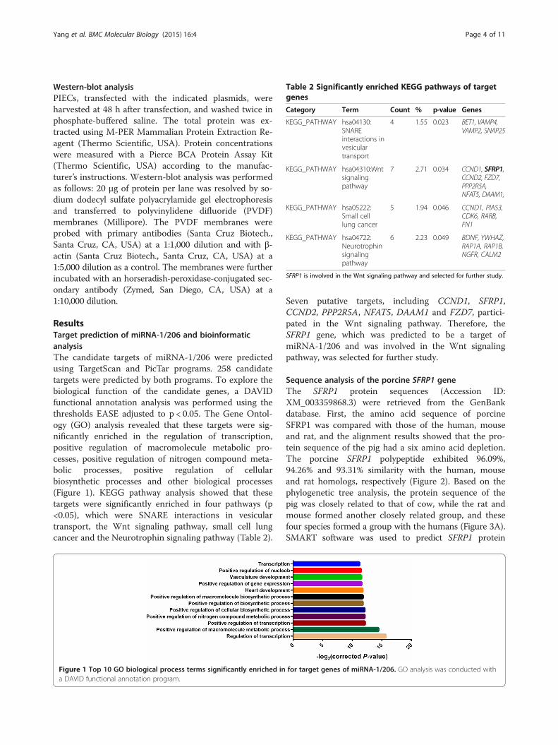
Table 2 Significantly enriched KEGG pathways of targetgenes
Category Term Count % p-value Genes
KEGG_PATHWAY hsa04130:SNAREinteractions invesiculartransport
4 1.55 0.023 BET1, VAMP4,VAMP2, SNAP25
KEGG_PATHWAY hsa04310:Wntsignalingpathway
7 2.71 0.034 CCND1, SFRP1,CCND2, FZD7,PPP2R5A,NFAT5, DAAM1,
KEGG_PATHWAY hsa05222:Small celllung cancer
5 1.94 0.046 CCND1, PIAS3,CDK6, RARB,FN1
KEGG_PATHWAY hsa04722:Neurotrophinsignalingpathway
6 2.23 0.049 BDNF, YWHAZ,RAP1A, RAP1B,NGFR, CALM2
SFRP1 is involved in the Wnt signaling pathway and selected for further study.
Yang et al. BMC Molecular Biology (2015) 16:4 Page 4 of 11
Western-blot analysisPIECs, transfected with the indicated plasmids, wereharvested at 48 h after transfection, and washed twice inphosphate-buffered saline. The total protein was ex-tracted using M-PER Mammalian Protein Extraction Re-agent (Thermo Scientific, USA). Protein concentrationswere measured with a Pierce BCA Protein Assay Kit(Thermo Scientific, USA) according to the manufac-turer’s instructions. Western-blot analysis was performedas follows: 20 μg of protein per lane was resolved by so-dium dodecyl sulfate polyacrylamide gel electrophoresisand transferred to polyvinylidene difluoride (PVDF)membranes (Millipore). The PVDF membranes wereprobed with primary antibodies (Santa Cruz Biotech.,Santa Cruz, CA, USA) at a 1:1,000 dilution and with β-actin (Santa Cruz Biotech., Santa Cruz, CA, USA) at a1:5,000 dilution as a control. The membranes were furtherincubated with an horseradish-peroxidase-conjugated sec-ondary antibody (Zymed, San Diego, CA, USA) at a1:10,000 dilution.
ResultsTarget prediction of miRNA-1/206 and bioinformaticanalysisThe candidate targets of miRNA-1/206 were predictedusing TargetScan and PicTar programs. 258 candidatetargets were predicted by both programs. To explore thebiological function of the candidate genes, a DAVIDfunctional annotation analysis was performed using thethresholds EASE adjusted to p < 0.05. The Gene Ontol-ogy (GO) analysis revealed that these targets were sig-nificantly enriched in the regulation of transcription,positive regulation of macromolecule metabolic pro-cesses, positive regulation of nitrogen compound meta-bolic processes, positive regulation of cellularbiosynthetic processes and other biological processes(Figure 1). KEGG pathway analysis showed that thesetargets were significantly enriched in four pathways (p<0.05), which were SNARE interactions in vesiculartransport, the Wnt signaling pathway, small cell lungcancer and the Neurotrophin signaling pathway (Table 2).
Figure 1 Top 10 GO biological process terms significantly enriched ina DAVID functional annotation program.
Seven putative targets, including CCND1, SFRP1,CCND2, PPP2R5A, NFAT5, DAAM1 and FZD7, partici-pated in the Wnt signaling pathway. Therefore, theSFRP1 gene, which was predicted to be a target ofmiRNA-1/206 and was involved in the Wnt signalingpathway, was selected for further study.
Sequence analysis of the porcine SFRP1 geneThe SFRP1 protein sequences (Accession ID:XM_003359868.3) were retrieved from the GenBankdatabase. First, the amino acid sequence of porcineSFRP1 was compared with those of the human, mouseand rat, and the alignment results showed that the pro-tein sequence of the pig had a six amino acid depletion.The porcine SFRP1 polypeptide exhibited 96.09%,94.26% and 93.31% similarity with the human, mouseand rat homologs, respectively (Figure 2). Based on thephylogenetic tree analysis, the protein sequence of thepig was closely related to that of cow, while the rat andmouse formed another closely related group, and thesefour species formed a group with the humans (Figure 3A).SMART software was used to predict SFRP1 protein
for target genes of miRNA-1/206. GO analysis was conducted with

Figure 2 Comparison of porcine SFRP1 sequence (GenBank accession ID: XP_003359916.2) with those of human (GenBank accessionID: NP_003003.3), mouse (GenBank accession ID: NP_038862.2) and rat (GenBank accession no. NP_001263641.1) sequence. Shadingshows identical and similar amino acid residues among the four species. Common structural domains are indicated by boxes including a Frizzleddomain and a Netrin C-terminal domain.
Yang et al. BMC Molecular Biology (2015) 16:4 Page 5 of 11
domains. The precursor protein of SFRP1 contained aFrizzled domain and a Netrin C-terminal domain(Figure 3B). The sites of SFRP1 protein localizationhad a 44.4% possibility of being extracellular, a22.2% possibility of being cytoplasmic and a 22.2%possibility of being in the endoplasmic reticulum.Additionally, the theoretical isoelectric point (pI)and molecular weight (Mw) of SFRP1 were 9.05 and34.79 KDa, respectively.
Distribution of porcine SFRP1 mRNA and miRNA-206 intissuesThe expression of SFRP1 and miRNA-206 was mea-sured in seven tissues from adult Tongcheng pigs. TheSFRP1 gene was highly expressed in the kidney, liver,lung, spleen and small intestine, moderately expressedin the heart, and weakly expressed in the longissimusdorsi muscle (Figure 4A). However, miRNA-206 wasabundantly expressed in the longissimus dorsi muscleand heart, and was weakly expressed in the other tis-sues (Figure 4B).
Figure 3 Phylogenetic tree and domains of porcine SFRP1 protein. (Aaccession numbers of those sequences are as follows: pig, XP_003359916.2mouse, NP_038862.2, and chicken, NP_989884.1. (B) Porcine SFRP1 proteindomain and a Netrin C-terminal domain.
Developmental expression of porcine SFRP1 mRNA andmiRNA-206We collected the longissimus dorsi muscle from 20 pre-natal and postnatal developmental stages of Tongchengpigs. Quantitative real-time PCR indicated that SFRP1was highly expressed at the E33 stage, and was thendown-regulated from E33 to E55. It was then up-regulated from E55 to E70 and down-regulated fromE70 to E85. Subsequently, it was up-regulated from theE85 to E95, and then down-regulated again from E95 toD0. The expression of SFRP1 was maintained at a stablelow level in postnatal skeletal muscle. In postnatal myo-genesis, SFRP1 was consistently expressed from days 0to 40. Subsequently, it decreased and reached a mini-mum at D100 (Figure 4C). The expression of miRNA-206 was weak in skeletal muscle in the early embryonicstages, but then remained stable with high levels of ex-pression in the remaining prenatal stages, although somefluctuations occurred. The expression level of miRNA-206 reached a peak at E100. In postnatal muscle,miRNA-206 was down-regulated from D0 to D40 andwas up-regulated from D40 to D100 (Figure 4D).
) Phylogenetic tree of SFRP1 from different species. The GenBank; human, NP_003003.3; cattle,NP_776885.1; rat, NP_001263641.1;domains. The porcine SFRP1 precursor protein contains a Frizzled

Figure 4 Relative expression of SFRP1and miRNA-206 in the different tissues and in skeletal muscle during development of Tongchengpigs. (A) Tissue distribution of SFRP1 in the adult Tongcheng pig. (B) Tissue distribution of miRNA-206 in the adult Tongcheng pig. (C) Relativeexpression of SFRP1 mRNA and (D) miRNA-1/206 in porcine skeletal muscle from Tongcheng pigs at different developmental stages (20 stages).miRNA-1 expression in Tongcheng pigs is cited from Tang et al. [35]. The values are the average (±SE) levels of SFRP1 mRNA and miRNA-1/206from three independent experiments normalized to GAPDH and U6, respectively. In each group, the lowest expression value was arbitrarily set to0 by log10 transformation to evaluate the relative expression levels.
Yang et al. BMC Molecular Biology (2015) 16:4 Page 6 of 11
Co-expression analysis of SFRP1 and microRNA-1/206Tang et al. have explored the spatial and dynamic ex-pression of miRNA-1 in the Tongcheng pig (Figure 4D)[35]. According to their study and this study, miRNA-1 and miRNA-206 were abundantly expressed in skel-etal muscle and weakly expressed in other tissues. Incontrast, SFRP1 was expressed at very low levels inadult skeletal muscle. The co-expression analysis re-vealed that the SFPR1 mRNA was significantly nega-tively correlated with miRNA-1 (Pearson’s RSFRP1/
miRNA-1 = −0.928, p value = 0.003) (Figure 5A) andmiRNA-206 (Pearson’s RSFRP1/miRNA-1 = −0.922, p value =0.003) (Figure 5B) at the mRNA level in different tissuesadult tissues.During skeletal muscle development, SFPR1 had a
higher expression level in the prenatal stages than inthe postnatal stages. However, in contrast to SFRP1,miRNA-1/206 exhibited a relatively higher level ofexpression in postnatal muscle compared with pre-natal muscle. SFRP1 mRNA was significantly nega-tively correlated with miRNA-1/206 [Pearson’sRSFRP1/miRNA-1 = −0.445, pvalue = 0.032 (Figure 5C);Pearson’s RSFRP1/miRNA-206 = −0.480, p value = 0.049(Figure 5D)]. These results indicated that SFRP1 ex-pression might be regulated by miR-1/206 in pigs.
SFRP1 is a putative target of microRNA-1/206The prediction from the miRNA-mRNA profiles andbioinformatics suggested that SFRP1 was potentially amiRNA-1/206 target in pigs. In the 3′-UTR region ofSFRP1 mRNA, a putative binding site was identified(7 bp conserved homology) (Figure 6A). The seed se-quence of miRNA-1/206 and the target-binding sitebetween SFRP1 and miRNA-1/ 206 were highly con-served across mammals (Figure 7).To validate whether SFRP1 was directly targeted
by miRNA-1/206 in pigs, we constructed thepsiCheck2-SFRP1-3′-UTR , a luciferase reporter vec-tor. Subsequently, miRNA-1and miRNA-206 mimicsand a normal control (NC) were co-transfected intoPIECs, and luciferase activity was detected. ThemiRNA-1 mimic-transfected group exhibited 68.29%less luciferase activity compared with the NC group(p < 0.01) and the miRNA-206 mimic-transfectedgroup exhibited 71.25% less luciferase activity comparedwith the control (p < 0.01) (Figure 8). To further validatethe specific target site, the binding region of the SFRP1 3′-UTR was mutated by bridge PCR (Figure 6B). The lucifer-ase activity of the psiCHECK-2-SFRP1–3′-UTR (mut) wasnot significantly decreased by both the miRNA-1 andmiRNA-206 mimics (12.39% of control for the miRNA-1

Figure 5 Correlation analyses of miRNA-1/206 and SFRP1 expression. (A) Pearson’s correlation between SFRP1 and miRNA-1 expression indifferent tissues. (B) Pearson’s correlation between SFRP1 and miRNA-206 expression in different tissues. (C) Pearson’s correlation between SFRP1and miRNA-1 expression during skeletal muscle development. (D) Pearson’s correlation between SFRP1 and miRNA-206 expression during skeletalmuscle development.
Yang et al. BMC Molecular Biology (2015) 16:4 Page 7 of 11
mimic-transfected group and 16.76% of control for themiRNA-206 mimic-transfected group) (p >0.05) (Figure 8).Then, we determined whether SFRP1 was affected by
miRNA-1/206 at the protein level. The overexpressionvector of SFRP1 was constructed and transfected intoPIECs, and the quantitative real-time PCR (qPCR) re-sults showed that the expression of SFRP1 was increasedapproximately 16-fold compared with the NC group.This result indicated that the SFRP1 overexpression vec-tor was successful (Figure 9A). The porcine SFRP1-CDS-3′-UTR-psiCHECK-2 vector was constructed, andit was co-transfected with miRNA-1 and miRNA-206mimics in PIECs. The Western blot results showed thatthe protein level of SFRP1 in the groups containingmiRNA-1/206 mimics was decreased compared with theNC group (Figure 9B). These results suggested that theSFRP1 gene was a target of miRNA-1/206.
DiscussionWe predicted the targets of miRNA-1/206 with the Tar-getScan and PicTar programs. Among the putative tar-gets, many genes had been validated by previous studies,such as HDAC4 [29], GJA1, KCNJ2 [36], Pola1 [28], andMet [37]. However, all these results were based on myo-blast C2C12 cells in vitro. Few reports have consideredmiRNA-1/206 targets during skeletal muscle development
in vivo, particularly in pigs. To discover potential targetsof miRNA-1/206 during swine myogenesis, we conductedGO and KEGG pathway analyses of targets based on theprediction data. The results suggested that these targetswere significantly enriched in the Wnt signaling pathway(p < 0.05). Wnts signaling proteins are secreted proteinsthat function in differentiation, embryonic developmentand cell proliferation [38]. The Wnt pathways play an im-portant role in the formation of muscle fibers during pre-natal [39] and postnatal myogenesis with the activation ofstem cells in the adult muscles [40,41]. SFRP1, a secretedantagonist of the Wnt-Frizzled pathway, was predicted tobe a target of miRNA-1/206 and to participate in the Wntsignaling pathway. The expression of SFRP1 mRNAwas up-regulated in muscle regeneration [42] and inskeletal muscle after denervation [43]. Additionally,an impairment of the Wnt-Frizzled pathway viaSFRP1 over-expression controlled proliferation andneovascularization after muscle ischemia [13]. A com-parison of the amino acid sequence of pig SFRP1with those of human, rat and mouse demonstratedremarkably high similarity across species, and thefrizzled domain and netrin C-terminal domain wereespecially highly conserved.Previous studies reported SFRP1 expression at the
mRNA level in various tissues, such as the brain, kidney

Figure 6 SFRP1 3′-UTR has miR-1/206 target binding sites. (A) SFRP1 was predicted as a target of microRNA-1/206. (B) Schematic of the predictedmiRNA-1 and miRNA-206 binding sites (underlined) in the 3′ UTR of SFRP1. The binding site region was deleted in the mutant 3′ -UTR reporters.
Yang et al. BMC Molecular Biology (2015) 16:4 Page 8 of 11
and heart, and that the transcript was present both inthe adult and during embryogenesis [7,44]. Moreover,SFRP1 was weakly expressed in matured skeletal muscles[45]. The qPCR results for SFRP1 in various adult por-cine tissues were mostly consistent with these reports.These results indicated that SFRP1 had almost no antag-onistic effect in adult skeletal muscle. Postnatal musclegrowth is largely determined by the total number of fi-bers, which is determined by two major waves of fibergeneration before birth: primary muscle fiber formationat 35–60 dpc and assemblage of secondary muscle fibersat 54–90 dpc [46-48]. The SFRP1 gene was down-regulatedin skeletal muscle from E33 to E55 in Tongcheng pigs, indi-cating that SFRP1 was primarily involved in the formationof primary muscle fibers. We also found that the expressionof SFRP1 was higher in embryonic skeletal muscle com-pared with postnatal skeletal muscle. miRNA-206, mean-while, was abundantly expressed in skeletal muscle andheart, and up-regulated from pre- to postnatal-stage skeletal
Figure 7 Predicted miRNA-1 and miRNA-206 binding sites (highlighte(A) Seed sequence of miRNA-1 showing species conservation. (B) Seed seq(highlighted in red) in the 3′-UTR of SFRP1 showing species conservation.
muscle. These results demonstrated that miR-206 plays akey role in skeletal muscle development in pigs. SFRP1 pri-marily affected skeletal muscle development in embryonicstages. Wnts signaling contributes to the overall process ofmyogenesis by activating myogenic regulatory factor genessuch as Myf5 and MyoD [49]. miRNA-1/206 promotedskeletal muscle satellite cell proliferation and differentiation[27] and SFRP1 might inhibit myoblast differentiation [12].The temporal expression patterns of miRNA-1/206 andSFRP1 in Tongcheng pigs were consistent with these previ-ous findings.The mRNA-miRNA co-expression correlation ana-
lysis was reported to identify the putative targets ofmiRNA [50,51], and this method could improve thepositive rate for identifying the mRNA target genesof miRNA. miRNA-1 and miRNA-206 were abun-dant in the postnatal stages and were at low levelsin the prenatal stages of muscle development, whileSFRP1 exhibited an opposite expression patterns.
d in red) in the 3′-UTR of SFRP1 showing species conservation.uence of miRNA-206 showing species conservation. (C) Binding sites

Figure 8 Validating SFRP1 as a positive target for miRNA-1 and miRNA-206. Cotransfection of porcine pre-miRNA-1 (A) and pre-miRNA-206(B) or control and porcine SFRP1 UTR-derived psiCHECK-2 construct or mutant in PIEC cells. Renilla activity at 48 h post-transfection shows a significantdecrease in normalized values compared with the control and mutant. Three replicates were performed for each group. **Indicates a p-value of lessthan 0.01 in Student’s t-test.
Yang et al. BMC Molecular Biology (2015) 16:4 Page 9 of 11
Correlation analysis revealed that the SFPR1 was sig-nificantly negatively correlated with miRNA-1/206(p < 0.05), and these results indicate that SFRP1 ispotentially regulated by miRNA-1/206.The interaction between SFRP1 and miRNA-1/206 in
pigs has not been previously reported. This study dem-onstrated that SFRP1 expression was regulated bymiRNA-1/206. The SFRP1 3′-UTR sequence around themiRNA-1/206 target sites and the seed sequence of ma-ture miRNA-1/206 are well conserved in mammals,which suggests that the target region is important inSFRP1 regulation and that the regulation of SFRP1 bymiRNA-1/206 may also exist in other species. Similar re-sults were observed in other miRNA studies [30,52].The luciferase activity of psiCHECK-2 containing
the SFRP1 3′-UTR sequence was significantly de-creased by co-transfection with miRNA-1/206mimics (p < 0.05). However, with the SFRP1 3′-UTRmutant sequence, activity was not significantly de-creased (p > 0.05). These results indicated that thetarget binding site was specific and unique in pigs,and that miRNA-1/206 might repress SFRP1 expres-sion by degrading the mRNA transcripts. We propose
Figure 9 The miRNA-1 and miRNA-206 regulate SFRP1 at the proteinindicates overexpression of SFRP1 48 h after transfection. (B) miRNA-1 and miSFRP1 was normalized against β-actin.
that SFRP1 is regulated by miRNA-1/206, is >involvedin the proliferation of muscle cells, and affects pre-natal skeletal muscle development. Moreover, we ex-plored the interactions between SFRP1 and miRNA-1/206 at the protein level, and Western-blot analysisconfirmed that SFRP1 was significantly down-regulated by miRNA-1/206.
ConclusionsIn summary, we predicted the target genes of miRNA-1/206and performed a functional annotation of the target genes.We performed a molecular characterization analysis of theporcine SFRP1 gene,, which was one of the predicted targetsfor miRNA-1/206. We also explored the spatial-temporalprofile of SFRP1 mRNA and miRNA-206 in adult tissuesand in skeletal muscle tissues during development in Tong-cheng pigs. The results indicated that SFRP1 was primarilyinvolved in prenatal skeletal muscle development. Finally, weverified that porcine SFRP1 was a target of miRNA-1/206using dual luciferase and Western blot assay. These resultsincrease our understanding of the biological functions ofSFRP1 and miRNA-1/206 in skeletal muscle development.
level. (A) Overexpression of porcine SFRP1 in PIEC cells. HistogramRNA-206 down-regulated the SFRP1 at the protein level. The expression of

Yang et al. BMC Molecular Biology (2015) 16:4 Page 10 of 11
AbbreviationsSFRP1: Secreted frizzled-related protein 1; MRFs: Myogenic regulatory factors;PIECs: Porcine iliac endothelial cells; HDAC4: Histone deacetylase 4; Pola1: thelargest subunit of DNA polymerase α; miRNA: microRNA; NC: Normal control;GAPDH: Glyceraldehyde-3-phosphate dehydrogenase; qPCR: quantitativereal-time PCR.
Competing interestsThe authors declare that they have no competing interests.
Authors’ contributionsYY, WS, CL and ZT conceived and designed the experiments. YY, WS and RWperformed the experiments. YY and RZ analyzed data. YY and WS drafted themanuscript. KL and ZT critically revised the manuscript. All authorscontributed to revision of the manuscript and approved the final version.
AcknowledgmentsThe authors are grateful to Dr. Jianhua Cao from Huazhong AgriculturalUniversity for providing the psiCheck-2 vector and to Sanping Xu in theTongcheng Animal Husbandry Bureau in Hubei Province for sample collection.This work was supported by the National Key Project (2014ZX08009-001), theNational Basic Research Program of China (2015CB943101, 2012CB124706), theNational Natural Science Foundation of China (31372295, 31330074) and theAgricultural Science and Technology Innovation Program (ASTIP-IAS05).
Author details1Key Laboratory of Farm Animal Genetic Resources and GermplasmInnovation of Ministry of Agriculture, Institute of Animal Science, ChineseAcademy of Agricultural Sciences, Beijing 100193, P.R. China. 2AgriculturalGenome Institute at Shenzhen, Chinese Academy of Agricultural Sciences,Shenzhen 518124, P.R. China. 3College of Animal Science and Technology,Northwest A & F University, No. 22 Xinong Road, 712100 Yangling, Shanxi,P.R. China.
Received: 29 October 2014 Accepted: 19 February 2015
References1. von Maltzahn J, Chang NC, Bentzinger CF, Rudnicki MA. Wnt signaling in
myogenesis. Trends Cell Biol. 2012;22(11):602–9.2. Cisternas P, Henriquez JP, Brandan E, Inestrosa NC. Wnt signaling in skeletal
muscle dynamics: myogenesis, neuromuscular synapse and fibrosis. MolNeurobiol. 2014;49(1):574–89.
3. Ridgeway AG, Petropoulos H, Wilton S, Skerjanc IS. Wnt signaling regulatesthe function of MyoD and myogenin. J Biol Chem. 2000;275(42):32398–405.
4. Rudnicki MA, Le Grand F, McKinnell I, Kuang S. The molecular regulation ofmuscle stem cell function. Cold Spring Harb Symp Quant Biol. 2008;73:323–31.
5. Vertino AM, Taylor-Jones JM, Longo KA, Bearden ED, Lane TF, McGehee JrRE, et al. Wnt10b deficiency promotes coexpression of myogenic and adipogenicprograms in myoblasts. Mol Biol Cell. 2005;16(4):2039–48.
6. Otto A, Schmidt C, Luke G, Allen S, Valasek P, Muntoni F, et al. CanonicalWnt signalling induces satellite-cell proliferation during adult skeletal muscleregeneration. J Cell Sci. 2008;121(Pt 17):2939–50.
7. Rattner A, Hsieh JC, Smallwood PM, Gilbert DJ, Copeland NG, Jenkins NA,et al. A family of secreted proteins contains homology to the cysteine-richligand-binding domain of frizzled receptors. Proc Natl Acad Sci U S A.1997;94(7):2859–63.
8. Xu Q, D'Amore PA, Sokol SY. Functional and biochemical interactions ofWnts with FrzA, a secreted Wnt antagonist. Development. 1998;125(23):4767–76.
9. Jones SE, Jomary C. Secreted Frizzled-related proteins: searching for relationshipsand patterns. Bioessays. 2002;24(9):811–20.
10. Chen Y, Stump RJ, Lovicu FJ, McAvoy JW. Expression of Frizzleds and secretedfrizzled-related proteins (Sfrps) during mammalian lens development. Int J DevBiol. 2004;48(8–9):867–77.
11. Lavergne E, Hendaoui I, Coulouarn C, Ribault C, Leseur J, Eliat PA, et al.Blocking Wnt signaling by SFRP-like molecules inhibits in vivo cell prolifera-tion and tumor growth in cells carrying active beta-catenin.Oncogene. 2010;30(4):423–33.
12. Descamps S, Arzouk H, Bacou F, Bernardi H, Fedon Y, Gay S, et al. Inhibitionof myoblast differentiation by Sfrp1 and Sfrp2. Cell Tissue Res. 2008;332(2):299–306.
13. Ezan J, Leroux L, Barandon L, Dufourcq P, Jaspard B, Moreau C, et al. FrzA/sFRP-1, a secreted antagonist of the Wnt-Frizzled pathway, controls vascularcell proliferation in vitro and in vivo. Cardiovasc Res. 2004;63(4):731–8.
14. Bartel DP. MicroRNAs: genomics, biogenesis, mechanism, and function. Cell.2004;116(2):281–97.
15. He L, Hannon GJ. MicroRNAs: small RNAs with a big role in gene regulation.Nat Rev Genet. 2004;5(7):522–31.
16. Carrington JC, Ambros V. Role of microRNAs in plant and animaldevelopment. Science. 2003;301(5631):336–8.
17. Chen X, Ba Y, Ma L, Cai X, Yin Y, Wang K, et al. Characterization ofmicroRNAs in serum: a novel class of biomarkers for diagnosis of cancerand other diseases. Cell Res. 2008;18(10):997–1006.
18. Ren J, Huang HJ, Gong Y, Yue S, Tang LM, Cheng SY. MicroRNA-206 suppressesgastric cancer cell growth and metastasis. Cell Biosci. 2014;4:26.
19. Tay Y, Zhang J, Thomson AM, Lim B, Rigoutsos I. MicroRNAs to Nanog, Oct4and Sox2 coding regions modulate embryonic stem cell differentiation.Nature. 2008;455(7216):1124–8.
20. Cimmino A, Calin GA, Fabbri M, Iorio MV, Ferracin M, Shimizu M, et al. miR-15and miR-16 induce apoptosis by targeting BCL2. Proc Natl Acad Sci U S A.2005;102(39):13944–9.
21. Krutzfeldt J, Stoffel M. MicroRNAs: a new class of regulatory genes affectingmetabolism. Cell Metab. 2006;4(1):9–12.
22. Kloosterman WP, Plasterk RH. The diverse functions of microRNAs in animaldevelopment and disease. Dev Cell. 2006;11(4):441–50.
23. McCarthy JJ. MicroRNA-206: the skeletal muscle-specific myomiR. BiochimBiophys Acta. 2008;1779(11):682–91.
24. McCarthy JJ, Esser KA. MicroRNA-1 and microRNA-133a expression are de-creased during skeletal muscle hypertrophy. J Appl Physiol. 2007;102(1):306–13.
25. Xu C, Lu Y, Pan Z, Chu W, Luo X, Lin H, et al. The muscle-specific microRNAsmiR-1 and miR-133 produce opposing effects on apoptosis by targetingHSP60, HSP70 and caspase-9 in cardiomyocytes. J Cell Sci. 2007;120(Pt 17):3045–52.
26. Baskerville S, Bartel DP. Microarray profiling of microRNAs reveals frequentcoexpression with neighboring miRNAs and host genes. RNA. 2005;11(3):241–7.
27. Chen JF, Tao Y, Li J, Deng Z, Yan Z, Xiao X, et al. microRNA-1 and microRNA-206regulate skeletal muscle satellite cell proliferation and differentiation by repressingPax7. J Cell Biol. 2010;190(5):867–79.
28. Kim HK, Lee YS, Sivaprasad U, Malhotra A, Dutta A. Muscle-specific microRNAmiR-206 promotes muscle differentiation. J Cell Biol. 2006;174(5):677–87.
29. Chen JF, Mandel EM, Thomson JM, Wu Q, Callis TE, Hammond SM, et al. Therole of microRNA-1 and microRNA-133 in skeletal muscle proliferation anddifferentiation. Nat Genet. 2006;38(2):228–33.
30. Hou X, Tang Z, Liu H, Wang N, Ju H, Li K. Discovery of MicroRNAs associatedwith myogenesis by deep sequencing of serial developmental skeletalmuscles in pigs. PLoS One. 2012;7(12):e52123.
31. da Huang W, Sherman BT, Lempicki RA. Systematic and integrative analysisof large gene lists using DAVID bioinformatics resources. Nat Protoc.2009;4(1):44–57.
32. Tamura K, Peterson D, Peterson N, Stecher G, Nei M, Kumar S. MEGA5:molecular evolutionary genetics analysis using maximum likelihood,evolutionary distance, and maximum parsimony methods. Mol Biol Evol.2011;28(10):2731–9.
33. Kozomara A, Griffiths-Jones S. miRBase: annotating high confidence microRNAsusing deep sequencing data. Nucleic Acids Res. 2014;42(Database issue):D68–73.
34. Ai J, Zhang R, Li Y, Pu J, Lu Y, Jiao J, et al. Circulating microRNA-1 as a potentialnovel biomarker for acute myocardial infarction. Biochem Biophys Res Commun.2010;391(1):73–7.
35. Tang Z, Liang R, Zhao S, Wang R, Huang R, Li K. CNN3 Is Regulated bymicroRNA-1 during Muscle Development in Pigs. Int J Biol Sci. 2014;10(4):377–85.
36. Yang B, Lin H, Xiao J, Lu Y, Luo X, Li B, et al. The muscle-specific microRNAmiR-1 regulates cardiac arrhythmogenic potential by targeting GJA1 andKCNJ2. Nat Med. 2007;13(4):486–91.
37. Taulli R, Bersani F, Foglizzo V, Linari A, Vigna E, Ladanyi M, et al. The muscle-specific microRNA miR-206 blocks human rhabdomyosarcoma growth inxenotransplanted mice by promoting myogenic differentiation. J Clin Invest.2009;119(8):2366–78.

Yang et al. BMC Molecular Biology (2015) 16:4 Page 11 of 11
38. Cadigan KM, Nusse R. Wnt signaling: a common theme in animaldevelopment. Genes Dev. 1997;11(24):3286–305.
39. van Amerongen R, Berns A. Knockout mouse models to study Wnt signaltransduction. Trends Genet. 2006;22(12):678–89.
40. Tsivitse S. Notch and Wnt signaling, physiological stimuli and postnatalmyogenesis. Int J Biol Sci. 2010;6(3):268–81.
41. Steelman CA, Recknor JC, Nettleton D, Reecy JM. Transcriptional profiling ofmyostatin-knockout mice implicates Wnt signaling in postnatal skeletalmuscle growth and hypertrophy. FASEB J. 2006;20(3):580–2.
42. Zhao P, Hoffman EP. Embryonic myogenesis pathways in muscleregeneration. Dev Dyn. 2004;229(2):380–92.
43. Svensson A, Norrby M, Libelius R, Tagerud S. Secreted frizzled relatedprotein 1 (Sfrp1) and Wnt signaling in innervated and denervated skeletalmuscle. J Mol Histol. 2008;39(3):329–37.
44. Finch PW, He X, Kelley MJ, Uren A, Schaudies RP, Popescu NC, et al.Purification and molecular cloning of a secreted, Frizzled-related antagonistof Wnt action. Proc Natl Acad Sci U S A. 1997;94(13):6770–5.
45. Melkonyan HS, Chang WC, Shapiro JP, Mahadevappa M, Fitzpatrick PA,Kiefer MC, et al. SARPs: a family of secreted apoptosis-related proteins. ProcNatl Acad Sci U S A. 1997;94(25):13636–41.
46. Picard B, Lefaucheur L, Berri C, Duclos MJ. Muscle fibre ontogenesis in farmanimal species. Reprod Nutr Dev. 2002;42(5):415–31.
47. Lefaucheur L, Edom F, Ecolan P, Butler-Browne GS. Pattern of muscle fibertype formation in the pig. Dev Dyn. 1995;203(1):27–41.
48. Wigmore PM, Stickland NC. Muscle development in large and small pigfetuses. J Anat. 1983;137(Pt 2):235–45.
49. Cossu G, Borello U. Wnt signaling and the activation of myogenesis inmammals. EMBO J. 1999;18(24):6867–72.
50. Nagalla S, Shaw C, Kong X, Kondkar AA, Edelstein LC, Ma L, et al. PlateletmicroRNA-mRNA coexpression profiles correlate with platelet reactivity.Blood. 2011;117(19):5189–97.
51. Ponsuksili S, Du Y, Hadlich F, Siengdee P, Murani E, Schwerin M, et al.Correlated mRNAs and miRNAs from co-expression and regulatory networksaffect porcine muscle and finally meat properties. BMC Genomics.2013;14:533.
52. Zhao S, Zhang J, Hou X, Zan L, Wang N, Tang Z, et al. OLFML3 expression isdecreased during prenatal muscle development and regulated bymicroRNA-155 in pigs. Int J Biol Sci. 2012;8(4):459–69.
Submit your next manuscript to BioMed Centraland take full advantage of:
• Convenient online submission
• Thorough peer review
• No space constraints or color figure charges
• Immediate publication on acceptance
• Inclusion in PubMed, CAS, Scopus and Google Scholar
• Research which is freely available for redistribution
Submit your manuscript at www.biomedcentral.com/submit