who list of priority medical devices for cancer management
TRANSCRIPT

WHO list of priority medical devices for cancer managementWHO Medical device technical series
Breast C
ervical Child
hood
Co
lon
Le
uke
mia
Lu
ng
Pro
state

WHO list of priority medical devices for cancer management
(WHO Medical device technical series)
ISBN 978-92-4-156546-2
© World Health Organization 2017
Some rights reserved. This work is available under the Creative Commons Attribution-NonCommercial-ShareAlike
3.0 IGO licence (CC BY-NC-SA 3.0 IGO; https://creativecommons.org/licenses/by-nc-sa/3.0/igo).
Under the terms of this licence, you may copy, redistribute and adapt the work for non-commercial purposes,
provided the work is appropriately cited, as indicated below. In any use of this work, there should be no
suggestion that WHO endorses any specific organization, products or services. The use of the WHO logo is
not permitted. If you adapt the work, then you must license your work under the same or equivalent Creative
Commons licence. If you create a translation of this work, you should add the following disclaimer along with
the suggested citation: “This translation was not created by the World Health Organization (WHO). WHO is not
responsible for the content or accuracy of this translation. The original English edition shall be the binding and
authentic edition”.
Any mediation relating to disputes arising under the licence shall be conducted in accordance with the mediation
rules of the World Intellectual Property Organization.
Suggested citation. WHO list of priority medical devices for cancer management. Geneva: World Health
Organization; 2017. Licence: CC BY-NC-SA 3.0 IGO.
Cataloguing-in-Publication (CIP) data. CIP data are available at http://apps.who.int/iris.
Sales, rights and licensing. To purchase WHO publications, see http://apps.who.int/bookorders. To submit
requests for commercial use and queries on rights and licensing, see http://www.who.int/about/licensing.
Third-party materials. If you wish to reuse material from this work that is attributed to a third party, such as
tables, figures or images, it is your responsibility to determine whether permission is needed for that reuse and
to obtain permission from the copyright holder. The risk of claims resulting from infringement of any third-party-
owned component in the work rests solely with the user.
General disclaimers. The designations employed and the presentation of the material in this publication do not
imply the expression of any opinion whatsoever on the part of WHO concerning the legal status of any country,
territory, city or area or of its authorities, or concerning the delimitation of its frontiers or boundaries. Dotted and
dashed lines on maps represent approximate border lines for which there may not yet be full agreement.
The mention of specific companies or of certain manufacturers’ products does not imply that they are endorsed
or recommended by WHO in preference to others of a similar nature that are not mentioned. Errors and omissions
excepted, the names of proprietary products are distinguished by initial capital letters.
All reasonable precautions have been taken by WHO to verify the information contained in this publication.
However, the published material is being distributed without warranty of any kind, either expressed or implied.
The responsibility for the interpretation and use of the material lies with the reader. In no event shall WHO be
liable for damages arising from its use.
Design and layout of the document - Jillian Reichenbach Ott (Genève Design).
Printed by the WHO Document Production Services, Geneva, Switzerland

WHO list of priority medical devices for cancer managementWHO Medical device technical series

Acknowledgemen
PHOTO CREDITS:
Vaccination photo: WHO/PAHO 2014. Young girls getting Human papillomavirus (HPV) vaccination in their
school, in São Paulo, Brazil.
Pathology photo: Dr Jagdish Butany, Toronto General Hospital, Histology Laboratory, Toronto, Canada.
Medical Imaging photo: Dr Adela Poitevin, Médica Sur, Tlalpan, Mexico City, Mexico.
Radiotherapy photo: Dr Adela Poitevin, Mexico, Médica Sur, Tlalpan, Mexico City, Mexico.
Nuclear Medicine photo: Dr Cecil Chow Robilotta, Centro de Medicina Nuclear, Instituto de Radiologia, São
Paulo, Brazil.
Palliative care photo: Anthony Greenwood, April 2016, Lancaster, UK. Photo provided by Dr Sheila Payne.
Surgery photo: Dr André Ilbawi, May 2016, Butaro Hospital, Rwanda.
Systemic therapy: Colleen Nixon, MSN RN CPHON, Nurse Educator, Boston Children’s Hospital Boston, MA.
Photo provided by Lisa Morrissey, RN.
Cover photo 1: Dr Diana Paez, IAEA. Photo was taken at the Department of Nuclear Medicine of the National
Cancer Institute of Mexico City.
Cover photo 2 - Pathology slide: Dr Jagdish Butany, Toronto General Hospital, Histology Laboratory,
Toronto, Canada.
Cover photo 3 - Nurse and child: Colleen Nixon, MSN RN CPHON, Nurse Educator, Boston Children’s
Hospital Boston, MA.
Cover photo 4 - Surgery: Dr André Ilbawi, May 2016, Butaro Hospital, Rwanda.

e
This project was developed by the World Health Organization (WHO) in response to the need for a model reference
list of basic and priority medical devices required for cancer management, with the goal of increasing access to
these medical devices especially in low- and middle-income countries . The project was developed under the overall
coordination of Adriana Velazquez Berumen, Senior Advisor and Focal Point on Medical Devices from the Innovation,
Access and Use unit of the Essential Medicines and Health Products Department, directed by Dr Suzanne Hill, under
the Health Systems and Innovation Cluster.
Special collaboration was received from Cherian Varghese, Coordinator, and Andre Ilbawi from the Noncommunicable
Diseases Management Department, as well as from James Campbell, Director, Rania Kawar, Tana Wuliji and Teena
Kunjumen of Health Workforce Department in WHO.
Technical support was provided mainly by Gabriela Jimenez Moyao, WHO temporary staff in 2015-2016, and Natalia
Rodriguez, intern in 2016. Additional support for the project was provided by Marina Alfons, Karin Diaconu, Mireille
Goetghebeur, Antonio Migliore, and Miriam Mikhail. Final editorial review by Maurice Page and Corrado Gemma.
WHO specially acknowledges May Abdel-Wahab and all staff from the Division of Human Health (NAHU) of the
International Atomic Energy Agency (IAEA) for all the technical input provided.
The primary financial support for this study was provided by the OPEC Fund for International Development (OFID).
WHO acknowledges and is most grateful to all the participants of the meetings and consultations, the Steering
Committee members and the experts who provided advice in the development of this document, as follows:
April 2015 - General Consultation Participants:
May Abdel-Wahab, Nicholas Adjabu, Benjamin O. Anderson, Damien Bertrand, Vikram Bhadrasain, Jolanta Bilinska,
Jagdish Butany, Eduardo Cazap, Nicole Denjoy, Ahmed Elzawawy, Alexandru Eniu, Temidayo Fadelu, Shamiram
Feinglass, John Flanigan, Birgit Fleurent, Xavier Franz, Ophira Ginsburg, Zelalem Gizachew, Mary Gospodarowicz,
S. W. Gunn, Leonie Hyme, Geoffrey Ibbott, Kinga Jamphel, Muditha Jayatilaka, Roberta Joppi, Mahlet Kifle,
Jitendar Kumar Sharma, Gilberto Lopes, Ratko Magjarevic, Alessandro Masellis, Joan McClure, Manuel Antonio
Munoz Aviles, Mulundi Mwanahamuntu, Dieter Nuernberg, Kofi Mensah Nyarko, Jackson Orem, Maurice Page,
Groesbeck Parham, Youlin Qiao, Sandra Rocha, Daniela Rodriguez, Ludo Scheerlinck, Julie Torode.
From WHO Regional Offices: Adham Ismail Abdel Moneim, Hanne Bak Pedersen.
From WHO Noncommunicable Diseases and Mental Health Cluster: Onyema Ajuebor, Marilys Corbex, Leanne
Riley, Andreas Ullrich, Cherian Varghese.
From WHO Health Systems and Innovation Cluster: Service Delivery and Safety Department: Katthayana
Aparicio Reyes, Marie-Charlotte Bouesseau, Meena Nathan Cherian, Nittita Prasopa-Plaizier; Health Workforce
Department: Annette Nkowane; Essential Medicines And Health Products: Marina Ashraf Alfons, Karin Daniela
Diaconu, Gilles Forte, Gabriela Jimenez Moyao, Nicola Magrini, Robyn Meurant, Antonio Migliore, Miriam Mikhail,
Megan Smith, Adriana Velazquez Berumen.
June-July 2015 - Field interviews in National Oncology Institute (INCAN) in Mexico:
Directors: Abelardo Meneses Garcia y Ángel Herrera Gómez; Gynaecology David Isla Ortiz; Urology, Miguel Ángel
Jiménez Ríos; Breast, Enrique Bargalló Rocha; Lung, Edgardo Jiménez Fuentes; Colo-rectal, Horacio Noé López
Basave and Itzel Vela; bone marrow, Ana Irma; Radiotherapy, Aida Mota García; Brachytherapy, Guadalupe
Elizabeth Trejo Durán; Clinical Laboratory , Patricia Montoya Pérez; Pathology, Hector Maldonado M; Cytology,.
Lorena Flores Hernández; Palliative care, Silvia Allende; Chemotherapy, Catalina Aguilar González; Systemic
Therapy, Odilia Tellez Miranda; Imaging , Yolanda Villaseñor Navarro; Nuclear Medicine, Osvaldo García Pérez;
Physicist, Jesús del Real; Endoscopy, Angélica Hernández Guerrero; Surgery, Gonzalo Montalvo Esquivel and
Biomedical Engineering, Sandra Rocha Nava.
September 2015 - Steering Committee Meeting Participants:
Benjamin O. Anderson, Ronald Bauer, Gouri Bhattacharyya, Bjorn Fahlgren, Mireille Goetghebeur, Mary
Gospodarowicz, Rosa Giuliani, Gilberto Lopes, Valentino Mvanga, Jitendar Sharma Kumar, Lawrence Shulman,
WHO Staff: Adham Ismail Abdel Moneim, Sheick Oumar Coulibaly, Suzanne Hill, Gabriela Jimenez Moyao, Cherian
Varghese; Adriana Velazquez Berumen. Observers: Ileana Freige, Miriam Mikhail, Ameel Mohammad, Julie Torode.
For further reference, see Annex 2.
Acknowledgements
f
WHO
list o
f prio
rity m
edica
l dev
ices f
or ca
ncer
man
agem
ent November–December 2015 - Experts, participants in teleconferences and
document editors:
May Abdel-Wahab, Nicholas Adjabu, Benjamin O. Anderson, Sizar Akoum, Ajai Basil, Angelika Bischof Delaloye,
Lori Buswell, Jagdish Butany, Bernadette Cappello, Eduardo Cazap, Laura Cedro, Kirti Chadha, Tobey Clark, Mary
Coffey, Ainhoa Costas-Chavarri, Cecil Chow Robilotta, Yadin David, Maurizio Dondi, Ahmed Elzawawy, Alexandru
Eniu, Sidnei Epelman, Temidayo Fadelu, Elena Fidarova, Patrick Fitzgibbons, John Flanigan, Ken Fleming, Claudia
Gamondi, Rosa Giuliani, Soehartati Gondhowiardjo, Surbhi Grover, Serigne Gueye, Brendan Healy, Geoffrey
Ibbott, André Ilbawi, Walter Johnson, Tom Judd, Ravi Kashyap, Michael Kawooya, Brendon Kearney, Paolo Lago,
Philip Larkin, Lai-Meng Looi, Guy Maddern, Hector Maldonado-Martinez, Melissa Martin, Miriam Mikhail, Danny
A. Milner, Jr, Lisa Morrissey, Manuel Antonio Munoz Aviles, Donna Newman, Dieter Nuernberg, Diana Paez,
Graciano Paulo, Sheila Payne, James Pepoon, Adela Poitevin, C.S. Pramesh, Madan Rehani, Sandra Rocha, Shuvro
H. Roy-Choudhury, Gustavo Sarria, Judith Shamian, Lawrence Shulman, Satchithanantham Somanesan, Gloria
Soto Giordani, Rathan Subramaniam, Richard Sullivan, Scott Triedman, Audrey Tsunoda, Jacob Van Dyk, Verna
Vanderpuye, David Watters, Michael Wilson, Gabrielle Wolff, Mei Ling Yap, Cheng-Har Yip, Eduardo Zubizarreta.
For further details can be found in Annex 2.
June 2016, Participants of the Regional consultation toStrenghen Health Systems response to address non communicable diseases in the South East Asia Region held in Colombo, Sri Lanka
From Ministries of Health: Bangladesh: Dr Abdul Alim, Mohammad Shameem Al Mamun; Bhutan: Kinga Jamphel,
Mr Tandin Dorji; DPR Korea, Kim Kyong Chol, Choe Sun Hui; India, Chinmoyee Das, Rinku Sharma; Indonesia: Aries
Hamzah, Monica Saraswati Sitepu, Mr Ibrahim Nizam, Shifaza Adam Shareef; Myanmar: Myint Shwe, Zaw Lin;
Nepal: Senendra Raj Upreti, Mohammad Daud; Sri Lanka: T. Siriwardena, DSV Mallawarachchi, D.S.D Samaraweera;
Thailand: Phattarapol Jungsomjatepaisal; Timor-Leste: Noel Gama Soares, Helder Juvinal Neto da Silva.
Invited participants: Habibullah Talukder, Chencho Dorjee, Rajesh Kumar, Hasbullah Thabrany, Ms Aishath
Shiruhana, Ko Ko, Arjun Karki, Walaiporn Pacharanaruemol, Don Matheson, Kumari Vinodhani Navaratne, Manju
Rani, Palitha Abeykoon, Salma Burton, Anika Singh. Observers: .S.Sirithunga, S. Perera. Other Agencies: Chitra
Weerakkody
WHO: WHO Country Offices: Syed Mahfuzul Huq, Tshering Dhendhup, Sadhana Bhagwat, Priska Apsari Primastuti.
WHO South-East Asia Regional Office: Thaksaphon Thamarangsi, Sunil Senanayake, Anita Kotwani, Gampo Dorji,
Puneet Dhingra, Mohita Dawar. WHO Headquarters: Cherian Varghese, Adriana Velazquez Berumen.
December 2016 - participants in Ghana Workshop on priority medical devices for cancer:
Afua Abrahams, Eric Addisson, Nicholas Adjabu, Ernest Adjei, Yvonne Dei Adomako, Samuel Kaba Akoriyea, Adu
Tutu Amankwah, Edith Annan, Francis Appillah, Adu Aryee, Judith Asiamah, Samuel Asiamah, Kwaku Ofori Atta,
Baffour Awuah, Joseph Y. B. Bennie, Ernest Osei Bonsu Baawuah, Nelson Damale, Augustine Faanu, Dayo Fadelu,
Oti Kwasi Gyamfi, JN Clegg-Lamptey, J. E. Mensah, William Addo Mills-Pappoe, Denis Ocansey, Edwina Addo
Opare Lokko, Kofi Opoku, Cathy Segbefia, Verna DNK Vanderpuye, Joel Yarney, John Zienaa.
WHO thanks the following organizations for their support to this project:
African Organisation for Research and Training in Cancer (AORTIC), American Society of Clinical Oncology
(ASCO), Breast Health Global Initiative (BHGI), Boston University’s Center for Future Technologies in Cancer
Care (CFTCC), Centro Nacional de Excelencia Tecnológica en Salud (CENETEC), Global Diagnostic Imaging,
Healthcare IT, and Radiation Therapy Trade Association (DITTA), European Society for Medical Oncology (ESMO),
EuroScan, Global Initiative for Emergency and Essential Surgical Care (GIEESC), Global Medical Technology
Alliance (GMTA), HUMATEM, Instituto Nacional de Cancerología de Mexico (INCAN), International Alliance
of Patients’ Organizations (IAPO), International Atomic Energy Agency (IAEA), International Federation of
Biomedical Laboratory Science (IFBLS), International Federation of Clinical Chemistry and Laboratory Medicine
(IFCC), International Federation of Hospital Engineering (IFHE), International Federation for Medical and
Biological Engineering (IFMBE), International Organization for Medical Physics (IOMP), International Society
of Radiographers and Radiological Technologists (ISRRT), International Society of Radiology (ISR), National
Comprehensive Cancer Network (NCCN), RAD-AID, Sociedad Latinoamericana y del Caribe de Oncología
Médica (SLACOM), Union for International Cancer Control (UICC), United Nations Children’s Fund (UNICEF),
United Nations Office for Project Services (UNOPS), World Association of Societies of Pathology and Laboratory
Medicine (WASPaLM), World Federation for Ultrasound in Medicine and Biology (WFUMB).

1
Table of contents
Acknowledgements e
Navigation diagram 4
Acronyms and abbreviations 5
Cancer colour codes 6
Executive summary 7
I. Background 8I.I WHO leadership priorities: increasing access to medical products . . . . . . . . . . . . . . . . . . .8
I.II WHO resolution on health technologies . . . . . . . . . . . . . . . . . . . . . . . . . . . . . . . . . . . . . . . . . . .8
I.III UN declaration and action plan for noncommunicable diseases . . . . . . . . . . . . . . . . . . . . . .9
I.IV The burden of cancer . . . . . . . . . . . . . . . . . . . . . . . . . . . . . . . . . . . . . . . . . . . . . . . . . . . . . . . . . . .9
I.V Availability of specific medical devices for cancer . . . . . . . . . . . . . . . . . . . . . . . . . . . . . . . . . . 11
II. Methodology 16
III. Priority medical devices by clinical areas 22
1. Vaccination, clinical assessment & endoscopy 261.1 General description of the unit . . . . . . . . . . . . . . . . . . . . . . . . . . . . . . . . . . . . . . . . . . . . . . . . . . . . . . 28
1.2 Priority medical devices for vaccination . . . . . . . . . . . . . . . . . . . . . . . . . . . . . . . . . . . . . . . . . . . 28
1.2.1 Specific medical devices for vaccination .. .. .. .. .. .. .. .. .. .. .. .. .. .. .. .. .. .. 28
1.3 Priority medical devices for clinical assessment . . . . . . . . . . . . . . . . . . . . . . . . . . . . . . . . . . . . 29
1.3.1 General medical devices for clinical assessment . .. .. .. .. .. .. .. .. .. .. .. .. .. .. 30
1.3.2 Specific medical devices for clinical assessment and minor procedures by cancer type. . . 31
1.4 Priority medical devices for endoscopy . . . . . . . . . . . . . . . . . . . . . . . . . . . . . . . . . . . . . . . . . . . 34
1.4.1 General medical devices for endoscopy .. .. .. .. .. .. .. .. .. .. .. .. .. .. .. .. .. .. .. 34
1.4.2 Specific medical devices for endoscopy by cancer type .. .. .. .. .. .. .. .. .. .. .. 36
1.4.3 Guidance documents . .. .. .. .. .. .. .. .. .. .. .. .. .. .. .. .. .. .. .. .. .. .. .. .. .. .. 37
1.5 Human resources for clinical assessment . . . . . . . . . . . . . . . . . . . . . . . . . . . . . . . . . . . . . . . . . 37
2. Medical imaging & nuclear medicine 382.1 General description of the medical imaging unit . . . . . . . . . . . . . . . . . . . . . . . . . . . . . . . . . . .40
2.2 Priority medical devices for medical imaging . . . . . . . . . . . . . . . . . . . . . . . . . . . . . . . . . . . . . .40
2.2.1 General medical devices for medical imaging .. .. .. .. .. .. .. .. .. .. .. .. .. .. .. .. 41
2.2.2 Specific medical devices for medical imaging by cancer type.. .. .. .. .. .. .. .. .. 46
2.3 Other health system components for medical imaging . . . . . . . . . . . . . . . . . . . . . . . . . . . . .49
2.3.1 Human resources for medical imaging .. .. .. .. .. .. .. .. .. .. .. .. .. .. .. .. .. .. .. 49
2.3.2 Infrastructure .. .. .. .. .. .. .. .. .. .. .. .. .. .. .. .. .. .. .. .. .. .. .. .. .. .. .. .. .. .. 50
2.3.3 Quality management.. .. .. .. .. .. .. .. .. .. .. .. .. .. .. .. .. .. .. .. .. .. .. .. .. .. .. 51
2.3.4 Guidance documents . .. .. .. .. .. .. .. .. .. .. .. .. .. .. .. .. .. .. .. .. .. .. .. .. .. .. 52
2.4 General description of the nuclear medicine unit . . . . . . . . . . . . . . . . . . . . . . . . . . . . . . . . . . . 56
2
WHO
list o
f prio
rity m
edica
l dev
ices f
or ca
ncer
man
agem
ent 2.5 Priority medical devices for nuclear medicine . . . . . . . . . . . . . . . . . . . . . . . . . . . . . . . . . . . . . 56
2.5.1 General medical devices for nuclear medicine.. .. .. .. .. .. .. .. .. .. .. .. .. .. .. .. 57
2.5.2 Specific medical devices for nuclear medicine by cancer type .. .. .. .. .. .. .. .. 60
2.6 Other health system components for nuclear medicine . . . . . . . . . . . . . . . . . . . . . . . . . . . . .61
2.6.1 Human resources for nuclear medicine .. .. .. .. .. .. .. .. .. .. .. .. .. .. .. .. .. .. .. 61
2.6.2 Infrastructure .. .. .. .. .. .. .. .. .. .. .. .. .. .. .. .. .. .. .. .. .. .. .. .. .. .. .. .. .. .. 62
2.6.3 Quality management.. .. .. .. .. .. .. .. .. .. .. .. .. .. .. .. .. .. .. .. .. .. .. .. .. .. .. 64
2.6.4 Guidance documents . .. .. .. .. .. .. .. .. .. .. .. .. .. .. .. .. .. .. .. .. .. .. .. .. .. .. 64
3. Surgery 683.1 General description of the unit . . . . . . . . . . . . . . . . . . . . . . . . . . . . . . . . . . . . . . . . . . . . . . . . . . . 70
3.2 Priority medical devices for surgery . . . . . . . . . . . . . . . . . . . . . . . . . . . . . . . . . . . . . . . . . . . . . . 70
3.2.1 General medical devices for surgery .. .. .. .. .. .. .. .. .. .. .. .. .. .. .. .. .. .. .. .. 71
3.2.2 Specific medical devices for surgery by cancer type .. .. .. .. .. .. .. .. .. .. .. .. .. 75
3.2.3 Surgical instrument sets, catheter and needle sizes .. .. .. .. .. .. .. .. .. .. .. .. .. 77
3.3 Other health system components . . . . . . . . . . . . . . . . . . . . . . . . . . . . . . . . . . . . . . . . . . . . . . . .83
3.3.1 Human resources for surgery .. .. .. .. .. .. .. .. .. .. .. .. .. .. .. .. .. .. .. .. .. .. .. 83
3.3.2 Infrastructure .. .. .. .. .. .. .. .. .. .. .. .. .. .. .. .. .. .. .. .. .. .. .. .. .. .. .. .. .. .. 84
3.3.3 Quality management.. .. .. .. .. .. .. .. .. .. .. .. .. .. .. .. .. .. .. .. .. .. .. .. .. .. .. 85
3.3.4 Guidance documents . .. .. .. .. .. .. .. .. .. .. .. .. .. .. .. .. .. .. .. .. .. .. .. .. .. .. 86
4. Clinical laboratory & pathology 884.1 General description of the unit . . . . . . . . . . . . . . . . . . . . . . . . . . . . . . . . . . . . . . . . . . . . . . . . . . .90
4.2 Priority medical devices for clinical laboratory . . . . . . . . . . . . . . . . . . . . . . . . . . . . . . . . . . . . .90
4.2.1 General medical devices for clinical laboratory .. .. .. .. .. .. .. .. .. .. .. .. .. .. .. 91
4.2.2 Specific medical devices for clinical laboratory by cancer type .. .. .. .. .. .. .. .. 97
4.3 Priority medical devices for pathology . . . . . . . . . . . . . . . . . . . . . . . . . . . . . . . . . . . . . . . . . . . .99
4.3.1 General medical devices for pathology.. .. .. .. .. .. .. .. .. .. .. .. .. .. .. .. .. .. .. 99
4.3.2 General medical devices for histopathology . .. .. .. .. .. .. .. .. .. .. .. .. .. .. .. .. 100
4.3.4 General medical devices for immunohistochemistry .. .. .. .. .. .. .. .. .. .. .. .. .. 103
4.3.5 Special stain (histochemical) .. .. .. .. .. .. .. .. .. .. .. .. .. .. .. .. .. .. .. .. .. .. .. 104
4.4 Other health system components . . . . . . . . . . . . . . . . . . . . . . . . . . . . . . . . . . . . . . . . . . . . . . . . 105
4.4.1 Human resources for clinical laboratory and anatomic pathology .. .. .. .. .. .. .. 105
4.4.2 Infrastructure .. .. .. .. .. .. .. .. .. .. .. .. .. .. .. .. .. .. .. .. .. .. .. .. .. .. .. .. .. .. 105
4.4.3 Quality management.. .. .. .. .. .. .. .. .. .. .. .. .. .. .. .. .. .. .. .. .. .. .. .. .. .. .. 107
4.4.4 Guidance documents . .. .. .. .. .. .. .. .. .. .. .. .. .. .. .. .. .. .. .. .. .. .. .. .. .. .. 109
5. Radiotherapy 1105.1 General description of the unit . . . . . . . . . . . . . . . . . . . . . . . . . . . . . . . . . . . . . . . . . . . . . . . . . . . 112
5.2 Priority medical devices . . . . . . . . . . . . . . . . . . . . . . . . . . . . . . . . . . . . . . . . . . . . . . . . . . . . . . . . 112
5.2.1 General medical devices for external radiotherapy.. .. .. .. .. .. .. .. .. .. .. .. .. .. 112
5.2.2 General medical devices for brachytherapy .. .. .. .. .. .. .. .. .. .. .. .. .. .. .. .. .. 115
5.2.3 Specific medical devices for brachytherapy by cancer type . .. .. .. .. .. .. .. .. .. 118
5.3 Other health system components . . . . . . . . . . . . . . . . . . . . . . . . . . . . . . . . . . . . . . . . . . . . . . . . 118
5.3.1 Human resources for radiotherapy.. .. .. .. .. .. .. .. .. .. .. .. .. .. .. .. .. .. .. .. .. 118
5.3.2 Infrastructure .. .. .. .. .. .. .. .. .. .. .. .. .. .. .. .. .. .. .. .. .. .. .. .. .. .. .. .. .. .. 120
5.3.3 Quality management.. .. .. .. .. .. .. .. .. .. .. .. .. .. .. .. .. .. .. .. .. .. .. .. .. .. .. 122
5.3.4 Guidance documents . .. .. .. .. .. .. .. .. .. .. .. .. .. .. .. .. .. .. .. .. .. .. .. .. .. .. 124

3
6. Systemic therapy 1286.1 General description of the unit . . . . . . . . . . . . . . . . . . . . . . . . . . . . . . . . . . . . . . . . . . . . . . . . . . . 130
6.2 Priority medical devices . . . . . . . . . . . . . . . . . . . . . . . . . . . . . . . . . . . . . . . . . . . . . . . . . . . . . . . . . 130
6.2.1 General medical devices for systemic therapy.. .. .. .. .. .. .. .. .. .. .. .. .. .. .. .. 131
6.2.2 Specific medical devices for systemic therapy by cancer type . .. .. .. .. .. .. .. .. 134
6.3 Other health system components . . . . . . . . . . . . . . . . . . . . . . . . . . . . . . . . . . . . . . . . . . . . . . . . 134
6.3.1 Human resources for systemic therapy .. .. .. .. .. .. .. .. .. .. .. .. .. .. .. .. .. .. .. 134
6.3.2 Infrastructure .. .. .. .. .. .. .. .. .. .. .. .. .. .. .. .. .. .. .. .. .. .. .. .. .. .. .. .. .. .. 135
6.3.3 Quality management.. .. .. .. .. .. .. .. .. .. .. .. .. .. .. .. .. .. .. .. .. .. .. .. .. .. .. 137
6.3.4 Guidance documents . .. .. .. .. .. .. .. .. .. .. .. .. .. .. .. .. .. .. .. .. .. .. .. .. .. .. 138
7. Palliative care & end of life care 1407.1 General description of the unit . . . . . . . . . . . . . . . . . . . . . . . . . . . . . . . . . . . . . . . . . . . . . . . . . . . 142
7.1.1 Definition . .. .. .. .. .. .. .. .. .. .. .. .. .. .. .. .. .. .. .. .. .. .. .. .. .. .. .. .. .. .. .. 142
7.1.2 Purposes .. .. .. .. .. .. .. .. .. .. .. .. .. .. .. .. .. .. .. .. .. .. .. .. .. .. .. .. .. .. .. .. 142
7.1.3 Palliative treatment. .. .. .. .. .. .. .. .. .. .. .. .. .. .. .. .. .. .. .. .. .. .. .. .. .. .. .. 142
7.2. Priority medical devices . . . . . . . . . . . . . . . . . . . . . . . . . . . . . . . . . . . . . . . . . . . . . . . . . . . . . . . . . 142
7.2.1 General medical devices for palliative care and end of life care .. .. .. .. .. .. .. .. 143
7.2.2 Specific medical devices for palliative care and end of life care by cancer type 146
7.3 Other health system components . . . . . . . . . . . . . . . . . . . . . . . . . . . . . . . . . . . . . . . . . . . . . . . . 147
7.3.1 Human resources for palliative care and end of life care .. .. .. .. .. .. .. .. .. .. .. 147
7.3.2 Infrastructure .. .. .. .. .. .. .. .. .. .. .. .. .. .. .. .. .. .. .. .. .. .. .. .. .. .. .. .. .. .. 148
7.3.3 Quality management.. .. .. .. .. .. .. .. .. .. .. .. .. .. .. .. .. .. .. .. .. .. .. .. .. .. .. 149
IV. Implementation strategy 150IV.I Approach for adapting the WHO list of priority medical devices for cancer management in each
setting . . . . . . . . . . . . . . . . . . . . . . . . . . . . . . . . . . . . . . . . . . . . . . . . . . . . . . . . . . . . . . . . . . . . . . . . . . . 150
IV.II Future activities . . . . . . . . . . . . . . . . . . . . . . . . . . . . . . . . . . . . . . . . . . . . . . . . . . . . . . . . . . . . . . . . 154
IV.III Concluding Remarks. . . . . . . . . . . . . . . . . . . . . . . . . . . . . . . . . . . . . . . . . . . . . . . . . . . . . . . . . . . 155
Glossary 156
Annex 1. Clinical interventions 160
Annex 2. Experts information 218
Annex 3. Terms of reference for experts groups 226
Annex 4. List of priority medical devices for cancer management, by categories 234

4
WHO
list o
f prio
rity m
edica
l dev
ices f
or ca
ncer
man
agem
ent
Navigation diagram
Click on the diagram to navigate to any section. You can click the icon in the top right corner to return to
this page.
PR
IMA
RY
(C
L,H
C),
SE
C-
ON
DA
RY
(D
H)
&
TE
RT
IAR
Y (
SH
)
Le
ve
l o
f C
are
*
* Appropriate level of care will depend on the particular intervention, setting, and available infrastructure and human resources.
SE
CO
ND
AR
Y (
DH
)
& T
ER
TIA
RY
(S
H)
TE
RT
IAR
Y (
SH
)
Prevention End of life care
TreatmentFollow-up/survivorship
care
Palliative care
Diagnosis
Screening
CL Community Level health post DH District Hospital HC Health Centre SH Specialized Hospital
Systemic therapy
Nuclear medicine
Surgery
Vaccination
Radio- therapy
Pathology Pathology
Clinical laboratory
Clinical laboratory
Clinical assessment
Palliative care
Medical imaging
Endoscopy
Clinical assessment
Nuclear medicine

5
Acronyms and abbreviations
ACR American College of RadiologyACS American Cancer SocietyAORTIC African Organisation for Research and
Training in CancerASCO American Society of Clinical OncologyBHGI Breast Health Global InitiativeBLQS Bureau of Laboratory Quality and
Standards, ThailandBSS Basic Safety StandardsC Difficile Clostridium difficile infection, also named
CDICBC Complete blood countCEA Carcinoembryonic antigenCGL Clinical guidelinesCKC cold knife conizationCL Community Level health postCT Computed TomographyDH District HospitalDIC Disseminated intravascular coagulationDICOM Digital Imaging and Communications in
MedicineDITTA Global Diagnostic Imaging, Healthcare IT,
and Radiation Therapy Trade AssociationDSM Department of Standards MalaysiaDSRS Digital specimen radiography system ECC Endocervical CurettageEQA External Quality assuranceESMO European Society for Medical OncologyEVIDEM Evidence and Value Impact on Decision
MakingFIT Faecal immunochemical testinggFOBT Guaiac faecal occult blood testGIEESC Global Initiative for Emergency and
Essential Surgical CareGMTA Global Medical Technology AllianceHC Health CenterHIC High-income countryHPV Human papillomavirusHTA Health Technology AssessmentHTAi Health Technology Assessment
InternationalHTM Health Technology ManagementIAEA International Atomic Energy AgencyIANZ International Accreditation New ZealandIAPO International Alliance of Patients’
OrganizationsIARC International Agency for Research on
CancerICEDOC International Campaign for
Establishment and Development of Oncology Centres
ICRP International Commission on Radiological Protection
ICT Information and communication technology
IFBLS International Federation of Biomedical Laboratory Science
IFCC International Federation of Clinical Chemistry and Laboratory Medicine
IFHE International Federation of Hospital Engineering
IFMBE International Federation for Medical and Biological Engineering
ILO International Labour OfficeIOMP International Organization for Medical
Physics ISR International Society of RadiologyISRRT International Society of Radiographers
and Radiological TechnologistsIUA International Union of Architects
LEEP/LLETZ Large loop excision of transformation zone
LMIC Low-middle income countryMCDA Multi criteria decision analysisMDT Medical Devices TeammHealth Mobile healthMRI Magnetic Resonance ImagingMRSA Methicillin resistant Staphylococcus
AureusNABL National Accreditation Board for Testing
and Calibration Laboratories, IndiaNATA National Association of Testing
AuthoritiesNCCN National Comprehensive Cancer NetworkNCD Noncommunicable diseaseNCI National Cancer InstituteNCRP National Council on Radiation Protection
and MeasurementsNEQAS National External Quality Assurance
ServiceNGO Non-Governmental OrganizationNIH National Institutes of HealthNordiQC Nordic immunohistochemical Quality
ControlOFID OPEC Fund for International
DevelopmentPACS Picture Archiving Communication
System PACT Programme of Action for Cancer Therapy PAHO Pan American Health OrganizationPEG Percutaneous endoscopic gastrostomyPEN Package of Essential Noncommunicable
diseases, WHOPET Positron Emission TomographyPICC Peripheral Inserted Central Catheter PMD Priority Medical DevicesPOC Point-of-carePPE Personal protective equipmentPT prothrombin timePTT partial thromboplastin timeQA Quality AssuranceQC Quality controlRCPA-QAP Royal College of Pathologists of Australia
- Quality Assurance ProgramRCR Royal College of RadiologistsRIS Radiological Information SystemRTT Radiation Therapist/Radiation Therapy
TechnologistSANAS South African National Accreditation
System SH Specialized HospitalSLACOM Sociedad Latinoamericana y del Caribe
de Oncología MédicaSPECT Single Photon Emission Computed
TomographyTPS Treatment Planning SystemTRUS Transrectal ultrasoundUICC Union for International Cancer Control UKAS United Kingdom Accreditation systemUN United NationsUNICEF United Nations Children’s FundUNOPS United Nations Office for Project
ServicesUPS Uninterruptible Power SystemWASPaLM World Association of Societies of
Pathology and Laboratory MedicineWFUMB World Federation for Ultrasound in
Medicine and BiologyWHA World Health AssemblyWHO World Health Organization

6
WHO
list o
f prio
rity m
edica
l dev
ices f
or ca
ncer
man
agem
ent
Cancer colour codes
Breast
Cervical
Childhood
Colorectal
Leukaemia
Lung
Prostate
All cancers

7
Medical devices are indispensable for health care provision, as emphasized in various WHO resolutions.
The rise of noncommunicable diseases (NCDs) demands that WHO identify appropriate, basic and priority
medical devices, and compile a WHO model list and clearing house that can serve as a reference to Member
States. The outcome of this process is the WHO list of priority medical devices for cancer management
publication, which describes the medical devices that are required to manage cancer, based on the list
of clinical interventions selected from clinical guidelines on prevention, screening, diagnosis, treatment,
palliative care, monitoring and end of life care. This publication addresses medical devices that can be used
for management of cancer and specifically describes medical devices for six types of cancer: breast, cervical,
colorectal, leukemia, lung and prostate.
The first section of this publication defines the global increase in cancer cases, the global goals to manage
NCDs and the WHO activities related to these goals.
The second section presents the methodology used for the selection of medical devices that support clinical
interventions required to screen, diagnose, treat and monitor cancer stages, as well as the provision of
palliative care, based on evidence-based information.
The third section lists the priority medical devices required to manage cancer in seven different units of health
care services: 1. Vaccination, clinical assessment and endoscopy, 2. Medical imaging and nuclear medicine, 3.
Surgery, 4. Laboratory and pathology, 5. Radiotherapy, 6. Systemic therapy and 7. Palliative and end of life
care. The lists include the basic technologies required to provide general services and the specific priority
medical devices to manage cancer. This section also examines other health system components such as
infrastructure, human resources and quality management requirements and guidance documents by service
unit. This is the core information presented by clinical service unit. It is very important to note the need for
effective communication with the patient and among clinicians of diagnostic results and treatments across
all components of the care pathway, of the different units and to note the interaction and sequencing of the
interventions as thus of all the medical devices required to perform them.
The last section proposes the activities required in a country or setting where the present guidance and
lists are to be implemented, on available clinical guidelines based on evidence and multidecision criteria by
international experts but the expensive and specialized technologies for specialized hospitals may require a
comprehensive health technology assessment considering local infrastructure, human resources and costing.
These activities include performing a needs assessment and cross-referencing and adjusting lists according
to country priorities, infrastructure, specialized human resources available and budget; a health technology
assessment for the prioritization of medical devices to be included in the country’s benefits package or to
cost them for reimbursement if this is applicable; and the selection and incorporation of the devices into the
healthcare system within a health technology management process.
Finally, this document mentions future activities in the development of the WHO list of priority medical
devices for cancer management where further investments are needed.
The annexes describe the clinical interventions considered in this study, the experts information as well as the
methodological tools used to develop these lists, including the three working tools to prioritize and select the
interventions and technologies and finally a compilation of all the medical devices listed in this publication,
by categories, for the users reference.
This book is intended for ministries of health, public health planners, health technology managers, disease
management, researchers, policy makers, funding and procurement agencies, and support and advocacy
groups for cancer patients.
Special acknowledgments go to all experts involved in the development of this document, who collaborated
with the main goal of helping Member States, NGOs, academia and the private sector to together address
the technological gap in order to improve management of cancer patients worldwide, and especially in
low-resource settings.
Executive summary

8
WHO
list o
f prio
rity m
edica
l dev
ices f
or ca
ncer
man
agem
ent
I.I WHO leadership priorities: increasing access to medical products
Equity in public health depends on access to essential, safe, high-quality, affordable and effective medical
technologies. Improving access to medical products is central to the achievement of universal health coverage,
and is one of six WHO Leadership Priorities (1) (Fig.1). To this end, the WHO Global Programme of Work
includes a section dedicated to increasing access to medicines and health technologies and strengthening
regulatory capacity. A target output for this work is to enable countries to develop or update, implement,
monitor, and evaluate national policies on better access to health technologies; and to strengthen evidence-
based selection and rational use of health technologies (2).
Fig. 1. WHO Leadership Priorities Infographic. (1)
I.II WHO resolution on health technologies
The first resolution on health technologies by the World Health Assembly was approved in May 2007
(WHA60.29). Through the passing of this resolution, delegations from Member States acknowledged the
importance of health technologies for the achievement of health-related development goals, urged the
expansion of expertise in the field of health technologies (in particular medical devices), and requested WHO
to take specific actions to support Member States (3).
The WHA60.29 Health Technology Resolution specifically requests WHO:
1. To provide support to Member States, where necessary in establishing mechanisms to assess national
needs for health technologies, and to assure their availability and safe use.
2. To provide technical guidance and support to Member States in analysing their needs and health
systems prerequisites for health technologies, in particular for medical devices.
3. To work jointly with other organizations of the United Nations system, international organizations,
academic institutions and professional bodies in order to provide support to Member States in the
prioritization, selection and use of health technologies, in particular medical devices.
4. To establish and regularly update an evidence- and web-based health technologies database to serve
as a clearing house which will provide guidance on appropriate medical devices according to levels of
care, setting, environment, and intended health intervention, tailored to the specific needs of countries
or regions.
5. To provide support to Member States with vulnerable health care systems to identify and put in place
appropriate health technologies, in particular medical devices, that facilitate access to quality services in
primary health care.
The International Health
environmental determinants
Heal
th-r
elat
ed M
illen
nium
Universal health coverage
Regulations ( 2005)Deve
lopm
ent G
oals
to m
edica
l pro
duct
s
including disabilities, mental health, violence and injuries
Social, economic and
Incre
asin
g ac
cess
Noncom
municable diseases
WHOleadershippriorities
I. Background

9
I. Backg
rou
nd
Other resolutions aim to ensure improved access, quality and use of medical products and technologies, as
does the WHO Global Programme of Work. For example, WHA67.20 (4) calls for medical devices regulations
and WHA67.23 (5) stresses the importance of health technology assessments to select technologies in order
to provide universal health coverage.
I.III UN declaration and action plan for noncommunicable diseases
NCDs – mainly cardiovascular diseases, cancers, chronic respiratory diseases and diabetes – are the biggest
cause of death worldwide. Approximately 38 million deaths occur annually from NCDs (68% of global
deaths), including 16 million people who die prematurely before the age of 70 (6). Most of these premature
deaths from NCDs occur in low- and middle-income countries (LMIC), where the burden of NCDs is rising
disproportionately. Most premature deaths are linked to common risk factors, namely tobacco use, unhealthy
diet, physical inactivity, and harmful consumption of alcohol.
To strengthen national efforts to address the burden of NCDs, the United Nations General Assembly adopted
the Political Declaration on NCDs in 2011. The 66th World Health Assembly endorsed the WHO Global
Action Plan for the Prevention and Control of NCDs 2013-2020 (resolution WHA66.10) (7) and the Global
Monitoring Framework for NCDs that tracks its implementation. The tracking will occur through monitoring
and reporting on the attainment of nine voluntary global targets to decrease premature mortality from
noncommunicable diseases by 25% by 2025 (“25 by 25”). One of these nine targets specifically concerns
access to medical technologies:
80% availability of the affordable basic technologies and essential medicines, including generics, required for treating major NCDs in both public and private facilities.
I.IV The burden of cancer
Cancer is a generic term for a large group of diseases that can affect any part of the body. Other terms used
are malignant tumours and neoplasms. One defining feature of cancer is the rapid creation of abnormal cells
that grow beyond their usual boundaries, which can then invade adjoining parts of the body and spread to
other organs. The latter process is referred to as metastasis (8). Metastases are the major cause of death from
cancer (9).
With approximately 14 million new cases, 8.2 million cancer related deaths, and 32.6 million people living with
cancer in 2012, cancer represents the leading cause of death worldwide (10, 11), killing more people than HIV/
AIDS, malaria and tuberculosis combined (Fig. 2). The number of new cases is expected to rise from 14 million
to 22 million by 2030; this is about a 70% increase in only two decades (11) and the number of global cancer
deaths is projected to increase by 45% in the period from 2007 to 2030.
The five most common sites of cancer diagnosed among men in 2012 were: lung, prostate, colorectal, stomach,
and liver cancer. Among women in 2012 they were: breast, colorectal, lung, cervix, and stomach cancer (9).
Five leading risk factors causing approximately one third of all cancers are: high body mass index, low fruit
and vegetable intake, lack of physical activity, tobacco use and alcohol use (9, 11). Tobacco use is the most
important risk factor and causes almost 20% of global cancer deaths and 70% of global lung cancer deaths
(9, 11). Viral infections such as Hepatitis B and C viruses (HBV/HCV) and Human papillomavirus (HPV) are
responsible for up to 20% of cancer deaths in many LMICs (9).
“ ”
10
WHO
list o
f prio
rity m
edica
l dev
ices f
or ca
ncer
man
agem
ent
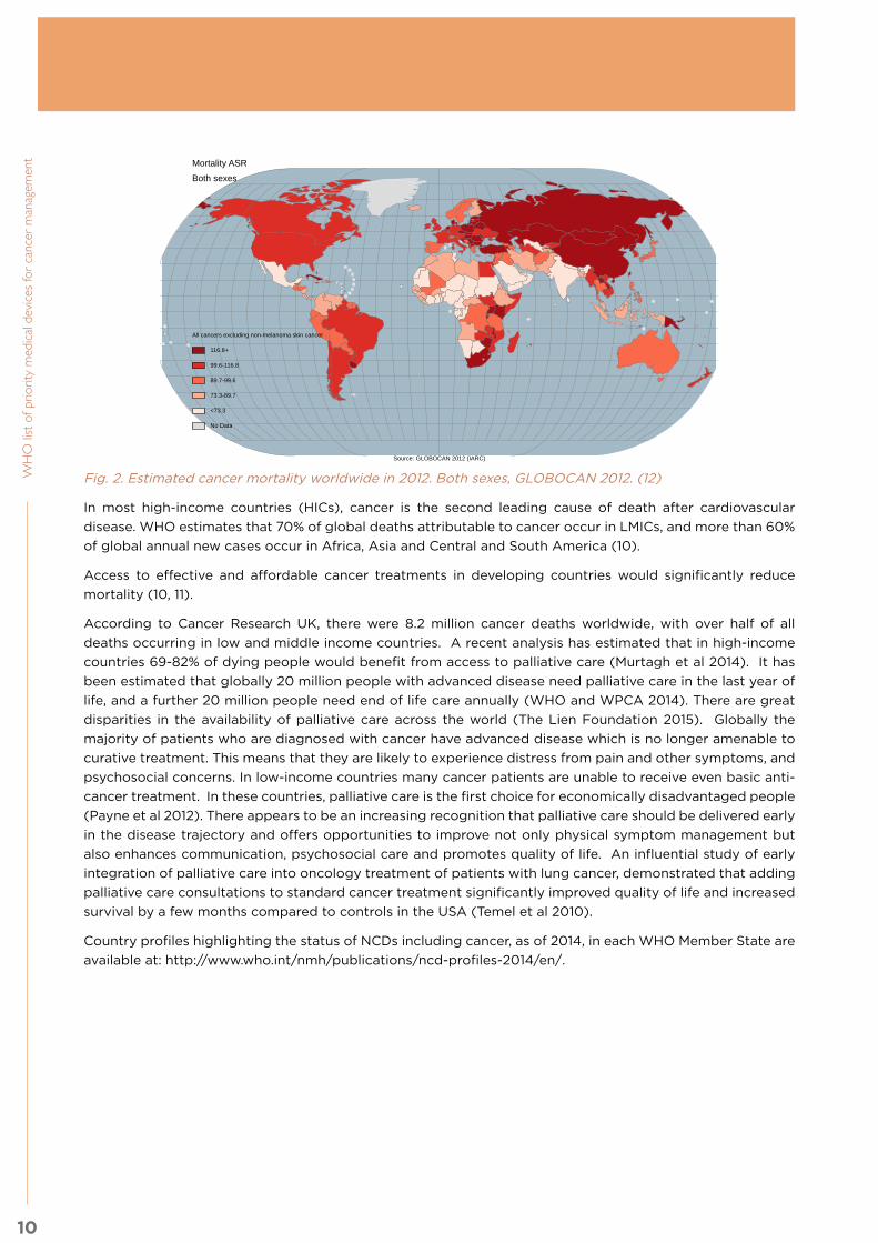
Fig. 2. Estimated cancer mortality worldwide in 2012. Both sexes, GLOBOCAN 2012. (12)
In most high-income countries (HICs), cancer is the second leading cause of death after cardiovascular
disease. WHO estimates that 70% of global deaths attributable to cancer occur in LMICs, and more than 60%
of global annual new cases occur in Africa, Asia and Central and South America (10).
Access to effective and affordable cancer treatments in developing countries would significantly reduce
mortality (10, 11).
According to Cancer Research UK, there were 8.2 million cancer deaths worldwide, with over half of all
deaths occurring in low and middle income countries. A recent analysis has estimated that in high-income
countries 69-82% of dying people would benefit from access to palliative care (Murtagh et al 2014). It has
been estimated that globally 20 million people with advanced disease need palliative care in the last year of
life, and a further 20 million people need end of life care annually (WHO and WPCA 2014). There are great
disparities in the availability of palliative care across the world (The Lien Foundation 2015). Globally the
majority of patients who are diagnosed with cancer have advanced disease which is no longer amenable to
curative treatment. This means that they are likely to experience distress from pain and other symptoms, and
psychosocial concerns. In low-income countries many cancer patients are unable to receive even basic anti-
cancer treatment. In these countries, palliative care is the first choice for economically disadvantaged people
(Payne et al 2012). There appears to be an increasing recognition that palliative care should be delivered early
in the disease trajectory and offers opportunities to improve not only physical symptom management but
also enhances communication, psychosocial care and promotes quality of life. An influential study of early
integration of palliative care into oncology treatment of patients with lung cancer, demonstrated that adding
palliative care consultations to standard cancer treatment significantly improved quality of life and increased
survival by a few months compared to controls in the USA (Temel et al 2010).
Country profiles highlighting the status of NCDs including cancer, as of 2014, in each WHO Member State are
available at: http://www.who.int/nmh/publications/ncd-profiles-2014/en/.
No Data
<73.3
73.3-89.7
89.7-99.6
99.6-116.8
116.8+
All cancers excluding non-melanoma skin cancer
Source: GLOBOCAN 2012 (IARC)
Mortality ASRBoth sexes

11
I. Backg
rou
nd
I.V Availability of specific medical devices for cancer
Medical devices are indispensable for effective screening, diagnosis, treatment, palliation and rehabilitation of
illness and disease. Cancer mortality can be significantly reduced if cases are detected and treated in a timely
manner, yet many preventable deaths occur as a result of lack of available technologies to screen, diagnose
and treat diseases (9).
Early diagnosis of cancer improves the outcome of treatment, however, many patients in low-income settings
do not have access to laboratory, pathology, radiology or other diagnostic methods for early diagnosis (9).
As a result, the majority of patients with malignant neoplasms in developing countries present at a late
stage with incurable disease. Point-of-care (POC) diagnostic tools allow for detection and monitoring at
the primary health care level, and are especially useful where patients would otherwise have to travel long
distances to reach a health facility with a well-equipped laboratory. The development of simple, affordable
diagnostic tools is needed to enable cancer screening in the places of highest cancer burden.
The most common cancer treatments are surgery, systemic therapy and radiotherapy, and curability is
attributed as follows: surgery (49%), radiotherapy (40%), and chemotherapy (11%) (11). Nonetheless, many of
the technologies required for these treatments are inaccessible to the developing world. Radiotherapy, for
example, has the potential to benefit approximately 60% of cancer patients during the course of their disease,
yet LMICs face the largest shortages of radiotherapy units, with approximately 30 countries in Africa and
Southeast Asia having no radiotherapy services available (11). The need for technologies to palliate incurable
disease and improve and prolong quality of life is also being addressed by this project (13).
An estimated 1.5 million different medical devices exist, in more than 10,000 types of generic device groups.
In 2008, WHO initiated the first global effort to identify global needs for medical devices, i.e. to prioritize
and select the essential and affordable medical devices of greatest importance considering the disease
burden of the individual country. In 2010, a very first global survey on medical devices revealed major gaps
in the availability of and access to medical devices in countries (14), as well as vast discrepancies between
countries in regards to the existence of regulatory capacities, national policies, national lists and technical
specifications for procurement and reimbursement of medical devices. As reported in the Global Health
Observatory (14), the following maps represent the density of high cost technologies, some of which are
indispensable for treating cancer (mammography, radiotherapy and computed tomography equipment) and
other technologies that are available almost exclusively in middle-high and high income countries (gamma
camera, magnetic resonance imaging, and positron emission tomography) (Fig. 3–8):
Note: The boundaries and names shown and the designations used on maps do not imply the expression
of any opinion whatsoever on the part of WHO concerning the legal status of any country, territory, city or
area or of its authorities, or concerning the delimitation of its frontiers or boundaries. Dotted lines on maps
represent approximate border lines for which there may not yet be full agreement.
Fig. 3. Mammography units per million females aged between 50 and 69 years old. 2014. Data from Global Health Observatory (14).
Mammography Units per 1,000,000 females***status as of July 2014
0.00 - 0.991.00 - 23.2023.21 - 82.5382.54 - 149.75149.76 - 599.30Data not availableNot applicable
*Medical Equipment: Mammography units per million females aged between 50 and 69 years old**
The boundaries and names shown and the designations used on this map do not imply the expression of any opinion whatsoeveron the part of the World Health Organization concerning the legal status of any country, territory, city or area or of its authorities, or concerning the delimitation of its frontiers or boundaries. Dotted and dashed lines on maps represent approximate border lines for which there may not yet be full agreement.
© WHO 2014. All rights reserved.
Data Source: Baseline country survey on medical devices, July 2014 updateMap Production: Policy, Acces, and Use (PAU unit)World Health Organization
0 2'500 5'000 7'500 10'000 Kilometers

12
WHO
list o
f prio
rity m
edica
l dev
ices f
or ca
ncer
man
agem
ent
Fig. 4. Radiotherapy units per million population. 2014. Data from Global Health Observatory (14).
Fig. 5. Computed Tomography (CT) units per million population. 2014.Data from Global Health Observatory (14).
Radiotheraphy Units per 1,000,000 population*status as of July 2014
0.000.01 - 0.390.40 - 0.991.00 - 3.513.52 - 26.43Data not availableNot applicable
*Medical Radiotheraphy Equipment: Radiotheraphy units (either Linear Accelerator or Telecobalt Unit) per million population**
The boundaries and names shown and the designations used on this map do not imply the expression of any opinion whatsoeveron the part of the World Health Organization concerning the legal status of any country, territory, city or area or of its authorities, or concerning the delimitation of its frontiers or boundaries. Dotted and dashed lines on maps represent approximate border lines for which there may not yet be full agreement.
© WHO 2014. All rights reserved.
Data Source: Baseline country survey on medical devices, July 2014 updateMap Production: Policy, Acces, and Use (PAU unit)World Health Organization
0 2'500 5'000 7'500 10'000 Kilometers**In the case the country did not answer and the quantity is available, we used the DIRA IAEA radiotherapy database as source.
Computed Tomography units per 1,000,000 population*status as of July 2014
0.00 - 0.360.37 - 0.991.00 - 6.886.89 - 13.8913.90 - 132.17Data not availableNot applicable
*Medical Equipment: Computed Tomography (CT) units per million population
The boundaries and names shown and the designations used on this map do not imply the expression of any opinion whatsoeveron the part of the World Health Organization concerning the legal status of any country, territory, city or area or of its authorities, or concerning the delimitation of its frontiers or boundaries. Dotted and dashed lines on maps represent approximate border lines for which there may not yet be full agreement.
© WHO 2014. All rights reserved.
Data Source: Baseline country survey on medical devices, July 2014 updateMap Production: Policy, Acces, and Use (PAU unit)World Health Organization
0 2'500 5'000 7'500 10'000 Kilometers

13
I. Backg
rou
nd
Fig. 6. Gamma Camera units per million population. 2014. Data from Global Health Observatory (14).
Fig. 7. Magnetic Resonance Imaging (MRI) units per million population. 2014. Data from Global Health Observatory (14).
Gamma Camera units per 1,000,000 population*status as of July 2014
0.000.01 - 0.160.17 - 0.991.00 - 3.823.83 - 26.43Data not availableNot applicable
Medical Equipment: Gamma Camera units per million population*
The boundaries and names shown and the designations used on this map do not imply the expression of any opinion whatsoeveron the part of the World Health Organization concerning the legal status of any country, territory, city or area or of its authorities, or concerning the delimitation of its frontiers or boundaries. Dotted and dashed lines on maps represent approximate border lines for which there may not yet be full agreement.
© WHO 2014. All rights reserved.
Data Source: Baseline country survey on medical devices, July 2014 updateMap Production: Policy, Acces, and Use (PAU unit)World Health Organization
0 2'500 5'000 7'500 10'000 Kilometers
Magnetic Res. Imaging units per 1,000,000 population*status as of July 2014
0.000.01 - 0.420.43 - 0.991.00 - 8.308.31 - 132.17Data not availableNot applicable
Medical Equipment: Magnetic Resonance Imaging (MRI) units per million population*
The boundaries and names shown and the designations used on this map do not imply the expression of any opinion whatsoeveron the part of the World Health Organization concerning the legal status of any country, territory, city or area or of its authorities, or concerning the delimitation of its frontiers or boundaries. Dotted and dashed lines on maps represent approximate border lines for which there may not yet be full agreement.
© WHO 2014. All rights reserved.
Data Source: Baseline country survey on medical devices, July 2014 updateMap Production: Policy, Acces, and Use (PAU unit)World Health Organization
0 2'500 5'000 7'500 10'000 Kilometers
14
WHO
list o
f prio
rity m
edica
l dev
ices f
or ca
ncer
man
agem
ent
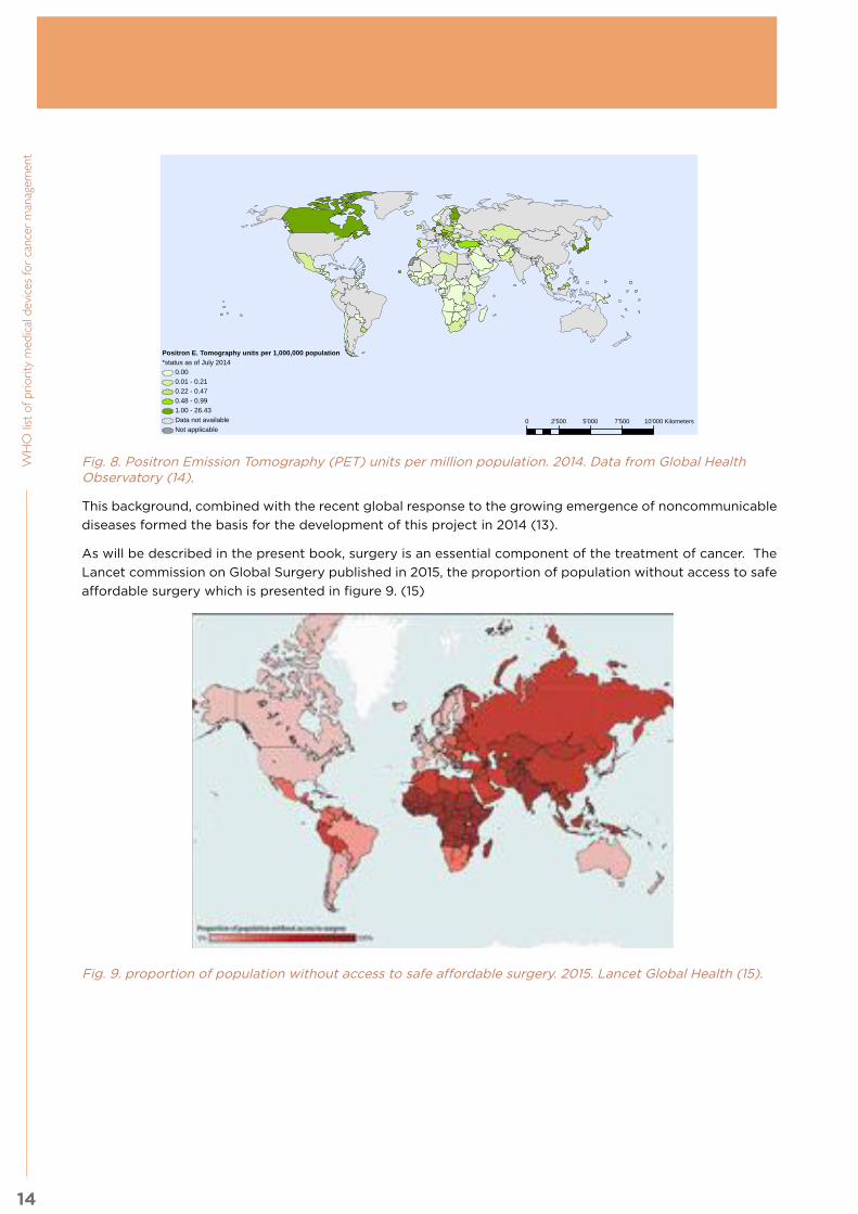
Fig. 8. Positron Emission Tomography (PET) units per million population. 2014. Data from Global Health Observatory (14).
This background, combined with the recent global response to the growing emergence of noncommunicable
diseases formed the basis for the development of this project in 2014 (13).
As will be described in the present book, surgery is an essential component of the treatment of cancer. The
Lancet commission on Global Surgery published in 2015, the proportion of population without access to safe
affordable surgery which is presented in figure 9. (15)
Fig. 9. proportion of population without access to safe affordable surgery. 2015. Lancet Global Health (15).
Positron E. Tomography units per 1,000,000 population*status as of July 2014
0.000.01 - 0.210.22 - 0.470.48 - 0.991.00 - 26.43Data not availableNot applicable
*Medical Equipment: Positron Emission Tomography (PET) units per million population
The boundaries and names shown and the designations used on this map do not imply the expression of any opinion whatsoeveron the part of the World Health Organization concerning the legal status of any country, territory, city or area or of its authorities, or concerning the delimitation of its frontiers or boundaries. Dotted and dashed lines on maps represent approximate border lines for which there may not yet be full agreement.
© WHO 2014. All rights reserved.
Data Source: Baseline country survey on medical devices, May 2014 updateMap Production: Policy, Acces, and Use (PAU unit)World Health Organization
0 2'500 5'000 7'500 10'000 Kilometers

15
I. Backg
rou
nd
Bibliography:
1. http://www.who.int/about/agenda/en/
2. http://www.who.int/about/resources_planning/twelfth-gpw/en/
3. Resolution WHA 60.29. Health technologies. In: Sixtieth World Health Assembly, Geneva, 2007. World Health Organization.
4. Resolution WHA67.20. Regulatory System Strengthening for Medical Products. In: Sixty-seventh World Health Assembly, Geneva, 2014. World Health Organization.
5. Resolution WHA67.23. Health Intervention and Technology Assessment in Support of Universal Health Coverage. In: Sixty-seventh World Health Assembly, Geneva, 2014. World Health Organization.
6. WHO Global status report on noncommunicable diseases, 2014 (http://apps.who.int/iris/).bitstream/10665/148114/1/9789241564854_eng.pdf?ua=1
7. Global Action Plan for the Prevention and Control of NCDs 2013-2020 (http://www.who.int/nmh/events/ncd_action_plan/en/).
8. World Health Organization, Comprehensive Cervical Cancer Control. A guide to essential practice 2014 (http://apps.who.int/iris/bitstream/10665/144785/1/9789241548953_eng.pdf).
9. World Health Organization. Cancer Fact sheet N°297. 2015 (http://www.who.int/mediacentre/factsheets/fs297/en/).
10. IARC, World Cancer Report 2014, B. Stewart and C. Wild, Editors. 2014, International Agency for Research on Cancer: Lyon, France.
11. Jemal, A., et al., The Cancer Atlas. Second edition ed. 2014.
12. IARC. All Cancers (excluding non-melanoma skin cancer) Estimated Mortality Worldwide in 2012 Globocan 2012. (http://globocan.iarc.fr/Pages/fact_sheets_cancer.aspx.)
13. http://www.who.int/medical_devices/en/
14. Global Health Observatory, Medical equipment (density per million population) (http://www.who.int/gho/health_technologies/medical_devices/medical_equipment/en/).
15. Lancet Global Health http://www.lancetglobalsurgery.org/
ReferencesMurtagh F, Bausewein C, Verne J, Groeneveld E.I, Kaloki Y, Higginson I.J. (2014) How many people need palliative care? A study developing and comparing methods for population-based estimates Palliative Medicine 28, 1: 49-58
Payne S, Chan N, Davies A, Poon E, Connor S and Goh C. Supportive, palliative, and end-of-life care for patients with cancer in Asia: resource-stratified guidelines from the Asian Oncology Summit 2012. The Lancet Oncology, 2012, (13): 492-500.
The Lien Foundation (2015) Quality of Death Index 2nd edition. The Lien Foundation: Singapore.
Temel J, et al (2010) Early palliative care for patients with metastatic non-small-cell lung cancer. New England Journal of Medicine 363:733-742
World Health Organisation. www.who.int/mediacentre/factsheet/fs310/en/intex2.html Accessed 17th February 2016.
World Health Organisation and Worldwide Palliative Care Alliance (2014) Global Atlas of Palliative Care at the End of Life. WPCA: New York.”

16
WHO
list o
f prio
rity m
edica
l dev
ices f
or ca
ncer
man
agem
ent The methodology used to select the priority medical devices (PMD) for cancer was based on the methodology
defined by WHO to select the Interagency list of priority medical devices for essential interventions for
reproductive, maternal, newborn and child health (1), which involved the revision of WHO guidelines to define
the interventions and the medical devices required to perform each intervention by levels of care. However,
for this specific cancer project, there were very few WHO guidelines available; thus the methodology had to
be modified accordingly, with supervision and approval of the WHO Guidelines Review Committee.
The following overview presents the milestones and the adaptation of the methodology throughout the
process, which led to the present publication.
Fig. 9. Overview of project methodology
The overall project methodology, as per Fig. 9 above, included the following steps and timeline:
Step 1: Development of preliminary medical devices list for cancer management (Nov 2014–Feb 2015)
Cancer types: Breast, cervical, colorectal, leukaemia, lung, and prostate
Guidelines: Systematic reviews of selected international guidelines for each cancer type
Interventions: Recommended procedures identified from guidelines
Technologies: Medical devices required for each intervention identified
Health conditions
Guidelines
Clinical interventions
Technologies
Expert review
Implementation
Neoplasms (breast, cervical, colorectal, leukaemia, lung, prostate)
Systematic review (of international clinical guidelines published 2008-2015)
Recommended procedures
Medical devices
Experts finalize list in working groups
Publication; in-country workshops
II. Methodology

17
II. Meth
od
olo
gy
Step 2: General consultation (April 2015)• Initial review of preliminary list and methodology
• Define action plan and timeline
• Activities recommended from this general consultation took place May–August 2015
Step 3: Advisory committee meeting (September 2015)• Define methodology for selection/classification of PMD list
• Define working groups and objectives for expert review of list
Step 4: Expert groups review (November 2015–February 2016)• Five expert groups reviewed the following subjects:
» Imaging and nuclear medicine
» Pathology and laboratory
» Surgery
» Radiotherapy
» Systemic therapy and palliative care and end of life care
• Teleconferences to finalize lists and discuss unit infrastructure, human resources, and quality
management (Nov-Dec 2015)
Step 5: Consultation to review the publication (March–April 2016)• Review of the document by all members of consultations and expert groups
Step 6: Country or regional review for implementation (June–December 2016)• Country workshops and dissemination of publication
The detailed activities performed during each step are described below:
Step 1: Development of preliminary medical devices list for cancer managementThe WHO Medical Devices Team (MDT) within the Policy Access and Use Unit of the Essential Medicines and
Health Products Department followed a stepwise approach to identify medical devices relevant for delivering
health care services for six cancer types.
This first step was to conduct literature searches to identify internationally available clinical guidelines for the
management of six cancer types: breast, cervical, colorectal, prostate, lung and leukaemia. To extract information
on the interventions along the continuum of care (prevention, screening, diagnosis, treatment, follow-up,
and palliative care) for the six types of cancer, a total of 27 clinical guidelines (CGL), provided by 6 different
Organizations, have been included (6 CGL for breast cancer; 1 CGL for cervical cancer; 5 CGL for colorectal
cancer; 6 CGL for leukaemia; 5 CGL for lung cancer; 4 CGL for prostate cancer), as listed in Table 1 below.

18
WHO
list o
f prio
rity m
edica
l dev
ices f
or ca
ncer
man
agem
ent
Type of Cancer Provider Organization
Publi-cationYear
Reference
Breast Cancer
World Health Organization 2014 WHO position paper on mammography screening (2014); ISBN
9789241507936The Breast Health Global Initiative 2008 Guidelines for International Breast Health and Cancer Control–
Implementation. Cancer 2008; 113(S8): i–ix, 2215–2371National Institute for Clinical Excellence 2014 Early and locally advanced breast cancer. NICE clinical guideline 80.
2014.National Institute for Clinical Excellence 2014 Advanced breast cancer. NICE clinical guideline 81. 2014.
National Comprehensive Cancer Network 2014 NCCN. Breast Cancer Screening and Diagnosis. Version 1.2014
National Comprehensive Cancer Network 2014 NCCN. Breast Cancer. Version 3.2014
Cervical Cancer
World Health Organization 2014 WHO Comprehensive cervical cancer control: a guide to essential
practice – 2nd ed. 2014.
Colorec-tal Cancer
National Institute for Clinical Excellence 2014 Colorectal cancer. NICE clinical guideline 131. 2014.
European Registration of Cancer Care 2014
van de Velde CJ, Boelens PG, Borras JM, et al. EURECCA colorectal: multidisciplinary management: European consensus conference colon & rectum. Eur J Cancer. 2014 Jan; 50(1):1.e1-1.e34
National Comprehensive Cancer Network 2014 NCCN. Colorectal Cancer Screening. Version 1.2014
National Comprehensive Cancer Network 2015 NCCN. Rectal Cancer. Version 2.2015
National Comprehensive Cancer Network 2015 NCCN. Colon Cancer. Version 2.2015
Leukae-mia
National Comprehensive Cancer Network 2014 NCCN. Acute Lymphoblastic Leukemia. Version 2.2014
National Comprehensive Cancer Network 2015 NCCN. Acute Myeloid Leukemia. Version 1.2015
National Comprehensive Cancer Network 2015 NCCN. Chronic Myelogenous Leukemia. Version 1.2015
European Society for Medical Oncology 2013
Fey MF, Buske C, on behalf of the ESMO Guidelines Working Group. Acute myeloblastic leukaemias in adult patients: ESMO Clinical Practice Guidelines for diagnosis, treatment and follow-up. Ann Oncol. 2013; 24(6): vi138–vi143
European Society for Medical Oncology 2011
Eichhorst B, Dreyling M, Robak T, Montserrat E, Hallek M, on behalf of the ESMO Guidelines Working Group. Chronic lymphocytic leukemia: ESMO Clinical Practice Guidelines for diagnosis, treatment and follow-up. Ann Oncol. 2011; 22(6): vi50–vi54
European Society for Medical Oncology 2012
Baccarani M, Pileri S, Steegmann JL, Muller M, Soverini S, Dreyling M; ESMO Guidelines Working Group. Chronic myeloid leukemia: ESMO Clinical Practice Guidelines for diagnosis, treatment and follow-up. Ann Oncol. 2012; 23(7): vii72-7
Lung Cancer
National Institute for Clinical Excellence 2011 Lung cancer. NICE clinical guideline 121. 2011.
European Society for Medical Oncology 2013
Vansteenkiste J, De Ruysscher D, Eberhardt WE, et al.; ESMO Guidelines Working Group. Early and locally advanced non-small-cell lung cancer (NSCLC): ESMO Clinical Practice Guidelines for diagnosis, treatment and follow-up. Ann Oncol. 2013; 24(6):vi89-98
European Society for Medical Oncology 2014
Reck M, Popat S, Reinmuth N, et al.; ESMO Guidelines Working Group. Metastatic non-small-cell lung cancer (NSCLC): ESMO Clinical Practice Guidelines for diagnosis, treatment and follow-up. Ann Oncol. 2014; 25(3):iii27-39
National Comprehensive Cancer Network 2015 NCCN. Lung Cancer Screening. Version 1.2015
National Comprehensive Cancer Network 2015 NCCN. Non-Small Cell Lung Cancer. Version 3.2015
Prostate Cancer
National Institute for Clinical Excellence 2014 Prostate cancer. NICE clinical guideline 175. 2014.
European Society for Medical Oncology 2013
Horwich A, Parker C, de Reijke T, et al.; ESMO Guidelines Working Group. Prostate cancer: ESMO Clinical Practice Guidelines for diagnosis, treatment and follow-up. Ann Oncol. 2013;24(6):vi106-14
National Comprehensive Cancer Network 2014 NCCN. Prostate Cancer Early Detection. Version 1.2014
National Comprehensive Cancer Network 2015 NCCN. Prostate Cancer. Version 1.2015
Table 1. Internationally available clinical guidelines identified for the management of six cancer types

19
II. Meth
od
olo
gy
Clinical interventions were extracted from each of the clinical guidelines identified, as described and listed in
Annex 1. The medical devices needed to carry out each of the interventions were identified from protocols,
guidelines and procedures. A non-exclusive list of medical devices associated with each intervention was
compiled in line with previous MDT methods. The medical devices were categorized according to intervention
and general clinical areas, e.g. Surgery, Laboratory, Radiotherapy, etc.
Step 2: General ConsultationFrom the 29-30th of April 2015, the MDT hosted an expert consultation on “Priority Medical Devices for
Cancer Management - Targeting Low and Middle-Income Countries” at WHO. The consultation brought
together over 70 participants from 28 countries. Participants included country representatives from national
Ministries of Health and cancer care professionals from teaching hospitals and research institutes in Bhutan,
China, Ethiopia, Ghana, India, Japan, Mexico, Sri Lanka, Uganda, and Zambia. Representatives of WHO
regional offices and headquarters were present along with experts from governmental agencies, academic
institutions, NGOs, professional associations, collaborating centres and UN Agencies.
Participants commented on the preliminary medical devices list and noted additions or deletions as necessary.
The key conclusions from the consultation that informed the further development of the document included:
(1) Use of generic names for medical devices (devices should be clearly identifiable by the descriptions
provided – avoiding provision of full technical specifications) and categorization of devices, (2) classification
of the devices by general use (the devices used for general management of cancers and other diseases in
each clinical area) and specific use for each cancer type (the devices specifically needed for a particular
neoplasm), (3) identification of the contextual elements that should be considered when using the list and
(4) clarification of the role of resource level stratification in this project.
Step 3: Advisory committee meetingFrom May to July 2015, WHO developed the actions requested in the April consultation. Then WHO called
for an advisory committee to define the methodology for selection, classification and presentation of the
medical devices list. This advisory committee meeting brought together 24 participants including 13 advisers,
two WHO regional officers, five WHO headquarters staff and four observers. The advisers provided expertise
in the following specialized areas: surgical oncology, medical oncology, radiation oncology, biomedical
engineering and health technology assessment.
The key conclusions from the advisory committee meeting that informed the further development of the
document included: (1) Expansion of the “basic medical devices” according to the NCD Action Plan Voluntary
Target 9, (2) definition of the core devices (high end or consumables) for cancer management, and (3)
the importance of setting/situation and each country’s need to define the implementation steps for the
acquisition of the technologies, in accordance with their own settings’ situation. The proposed methodology
for devices selection was defined as follows:
i. Definition of the core services/ interventions.
ii. Definition of the basic devices that could achieve the basic interventions in these core services.
iii. When there is more than one multiple general approach or/and to provide data on devices considered,
the working groups will use a pragmatic qualitative multi-criteria decision analysis (MCDA) approach.
iv. The committee decided against stratifying the devices and to have just one basic level for the PMD
lists, thereby encouraging the countries to purchase at least the basic requirements and then continue
improving as per availability of resources (human, infrastructure, financial) and based on local needs.
Moreover, it was agreed that six working groups (Imaging and Nuclear Medicine, Surgery, Pathology and
Laboratory, Radiotherapy, Systemic Therapy and Palliative care) would follow the methodology with
members of different disciplines for each group (oncologic surgeon, radiation oncologists, medical physicist,
pathologist, laboratory specialist, biomedical engineer, etc.) from different regions of the world, from different
income groups and with gender balance.

20
WHO
list o
f prio
rity m
edica
l dev
ices f
or ca
ncer
man
agem
ent Further actions for WHO included the development of: instructions and terms of reference for the working
groups, a nomination template for selection of members of the groups, working tools for device selection and
MCDA, and coordination of teleconferences for each group to generate a final document.
Step 4: Expert groups reviewFive expert groups instead of six were defined based on key disciplines (systemic therapy and palliative care
were merged into one group). The advisory committee nominated the experts to form the expert groups.
Additional consultants and internal WHO personnel were included. Eighty-three nominations were received
and 60 experts were selected based on the criteria to form the five expert groups. The experts came from
29 Member States distributed across the six WHO regions, from all income countries and with 38% female
participants. Experts’ contact information, affiliations, and declarations of interest are available in Annex 2.
Through 25 daily conferences with the 5 groups, the experts were asked to perform the following activities based
on their background, expertise, knowledge and the relevant scientific information, using three WHO working tools
(experts terms of reference and working tool examples included in Annex 3) developed for this review:
1. Review the list of general basic medical devices per clinical area (using working tool 1).
2. Identify the basic services, functions/interventions and specific basic medical devices to be included
in the WHO list of medical devices for cancer management, and include key implementation criteria,
including human resources, interdependencies, infrastructure and quality management requirements
(using working tool 2, adapted from the contextual tool of the MCDA EVIDEM framework).
3. Use the MCDA tool to complement the selection and prioritization exercise in case of contentious
options or/and to provide data on devices considered, using the following value criteria specifically
designed for this project, including effectiveness, safety, patient-reported outcomes, therapeutic benefit,
multi-disease, multi-cancer, ease of use, ease of training, telemedicine capabilities, affordability, positive
consequences on healthcare resource utilization, and quality of evidence (working tool 3, adapted from
the Core Model of the MCDA EVIDEM framework).
The working tools 1, 2 and 3 completed by the experts for the priority devices are available in the
supplementary material (Working tools data) posted on the WHO web site on medical devices for cancer.
(http://www.who.int/medical_devices/en/). Following the expert groups’ reviews, the PMD lists were divided
by clinical services into seven subchapters. For each chapter, a main author was designated to identify the
contextual elements that should be considered when using the list for implementation purposes (special
notes). This included references to infrastructure requirements, quality management, human resource, and/
or other necessary capacity. The members of each group, who had participated in the teleconferences,
complemented the information.
Step 5: Consultation to review the final publicationA consultation to review the outcome lists and the complete document was performed, with all participants
of the general consultation meeting, advisory committee meeting and expert groups. All reviewers’ edits
and comments were integrated into the document during the WHO internal review. As a result of feedback
received during this consultation, the PMD lists were divided into two levels: those absolutely necessary
for cancer management in district hospitals at the second level of care, and those considered standard
for cancer care but due to financial and infrastructure requirements may be better suited for a specialized
hospital at the third level.
Step 6: Country or regional review for implementationThe publication will also undergo country and/or regional review for implementation strategy development
on the basis of performing a needs assessment unique to their setting and priorities.

21
III. Prio
rity m
ed
ical d
evic
es b
y c
linic
al a
reas
Bibliography:1) Interagency list of medical devices for essential interventions for reproductive, maternal, newborn and
child health. World Health Organization, 2015. Available at: http://www.who.int/medical_devices/md_maternal_v12_web.pdf
Guidance documents:
Package of Essential Noncommunicable ( PEN) Disease interventions for Primary Health Care in Low Resource Settings, 2010 www.who.int/nmh/publications/essential_ncd_interventions_lr_settings.pdf
Clinical Practice Guidelines from CENETEC, Mexico: http://www.cenetec.salud.gob.mx/contenidos/gpc/catalogoMaestroGPC.html
Clinical Practice Guidelines from Ministry of Health and Social Protection, Colombia: http://gpc.minsalud.gov.co/Pages/Default.aspx
National Cancer Treatment Guidelines. Ministry of Public Health-Tokten Lebanon-UNDP- Council of Development and Reconstruction. http://www.moph.gov.lb/Drugs/Documents/16UNDPcancerbook%202012web.pdf
Canadian Association of Radiologists Diagnostic Imaging Referral Guidelines for Cancer: http://www.car.ca/uploads/standards%20guidelines/car-referralguidelines-k-en-20130726.pdf
National Guidelines for Cancer Management, Kenya: http://kehpca.org/wp-content/uploads/National-Cancer-Treatment-Guidelines2.pdf
Standard Treatment Guidelines, National Health Mission, India: http://nrhm.gov.in/nhm/nrhm/guidelines/nrhm-guidelines/standard-treatment-guidelines.html
22
WHO
list o
f prio
rity m
edica
l dev
ices f
or ca
ncer
man
agem
ent This chapter includes the list of priority medical devices for cancer management of primarily six cancer
types (breast, cervical, colorectal, leukaemia, lung, and prostate); however, as detailed below, much of
the information in these units including medical devices lists, infrastructure requirements and guidance
documents are pertinent to other cancers as well as other diseases. The list is grouped into specific clinical
units as presented in the following sub-chapters:
III. Priority medical devices by clinical areas
Each subchapter includes the following sections:
1. General description of the unit: the general purpose of the clinical area within the health service delivery
sequence is described and the relevant clinical interventions are listed.
2. Priority medical devices: the list of medical devices is presented in two parts:
i. General Medical Devices that need to be available for basic use of the clinical unit to perform the
majority of interventions, and can be used for various diseases including cancers (Radiotherapy and
Systemic Therapy chapters are specific to cancer treatment).
ii. Specific Medical Devices for diagnosis or treatment of a specific neoplasm or type of cancer.
It is important to note that this is a non-exclusive list and that the devices could differ according to different
methods or techniques for the same intervention or procedure. The intended purpose of these lists is to present
the medical devices needed for a general approach to the defined cancer interventions within each clinical area.
Throughout this publication, the priority medical devices may include any of the following subcategories:
1. Vaccination, clinical assessment & endoscopy
5. Radiotherapy
2. Medical imaging & nuclear medicine
6. Systemic therapy
3. Surgery 7. Palliative care & end of life care
4. Clinical laboratory & pathology
• Laboratory and pathology equipment
• Medical equipment
• Medical furniture
• Other (glassware, utensils, etc.)
• Personal protective equipment and clothing (PPE)
• Quality assurance devices
• Radiation protection devices
• Single use devices/disposables/medical supplies
• Solutions and reagents
• Software
• Surgical instruments.
For the purpose of this publication, the medical devices were classified upon :
3. Capital investment (equipment, furniture and surgical instruments) all of the above are reused and
requires maintenance through out the life cycle.
4. Operational costs ( consumables, single use devices ) as per the national health accounts, to facilitate
tracking and quantification of expenses.
Every country can re- classify as needed and adapt the lists. it is important to note that also capital equipment
can be leased or rented.
5. Human resources: a list of the specialized human resources required to use the devices listed and
perform the interventions, and alternative options in case these are not available.
6. Infrastructure requirements: a general description of the layout distribution, shielding and radiation
protection (if applicable), electrical systems, heating, ventilation and air conditioning systems, safety,
and information and communication system requirements.
7. Quality management: description of activities within a quality policy framework to guarantee quality in
the clinical area.
8. Guidance documents: guidelines and documents from UN Agencies, NGOs, publications and other
relevant information to expand upon the topics already generally described in the subchapter.

23
III. Prio
rity m
ed
ical d
evic
es b
y c
linic
al a
reas
Cancer patient continuum of care
The clinical units in the subchapters correspond to the cancer patient continuum of care. Prevention and early
detection should ideally take place in primary health care settings, with referral to a secondary level for
diagnosis and treatment capabilities, and to the more specialized tertiary levels whenever needed. However,
in many LMICs there are limited early detection and diagnostic capabilities, and the patient may arrive with
an advanced stage of cancer and an increased mortality risk. These cases often require direct referral to
tertiary level facilities where available, and burdensome costs for patients and healthcare systems.
It is important to note that the type of hospitals that correspond
to each healthcare system level is a contextual decision that
each country should make. Generally, the district hospital
corresponds to a second level of care (general hospital)
and specialized hospitals (regional, national/teaching with
specialized oncological human resources and infrastructure)
correspond to the third level.
The following health care service delivery overview diagram
shows how the clinical units mentioned in this document are
interrelated and interdependent (Fig. 10). After proper diagnosis
and staging, it is recommended that the treatment or therapeutic
decisions be made in a multidisciplinary tumour board. This will
ensure that doctors and patients decide on the best available treatment, integrating all necessary disciplines.
This graph is merely a representation of some generic interventions and does not intend to include all possible
pathways, which will be different depending on the patient, the stage the type of cancer and the diagnostic
and treatment available in their setting.Many of the clinical services can be used for multiple functions,
example screening, diagnosis and monitoring patients. Other treatments as radiotherapy, chemotherapy or
surgery can be used for treatment but also as palliative care, which are represented in this graphic with the
multiple colors to indicate different uses of the same technology.
Fig. 10. Health care service delivery overview
The medical devices lists presented in
each clinical unit subchapter are defined as
those necessary for cancer management in
district hospitals at the second level of care,
or as those considered standard for cancer
care but due to financial and infrastructure
requirements may be better suited for a
specialized hospital at the third level. Nuclear
Medicine, Radiotherapy and Systemic
Therapy units are considered at the third level
of specialized care.
PR
IMA
RY
(C
L,H
C),
SE
C-
ON
DA
RY
(D
H)
&
TE
RT
IAR
Y (
SH
)
Le
ve
l o
f C
are
*
* Appropriate level of care will depend on the particular intervention, setting, and available infrastructure and human resources.
SE
CO
ND
AR
Y (
DH
)
& T
ER
TIA
RY
(S
H)
TE
RT
IAR
Y (
SH
)
Prevention End of life care
TreatmentFollow-up/survivorship
care
Palliative care
Diagnosis
Screening
CL Community Level health post DH District Hospital HC Health Centre SH Specialized Hospital
Systemic therapy
Nuclear medicine
Surgery
Vaccination
Radio- therapy
Pathology Pathology
Clinical laboratory
Clinical laboratory
Clinical assessment
Palliative care
Medical imaging
Endoscopy
Clinical assessment
Nuclear medicine

24
WHO
list o
f prio
rity m
edica
l dev
ices f
or ca
ncer
man
agem
ent Medical devices considerations:
The tables presented in the different sections include the priority medical devices required to perform the
health interventions listed. The selection and prioritization of medical devices will be determined according
to the country’s different levels of health care (primary, secondary and tertiary), specialized human resources
and available infrastructure, and patient flow, according to the local needs. This will be presented in greater
detail in Chapter IV, Implementation strategy.
All the medical devices that are listed in this document need to be regulated, assessed and managed properly.
The issues that need to be considered and will be expanded upon in Chapter IV, include the following:
1. Selection: If the devices included in these lists are approved by the ministries of health of the Member
States, they can be added to the priority/basic medical devices list of the country.
2. Regulatory clearance: These devices should have regulatory clearance to be marketed in the country,
after analysing the safety and effectiveness of the technology, and should be approved by a national
regulatory authority related to the ministry of health.
3. Assessment: The priority medical devices, especially the ones that are most costly and complex, require
specific infrastructure and trained human resources, and need an assessment process to evaluate if these
can be placed for public procurement or considered in the package of interventions as a reimbursable
procedure/product. These assessments consider not only the safety and efficacy of the technology, but
also feasibility, cost effectiveness, ethical, organizational and human resources requirements.
4. Acquisition or incorporation: In order to procure or acquire these medical devices once the need is
defined, a tender process is required which will include the definition, technical specifications, request
for regulatory approval, standards compliance, delivery times, and aspects such as installation, warranty,
technical support costs and training to be included in the procurement package. An extended warranty
is required if maintenance is a problem in the country of use. Acquisition requirements should include
software licenses, upgrade policy, commissioning (acceptance) – attributes and costs.
5. Installation: Once the bidding and contract has been completed, the mechanical guidelines must be
available to prepare for installation. This is particularly specific to diagnostic imaging, and is costly and
complex for radiotherapy equipment. The installation has to be to the complete satisfaction of the final
user. This applies to medical equipment in general and is of very high importance for equipment required
in diagnostic imaging, nuclear medicine, surgery and radiotherapy.
6. Supply chain and logistics: Reliable supply chain and logistics management are needed to ensure the
right quality product is available, in the right quantities, at the right time, and for a reasonable cost.
7. Maintenance: In every area, the maintenance and repair of medical equipment are significant challenges
and potentially costly, and it is important to consider service documentation, software diagnostics, special
tools & critical parts to be available at the hospital site and these costs upfront, as well as to verify local
or regional suppliers that can provide continued technical support. In some cases, especially for high cost
equipment, multiyear service agreements are a good option to consider.
8. Training: The training of the user ensures the safe and appropriate use of most health technologies and
should be requested of the manufacturer/supplier of the device when drawing up the purchase agreement.
9. Follow-up: It is important to ensure appropriate follow-up to the safe use of medical equipment and
reporting of any adverse event to the regulatory authorities, as well as proper final decommissioning,
according to national and global guidelines.
10. Donations: Device donations from high-resource settings should only be deployed to other settings
after careful deliberation on appropriateness, costs of maintenance, and relevance. When a donation is
received, care should be taken that the equipment or technology is in good condition and complies with
standards of safety and quality.
11. Infrastructure considerations: It is important to note that in low- and middle-income countries,
infrastructure concerns like stable power supply, availability of running water, oxygen supply or air
conditioning might be limited and humidity and high temperatures could affect the performance of the
medical technologies.
12. Telemedicine: When connectivity is available, telemedicine can be a resource for training users, sending images
or information for diagnosis or treatment alternatives, or to have a second opinion or to send to experts.

25
III. Prio
rity m
ed
ical d
evic
es b
y c
linic
al a
reas
13. Mobile health: Mobile health (mHealth) solutions might also be available and should be considered.
14. Data security: Data storage and patient data confidentiality should also be addressed. Laboratory,
radiology or hospital information systems and electronic health records could be available in different
settings, and it is important to consider interoperability to ensure data management.
For all these steps, WHO has developed reference resources available at www.who.int/medical_devices/en.
The WHO medical devices technical series (Fig. 11) provides specific guidance on each of the above steps.
WHO MEDICAL DEVICE TECHNICAL SERIES: TO ENSURE IMPROVED ACCESS, QUALITY AND USE OF MEDICAL DEVICES
WHO Medical device technical series
HUMAN RESOURCES FOR MEDICAL DEVICES
The role of biomedical engineers
DEVELOPMENT OF MEDICAL
DEVICE POLICIES, STRATEGIES AND
ACTION PLANSWHO MEDICAL DEVICE TECHNICAL SERIES
Rese
arch
and
dev
elop
men
t
Med
ical
dev
ices
Asse
ssm
ent
Regu
latio
n
Man
agem
ent
Global Model Regulatory Framework for Medical
Devices including in vitro diagnostic medical devices
WHO Medical device technical series
HEALTH TECHNOLOGY ASSESSMENT OF
MEDICAL DEVICESWHO MEDICAL DEVICE TECHNICAL SERIES
Assessment
Regulation
NEEDS ASSESSMENT PROCESS
WHO MEDICAL DEVICE TECHNICAL SERIES
Management
WHO list of priority medical devices for cancer management
Breast C
ervical Child
hood
Co
lon
Le
uke
mia
Lu
ng
Pro
state
Medical devices
MEDICAL DEVICE NOMENCLATURE
WHO MEDICAL DEVICE TECHNICAL SERIES
Interagency list of priority medical devices for essential interventions for reproductive, maternal, newborn and child health
PROCUREMENT PROCESS
RESOURCE GUIDEWHO MEDICAL DEVICE TECHNICAL SERIES
MEDICAL DEVICE DONATIONS:
CONSIDERATIONS FOR SOLICITATION AND
PROVISIONWHO MEDICAL DEVICE TECHNICAL SERIES
COMPUTERIZED MAINTENANCE MANAGEMENT
SYSTEM WHO MEDICAL DEVICE TECHNICAL SERIES
MEDICAL EQUIPMENT MAINTENANCE
PROGRAMME OVERVIEW
WHO MEDICAL DEVICE TECHNICAL SERIES
INTRODUCTION TO MEDICAL EQUIPMENT
INVENTORY MANAGEMENT
WHO MEDICAL DEVICE TECHNICAL SERIES
SAFE USE OF MEDICAL DEVICES
WHO MEDICAL DEVICE TECHNICAL SERIES
MEASURING CLINICAL
EFFECTINESS WHO MEDICAL DEVICE TECHNICAL SERIES
DECOMMISSIONING MEDICAL DEVICES
WHO MEDICAL DEVICE TECHNICAL SERIES
POST-MARKET SURVEILLANCE AND
ADVERSE EVENT REPORTING
WHO MEDICAL DEVICE TECHNICAL SERIES
MEDICAL DEVICE INNOVATION
WHO MEDICAL DEVICE TECHNICAL SERIES
Research and development
Global atlas of medical devices
Fig. 11. WHO medical device technical series
Vaccination, clinical assessment & endoscopy
1.

27
1. Vaccin
atio
n, c
linic
al a
ssessm
en
t & e
nd
osc
op
y Health service delivery sequence overview:
This diagram expresses the flow of cancer patient to and from the vaccination, clinical assessment and
endoscopy units, and the elements to consider as support for the unit (Fig 10.1).
Fig. 10.1. Health care service delivery overview – vaccination, clinical assessment and endoscopy
PR
IMA
RY
(C
L,H
C),
SE
C-
ON
DA
RY
(D
H)
&
TE
RT
IAR
Y (
SH
)
Le
ve
l o
f C
are
*
* Appropriate level of care will depend on the particular intervention, setting, and available infrastructure and human resources.
SE
CO
ND
AR
Y (
DH
)
& T
ER
TIA
RY
(S
H)
TE
RT
IAR
Y (
SH
)
Prevention End of life care
TreatmentFollow-up/survivorship
care
Palliative care
Diagnosis
Screening
CL Community Level health post DH District Hospital HC Health Centre SH Specialized Hospital
Systemic therapy
Nuclear medicine
Surgery
Vaccination
Radio- therapy
Pathology Pathology
Clinical laboratory
Clinical laboratory
Clinical assessment
Palliative care
Medical imaging
Endoscopy
Clinical assessment
Nuclear medicine
1.1 General description of the unit
1.2 Priority medical devices for vaccination
1.2.1 Specific medical devices for vaccination
1.3 Priority medical devices for clinical assessment
1.3.1 General medical devices for clinical assessment
1.3.2 Specific medical devices for clinical assessment and minor procedures by cancer type
1.4 Priority medical devices for endoscopy
1.4.1 General medical devices for endoscopy
1.4.2 Specific medical devices for endoscopy by cancer type
1.4.3 Guidance documents
1.5 Human resources for clinical assessment

28
WHO
list o
f prio
rity m
edica
l dev
ices f
or ca
ncer
man
agem
ent
1.1 General description of the unit
This unit includes three sections, two of which can be done starting at primary health care level, these are:
first the preventive measure, which is the HPV vaccination for cervical cancer. and secondly the initial clinical
assessments.
As indicated in the WHO cervical cancer control guidelines, it is recommended that the HPV vaccine is applied
for girls from 9 to 13 years, as cervical cancer is caused by high risk types of HPV, therefore vaccination is a very
important prevention intervention. The same applies to Hepatitis B for liver cancer, which has a high prevalence
in many LMIC. The other public health solutions for prevention of NCDs are healthy behaviour, including physical
activity, adequate nutrition, and avoiding tobacco and consumption of alcohol, among others.
Some screening and diagnosis, can be done in health clinics and district hospitals. In the case of cervical
cancer WHO recommends HPV testing, cytology and visual inspections with acetic acid. Then the women
with positive screening test must receive effective treatment.
As indicated in the Package of Essential Noncommunicable interventions for primary health care, “ Efficient use
of limited health care resources, sustainable health financing, access to basic diagnostics, essential medicines,
and organized medical information and referral systems are indispensable for the delivery of equitable care
for persons with and at risk of NCDs. Therefore, every chapter contains health systems requirements to
support the complete approach to tackle NCDs.
The annex A of the PEN, indicates specifically three cost effective interventions for primary health care:
1. One visit VIA (screening and treatment for woman between 30 to 49, and can be earlier if needed)
2. Three visits cytology for woman between 30 to 49
3 Examination of breast by health care worker in clinical annually
The third section is on endoscopy, usually performed in secondary and tertiary level.
1.2 Priority medical devices for vaccination
The following table present the priority medical devices required to perform the following health
interventions:
Cancer type Interventions
Cervical cancer • HPV vaccination
1.2.1 Specific medical devices for vaccination
1.2.1.1 Cervical
Procedure Medical devices category Capital equipment Accessories/hardware/software/
consumables/single use devices
Human papillomavirus vaccination
Personal protective equipment and clothing
Gloves, examination, non-sterile, single use (various sizes)
Single use devices/disposables/medical supplies
Safety box, for used syringes/needlesCotton wool, 500g, roll, non-sterileSyringes, autodisable (AD), (various capacities)

29
1. Vaccin
atio
n, c
linic
al a
ssessm
en
t & e
nd
osc
op
y
1.3 Priority medical devices for clinical assessment
The following tables present the general medical devices for clinical assessment and minor procedures, which
can be used for screening, diagnosis, staging, and follow-up of many diseases including several cancers, and
specific medical devices, which are used for interventions for specific cancer types.
Please note: Components of all surgical instrument sets as well as needle and catheter gauge sizes (G) are
listed in the chapter annex at the end of this chapter.
The following tables present the priority medical devices required to perform the following health
interventions:
Cancer type Interventions
All cancers • Clinical assessment and minor procedures
Breast cancer • Clinical examination of breast
Cervical cancer • Gynaecological examination and gynaecological procedures• Colposcopy• Cryotherapy • Endocervical curettage ECC• Large loop excision of transformation zone (LEEP/LLETZ)• Pap smear• Visual inspection with acetic acid
Colorectal cancer • Dietary counselling• Digital rectal examination and examination of abdomen, liver and lymph nodes• Rigid sigmoidoscopy• Proctoscopy• Faecal ocult blood test
Lung cancer • Spirometry • Bronchoscopy
Prostate cancer • Digital rectal examination• PSA
Cervical and prostate cancers
• Cystoscopy

30
WHO
list o
f prio
rity m
edica
l dev
ices f
or ca
ncer
man
agem
ent 1.3.1 General medical devices for clinical assessment
These are general as they can be used for various diseases including all types of cancer excluding Leukaemia.
Please note they are organized by clinical procedure.
Procedure Medical devices category Capital equipment Accessories/hardware/software/
consumables/single use devices
Clinical assessment and minor procedures
Medical equipment
Aneroid sphygmomanometer Blood pressure cuffs
Stethoscope
Thermometer
Examination table
Resuscitation trolley, equipped, with medicines and defibrillator
Fixed examination/treatment light
Pulse oximeter Spare probes, adult and paediatric
Electrocardiography system (Assessment for cardiological disease)
• Electrodes• Leads• Gel, etc.
Blood glucometer (Assessment for diabetes, glucose measurement)
Micro cuvettes, strips or similarLancet, blood, safety, sterile (various sizes)
Floor scale and stadiometer Tape measurement
Laryngoscope
Medical furniture X-ray, viewer (negatoscope)
Stand, infusion, double hook, on casters
Tray, dressing, stainless steel, approx. 300 x 200 x 30 mm
Trolley, dressing, stainless steel, 2 trays
Cabinet, instruments, double door
Personal protective equipment and clothing
Gloves, examination, non-sterile, single use (various sizes)
Single use devices/disposables/medical supplies
Syringes with needles
Compress, gauze, sterile & non-sterile, single use
Skin-cleaning wipe
Bags for contaminated supplies
Transparent film dressings with a gel pad
Infusion giving set, sterile, single use
Intravenous cannulas or catheters
Solutions and reagents
Alcohol isopropyl 70%
IV solution
Iodine povacrylex and isopropyl alcohol solution or similar
High level disinfectant, 0.5% chlorine solution for decontaminating instruments
Distilled water
Other Brush, hand, scrubbing, plastic
Bowl, polypropylene
Basin, kidney, polypropylene
Safety box, for used syringes/needles
Receptacle, waste, stainless steel, pedal action
Arm/leg tourniquet

31
1. Vaccin
atio
n, c
linic
al a
ssessm
en
t & e
nd
osc
op
y
1.3.2 Specific medical devices for clinical assessment and minor procedures by cancer type
1.3.2.1 Cervical
Procedure Medical devices category Capital equipment Accessories/hardware/software/
consumables/single use devices
Gynaecological examination and gynaecological procedures1
Medical equipment Bright light source
Gynaecological examination/treatment table
Instruments Forceps tissue-long
Cheron forceps
Long needle holders
Cervical punch biopsy forceps
Ring forceps
Vaginal sidewall retractors
Vaginal speculum, reusable
Compress, gauze, sterile & non-sterile, single use
Specimen container
Absorbent tipped applicator/large
Tongue depressor, single use (wooden or plastic spatula)
Examination table paper cover
Personal protective equipment and clothing
Gloves, examination, non-sterile, single use (various sizes)
Solutions and reagents
Formalin 10%, or tissue fixation reagents. Phosphate buffered
Lubricating jelly (K-Y)
Monsel’s paste
Saline solution
Lugol iodine, bottle/Acetic acid solution 3–5%
0.5% chlorine solution for decontaminating instruments
Other Container for warm water
Bags for contaminated disposable supplies
Colposcopy Medical equipment Colposcope
Cryotherapy Medical equipment Cryosurgery unit, with all parts and accessories listed
Probe, trigger, handle grip, yoke, inlet of gas cylinder, tightening knob, pressure gauge showing cylinder pressure, silencer outlet, gas-conveying tube probe tip
Colposcope
Endocervical curettage ECC
Medical equipment Colposcope
Instruments Endocervical curette
1 The devices listed in this procedure should be considered in addition to the equipment enlisted for the following procedures: cold knife conization (CKC), colposcopy, large loop excision of transformation zone (LEEP/LLETZ) and cryotherapy, visual inspection with acetic acid, endocervical curettage, pap smear and proctoscopy.

32
WHO
list o
f prio
rity m
edica
l dev
ices f
or ca
ncer
man
agem
ent
Procedure Medical devices category Capital equipment Accessories/hardware/software/
consumables/single use devices
Large loop excision of transformation zone (LEEP/LLETZ)
Medical equipment Electrosurgical unit, with all parts and accessories listed
Electrocautery system electrode for LEEPReturn electrodeRound and square loops of different sizes (rarely deeper than 10 mm)Wire electrodes of several sizes (square loop electrode, semi-circular loop electrode)Coagulating/ball electrode
Smoke evacuator
Colposcope
Instruments Vaginal speculum, Non conducting preferably with side retractors
Papanicolaou Test (Pap smear)
Instruments Vaginal speculum, reusable Local anaesthetic, syringes
Personal protective equipment and clothing
Gloves, examination, non-sterile, single use (various sizes)
Single use devices/disposables/medical supplies
Microscope slides frosted or liquid-based container (tube containing a special preservative solution)
Tongue depressor, single use (wooden or plastic spatula)
Cervical cytology brush or cervical cytology scraper (optional)
Examination table paper cover
Solutions and reagents
0.5% chlorine solution for decontaminating instruments
Fixative spray or solution for pap smear (if slides are used)
Other Container for warm water
Bags for contaminated disposable supplies
Visual inspection with acetic acid
Instruments Vaginal speculum, reusable
Single use devices/disposables/medical supplies
Examination table paper cover
Absorbent tipped applicator/large
Personal protective equipment and clothing
Gloves, examination, non-sterile, single use (various sizes)
Solutions and reagents
Lugol iodine, bottle/Acetic acid solution 3–5%
0.5% chlorine solution for decontaminating instruments
Other Container for warm water
Bags for contaminated disposable supplies
1.3.2.2 Colorectal
Procedure Medical devices category Capital equipment Accessories/hardware/software/
consumables/single use devices
Dietary counselling
Medical equipment Floor scale with stadiometer
Digital rectal examination and examination of abdomen, liver and lymph nodes.
Personal protective equipment and clothing
Gloves, examination, non-sterile, single use (various sizes)
Single use devices/disposables/medical supplies
Lubricating jelly (K-Y)
Rigid sigmoidoscopy
Medical equipment Rigid sigmoidoscope

33
1. Vaccin
atio
n, c
linic
al a
ssessm
en
t & e
nd
osc
op
y
Procedure Medical devices category Capital equipment Accessories/hardware/software/
consumables/single use devices
Proctoscopy Medical equipment Proctoscope (see Endoscopy tower system in General devices for endoscopy table 1.4.1.)
1.3.2.3 Lung
Procedure Medical devices category Capital equipment Accessories/hardware/software/
consumables/single use devices
Spirometry Medical equipment Diagnostic spirometer Respiratory, noseclipsfilter, pulmonary function filter, mouthpiece
Bronchoscopy Medical equipment Bronchoscope
Laryngeal mask
Endotracheal tube
1.3.2.4 Prostate
Procedure Medical devices category Capital equipment Accessories/hardware/software/
consumables/single use devices
Digital rectal examination
Personal protective equipment and clothing
Gloves, examination, non-sterile, single use (various sizes)
Single use devices/disposables/medical supplies
Lubricating jelly (K-Y)
1.3.2.5 Cervical and Prostate
Procedure Medical devices category Capital equipment Accessories/hardware/software/
consumables/single use devices
Cystoscopy Medical equipment Rigid cystoscope (strongly recommended to consider an endoscopy tower)
Instruments Cystoscopy biopsy forceps
Cystoscope sheath
Single use devices/disposables/medical supplies
Lubricating jelly (K-Y)
Biopsy needle
Personal protective equipment and clothing
Urological irrigation kit
Specimen container
Cryptographic/urethrographic catheter, female
Gloves, examination, non-sterile, single use (various sizes)
Solutions and reagents
Transurethral-instrument lubricant
Formalin 10%, or tissue fixation reagents

34
WHO
list o
f prio
rity m
edica
l dev
ices f
or ca
ncer
man
agem
ent
1.4 Priority medical devices for endoscopy
The following tables present the general medical devices for endoscopy, which can be used for screening,
diagnosis, staging, and follow-up of many diseases including several cancers, and specific medical devices,
which are used for surgical interventions for specific cancer types.
Please note: Components of all surgical instrument sets as well as needle and catheter gauge sizes (G) are
listed in the chapter annex at the end of this chapter.
The following tables present the priority medical devices required to perform the following health interventions:
Cancer type Interventions
All applicable cancers • General endoscopy procedures
Colorectal cancer • Colonoscopy• Endoscopic biopsy• Flexible sigmoidoscopy
1.4.1 General medical devices for endoscopy
Endoscopy procedures can be performed outside the surgical unit.
Procedure Medical devices category
Capital equipment (for specialized hospitals)
Accessories/hardware/software/consumables/single use devices
General endoscopic procedures
Medical equipment Endoscopy tower system including: Insufflator Light source Irrigation/ aspiration pumpProcessing unitVideo image display monitor
Endoscope system
Single-patient physiologic monitoring system for ECG, Capnography, SpO2, B.P. Thermometer
ElectrodesSpO2 sensorsBlood pressure cuffTemperature sensor
Resuscitation trolley, equipped with medicines and defibrillator/with laryngoscope
Endoscope sterilization/disinfection support set
Endoscope washer/disinfector
Ultrasonic washer
Adequate containers to clean and disinfect endoscopes and to transport them
Endoscope cleaning brush
Endoscope leak tester, mechanical Endoscope lens cleaner
Rubber bulb
Specimen container
Endoscope lens cleaner
Medical furniture Cabinet, instruments, double door
Cabinets for accessories and clothes
Endoscope drying/storage cabinet
Footstool, two steps
Intravenous pole
Stretcher, foldable
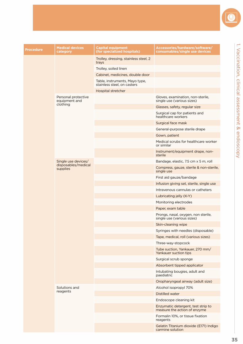
35
1. Vaccin
atio
n, c
linic
al a
ssessm
en
t & e
nd
osc
op
y
Procedure Medical devices category
Capital equipment (for specialized hospitals)
Accessories/hardware/software/consumables/single use devices
Trolley, dressing, stainless steel, 2 trays
Trolley, soiled linen
Cabinet, medicines, double door
Table, instruments, Mayo type, stainless steel, on casters
Hospital stretcher
Personal protective equipment and clothing
Gloves, examination, non-sterile, single use (various sizes)
Glasses, safety, regular size
Surgical cap for patients and healthcare workers
Surgical face mask
General-purpose sterile drape
Gown, patient
Medical scrubs for healthcare worker or similar
Instrument/equipment drape, non-sterile
Single use devices/disposables/medical supplies
Bandage, elastic, 7.5 cm x 5 m, roll
Compress, gauze, sterile & non-sterile, single use
First aid gauze/bandage
Infusion giving set, sterile, single use
Intravenous cannulas or catheters
Lubricating jelly (K-Y)
Monitoring electrodes
Paper, exam table
Prongs, nasal, oxygen, non sterile, single use (various sizes)
Skin-cleaning wipe
Syringes with needles (disposable)
Tape, medical, roll (various sizes)
Three-way-stopcock
Tube suction, Yankauer, 270 mm/ Yankauer suction tips
Surgical scrub sponge
Absorbent tipped applicator
Intubating bougies, adult and paediatric
Oropharyngeal airway (adult size)
Solutions and reagents
Alcohol isopropyl 70%
Distilled water
Endoscope cleaning kit
Enzymatic detergent, test strip to measure the action of enzyme
Formalin 10%, or tissue fixation reagents
Gelatin Titanium dioxide (E171) Indigo carmine solution

36
WHO
list o
f prio
rity m
edica
l dev
ices f
or ca
ncer
man
agem
ent
Procedure Medical devices category
Capital equipment (for specialized hospitals)
Accessories/hardware/software/consumables/single use devices
Glutaraldehyde 3.4% (Cidex, Maxicide, Wavicide)
Methylene blue, bottle
IV solutions
Lugol iodine, bottle/Acetic acid solution 3–5%
Peracetic acid disinfectant anticorrosion additive for endoscopes or similar
Cleaning systems for endoscopes
Arm/leg tourniquet Bedpan
Sponge bowl
Bowl/Emesis bowl
Other Safety box, for used syringes/needles
1.4.2 Specific medical devices for endoscopy by cancer type
1.4.2.1 Colorectal
Procedure Medical devices category
Capital equipment (for specialized hospitals)
Accessories/hardware/software/consumables/single use devices
Colonoscopy Medical equipment Rectal irrigation system
Video-colonoscope (see Endoscopy Tower System in General Devices for Endoscopy, Table 1.4.1.)
Endoscopic biopsy
Medical equipment Rectal irrigation system (see Endoscopy Tower System in General Devices for Endoscopy Table 1.4.1.)
Video-colonoscope (see Endoscopy Tower System in General Devices for Endoscopy Table 1.4.1.)
Single use devices/disposables/medical supplies
Endoscopic hemoclip
Polypectomy snare
Sclerotherapy endoscopic needles
Wire oval snare
Surgical instruments Biopsy forceps
Flexible sigmoidoscopy
Medical equipment Flexible sigmoidoscope (see Endoscopy Tower System in General Devices for Endoscopy Table1.4.1.)

37
1. Vaccin
atio
n, c
linic
al a
ssessm
en
t & e
nd
osc
op
y
1.4.3 Guidance documents
Title Link Description
Package of Essential Noncommunicable ( PEN) Disease interventions for Primary Health Care in Low Resource Settings
www.who.int/nmh/publications/essential_ncd_interventions_lr_settings.pdf
WHO PEN describes the minimum standard for NCDs to strengthen national capacity to integrate and scale up care of heart disease, diabetes, cancer asthma and chronic obstructive pulmonary disease in primary care in low resource settings.
Global Action Plan for the Prevention and Control of NCDs 2013-2020ASCO-ONS Standards for Safe Chemotherapy Administration
www.who.int/nmh/events/ncd_action_plan/en/
The 66th World Health Assembly endorsed the action plan for NCDs. It includes 9 voluntary global targets of which 9 is availability of affordable technologies to manage NCDs by 2015 and annex 3 on the essential interventions.
World Health Organization, Comprehensive Cervical Cancer Control. A guide to essential practice 2014
http://apps.who.int/iris/bitstream/10665/ 144785/1/9789241548953_eng.pdf
Provides a broad vision of a comprehensive approach and includes developments in technologies and strategies for cervical cancer prevention and control
1.5 Human resources for clinical assessment
Depending on national context including workforce availability and models, different occupations can fulfil
the roles that may be required for clinical assessments. A prerequisite is having the required competencies
to perform these roles and being officially recognized for it as per the scope of practice. The ISCO2 codes are
added as illustrative examples based on current practice for further reference. This should not be interpreted
as guidance on occupations that should perform each role but rather indicative occupations that are currently
performing these roles in different country contexts.
Sample role Sample occupations (ISCO-08 codes)
General clinical assessment, • General medical practitioner, (2211) • Nursing professional (2221)• Nursing associate professional (3221), supervised: healthcare assistant (5321)• Nurse-midwife (2221)• Haematology, clinical oncologist, Specialist medical practitioner (2212)
Blood clinical assessment • Hematologist (2212)• Specialist medical practitioner (2212)• Biomedical Laboratory Scientist, Medical and pathology laboratory technicians
(3212)
Clinical examination of breastGynaecological examination Colposcopy
• Gynaecologist, Clinical oncologist, Specialist medical practitioner , (2212)• General medical practitioner ( 2211)• Nursing professional ( 2221)• Radiation Oncologist, Specialist medical practitioner (2212)
Prostate assessments • Urologist, Clinical oncologist, Radiation Oncologist,Specialist medical practitioner (2212)
• General medical practitioner (2211)
Lung assessmentsSpirometry
• Neumologist, Specialist medical practitioner (2212) • General medical practitioner (2211)r
Colonoscopy, Bronchoscopy, cystoscopy,
• Endoscopist, Urologist, Neumologist, Gastroenterologist, Specialist medical practitioner (2212)
• General medical practitioner ( 2211)
2 International Standard Classification of Occupations, http://www.ilo.org/public/english/bureau/stat/isco/docs/publication08.pdf

2.
Medical imaging & nuclear medicine

39
2. M
ed
ical im
ag
ing
& n
ucle
ar m
ed
icin
e
Health service delivery sequence overview:
This diagram expresses the flow of cancer patient to and from the medical imaging unit, and the elements to
consider as support for the unit.
Fig. 10.2. Health care service delivery overview – medical imaging and nuclear medicine
PR
IMA
RY
(C
L,H
C),
SE
C-
ON
DA
RY
(D
H)
&
TE
RT
IAR
Y (
SH
)
Le
ve
l o
f C
are
*
* Appropriate level of care will depend on the particular intervention, setting, and available infrastructure and human resources.
SE
CO
ND
AR
Y (
DH
)
& T
ER
TIA
RY
(S
H)
TE
RT
IAR
Y (
SH
)
Prevention End of life care
TreatmentFollow-up/survivorship
care
Palliative care
Diagnosis
Screening
CL Community Level health post DH District Hospital HC Health Centre SH Specialized Hospital
Systemic therapy
Nuclear medicine
Surgery
Vaccination
Radio- therapy
Pathology Pathology
Clinical laboratory
Clinical laboratory
Clinical assessment
Palliative care
Medical imaging
Endoscopy
Clinical assessment
Nuclear medicine
2.1 General description of the medical imaging unit
2.2 Priority medical devices for medical imaging
2.2.1 General medical devices for medical imaging
2.2.2 Specific medical devices for medical imaging by cancer type
2.3 Other health system components for medical imaging
2.3.1 Human resources for medical imaging
2.3.2 Infrastructure
2.3.3 Quality management
2.3.4 Guidance documents
2.4 General description of the nuclear medicine unit
2.5 Priority medical devices for nuclear medicine
2.5.1 General medical devices for nuclear medicines
2.5.2 Specific medical devices for nuclear medicine by cancer type
2.6 Other health system components for nuclear medicine
2.6.1 Human resources for nuclear medicine
2.6.2 Infrastructure
2.6.3 Quality management
2.6.4 Guidance documents

40
WHO
list o
f prio
rity m
edica
l dev
ices f
or ca
ncer
man
agem
ent
2.1 General description of the medical imaging unit
The purpose of the medical imaging unit is to perform screening, initial diagnosis, staging, treatment planning,
image guided diagnosis, therapies and follow-up, using a wide variety of medical imaging equipment and
techniques including: interventional radiology for treatment, conventional radiography, angiography, and
nuclear medicine techniques as with positron emission tomography (PET) and gamma cameras. The latter
will be described in detail in the nuclear medicine section.
Over the last decades, major technical innovations have contributed to enhancing diagnostic accuracy and
have allowed for continuous improvement of imaging protocols. This has expanded the role and applications
of nuclear medicine and diagnostic imaging for early detection, diagnosis and follow up of cancer patients.
The complexity of infrastructure, equipment and human resources for medical imaging services should be
consistent with the capabilities of the healthcare level in which they are placed (primary, secondary or tertiary).
The expansion of the applicability of modalities that use ionizing radiation in medical imaging has resulted
in a significant increase in the medical exposure of the population, especially due to the use of computed
tomography (CT) scans and fluoroscopy image-guided interventional procedures. Concerning patient safety,
there is a need to implement appropriate quality assurance and radiation protection programmes for patients,
public and staff in all diagnostic-imaging units and interventional radiology.
Medical imaging is also indispensable for the performance of a spectrum of image-guided interventional
procedures including: minimally invasive biopsies, placement of catheters for administration of chemotherapy
and other medications or infusions, ablation of tumours (as with radiofrequency), among others. Specifically,
image-guided procedures can be used for diagnostic purposes as well as for curative or palliative intent. The
guidance can be performed using ultrasonography, computed tomography, fluoroscopy and/or angiography
equipment depending on the clinical need.
The following section presents the medical devices used in imaging for the selected interventions/procedures
(refer to the methodology section). A general description of infrastructure, human resources and quality
management follows and guidance documents are given, in the understanding that medical devices are just
one piece of the health delivery process.
2.2 Priority medical devices for medical imaging
The use of the following devices in coordination with defined evidence-based clinical procedures and
protocols is very important in order to achieve the best patient outcomes. Existing imaging referral guidelines
can be used to enhance appropriateness of referral. These decision support tools can inform referrers’ and
imaging specialists’ choice of the appropriate examination (1).
The minimum technical characteristics required to respond to the clinical needs are mentioned in the tables,
but detailed technical specifications should be developed according to local needs.
Compliance with DICOM (Digital Imaging and Communications in Medicine) standard is required to have a
functional network of medical imaging devices, along with visualization devices and printers as needed, and
digital information that can be used in teleradiology applications.
It is important to mention that teleradiology with main centres and peripheral institutions could be
implemented to support remote diagnosis and should be evaluated according to local needs and regulations.
The following tables present general medical devices which can be used for screening, diagnosis and
treatment of many diseases including several cancers, and specific medical devices, which are used for
interventions for specific cancer types. They are all working within a RIS (Radiological Information Systems)
and using PACS (Picture Archiving Computerized System). Should be noted that PACS, RIS and DICOM
should be considered as the ICT support for medical imaging centres locally, remotely and to support also
teleradiology applications. It is important to note that besides the capital expenses of the equipment, having
a DICOM license and the expected cost of maintenance/updating of the license should be considered when
setting a medical imaging unit.
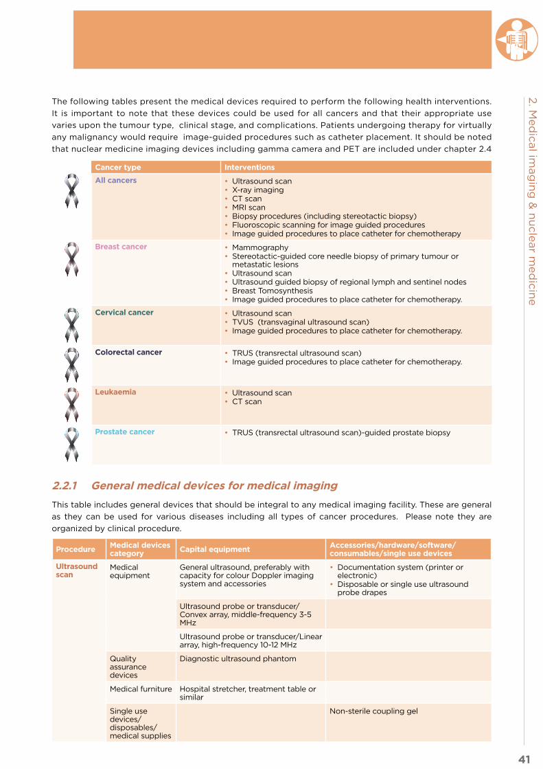
41
2. M
ed
ical im
ag
ing
& n
ucle
ar m
ed
icin
e
The following tables present the medical devices required to perform the following health interventions.
It is important to note that these devices could be used for all cancers and that their appropriate use
varies upon the tumour type, clinical stage, and complications. Patients undergoing therapy for virtually
any malignancy would require image-guided procedures such as catheter placement. It should be noted
that nuclear medicine imaging devices including gamma camera and PET are included under chapter 2.4
2.2.1 General medical devices for medical imaging
This table includes general devices that should be integral to any medical imaging facility. These are general
as they can be used for various diseases including all types of cancer procedures. Please note they are
organized by clinical procedure.
Procedure Medical devices category Capital equipment Accessories/hardware/software/
consumables/single use devices
Ultrasound scan
Medical equipment
General ultrasound, preferably with capacity for colour Doppler imaging system and accessories
• Documentation system (printer or electronic)
• Disposable or single use ultrasound probe drapes
Ultrasound probe or transducer/Convex array, middle-frequency 3-5 MHz
Ultrasound probe or transducer/Linear array, high-frequency 10-12 MHz
Quality assurance devices
Diagnostic ultrasound phantom
Medical furniture Hospital stretcher, treatment table or similar
Single use devices/disposables/medical supplies
Non-sterile coupling gel
Cancer type Interventions
All cancers • Ultrasound scan• X-ray imaging• CT scan • MRI scan • Biopsy procedures (including stereotactic biopsy)• Fluoroscopic scanning for image guided procedures • Image guided procedures to place catheter for chemotherapy
Breast cancer • Mammography• Stereotactic-guided core needle biopsy of primary tumour or
metastatic lesions• Ultrasound scan• Ultrasound guided biopsy of regional lymph and sentinel nodes• Breast Tomosynthesis• Image guided procedures to place catheter for chemotherapy.
Cervical cancer • Ultrasound scan• TVUS (transvaginal ultrasound scan)• Image guided procedures to place catheter for chemotherapy.
Colorectal cancer • TRUS (transrectal ultrasound scan)• Image guided procedures to place catheter for chemotherapy.
Leukaemia • Ultrasound scan• CT scan
Prostate cancer • TRUS (transrectal ultrasound scan)-guided prostate biopsy

42
WHO
list o
f prio
rity m
edica
l dev
ices f
or ca
ncer
man
agem
ent
Procedure Medical devices category Capital equipment Accessories/hardware/software/
consumables/single use devices
Solutions and reagents
Antimicrobial solution to disinfect the transducers
X-Ray imaging
Medical equipment
General-purpose digital radiography system with 3-megapixels monitor for interpretation
Fluoroscop-ic imaging process
Medical equipment
Fluoroscopy unit
CT scan Medical equipment
Computed Tomography (CT) System (multi-slice) including• workstation, preferably with ability
to perform 3-D reconstruction, computers
• 3-megapixels monitor for Interpretation
• Laser/thermal printer (optional)• PACS and RIS to share images• Dual head injector
Appropriate dedicated coils and pediatric if needed.3-megapixels monitor for interpretation
Quality assurance devices
CT phantom, CT quality control devices, including pencil style CT dose detector
MRI scan (for specialized hospitals)
Medical equipment
Magnetic Resonance Imaging (MRI) System (1.5 T)
Software package
Magnetic metal detector
MRI compatible anaesthesia machine
MRI compatible infusion pump
MRI compatible patient physiologic monitoring system
MRI compatible resuscitation trolley equipped with medicines and defibrillator
MR-safe stethoscope and sphygmomanometer
MRI compatible general-purpose suction system
MR compatible biopsy / procedure equipment (optional)
Quality assurance devices
MRI system quality assurance device
Medical furniture MRI compatible assistive footstool
MRI compatible cart
MRI compatible intravenous pole
MRI compatible wheelchair
MRI compatible stretcher
Solutions and reagents
MRI contrast media, injectable
Other MRI compatible oxygen canisters
General procedures for the area
Software Picture Archiving Communication System (PACS)
RIS (Radiological Information Systems)
3 Components of all surgical instrument sets as well as needle and catheter gauge sizes (G) are listed in the chapter annex at the end of the Surgery chapter

43
2. M
ed
ical im
ag
ing
& n
ucle
ar m
ed
icin
e
Procedure Medical devices category Capital equipment Accessories/hardware/software/
consumables/single use devices
Biopsy, drainage or ablation procedures
Medical furniture Patient Procedure Table / Couch
Table, instruments, Mayo type, stainless steel, on casters
Drug cupboard (local anesthesia for pain, contrast reaction)
Instruments Scalpel with blades
Dissection forceps
Personal protective equipment
Surgical cap for patients and healthcare worker
Surgical face mask
Eye protective wear
Operator sterile gown
General-purpose sterile drape
Single use devices/disposables/medical supplies
Cannulas, intravenous (IV) short, sterile, single use (sizes G )1
Compress, gauze, sterile & non-sterile, single use
Skin-cleansing wipe
Skin markers
Syringes with needles (disposable)
Absorbent tipped applicator
Sterile ultrasound coupling gel
Non-implantable needle guide
First aid gauze/bandage
Skin-cover adhesive strip
Microscope slides frosted
Tongue depressor, single use
Needles, sterile, single use:• 20–24G (for fine needle aspiration)• 11–14 G (for bone biopsy)• 16–20G (for other tissue biopsy)
ICD set / Thoracic tube insertion set (see Surgery subchapter for instrument sets)
Gelfoam (for plugged biopsy)
Solutions and reagents
Formalin 10% or tissue fixation reagents
Alcohol or iodine preparation cleansing agent
Oxygenated water
Other Ruler
Specimen container
Label or pen for labelling specimen containers
Basic sedation equipment
Safety box, for used syringes/needles
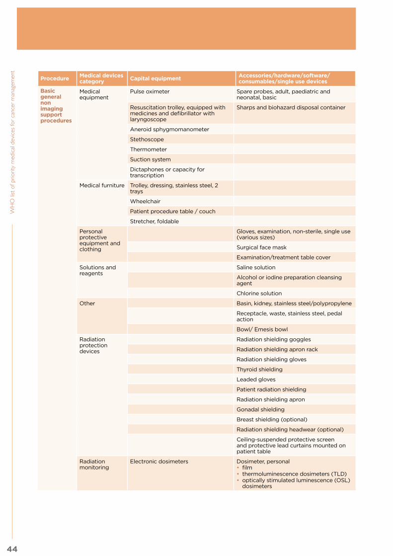
44
WHO
list o
f prio
rity m
edica
l dev
ices f
or ca
ncer
man
agem
ent
Procedure Medical devices category Capital equipment Accessories/hardware/software/
consumables/single use devices
Basic general non imaging support procedures
Medical equipment
Pulse oximeter Spare probes, adult, paediatric and neonatal, basic
Resuscitation trolley, equipped with medicines and defibrillator with laryngoscope
Sharps and biohazard disposal container
Aneroid sphygmomanometer
Stethoscope
Thermometer
Suction system
Dictaphones or capacity for transcription
Medical furniture Trolley, dressing, stainless steel, 2 trays
Wheelchair
Patient procedure table / couch
Stretcher, foldable
Personal protective equipment and clothing
Gloves, examination, non-sterile, single use (various sizes)
Surgical face mask
Examination/treatment table cover
Solutions and reagents
Saline solution
Alcohol or iodine preparation cleansing agent
Chlorine solution
Other Basin, kidney, stainless steel/polypropylene
Receptacle, waste, stainless steel, pedal action
Bowl/ Emesis bowl
Radiation protection devices
Radiation shielding goggles
Radiation shielding apron rack
Radiation shielding gloves
Thyroid shielding
Leaded gloves
Patient radiation shielding
Radiation shielding apron
Gonadal shielding
Breast shielding (optional)
Radiation shielding headwear (optional)
Ceiling-suspended protective screen and protective lead curtains mounted on patient table
Radiation monitoring
Electronic dosimeters Dosimeter, personal • film • thermoluminescence dosimeters (TLD) • optically stimulated luminescence (OSL)
dosimeters

45
2. M
ed
ical im
ag
ing
& n
ucle
ar m
ed
icin
e
Procedure Medical devices category Capital equipment Accessories/hardware/software/
consumables/single use devices
Contrast injection procedures (and man-agement of contrast media reac-tions)
Medical equipment
Contrast medium injection system
Single-patient physiologic monitoring system
Defibrillator
Emergency cart with medicines and defibrillator or automated external defibrillator (AED), blood pressure/pulse monitor, pulse oximeter
Suction system
Warming cabinet for contrast media
Medical furniture Stand, infusion, double hook, on casters
Table, instruments, Mayo type, stainless steel, on casters
Personal protective equipment and clothing
Operator sterile gown
Protective cotton gown reusable
Gown, patient
General-purpose sterile drape
Surgical cap for patients and healthcare worker
Single use devices/disposables/medical supplies
Catheter, Foley, sterile, single use (sizes G)3
Catheter, urethral, sterile, single use (sizes G)3
Compress, gauze, sterile & non-sterile, single use
Infusion set, sterile, single use
Skin-cleansing wipe
Syringes with needles (disposable)
Tape, medical, roll (various sizes)
Catheter (18 gauge or larger)1
Septo syringe
First aid gauze/bandage
Skin-cover adhesive strip
Cannulas, intravenous (IV) single use (sizes G)3 , intubation equipment
Solutions and reagents
Contrast media, injectable
Contrast media, oral
Sterile saline for IV
Other Arm/leg tourniquet Oxygen supply
Safety box, for used syringes/needles

46
WHO
list o
f prio
rity m
edica
l dev
ices f
or ca
ncer
man
agem
ent 2.2.2 Specific medical devices for medical imaging by cancer type
This section describes the medical imaging devices or accessories required that complement the above list of
general devices. For metastatic breast, cervical, colorectal, and prostate cancer, serial imaging staging is most
commonly through CT, PET/CT, and/or potentially bone scans (gamma camera) as relevant to the case which
will be described in the next section. It is important to have preparation sites for all the following procedures.
2.2.2.1 Breast
Procedure Medical devices category Capital equipment Accessories/hardware/software/
consumables/single use devices
Mammography Medical equipment Mammographic X-ray system Compression devices for additional projections e.g. cone compression (compatible with the collimators)
Stereotactic-guided core needle biopsy of primary tumor or metastatic lesions
Medical equipment Mammographic stereotactic biopsy system (cone compression devices could be used with normal mammographic system) Optional: vacuum-assisted biopsy device and driver
Single use devices/disposables/medical supplies
Biopsy needle
Wire localization needle ( e.g. Kopan's Needle 21G, 20G)
Fiducial markers/soft tissue markers
Ultrasound guided biopsy of regional lymph and sentinel nodes3 (See General Medical Devices table 2.2.1 above, ultrasound procedures)
Medical equipment Biopsy gun Biopsy needle (semi and automatic devices)
Ultrasound probe or transducer/Linear array, high-frequency transducers, small-footprint, large-bandwidth transducers with central frequency above 10 MHz are ideal
Ultrasound probe cover
Single use devices/disposables/medical supplies
Sterile ultrasound coupling gel
Core needle biopsy4
Medical equipment Biopsy Gun Biopsy needle
Single use devices/disposables/medical supplies
Specimen container
Needles and syringes for local anaesthetic
Skin-cleaning wipe
Skin-cover adhesive strip
Solutions and reagents
Formalin 10%, or tissue fixation reagents
Alcohol or iodine preparation cleansing agent
Other Label or pen for labelling sample
Ruler
Fine needle aspiration (FNA)4
Single use devices/disposables/medical supplies
Specimen container
Needles and syringes for local anaesthetic
Syringes for the biopsy and holder (optional)
3 Can be image-guided; i.e. breast ultrasound-guided biopsy or mammogram-guided (refer to the imaging section) although core needle biopsy is more common for mammographic biopsies than fine needle aspiration (FNA).

47
2. M
ed
ical im
ag
ing
& n
ucle
ar m
ed
icin
e
Procedure Medical devices category Capital equipment Accessories/hardware/software/
consumables/single use devices
Biopsy needle (23 or 25 gauge needle)
Microscope slides frosted
Skin-cleaning wipe
Skin-cover adhesive strip
Solutions and reagents
Formalin 10%, or tissue fixation reagents
Alcohol or iodine preparation cleansing agent
Other Label or pen for labelling sample
Ruler
Procedure Medical devices category
Capital equipment (For specialized hospitals)
Accessories/hardware/software/consumables/single use devices
MRI scan Medical equipment (see General Medical Devices table 2.2.1 above) with breast coil
Vascular access for chemotherapy, ports and PICCs
2.2.2.2 Cervical
Procedure Medical devices category Capital equipment Accessories/hardware/software/
consumables/single use devices
Ultrasound scan Single use devices/disposables/medical supplies
(see General Medical Devices table 2.2.1 above) with vaginal ultrasound imaging system transducer
Ultrasound probe cover Sterilization solution for US transducer
Interventional radiology for palliative care
Medical equipment General-purpose digital radiography system
2.2.2.3 Colorectal Only a small fraction of colorectal lesions can be sampled under TRUS, only the distal-most ones, and mention
should be made that endoscopy is required for sampling of the majority of colorectal lesions. See endoscopy
chapter for more information.
Procedure Medical devices category Capital equipment Accessories/hardware/software/
consumables/single use devices
TRUS (transrectal ultrasound) (See General Medical Devices table 2.2.1 above, ultrasound procedures)
Medical equipment Ultrasound system (as mentioned in the ultrasound procedure section) or ultrasound imaging system specific for TRUS, line-powered if possible, specific for this purpose including intracavitary probe for TRUS/Rectal ultrasound imaging system transducer(see General Medical Devices table 2.2.1 above)
Sterilization solution for US transducer
Biopsy gun Biopsy needle
CT Colonography
Medical equipment Computed Tomography (CT) System (multi-slice)
Foley catheter, appropriate reading software
Air or CO2 insufflator,
Procedure Medical devices category
Capital equipment (For specialized hospitals)
Accessories/hardware/software/consumables/single use devices
MRI scan Medical equipment (see General Medical Devices table 2.2.1 above)
Rectal coil
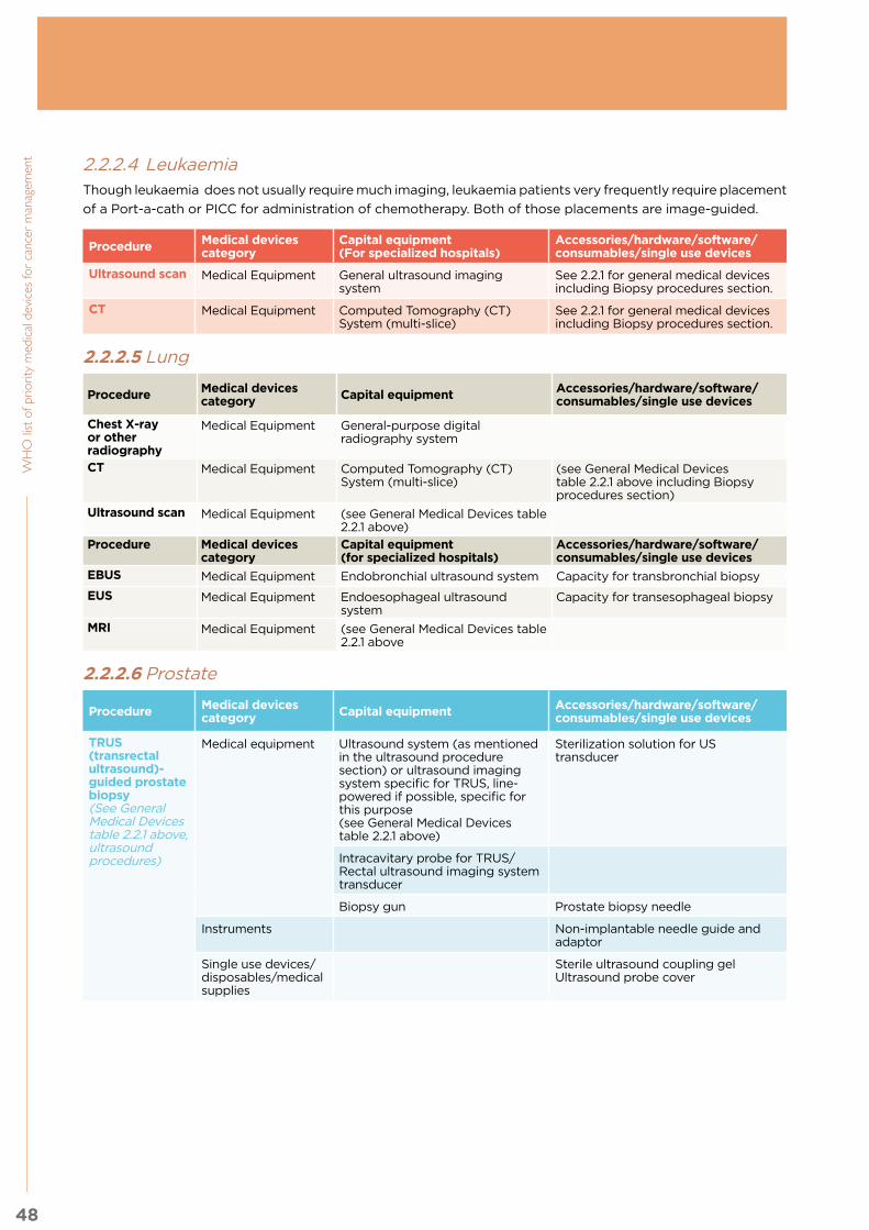
48
WHO
list o
f prio
rity m
edica
l dev
ices f
or ca
ncer
man
agem
ent
2.2.2.4 LeukaemiaThough leukaemia does not usually require much imaging, leukaemia patients very frequently require placement
of a Port-a-cath or PICC for administration of chemotherapy. Both of those placements are image-guided.
Procedure Medical devices category
Capital equipment(For specialized hospitals)
Accessories/hardware/software/consumables/single use devices
Ultrasound scan Medical Equipment General ultrasound imaging system
See 2.2.1 for general medical devices including Biopsy procedures section.
CT Medical Equipment Computed Tomography (CT) System (multi-slice)
See 2.2.1 for general medical devices including Biopsy procedures section.
2.2.2.5 Lung
Procedure Medical devices category Capital equipment Accessories/hardware/software/
consumables/single use devices
Chest X-ray or other radiography
Medical Equipment General-purpose digital radiography system
CT Medical Equipment Computed Tomography (CT) System (multi-slice)
(see General Medical Devices table 2.2.1 above including Biopsy procedures section)
Ultrasound scan Medical Equipment (see General Medical Devices table 2.2.1 above)
Procedure Medical devices category
Capital equipment (for specialized hospitals)
Accessories/hardware/software/consumables/single use devices
EBUS Medical Equipment Endobronchial ultrasound system Capacity for transbronchial biopsy
EUS Medical Equipment Endoesophageal ultrasound system
Capacity for transesophageal biopsy
MRI Medical Equipment (see General Medical Devices table 2.2.1 above
2.2.2.6 Prostate
Procedure Medical devices category Capital equipment Accessories/hardware/software/
consumables/single use devices
TRUS (transrectal ultrasound)-guided prostate biopsy(See General Medical Devices table 2.2.1 above, ultrasound procedures)
Medical equipment Ultrasound system (as mentioned in the ultrasound procedure section) or ultrasound imaging system specific for TRUS, line-powered if possible, specific for this purpose (see General Medical Devices table 2.2.1 above)
Sterilization solution for US transducer
Intracavitary probe for TRUS/Rectal ultrasound imaging system transducer
Biopsy gun Prostate biopsy needle
Instruments Non-implantable needle guide and adaptor
Single use devices/disposables/medical supplies
Sterile ultrasound coupling gelUltrasound probe cover

49
2. M
ed
ical im
ag
ing
& n
ucle
ar m
ed
icin
e
2.3 Other health system components for medical imaging
2.3.1 Human resources for medical imaging
A coordinated team of radiologists, and clinicians (clinical physicians) experienced in ultrasonography,
radiological technologists/radiographers and sonographers, is essential in the process of producing and
interpreting diagnostic images. Medical physicists have an important role in the optimization of the studies
and in the development of quality assurance, dosimetry and radiation protection programmes that ensure safe
and effective medical imaging. Additionally, biomedical engineers are needed for the technical management
of equipment and IT specialists are needed if the establishment and maintenance of teleradiology systems.
Multidisciplinary team collaboration is very important, including the participation of imaging specialists in
clinical conferences such as “tumour boards” around the world.
Some of these health professionals (e.g. radiologists, nuclear medicine specialists, radiographers and medical
physicists) are more familiar with medical uses of ionizing radiation, as the requirements for the above
mentioned their education, training, qualification and competencies usually include diagnostic imaging
also includes and radiation protection. This is not the case for other specialists (e.g. orthopaedic surgeons,
cardiologists, gastroenterologists, rheumatologists and neurosurgeons, among others) who also use ionizing
radiation in some situations, but for whom radiation protection is not traditionally part of their education,
training, qualification and competencies. Guidance on education and training in radiation protection of health
professionals is available elsewhere (2, 3).
Depending on national context including workforce availability and models, different occupations can fulfil
the roles that may be required for medical imaging. A prerequisite is having the required competencies to
perform these roles and being officially recognized for it as per the scope of practice. The ISCO2 codes are
added as illustrative examples based on current practice for further reference. This should not be interpreted
as guidance on occupations that should perform each role but rather indicative occupations that are currently
performing these roles in different country contexts.
Sample role Sample occupations (ISCO-08 codes)
• Responsible for the medical aspects of all the procedures and installations including justification and optimization of studies
• Acquisition and interpretation of US and fluoroscopic studies, interpretation of conventional radiology, mammography, CT and MR studies
• Performance of image-guided, minimally invasive diagnostic and therapeutic procedures (special training required)
• Responsible for regular and independent audits of the programme of QA for medical exposures (in coordination with radiographer and medical physicist or related occupation)
• Radiologist , Specialist medical practitioner (2212)
• Responsible of performing the ultrasound procedures with the different probes depending on organ scanned.
• Radiologist , Specialist medical practitioner (2212)
• General medical practitioner (2211)• Ultrasonographist, Medical
imaging and therapeutic equipment technician (3211)
• Plan and perform the medical imaging procedure (eg. CT, MR, mammography and conventional radiology)
• Responsible for overseeing team approach to protocol development with technologist and physicist
• Support radiologists in performance of minimally invasive diagnostic and therapeutic image-guided procedures.
• Optimization of medical imaging procedures• Implementation of radiation protection protocols (incoordination with
medical physicists and radiologists).• Administrative issues of radiological services (in coordination with
radiologists and administrative personnel)
• Radiographer, Medical imaging and therapeutic equipment technicians(3211)
2 International Standard Classification of Occupations, http://www.ilo.org/public/english/bureau/stat/isco/docs/publication08.pdf

50
WHO
list o
f prio
rity m
edica
l dev
ices f
or ca
ncer
man
agem
ent
Sample role Sample occupations (ISCO-08 codes)
• Responsibility towards radiation protection in compliance with national and international (BSS) requirements
• Responsibility to contribute to the imaging team to ensure that patients do not receive additional exposure due to inappropriate or suboptimal procedures
• Optimization of studies• Establishment of comprehensive QA programmes for medical exposures
including calibration and dosimetry of radiological equipment and radiation protection programmes for workers and patients
• Calibration and verification of measurement instruments• Technical supervision of equipment operation and maintenance
• Medical physicist (2111)
• Scheduling, reception of patients, all other administrative tasks • Clerical support workers ( 4412)• Technologists(3211),• Nurses (2221)
• Management of medical devices: planning, procurement, supervision of installation and maintenance
• Biomedical Engineer (2149)• Biomedical technician / physical
and engineering science technician (3119)
2.3.2 Infrastructure
Diagnostic imaging services require several utilities and services such as: water, stable electricity, installations
with proper infrastructure and adequate sanitary conditions, among others, for the protection of patients,
workers and the general public.
Warning devices (e.g. signs, lights) to prevent inadvertent entry into controlled or supervised areas must
be posted. Additional design features are needed for proper performance of diagnostic and interventional
procedures in children. More information regarding medical radiological equipment and related software
for diagnostic radiology and image guided interventional procedures are provided elsewhere, such as in the
IAEA-PAHO-WHO Safety Guide on Radiological Protection for Medical Exposure to Ionizing Radiation (4).
Appropriate steps should also be taken for safe disposal of both biological waste and disposable consumable
items used in these procedures.
If available, national radiology guidelines should be followed. Codes and standards such as NFPA (National
Fire Protection Association), ISO, NEC (National Electrical Code), emergency power systems, ASHRAE for
Ventilation of Health Care, washing facilities and special attention to temperature and humidity for each
equipment installation are needed, as required by national regulatory body both, one related to Ministry of
Health and the second related to radiation energy in the country.
Facilities layout distribution considerationsThe weight and dimensions of medical equipment should be considered when designing facilities, as well
as the planning of how to move equipment in prior to installation. Structural considerations should inform
decisions regarding equipment placement in existing facilities. Facility planning and design should incorporate
convenient and dedicated placement of imaging equipment, considering the requirements of floor space (if
the medical equipment is fixed), mechanical connections and the voltage required for electrical connections.
Commonly, medical imaging devices are installed by the manufacturer or vendor; therefore, close coordination
between all stakeholders (designers, installers, construction contractors and others) is required.
The size of all equipment areas should comply with manufacturers’ recommendations and radiation protection
requirements of the country or if not available, then international recommendations should be followed. Larger
rooms are preferable to facilitate equipment and patient movement during the procedure. Larger rooms are
also preferable for radiation protection purposes since increased distance from the radiation source confers
an exponentially decreased radiation dose, as explained by the inverse square law for radiation.”
Rooms and surrounding areas of MRI installations must be shielded against radiofrequency fields (Faraday
cage) and must comply with safety issues related to the use of the magnetic field (ferrous metal interferes
with image quality; construction must therefore minimize interference).
51
2. M
ed
ical im
ag
ing
& n
ucle
ar m
ed
icin
e
Moreover, MR suite entry security must be maintained since any ferromagnetic object will act as a guided
projectile and will harm a patient undergoing imaging. As an example, patients have been killed by non-MR-
safe oxygen canisters inadvertently brought into MR suites; those canisters subsequently acted as guided
missiles. Even cleaning staff with non-MR-safe floor buffers may not enter the MR suites after-hours as those,
too, rapidly pull into the bore of the magnet. An MR safety committee for the facility is advisable. All facility
staff should be educated that the magnet is always on. In this spirit, prior to MR imaging, patients must
undergo routine questioning as to potential ferromagnetic materials intrinsic to them: e.g. non-MR-safe
implants or clips, shrapnel, and more.
Shielding and radiation protectionAppropriate structural shielding to protect workers, patients and general public from ionizing radiation must
be installed to ensure adequate protection according to national and international guidelines (4).
Shielding requirements including both structural and ancillary protective barriers must be considered at
the design stage of planning a diagnostic imaging facility or department. A certified medical physicist or a
qualified expert should specify the type, location and amount of protective barriers to be installed taking into
account, amongst other things, the department layout (areas distribution) and medical equipment installed,
the workload of the equipment and the occupancy factors of the surrounding areas. In rooms performing
fluoroscopy- or angiography-guided interventional procedures, ceiling mounted protective screens, mobile
protective barriers, table mounted leaded curtains and protective eye wears should be considered.
The patient should be visible at all times through the window or operator shielding, even when the patient is in
the tilt position, and there should be some means of communication with the patient during the examination.
MRI installations must be shielded against radiofrequency fields (Faraday cage) and must comply with safety
and image quality issues related to the use of magnetic fields.
In medical imaging units, the safety of patients and healthcare workers is very important to take into account;
therefore, personnel dosimeters (passive and active if needed), devices for radiation protection should be
included, such as lead glass eye wears (goggles), protective lead aprons, thyroid shielding for workers, breast,
thyroid, eyes and gonadal shielding for patients, among others. Radiation survey meter should be available.
Adequate equipment to perform quality control tests should also be included as part of the lists of required
technologies, including suitable dosimetry instrumentation and calibration devices (5).
Heating, ventilation and air conditioning systemsThe ventilation should be adequate for the area and must comply with the manufacturers’ specifications for
the imaging equipment, but at the same time it should be consistent with the requirements for safety and
comfort of the patient. An helium extraction pipe system must be installed in case of quenching, as well as
an O2 monitor.
Information and communications technology (ICT)RIS, PACS, DICOM and workstation areas, including the storage, privacy and security of medical records should
be considered and are described further in the IAEA Human Health Series No. 28 Worldwide Implementation
of Digital Imaging in Radiology (6). If the use of teleradiology, PACS or RIS is considered, adequate bandwidth,
image quality and information infrastructure and/or web-based platforms should be available.
It is important to mention expected DICOM and PACS license renewals/updates and incumbent costs which
can potentially be negotiated or at least clarified as part of the initial contract. Omission of clear long-
term hardware maintenance and software contracts has proven troublesome, with multiple examples in low
resource settings where subsequent long-term costs post-procurement have been unexpected.
2.3.3 Quality management
Quality management, both generally and specifically within radiology, demands a quality culture that includes
a systematic approach to the elements that govern the delivery of that service. Therefore, the concept of
quality within a radiological facility covers, in its widest sense, all those factors that affect the intended
outcome, such as: generation of differential diagnostic possibilities, a tissue-based diagnosis from an image-

52
WHO
list o
f prio
rity m
edica
l dev
ices f
or ca
ncer
man
agem
ent guided biopsy, clinical restaging of cancer based upon serial imaging, assessment of response to therapy (as
after radiotherapy), or palliation from an interventional radiology procedure such as radiofrequency ablation.
WHO defines quality assurance (QA) programmes in diagnostic imaging as a coordinated effort by the staff
operating a facility to ensure that the images produced are of sufficiently high quality to consistently provide
adequate diagnostic and/or intra-procedural information at the lowest possible cost and with the least possible
exposure of the patient, personnel, and public to radiation. QA programmes related to medical exposures are
mandatory and the conditions for their implementation must be ensured. Calibration, dosimetry and QA of
radiological equipment must be performed by, under the oversight of, or with the documented advice of a
medical physicist, along with regular independent audits of these programmes.
If teleradiology is used, the quality of the transferred images, identification and confidentiality of the patients’
data, adequate transmission of clinical information, and relevant previous and/or complementary images
must be audited on a regular basis.
One of the most important components of this quality framework is the comprehensive clinical audit,
which involves evaluation of data, documents and resources to check performance against standards. It
is essentially a process of fact-finding and interpretation and, as such, provides an efficient tool for quality
improvement. Please see the IAEA Quality Assurance Audit for Diagnostic Radiology Improvement and
Learning (QUAADRIL) publication for more information (7).
2.3.4 Guidance documents
Title Link Description
WHO-ICRQS Referral Guidelines Project IRQN “Referral Guidelines for Diagnostic Imaging”
http://www.isradiology.org/isr/quality_guidelines.php
The Project aims to promote safe use and appropriate selection of diagnostic imaging, and to ensure these procedures are justified and indicated by providing a practical guidance tool and supporting its implementation. The Project includes activities to improve clinical referral guideline awareness, access and use. The use of clinical referral guidelines supports good medical practice, patient-centered care, procedure justification, and radiation safety.
ACR (2015) American College of Radiology’s Appropriateness Criteria®
http://www.acr.org/Quality-Safety/Appropriateness-Criteria
The ACR Appropriateness Criteria® (AC) are evidence-based guidelines to assist referring physicians and other providers in making the most appropriate imaging or treatment decision for a specific clinical condition. Employing these guidelines helps providers enhance quality of care and contributes to the most efficacious use of radiology.
European Qualifications Framework (EQF) Benchmarking Document: Radiographers
http://www.efrs.eu/publications/see/EFRS_EQF_level_6_benchmark?file=749
The purpose of the document is to serve as point of reference and benchmark for educational institutions, employers and professional bodies in Europe.
Radioisotope Handling Facilities (Atomic Energy Regulatory Board of India)
http://www.aerb.gov.in/AERBPortal/pages/English/t/publications/CODESGUIDES/SG-RF-RS.pdf
The objective of this Atomic Energy Regulatory Board of India safety guide is to provide guidance for implementing radiation safety requirements in facilities handling radioisotopes in order to: (a) ensure that workers and members of the public are not exposed to radiation, in excess of limits, specified by the competent authority, under the Atomic Energy (Radiation Protection) Rules, 2004, and safety directives issued by the competent authority from time to time; (b) reduce such radiation exposures to levels as low as reasonably achievable (ALARA); (c) ensure safe handling, physical security of radioactive materials and management of radioactive wastes; (d) provide means for detecting/assessing hazardous situations and initiating prompt remedial measures towards mitigating consequences; and (e) ensure accounting and recording of use of radioactive materials.

53
2. M
ed
ical im
ag
ing
& n
ucle
ar m
ed
icin
e
Title Link Description
ICRP Publication 127 on Radiological Protection in Ion Beam Radiotherapy
http://www.icrp.org/publication.asp?id=ICRP%20Publication%20127
Radiological protection and safety management should always conform with regulatory requirements. The current regulations for occupational exposures in photon radiotherapy are applicable to ion beam radiotherapy with protons or carbon ions. However, ion beam radiotherapy requires a more complex treatment system than conventional radiotherapy, and appropriate training of staff and suitable quality assurance programmes are recommended to avoid possible accidental exposure of patients, to minimise unnecessary doses to normal tissue, and to minimise radiation exposure of staff
Federal Guidance Report No. 14: Radiation Protection Guidance for Diagnostic and Interventional X-Ray Procedures
https://www.epa.gov/sites/production/files/2015-05/documents/fgr14-2014.pdf
Federal Guidance Report No. 14 provides federal facilities that use diagnostic and interventional X-ray equipment with recommendations for keeping patient doses as low as reasonably achievable without compromising the quality of patient care. This guidance is an update of Federal Guidance Report No. 9, which was issued in 1976.
ICRP Publication 129 on Radiological Protection in Cone Beam Computed Tomography (CBCT)
http://www.icrp.org/publication.asp?id=ICRP%20Publication%20129
The perception that CBCT involves lower doses was only true in initial applications. CBCT is now used widely by specialists who have little or no training in radiological protection. This publication provides recommendations on radiation dose management directed at different stakeholders, and covers principles of radiological protection, training, and quality assurance aspects. The recommendations provided in this publication may evolve in the future as CBCT equipment and applications evolve. As with previous ICRP publications, the Commission hopes that imaging professionals, medical physicists, and manufacturers will use the guidelines and recommendations provided in this publication for implementation of the Commission’s principle of optimisation of protection of patients and medical workers, with the objective of keeping exposures as low as reasonably achievable, taking into account economic and societal factors, and consistent with achieving the necessary medical outcomes.
Radiation Therapy Sources, Equipment and Installations (Atomic Energy Regulatory Board of India)
http://www.aerb.gov.in/AERBPortal/pages/English/t/publications/CODESGUIDES/SC-MED-01R.pdf
The objective of this Atomic Energy Regulatory Board of India code is to stipulate the radiation safety requirements in the design, installation and operation of radiation therapy sources, equipment and installations in order to ensure: (a) that radiation workers and members of the public do not receive radiation dose in excess of the limits specified by the competent authority under the Atomic Energy (Radiation Protection) Rules, 2004, and the safety notifications/directives issued thereunder from time to time; (b) that radiation doses to workers and members of the public are optimised to levels as low as reasonably achievable; (c) availability of appropriate instruments, tools and accessories, personnel and expertise, for safe handling of equipment and sources; (d) patient protection; (e) security, safe custody, transportation and disposal of sources; (f) timely detection and prompt rectification of malfunctions of radiation safety related equipment; and (g) initiation of appropriate actions to mitigate consequences of radiation emergencies.
Safety Code For Medical Diagnostic X-Ray Equipment and Installations (Atomic Energy Regulatory Board of India)
http://www.aerb.gov.in/AERBPortal/pages/English/t/publications/CODESGUIDES/SC-MED-02-REV1.PDF
This Atomic Energy Regulatory Board of India code is intended to govern radiation safety in design, installation and operation of X-ray generating equipment for medical diagnostic purposes in order to: (a) ensure that radiation workers and members of the public are not exposed to radiation in excess of limits specified by the competent authority under the Radiation Protection Rules, 1971, and by safety directives issued from time to time; (b) reduce radiation exposures below these limits to levels as low as reasonably achievable; (c) ensure availability of appropriate equipment, personnel and expertise for safe use of the equipment and for patient protection; and (d) ensure timely detection and prompt rectification of radiation safety-related defects or malfunctioning of the equipment.
Communicating radiation risks in paediatric imaging- information to support health care discussions about benefit and risk. WHO. 2016. ISBN: 9789241510349
http://apps.who.int/iris/bitstream/10665 /205033/1/ 9789241510349_ eng.pdf
The document is intended to serve as a tool for health care providers to communicate known or potential radiation risks associated with paediatric imaging procedures, to support risk-benefit dialogue in health care settings.
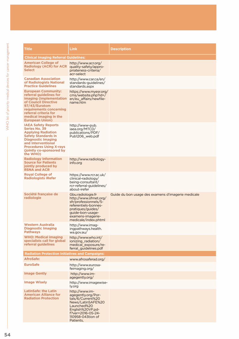
54
WHO
list o
f prio
rity m
edica
l dev
ices f
or ca
ncer
man
agem
ent
Title Link Description
Clinical Imaging Referral Guidelines:
American College of Radiology (ACR) for ACR Select
http://www.acr.org/quality-safety/appro-priateness-criteria/acr-select
Canadian Association of Radiologists National Practice Guidelines
http://www.car.ca/en/standards-guidelines/standards.aspx
European Community: referral guidelines for imaging (implementation of Council Directive 97/43/Euratom requirements concerning referral criteria for medical imaging in the European Union)
https://www.myesr.org/cms/website.php?id=/en/eu_affairs/newfile-name.htm
IAEA Safety Reports Series No. 39 Applying Radiation Safety Standards in Diagnostic Imaging and Interventional Procedures Using X-rays (jointly co-sponsored by the WHO)
http://www-pub.iaea.org/MTCD/publications/PDF/Pub1206_web.pdf
Radiology Information Source for Patients jointly produced by RSNA and ACR
http://www.radiology-info.org
Royal College of Radiologists iRefer
https://www.rcr.ac.uk/clinical-radiology/being-consultant/rcr-referral-guidelines/about-irefer
Société française de radiologie
Gbu.radiologie.frhttp://www.sfrnet.org/sfr/professionnels/5-referentiels-bonnes-pratiques/guides/guide-bon-usage-examens-imagerie-medicale/index.phtml
Guide du bon usage des examens d’imagerie medicale
Western Australia Diagnostic Imaging Pathways
http://www.imag-ingpathways.health.wa.gov.au/
WHO: Medical imaging specialists call for global referral guidelines
http://www.who.int/ionizing_radiation/medical_exposure/re-ferral_guidelines.pdf
Radiation Protection Initiatives and Campaigns:
AfroSafe: www.afrosaferad.org/
EuroSafe http://www.eurosa-feimaging.org/
Image Gently http://www.im-agegently.org/
Image Wisely http://www.imagewise-ly.org
LatinSafe: the Latin American Alliance for Radiation Protection
http://www.im-agegently.org/Por-tals/6/Current%20News/LatinSAFE%20Launched%20English%20VF.pd-f?ver=2016-05-24-110958-043tion of Patients,

55
2. M
ed
ical im
ag
ing
& n
ucle
ar m
ed
icin
e
Title Link Description
Cancer staging by NCI and AJCC:
http://www.cancer.gov/about-cancer/diagno-sis-staging/staging
https://cancerstaging.org/references-tools/Pages/What-is-Cancer-Staging.aspx
Management of Contrast Media Reactions:
American College of Radiology (ACR) Manual on Contrast Media Version 10.2 (2016)
http://www.acr.org/~/media/37D84428B-F1D4E1B9A3A-2918DA9E27A3.pdf
Informational link on CT Colonography:
From IAEA RPOP https://rpop.iaea.org/rpop/rpop/content/informationfor/health-professionals/1_radiol-ogy/computedtomog-raphy/ctcolonography.htm
Links to all the radiology-related NGOs in “official relations” with WHO
DITTA http://globalditta.org/
IOMP http://www.iomp.org/
ISR http://www.isradiology.org/isr/index.php
ISRRT http://www.isrrt.org/isrrt/default.asp
RAD-AID http://www.rad-aid.org/
WFUMB http://www.wfumb.org/
Bibliography:
1) ACR (2015) American College of Radiology’s Appropriateness Criteria® (http://www.acr.org/Quality-Safety/Appropriateness-Criteria) & RCR (2012). Royal College of Radiologists. iRefer: Making the best use of clinical radiology, 7th edition. London: The Royal College of Radiologists, 2012; (http://www.rcr.ac.uk/content.aspx?PageID=995).
2) ICRP, 2009. Education and Training in Radiological Protection for Diagnostic and Interventional Procedures. ICRP Publication 113. Ann. ICRP 39(5) and EC, 2014 Radiation Protection No 175- European guidance on radiation protection education and training of medical professionals in the EU, European Commission; (http://ec.europa.eu/energy/sites/ener/files/documents/175.pdf).
3) ICRP Publication 117 on Radiological Protection in Fluoroscopically Guided Procedures outside the Imaging Department. Ann. ICRP 40(6), 2010
4) IAEA-PAHO-WHO Safety Guide on Radiological Protection for Medical Exposure to Ionizing Radiation (ISBN:92-0-111302-1).
5) BSS, 2014 - Radiation protection and safety of radiation sources: International Basic Safety Standards. - General safety requirements Part 3 Series No. GSR Part 3. Vienna.
6) IAEA Human Health Series No 28. Worldwide Implementation of Digital Imaging in Radiology, 2015.
7) Quality Assurance Audit For Diagnostic Radiology Improvement And Learning (QUAADRIL). International Atomic Energy Agency, Vienna, 2010; (http://www-pub.iaea.org/MTCD/Publications/PDF/Pub1425_web.pdf).

56
WHO
list o
f prio
rity m
edica
l dev
ices f
or ca
ncer
man
agem
ent
2.4 General description of the nuclear medicine unit
Nuclear medicine is a field of medicine that uses small amounts of radioactive substances called
radiopharmaceuticals for the diagnosis and treatment of many health conditions including certain types of
cancer. Nuclear medicine procedures are usually performed in the medical imaging unit, and allow for the
characterization of physiologic processes inside the body, down to the molecular level, offering the potential
to identify diseases at early stages and to assess the response to therapeutic interventions. The information
produced is important for doctors to determine the status and function of different organs and tissues. It
helps doctors make critical decisions and tailor the treatment to the patient’s needs.
Nuclear medicine studies are applicable for diagnosis, careful prognostic assessments and appropriate
therapeutic decisions. They also allow the monitoring of treatment effects in a plethora of clinical conditions.
Nuclear medicine is considered an important tool for cancer management. In addition to its diagnostic
applications it can be used as an integral component for the treatment of some types of cancer, such as
thyroid, neuroblastoma, neuroendocrine tumours and lymphoma. It is also used for radioembolisation of liver
tumours and is very effective for palliation of bone pain caused by metastases, which can occur in patients
with breast, prostate and lung cancer.
Different radiopharmaceuticals may be used and their choice depends on the type of exam and the organ/
structure to be evaluated. For almost all types of examinations, radiopharmaceuticals are typically injected
intravenously and then taken up by the organ/structure(s) of interest. Radioactive emissions from the
radiotracer are detected by the imaging device – gamma camera or PET scanner – which produces images
of the target organ/structure(s).
Nuclear imaging, particularly if coupled with computed tomography (CT) or magnetic resonance imaging
(MRI) can show anatomical details and reveal how the targeted body part functions. These new modalities
are called hybrid imaging, including Positron Emission Tomography (PET) and, Single-photon Emission
Computed Tomography (SPECT), therefore leading to SPECT/CT, PET/CT, PET/MRI.
In conclusion, nuclear medicine imaging is crucial for the diagnosis and therapeutic management of chronic
diseases. Cancer diagnosis requires clinical assessment through appropriate diagnostic services such as those
provided by nuclear medicine, combined with an optimal treatment.
2.5 Priority medical devices for nuclear medicine
The following tables present general medical devices, which can be used for screening, diagnosis, staging,
and follow-up of many diseases including several cancers, and specific medical devices, which are used for
interventions for specific cancer types.
Radiopharmaceuticals can be prepared on site or ordered from a dedicated provider either as unidoses,
calibrated and ready for use, or in multidose vials, from which the appropriate activity is withdrawn for
several patients. It is important to have cell labelling methodologies to distinguish between infectious and
malignancies. All areas, procedures and radiation protection should follow guidance and overview from
national regulatory authorities, as appropriate.

57
2. M
ed
ical im
ag
ing
& n
ucle
ar m
ed
icin
e
Nuclear medicine interventions using gamma camera or PET/CT scanner can be used for all selected
cancer types and their appropriate use varies upon the tumour type and clinical stage. The following
tables present the priority medical devices required to perform the following health interventions:
2.5.1 General medical devices for nuclear medicine
These are general as they can be used for various diseases including all types of cancer. Please note they are
organized by clinical procedure.
Procedure Medical devices category
Capital equipment (for specialized hospitals)
Accessories/hardware/software/consumables/single use devices
Nuclear medicine scan
Medical equipment
Gamma camera system with SPECT capability
Quality assurance devices
QA/QC phantoms and accessories for all existing systems in the unit
Dose calibrator
Radionu-clide injection (including patient monitoring)
Medical equipment
Blood glucometer • Microcuvettes, strips or similar• Lancet, blood, safety, sterile (various
sizes)• Accessories
Infusion pump Infusion pump administration set
Resuscitation trolley, equipped with medicines and defibrillator. Include laryngoscope
Stethoscope
Weighing scale, range 0 –150 Kg
Aneroid sphygmomanometer Blood pressure cuffs
Single-patient physiologic monitoring system (optional)
Refrigerator
Medical furniture
Adequate furniture for patients, in the waiting area, non-absorbent surfaces
Trolley, dressing, stainless steel, 2 trays
Blood draw chairs
Stretcher, foldable
Cancer type Interventions
All cancers • Nuclear medicine scan and radionuclide injection (including patient monitoring)
• Preparing and dispensing radiopharmaceuticals
Breast cancer • Sentinel node mapping prior to surgical intervention
Lung, Breast, Prostate and Cervical cancer
• Bone scan
Lung, Breast, Prostate, Cervical and Colorectal cancer
• Staging, response assessment
3 Components of all surgical instrument sets as well as needle and catheter gauge sizes (G) are listed in the chapter annex at the end of the Surgery chapter

58
WHO
list o
f prio
rity m
edica
l dev
ices f
or ca
ncer
man
agem
ent
Procedure Medical devices category
Capital equipment (for specialized hospitals)
Accessories/hardware/software/consumables/single use devices
Single use devices/disposables/medical supplies
Cannulas, intravenous (IV) short, sterile, single use (sizes G)3
Compress, gauze, sterile & non-sterile, single use
Infusion giving set, sterile, single use
Needles, luer, sterile, single use (sizes G)3
Needles, scalp vein, sterile, single use (sizes G)3
Prongs, nasal, oxygen, non-sterile, single use (various sizes)
Skin-cleaning wipe
Tape, medical, roll (various sizes)
Transparent film dressings
Lead sharps container according to isotope energy used
Syringes, luer, sterile, single use (various capacities)
Instrument/equipment drape, single-use, non-sterile
Skin-cover adhesive strip First aid gauze/bandage
Solutions and reagents
IV solutions
Nuclear medicine radiation protection devices
Geiger-Müller counter radiation survey meter and measuring probe
Dosimeter, personal
Area monitoring devices (area survey meter)
Ring dosimeter
Mobile radiation protection shields
Radiation shielding container, transport/transfer
Syringe radiation shield (Gamma, Beta and Alpha)
Vial radiation shield
Radiation protection blocks
Radiation protection shields with glass
Shielded storage bins/recipients for radioactive wastes
Locked and adequately shielded and/or isolated area for radioactive wastes decay
Decontamination kits
Radiation protection gloves when managing relatively high doses, such as those for therapeutic purposes
Radiation shielding apron
Glasses, safety shield. All these devices need to be adapted to the radionuclides used (type of radiation, energy, activity, etc.)

59
2. M
ed
ical im
ag
ing
& n
ucle
ar m
ed
icin
e
Procedure Medical devices category
Capital equipment (for specialized hospitals)
Accessories/hardware/software/consumables/single use devices
Radiation safety instruments
Forceps Tongs
Needle sheath holder Aspiration tray
Drip trays
Personal protective equipment
Gloves, examination, non-sterile, single use (various sizes)
Gown, patient
Other Arm/leg tourniquet
Prepar-ing and dispensing radiophar-maceuticals
Medical equipment
Negative Pressure Fume Hood Require negative pressure as volatile radioisotopes (such as I-131) are manipulated
Laboratory equipment
High performance liquid chromatography (if radiopharmaceuticals are prepared on site)
Thin layer chromatography scanner (if radiopharmaceuticals are prepared on site)
Radionuclide generator (adequately shielded); Mo99-Tc99m; Ga68 (if radiopharmaceuticals are prepared on site)
Positive pressure Laminar Flow cabinet for radiopharmaceutical preparation
Require positive pressure to keep pyrogens out of the working area where radiopharmaceuticals are prepared and compounded
Refrigerator
Hot plate, with stirrer
Water bathThermometer
Medical furniture
Adequate furniture for receiving, handling and preparing radiopharmaceuticals.
Single use devices/disposables/medical supplies
Needles, luer, sterile, single use (sizes G)3
Syringes, luer, sterile, single use (various capacities)
Sterile absorbing paper
Quality assurance devices
Calibrated radioactive reference sources for quality control of activity measuring systems

60
WHO
list o
f prio
rity m
edica
l dev
ices f
or ca
ncer
man
agem
ent
Procedure Medical devices category
Capital equipment (for specialized hospitals)
Accessories/hardware/software/consumables/single use devices
Solutions and reagents radi-opharmaceuti-cal components
Non generator produced radiopharmaceuticals (I-131; Tl-201 etc.); PET tracers: multidose vials or unit doses (F18-FDG)
Labelling kits (MDP; MAA, pyrophosphate, MIBI, MAG3, DMSA, Nanocolloids, etc.)
Distilled water
Saline solution
Radiation protection devices
L-Shield
Radiation shielding container, general-purpose (adapt thickness according to radiation energies used)
Radioactive waste receptacle
Personal protective equipment
Gloves, examination, non-sterile, single use (various sizes)
Gown for staff
Non-conductive shoe cover
Coat, medical, woven (various sizes)
Radiation decontamination spill kit
PET/CT Medical equipment.
PET/CT system See 2.5.2.3 for radiopharmaceutical components and QA devices.
2.5.2 Specific medical devices for nuclear medicine by cancer type
2.5.2.1 Breast
Procedure Medical devices category
Capital equipment (for specialized hospitals)
Accessories/hardware/software/consumables/single use devices
Sentinel node mapping prior to surgical intervention
Medical equipment Gamma camera system with SPECT capability (see General Medical Devices Table 2.5.1.)
Gamma probes (for intraoperative use)
Radiopharmaceutical components
Tc99m colloid
2.5.2.2 Lung, Breast, Prostate and Cervical
Procedure Medical devices category
Capital equipment (for specialized hospitals)
Accessories/hardware/software/consumables/single use devices
Bone scan Medical equipment Gamma camera system (as per table General Medical equipment, 2.5.1 with SPECT capability)
Radiopharmaceutical components
Tc99m diphosphonate (MDP, HMDP, DPD ...)

61
2. M
ed
ical im
ag
ing
& n
ucle
ar m
ed
icin
e
2.5.2.3 Lung, Breast, Prostate, Cervical and Colorectal
Procedure Medical devices category
Capital equipment (for specialized hospitals)
Accessories/hardware/software/consumables/single use devices
Staging, response assessment
Medical equipment PET/CT system
Quality assurance devices
QA/QC phantoms and accessories for PET/CT System
Radiopharmaceutical components
FDG and other radiopharmaceuticals labeled with positron emitters Radiopharmaceutical preparations using centralized radio pharmacy or from in-house radio pharmacy facility having medical cyclotron
There is strong evidence to support the use of PET/CT in diagnosis, staging, re-staging, follow-up and
evaluation of therapy response and therapy planning in a variety of cancers. The use of PET/CT is considered
a standard of care for several types of cancer, supported by extensive scientific literature and technology
assessment reports (1, 2). In addition, constant research is generating medical evidence on its usefulness in
a variety of other clinical conditions. The ongoing development of new tracers is continuously revealing new
clinical applications in the medical community. Nonetheless, the use of this equipment involves significant
technical difficulties, financial, logistical, educational and quality management investments that should be
determined be each individual country. They are mainly due to the very short half-life of the isotopes (less
than 2 hours) and the very high energy (511 KeV) of the emitted radiation. Depending upon the country,
isotopes may have to be produced on site, in a cyclotron.
2.6 Other health system components for nuclear medicine
2.6.1 Human resources for nuclear medicine
Nuclear medicine encompasses diagnostic and therapeutic procedures applicable to several clinical
conditions, covered by many clinical specialties. Close interaction with referring clinicians is necessary in
order to increase the effectiveness of patient management. This interaction should be extended to educate
the medical community on the appropriate use of nuclear medicine applications and to expand research
pportunities, when applicable. Nuclear medicine has an inherent interdisciplinary nature. Aside from nuclear
medicine physicians and other medical personnel (e.g. nurses, radiographer/nuclear medicine technologist),
it requires cooperation with other professionals such as radio pharmacists, radiochemists, medical physicists,
engineers and IT specialists. Also, administrative and support services play an essential role. Responsibilities
must be defined and communication methods need to be in place.
Depending on national context including workforce availability and models, different occupations can fulfil
the roles that may be required for nuclear medicine. A prerequisite is having the required competencies to
perform these roles and being officially recognized for it as per the scope of practice. The ISCO2 codes are
added as illustrative examples based on current practice for further reference. This should not be interpreted
as guidance on occupations that should perform each role but rather indicative occupations that are currently
performing these roles in different country contexts (3,4).
2 International Standard Classification of Occupations, http://www.ilo.org/public/english/bureau/stat/isco/docs/publication08.pdf
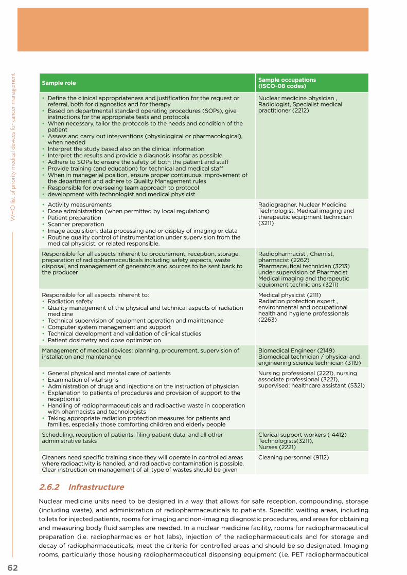
62
WHO
list o
f prio
rity m
edica
l dev
ices f
or ca
ncer
man
agem
ent
Sample role Sample occupations (ISCO-08 codes)
• Define the clinical appropriateness and justification for the request or referral, both for diagnostics and for therapy
• Based on departmental standard operating procedures (SOPs), give instructions for the appropriate tests and protocols
• When necessary, tailor the protocols to the needs and condition of the patient
• Assess and carry out interventions (physiological or pharmacological), when needed
• Interpret the study based also on the clinical information• Interpret the results and provide a diagnosis insofar as possible.• Adhere to SOPs to ensure the safety of both the patient and staff• Provide training (and education) for technical and medical staff• When in managerial position, ensure proper continuous improvement of
the department and adhere to Quality Management rules• Responsible for overseeing team approach to protocol• development with technologist and medical physicist
Nuclear medicine physician , Radiologist, Specialist medical practitioner (2212)
• Activity measurements• Dose administration (when permitted by local regulations)• Patient preparation• Scanner preparation• Image acquisition, data processing and or display of imaging or data• Routine quality control of instrumentation under supervision from the
medical physicist, or related responsible.
Radiographer, Nuclear Medicine Technologist, Medical imaging and therapeutic equipment technician (3211)
Responsible for all aspects inherent to procurement, reception, storage, preparation of radiopharmaceuticals including safety aspects, waste disposal, and management of generators and sources to be sent back to the producer
Radiopharmacist , Chemist, pharmacist (2262)Pharmaceutical technician (3213) under supervision of PharmacistMedical imaging and therapeutic equipment technicians (3211)
Responsible for all aspects inherent to:• Radiation safety• Quality management of the physical and technical aspects of radiation
medicine• Technical supervision of equipment operation and maintenance• Computer system management and support• Technical development and validation of clinical studies• Patient dosimetry and dose optimization
Medical physicist (2111)Radiation protection expert , environmental and occupational health and hygiene professionals (2263)
Management of medical devices: planning, procurement, supervision of installation and maintenance
Biomedical Engineer (2149)Biomedical technician / physical and engineering science technician (3119)
• General physical and mental care of patients• Examination of vital signs• Administration of drugs and injections on the instruction of physician• Explanation to patients of procedures and provision of support to the
receptionist• Handling of radiopharmaceuticals and radioactive waste in cooperation
with pharmacists and technologists• Taking appropriate radiation protection measures for patients and
families, especially those comforting children and elderly people
Nursing professional (2221), nursing associate professional (3221), supervised: healthcare assistant (5321)
Scheduling, reception of patients, filing patient data, and all other administrative tasks
Clerical support workers ( 4412)Technologists(3211),Nurses (2221)
Cleaners need specific training since they will operate in controlled areas where radioactivity is handled, and radioactive contamination is possible. Clear instruction on management of all type of wastes should be given
Cleaning personnel (9112)
2.6.2 Infrastructure
Nuclear medicine units need to be designed in a way that allows for safe reception, compounding, storage
(including waste), and administration of radiopharmaceuticals to patients. Specific waiting areas, including
toilets for injected patients, rooms for imaging and non-imaging diagnostic procedures, and areas for obtaining
and measuring body fluid samples are needed. In a nuclear medicine facility, rooms for radiopharmaceutical
preparation (i.e. radiopharmacies or hot labs), injection of the radiopharmaceuticals and for storage and
decay of radiopharmaceuticals, meet the criteria for controlled areas and should be so designated. Imaging
rooms, particularly those housing radiopharmaceutical dispensing equipment (i.e. PET radiopharmaceutical

63
2. M
ed
ical im
ag
ing
& n
ucle
ar m
ed
icin
e
and radioactive gas and aerosol dispenser devices), as well as waiting rooms dedicated to patients who have
been injected with radiopharmaceuticals (e.g. uptake rooms in a PET facility) should also be considered
controlled areas. Rooms with patients undergoing radiopharmaceutical therapy should be controlled
areas. Rooms housing hybrid machines that have an X-ray component (SPECT/CT and PET/CT) should be
considered controlled areas. A warning light at the entry to the room should indicate the machine is on, to
prevent unintended passage. Supervised areas may include examination rooms with probes, corridors and
other areas where there are patients who have been administered with radiopharmaceuticals (5). Access to
all controlled areas needs to be restricted to authorised persons (nuclear medicine staff, patients, caregivers)
in order to avoid unnecessary radiation exposure of the general public.
In units that administer radiopharmaceuticals for therapeutic purposes, a dedicated ward for confinement may
be necessary, depending on the type of treatment, administered activity, patient needs and conditions and local
rules and regulations, whilst also taking into account recommendations from IAEA Basic Safety Standards (5).
The establishment of nuclear medicine units able to provide diagnostic and therapeutic services, should take
into consideration the permits or sanitary authorization by local regulatory authorities, as well as instalment
costs and sustainability aspects.
Layout distributionThe siting and layout of a nuclear medicine department should take into account workload and patient
flow, both within the nuclear medicine facility and, in cases where the nuclear medicine facility is part of a
larger hospital or medical centre, with other departments of the wider facility. The nuclear medicine facility
should be readily accessible, especially for outpatients, who constitute the majority of patients. Enough
space should be allocated to facilitate the operation according to the workload. It should be built in a way
to minimize radiation exposure to staff and members of the public and contamination of the environment
(e.g. distance, wall thickness/composition, appropriate ventilation outlets, fluid drainage, waste management
and storage). Areas where radioactive material is stored, handled or administered to patients as well as all
rooms where patients stay after administration of radiopharmaceuticals (e.g. waiting rooms, changing areas,
toilets, uptake, investigation, stress rooms, imaging suites etc.) need to be classified. Access to controlled
areas should be regulated and is normally limited to patients and involved personnel. Security systems should
be installed to prevent the loss or theft of radioactive sources, or contaminated lead. The nuclear medicine
department should have source security systems and procedures to ensure continuity in the control and
accountability of each radioactive source at all times and to prevent theft, loss, unauthorized withdrawal of
radioactive materials or entrance of unauthorized personnel to the controlled areas. Please refer to IAEA
Basic Safety Standards for further information (5).
Electrical systemsTo ensure continuity of services, stable power supply and back-up systems should be installed.
Heating, ventilation and air conditioning systemsAir conditioning may be necessary to ensure correct functioning of imaging and other electronic equipment
(e.g. gamma cameras, PET/CT scanners, IT). Special care is to be given to the ventilation, with airflow
going from the least to the highest contamination risk areas. In fume hoods, negative pressure should be
installed to allow contaminated air with volatile radioisotopes to be evacuated, whereas in other areas in
which radiopharmaceuticals are prepared for administration to patients, laminar flow hoods are required with
positive pressures to keep pyrogens out of the working area.
Air conditioning, with proper humidity and temperature control should be planned, in order to avoid possible
damage to imaging systems, especially to radiation detectors and related components.
Radiation protection and shieldingStructural shielding should be designed to meet the requirements for optimization of protection, taking
into consideration the classification of the areas within the facility, the type of work to be done and the
radionuclides (and their activity) intended to be used. When SPECT/CT or PET/CT scanners are installed,
appropriate room shielding should be provided for the X-ray radiation emitted from the CT component.

64
WHO
list o
f prio
rity m
edica
l dev
ices f
or ca
ncer
man
agem
ent However, shielding might be required even for an imaging room with a normal gamma camera to avoid
interferences with other sources in the vicinity (injected patients, other camera). Shielding requirements must
be assessed on a case-by-case basis.
Special systemsIf telenuclear medicine is considered, the adequate bandwidth, information infrastructure or web based
platforms should be available, and appropriate devices (video surveillance) should be installed for patient
surveillance in diagnostic rooms during procedures and in confinement rooms for therapy.
Other considerationsWherever radioactive contamination might occur, floors and walls should be covered with non-porous,
washable material, curved to the walls, with all joints sealed and glued to the floor. The weight of equipment
to be installed should be considered during planning.
2.6.3 Quality management
Quality management in nuclear medicine aims at continuous improvement of services offered to patients and
referring physicians as well as insurances and providers of material and equipment. It is based on regular and
systematic reviewing of all involved processes, internal and external audits with the intent of improving quality
of services offered while maintaining highest ethical (appropriateness of procedures, patient information and
consent, communication, etc.) and safety standards (clinical, radiation, technical, chemical, pharmacological,
etc.) for patients, personnel, relatives, caregivers, members of the public and the environment.
To maximize the efficiency of the technology and ensure the accuracy of nuclear medicine devices, planning
must take place prior to purchase of equipment in the areas of maintenance and quality assurance (6-7).
Options for maintenance including vendor service contracts and training in-house or regional health system
staff, must be evaluated. Requirements for service and calibration, along with required service documentation,
ancillary equipment, Quality Control tools, spare parts and diagnostics must be included in tenders (request
for proposals). If the vendor does not meet service requirements in the tender, the service attributes desired
must be negotiated prior to purchase, due to the difficulty and expense of post-purchase servicing.
Regular quality audits and assessments are vital for modern nuclear medicine services. More importantly,
the entire quality management and audit process has to be systematic, patient orientated and outcome
based. The management of services should also take into account the diversity of nuclear medicine services
around the world and should invite multidisciplinary contributions. The latter include clinical, technical,
radiopharmaceutical, medical physics and radiation safety procedures.
Please see the IAEA Quality Management Audits in Nuclear Medicine Practices (QUANUM 2.0) for more
information, including QA/QC of equipment, an integral part of nuclear medicine quality management (8).
2.6.4 Guidance documents
Title Link Description
Radiation Protection and Safety of Radiation Sources: International Basic Safety Standards 2014
www-pub.iaea.org/MTCD/publications/PDF/Pub1578_web-57265295.pdf
This publication is the new edition of the International Basic Safety Standards. The edition is co-sponsored by seven other international organizations — European Commission (EC/Euratom), FAO, ILO, OECD/NEA, PAHO, UNEP and WHO. It replaces the interim edition that was published in November 2011 and the previous edition of the International Basic Safety Standards, which was published in 1996. It has been extensively revised and updated to take account of the latest findings of the United Nations Scientific Committee on the Effects of Atomic Radiation, and the latest recommendations of the International Commission on Radiological Protection. The publication details the requirements for the protection of people and the environment from harmful effects of ionizing radiation and for the safety of radiation sources. All circumstances of radiation exposure are considered.

65
2. M
ed
ical im
ag
ing
& n
ucle
ar m
ed
icin
e
Title Link Description
Planning a Clinical PET Centre
www-pub.iaea.org/MTCD/Publications/PDF/Pub1457_web.pdf
This publication presents a comprehensive overview of the steps involved in the establishment of a clinical PET facility, from strategy formulation to cyclotron implementation, radiopharmaceutical production and clinical applications. Also covered are staff requirements and radiation protection issues. It is intended for healthcare administrators, project and site planners, as well as professionals involved in providing PET services.
Quality Management Audits in Nuclear Medicine Practices
www-pub.iaea.org/MTCD/Pub-lications/PDF/Pub 1683Web-68161172.pdf
Quality management systems are essential and should be maintained with the intent to continuously improve effectiveness and efficiency, enabling nuclear medicine to achieve the expectations of its quality policy, satisfy its customers and improve professionalism. The quality management (QM) audit methodology in nuclear medicine practice, introduced in this publication, is designed to be applied to a variety of economic circumstances. A key outcome is a culture of reviewing all processes of the clinical service for continuous improvement in nuclear medicine practice. Regular quality audits and assessments are vital for modern nuclear medicine services. More importantly, the entire QM and audit process has to be systematic, patient oriented and outcome based. The management of services should also take into account the diversity of nuclear medicine services around the world and multidisciplinary contributions. The latter include clinical, technical, radiopharmaceutical, medical physics and radiation safety procedures.
Standard Operating Procedures for PET/CT: A Practical Approach for Use in Adult Oncology
www-pub.iaea.org/MTCD/Publications/PDF/Pub1616_web.pdf
Proper cancer management requires highly accurate imaging for characterizing, staging, restaging, assessing response to therapy, prognosticating and detecting recurrence of disease. The ability to provide, in a single imaging session, detailed anatomical and metabolic/functional information has established positron emission tomography/computed tomography (PET/CT) as an indispensable imaging procedure in the management of many types of cancer. The reliability of the images acquired on a PET/CT scanner depends on the quality of the imaging technique. This publication addresses this important aspect of PET/CT imaging, namely, how to perform an 18F-fluorodeoxyglucose (FDG) PET/CT scan in an adult patient with cancer. It provides a comprehensive overview that can be used both by new PET/CT centres in the process of starting up and by established imaging centres for updating older protocols.
Nuclear Medicine Resources Manual
www-pub.iaea.org/mtcd/publications/pdf/pub1198_web.pdf
This manual provides comprehensive guidance, at the international level, on many aspects of nuclear medicine practice, including education, training, facilities and equipment, quality systems, and radiopharmacy and clinical practice. It will be of use to those working in both new and more developed nuclear medicine centres.
Atlas of Bone Scintigraphy in the Developing Paediatric Skeleton: The Normal Skeleton Variants and Pitfalls
www-pub.iaea.org/MTCD/Publications/PDF/Pub1491_web.pdf
Bone scintigraphy is a widely accepted method for the evaluation of paediatric bone metabolism. Its strengths are high sensitivity and the capability to investigate the entire skeleton in a single examination. The interpretation of bone scanning in children is challenging and requires knowledge of the appearance of the maturing skeleton. The atlas provides an overview of issues related to bone physiology and variants, and points out pitfalls that might be encountered in daily routine work. Specific suggestions and hints assist in establishing adequate imaging protocols to deliver optimal image quality adjusted to the needs of each age group. The atlas will serve as a valuable reference for nuclear medicine physicians, radiologists and orthopaedic surgeons, and for those involved in teaching and performing paediatric bone scan imaging.
Appropriate Use of FDG-PET for the Management of Cancer Patients
www-pub.iaea.org/MTCD/Publications/PDF/Pub1438_web.pdf
This book provides guidance on the value and appropriateness of the use of positron emission tomography (PET), either alone or in combination with computed tomography (CT) scanners using 2-fluoro-2-deoxy-D-glucose (FDG) labelled with fluorine-18, in the management of patients affected by cancer. The concept of appropriateness provides a tool for determining which diagnostic investigations and therapies should be implemented, with the overall aim of optimizing health resource allocation, recognizing not only the cost of the intervention but also the consequences of failure to implement innovations of proven effectiveness. The book includes clinical scenarios for FDG-PET/CT indications; in all, 21 different types of cancer are considered, with seven different possible indications each.

66
WHO
list o
f prio
rity m
edica
l dev
ices f
or ca
ncer
man
agem
ent
Title Link Description
Practical Guidance on Peptide Receptor Radionuclide Therapy (PRRNT) for Neuroendocrine Tumours
www-pub.iaea.org/MTCD/Publications/PDF/P1560_web.pdf
This publication provides comprehensive, multidisciplinary guidance on the use of peptide receptor radionuclide therapy (PRRNT) in the treatment of patients with neuroendocrine tumours (NETs) and gastroenteropancreatic cancers, taking into account the recent international classifications of NETs. It provides comprehensive protocols for employing 90Y or 177Lu tagged somatostatin receptor targeting peptides as well as clinically assessed protocols for renal protection. It provides comprehensive, evidence based clinical guidelines, with input from experienced and renowned medical professionals in the field. The various sections of the book cover clinical presentation, patient eligibility criteria and means of assessing the effectiveness of therapy using molecular and morphological medical imaging techniques.
Guided Intraoperative Scintigraphic Tumour Targeting (GOSTT) Implementing Advanced Hybrid Molecular Imaging and Non-imaging Probes for Advanced Cancer Management
www-pub.iaea.org/MTCD/Publi-cations/PDF/Pub1648 web-19833477.pdf
The objective of this publication is to provide an updated source for professionals using guided intra- or perioperative scintigraphic tumour targeting (GOSTT). It supports and facilitates both the clinical decision making process and the implementation of minimally invasive surgical procedures; the ultimate aim of both approaches is to improve the standard of health care received by patients with cancer. This publication provides an update on innovations in the use of radiopharmaceuticals for SLNM and SLNB, in combination with vital dyes, when appropriate, to facilitate the detection of sentinel lymph nodes. In addition, it provides an update on advances in the implementation of hybrid imaging technologies for the surgical management of patients with cancer in conjunction with intraoperative regional lymph node mapping. Experience with small-field scintigraphic imaging devices in the operating theatre is also presented.
Clinical PET/CT Atlas: A Casebook of Imaging in Oncology
www-pub.iaea.org/MTCD/Publi-cations/PDF/Pub1680Web.pdf
Integrated positron emission tomography and computed tomography (PET/CT) has evolved since its introduction into the commercial market in 2001 into a major imaging procedure, particularly in oncological imaging. In clinical routine service, PET/CT has shown a significant impact on diagnosis, treatment planning, staging, therapy and the monitoring of treatment response, and has played an important role in the care of cancer patients. The high sensitivity from the PET component and the specificity of the CT component give this hybrid imaging modality the unique characteristics that make PET/CT one of the fastest growing imaging modalities, even 14 years after its clinical introduction. This PET/CT atlas combines nearly one hundred comprehensive cases covering all major indications of FDG–PET/CT as well as some cases of clinically relevant special tracers. The cases provide an overview of what the specific disease can look like in PET/CT, the typical pattern of the disease’s spread, as well as common pitfalls and teaching points. This PET/CT atlas will be of help to all professionals working with and interested in PET/CT imaging. It contains a variety of oncological imaging and provides clinically relevant teaching files on the effectiveness and diagnostic quality of FDG–PET/CT imaging in routine applications.
PET/CT Atlas On Quality Control And Image Artefacts
www-pub.iaea.org/MTCD/Publications/PDF/Pub1642web -16821314.pdf
This atlas on quality control (QC) and PET/CT artefacts provides guidance on typical image distortions in clinical PET/CT usage scenarios. A number of cases are presented to provide nuclear medicine and radiology professionals with an assortment of examples of possible image distortions and errors in order to support the correct interpretation of images. This atlas is intended to be used as a guide on how to take proper QC measures, on performing situation and problem analysis, and on problem prevention, and will be especially useful to medical physicists, physicians, technologists and service engineers in the clinical field.
Human Health Campus Website
https://humanhealth.iaea.org/hhw/
The Human Health Campus is an educational and resources website for health professionals in Radiation Medicine and Nutrition. This website is a valuable tool used by the IAEA Division of Human Health to achieve its mission to enhance the capabilities in Member States to address needs related to the prevention, diagnosis and treatment of health problems through the application of nuclear techniques.
Quality AssuranceFor SPECT Systems
www-pub.iaea.org/MTCD/publications/PDF/Pub1394_web.pdf
The current publication is intended to be a resource for medical physicists, technologists and other healthcare professionals who are responsible for ensuring optimal performance of imaging instruments, particularly SPECT systems, in their respective institutions. It is intended for managers, clinicians and other decision makers who are responsible for implementing quality assurance/quality control programmes in nuclear medicine centres. It is hoped that it will play an important role in helping maintain image quality and lead to better utilization of nuclear medicine imaging instruments worldwide.
Quality Assurance For PET and PET/CT Systems
www-pub.iaea.org/MTCD/publications/PDF/Pub1393_web.pdf
This publication provides guidelines for the implementation of QA and QC programmes concerning the combined medical diagnostic modality of PET and CT technologies. The use of these independent, but complementary, imaging techniques is frequent and growing within the fields of diagnostic imaging, oncology, cardiology and neurology, where they allow physicians to locate and diagnose malignant diseases accurately. Specific topics of discussion include the frameworks for reference values, tolerances and action levels, minimal required configurations with corresponding performance characteristics, and the management of ancillary equipment.

67
2. M
ed
ical im
ag
ing
& n
ucle
ar m
ed
icin
e
Title Link Description
VA Design Guide for Nuclear Medicine
http://www.wbdg.org/FFC/VA/VADEGUID/nucmed.pdf
The Nuclear Medicine Design Guide was developed as a tool to assist Contracting Officers, Medical Center Staff, and Architects and Planners with the design and construction of Nuclear Medicine facilities
A Guide to Clinical PET in Oncology: Improving Clinical Management of Cancer Patients
www-pub.iaea.org/MTCD/publications/PDF/te_1605_web.pdf
Clinical PET, in particular with fluorine-18-fluorodeoxyglucose (18F-FDG), has already proven itself to have considerable value in oncology. The indications include malignant lymphoma and melanoma, head and neck cancers, oesophageal cancer, breast cancer, lung cancer and colorectal cancer, and it is still being expanded. The roles of clinical PET could be for 1) preoperative staging of cancers, 2) differentiation between residual tumour and scarring, 3) demonstration of suspected recurrences, 4) monitoring response to therapy, 5) prognosis and 6) radiotherapy treatment planning.
Clinical Applications of SPECT/CT: New Hybrid Nuclear Medicine Imaging System
www-pub.iaea.org/MTCD/publications/PDF/TE_1597_Web.pdf
This report presents an overview of clinical applications of SPECT/CT and a relevant source of information for nuclear medicine physicians, radiologists and clinical practitioners. This information may also be useful for decision making when allocating resources dedicated to the health care system, a critical issue that is especially important for the development of nuclear medicine in developing countries. In this regard, the IAEA may be heavily involved in the promotion of programmes aimed at the IAEA’s coordinated research projects and Technical Cooperation projects.
Criteria for Palliation of Bone Metastases – Clinical Applications
www-pub.iaea.org/MTCD/publications/PDF/te_1549_web.pdf
Bone metastases are a frequent complication of cancer. It is estimated that they arise in 14–70% of all tumour patients, while it was reported that they occur in 70–85% patients in autopsy material. Although they may arise from any primary malignant tumour, certain tumours such as breast, prostate, lung, thyroid, kidney and myeloma have a predilection for a spread to bone. Bone metastases frequently cause pain, but there are also clinical situations with bone metastases causing no pain at all. The overall importance of the problem of bone metastases is well recognized by the fact that each year hundreds of thousands of cancer patients develop bone metastases. For example, more than 100 000 new patients develop this condition in the United States of America, although the prevalence is estimated to be double the number of new cases. While it is virtually unknown how many cancer patients in developing countries develop bone metastases, it is not unrealistic to expect that these figures largely surpass those coming from the developed countries.
Nuclear Medicine in Thyroid Cancer Management: A Practical Approach
www-pub.iaea.org/MTCD/publications/PDF/te_1608_web.pdf
Thyroid cancers are now being diagnosed at an earlier stage and treatments, together with follow-up strategies, are more effective. However this is not consistent throughout the world. The practice does differ considerably from country to country and region to region. Functional imaging using nuclear medicine procedures has become an indispensable tool for the diagnosis, treatment planning and management of patients. In terms of treatment, the use of radioiodine (131I) has been central to thyroid cancer and has been successfully used for over six decades. This publication is a culmination of efforts by more than twenty international experts in the field to produce a global perspective on the subject. Views expressed are those of individual experts involved and are intended to assist national or regional authorities in decisions regarding the frameworks for effective treatment of thyroid cancer.
Bibliography:1) Overview of the clinical effectiveness of positron emission tomography imaging in selected cancers. K
Facey, I Bradbury, G Laking and E Payne. Health Technology Assessment 2007; Vol. 11: No. 44, Health Technology Assessment NHS R&D HTA Programme. Available at: http://ushamp-build.squiz.co.uk/__data/assets/pdf_file/0010/58987/ExecutiveSummary-hta11440.pdf
2) Appropriate use of FDG-PET for the management of cancer patients. Human Health Series no 9. INTERNATIONAL ATOMIC ENERGY AGENCY VIENNA, 2010
3) Planning a clinical PET center. Human Health Series no 11. INTERNATIONAL ATOMIC ENERGY AGENCY VIENNA, 2010
4) Nuclear Medicine Resources Manual Ver 2.0. A guide for decision makers. International Atomic Energy (in press), Vienna 2016
5) Radiation Protection and Safety Of Radiation Sources: International Basic Safety Standards. International Atomic Energy Agency Vienna, 2014].
6) Quality Assurance for SPECT systems INTERNATIONAL ATOMIC ENERGY AGENCY VIENNA, 2009
7) Quality Assurance for PET and PET/CT systems INTERNATIONAL ATOMIC ENERGY AGENCY VIENNA, 2009
8) Quality Management Audits In Nuclear Medicine Practices Second Edition International Atomic Energy Agency, Vienna, 2015. http://www-pub.iaea.org/MTCD/Publications/PDF/Pub1683Web-68161172.pdf.

Surgery3.

69
3. S
urg
ery
Health service delivery sequence overview:
This diagram expresses the flow of cancer patient to and from the clinical assessment and surgery unit, and
the elements to consider as support for the unit (Fig 10.3).
Fig. 10.3. Health care service delivery overview – surgery
PR
IMA
RY
(C
L,H
C),
SE
C-
ON
DA
RY
(D
H)
&
TE
RT
IAR
Y (
SH
)
Le
ve
l o
f C
are
*
* Appropriate level of care will depend on the particular intervention, setting, and available infrastructure and human resources.
SE
CO
ND
AR
Y (
DH
)
& T
ER
TIA
RY
(S
H)
TE
RT
IAR
Y (
SH
)
Prevention End of life care
TreatmentFollow-up/survivorship
care
Palliative care
Diagnosis
Screening
CL Community Level health post DH District Hospital HC Health Centre SH Specialized Hospital
Systemic therapy
Nuclear medicine
Surgery
Vaccination
Radio- therapy
Pathology Pathology
Clinical laboratory
Clinical laboratory
Clinical assessment
Palliative care
Medical imaging
Endoscopy
Clinical assessment
Nuclear medicine
3.1 General description of the unit
3.2 Priority medical devices for surgery
3.2.1 General medical devices for surgery
3.2.2 Specific medical devices for surgery by cancer type
3.2.3 Surgical instrument sets, catheter and needle sizes
3.3 Other health system components
3.3.1 Human resources for surgery
3.3.2 Infrastructure
3.3.3 Quality management
3.3.4 Guidance documents
70
WHO
list o
f prio
rity m
edica
l dev
ices f
or ca
ncer
man
agem
ent
3.1 General description of the unit
The objective of a surgical unit is to provide general and specialized surgical care for patients. An important
goal of surgical care is for an interventional curative treatment. Other reasons include diagnosis, palliation of
symptoms, cancer prevention and reconstructive care.
It should be noted that in 2015, the Lancet Commission on Global Surgery stated that 5 billion people do
not have access to safe surgery and this was recognized by all Member States in the World Health Assembly
resolution 68. to Strengthen Emergency and Essential Surgical Care. ( see guidance documents section 3.3.4).
Services provided by a surgical unit must be safe, high-quality, founded on guidelines or established standards
such as the WHO Safe Surgery Saves Lives programme (1), and coordinated with other modalities of cancer
management. Multidisciplinary decision-making and treatment should be promoted. Moreover, there should
be a quality management framework that addresses equity and accessibility, workload and demand, core
processes and outcomes.
The surgery unit provides core services in cancer management and has a critical link with diagnostic services
– particularly pathology, radiology and endoscopy. Over 80% of cancer patients will need surgery at some
time (2). Similar to other cancer services, surgery must be viewed as a part of the larger health system,
recognizing that a surgery unit consists of broad human resource expertise, is linked to other critical health
services and contributes to wide-ranging service delivery across health disciplines.
The flow of patients and information from diagnostic support services (as can be seen in Fig. 10.3 on the
health services delivery process) must be timely and equitable. Primary care services may be a component
of the surgery unit. Primary care services should be capable of recognising cancer signs and symptoms,
performing appropriate simple biopsies, or referring for diagnostic procedures such as endoscopy.
The surgery unit extends beyond the operating room and is also linked to pre- and post-operative services
involving anaesthesia, pain management and nursing services. Post-operative nursing care, for example, must
be effective in identifying post-operative abnormalities and recognising changes or deterioration in patient
status, escalating levels of care appropriately.
Surgical units treating cancer differ in their ability to carry out procedures depending on the available
infrastructure and expertise. At its most basic, a surgical unit needs appropriately trained surgical healthcare
professionals, sufficient physical infrastructure to ensure safe surgery, surgical supplies, and procedures that
ensure safety and quality for both patients and staff.
3.2 Priority medical devices for surgery
It is very important to consider the four components of the customary supply chain process: product selection,
procurement, distribution and se. This process requires input from all stakeholders in surgical care – from
programme managers to clinical staff. Reliable supply chain and logistics management are needed to ensure
the right quality product is available, in the right quantities, at the right time, and for a reasonable cost. This
includes devices listed in this document, sutures, solutions, consumables and the related essential medicines
in perioperative care (such as analgesics, anaesthetics and antibiotics).
The following tables present the general medical devices for surgery, which can be used for diagnosis, staging,
treatment, follow-up and/or palliative care of many diseases including several cancers, and specific medical
devices, which are used for surgical interventions for specific cancer types.
Please note: Components of all surgical instrument sets as well as needle and catheter gauge sizes (G) are
listed in the chapter annex at the end of this chapter.
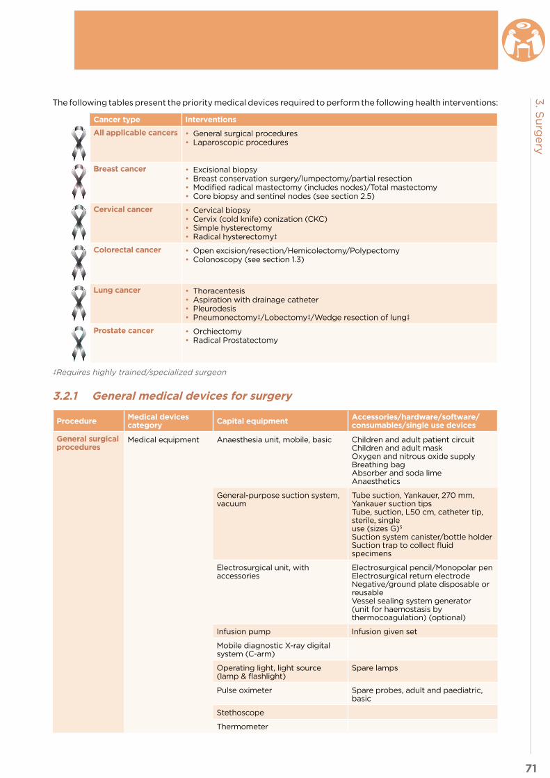
71
3. S
urg
ery
The following tables present the priority medical devices required to perform the following health interventions:
Cancer type Interventions
All applicable cancers • General surgical procedures• Laparoscopic procedures
Breast cancer • Excisional biopsy• Breast conservation surgery/lumpectomy/partial resection• Modified radical mastectomy (includes nodes)/Total mastectomy• Core biopsy and sentinel nodes (see section 2.5)
Cervical cancer • Cervical biopsy• Cervix (cold knife) conization (CKC)• Simple hysterectomy • Radical hysterectomy‡
Colorectal cancer • Open excision/resection/Hemicolectomy/Polypectomy• Colonoscopy (see section 1.3)
Lung cancer • Thoracentesis• Aspiration with drainage catheter• Pleurodesis• Pneumonectomy‡/Lobectomy‡/Wedge resection of lung‡
Prostate cancer • Orchiectomy• Radical Prostatectomy
‡Requires highly trained/specialized surgeon
3.2.1 General medical devices for surgery
Procedure Medical devices category Capital equipment Accessories/hardware/software/
consumables/single use devices
General surgical procedures
Medical equipment Anaesthesia unit, mobile, basic Children and adult patient circuitChildren and adult maskOxygen and nitrous oxide supplyBreathing bagAbsorber and soda limeAnaesthetics
General-purpose suction system, vacuum
Tube suction, Yankauer, 270 mm, Yankauer suction tipsTube, suction, L50 cm, catheter tip, sterile, single use (sizes G)3
Suction system canister/bottle holderSuction trap to collect fluid specimens
Electrosurgical unit, with accessories
Electrosurgical pencil/Monopolar penElectrosurgical return electrodeNegative/ground plate disposable or reusableVessel sealing system generator (unit for haemostasis by thermocoagulation) (optional)
Infusion pump Infusion given set
Mobile diagnostic X-ray digital system (C-arm)
Operating light, light source (lamp & flashlight)
Spare lamps
Pulse oximeter Spare probes, adult and paediatric, basic
Stethoscope
Thermometer
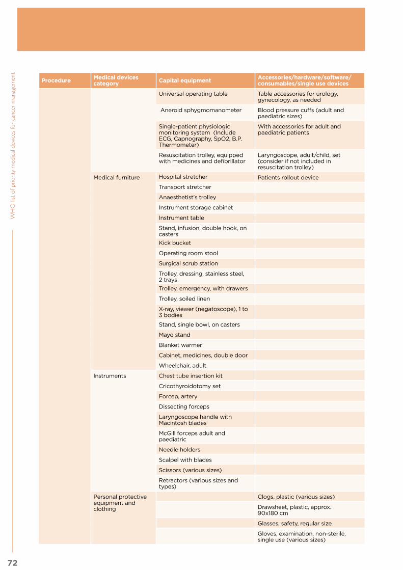
72
WHO
list o
f prio
rity m
edica
l dev
ices f
or ca
ncer
man
agem
ent
Procedure Medical devices category Capital equipment Accessories/hardware/software/
consumables/single use devices
Universal operating table Table accessories for urology, gynecology, as needed
Aneroid sphygmomanometer Blood pressure cuffs (adult and paediatric sizes)
Single-patient physiologic monitoring system (Include ECG, Capnography, SpO2, B.P. Thermometer)
With accessories for adult and paediatric patients
Resuscitation trolley, equipped with medicines and defibrillator
Laryngoscope, adult/child, set (consider if not included in resuscitation trolley)
Medical furniture Hospital stretcher Patients rollout device
Transport stretcher
Anaesthetist's trolley
Instrument storage cabinet
Instrument table
Stand, infusion, double hook, on casters
Kick bucket
Operating room stool
Surgical scrub station
Trolley, dressing, stainless steel, 2 trays
Trolley, emergency, with drawers
Trolley, soiled linen
X-ray, viewer (negatoscope), 1 to 3 bodies
Stand, single bowl, on casters
Mayo stand
Blanket warmer
Cabinet, medicines, double door
Wheelchair, adult
Instruments Chest tube insertion kit
Cricothyroidotomy set
Forcep, artery
Dissecting forceps
Laryngoscope handle with Macintosh blades
McGill forceps adult and paediatric
Needle holders
Scalpel with blades
Scissors (various sizes)
Retractors (various sizes and types)
Personal protective equipment and clothing
Clogs, plastic (various sizes)
Drawsheet, plastic, approx. 90x180 cm
Glasses, safety, regular size
Gloves, examination, non-sterile, single use (various sizes)

73
3. S
urg
ery
Procedure Medical devices category Capital equipment Accessories/hardware/software/
consumables/single use devices
Gloves, surgical sterile, ( various sizes)
Non-conductive shoe cover
Surgical cap for patients and healthcare workers
Gown, protective for health care workers
Aprons, impermeable
Surgical face mask
General-purpose sterile drape
Gown, patient
Medical scrubs for healthcare workers or similar
Single use devices/disposables/medical supplies
Anaesthesia breathing circuit
Compress, gauze, sterile & non-sterile, single use
Elastic bandage
Endotracheal tubes adult and paediatric
Infusion giving set, sterile, single use
IV infusor bags/sets
Mask and tubing for oxygen
Monitoring electrodes
Nasogastric tubes
Operating room laundry bag
Organ bag
Bags (various colors)
Skin-cleaning wipe
Sutures
Syringes with needles (disposable)
Tape, medical, roll (various sizes)
Three-way-stopcock
Ureteral catheter connector and other connectors as required
Urinary catheter (Foley)
Urine drain bag
Surgical scrub sponge
Intravenous cannulas or catheters
Laryngeal mask airways sizes 2, 3, 4, three sets per O.R, Oropharyngeal airway (adult size and paediatric)
Intubating bougies, adult and paediatric
Operating table cover
Bedmat, absorbable
Solutions and reagents
IV solutions
Formalin 10%, Phospate buffered or tissue fixation reagents

74
WHO
list o
f prio
rity m
edica
l dev
ices f
or ca
ncer
man
agem
ent
Procedure Medical devices category Capital equipment Accessories/hardware/software/
consumables/single use devices
Iodine povacrylex and isopropyl alcohol solution, chlorhexidine, or similar
Other Arm/leg tourniquet
Bedpan
Waste disposal container
Sponge bowl
Skin marker pen
Bowl/ Emesis bowl
Resuscitator bag valve and mask (adult and paediatric)
Safety box, for used syringes/needles
Laparoscopic procedures
Medical equipment Laparoscopic tower system, including: Video recorder, video monitor, laparoscopic light source, laparoscopic processing unit, CCD (Charge-coupled device), high-flow laparoscopic insufflator with C02 gas source and pressure regulation unit (Laparoscope can be 5 mm, 10 mm, 30 degree angled or straight)
High-frequency generator, both mono and bipolar for laparoscopic procedures
Monopolar and Bipolar coagulation forceps/dissector
Instruments Laparoscopic multi-instrument access port (optional for single-incision laparoscopic surgery)
Laparoscopic irrigation/aspiration cannula
Laparoscopic needle holder
Trocars with safety sheath (multiple sizes)
Specific instruments for the laparoscopic intervention: (for dissection or haemostasis, e.g. Laparoscopic dissection spatula, Internally-anchored endotherapy retractor, Laparoscopic grasping forceps, Laparoscopic swab forceps, Laparoscopic biopsy forceps, Endoclip-applicator, Laparotomy ring)
Staples, Implantable (optional)
Laparoscope holder (optional)
Suction and irrigation device
Laparoscopic electrosurgical blunt dissector
Laparoscopic grasper
Single use devices Veress needle (optional, for transperitoneal access only)‡
‡ Requires highly trained/specialized surgeon

75
3. S
urg
ery
3.2.2 Specific medical devices for surgery by cancer type
3.2.2.1 Breast
Procedure Medical devices category Capital equipment Accessories/hardware/software/
consumables/single use devices
Excisional biopsy
Instruments Excisional breast biopsy set
Breast-conservation surgery-lumpectomy/partial resection
Instruments Mastectomy set3
Single use devices/disposables/medical supplies
Surgical clip
Core needle biopsy4
Medical equipment Biopsy Gun Biopsy needle
Single use devices/disposables/medical supplies
Specimen container
Needles and syringes for local anaesthetic
Skin-cleaning wipe
Skin-cover adhesive strip
Solutions and reagents
Formalin 10%, or tissue fixation reagents
Alcohol or iodine preparation cleansing agent
Other Label or pen for labelling sample
Ruler
Procedure Medical devices category
Capital equipment (for specialized hospitals)
Accessories/hardware/software/consumables/single use devices
Modified radical mastectomy (includes nodes)/Total mastectomy
Instruments Mastectomy set3
Single use devices/disposables/medical supplies
Closed-wound drainage reservoir system with closed wound drain connector
3.2.2.2 Cervical
Procedure Medical devices category Capital equipment Accessories/hardware/software/
consumables/single use devices
Cervical biopsy Instruments Gynaecologic biopsy set3
Cervical Conization
Medical equipment Electrosurgical unit, with accessories, smoke evacuator
Loop electrodes, ball electrodes, ferric subsulfate solution (Monsel’s solution)
Instruments Cervix conization set3
Vaginal speculum, Non-conducting preferably with side retractors
Simple Hysterectomy
Instruments Vaginal hysterectomy set3 and Abdominal Hysterectomy set3
Single use devices/disposables/medical supplies
Cervical aspiration catheter
Closed-wound drainage reservoir system with closed wound drain connector
Procedure Medical devices category
Capital equipment (for specialized hospitals)
Accessories/hardware/software/consumables/single use devices
Radical hysterectomy‡
Instruments Vaginal Hysterectomy set3 and Abdominal Hysterectomy set3
Single use devices/disposables/medical supplies
Cervical aspiration catheter
Closed-wound drainage reservoir system with closed wound drain connector
‡ Requires highly trained/specialized surgeon
3. Components of all surgical instrument sets as well as needle and catheter gauge sizes (G) are listed in the chapter annex at the end of the Surgery chapter

76
WHO
list o
f prio
rity m
edica
l dev
ices f
or ca
ncer
man
agem
ent
3.2.2.3 Colorectal
Procedure Medical devices category Capital equipment Accessories/hardware/software/
consumables/single use devices
Open excision/resection/ Hemicolecto-my/ Polypectomy
Instruments Basic Colon Surgery set3
Laparotomy set3
3.2.2.4 Lung
Procedure Medical devices category Capital equipment Accessories/hardware/software/
consumables/single use devices
Lung procedures
Medical equipment Single-patient physiologic monitoring system for ECG, Capnography, SpO2, B.P.
Cables and sensor
Thoracentesis Instruments Thoracentesis set Underwater seal drainage
Single use devices/disposables/medical supplies
Sterile sample container
Needles and syringes for local anaesthetic
Flexible silicon catheter with needle
Aspiration with drainage catheter
Instruments Chest tube set (clamps included)
Single use devices/disposables/medical supplies
Chest tube
Sterile sample container
Needles and syringes for local anaesthetic
Drainage bag including gravitational IV tube or connect to suction system.
Pleurodesis Instruments Chest tube set3 (clamps included)
Single use devices/disposables/medical supplies
Chest tube
Sterile sample container
Needles and syringes for local anaesthetic
Drainage bag including gravitational IV tube or connect to suction system.
Solutions and reagents
Sclerodesis agent (e.g. talc)
Procedure Medical devices category
Capital equipment (for specialized hospitals)
Accessories/hardware/software/consumables/single use devices
Open Lobec-tomy/Pneu-monectomy/ Wedge resec-tion of lung (Thoracotomy)‡
Medical equipment Endoscopy tower system (Insufflator, Light source, Irrigation/ aspiration pump, Processing unit, Video image display monitor, Bronchoscope
Instruments Thoracotomy set3
Lobectomy and segmental lung set3
Single use devices Surgery table, padded accessories for patient positioning or similar
Safety straps
Chest tubes drainage
Hemoclips/clip cartridge or similar for cardiothoracic surgery
Staplers (linear and thoracotomy) with staples reloads
Epidural catheter
Pneumonectomy pleurevacs only for Pneumonectomy
‡ Requires highly trained/specialized surgeon

77
3. S
urg
ery
3.2.2.5 Prostate
Procedure Medical devices category Capital equipment Accessories/hardware/software/
consumables/single use devices
Orchiectomy Instruments Basic Surgery set1/ Minor tray
Radical Prostatectomy
Instruments Prostatectomy set1
3.2.3 Surgical instrument sets, catheter and needle sizes
Basic Surgery set / Minor tray
4 x Clamp, towel, Backhaus, 130 mm
2 x Forceps, tissue seizing, Allis, 150 mm
6 x Forceps, artery, Halsted-Mosquito, 125 mm, curved
1 x Forceps, artery, Kocher, 140 mm, straight
1 x Forceps, dressing, standard, 155 mm, straight
1 x Forceps, tissue holding, Collin, 160 mm
1 x Forceps, tissue, standard, 145 mm, straight
1 x Forceps, dressing & polypus, Cheron, 250 mm
1 x Needle holder, Mayo-Hegar, 180 mm, straight
1 x Probe, double-ended, 145 mm
1 x Retractor, Farabeuf, double-ended, 120 mm, pair
2 x Scalpel handles: number 3 and 4, one each
1 x Scissors, Metzenbaum, 140 mm, curved, blunt/blunt
1 x Scissors, Mayo, 140 mm, curved, blunt/blunt
1 x Bowl, stainless steel, 180 ml
2 x Dishes, kidney type
1 x Forceps Rampleys, sponge holding
Dressing set
1 x Forceps, artery, Kocher, 140 mm, str
1 x Forceps, dressing, standard, 155 mm, str
1 x Scissors, Deaver, 140 mm, str, s/b
2 x Dishes, kidney type
Laparotomy set
4 x Forceps, Ring c and r
2 x Babcock clamps
1 x Needle holder, Mayo-Hegar, straight, small, medium and large
1 x Forceps, dressing, standard, 155 mm, straight
1 x Forceps, dressing, standard, 250 mm, straight
1 x Dissecting Forceps, teeth, fenestrated, long and short
1 x Dissecting Forceps, large non teeth
1 x Retractor, Farabeuf, double-ended, 180 mm, pair
1 x Retractor, abdominal, Balfour, 3 blades
1 x Forceps, artery, Kelly, 140 mm, curved (long and short)
2 x Forceps, tissue seizing, Allis, 150 mm (short and long)
6 x Forceps, artery, Halsted-Mosquito, 125 mm, curved
1 x Tube suction, Yankauer, 270 mm
2 x Bowl, stainless steel, 600 ml
2 x Dishes, kidney type
1 x Forceps Rampley’s, sponge holding
Coucher Forceps
Richardson retractor
Allis clamps, extra large
Diverse scissors
Snaider clamps long
Cistic clamp long
2 x Spatula, abdominal, malleable, 270 mm
1 x Retractor, abdominal, Collin, 3 blades
Deavers Abdominal Retractors
Doyen Abdominal Retractor
4 x Clamp, towel, Backhaus, 130 mm
1 x Scalpel handle, no.4
Suture set
1 x Scissors, Deaver, 140 mm, curved, sharp/blunt
1 x Needle holder, Mayo-Hegar, 180 mm, straight
1 x Forceps, artery, Kocher, 140 mm, straight
1 x Scalpel handle, no. 4
1 x Forceps, tissue, standard, 145 mm, straight
1 x Probe, double-ended, 145 mm
2 x Dishes, kidney type
1 x Forceps, dissecting w tooth
1 x Forceps, dissecting , non tooth
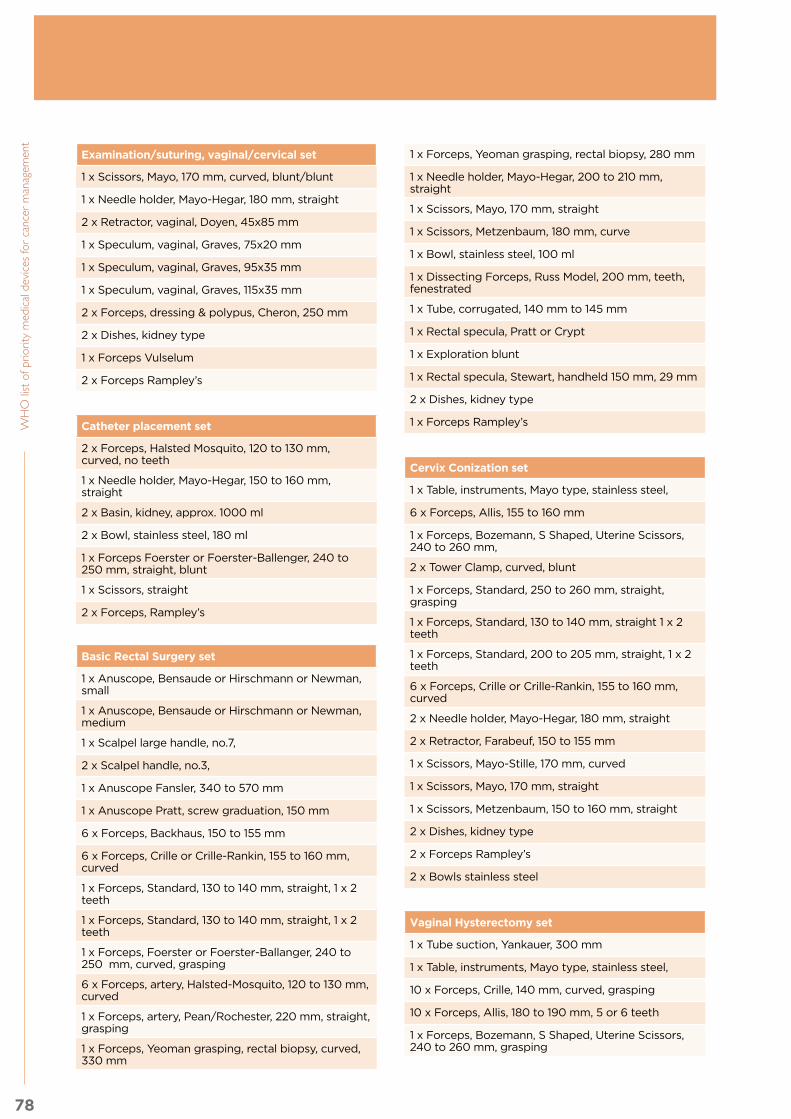
78
WHO
list o
f prio
rity m
edica
l dev
ices f
or ca
ncer
man
agem
ent
Examination/suturing, vaginal/cervical set
1 x Scissors, Mayo, 170 mm, curved, blunt/blunt
1 x Needle holder, Mayo-Hegar, 180 mm, straight
2 x Retractor, vaginal, Doyen, 45x85 mm
1 x Speculum, vaginal, Graves, 75x20 mm
1 x Speculum, vaginal, Graves, 95x35 mm
1 x Speculum, vaginal, Graves, 115x35 mm
2 x Forceps, dressing & polypus, Cheron, 250 mm
2 x Dishes, kidney type
1 x Forceps Vulselum
2 x Forceps Rampley’s
Catheter placement set
2 x Forceps, Halsted Mosquito, 120 to 130 mm, curved, no teeth
1 x Needle holder, Mayo-Hegar, 150 to 160 mm, straight
2 x Basin, kidney, approx. 1000 ml
2 x Bowl, stainless steel, 180 ml
1 x Forceps Foerster or Foerster-Ballenger, 240 to 250 mm, straight, blunt
1 x Scissors, straight
2 x Forceps, Rampley’s
Basic Rectal Surgery set
1 x Anuscope, Bensaude or Hirschmann or Newman, small
1 x Anuscope, Bensaude or Hirschmann or Newman, medium
1 x Scalpel large handle, no.7,
2 x Scalpel handle, no.3,
1 x Anuscope Fansler, 340 to 570 mm
1 x Anuscope Pratt, screw graduation, 150 mm
6 x Forceps, Backhaus, 150 to 155 mm
6 x Forceps, Crille or Crille-Rankin, 155 to 160 mm, curved
1 x Forceps, Standard, 130 to 140 mm, straight, 1 x 2 teeth
1 x Forceps, Standard, 130 to 140 mm, straight, 1 x 2 teeth
1 x Forceps, Foerster or Foerster-Ballanger, 240 to 250 mm, curved, grasping
6 x Forceps, artery, Halsted-Mosquito, 120 to 130 mm, curved
1 x Forceps, artery, Pean/Rochester, 220 mm, straight, grasping
1 x Forceps, Yeoman grasping, rectal biopsy, curved, 330 mm
1 x Forceps, Yeoman grasping, rectal biopsy, 280 mm
1 x Needle holder, Mayo-Hegar, 200 to 210 mm, straight
1 x Scissors, Mayo, 170 mm, straight
1 x Scissors, Metzenbaum, 180 mm, curve
1 x Bowl, stainless steel, 100 ml
1 x Dissecting Forceps, Russ Model, 200 mm, teeth, fenestrated
1 x Tube, corrugated, 140 mm to 145 mm
1 x Rectal specula, Pratt or Crypt
1 x Exploration blunt
1 x Rectal specula, Stewart, handheld 150 mm, 29 mm
2 x Dishes, kidney type
1 x Forceps Rampley’s
Cervix Conization set
1 x Table, instruments, Mayo type, stainless steel,
6 x Forceps, Allis, 155 to 160 mm
1 x Forceps, Bozemann, S Shaped, Uterine Scissors, 240 to 260 mm,
2 x Tower Clamp, curved, blunt
1 x Forceps, Standard, 250 to 260 mm, straight, grasping
1 x Forceps, Standard, 130 to 140 mm, straight 1 x 2 teeth
1 x Forceps, Standard, 200 to 205 mm, straight, 1 x 2 teeth
6 x Forceps, Crille or Crille-Rankin, 155 to 160 mm, curved
2 x Needle holder, Mayo-Hegar, 180 mm, straight
2 x Retractor, Farabeuf, 150 to 155 mm
1 x Scissors, Mayo-Stille, 170 mm, curved
1 x Scissors, Mayo, 170 mm, straight
1 x Scissors, Metzenbaum, 150 to 160 mm, straight
2 x Dishes, kidney type
2 x Forceps Rampley’s
2 x Bowls stainless steel
Vaginal Hysterectomy set
1 x Tube suction, Yankauer, 300 mm
1 x Table, instruments, Mayo type, stainless steel,
10 x Forceps, Crille, 140 mm, curved, grasping
10 x Forceps, Allis, 180 to 190 mm, 5 or 6 teeth
1 x Forceps, Bozemann, S Shaped, Uterine Scissors, 240 to 260 mm, grasping

79
3. S
urg
ery
1 x Forceps, Standard, 100 to 110 mm, straight, 1 x 2 teeth
1 x Forceps, Standard, 240 to 250 mm, straight, grasping
2 x Forceps Foerster or Foerster-Ballenger, 240 to 250 mm, curved, grasping
6 x Forceps, Heaney, 205 to 210 mm, 2 teeth
6 x Forceps, artery, Pean/Rochester, 180 to 185 mm, curved, grasping
1 x Needle holder, Heaney, 200 to 210 mm, curved
1 x Needle holder, Hegar or Mayo-Hegar, 160 mm, straight
1 x Needle holder, Hegar or Mayo-Hegar, 200 to 210 mm, straight
1 x Retractor, vaginal, Doyen, 45x85 mm, length 240 mm
1 x Retractor, vaginal, Doyen, 45x85 mm, length 240 mm
1 x Scissors, Mayo, 170 mm, curved, blunt/blunt
1 x Scissors, Metzenbaum, 200 to 205 mm, curved
1 x Scissors, Metzenbaum, 150 to 160 mm, straight
1 x Bowl, stainless steel, 30 ml
1 x Dissecting forceps, Standard, 130 to 140 mm, grasping
1 x Scissors, Mayo, 170 mm, straight, blunt/blunt
2 x Basin, kidney, approx. 500 ml
2 x Scalpel handle, no. 4
6 x Forceps, Backhaus, 150 mm to 155 mm
2 x Forceps, Pozzi, 240 mm
2 x Forceps Rampley’s
1 x Forceps Vulselum
Abdominal Hysterectomy set
2 x Scalpel handle, no. 4
1 x Tube suction, Yankauer, 300 mm
1 x Tube suction, Yankauer, 22.8 mm, screw off button
1 x Table, instruments, Mayo type, stainless steel,
1 x Forceps, Aldlerkreutz, 200 mm, straight,
8 x Forceps, Allis, 250 to 260 mm, 5 or 6 teeth
6 x Forceps, Backhaus, 150 to 155 mm
1 x Forceps, Bozemann, S Shaped, grasping
10 x Forceps, Crille or Crille-Rankin, 155 to 160 mm, curved
1 x Forceps, Dartigues or Hiterolabo, 255 to 270 mm, curved
1 x Dissecting Forceps, Standard, 130 to 140 mm, teethless, graves
1 x Forceps, Standard, 250 to 260 mm, straight, grasping
1 x Forceps, Standard, 130 to 140 mm, straight, 1 x 2 teeth
2 x Forceps, Standard, 250 to 260 mm, straight, grasping
2 x Forceps Foerster or Foerster-Ballenger, 240 to 250 mm, curved, grasping
2 x Forceps Foerster or Foerster-Ballenger, 240 to 250 mm, straight, grasping
6 x Forceps, Heaney, 220 mm, 2 teeth, curved, grasping
6 x Forceps, artery, Pean/Rochester, 220 to 225 mm, curved, grasping
2 x Forceps, artery, Rochester Ochsner or Kocher-Ochsner, 240 to 225 mm, curved, teeth
1 x Needle holder, Mayo-Hegar, 180 mm, straight
1 x Needle holder, Hegar or Mayo-Hegar, 240 mm, straight
1 x Deaver Bladder, valve 25 x 300 mm
1 x Deaver Bladder, valve 75 x 300 to 310 mm
1 x Farabeuf Bladder, 2 x 150 to 155 mm
1 x O´Sullivan O´Connor, 3 sleves
1 x Scissors, Mayo, 170 mm, curved, blunt/blunt
1 x Scissors, Mayo, 170 mm, straight, blunt/blunt
1 x Scissors, Mayo, 170 mm, straight, blunt/blunt
1 x Scissors, Mayo, 230 mm, straight, blunt/blunt
1 x Scissors, Mayo-Harrington, 225 to 230 mm, curved, blunt/blunt
1 x Scissors, Metzenbaum, 180 mm, curved
1 x Scissors, Metzenbaum, 230 mm, curved, blunt
2 x Basin, kidney, approx. 1000 ml
1 x Bowl, stainless steel, 250 ml
Prostatectomy set
2 x Tube suction, Yankauer, 22.8 mm, screw off button
1 x Table, instruments, Mayo type, stainless steel,
2 x Scalpel handle, one no.3 and one no.4
10 x Forceps, Allis, 180 to 190 mm, 5 or 6 teeth
10 x Forceps, Crille, 140 mm, curved, grasping
2 x Forceps, Bakey, dissection, straight, branches 2 mm, 190 to 200 mm length
1 x Forceps, Standard, 130 to 140 mm, straight, 1 x 2 teeth
2 x Standard, 250 to 260 mm, straight, grasping
2 x Forceps Foerster or Foerster-Ballenger, 180 mm to 200 mm, curved, grasping
2x Forceps, Mixter, 230 mm, grasping
4 x Forceps, artery, Pean/Rochester, 200 to 205 mm, curved, grasping
2 x Forceps, Potts Smith, 230 mm, grasping

80
WHO
list o
f prio
rity m
edica
l dev
ices f
or ca
ncer
man
agem
ent
2 x Forceps, Satinsky, 255 to 265 mm, double angulation
1 x Needle holder, Mayo-Hegar, 180 mm, straight
1 x Needle holder, Hegar or Mayo-Hegar, 180 to 185 mm, straight
1 x Needle holder, Hegar or Mayo-Hegar, 300 mm, straight
1 x Balfour Bladder, central valve of 65 to 80 mm x 80 to 85 mm, lateral valves, fenestrated, maximum 250 to 255 mm
1 x Deaver Bladder, valve 19 x 180 mm
1 x Deaver Bladder, valve 25 x 300 mm
1 x Desmarres Bladder, valve 13 x 14 mm, 130 to 140 mm length
1 x Richardson Bladder, valve 38 x 44 x 30–38 mm, length 240 to 245 mm
1 x Basin, kidney, approx. 1000 ml
1 x Basin, kidney, approx. 500 ml
1 x Farabeuf Bladder, 2, 150 to 155 mm length
2 x Scissors, Bakey, angulated 60°, 230 to 240 mm length
2 x Scissors, Mayo, 150 to 155 mm, straight, blunt/blunt
4 x Standard, 250 to 260 mm, straight, grasping
4 x Forceps Foerster or Foerster-Ballenger, 180 to 200 mm, curved, grasping
4 x Forceps, Mixter, 230 mm, grasping
4 x Forceps, artery, Pean/Rochester, 200 to 205 mm, curved, grasping
4 x Forceps, Potts Smith, 180 to 240 mm length
Diverse retractors
Babcock forceps
Curved needle holder
Bull dog clamps
Hoitgrewe malleable blade
McDougal clamp
Right angle clamp
2 x Forceps Rampleys
Tracheostomy set
5 x Forceps, Field
2 x Forceps, Mosco, curved
2 x Forceps, artery, Kelly, short
1 x Forceps, Allis, large
2 x Farabeu Bladder
2 x hook
2 x Dissecting forceps, small
1 x Needle holder
1 x Scalpel handle, no.7
1 x Dishes, kidney type
2 x Forceps Rampley
2 x Bowl, stainless steel
Gynecological and Kidney set
1 x Forceps, uterine
1 x Speculum, vaginal,
1 x Basin, kidney, approx. 500 ml
Urology set
Sterilization container for the set of instruments, with cover, perforation and filter
1 x Urethral sounds metal Otis-Clutton curved with olive ends (Sizes 4, 6, 8, 10, 12 FG)
1 x Bougies neoplex filiform (smaller sizes olive tipped)
1 x Sounds metal large Powell’s straight blunt plain end
1 x Cystoscope fibre-lit 30 deg telescope with sheath and obturator, operated by standard Heine battery handle
Tiemann brown rubber catheters Ch. 12-24
1 x Foley-Tiemann urinary catheters Ch. 12-24
1 x Introducer for catheterisation 460 mm
1 x kidney dishes assorted 6’’, 8’’, 10’’
3 x Gallipots stainless steel 70 mm diameter
2 Lotion bowls stainless steel 105 mm diameter x 40 mm
Lumbar Puncture set, Adult
1 x Instrument tray
1 x Kidney dish, 20 cm
1 x Galipot, 3 oz
1 x Sponge holder, 17 cm
1 x Lumbar puncture needles with stilettoes, different sizes for adult
1 x Spinal manometer
1 x 3-way Stopcock, Luer Lock
1 x Adapter
1 x Syringe Luer Lock
1 x Bijou sterile bottle
1 x Hypodermic needles, lock, 22 G
1 x Spinal needle (trocar & cannula), 22G
1 x Spinal needle (trocar & cannula), 20G
1 x Spinal needle (trocar & cannula), 23G
1 x Spinal needle (trocar & cannula), 18G

81
3. S
urg
ery
Lumbar Puncture set, Paediatrics
1 x Instrument tray
1 x Kidney dish, 20 cm
1 x Galipot, 3 oz
1 x Sponge Holder, 17 cm
1 x Lumbar puncture needles with stilettoes, different sizes for child
1 x Spinal manometer
3-way Stopcock, Luer Lock
1 x Adapter
1 x Syringe Luer Lock
1 x Bijou sterile bottle
1 x Hypodermic needles, Lock, 25 G
1 x Spinal needle (trocar & cannula), 25 G
Pleural Biopsy set
1 x Sterilization container for the set of instruments, with cover, perforation and filter
2 x Kidney dish, 20 cm
2 x Galipot, 6 oz
4 x Sponge holder, 17 cm
2 x Dissecting forceps, plain, 15 cm
2 x Scalpel handle, no. 3
8 x Towel clips, Backhaus, 8 cm
2 x Set Abram’s pleural biopsy needles (8 G x 3½”)
5 x Two way adaptor
2 x Syringe, with metal tip, Luer lock
10 x Hypodermic needle, Luer lock, 23 G
10 x Hypodermic needle, Luer lock, 21 G
2 x Galipot, 3 oz
Mastectomy set
2 x Needle holder, Mayo-Hegar, 180 mm, straight
1 x Retractor, Farabeuf, double-ended, 120 mm, pair
1 x Tube suction, Yankauer, 270 mm
10 x Forceps, artery, Kelly, 140 mm, curved
5 x Forceps, tissue seizing, Allis, 150 mm
4 x Richardson Bladder, valve 38x44x30–38 mm, length 240 to 245 mm
10 x Forceps, artery, Halsted-Mosquito, 125 mm, curved
2 x Forceps, dressing, ring
2 x Forceps grasper short, 5 mm
1 x Volkman retractor
1 x Scalpel No 4 and 7 with blade
1 x Gerald Dressing Forceps, 18 cm, straight
2 x Dishes, kidney type
Chest Aspirations set
1 x Instrument tray, 24 x 5 cm
1 x Kidney dish, 20 cm
1 x Galipot, 6 oz
1 x Sponge holder, 17 cm
1 x Marting Aspiration Needles, 17 G
1 x Trocar and Cannulae, size 11 G and 14 G
1 x Two way Adaptor
1 x Doz. Hypodermic Needles, Luer Lock, 21 G x 1½
1 x Doz. Hypodermic Needles, Luer Lock, 23 G x 1½
1 x Galipot, 3 oz
Gynecologic biopsy set
1 x Forceps, uterine, ovum, Bierer, large
1 x Forceps, uterine, ovum, Bierer, small
1 x Forceps, uterine, ovum, Sopher, small
1 x Posi-Locking Instrument Holder
1 x Speculum, vaginal, Graves, wide mouth
1 x Fixed-diameter cervical dilator, reusable
1 x Disposable self-retaining retraction system ring
2 x Dishes, kidney type
Excisional breast biopsy set
1 x Retractor, Farabeuf, double-ended, 120 mm, pair
2 x Needle holder, Mayo-Hegar, 180 mm, straight
2 x Forceps, tissue seizing, Allis, 150 mm
1 x Forceps, artery, Kelly, 140 mm, curved (long and short)
2 x Forceps, Halsted Mosquito, 120 to 130 mm, curved, no teeth
2 x Forceps, Ring c and r
1 x Forceps, dressing, standard, 155 mm, straight
1 x Forceps, tissue holding, Collin, 160 mm
2 x Forceps, tissue, standard, 145 mm, straight
2 x Dissecting Forceps, teeth and non teeth, fenestrated, long and short
2 x Dishes, kidney type
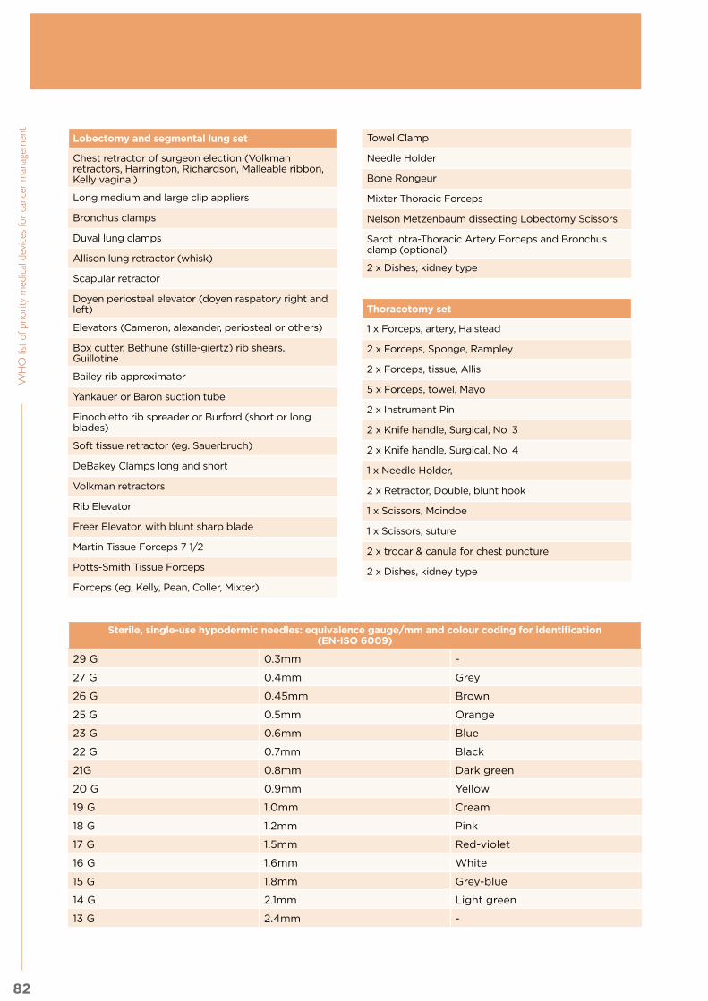
82
WHO
list o
f prio
rity m
edica
l dev
ices f
or ca
ncer
man
agem
ent
Lobectomy and segmental lung set
Chest retractor of surgeon election (Volkman retractors, Harrington, Richardson, Malleable ribbon, Kelly vaginal)
Long medium and large clip appliers
Bronchus clamps
Duval lung clamps
Allison lung retractor (whisk)
Scapular retractor
Doyen periosteal elevator (doyen raspatory right and left)
Elevators (Cameron, alexander, periosteal or others)
Box cutter, Bethune (stille-giertz) rib shears, Guillotine
Bailey rib approximator
Yankauer or Baron suction tube
Finochietto rib spreader or Burford (short or long blades)
Soft tissue retractor (eg. Sauerbruch)
DeBakey Clamps long and short
Volkman retractors
Rib Elevator
Freer Elevator, with blunt sharp blade
Martin Tissue Forceps 7 1/2
Potts-Smith Tissue Forceps
Forceps (eg, Kelly, Pean, Coller, Mixter)
Towel Clamp
Needle Holder
Bone Rongeur
Mixter Thoracic Forceps
Nelson Metzenbaum dissecting Lobectomy Scissors
Sarot Intra-Thoracic Artery Forceps and Bronchus clamp (optional)
2 x Dishes, kidney type
Thoracotomy set
1 x Forceps, artery, Halstead
2 x Forceps, Sponge, Rampley
2 x Forceps, tissue, Allis
5 x Forceps, towel, Mayo
2 x Instrument Pin
2 x Knife handle, Surgical, No. 3
2 x Knife handle, Surgical, No. 4
1 x Needle Holder,
2 x Retractor, Double, blunt hook
1 x Scissors, Mcindoe
1 x Scissors, suture
2 x trocar & canula for chest puncture
2 x Dishes, kidney type
Sterile, single-use hypodermic needles: equivalence gauge/mm and colour coding for identification (EN-ISO 6009)
29 G 0.3mm -
27 G 0.4mm Grey
26 G 0.45mm Brown
25 G 0.5mm Orange
23 G 0.6mm Blue
22 G 0.7mm Black
21G 0.8mm Dark green
20 G 0.9mm Yellow
19 G 1.0mm Cream
18 G 1.2mm Pink
17 G 1.5mm Red-violet
16 G 1.6mm White
15 G 1.8mm Grey-blue
14 G 2.1mm Light green
13 G 2.4mm -
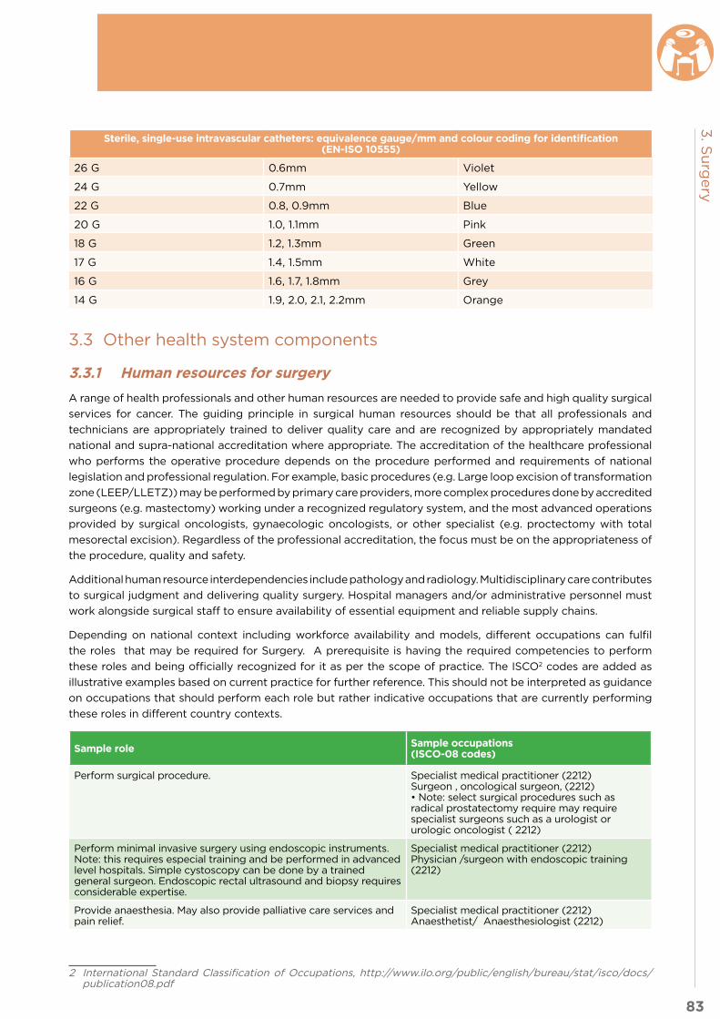
83
3. S
urg
ery
Sterile, single-use intravascular catheters: equivalence gauge/mm and colour coding for identification (EN-ISO 10555)
26 G 0.6mm Violet
24 G 0.7mm Yellow
22 G 0.8, 0.9mm Blue
20 G 1.0, 1.1mm Pink
18 G 1.2, 1.3mm Green
17 G 1.4, 1.5mm White
16 G 1.6, 1.7, 1.8mm Grey
14 G 1.9, 2.0, 2.1, 2.2mm Orange
3.3 Other health system components
3.3.1 Human resources for surgery
A range of health professionals and other human resources are needed to provide safe and high quality surgical
services for cancer. The guiding principle in surgical human resources should be that all professionals and
technicians are appropriately trained to deliver quality care and are recognized by appropriately mandated
national and supra-national accreditation where appropriate. The accreditation of the healthcare professional
who performs the operative procedure depends on the procedure performed and requirements of national
legislation and professional regulation. For example, basic procedures (e.g. Large loop excision of transformation
zone (LEEP/LLETZ)) may be performed by primary care providers, more complex procedures done by accredited
surgeons (e.g. mastectomy) working under a recognized regulatory system, and the most advanced operations
provided by surgical oncologists, gynaecologic oncologists, or other specialist (e.g. proctectomy with total
mesorectal excision). Regardless of the professional accreditation, the focus must be on the appropriateness of
the procedure, quality and safety.
Additional human resource interdependencies include pathology and radiology. Multidisciplinary care contributes
to surgical judgment and delivering quality surgery. Hospital managers and/or administrative personnel must
work alongside surgical staff to ensure availability of essential equipment and reliable supply chains.
Depending on national context including workforce availability and models, different occupations can fulfil
the roles that may be required for Surgery. A prerequisite is having the required competencies to perform
these roles and being officially recognized for it as per the scope of practice. The ISCO2 codes are added as
illustrative examples based on current practice for further reference. This should not be interpreted as guidance
on occupations that should perform each role but rather indicative occupations that are currently performing
these roles in different country contexts.
Sample role Sample occupations (ISCO-08 codes)
Perform surgical procedure. Specialist medical practitioner (2212) Surgeon , oncological surgeon, (2212) • Note: select surgical procedures such as radical prostatectomy require may require specialist surgeons such as a urologist or urologic oncologist ( 2212)
Perform minimal invasive surgery using endoscopic instruments. Note: this requires especial training and be performed in advanced level hospitals. Simple cystoscopy can be done by a trained general surgeon. Endoscopic rectal ultrasound and biopsy requires considerable expertise.
Specialist medical practitioner (2212) Physician /surgeon with endoscopic training (2212)
Provide anaesthesia. May also provide palliative care services and pain relief.
Specialist medical practitioner (2212) Anaesthetist/ Anaesthesiologist (2212)
2 International Standard Classification of Occupations, http://www.ilo.org/public/english/bureau/stat/isco/docs/publication08.pdf
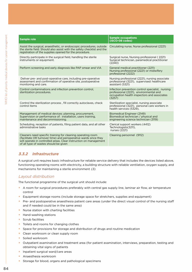
84
WHO
list o
f prio
rity m
edica
l dev
ices f
or ca
ncer
man
agem
ent
Sample role Sample occupations (ISCO-08 codes)
Assist the surgical, anaesthetic, or endoscopic procedures, outside the sterile field. Should also assist with the safety checklist and the registration of the supplies opened for the procedure.
Circulating nurse, Nurse professional (2221)
Directly participate in the surgical field, handling the sterile instruments or equipment.
Surgical nurse, Nursing professional ( 2221)Surgical technician, paramedical practitioner (2240)
Perform screening and early diagnosis like PAP smear and VIA General medical practitioner (2211)Nursing professional (2221) or midwifery professional (2222)
Deliver pre- and post-operative care, including pre-operative assessment and confirmation of operative site, postoperative monitoring and care.
Nursing professional (2221), nursing associate professional (3221), supervised: healthcare assistant (5321)
Control contaminations and infection prevention control, sterilization procedures.
Infection prevention control specialist, nursing professional (2221), environmental and occupation health inspectors and associates (3257)
Control the sterilization process , fill correctly autoclaves, check control items
Sterilization specialist, nursing associate professional (3221), personal care workers in health services (5329),
Management of medical devices: planning, procurement,Supervision or performance of: installation, users training, maintenance and decommissioning.
Biomedical Engineer (2149)Biomedical technician / physical and engineering science technician (3119)
Scheduling, reception of patients, filing patient data, and all other administrative tasks
Clerical support workers (4412)Technologists(3211), nurses (2221)
Cleaners need specific training for cleaning operating room (facilitate OR turnover time) and perioperative wards since they will operate in controlled areas. Clear instruction on management of all type of wastes should be given
Cleaning personnel (9112)
3.3.2 Infrastructure
A surgical unit requires basic infrastructure for reliable service delivery that includes the devices listed above,
functioning operating rooms with electricity, a building structure with reliable ventilation, oxygen supply, and
mechanisms for maintaining a sterile environment (3).
Layout distributionThe functional programme of the surgical unit should include:
• A room for surgical procedures preferably with central gas supply line, laminar air flow, air temperature
control
• Equipment storage rooms (include storage space for stretchers, supplies and equipment)
• Pre- and postoperative anaesthesia patient care areas (under the direct visual control of the nursing staff
and if needed could be in the same area)
• Nurse station with charting facilities
• Hand-washing stations
• Scrub facilities
• Toilets and rooms for changing clothes
• Space for provisions for storage and distribution of drugs and routine medication
• Clean workroom or clean supply room
• Soiled workroom
• Outpatient examination and treatment area (for patient examination, interviews, preparation, testing and
obtaining vital signs of patients
• Inpatient surgical ward/care areas
• Anaesthesia workroom
• Storage for blood, organs and pathological specimens

85
3. S
urg
ery
• Area for preparation and examination of frozen sections (if the laboratory isn’t near the surgical unit)
• Sterilization facilities.
The size, location, and configuration of the surgical suite and support areas should reflect the projected
volume of patients (4).
Surgical infrastructure also relies on a strong referral mechanism that promotes links between and integration of
services. This includes communication between primary and specialty care, cancer surgery and multidisciplinary
services including pathology and radiotherapy, and links among facilities providing cancer care.
An adequate preoperative assessment is directly related to better surgical outcomes. Access to reliable basic
imaging, laboratory, pathology and clinical evaluation is important in order to plan the surgical procedure,
and to prevent surgical and anaesthetic complications.
Moreover, access to safe, quality, and efficacious blood products is an objective of the WHO programme on
Blood Transfusion Safety and relevant to the delivery of cancer surgery service. Perioperative transfusion may
be needed due to a patient’s underlying condition, co-morbid diseases, or the complexity of surgical care.
The availability of facilities that provide safe blood enables cancer surgical care.
Electrical systemsOperating rooms should have appropriate electrical systems, such as emergency outlets supplied by backup
generators in case of grid failure, and isolated power systems.
Heating, ventilation and air conditioning systemsThe ventilation system should be designed to protect the suite from the environment but above of all to
protect the patient from the environment. Ideally, the temperature of the operating room should be maintained
between 22°C and 26°C. There should be a reference supply airflow. There should be positive pressure in
relationship to other areas.
Communication systemsEach room should have a system for emergency communication with the surgical suite control station.
Special systemsAspects to be considered include oxygen availability (oxygen concentrator, oxygen/gas pipeline, cylinder)
in surgery and endoscopy, and vacuum availability (suction pump, vacuum central pump). If applicable,
sufficient outlets for oxygen, vacuum and medical air systems should be considered.
Regarding architectural details, the operating room perimeter: walls, ceilings, and floors, including penetrations,
should be sealed and washable.
3.3.3 Quality management
Six domains of quality should be monitored, as articulated in WHO Quality of Care: A Process for Making
Strategic Choices in Health Systems (2006): quality of care should be effective, efficient, accessible,
acceptable/patient-centred, equitable, and safe. Each of these domains should be evaluated in quality
assurance programmes implemented with key stakeholders in interdependent fields. Surgical care is based
on a team model that requires commitment from all partners to improve quality and outcomes.
Broad strategies used to improve quality include establishing quality management guidelines, establishing
surgical standards, utilizing forums like Morbidity & Mortality conferences (M&M), collecting and monitoring
indicators of quality, and performing comprehensive audits of surgical procedures done at the regional or
national level. While outcome data generally reflect the overall effectiveness and quality of care (such as
percentage of cancer patients who survive at one year), additional factors should be assessed that can
impact outcomes, such as delays in care or resource limitations.

86
WHO
list o
f prio
rity m
edica
l dev
ices f
or ca
ncer
man
agem
ent Quality care must be accessible and delivered in a timely manner. Sample quality indicators proposed in
WHO Cancer Control Modules: Diagnosis & Treatment (2008) include the number of patients receiving timely
treatment and trained human resources (5). Treatment guidelines can also improve quality of care and can be
measured through indicators such as percentage of patients treated according to guidelines, as suggested in
WHO National Cancer Control Programmes (2002) (6).
Quality and safety are fundamental to effective cancer surgery. Surgical and anaesthesia services have a
long history of promoting safety and include the WHO Safe Surgery programme. The Safe Surgery Saves
Lives programme aims to improve the quality of surgical care by defining a core set of safety standards
to be applied in all facilities. Safe surgery requires the participation of all members of the surgical team,
including nurses, anaesthesia providers, and surgeons, who together promote a culture of safety. High quality,
appropriate and safe anaesthesia and nursing are critical to effective operative care.
Monitoring and evaluation programmes must be fed back to the surgical team for ongoing quality improvement.
For example, a surgical team receiving feedback on a disproportionately high incidence of surgical site
infections can evaluate potential etiologies such as poor sterilization, incorrectly timed or administered
antimicrobials, improperly prepared skin with antiseptics, or other factors. Continuous quality improvement
linked to service delivery, safety, and integrated care is critical to successful cancer surgery.
3.3.4 Guidance documents
Title Link Description
World Health Assembly resolution on Strengthening Emergency and Essential Surgical Care and Anaesthesia, 2015
http://apps.who.int/gb/ebwha/pdf_files/WHA68/A68_R15-en.pdf
Recognising the crucial importance of surgical services, and the huge gaps in global access, the resolution underlines the importance of building political commitment to expand access, and of strengthening the surgical workforce through training and knowledge exchange.
Surgical Services for Cancer Care
http://dcp-3.org/sites/default/files/chapters/DCP3%20Cancer_Ch%2013.pdf
DCP3 Chapter from Cancer Volume, Part II: Interventions
WHO Safe Surgery Saves Lives
http://apps.who.int/iris/bitstream/10665/ 44185/1/ 9789241598552_eng.pdf
Guidelines for implementation of the Surgical Safety Checklist.
Integrated Management for Emergency and Essential Surgical Care (IMEESC) toolkit
http://www.who.int/surgery/publications/imeesc/en/
The WHO Integrated Management for Emergency & Essential Surgical Care e-learning toolkit (CD) was developed by the Clinical Procedures Unit in collaboration with the Global Initiative for Emergency & Essential Surgical Care members. This tool targets policy-makers, managers and health-care providers (surgeons, anaesthetists, non-specialist doctors, health officers, nurses, and technicians). This tool contains WHO recommendations for minimum standards to improve quality and safety of emergency, surgery, trauma, obstetrics and anaesthesia at first-referral level health-care facilities.
The Lancet Commission on Global Surgery
http://www.thelancet.com/commissions/global-surgery
The global burden of disease amenable to surgical intervention, such as trauma, cancer, and complications from childbirth, is substantial and growing. Despite this, there are currently gross disparities in access to safe surgical care worldwide. Surgery is an integral, indivisible component of a properly functioning health system, and all people should have access to safe, high-quality surgical and anaesthesia care with financial protection when needed. The purpose of The Lancet Commission on Global Surgery is to make this vision a reality by embedding surgery within the global health agenda, catalysing political change, and defining scalable solutions for provision of quality surgical and anaesthesia care for all.

87
3. S
urg
ery
Bibliography:
1) WHO guidelines for safe surgery.
Available at: http://www.who.int/patientsafety/safesurgery/en/
2) Sullivan, Richard, et al. “Global cancer surgery: delivering safe, affordable, and timely cancer surgery.”
The Lancet Oncology 16.11 (2015): 1193-1224.
Available at: http://www.thelancet.com/commissions/global-cancer-surgery
3) WHO Surgical Care at the District Hospital.
Available at: http://www.who.int/surgery/publications/en/SCDH.pdf
4) Guidelines for design and construction of Health Care Facilities. The Facility Guidelines Institute.
Available at: http://www.ashe.org/advocacy/orgs/fgi.shtml
5) WHO Cancer Control Modules: Diagnosis & Treatment (2008).
Available at: http://www.who.int/cancer/publications/cancer_control_diagnosis/en/
6) WHO National Cancer Control Programmes (2002).
Available at: http://www.who.int/cancer/publications/nccp2002/en/

4. Clinical laboratory & pathology

89
4. C
linic
al la
bo
rato
ry &
path
olo
gy
Clinical laboratory & pathology
Health service delivery sequence overview:
This diagram expresses the flow of cancer patients to and from the clinical laboratory and pathology unit, and
the elements to consider as support for the unit.
Fig. 10.4 Health care service delivery overview – clinical laboratory and pathology
PR
IMA
RY
(C
L,H
C),
SE
C-
ON
DA
RY
(D
H)
&
TE
RT
IAR
Y (
SH
)
Le
ve
l o
f C
are
*
* Appropriate level of care will depend on the particular intervention, setting, and available infrastructure and human resources.
SE
CO
ND
AR
Y (
DH
)
& T
ER
TIA
RY
(S
H)
TE
RT
IAR
Y (
SH
)
Prevention End of life care
TreatmentFollow-up/survivorship
care
Palliative care
Diagnosis
Screening
CL Community Level health post DH District Hospital HC Health Centre SH Specialized Hospital
Systemic therapy
Nuclear medicine
Surgery
Vaccination
Radio- therapy
Pathology Pathology
Clinical laboratory
Clinical laboratory
Clinical assessment
Palliative care
Medical imaging
Endoscopy
Clinical assessment
Nuclear medicine
4.1 General description of the unit4.2 Priority medical devices for clinical laboratory
4.2.1 General medical devices for clinical laboratory
4.2.2 Specific medical devices for clinical laboratory by cancer type
4.3 Priority medical devices for pathology4.3.1 General medical devices for pathology
4.3.2 General medical devices for histopathology
4.3.3 General medical devices for cytology
4.3.4 General medical devices for immunohistochemistry
4.4 Other health system components4.4.1 Human resources for clinical laboratory and pathology
4.4.2 Infrastructure
4.4.3 Quality management
4.4.4 Guidance documents

90
WHO
list o
f prio
rity m
edica
l dev
ices f
or ca
ncer
man
agem
ent
4.1 General description of the unit
The purpose of the pathology and laboratory medicine unit is to perform accurate testing in a timely fashion
on patient samples and to analyse and interpret the results, thereby guiding the diagnosis, treatment and
management of the patient. The intention is to provide these results based on examination of the patient’s cells,
tissues, blood and other body fluids, while ensuring the safety of personnel, public and patients. Timeliness,
accuracy of results, quality management and universal safety are critical elements in medicine and in the safe
functioning of the pathology and laboratory medicine unit.
The Pathology laboratory handles tissues, body fluids and other specimen types. Specimens submitted for
pathologic examination may be solid tissues from surgical excisions or biopsies (histopathology) or cells in
smears, scrapings or fluid specimens (cytopathology). The significance of the pathology laboratory (in its
broadest terms) is that in many cases, patient management is based on a definitive histologic or cytologic
diagnosis, especially in the case of neoplasms. Future management of a patient depends on an assessment of
various pathologic parameters including the histopathologic type of tumour, its grade, its extent and its stage
and additional ancillary studies which insure correct treatment or inform prognosis. A complete and detailed
examination of the excised tissues is therefore critical, especially in this age of personalized medicine. To ensure
that all specimens are appropriately handled, guidelines and standard operating procedures must be in place
for the handling of all specimens. This will prevent misdiagnosis, save time, anxiety and possible harm.
Tissues must be handled appropriately, immediately after collection by the clinician, so that the quality of the
tissues available does not deteriorate and optimum preservation is maintained for accurate diagnoses and
possible biomarker testing.
Effective timely communication, however, is not just crucial in reporting of pathology results, but also is vital
for exchange of information about the patient and their management at all points during the pathway of care.
In addition, the clinical laboratories (laboratory medicine) provide testing in haematology, chemistry, immunology,
and microbiology that are also essential for diagnosis and management of many diseases including cancers.
4.2 Priority medical devices for clinical laboratory
The following tables present general medical devices for clinical laboratory medicine, which can be used for
screening, diagnosis, staging, and follow-up of many diseases including several cancers, and specific medical
devices, which are used for interventions for specific cancer types.
The following tables present the priority medical devices required to perform the following health interventions:
Cancer type Interventions
All cancers • Pre-analytical procedure: phlebotomy, sample reception and distribution• General analytical procedures• Blood chemistry tests (liver and renal function tests, alkaline phosphatase and calcium) • Tumour lysis syndrome panel: lactate dehydrogenase (LDH), uric acid, potassium (K),
Calcium (Ca), Phosphate• Complete blood count (CBC) - total white blood count with differential, platelet count, and
haemoglobin level and red blood cell indices• Disseminated intravascular coagulation (DIC) panel: D-dimer, fibrinogen, prothrombin time
(PT), partial thromboplastin time (PTT) • Urine free light chains • Serum free light chains• Serum protein electrophoresis• Immunoassay test
Breast cancer
• Biomarker analysis
Cervical cancer
• Baseline CD4 count test (for HIV(+) patients)• HPV Test

91
4. C
linic
al la
bo
rato
ry &
path
olo
gy
Cancer type Interventions
Colorectal cancer
• Carcinoembryonic antigen (CEA) test• Faecal immunochemical testing (FIT)• Guaiac faecal occult blood test (gFOBT)
Leukaemia • Bone marrow aspiration• Bone marrow biopsy• Cerebrospinal fluid aspirate and lumbar puncture• Comprehensive flow cytometric immunophenotyping• Optional: Cytogenetics, Cytochemistry eg myeloperoxidase, specific and non-specific
esterase, sudan B and In situ hybridisation (FISH, SISH)
Prostate cancer
• PCA3 test• Prostate specific antigen PSA
4.2.1 General medical devices for clinical laboratory
Choosing equipment for laboratory services depends mainly upon volume and resources. The type of equipment
needed should be considered (automated, semi-automated) according to facility capacity, and options for
purchasing, leasing or renting the devices along with the relevant consumables, accessories and software.
Procedure Medical devices category Capital equipment Accessories/hardware/software/
consumables/single use devices
Pre-analytical procedure- Phlebotomy, sample reception and distribution
Laboratory and pathology equipment
Table top centrifuge
Medical furniture Adequate furniture for the laboratory devices
Blood draw chairs
Sample distribution trolley
Personal protective equipment
Coat, medical, woven, white (various sizes)
Gloves, examination, non-sterile, single use (various sizes)
Disposables/medical supplies
Bandage, adhesive, 3.0 cm, box/100
Compress, gauze, antiseptic, 6x3 cm, sterile
Compress, gauze, sterile & non-sterile, single use
Needles, luer, sterile, single use (sizes G)3
Paper towels
Swab-pad, alcohol
Syringes
Tape, medical, roll (various sizes)
Needle, vacuum tube, 20 G/ 22 G, sterile
Needle holder, vacuum tubes, sterile
Sheet, absorbent, bench, 50x40 cm
Tube, vacuum, Ethylene Diamine Tetra-acetic Acid (EDTA), sterile (various capacities)
Tube, vacuum, plain/dry, sterile (various capacities)
Tube, vacuum, serum, sterile
Lancet, blood, safety, sterile (various sizes)

92
WHO
list o
f prio
rity m
edica
l dev
ices f
or ca
ncer
man
agem
ent
Procedure Medical devices category Capital equipment Accessories/hardware/software/
consumables/single use devices
Solutions and reagents
Distilled water
Ethanol
Other Rack, test tubes
Tourniquet, with buckle
Sample distribution container
Thermic containers
Safety box, for used syringes/needles
General analytical procedures
Laboratory and pathology equipment
Refrigerator, laboratory
Refrigerator/freezer, laboratory
Freezer, laboratory
Microscope, binocular
Camera for microscope (for telemedicine and documentation)
Timer, digital
Timer, 60 min, mechanical
Thermometer, glass, min/max -20°C/100°C
Thermometer, min/max -30°C/60°C
Magnifying glass
Centrifuge Accessories for serology
Centrifuge, micro - haematocrit
Distillation unit, 2 L/h, with tank
Hot plate, with stirrer
Incubator, 30 L, up to 80° C
pH meter
Rotator, agglutination test
Scale, digital, 1500 g/0.1 g
Scale, precision, digital, 500 g/0.01 g
Basic laboratory mixer/Laboratory shaker vortex
Shaker, orbital
Spectrophotometer, ultraviolet/visible
Sterilizer steam autoclave, 24 L
Water bath, 7 L
Hygrometer Microplate, ELISA, 96 U-well
Instruments Forceps, dressing, 155 mm, straight
Spatula, stainless steel (various sizes)
Clamp, test tubes
Medical furniture Adequate furniture for the laboratory devices
Cabinet for microscope
Personal protective equipment
Coat, medical, woven, white (various sizes)
Gloves, examination, latex, non-sterile, single use (various sizes)

93
4. C
linic
al la
bo
rato
ry &
path
olo
gy
Procedure Medical devices category Capital equipment Accessories/hardware/software/
consumables/single use devices
Gloves, nitrile, powder-free, non-sterile, single use
Mask, surgical, non-woven
Glasses, safety, regular size
Disposables/medical supplies
Bag, re-sealable, plastic
Compress, gauze, 10x10 cm, non-sterile
Cover glass, slides
Envelope, packing, 27x36 cm
Lancet, 2 mm, safety, sterile
Paper towels
Paper, dry blood spot
Paper, lens
Rack, drying DBS cards, 10 positions
Slide, microscope
Slide, microscope, frosted
Swab, cotton-tip, tube, sterile
Vortex, test tube
Wooden or plastic applicator sticks
Bag, biohazard, 20 L
Blood collection tube, neonatal cord blood, sterile
Container, sample, 50 ml
Cotton wool, 500 g, roll, non-sterile
Dressing strip, adhesive, diameter 3.0 cm, sterile
Film, sealing, flexible, 10 cm x 38 m, roll
Inoculation loop, plastic, sterile
Kato-Katz, kit, stool sample preparation
Monitor card, humidity, passive/cumulative
Paper, filter #1
Paper, pH indicator 2.0 to 9.0
Paper, weighing
Pipette, repeat, tip 2.5/5.0 ml, 10/25 ml
Pipette, tip, barrier, 200 ul / 1000 ul, sterile
Pipette, tip, blue, 100–1000 ul
Pipette, tip, white, 2–20 ul
Pipette, tip, yellow, 10–100 ul / 20–200 ul
Pipette, transfer, 3 ml, non-sterile
Pipette, transfer, 3 ml, sterile
Reservoir, reagent, 60 ml
Sealant, compound
Tape

94
WHO
list o
f prio
rity m
edica
l dev
ices f
or ca
ncer
man
agem
ent
Procedure Medical devices category Capital equipment Accessories/hardware/software/
consumables/single use devices
Tube, push cap, 0.2 ml, PCR, sterile
Tube, push cap, 5.0 ml, non-sterile
Tube, screw cap, 0.2 ml / 0.5 ml / 2.0 ml / 5.0 ml, non-sterile
Tube, screw cap, 0.2 ml / 0.5 ml / 2.0 ml / 5.0 ml, sterile
Tube, screw cap, conic, 15/50 ml, non-sterile
Tube, vacuum, EDTA, 2 ml / 4 ml / 6 ml, sterile
Tube, vacuum, plain/dry, 4 ml / 6 ml, sterile
Tube, vacuum, serum, 4 ml / 6 ml, sterile
Parafilm paper
Reservoir, reagent, 60 ml
Solutions and reagents
Acetic acid, 36%, bottle
Acetone, bottle
Bromine solution
Buffer, tablets, pH 7.2, box
Cytology stain kit
Diethyl ether, bottle
Distilled water
Ethanol, denatured, 70%, bottle
Ethanol, denatured, 95%, bottle
Formaldehyde, 10%, 10 ml, ampoule
Gentian violet, solution, bottle
Glycerol, bottle
Hydrochloric acid, 40%, bottle
Indian ink, black, bottle
Isopropyl alcohol
KI starch solution
Lugol iodine, bottle
Methanol, bottle
Methylene blue, bottle
Nitric acid
Oil, immersion, bottle
Oxidase test
Petroleum gel, paraffin, bottle
Potassium iodide
Silica gel (desiccant for DBS), pouch
Sodium bicarbonate
Sodium chloride, powder, bottle
Sodium hypochlorite, tablets
Sodium persulfate
Stain, Field A, solution
Stain, Field B, solution

95
4. C
linic
al la
bo
rato
ry &
path
olo
gy
Procedure Medical devices category Capital equipment Accessories/hardware/software/
consumables/single use devices
Stain, Giemsa, solution
Stain, Gram, set
Stain, May-Grunwald Giemsa, set
Stain, Ziehl–Neelsen, solution, bottle
Sugar fermentation tests
Sulphuric acid, 95%
Test, Nickerson or sabouraud medium, kit
Test, potassium hydroxide KOH, preparation
Trichloroacetic acid, crystals, bottle
Pipettes cleaning solution
Substrate, adamantyl phosphate oxetane
Wash solution
Xylene, bottle
Other Safety box, for used syringes/needles
Beaker, glass, 100 ml / 250 ml
Bottle, amber, dropper, 30 ml
Bottle, amber, screw cap, 100 ml / 250 ml / 1000 ml
Bottle, plastic, 1 L
Box, refill, pipette tips, empty
Box, specimen transport, 2 L/4 L
Box, storage, 100 slides
Box, storage 0.5/2/5 ml tubes, 100 positions
Brush, bottles and flasks (various sizes)
Brush, test tubes (various sizes)
Containers for hazardous waste (solutions and others)
Cylinder, measuring, glass, 10 ml /100 ml / 500 ml/ 100 ml
Funnel, glass
Funnel, plastic
Jar, Coplin, staining
Label, biohazard, adhesive, 3x4 cm
Label, self-adhesive, freezer
Label, self-adhesive, 5x10 cm
Marker pen, cryoware
Marker pen, glassware
Marker, diamond
Petri dish, glass, with lid
Pipette, digital, 10–100 ul
Pipette, digital, 100–1000 ul
Pipette, digital, 2–20 u

96
WHO
list o
f prio
rity m
edica
l dev
ices f
or ca
ncer
man
agem
ent
Procedure Medical devices category Capital equipment Accessories/hardware/software/
consumables/single use devices
Pipette, digital, 20–200 ul
Pipette, digital, 8 channel, 20-200 ul
Pipette, digital, 8 channel, 5–50 ul
Pipette, filler, wheel-run, set/2
Pipette, repeating, 5 volume
Pipette, stand, 4 positions
Pipettes, blood graduated, 0.05 ml
Rack, drying glass & plastic ware
Rack, drying slides, 12 positions
Rack, staining slides, horizontal, 12 positions
Rack, test tubes, 24 positions
Rack, tubes, 0.5/2.0/5.0 ml, 24 positions
Rod, glass
Wash bottle, 250 ml
Serological pipette
Pasteur Pipette
Single use devices Punch, Dry Blood Spot (DBS), 3.0 mm
Blood chemistry tests (liver and renal function tests, alkaline phosphatase and calcium) Tumour lysis syndrome panel: lactate dehydrogenase (LDH), uric acid, potassium (K), Calcium (Ca), Phosphates
Laboratory and pathology equipment
Clinical chemistry Analyser Reagents, calibrators and controls for clinical chemistry analyzer
Ultrasonic washer
Table top centrifuge
Complete blood count and - Complete blood count with differential and platelets (including full blood count and haemoglobin levels)
Laboratory and pathology equipment
Analyser Laboratory Haematology, Manual or Automated
Reagents calibrators and controls haematology analyserIf not automated: Buffer solution, Wright stain, wash solution, dropper, Pasteur pipette, Wintor tube, ESR (Erythrocyte Sedimentation Rate) Analyser, Integrated slide marker/stainer system, Rack, ESR, 5 positions, Counter, cell, manual differential, Counter, hand tally, mechanical, Counting chamber, Neubauer, Haemoglobin colour scale (starter kit), Hemoglobinometer, with accessories
Disseminated intravascular coagulation (DIC) panel: D-dimers, fibrinogen, prothrombin time (PT), partial thromboplastin time (PTT)
Laboratory and Pathology Equipment
Coagulation analyser, Manual or Automated
Reagents calibrators and controls

97
4. C
linic
al la
bo
rato
ry &
path
olo
gy
4.2.2 Specific medical devices for clinical laboratory by cancer type
4.2.2.1 Breast
Procedure Medical devices category
Capital equipment (for specialized hospitals)
Accessories/hardware/software/consumables/single use devices
Biomarker analysis
Laboratory and pathology equipment
Immunoassay unit Reagents for immunohistochemistry lab (ie. HER2, estrogen receptor, progesterone receptor) solutions and controls, secondary antibodies
Automated IHC staining system
Direct visualization of antibody staining for estrogen and progesterone receptor (for anti-estrogen therapy) and for Her2 antigen (for anti-Her2 therapy).
4.2.2.2 Cervical
Procedure Medical devices category
Capital equipment (for specialized hospitals)
Accessories/hardware/software/consumables/single use devices
Baseline CD4 count test (for HIV+ patients)
Laboratory and pathology equipment
Flow cytometer
HPV Test Laboratory and pathology equipment
HPV DNA Test
4.2.2.3 Colorectal
Procedure Medical devices category
Capital equipment (for specialized hospitals)
Accessories/hardware/software/consumables/single use devices
Carcinoembry-onic antigen (CEA) test
Laboratory and pathology equipment
Immunoassay unit Depending on the technique, may include reagent sample diluent and reagents (ie.CEA test)
Faecal immuno-chemical testing (FIT)
Laboratory and pathology equipment
iFOB immunochemical analyzer, automated
Guaiac faecal occult blood test (gFOBT)
Laboratory and pathology equipment
Faecal occult blood test (FOBT) rapid test kit (slides and applicator sticks)

98
WHO
list o
f prio
rity m
edica
l dev
ices f
or ca
ncer
man
agem
ent
4.2.2.4 Leukaemia
Procedure Medical devices category
Capital equipment (for specialized hospitals)
Accessories/hardware/software/consumables/single use devices
Bone marrow aspiration
Instruments Bone marrow aspiration set
Scalpel with blades
Single use devices/disposables/medical supplies
Aspiration tray
Bone marrow aspiration needles 15 or 16 several lengths
Tube containing EDTA anticoagulant
Microscope slides frosted
Spinal needle
Solutions and reagents
Preservative solutions
Bone marrow biopsy
Instruments Blunt obturator
Single use devices/disposables/medical supplies
Iliac crest aspiration needles
Microscope slides frosted
Jamshidi needle (sizes: Adult 4 inch, 11 gauge; Paediatric 3 inch, 13 gauge)orBone marrow biopsy needles with specially sharpened cutting edges and core securing devices
Solutions and reagents
Formalin 10%, or tissue fixation reagents like B5, Boiuns, etc
Antiseptic skin cleansing agent
Cerebrospinal fluid aspirate and lumbar puncture
Medical equipment Cerebrospinal fluid manometer
Instruments Lumbar Puncture set, Adult or Child
Single use devices/disposables/medical supplies
Grey bottle, sterile universal specimen bottles
Collection tube/sterile plastic tubes
Skin-cover adhesive strip
Spinal needle
Spinal anaesthesia needle, single-use
Syringes
Hypodermic needles: gauge 25 G, 23 G, 21 G
Gauze strip antimicrobial
First aid gauze/bandage
Solutions and reagents
Antiseptic skin cleansing agent
Utensils Skin marker pen
Comprehensive flow cytometric immunopheno-typing
Laboratory and pathology equipment
Flow cytometerMicroscope, binocular

99
4. C
linic
al la
bo
rato
ry &
path
olo
gy
4.2.2.5 Prostate
Procedure Medical devices category
Capital equipment (for specialized hospitals)
Accessories/hardware/software/consumables/single use devicess/single use devices
PCA3 test, Prostate specific antigen PSA
Laboratory and pathology equipment
Immunoassay unit, automated Sample diluent and reagents ( ie.PSA test)
4.3 Priority medical devices for pathology
The following tables present general medical devices for pathology, which can be used for screening, diagnosis,
staging, and follow-up of many diseases including several cancers, and specific medical devices, which are used
for interventions for specific cancer types.
The following tables present the priority medical devices required to perform the following health interventions:
Cancer type Interventions
All cancers • General pathology procedures: morphological assessment of tumour biopsy and surgical resection specimens, including assessment of surgical margins (includes histopathology, cytopathology, immunohistochemistry and histochemical stains)
4.3.1 General medical devices for pathology
Procedure Medical devices category
Capital equipment (for specialized hospitals)
Accessories/hardware/software/consumables/single use devicess/single use devices
Pathology procedures
Personal protective equipment
Glasses, safety, regular size
Heat-resistant gloves
Disposable apron or heavy duty plastic apron (washable, reusable)
Medical scrubs
Face masks N95
Plastic face shield
Gloves, examination, non-sterile, single use (various sizes)
Coat, medical, woven (various sizes)
Single use devices/disposables/medical supplies
Cover glass/cover slips
Microscope slides frosted
Markers, fine point, permanent black, for glassware and slides
Compress, gauze, sterile & non-sterile, single use
Pipette tip
Sponges

100
WHO
list o
f prio
rity m
edica
l dev
ices f
or ca
ncer
man
agem
ent
Procedure Medical devices category
Capital equipment (for specialized hospitals)
Accessories/hardware/software/consumables/single use devicess/single use devices
Syringes and needles (various sizes, G) 3
Swab-pad, alcohol
Tape, medical, roll (various sizes)
Tube, push cap, 0.2 ml, PCR, sterile
Tube, push cap, 5.0 ml, non-sterile
Tube, screw cap, conic, 15/50 ml, non-sterile
Tube, screw cap, 0.2 ml / 0.5 ml / 2.0 ml / 5.0 ml, non-sterile
Tube, screw cap, 0.2 ml / 0.5 ml / 2.0 ml / 5.0 ml, sterile
Wooden or plastic applicator sticks
Cassettes
Micro-vial tubes
Other Label, self-adhesive, different sizes
4.3.2 General medical devices for histopathology
Procedure Medical devices category
Capital equipment (for specialized hospitals)
Accessories/hardware/software/consumables/single use devices
Reception Laboratory and pathology equipment
TableLabel printer attach with LIS (optional but very useful)
Date chop/stampStamp pad BasketsRecord bookLaboratory registration labels
Software Laboratory information system ( LIS ) (optional but very useful)
Grossing/Prosection
Laboratory and pathology equipment/medical equipment
Professional grossing bench with sink and exhaust system/ grossing stationRefrigerator/freezer laboratoryCassette printer (Optional)Permanent marker pen (for cassette)StrainerOrgan balance,Ruler or measuring tapeCutting board
Magnifier Dictation machine TapesBiohazard Bin
Instruments Forceps,Knife for specimens,Rotary saw (optional),Scissors,Spatula,Scalpel handle with blades,
Solution and reagents
Inks (for surgical margins)
Single use devices Paper lens
Fixation of specimens
Solution and reagents
10% Neutral Buffered Formalinadjusted for specimen volume5% 20% Formic Acid
Medical furniture Cabinet, Table Container for specimen immersion (various sizes)Universal bottle for small specimens
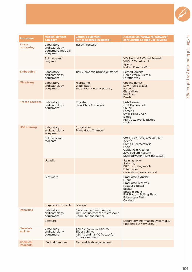
101
4. C
linic
al la
bo
rato
ry &
path
olo
gy
Procedure Medical devices category
Capital equipment (for specialized hospitals)
Accessories/hardware/software/consumables/single use devices
Tissue processing
Laboratory and pathology equipment, medical equipment
Tissue Processor
Solutions and reagents
10% Neutral Buffered Formalin100% 95% AlcoholXyleneMelted Paraffin Wax
Embedding Laboratory and pathology equipment
Tissue embedding unit or station Heated forcepsMould (various sizes)Paraffin Wax
Microtomy Laboratory and pathology equipment
Microtome,Water bath, Slide label printer (optional)
Cooling device Low Profile BladesForceps Glass slidesHot PlateBrush
Frozen Sections Laboratory and pathology equipment
Cryostat, Stool Chair (optional)
Histofreezer OCT CompoundChuckForcepsSmall Paint Brush SlidesHigh/Low Profile BladesRacks
H&E staining Laboratory and pathology equipment
AutostainerFume Hood Chamber
Solutions and reagents
100%, 95%, 80%, 70% AlcoholXyleneHarris’s HaematoxylinEosin0.25% Acid Alcohol20% Sodium AcetateDistilled water (Running Water)
Utensils Staining racksSlide trayDPX mounting media Filter paper Coverslips ( various sizes)
Glassware Graduated cylinderFunnelGraduated pipettesPasteur pipettesBeakerBottle reagentFlat Bottom Boiling FlaskErlenmeyer flaskCoplin jar
Surgical instruments Forceps
Reporting Laboratory and pathology equipment
Binocular light microscope,Immunofluorescence microscope,Computer and printer
Software Laboratory Information System (LIS)(optional but very useful)
Materials archive
Laboratory and pathology equipment
Block or cassette cabinet,Slides cabinet,- 20 ˚C and - 80˚C freezer for frozen specimens
Chemical Reagents
Medical furniture Flammable storage cabinet

102
WHO
list o
f prio
rity m
edica
l dev
ices f
or ca
ncer
man
agem
ent 4.3.3 General medical devices for cytology
Procedure Medical devices category
Capital equipment (for specialized hospitals)
Accessories/hardware/software/consumables/single use devices
Sample processing
Laboratory and pathology equipment
Biosafety cabinet Class II,Cytocentrifuge,Slide label printer (optional),Centrifuge,Refrigerator
Instruments Forceps Glass slidesThinPrep slidesPasteur pipetteCoplin jarRacksScrew cap tube 15 ml / 20 mlBiohazard bin
Solutions or reagents Plasma ThrobimMethanol
Single use devices Paper, lens
Staining Laboratory and pathology equipment
AutoStainer
Solutions and reagents
50%, 70%, 80%, 95% ,100% AlcoholDistilled water ( running water )HaematoxylinEosinOrange Gelb-60.5% Acid AlcoholMay Grunewald StainGiemsa StainMethanolXylene
Utensils Staining racks Slide TrayDPX mounting media Filter Paper Coverslips ( various sizes )
Glassware Graduated CylinderFunnelGraduated pipettesPasteur pipettesBeakerBottle reagentFlat Bottom Boiling FlaskErlenmeyer flaskCoplin jar
Surgical instruments Forceps
Reporting Laboratory and pathology equipment
Binocular light microscope,Computer and printer
Software Laboratory Information System (LIS)(optional but very useful)
Materials archives
Laboratory and pathology equipment
Refrigerator,Slides cabinet,
Chemical Reagents
Medical furniture Flammable storage cabinet

103
4. C
linic
al la
bo
rato
ry &
path
olo
gy
4.3.4 General medical devices for immunohistochemistry
Procedure Medical devices category
Capital equipment (for specialized hospitals)
Accessories/hardware/software/consumables/single use devices
IHC staining Laboratory and pathology equipment
Gravity–convection laboratory ovenRefrigerator-20˚C FreezerOven 37˚C and 60˚CMechanical balanceScale precision digital 500 g/0.01 gMechanical and Digital TimerMicrowave oven Pressure cookerpH meterImmunohistochemistry (IHC) / In situ hybridization (ISH) staining platform Slides label printer for Immunohistochemistry (IHC) / In situ hybridization (ISH) staining platform
Micropipettes (different microliters)Staining Rack
Instruments Forceps
Solutions and reagents
Distilled waterXylene 100% 95 % 80% 70 % AlcoholWashing BufferPeroxidaseBlocking protein solutionAntibodies diluentsPrimary antibodiesSecondary antibodiesHarris’s HaematoxylinBluing ReagentDABEnzyme solution Antibodies detection kit / Amplification Kit and Prep Kit dispenser or similar for IHC staining platformImmunohistochemistry (IHC) / In situ hybridization (ISH) staining platform reagents and buffers
Other Slide TrayBiohazard BinMarkers / fine point / permanent black for glasswareRack for drying glassware and plastic wareRack for drying slidesRack for tubesCover plates Cover slip ( various sizes )
Glassware BeakerBottle ReagentFunnelGraduated cylinderGraduated pipetteCoplin JarDropper bottle
Chemical Reagents
Medical furniture Flammable storage cabinet

104
WHO
list o
f prio
rity m
edica
l dev
ices f
or ca
ncer
man
agem
ent 4.3.5 Special stain (histochemical)
Procedure Medical devices category
Capital equipment (for specialized hospitals)
Accessories/hardware/software/consumables/single use devices
Special staining Laboratory and pathology equipment
Gravity–convection laboratory ovenRefrigerator-20˚C and - 80˚C FreezerOven 37˚C and 60˚CMechanical balancePrecision electronic balancepH meterMechanical and Digital TimerHot plateMagnetic stirrerMicrowave oven Pressure cooker
Micropipettes (different microliters)
Instruments ForcepsSpatula
Solutions and reagents
Harris’s HaematoxylinEosin0.5 % Periodic AcidSchiff’s ReagentCarbol Fuchsin0.5% Acid alcohol10% Sulphuric AcidMethylene Blue3% Acetic AcidAlcian BlueSodium ChlorideConge RedPotassium PermanganateSodium Hydroxide2% Oxalic AcidFouchet’s ReagentNuclear Fast Red5% Chromic AcidSodium MetabisulfiteMethenamine SilverGold Chloride Sodium ThiosulphateBouin’s FixativeCelestine BlueAcid Fuchsin5% Phosphotungstic acidLight Green
Other Staining RackMagnetic stirrerMarkers / fine point / permanent black for glasswareRack for drying glassware and plastic wareRack for drying slidesCover plates Cover slip ( various sizes )
Glassware BeakerBottle ReagentFunnelGraduated cylinderGraduated pipetteCoplin JarFilter paperLaboratory mortarErlenmeyer FlaskFlat Bottom Boiling FlaskPasteur pipetteDropper bottle
Chemical Reagents
Medical furniture Flammable storage cabinet

105
4. C
linic
al la
bo
rato
ry &
path
olo
gy
4.4 Other health system components
4.4.1 Human resources for clinical laboratory and anatomic pathology The personnel required depend on the anticipated volume of material and process to be handled by the specific
laboratory unit.
Depending on national context including workforce availability and models, different occupations can fulfil the
roles that may be required for clinical laboratory and anatomic pathology. A prerequisite is having the required
competencies to perform these roles and being officially recognized for it as per the scope of practice. The
ISCO2 codes are added as illustrative examples based on current practice for further reference. This should not
be interpreted as guidance on occupations that should perform each role but rather indicative occupations that
are currently performing these roles in different country contexts.
Sample role Sample occupations (ISCO-08 codes)
Tissue handling/transportation, tissue assessment, sectioning and staining, analysis of tissue sections
Biomedical Laboratory Scientist, Medical and pathology laboratory technicians (3212)
Gross and microscopic analysis of tissue and cytologic specimens with diagnosis
Specialist medical practitioner (2212) Anatomic Pathologist/ Cytopathologist (2212)
Review Pap smears and non-gynaecologic cytology preparations
Cytotechnologist (3212)
Management of medical devices: planning, procurement,Supervision or performance of: installation, users training, maintenance and decommissioning.
Biomedical Engineer (2149)Biomedical technician / physical and engineering science technician (3119) under supervision of biomedical engineer (2149)
Ensure Quality of all Procedures, that relevant Standard Operating Procedures (SOPs) are followed, and that all staff maintain professional standards and certificationEnsure that sufficient internal and external quality control procedures are in place and followed.• Provide employees with orientation and training.• Include policies relevant to personnel in the quality manual.
Specialist medical practitioner (2212) Anatomic Pathologist (2212),Biomedical Laboratory Scientist, Medical and pathology laboratory technicians (3212)Laboratory quality manager
Administrative tasks Clerical support workers ( 4412)
4.4.2 Infrastructure
Even at the most primary or basic level, the pathology laboratory facility must meet the ISO certification and
local building criteria required for its type of use and equipment, structure, ventilation and storage capacities.
Layout distributionThe laboratory should have the essential infrastructure for the collection and safe handling of the specimens
(tissue, blood and other human samples), tissue processing, and diagnosis of the lesion and in the case of
pathology (cytology and haematology), the appropriate storage of paraffin blocks and slides to ensure their
future use for the benefit of the patient and as required by local laws.
It is important to consider procurement of the sample, which is likely performed offsite from the central institution,
with subsequent transportation of the sample to the pathology laboratory. The personnel from remote sites
must be instructed about the need for appropriate and timely fixation of the tissues, and appropriate handling
of the tissues in the appropriate fixative solutions.
The size of the facility (area) should be commensurate with the volume of material handled, the equipment
needed, and the staff to be accommodated on site at any given time. The area must have the capacity to
accommodate all of the equipment needed for the unit (see the list of devices necessary for the laboratory),
and also appropriate and adequate space for storage of pathological specimens.
The laboratory premises should be appropriately identified and should comply with local/national building
regulations. There must be sufficient space and facilities for the safe and satisfactory operation of the laboratory
service. Specifically, there should be facility for:
2 International Standard Classification of Occupations, http://www.ilo.org/public/english/bureau/stat/isco/docs/publication08.pdf

106
WHO
list o
f prio
rity m
edica
l dev
ices f
or ca
ncer
man
agem
ent 1. the reception and handling of specimens
2. the handling of hazardous materials
3. space for performance of all licensed tests
4. space for maintenance of all equipment
5. the safe storage of reagents
6. the storage of tissues including blood and other body fluids
7. storage of residual tissues from specimens, tissue blocks and glass slides
8. space for administrative staff and professional staff to work comfortably.
Safety equipment including eye wash/shower, hand basins and personal protective equipment must be available.
Considerations for pre-analytical and analytical phases of specimen handling:
Adequate and clean facilities are required with clean water supply and adequate electrical outlets, with surge
protection and/or universal power supply systems to support analyzers and tissue processors All specimens
received in the laboratory must be immediately and appropriately labelled. A written policy should be
developed for the management and handling of inadequately-labelled specimens or incomplete request forms.
All specimens coming to pathology laboratories or being transported from one laboratory to another must
be packaged appropriately with appropriate labels and must ensure the integrity of the specimen as well as
the safety of the public. If a specimen’s integrity is compromised, this must be recorded and the appropriate
individuals informed immediately.
Considerations for post-analytical phase of specimen handling:
The post-analytical phase involves the transfer of information back to the appropriate physician and patient.
Failure to have a post-analytical phase policy in place can result in adverse events and patient harm.
Electrical systemsMost tissue handling equipment runs on electric power and this makes an adequate electrical system essential.
To facilitate the functionality of the devices, a continuous and uninterrupted electrical supply of the appropriate
electrical voltage is required. The continuous electrical supply for equipment should be reinsured by a backup
electrical supply for essential equipment, in case of power failure.
Heating, ventilation and air conditioning systemsLaboratories should have appropriate ventilation and adequate humidity and temperature conditions. In tropical
and subtropical climates, air conditioning would be an asset. All these should conform to national guidelines.
The area requires ambient temperature comfortable for all staff including those who don protective materials
(at a minimum, a gown and a plastic apron). Additionally, specific ventilation must be installed in areas where
surgical specimens fixed in preservatives, such as formalin (10%), are being examined and sectioned. For this,
an appropriate workstation (see list of devices) is necessary.
Air from this suite may need to be exhausted externally, to prevent recirculation with the rest of the facility. The
pressure should be negative in relationship to adjacent areas.
Safety and security Special safety and security considerations should be taken into account as biohazardous materials, flammable
materials, toxic materials and waste and excess formalin tissues within the laboratory can be reactive and
potentially cause harm to personnel in and surrounding the laboratory.
Considering these potential dangers, special criteria for fire protection should be installed within the facility.
Special SystemsThe walls, ceilings and floors should be of a material suitable for regular cleaning and resistant to a potential
biohazardous material spill. Moreover, the area should have adequate systems to provide both water and
drainage. Lighting and noise levels should conform to national guidelines and all of the above materials and
equipment should be securely stored. There should also be appropriate toilet facilities for staff.

107
4. C
linic
al la
bo
rato
ry &
path
olo
gy
4.4.3 Quality management
Quality assurance promotes the safe, effective and appropriate functioning of a laboratory, and the safety of staff
and patients. With the intention of ensuring personnel, public and patient safety, the appropriate functioning
of a laboratory will be greatly enhanced by the availability of a good laboratory information system, one which
can track specimens from arrival to final report and which can be integrated into the wider electronic patient
records system. There are many purveyors of these systems and the choice of an appropriate one would help in
many ways, as will be described later in this section. A good system ensures that all specimens are appropriately
labelled with at least two patient identifiers. The unique identifier applied to a patient’s tissue sample follows the
sample from the beginning to the end. An example of this is the bar code system. Quality assurance standards
should be based on local standards for Quality and Competence. If not available, standards can be based on
the quality assurance recommendations of the RCPA (Royal College of Pathologists of Australasia) or the CAP
(College of American Pathologists).
Basic requirements:
All patient specimens or body parts must be treated with respect at all times, with adherence to and
compliance with the highest ethical standards and with appropriate consideration of patient safety. In all
cases, there must be an appropriate patient consent for the examination of the tissues and the performance
of special studies (example HIV and molecular testing). It is the responsibility of the treating physician to
ensure that a consent is obtained.
Governance:
Each laboratory must have documented systems of governance, including guidelines for patient and staff safety
and the control/regulation of risks involved in the delivery of laboratory services. The laboratory must have
appropriate supervision, by qualified individuals and appropriately trained certified laboratory directors. The
training of all laboratory staff must include safe and ethical laboratory practice. They must be given the authority
to ensure that the standards are met. The standards may be the ISO 15189 Medical Laboratory Standards or
equivalent standards set by a National Association or a national government of that jurisdiction, e.g. National
Association of Testing Authorities (NATA), Australia; South African National Accreditation System, South
Africa (SANAS); United Kingdom Accreditation Service, United Kingdom (UKAS); International Accreditation
New Zealand, New Zealand (IANZ); or those set by other authorities, such as: National Accreditation Board
for Testing and Calibration Laboratories (NABL), India; Bureau of Laboratory Quality and Standards (BLQS),
Thailand; Department of Standards Malaysia (DSM), Malaysia.
The standards must take into account the volume of work and set operational practices and requirements for
staffing, staff training, and continuing professional development. The standards must plan and regulate the
tests to be provided either at the laboratory or off site, as well as safeguard quality assurance via organization
of a referral program. These requirements for the above tests must document the methods and the procedures
to be followed and ensure that the personnel are trained appropriately in these procedures. The personnel’s
competence to perform these tests should be determined on an ongoing basis. If the laboratory performs
services during one shift per working day, it should be appropriately staffed for this. If it has more than one shift,
a shift supervisor must be appointed, trained and empowered to make appropriate decisions.
Internal Quality Assurance:
As previously stated, appropriate procedures must be in place for the handling of tissues sent to the laboratory
from the time that they are removed from the patient by the surgeon/practitioner and sent to the laboratory.
Procedures for the appropriate fixation, storage, and disposal of tissues, must be in place, and a log maintained
of these practices, detailing when and which patients’ tissues were “discarded”.
All tests that laboratory personnel are required to perform must have documented standard operating
procedures (SOP) in place, which are readily accessible and strictly followed. The materials needed for the
appropriate performance of these tests must be easily accessible (day or night).

108
WHO
list o
f prio
rity m
edica
l dev
ices f
or ca
ncer
man
agem
ent Since many potentially toxic chemicals are used in the laboratory for tissue fixation, processing and staining,
their appropriate storage is essential (prior to use and when they are ready for discard). There must be policies,
protocols and documentation for chemicals as they come in and are ultimately destroyed. Areas where these
materials are used must have appropriate exhaust systems.
There must be a tracking system to monitor the movement of tissue from the operating room or the physicians’
office to the pathology laboratory, including the removal of slides and storage of the residual tissues (until
they are discarded). All of the above materials and equipment should be securely stored. This paper trail (or
electronic trail) is essential.
The above pertains to the first two stages of tissue handling – preanalytic and analytic. The final stage is the
evaluation of the tissues and the making of an appropriate diagnosis. The professional staff must be appropriately
trained, qualified, certified and have the opportunity for adequate continuous professional development.
External Quality Assurance:
A good external quality assurance (EQA) system provides an interlaboratory comparison of technical testing
and interpretations of shared batches of samples by participating laboratories on a regular basis. For
laboratories providing services for cancer management, the EQA program participated should have neoplastic
tissues represented. EQA serves as an early warning system when the laboratory performs out-of-range of the
majority of other participating laboratories. A good EQA program also provides educational instruction to help
laboratories address areas of weaknesses. A log of this practice should be maintained.
Immunohistochemistry:
Where a laboratory performs immunohistochemical stains, it should be part of a quality assurance system,
such as the NordiQC (Nordic immunohistochemical Quality Control), NEQAS, RCPA-QAP or other similar
proficiency testing systems. To ensure staff qualifications and competence, all qualified staff should be
given the opportunity to participate in ongoing professional development to ensure that their skills are
maintained and updated regularly. Where a laboratory offers consulting services to smaller laboratories, an
information system that allows for tracking of cases from the time they come in to the time the final report
is issued, must be available.
Critical alert:
Where there is a significant result (convergent with the clinical or especially one divergent from the clinical
diagnosis) the result should be treated as a critical incident and a system in place to report it to the appropriate
staff. This effort should be recorded. All diagnosis of malignancy, whether suspected or not, should be reported
immediately to the requesting physician and treated as a critical result.
Employee Relations:
It is the responsibility of the institution and the laboratory director, to ensure that the work environment is
appropriate for hassle-free work. Staff should have appropriate work clothes and footwear while inside the
laboratory (open footwear such as sandals should be prohibited). Clothes (outerwear such as lab coats) worn
inside the laboratory should preferably not be worn outside the lab. An appropriate facility to handle the
outerwear should be available. Hazardous materials must be appropriately stored and handled. Staff should be
aware of the handling of hazardous materials spills. Regular training in this procedure should be offered to all
laboratory staff.

109
4. C
linic
al la
bo
rato
ry &
path
olo
gy
4.4.4 Guidance documents
Title Link Description
Laboratory Quality Management System
https://globalhealthlabo-ratories.tghn.org/site_media/media/articles/WHO_Lab-oratory_quality_management_sys-tem_1.pdf
Achieving, maintaining and improving accuracy, timeliness and reliability are major challenges for health laboratories. This handbook is intended to provide a comprehensive reference on Laboratory Quality Management Systems for all stakeholders in health laboratory processes, from management, to administration, to biomedical laboratory scientists. This handbook covers topics that are essential for quality management of a public health or clinical laboratory. They are based on both ISO 15189 and CLSI GP26-A3 documents.
Laboratory Assessment Tool
https://globalhealthlabo-ratories.tghn.org/site_media/media/articles/WHO_Lab-oratory_Assess-ment_Tool_1.pdf
This document offers guidance to assess individual laboratories and the national laboratory system. It describes a general process for assessing laboratories and provides questionnaires to help assess laboratories. The document and its questionnaires can be used as is or after an adaptation to meet local requirements or specificities to better fit the assessment context. The intended audience of the document is any stakeholder performing laboratory assessments, including: national health authorities, multilateral agencies, non-governmental organizations (NGOs), laboratory managers, etc.
The Pathology Request-Test-Report Cycle - Guidelines for Requesters and Pathology Providers
https://www.rcpa.edu.au/getattach-ment/cb14bc34-0a01-4c09-839c-614e098c84b6/Pathology-Re-quest-Test-Re-port-Cycle-Guide-lines.aspx
These guidelines have been developed for use by medical practitioners when requesting pathology tests and by Pathology Providers operating in both public and private practice.
Australian Commission on Safety and Quality in Healthcare, National Safety and Quality Service Standards, 2011, Government of Australia
http://www.safet-yandquality.gov.au/wp-content/uploads/2011/01/NSQHS-Standards-Sept2011.pdf
Contains safety and quality standards for various topics, including handling of blood and blood products.
Quality Systems for Medical Laboratories. Guidelines for Implementation and Monitoring. WHO Regional Publications, Eastern Mediterranean Series. (1995)
http://applications.emro.who.int/dsaf/dsa33.pdf
The document provides guidance for laboratories to introduce, maintain and improve appropriately specified levels of service quality, and to provide a basis for assuring an adequate quality of services, quality system and its specifications.
References
Warner LC. Lab-in-a-Box: a guide to building anatomic pathology networks in resource-limited settings. Am J
Clin Pathol 2017;147(1):8-14. doi: 10.1093/ajcp/aqw217

5. Radiotherapy

111
5. R
ad
ioth
era
py
Health service delivery sequence overview: This diagram expresses the flow of cancer patient to and from the radiotherapy unit, and the elements to
consider as support for the unit (Fig. 10.5).
Fig. 10.5 Health care service delivery overview – radiotherapy
PR
IMA
RY
(C
L,H
C),
SE
C-
ON
DA
RY
(D
H)
&
TE
RT
IAR
Y (
SH
)
Le
ve
l o
f C
are
*
* Appropriate level of care will depend on the particular intervention, setting, and available infrastructure and human resources.
SE
CO
ND
AR
Y (
DH
)
& T
ER
TIA
RY
(S
H)
TE
RT
IAR
Y (
SH
)
Prevention End of life care
TreatmentFollow-up/survivorship
care
Palliative care
Diagnosis
Screening
CL Community Level health post DH District Hospital HC Health Centre SH Specialized Hospital
Systemic therapy
Nuclear medicine
Surgery
Vaccination
Radio- therapy
Pathology Pathology
Clinical laboratory
Clinical laboratory
Clinical assessment
Palliative care
Medical imaging
Endoscopy
Clinical assessment
Nuclear medicine
5.1 General description of the unit
5.2 Priority medical devices
5.2.1 General medical devices for external radiotherapy
5.2.2 General medical devices for brachytherapy
5.2.3 Specific medical devices for brachytherapy by cancer type
5.3 Other health system components
5.3.1 Human resources for radiotherapy
5.3.2 Infrastructure
5.3.3 Quality management
5.3.4 Guidance documents

112
WHO
list o
f prio
rity m
edica
l dev
ices f
or ca
ncer
man
agem
ent
5.1 General description of the unitRadiation therapy is a clinical modality that uses ionizing radiation in the treatment of patients with malignant
neoplasia (and occasionally benign disease). It is an important component of multimodality cancer therapy,
surgery and chemotherapy. More than 50% of new cancer cases require radiotherapy as their definitive or
adjuvant therapy (1).
Radiotherapy services should be available at specialized oncology hospitals, and it should be noted that they
require very specialized infrastructure and human resources. It is important to analyse which cancer centres
will have the radiation oncology unit, depending on the human, financial and technical resources available, as
well as the local demand. Although linear accelerators are the preferred technology, Cobalt 60 unites can be
used in limited resource settings as they are easier to operate and maintain.
Radiation safety must always be considered for patients, workers and the public. The role of imaging in
radiation therapy is a determinant of the success of the therapy, and the targeting, particularly when there
is motion (e.g. diaphragmatic or breast tumors), which affects the location of the tumour relative to healthy
tissue, makes imaging a determinant to enhance the therapy and reduce the collateral damage.
Radiotherapy cannot be delivered without a treatment planning system, which is used to determine target
volumes and other critical structures, and, subsequently, to determine how the radiotherapy system will
deliver the treatment as required. Additionally, oncology information systems combine all oncology data
pertaining to a patient for easy access and reference. This enables clinicians to perform essential quality
assurance measures in order to facilitate best outcomes for patient care.
5.2 Priority medical devices The following tables present general medical devices, which can be used for the treatment, follow-up and/or
palliative care of many diseases including several cancers, and specific medical devices, which are used for
interventions for specific cancer types.
The following tables present the priority medical devices required to perform the following health interventions:
Cancer type Interventions
All cancers • Imaging and treatment planning• External beam radiotherapy • Brachytherapy
Cervical cancer & prostate cancer
• High dose rate brachytherapy
5.2.1 General medical devices for external radiotherapy
Procedure Medical devices category
Capital equipment(for specialized hospitals)
Accessories/hardware/software/consumables/single use devices
Radiotherapy treatment delivery
Medical equipment Linear Accelerator (LINAC) (otherwise, Cobalt-60 Unit)
• 3D Conformal therapy • At least 6 MV with multileaf
collimator and electronic portal imaging.
Patient positioning/tracking system
Single-patient physiologic monitoring system
• Blood pressure cuffs for adults and infants
• Thermometer probes (in case it is needed)
Resuscitation trolley, equipped with medicines and defibrillator
• Laryngoscope for adults and infants
Radiation safety devices
Personal Dosimeter
Geiger–Müller (GM) survey meter
Large volume ionization chamber (consider requirements from IAEA)
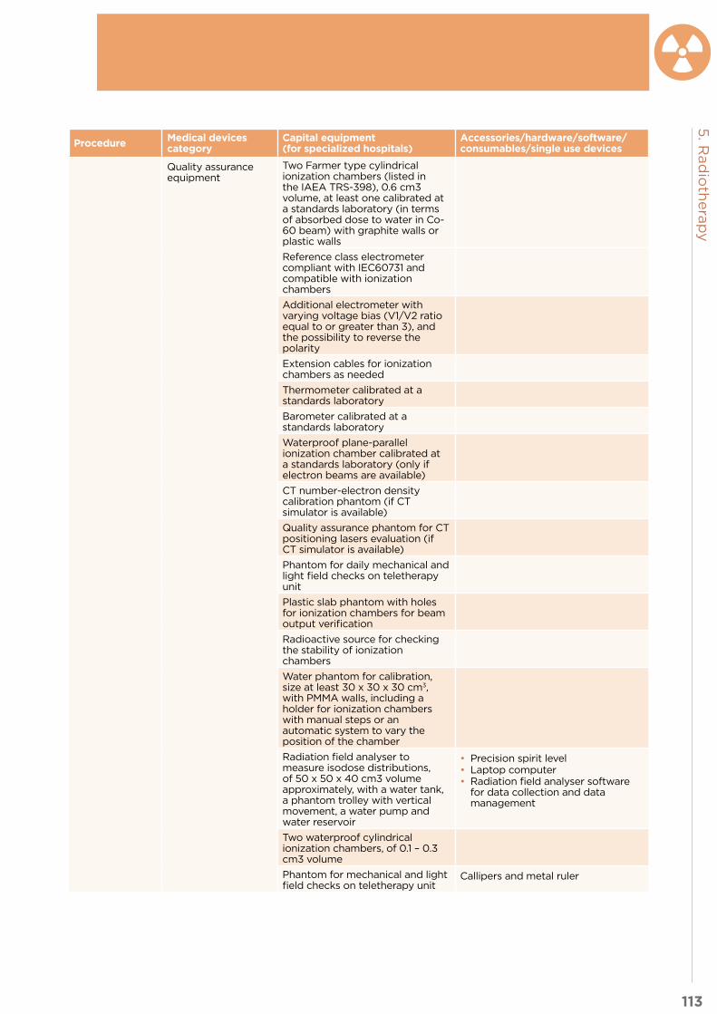
113
5. R
ad
ioth
era
py
Procedure Medical devices category
Capital equipment(for specialized hospitals)
Accessories/hardware/software/consumables/single use devices
Quality assurance equipment
Two Farmer type cylindrical ionization chambers (listed in the IAEA TRS-398), 0.6 cm3 volume, at least one calibrated at a standards laboratory (in terms of absorbed dose to water in Co-60 beam) with graphite walls or plastic walls
Reference class electrometer compliant with IEC60731 and compatible with ionization chambers
Additional electrometer with varying voltage bias (V1/V2 ratio equal to or greater than 3), and the possibility to reverse the polarity
Extension cables for ionization chambers as needed
Thermometer calibrated at a standards laboratory
Barometer calibrated at a standards laboratory
Waterproof plane-parallel ionization chamber calibrated at a standards laboratory (only if electron beams are available)
CT number-electron density calibration phantom (if CT simulator is available)
Quality assurance phantom for CT positioning lasers evaluation (if CT simulator is available)
Phantom for daily mechanical and light field checks on teletherapy unit
Plastic slab phantom with holes for ionization chambers for beam output verification
Radioactive source for checking the stability of ionization chambers
Water phantom for calibration, size at least 30 x 30 x 30 cm3, with PMMA walls, including a holder for ionization chambers with manual steps or an automatic system to vary the position of the chamber
Radiation field analyser to measure isodose distributions, of 50 x 50 x 40 cm3 volume approximately, with a water tank, a phantom trolley with vertical movement, a water pump and water reservoir
• Precision spirit level• Laptop computer• Radiation field analyser software
for data collection and data management
Two waterproof cylindrical ionization chambers, of 0.1 – 0.3 cm3 volume
Phantom for mechanical and light field checks on teletherapy unit
Callipers and metal ruler

114
WHO
list o
f prio
rity m
edica
l dev
ices f
or ca
ncer
man
agem
ent
Procedure Medical devices category
Capital equipment(for specialized hospitals)
Accessories/hardware/software/consumables/single use devices
In-vivo dosimetry system (for example, diode dosimeters, thermoluminescent dosimetry (TLD), or optically stimulated luminescence dosimetry (OSLD))
Array of diodes or ion chambers for routine quality assurance checks
Film dosimetry system including radiochromic film, film scanner and dedicated software
Radiochromic film
EPID image quality phantom
Radiation safety devices
Area radiation monitor with audible alarm (only if Co-60 teletherapy is available)
Survey meter
Imaging and Treatment Planning
Medical equipment Computed Tomography (CT)System (16 slices minimum)
CT Overlay flat table
Laser patient positioning system
Contrast media injection system (optional)
Conventional simulator (only if a CT simulator is not available)
Computerized treatment planning systems (three-dimensional) including
Colour printer
Hardware and software, virtual simulation software, and plan review software
Digitiser
Mould make process (Immobilization and patient positioning system)
Equipment Hot water bath for thermoplastic immobilization system
Hot wire cutter
Drill
Pot for cerrobend cadmium free low melting point alloy (if needed according to the technique and the type of accelerator)
Furniture Adequate furniture for the moulding process
Instruments Tray, dressing, stainless steel
Personal protective equipment and clothing
Apron, protection, plastic
Eye protection glasses, safety, regular size
Gloves, examination, latex, non-sterile, single use (various sizes)
Coat, medical, woven (various sizes)
Other Receptacle, waste, stainless steel, pedal action
Safety box for used syringes/needles
Brush
Acetate sheets
Rubber hammer
Mould materials Bolus
Chosen material for the mould: Foam blocks/Styrofoam/Polystyrene, Thermoplastic, vacuum bag or similar as needed
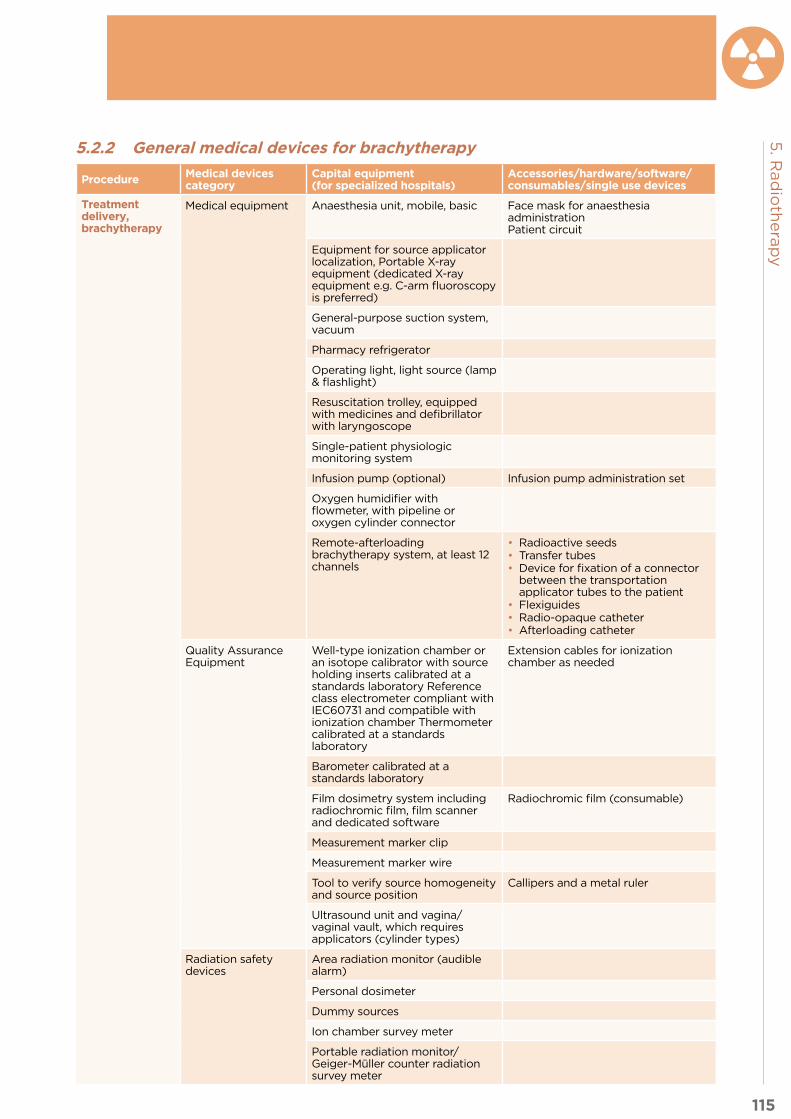
115
5. R
ad
ioth
era
py
5.2.2 General medical devices for brachytherapy
Procedure Medical devices category
Capital equipment (for specialized hospitals)
Accessories/hardware/software/consumables/single use devices
Treatment delivery, brachytherapy
Medical equipment Anaesthesia unit, mobile, basic Face mask for anaesthesia administrationPatient circuit
Equipment for source applicator localization, Portable X-ray equipment (dedicated X-ray equipment e.g. C-arm fluoroscopy is preferred)
General-purpose suction system, vacuum
Pharmacy refrigerator
Operating light, light source (lamp & flashlight)
Resuscitation trolley, equipped with medicines and defibrillator with laryngoscope
Single-patient physiologic monitoring system
Infusion pump (optional) Infusion pump administration set
Oxygen humidifier with flowmeter, with pipeline or oxygen cylinder connector
Remote-afterloading brachytherapy system, at least 12 channels
• Radioactive seeds • Transfer tubes• Device for fixation of a connector
between the transportation applicator tubes to the patient
• Flexiguides• Radio-opaque catheter• Afterloading catheter
Quality Assurance Equipment
Well-type ionization chamber or an isotope calibrator with source holding inserts calibrated at a standards laboratory Reference class electrometer compliant with IEC60731 and compatible with ionization chamber Thermometer calibrated at a standards laboratory
Extension cables for ionization chamber as needed
Barometer calibrated at a standards laboratory
Film dosimetry system including radiochromic film, film scanner and dedicated software
Radiochromic film (consumable)
Measurement marker clip
Measurement marker wire
Tool to verify source homogeneity and source position
Callipers and a metal ruler
Ultrasound unit and vagina/vaginal vault, which requires applicators (cylinder types)
Radiation safety devices
Area radiation monitor (audible alarm)
Personal dosimeter
Dummy sources
Ion chamber survey meter
Portable radiation monitor/Geiger-Müller counter radiation survey meter

116
WHO
list o
f prio
rity m
edica
l dev
ices f
or ca
ncer
man
agem
ent
Procedure Medical devices category
Capital equipment (for specialized hospitals)
Accessories/hardware/software/consumables/single use devices
Medical Furniture Hospital stretcher, with side rails
Reclining chair
Stand, infusion, double hook, on casters
Trolley, dressing, stainless steel, 2 trays
Trolley, medicines
Trolley, soiled linen
Wheelchair, adult
Table, instruments, Mayo type, stainless steel, on casters
Cabinet for the necessary instruments, equipment, treatment aid and the required documents
Cabinet, medicines, double door
Instruments Dressing set
General-purpose surgical scissors, reusable
Measurement ruler
Metzenbaum scissors
Suture set
Rectal probe or rectal catheter
Personal protective equipment and clothing
Gloves, examination, non-sterile, single use (various sizes)
Gown, patient, woven
Non-conductive shoe cover
Operating room gown, reusable
Surgical cap for patients and healthcare worker
Surgical face mask
Surgical gloves
Single use devices/disposables/medical supplies
Compress, gauze, sterile & non-sterile, single use
Infusion giving set, sterile, single use
Prongs, nasal, oxygen, non-sterile, single use (various sizes)
Radiographic or radiochromic film
Safety box, for used syringes/needles
Silicone adhesive tape
Skin-cleaning wipe
Surgical scrub brush, single-use
Sutures
Syringes with needles (disposable)
Tape, medical, roll (various sizes)
Catheter, Foley, sterile, single use (sizes G)3
Skin-cover adhesive strip
General-purpose sterile drape

117
5. R
ad
ioth
era
py
Procedure Medical devices category
Capital equipment (for specialized hospitals)
Accessories/hardware/software/consumables/single use devices
Solutions and reagents
Aqueous antibacterial solution
Contrast media
Hydrogen peroxide
Saline solution
Lubricating jelly (K-Y)
Iodine povacrylex and isopropyl alcohol solution or similar
Other Arm/leg tourniquet
Basin, kidney, stainless steel/polypropylene
Bowl (dressing changes), stainless steel/polypropylene
Kick bucket
Manual brachytherapy source, temporary placement
Radiation protection devices
Clamps for the source management
Emergency container and emergency source handling instruments (Retrieval instrument)
Portable radiation protection barriers
Radioactive waste storage
Source handling instruments and accessories (in source preparation room and patient loading room)
Source loading and cutting devices
Source storage and transport containers within the department
Imaging and treatment planning
Medical equipment Imaging X-Ray or Computed tomography (CT) (if not available in external radiotherapy)
Include appropriate brachytherapy software
3. Components of all surgical instrument sets as well as needle and catheter gauge sizes (G) are listed in the chapter annex at the end of the Surgery chapter

118
WHO
list o
f prio
rity m
edica
l dev
ices f
or ca
ncer
man
agem
ent 5.2.3 Specific medical devices for brachytherapy by cancer type
5.2.3.1 Cervical
Procedure Medical devices category
Capital equipment (for specialized hospitals)
Accessories/hardware/software/consumables/single use devices
Treatment delivery, High dose rate Brachytherapy
Medical equipment Universal operating table-radiopaque, (leg rests, anaesthesia requirements, etc) or Brachytherapy table
If patients are prepared in a separate room, then movable, interchangeable patient tables are generally supplied so that the patient is not moved unnecessarily between applicator insertion and treatment delivery
Instruments Gynaecologic applicators sets (for intracavitary and interstitial treatments e.g. Henschke, Fletcher–Suit, Manchester or Delouche type) / Cervical/intrauterine brachytherapy system applicator
Scissors
Hysterometer
Manual expandable cervical dilator
Ring forceps
Uterine forceps
Vaginal Speculum, reusable
5.2.3.2 Prostate
Procedure Medical devices category
Capital equipment (for specialized hospitals)
Accessories/hardware/software/consumables/single use devices
Treatment delivery, High dose rate Brachytherapy
Medical equipment Ultrasound unit with biplanar transducer
Stepping unit and stabilizer
Instruments Needle cradle
5.3 Other health system components
5.3.1 Human resources for radiotherapyThe staff would include the following or related areas : radiation oncologists, medical physicists, dosimetrists,
radiographers/radiation therapy technologists (RTT),nurses, and other professionals involved in the
preparation and delivery of treatment as biomedical engineers, equipment technicians and administrative
personnel. In some countries and in larger radiotherapy departments, the additional roles of physician
assistants and medical physics assistants might be found effective.
Depending on national context including workforce availability and models, different occupations can fulfil
the roles that may be required for radiotherapy. A prerequisite is having the required competencies to
perform these roles and being officially recognized for it as per the scope of practice. The ISCO2 codes are
added as illustrative examples based on current practice for further reference. This should not be interpreted
as guidance on occupations that should perform each role but rather indicative occupations that are currently
performing these roles in different country contexts.
2 International Standard Classification of Occupations, http://www.ilo.org/public/english/bureau/stat/isco/docs/publication08.pdf

119
5. R
ad
ioth
era
py
Sample role Sample occupations (ISCO-08 codes)
Sets up the overall policy for the radiation therapy programme and implementation; Participates in the evaluation of the proposed departmental clinical load; Participates in multidisciplinary cancer team;Responsible for the overall care of the patient including:• Clinical evaluation• Therapeutic decision making• Establishment of treatment plan, including dose prescription, contouring
volumes, OAR and restrictive doses, plan evaluation, treatment execution• On-treatment evaluations and patient monitoring• Treatment summary• Follow-up and response assessment of overall treatment
Radiation Oncologist, Specialist medical practitioner (2212)
Provides clinical physics service and supervision. Responsible for:• Specification of therapy equipment (external beam, brachytherapy,
simulators, CT and imaging systems, and treatment planning systems) and assuring their radiation safety
• Acceptance testing, commissioning and quality assurance (including calibration) of therapy equipment
• Measurement and analysis of beam data, and tabulation of beam data for clinical use
• Establishment of dose calculation procedures• Establishment of technical aspects of treatment planning and treatment
procedures• Supervision, evaluation and optimization of treatment planning• Establishment and supervision of quality assurance procedures in
radiotherapy regarding delivery of the treatment, radiation safety, quality control and regulatory compliance
• Supervision of maintenance of radiotherapy equipment• Education of staff in radiation safety principles.
Medical Physicist (2111)
• Management of the daily workload of the treatment units and simulators; • Operates simulator and other imaging devices for therapy purposes (CT
scan, C-arm, and simulators); • Operates HDR brachytherapy machines; • Records and checks all the parameters needed to repeat or reconstruct the
activities undertaken; • Explanation of treatment and daily checking of patients undergoing
radiation treatment and reporting new or unusual conditions of patient to radiation oncologist;
• Acquisition and verification of images consistent with departmental protocols.
Radiation Therapy Technologist (RTT), medical radiation therapist, medical imaging and therapeutic equipment technicians (3211)
• Provides nursing assistance to radiation oncologist in clinic and brachytherapy room;
• Provides nursing intervention for the actual and potential problems that the patient and family may experience related to the disease process, treatment course and follow-up period;
• Provides teaching, counselling and support functions needed to assist the patient and family to cope with and adjust to the diagnosis and treatment of cancer
Oncology Nurse, Nursing professional (2221) Nursing associate professional (3221), supervised: healthcare assistant (5321)
Responsible for general management of equipment including:• Procurement specifications, planning of bunker and special infrastructure
along with physicists and facilities engineer; • Supervision of installation, compliance with requirements, relations with
manufacturer on preventive and corrective maintenance; • Participation in installation of new radiotherapy equipment or major
reparation of radiotherapy equipment by vendor.
Biomedical Engineer (2149)
• Designs the radiation protection and safety programme, and evaluates compliance with it;
• Prepares the licence application, especially the safety assessment for radiotherapy sources;
• Measures accident prevention and mitigation.
Radiation protection officer, Radiation protection expert , environmental and occupational health and hygiene professionals (2263)
Provides administrative support to radiotherapy team in maintaining radiotherapy programme, patients appointments, etc.
Admission/, administrative Staff , Clerical support workers ( 4412)Technologists (3211),
Support medical physics procedures Physics assistant, technician (3111)

120
WHO
list o
f prio
rity m
edica
l dev
ices f
or ca
ncer
man
agem
ent A quality radiotherapy service is a multifaceted process involving several distinct groups of health experts
and supporting staff. In order to improve radiotherapy service, continuous development is essential. Staff/
human resource development is aimed to achieve two objectives as follows:
1. Match staff headcount with radiotherapy unit requirement analysis
2. Enhance staff competency in accordance with advancements in technology and science through training
and education
The education and training programmes are intended for all staff: radiation oncologists, medical physicists,
dosimetrists, physics assistants, RTTs, in house technicians, and nurses.
5.3.2 InfrastructureA radiotherapy centre usually comprises an admission area, administrative offices, clinics, CT simulation
room, a treatment aid fabrication mould room, a treatment planning room, an operating theatre or procedure
room for brachytherapy, and one or more treatment rooms for external radiotherapy and brachytherapy.
The radiotherapy unit placement should be carefully planned and designed to provide good patient flow,
radiation safety and be supported by a well-established information technology (IT) system. It should
include physician work room and meeting room, which will be used to conduct vital quality assurance
meetings and other staff meetings.
The housing for a linear accelerator is very specific and requires long term planning for construction to meet
the requirements of the shielding for the specific equipment to ensure the safety of people in surrounding
areas (3, 8). This bunker is an expensive setting that requires radiation calculations and specialized studies.
It is important to consider all recommendations from IAEA to set up a radiotherapy unit (4), as well as follow
local national laws and guidelines regarding radiotherapy services (if available).
In reference to the equipment, contracts for maintenance by vendors are beneficial to provide ongoing
sustainability of the treatment, simulation and planning equipment. Routine maintenance should be scheduled
in order to reduce the risk of equipment downtime, which causes patient treatment delays and lost revenue.
The infrastructure of the radiotherapy and brachytherapy unit is discussed generally in this section but further
information should be consulted in the IAEA references mentioned below.
Layout distribution
Reception and waiting area
The reception should be located at the main entrance to the department and act as distribution point for all
the different sections in the department.
• Sufficient waiting areas should be provided for patients attending clinics and those awaiting treatment
(in larger departments, smaller waiting areas should be provided close to the treatment machines).
• An area should be provided for patients on stretchers with electrical power and medical gas
connections.
Examination room (clinics)
• A patient changing area should be provided.
• The examination rooms should be large enough to include standard and gynaecological examination
tables, appropriate examination instruments and medical supplies.
External beam radiotherapy - Simulator or CT Simulator room
• A door interlock or other suitable means to prevent unauthorized access is needed.
• A sign should be posted at the entrance warning of the radiation hazard.
• The room should be large enough to accommodate the simulator, allowing the full range of motion of
the treatment table.
• Secure mounting of patient positioning lasers to the wall at points appropriate for projection of lines
through the isocentre should be included in the plans. Adjustable lasers intersecting at a known
distance from the reference plane are required for CT simulators.
• A means for dimming the room lights is essential.

121
5. R
ad
ioth
era
py
• Enough space should be provided for storage of treatment devices and quality assurance equipment.
• If the immobilization devices are to be made in the simulator room, cabinet space to store supplies
for their fabrication will be required. A sink and counter-top space for a heated water bath should be
provided in this room.
• The control/console area should be of adequate size to accommodate the RTT, a physician and at least
one additional person. It should have access to both the simulator room and the hallway.
• A viewing window for the control room is required.
External beam radiotherapy - Mould room
• Space for tools, a block cutter and counter-top is needed, along with adequate workspace for pouring
and mounting the blocks.
• Large enough space for storage of Styrofoam, trays and shielding material for custom blocking is also required.
• If immobilization devices are fabricated in the mould room, space for a patient couch will be required,
and lasers mimicking those in the treatment and simulator rooms should be provided.
External beam radiotherapy - Treatment planning room
• Large enough space is required to house the treatment-planning computer with its video monitors, a
printer, a plotter and a digitizer (if used) and other required computer equipment.
• Space is also needed for supplies of paper and pens or ink for the printer and plotter (if required).
External beam radiotherapy – Treatment room
• The room should be large enough to accommodate the treatment machine, allowing the full range of
motion of the treatment table and access for beds if necessary.
• A heavy, electrically operated door should be provided at the entrance to the room or an appropriately
designed extended corridor (called a maze or labyrinth) leading into the room, and a sign should be
posted at the entrance warning of the radiation hazard.
• A means for dimming the room lights should be included.
• Adequate space should be provided for storage of treatment devices, immobilization devices, blocks
and quality assurance equipment.
• A wash hand basin should be installed in all treatment rooms.
• A ‘last man out’ button should be installed.
• A door interlock must be provided.
High Dose Rate (HDR) Brachytherapy - Operating theatre/treatment room and radiographic imaging system
• A brachytherapy unit should include the shielded treatment room, a control area, a procedure/
preparation room, a recovery area, and an imager or film processing area.
• An independent radiation monitor should be provided that indicates, through a measurement of
radiation levels, whenever the source is moved out of the shielded safe zone.
• A door interlock must be provided.
• A sign should be posted at the entrance warning of the radiation hazard, in accordance with local or
national regulations and the Basic Safety Standard (BSS) (2).
High Dose Rate Brachytherapy - Treatment planning room (TPS)
• Treatment planning for HDR brachytherapy could be performed on a general TPS room for teletherapy
and brachytherapy using a separate brachytherapy planning system, or in a separate TPS room more
convenient to the HDR brachytherapy suite.

122
WHO
list o
f prio
rity m
edica
l dev
ices f
or ca
ncer
man
agem
ent
Shielding• The shielding of the simulator room and the treatment room should be designed according to the
requirements of the national regulatory body, paying due regard to the requirements of the BSS (2).
• The brachytherapy treatment room should have adequate shielding for a high dose rate remote
afterloading unit, designed in accordance with the recommendations of IAEA SRS No 47 (3), paying due
regard to the requirements of the BSS (2) and the regulatory authority
Heating, ventilation and air conditioning systems• The mould room should have adequate ventilation and there should be adequate temperatures to keep
equipment running as specified by the manufacturer. The treatment and control rooms as well, as this
is something that can be planned ahead, even if the equipment provider has not been selected. Certain
equipment needs direct cooling (linear accelerators).
Communication systems• Provision for the system management of the IT equipment is critical, but control of the system must be
retained by the radiotherapy department.
• The console/control area for the treatment room should be provided with a patient intercommunication
system and at least two closed circuit television monitors. Sufficient space should be provided for two
operators, a physician and at least one additional person to be present.
• It is important to have a patient information system (or treatment record and verify system).
Electrical systemsIn countries where power is not readily available, installation of uninterrupted power systems (UPS),
stabilizers or generators should be considered (for the linear accelerator circuit). This would guarantee that
ongoing treatments can be finished in the event of a loss of power. Special electrical systems for radiotherapy
equipment need to be considered.
Special systems• Sufficient washroom facilities should be strategically placed throughout the facility.
• A supply of medical gasses should be considered in the treatment room.
5.3.3 Quality managementMost of the information in this section on Quality Management comes directly from the following publications:
• Comprehensive Audits of Radiotherapy Practices: A tool for Quality Improvement (5)
• Practical Guidelines for implementation of a quality system in Radiotherapy (6)
• Setting Up a Radiotherapy Programme (7)
Quality Assurance and Quality Control in Radiotherapy
Radiotherapy is a specialized discipline using complex equipment and involving interlinked stages and
processes, which require careful and accurate application. Hence, quality assurance is vital to ensure the
delivery of safe and effective treatment. Quality assurance in radiotherapy consists of procedures that ensure
a consistent and safe fulfilment of the dose prescription to the target volume with minimal dose to normal
tissues and minimal exposure to personnel and the public. It involves both clinical and physics aspects. The
main aspects include clinical policies, maintenance and review of records, treatment planning and delivery,
a quality control programme for machine and equipment performance, maintenance programmes and
investigative procedures for accidental medical exposures. The establishment of such a comprehensive
quality assurance programme shall be in accordance with the Basic Safety Standard (BSS) and the guidelines
given by the WHO and PAHO (2).
Quality control in a radiation therapy department covers a wide range of activities, and the treatment process
can be viewed in many different ways. Four main areas have been identified, which are: external beam
treatments, brachytherapy treatments, measurement equipment, and clinical aspects of the treatments. In

123
5. R
ad
ioth
era
py
many countries, the specification, performance and quality control of teletherapy units may be subject to
government regulations. If this is the case, these government regulations must be adhered to.
Independent external audits are a necessary part of a comprehensive quality assurance (QA) programme in
radiation oncology. Quality audits can be of various types and levels, either reviewing specific critical parts
of the radiotherapy process (partial audits) or assessing the whole process (comprehensive audits). The
objective of a comprehensive clinical audit is to review and evaluate the quality of all of the components of
the practice of radiotherapy at an institution, including its professional competence, with a view to quality
improvement. A multidisciplinary team, comprising a radiation oncologist, a medical physicist and a RTT,
carries out the audit.
Radiation Protection and Safety
Radiation safety should be provided for all radiotherapy centres from the beginning of building the
infrastructure until the very end of patient treatments, including considerations for staff and general public.
In order to achieve that safety, there are many standards that have been developed around the world, most
of them provided by IAEA.
1. Patient Safety
The objective of patient safety is to manage the radiation dose to the patient commensurate with the medical
purpose. The measures to ensure quality of a radiotherapy treatment inherently provide for patient safety and
for the avoidance of accidental exposure. The safety of the patient is integrated, therefore, with the quality
assurance of the radiotherapy treatments.
2. Healthcare worker
a. Healthcare investigation levels for staff exposure in radiotherapy
The establishment of investigation levels is a tool used to provide a ‘warning’ of the need to review procedures
and performance, to investigate what is not working as expected and to take timely corrective action. In
radiotherapy, a suitable quantity for use as the investigation level is the monthly effective dose itself, but
the dose to the hands can be used as a quantity to establish the investigation level for staff in manual
brachytherapy. In addition, the limitation dosage for health worker will vary for each country; reference should
be made to each country’s regulations.
b. Pregnant workers
The BSS establishes that “A female worker should, on becoming aware that she is pregnant, notify the
employer in order that her working conditions may be modified if necessary.” The notification of pregnancy
shall not be considered a reason to exclude a female worker from work; however, the employer shall adapt the
working conditions in respect of occupational exposure so as to ensure that the embryo or foetus is afforded
the same broad level of protection as required for members of the public. The limitation of the dose to the
conceptus does not mean that it is necessary for pregnant women to avoid work with radiation, but it does
imply that it is necessary for the employer to carefully review the exposure conditions with regard to both
normal exposure and potential exposure.
3. Public Safety
Public exposure is controlled by proper design of shielding and, in large part, by ensuring that radiation
sources are shielded and secured (e.g. located in a locked area), and that keys to equipment control panels
are secured to prevent unauthorized access or use. Presence of members of the public in and near the
radiotherapy department should be taken into account when designing the shielding of storage and treatment
facilities.
Relevant areas of a practice can be classified as controlled or supervised. A controlled area is defined as an
area in which specific protection measures and safety provisions are needed to control normal exposure and
to prevent potential exposure. In radiotherapy practice, the department may be divided into three areas:

124
WHO
list o
f prio
rity m
edica
l dev
ices f
or ca
ncer
man
agem
ent • Controlled areas (areas requiring specific protection measures) include:
i. External beam therapy room
ii. Remote afterloading brachytherapy
iii. Operating rooms during brachytherapy procedures using real sources
iv. Brachytherapy patient rooms
v. Radioactive source storage and handling areas.
It is preferable to define controlled areas by physical boundaries such as walls or other physical barriers
marked or identified with ‘radiation area’ signs. The area of the control panel could be considered a controlled
area, not because of normal exposure, which can be reduced by shielding, but rather for reasons of preventing
accidental exposure of patients, by restriction of access to non-related persons, to prevent distraction of the
operator of a radiotherapy. Note that in some countries, national regulations define the term controlled area
differently, and do not use the term supervised area.
• A supervised area is any area not already designated as a controlled area but where occupational
exposure conditions need to be kept under review even though specific protection measures and safety
provisions are not normally needed. Supervised areas may involve areas surrounding brachytherapy
patients’ rooms or around radioactive source storage and handling areas.
• All areas not designated as controlled or supervised areas should be such that persons in them are
afforded the same level of protection as members of the public.
5.3.4 Guidance documents
Title Link Description
Commissioning of Radiotherapy Treatment Planning Systems? Testing for Typical External Beam Treatment TechniquesIAEA-TECDOC-1583
http://www-pub.iaea.org/MTCD/publications/PDF/te_1583_web.pdf
This publication is intended as a guide for the clinical commissioning of radiotherapy treatment planning systems (RTPSs) and provides a simple protocol for these tasks. The procedures for clinical commissioning tests cover typical treatment techniques used in radiotherapy hospitals and are based on the use of a specific phantom. The purpose of this testing is twofold. Firstly, the tests will provide an educational opportunity for the user to become familiar with the operation of the RTPS. Secondly, the tests will demonstrate to the user that the logistic chain starting from CT scanning, anatomic modelling, treatment planning and MU calculation is operable and leads to the desired results with sufficient accuracy.
Setting Up a Radiotherapy Programme: Clinical, Medical Physics, Radiation Protection and Safety Aspects
http://www-pub.iaea.org/books/iaeabooks/10740/Setting-Up-a-Radiotherapy-Programme
This publication provides guidance for designing and implementing radiotherapy programmes, taking into account clinical, medical physics, radiation protection and safety aspects. It reflects current requirements for radiotherapy infrastructure in settings with limited resources. It will be of use to professionals involved in the development, implementation and management of radiotherapy programmes.
Planning National Radiotherapy Services: A Practical ToolIAEA Human Health Series No. 14
http://www-pub.iaea.org/books/IAEABooks/8419/Planning-National-Radiotherapy-Services-A-Practical-Tool
The current and future burden of cancer incidence in developing countries requires the planning, establishment and upgrading of radiotherapy services at the national level. This publication is a practical guide outlining the main issues at stake when planning national radiotherapy services. It provides an assessment of the cancer burden, evaluates the existing resources, and determines what is needed and how to cover the gap in a resource oriented rational way. The publication will be of practical value to decision makers and programme managers in public health facing the organization or reorganization of radiotherapy services in their countries.
Radiotherapy Facilities: Master Planning and Concept Design ConsiderationsIAEA Human Health Reports No. 10
http://www-pub.iaea.org/books/IAEABooks/10561/Radiotherapy-Facilities-Master-Planning-and-Concept-Design-Considerations
The current and future burden of cancer incidence in developing countries requires the planning, establishment and upgrading of radiotherapy services at the national level. This publication is a practical guide outlining the main issues at stake when planning national radiotherapy services. It provides an assessment of the cancer burden, evaluates the existing resources, and determines what is needed and how to cover the gap in a resource oriented rational way. The publication will be of practical value to decision makers and programme managers in public health facing the organization or reorganization of radiotherapy services in their countries.

125
5. R
ad
ioth
era
py
Title Link Description
Quality Assurance in RadiotherapyIAEA TECDOC No. 989
http://www-pub.iaea.org/books/IAEABooks/5644/Quality-Assurance-in-Radiotherapy
Major efforts have been made to develop and implement QA methodologies, aimed at reducing various sources of errors to ensure not only a high standard of radiation treatment, but first and foremost to prevent radiation accidents. One of the main goals of this publication was to deal with the design, harmonization and structures of QA programmes in different countries, as well as with implementation of these programmes at the institutional, national, regional and international levels.
Applying Radiation Safety Standards in RadiotherapySafety Reports Series No. 38
http://www-pub.iaea.org/books/IAEABooks/7114/Applying-Radiation-Safety-Standards-in-Radiotherapy
The International Basic Safety Standards for Protection against Ionizing Radiation and for the Safety of Radiation Sources (BSS), jointly sponsored, inter alia, by the IAEA, ILO, WHO and PAHO, establish requirements on the legal persons responsible for designing, running and decommissioning practices involving ionizing radiation. These requirements are basic and general in nature. This report is intended to be of assistance to both regulators and users of radiation sources in radiotherapy in applying the BSS to this practice. Regulators will find it useful for reviewing applications for authorization and for inspection of the practice. Users of radiation in radiotherapy may follow the guidance provided in order to comply with BSS requirements or equivalent national requirements. Experts recruited on IAEA missions to advise on the implementation of the BSS for the practice of radiotherapy are expected to use the guidance given in this report rather than their own national regulations and guidance.
Staffing in Radiotherapy: An Activity Based ApproachIAEA Human Health Reports (CD-Rom) No. 13
http://www-pub.iaea.org/books/IAEABooks/10800/Staffing-in-Radiotherapy-An-Activity-Based-Approach
Radiotherapy requires competent professional staff to ensure safe and effective patient treatment and management. There is a need to provide guidelines that recommend appropriate staffing levels to support the initiation of new services as well as the expansion or upgrade of existing services as even simple upgrades or replacement of existing equipment may have a significant impact on staffing needs. Similarly, the introduction of education and training programmes will require staffing adjustments. A calculation algorithm was developed to predict staffing levels based on the inputs that are known or can be easily estimated. This publication complements other IAEA publications used to support the initiation of basic radiation medicine services including setting up a Radiotherapy Programme: Clinical, Medical Physics, Radiation Protection and Safety Aspects.
IAEA Syllabus for the Education and Training of Radiation Oncologists Endorsed by the American Society for Radiation Oncology (ASTRO) and the European Society for Therapeutic Radiology and Oncology (ESTRO)Training Course Series No. 36
http://www-pub.iaea.org/books/IAEABooks/8159/IAEA-Syllabus-for-the-Education-and-Training-of-Radiation-Oncologists-Endorsed-by-the-American-Society-for-Radiation-Oncology-ASTRO-and-the-European-Society-for-Therapeutic-Radiology-and-Oncology-ESTRO
The lack of sufficiently trained staff is a critical problem for the establishment of adequate radiotherapy services in the developing world. The importance of addressing and eventually solving this problem cannot be overemphasized. The appropriate training and subsequent retention of professionals is essential for planned radiotherapy services to be effective in dealing with this ‘silent crisis’ of cancer in the developing world. This publication is aimed at programme directors of radiation oncology training programmes, as well as institution managers and teaching staff involved in the planning and implementation of educational activities.
Clinical Training of Medical Physicists Specializing in Radiation OncologyTraining Course Series No. 37
http://www-pub.iaea.org/books/IAEABooks/8222/Clinical-Training-of-Medical-Physicists-Specializing-in-Radiation-Oncology
The application of radiation in human health, for both diagnosis and treatment of disease, is an important component of the work of the IAEA. The responsibility for the increasingly technical aspects of this work is undertaken by the medical physicist. To ensure good practice in this vital area, structured clinical training programmes are required to complement academic learning. This publication is intended to be a guide to the practical implementation of such a programme for radiation therapy.
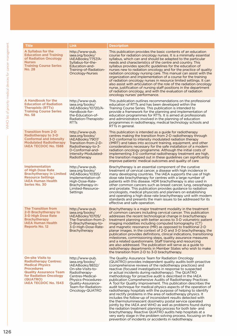
126
WHO
list o
f prio
rity m
edica
l dev
ices f
or ca
ncer
man
agem
ent
Title Link Description
A Syllabus for the Education and Training of Radiation Oncology NursesTraining Course Series No. 28
http://www-pub.iaea.org/books/IAEABooks/7757/A-Syllabus-for-the-Education-and-Training-of-Radiation-Oncology-Nurses
This publication provides the basic contents of an education course for radiation oncology nurses. It is a minimally essential syllabus, which can and should be adapted to the particular needs and characteristics of the centre and country. This syllabus provides specific guidelines for the education of nurses new to radiation oncology and for the practice of quality radiation oncology nursing care. This manual can assist with the organization and implementation of a course for the training of radiation oncology nurses in resource limited settings. It can also assist with articulation of the role of the radiation oncology nurse, justification of nursing staff positions in the department of radiation oncology, and with the evaluation of radiation oncology nurses’ performance.
A Handbook for the Education of Radiation Therapists (RTTs)Training Course Series No. 58
http://www-pub.iaea.org/books/IAEABooks/10720/A-Handbook-for-the-Education-of-Radiation-Therapists-RTTs
This publication outlines recommendations on the professional education of RTTs and has been developed within the Training Course Series. This publication is intended to provide a framework for the planning and implementation of education programmes for RTTs. It is aimed at professionals and administrators involved in the planning of education programmes in radiotherapy, medical technology schools and RTTs in general.
Transition from 2-D Radiotherapy to 3-D Conformal and Intensity Modulated RadiotherapyIAEA TECDOC No. 1588
http://www-pub.iaea.org/books/IAEABooks/7907/Transition-from-2-D-Radiotherapy-to-3-D-Conformal-and-Intensity-Modulated-Radiotherapy
This publication is intended as a guide for radiotherapy centres making the transition from 2-D radiotherapy through 3-D conformal to intensity modulated radiation therapy (IMRT) and takes into account training, equipment, and other considerations necessary for the safe installation of a modern radiation oncology programme. Although the initial costs of implementing 3-D conformal radiotherapy treatment are high, the transition mapped out in these guidelines can significantly improve patients’ medical outcomes and quality of care
Implementation of High Dose Rate Brachytherapy in Limited Resource SettingsIAEA Human Health Series No. 30
http://www-pub.iaea.org/books/IAEABooks/10355/Implementation-of-High-Dose-Rate-Brachytherapy-in-Limited-Resource-Settings
Brachytherapy is an essential component of the curative treatment of cervical cancer, a disease with high incidence in many developing countries. The IAEA supports the use of high dose rate brachytherapy for centres with a large number of patients with this disease. HDR brachytherapy is also used in other common cancers such as breast cancer, lung, oesophagus and prostate. This publication provides guidance to radiation oncologists, medical physicists and planners on establishing and operating a high dose rate brachytherapy unit with modern standards and presents the main issues to be addressed for its effective and safe operation.
The Transition from 2-D Brachytherapy to 3-D High Dose Rate BrachytherapyIAEA Human Health Reports No. 12
http://www-pub.iaea.org/books/IAEABooks/10705/The-Transition-from-2-D-Brachytherapy-to-3-D-High-Dose-Rate-Brachytherapy
Brachytherapy is a major treatment modality in the treatment of common cancers including cervical cancer. This publication addresses the recent technological change in brachytherapy treatment planning with better access to 3-D volumetric patient imaging modalities including computed tomography (CT) and magnetic resonance (MR) as opposed to traditional 2-D planar images. In the context of 2-D and 3-D brachytherapy, the publication provides definitions, clinical indications, transitioning milestones, commissioning steps, quality assurance measures and a related questionnaire. Staff training and resourcing are also addressed. The publication will serve as a guide to radiotherapy departments in Member States who wish to make the transition from 2-D to 3-D brachytherapy.
On-site Visits to Radiotherapy Centres: Medical Physics ProceduresQuality Assurance Team for Radiation Oncology (QUATRO)IAEA TECDOC No. 1543
http://www-pub.iaea.org/books/IAEABooks/8832/On-site-Visits-to-Radiotherapy-Centres-Medical-Physics-Procedures-Quality-Assurance-Team-for-Radiation-Oncology-QUATRO
The Quality Assurance Team for Radiation Oncology (QUATRO) provides independent quality audits both proactive (comprehensive reviews of the radiotherapy practices) and reactive (focused investigations in response to suspected or actual incidents during radiotherapy). The QUATRO methodology for proactive audit is described in the IAEA publication Comprehensive Audits of Radiotherapy Practices: A Tool for Quality Improvement. This publication describes the audit technique for medical physics aspects of the operation of radiotherapy hospitals with the purpose of helping to identify and rectify problems in the area of radiotherapy physics. It includes the follow-up of inconsistent results detected with the thermoluminescent dosimetry postal service operated jointly by the IAEA and WHO as well as problems found during the radiation treatment planning process for both tele- and brachytherapy. Reactive QUATRO audits help hospitals at a very early stage in the problem solving process, focusing on the prevention of incidents or accidents in radiotherapy.

127
5. R
ad
ioth
era
py
Title Link Description
Absorbed Dose Determination in External Beam RadiotherapyIAEA Technical Report Series No. 398
http://www-pub.iaea.org/books/IAEABooks/5954/Absorbed-Dose-Determination-in-External-Beam-Radiotherapy
This Code of Practice, which has also been endorsed by WHO, PAHO and ESTRO, fulfils the need for a systematic and internationally unified approach to the calibration of ionization chambers in terms of absorbed dose to water and to the use of these detectors in determining the absorbed dose to water for the radiation beams used in radiotherapy. It provides a methodology for the determination of absorbed dose to water in the low, medium and high energy photon beams, proton beams and heavy ion beams used for external radiation therapy.
“Safety is no accident” https://www.astro.org/uploadedFiles/Main_Site/Clinical_Practice/Patient_Safety/Blue_Book/SafetyisnoAccident.pdf
The document provides guidance on the requirements and processes needed to run a modern radiotherapy department.
European Higher Education Area Level 6 Benchmarking Document for Radiation TherapisTs (RTTs)
http://www.estro.org/binaries/content/assets/estro/about/rtt/rtt-benchmarking.pdf
The ESTRO, through the Radiation TherapisT (RTT) Committee has sought, over a twenty - five year period, to address the educational and professional issues of the group of healthcare professionals responsible for the delivery of the radiotherapy prescription accurately and safely. This document defines the competences that RTTs should have on graduation from their basic education programme.
Roles and Responsibilities, and Education and Training Requirements for Clinically Qualified Medical Physicists
http://www-pub.iaea.org/books/iaeabooks/10690/Roles-and-Responsibilities-and-Education-and-Training-Requirements-for-Clinically-Qualified-Medical-Physicists
This publication addresses the shortfall of well trained and clinically qualified medical physicists working in radiation medicine. The roles, responsibilities and clinical training requirements of medical physicists have not always been well defined or well understood by health care professionals, health authorities and regulatory agencies. To fill this gap, this publication provides recommendations for the academic education and clinical training of clinically qualified medical physicists, including recommendations for their accreditation certification and registration, along with continuous professional development. The goal is to establish criteria that support the harmonization of education and clinical training worldwide.
Bibliography:1) Expanding global access to radiotherapy. Atun, Rifat et al. The Lancet Oncology , Volume 16 , Issue 10 ,
1153 - 1186
2) BSS, 2014 - Radiation protection and safety of radiation sources: International Basic Safety Standards. -
General safety requirements Part 3 Series No. GSR Part 3. Vienna
3) IAEA SRS No. 47. Radiation Protection in the Design of Radiotherapy Facilities. Available at http://www-
pub.iaea.org/books/IAEABooks/7197/Radiation-Protection-in-the-Design-of-Radiotherapy-Facilities
4) Radiotherapy Facilities: Master Planning and Concept Design Considerations. IAEA Human Health
Reports No. 10. Available at: http://www-pub.iaea.org/books/IAEABooks/10561/Radiotherapy-Facilities-
Master-Planning-and-Concept-Design-Considerations
5) Comprehensive Audits of Radiotherapy Practices: A Tool for Quality Improvement. Available at:
http://www-pub.iaea.org/books/IAEABooks/7680/Comprehensive-Audits-of-Radiotherapy-Practices-A-
Tool-for-Quality-Improvement-Quality-Assurance-Team-for-Radiation-Oncology-QUATRO
6) Practical Guidelines for the Implementation of A Quality System in Radiotherapy. Available at: http://
www.estro.org/binaries/content/assets/estro/school/publications/booklet-4---practical-guidelines-for-
the-implementation-of-a-quality-system-in-radiotherapy.pdf
7) Setting Up a Radiotherapy Programme: Clinical, Medical Physics, Radiation Protection and Safety
Aspects. Available at: http://www-pub.iaea.org/books/IAEABooks/7694/Setting-Up-a-Radiotherapy-
Programme
8) NCRP Report No. 151 - Structural Shielding Design and Evaluation for Megavoltage X- and Gamma-Ray
Radiotherapy Facilities (2005); http://www.ncrppublications.org/Reports/151.

6. Systemic therapy

129
6. S
yste
mic
thera
py
Health service delivery sequence overview:
This diagram expresses the flow of cancer patients to and from the systemic therapy unit, and the elements
to consider as support for the unit.
Fig. 10.6. Health care service delivery overview – systemic therapy
PR
IMA
RY
(C
L,H
C),
SE
C-
ON
DA
RY
(D
H)
&
TE
RT
IAR
Y (
SH
)
Le
ve
l o
f C
are
*
* Appropriate level of care will depend on the particular intervention, setting, and available infrastructure and human resources.
SE
CO
ND
AR
Y (
DH
)
& T
ER
TIA
RY
(S
H)
TE
RT
IAR
Y (
SH
)
Prevention End of life care
TreatmentFollow-up/survivorship
care
Palliative care
Diagnosis
Screening
CL Community Level health post DH District Hospital HC Health Centre SH Specialized Hospital
Systemic therapy
Nuclear medicine
Surgery
Vaccination
Radio- therapy
Pathology Pathology
Clinical laboratory
Clinical laboratory
Clinical assessment
Palliative care
Medical imaging
Endoscopy
Clinical assessment
Nuclear medicine
6.1 General description of the unit
6.2 Priority medical devices
6.2.1 General medical devices for systemic therapy
6.2.2 Specific medical devices for systemic therapy by cancer type
6.3 Other health system components
6.3.1 Human resources for systemic theraphy
6.3.2 Infrastructure
6.3.3 Quality management
6.3.4 Guidance documents

130
WHO
list o
f prio
rity m
edica
l dev
ices f
or ca
ncer
man
agem
ent
6.1 General description of the unit
Chemotherapy units (or oncology units) are required to administer systemic therapies to patients with cancer,
both in adjuvant and metastatic settings. The usual route of chemotherapy administration is intravenous
(bolus or continuous infusion); other routes of infusion include intramuscular or subcutaneous injection.
Although less common, intraperitoneal, intrathecal, intra-arterial may also be used. Oral formulations are
generally used for outpatients.
The side effects of anticancer drugs are well known. Patient safety and occupational risks to health care
workers who prepare, handle or dispose of cytotoxic agents must be considered in the design and operation
of medical oncology units. Internal distribution and disposal of waste must also be planned for carefully.
Activities within the unit include:
• Managing the scheduling of patient appointments;
• Medication administration;
• Documenting the patients’ demographic information; and
• Documenting all treatments administered, including date, time, treatments, drugs, doses, and patient
response.
Additionally, an assessment of how the patient tolerated the prior therapy should be documented in case there
is a need to reduce the dose of chemotherapy due to unacceptable toxicity. Some chemotherapy agents may
cause an immediate reaction; all units should have an emergency box so response to the immediate reaction
can be mitigated by a rapid response.
6.2 Priority medical devices
The following tables contain general medical devices, which can be used for preparation and treatment/
infusion of chemotherapy of several cancers, and specific medical devices, which are used for interventions
for specific cancer types. The use of central venous catheters versus peripheral is discussed at the end of
the list. It is important to include, if applicable, the medical devices for paediatric patients as recommended
below. If the chemotherapy is delivered in an outpatient unit, purchasing an ambulance for transportation of
patients in case of an emergency is desirable.
The main intervention addressed in this chapter is the systemic therapy infusion of the different specialized
treatments, for all cancer types. Other interventions specific to Leukaemia are also addressed, as listed below:
Cancer type Interventions
All cancers • Drug infusion, chemotherapy administration• Hazardous drug preparation• Intrathecal chemotherapy

131
6. S
yste
mic
thera
py
6.2.1 General medical devices for systemic therapy
Procedure Medical devices category
Capital equipment (for specialized hospitals)
Accessories/hardware/software/consumables/single use devices
Drug Infusion, chemotherapy administration
Medical equipment Fixed examination/treatment light
Pharmacy refrigerator
Stethoscope, adult, binaural and paediatric
Thermometer, clinical, digital 32–43°C
Tympanic probe covers (if tympanic thermometers are used)
Resuscitation trolley, equipped with medicines and defibrillator
With laryngoscope (for adult and paediatric patients)
Sphygmomanometer (include paediatric size tubes if applicable)
Infant/paediatric blood pressure cuffs
Basic vital signs monitor (availability in the setting)
Vein finder device (optional)Monitoring electrodes
General physical examination set Ophthalmoscope, Otoscope, Lamp)
Adult stand up scale
Stadiometer (wall mounted)
Oxygen therapy flowmeter, dial-type (if pipeline available)
Suction availability (accessories for wall or portable equipment)
Infusion pump (optional) Infusion pump administration set
Elastomeric pump (optional)
Tympanic thermometer
Infant scale
Medical furniture Reclining chair
Patient lifting hoist
Hospital stretcher with side rails
General cabinet
Stand, infusion, double hook, on casters
Table, instruments, Mayo, stainless steel, on castors
Trolley, dressing, stainless steel, 2 trays
Trolley, soiled linen
Wheelchair, adult/child
Cabinet, medicine, with lock (consider national regulations)
Cribs
Bedside tables/commodes
Instruments Dressing set
Personal protective equipment and clothing
Glasses, safety, regular size
Gloves, nitrile non-sterile, single-use
Gloves, non-sterile, single-use
General purpose sterile drape
Surgical face mask
Gown, impermeable single use
Apron impermeable
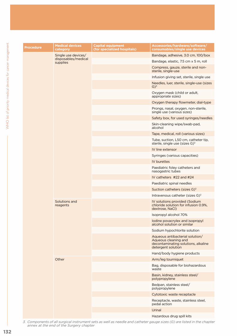
132
WHO
list o
f prio
rity m
edica
l dev
ices f
or ca
ncer
man
agem
ent
Procedure Medical devices category
Capital equipment (for specialized hospitals)
Accessories/hardware/software/consumables/single use devices
Single use devices/disposables/medical supplies
Bandage, adhesive, 3.0 cm, 100/box
Bandage, elastic, 7.5 cm x 5 m, roll
Compress, gauze, sterile and non-sterile, single-use
Infusion giving set, sterile, single use
Needles, luer, sterile, single-use (sizes G)3
Oxygen mask (child or adult, appropriate sizes)
Oxygen therapy flowmeter, dial-type
Prongs, nasal, oxygen, non-sterile, single use (various sizes)
Safety box, for used syringes/needles
Skin-cleaning wipe/swab-pad, alcohol
Tape, medical, roll (various sizes)
Tube, suction, L50 cm, catheter tip, sterile, single use (sizes G)3
IV line extensor
Syringes (various capacities)
IV burettes
Paediatric foley catheters and nasogastric tubes
IV catheters #22 and #24
Paediatric spinal needles
Suction catheters (sizes G)3
Intravenous catheter (sizes G)3
Solutions and reagents
IV solutions provided (Sodium chloride solution for infusion 0.9%, dextrose, NaCl)
Isopropyl alcohol 70%
Iodine povacrylex and isopropyl alcohol solution or similar
Sodium hypochlorite solution
Aqueous antibacterial solution/Aqueous cleaning and decontaminating solutions, alkaline detergent solution
Hand/body hygiene products
Other Arm/leg tourniquet
Bag, disposable for biohazardous waste
Basin, kidney, stainless steel/polypropylene
Bedpan, stainless steel/polypropylene
Cytotoxic waste receptacle
Receptacle, waste, stainless steel, pedal action
Urinal
Hazardous drug spill kits
3. Components of all surgical instrument sets as well as needle and catheter gauge sizes (G) are listed in the chapter annex at the end of the Surgery chapter

133
6. S
yste
mic
thera
py
Procedure Medical devices category
Capital equipment (for specialized hospitals)
Accessories/hardware/software/consumables/single use devices
Hazardous drug preparation (pharmacy, special isolated room inside or outside the facility to provide the chemotherapy)
Medical equipment Class-II biological safety cabinet (the cabinet is needed in cases when the reconstitution of chemo is done in the unit)
Respirators (If reconstitution is done in the unit)
Furniture Adequate furniture to prepare the hazardous drugs including chairs, tables, cabinets
Personal protective equipment and clothing
Coat, medical, woven (various sizes)
Glasses, safety, regular size
Gloves, nitrile non-sterile, single-use
Gloves, non-sterile, single-use
Non-conductive shoe cover
Protective head cover
General-purpose sterile drape
Surgical face mask
Gown, impermeable single-use
Single use devices/disposables/medical supplies
Bag, disposable for biohazardous waste
Filter Discs (if available)
Needles, luer, sterile, single-use (sizes G)3
Scalp vein intravenous administration set
Secondary set with drip chamber
Closed system drug transfer devices (if available)
Filter needles (if available)
Filter Venting devices (if available)
Syringes, luer lock, sterile, single-use (various capacities)
IV fluid and blood giving sets
Solutions and reagents
Isopropyl alcohol 70%
Other Compress, gauze, sterile and non-sterile, single use
Labels for the drugs identification
Intrathecal chemotherapy
Lumbar puncture set
Single use devices/disposables/medical supplies
Needles, spinal, sterile, single-use (sizes)3
Sterile culture tube
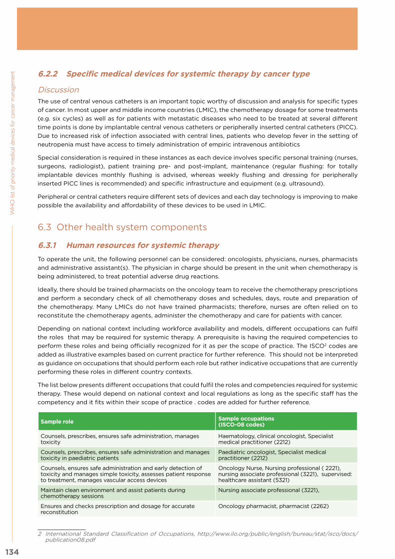
134
WHO
list o
f prio
rity m
edica
l dev
ices f
or ca
ncer
man
agem
ent 6.2.2 Specific medical devices for systemic therapy by cancer type
DiscussionThe use of central venous catheters is an important topic worthy of discussion and analysis for specific types
of cancer. In most upper and middle income countries (LMIC), the chemotherapy dosage for some treatments
(e.g. six cycles) as well as for patients with metastatic diseases who need to be treated at several different
time points is done by implantable central venous catheters or peripherally inserted central catheters (PICC).
Due to increased risk of infection associated with central lines, patients who develop fever in the setting of
neutropenia must have access to timely administration of empiric intravenous antibiotics
Special consideration is required in these instances as each device involves specific personal training (nurses,
surgeons, radiologist), patient training pre- and post-implant, maintenance (regular flushing: for totally
implantable devices monthly flushing is advised, whereas weekly flushing and dressing for peripherally
inserted PICC lines is recommended) and specific infrastructure and equipment (e.g. ultrasound).
Peripheral or central catheters require different sets of devices and each day technology is improving to make
possible the availability and affordability of these devices to be used in LMIC.
6.3 Other health system components
6.3.1 Human resources for systemic therapy
To operate the unit, the following personnel can be considered: oncologists, physicians, nurses, pharmacists
and administrative assistant(s). The physician in charge should be present in the unit when chemotherapy is
being administered, to treat potential adverse drug reactions.
Ideally, there should be trained pharmacists on the oncology team to receive the chemotherapy prescriptions
and perform a secondary check of all chemotherapy doses and schedules, days, route and preparation of
the chemotherapy. Many LMICs do not have trained pharmacists; therefore, nurses are often relied on to
reconstitute the chemotherapy agents, administer the chemotherapy and care for patients with cancer.
Depending on national context including workforce availability and models, different occupations can fulfil
the roles that may be required for systemic therapy. A prerequisite is having the required competencies to
perform these roles and being officially recognized for it as per the scope of practice. The ISCO2 codes are
added as illustrative examples based on current practice for further reference. This should not be interpreted
as guidance on occupations that should perform each role but rather indicative occupations that are currently
performing these roles in different country contexts.
The list below presents different occupations that could fulfil the roles and competencies required for systemic
therapy. These would depend on national context and local regulations as long as the specific staff has the
competency and it fits within their scope of practice . codes are added for further reference.
Sample role Sample occupations (ISCO-08 codes)
Counsels, prescribes, ensures safe administration, manages toxicity
Haematology, clinical oncologist, Specialist medical practitioner (2212)
Counsels, prescribes, ensures safe administration and manages toxicity in paediatric patients
Paediatric oncologist, Specialist medical practitioner (2212)
Counsels, ensures safe administration and early detection of toxicity and manages simple toxicity, assesses patient response to treatment, manages vascular access devices
Oncology Nurse, Nursing professional ( 2221),nursing associate professional (3221), supervised: healthcare assistant (5321)
Maintain clean environment and assist patients during chemotherapy sessions
Nursing associate professional (3221),
Ensures and checks prescription and dosage for accurate reconstitution
Oncology pharmacist, pharmacist (2262)
2 International Standard Classification of Occupations, http://www.ilo.org/public/english/bureau/stat/isco/docs/publication08.pdf

135
6. S
yste
mic
thera
py
Sample role Sample occupations (ISCO-08 codes)
Reconstitutes chemotherapy drugs for administration under supervision of pharmacist
Pharmaceutical technician or assistant (3213)
Management of medical devices: planning, procurement,Supervision or performance of: installation, users training, maintenance and decommissioning.
Biomedical Engineer (2149)Biomedical technician / physical and engineering science technician (3119) under supervision of biomedical engineer (2149)
Scheduling, reception of patients, filing patient data, and all other administrative tasks
Clerical support workers ( 4412)Technologists(3211),Nurses (2221)
Cleaners need specific training for cleaning systemic therapy area and pharmacy where chemotherapies are prepared.
Cleaning personnel (9112)
An ideal unit will have all the personnel listed above to improve the efficiency and safety of chemotherapy
administration.
The minimum requirement for a chemotherapy unit is to have a combination that includes:
1. A consulting oncologist (may be non-residential, but must be reachable in all cases)
2. Physician with experience in prescribing chemotherapy, ensuring safety, monitoring and managing toxicity
3. Pharmacist or registered nurse with experience in reconstitution of chemotherapy
4. A registered nurse with proper training and experience in administering chemotherapy.
6.3.2 Infrastructure
A patient care area where chemotherapy is administered must have a patient waiting area; a treatment room
with comfortable reclining chairs and television set (if possible); a clinical area equipped with a refrigerator;
clinical supplies for mixing drugs (syringes, needles, etc.); a containment cabinet or hood for mixing
chemotherapy; designated containers and area for disposal of cytotoxic waste and pharmacy for the storage
of drugs. According to the level of complexity of the unit it may have a unit for storage of the medication, a
central pharmacy in the hospital, or optional drug preparations provided by external suppliers.
The facility could be integrated with an oncology or general hospital or it could be a specialized outpatient unit.
Layout distributionBasic requirements for a facility would include:
1. A pharmacy
2. Consulting room for patient evaluation
3. Access to laboratory diagnostic tests such as haematology test and clinical chemistry
4. Chemotherapy mixing room with vents/extractors and ability to maintain aseptic conditions (controlled area)
5. Chemotherapy suite for administration or a ward
6. Nurse station
7. Toilet facilities
In the ward there should be sufficiently clear dimension between beds and lounge chairs used for chemotherapy
treatment/infusion and during administration. Access to the area should be limited to patients receiving
therapy and essential personnel.
Electrical systemsIf infusion pumps are used to infuse chemotherapy, stable power supply and back-up systems should be
installed. Emergency and standby power systems should be considered.

136
WHO
list o
f prio
rity m
edica
l dev
ices f
or ca
ncer
man
agem
ent
Heating, ventilation and air conditioning systemsThe mixing room inside the pharmacy or the ward should maintain aseptic conditions but a contained
environment where air pressure is negative to the surrounding areas or that is protected by an airlock or
anteroom, is preferred (1). Ideally, a biological safety cabinet should be available.
Ventilation and air conditioning systems are important particularly with patients with an extreme susceptibility
of infection.
Communication systemsCommunication systems linking the pharmacy and the oncology unit should be installed if they are located a
considerable distance apart.
Special systemsThe availability of an oxygen supply for example via an oxygen concentrator, oxygen cylinder or pipeline, is
highly recommended.
Cleaning and decontamination of hazardous chemotherapy drug equipment and work surfacesDecontamination may be defined as cleaning or deactivating. Deactivating a hazardous substance is
preferred, but no single process has been found to deactivate all currently available hazardous drugs (HDs).
The use of alcohol for disinfecting a primary engineering control or other contaminated surface will not
deactivate any hazardous chemotherapy drugs and may result in the spread of contamination rather than in
any actual cleaning. Many recommend sodium hypochlorite solution as an appropriate deactivating agent.
Research has shown that strong oxidizing agents, such as sodium hypochlorite, are effective deactivators of
many hazardous chemotherapy drugs. A 2% sodium hypochlorite solution with detergent may be wiped onto
contaminated surfaces, and then rinsed; this is followed by a neutralizing solution of 1% sodium thiosulfate,
wiped on and off, followed by a rinse solution of water, then alcohol. Surface contact for each solution should
be at least 30 seconds. Some studies have shown good analytical results with this technique. The hazardous
chemotherapy drugs may not be fully deactivated, but the wiping action followed by rinsing appears to be
effective in cleaning. This technique may be used on any surface that will not be harmed by a bleach solution.
All cleaning solutions, wipers, and rinsates must be contained and discarded as hazardous (2).
Extravasation managementIn terms of cancer therapy, extravasation refers to the inadvertent infiltration of chemotherapy into the
subcutaneous or subdermal tissues surrounding the intravenous or intra-arterial administration site. The risk
is lower in experienced institutions with well-trained personnel. In general hospitals or when non-trained
nurses or physicians apply infusions, the risk is much higher. Most extravasations can be prevented with the
systematic implementation of careful, standardized, evidence-based administration techniques. In order to
minimize the risk of extravasation, the staff involved in the infusion and management of cytotoxic drugs must
be trained to implement several preventive protocols. Regardless of the chemotherapy drug, early initiation
of treatment is considered mandatory. In this context, patient education is crucial for a prompt identification
of the extravasation.
An extravasation kit containing instructions, materials and medication to handle any incidence should be
available. An early multidisciplinary evaluation by nurses, physicians and surgeons is recommended.
Various suggestions have been published with possible topical or injected pharmacologic methods for certain
vesicant chemotherapy drugs. One should be aware that many are considered ineffective or further damage
the extravasated area. It should be noted that many of these substances are not available or at best have
limited access for use in many countries.
Local injection or topical corticosteroids are not recommended due to the increased need for surgical
debridement. Topical dimethyl sulfoxide (DMSO) is a treatment option in extravasations occurred during
anthracyclines, mitomycin C or platin salts infusions. The application must be repeated every 8 hours for 1
week and the first administration of DMSO performed in the first 10 minutes after extravasation.

137
6. S
yste
mic
thera
py
Dexrazoxane prevented severe tissue damage following anthracycline extravasation, but is not available in
many countries. Hyaluronidase might be efficacious in preventing skin necrosis by extravasation due to vinca
alkaloids (3).
OthersThe design of this area must include surfaces that are compatible with cleaning and decontaminating agents.
Upholstered and carpeted surfaces should be avoided.
6.3.3 Quality management
Patient safety
There are several published guidelines ensuring safety for patients undergoing chemotherapy. All patients
should be fully informed about their condition, indication for use of specific drugs, expected side effects and
outcomes as well as treatment options. These should be captured in a signed consent form. Record keeping
is of high priority to cross check prescriptions, make sure patients have been evaluated for toxicity, review
laboratory parameters and document any dose adjustments prior to the next cycle. The labelling of drugs,
cross checking of labels and use of appropriate infusion mixture by several other health care workers are
important to avoid misadministration such as over dosage. A secondary check system should be put in place
and should include a double check of the order to assure right cycle, day, drug, height, weight, body surface
area (BSA), dose, route and patient. Additional checks should be performed during the reconstitution and
mixing of the drug and upon administration, to assure the right patient is receiving the right drugs and dose,
on the right day and time. Accidental exposure can occur in patients as well during administrations. The
best patient safety outcomes are achieved with adequate and quality staffing as well as adherence to safety
recommendations.
Oncology nurses need specialized training to assure the proper administration techniques for drugs that are
called vesicants. If not administered properly accidental extravasation can happen causing severe harm, if
these drugs infiltrate into the patient subcutaneous tissue. This can be a devastating occurrence, highlighting
the importance of training and skill development.
Public safety
Chemotherapy drugs are prescribed by physicians only for use by a specific patient. The general public
should not be exposed to these drugs during or following administration. Exposure to bodily fluids of a
patient immediately or a few days following administration may have adverse consequences. Patients need
to be educated about the effect of their medication on others close to them. Topics such as the use of
washroom facilities, vomitus, sweat and sperm or vaginal fluids should be discussed to ensure appropriate
measures are in place to prevent contamination. Most importantly, the safety of pregnant women and young
children is paramount.
If a patient receiving chemotherapy requires care, care givers, including health care professionals, should wear
gloves and wash their hands with soap and water before donning and after removing gloves. All reusable and
disposable containers for bodily fluids should be washed several times with detergent prior to disposal into
designated plastic bags. Used needles and blades should be placed into puncture proof, labelled containers.
All other tubing and containers containing medication should be placed in hazardous waste labelled bins to
be disposed of appropriately.
These recommendations apply to oral chemotherapy as it applies to the home environment.
Health care workers
Pharmacists, pharmacy technicians and oncology nurses are at risk of direct contact with chemotherapy
agents during preparation and administration. Use of impermeable gloves, gowns, goggles and shoes should
be mandatory without any exceptions. Most commonly, skin and eye contamination can occur as well as
needle stick injuries. In case of external exposure, immediate washing with soap and water several times
is recommended and the incident should then be reported to a physician. Institutions should enforce that
only highly skilled and qualified staff operate the chemotherapy administration suite. Continuous education
programmes on the safe preparation and administration and awareness and management of exposure should

138
WHO
list o
f prio
rity m
edica
l dev
ices f
or ca
ncer
man
agem
ent be mandatory. A log should be kept of the incidences of accidental exposures in order to improve procedures
to reduce such incidents.
Competencies relating to administering cytotoxic and biotherapy agents to paediatric oncology patients
The advent and use of chemotherapy tremendously impacted the survival of children with cancer. In the
past 60 years, the survival rates of children with cancer have dramatically improved with 5-year survival
rates of the most common types of childhood cancer nearing 90% in high income countries. In low and mid
income countries, however, survival rates continue to be much lower. Use of chemotherapeutic agents is
not without risk as they are administered in a variety of routes and doses. It is important for nurses to have
the knowledge needed to safely administer antineoplastic agents to all patients, but in particular paediatric
oncology patients (4-7).
Competencies: To administer cytotoxic and biotherapy agents to oncology paediatric patients, the nurse must:
• Possess knowledge of safe handling guidelines for cytotoxic agents and waste products;
• Familiarize self with actions, administration guidelines, and side effects of cytotoxic medications as
outlined in pharmacy manual and other resources;
• Be able to provide patient/family teaching in relation to side effects and management of side effects;
• Be knowledgeable about paediatric cancer diagnosis and treatment, haematopoiesis and immune
response, treatment modalities, and psychosocial issues in paediatric oncology, attained through
education and experience;
• Be able to teach patients/families about their diagnosis, treatment and the cancer experience; and
• List and describe chemotherapy and biotherapy agents and classifications and their mode of action,
administration considerations, toxicity and symptom management, and late effects.
Related competencies include:
• Care of central venous catheters and venous access devices; and
• Administration of intravenous, intramuscular, subcutaneous and oral medications to paediatric patients.
6.3.4 Guidance documents
Title Link Description
Central venous access in oncology: ESMO Clinical Practice Guidelines
http://www.esmo.org/Guidelines/Supportive-Care/Central-Venous-Access-in-Oncology
Central venous access plays a critical role in the management and care of cancer patients. These new ESMO Clinical Practice Guidelines apply to central venous access in adult cancer patients and cover the use of peripherally inserted central catheters, tunnelled central catheters and totally implantable devices. The guidelines cover diagnosis and treatment of infections as well as complications requiring the removal of central venous access devices.
ASCO-ONS Standards for Safe Chemotherapy Administration
http://www.instituteforquality.org/asco-ons-standards-safe-chemotherapy-administration
The ASCO/ONS chemotherapy safety standards are intended to reduce the risk of errors when providing adult patients with chemotherapy, and to provide a framework for best practices in cancer care. Specifically, they can inform practice policies and procedures, internal quality assessment, and external quality monitoring.
139
6. S
yste
mic
thera
py
Bibliography:
1) https://www.ashp.org/DocLibrary/BestPractices/PrepGdlHazDrugs.aspx
2) Safe Handling Of Hazardous Chemotherapy Drugs In Limited-Resource Settings, PAHO; Touzin et al.
2010; Zock, Soefje, and Rickabaugh 2011
3) J. A. Pérez Fidalgo et al; Management of chemotherapy extravasation: ESMO–EONS Clinical Practice
Guidelines-Annals of Oncology 23 (Supplement 7): vii167–vii173, 2012
4) http://www.bcchildrens.ca/Oncology-Site/Documents/
ChemotherapyAdministrationCompetenciesforCommunity.pdf
5) Bertram, Leslee A., “Pediatric Oncology Nurses’ Knowledge of Safe Administration of Chemotherapy/
Biotherapy” (2015). DNP Practice Inquiry Projects. Paper 60.
6) http://www.asco.org/international-programs/multidisciplinary-cancer-management-courses
7) http://www.asco.org/international-programs/global-curriculum
7. Palliative care & end of life care

141
7. Pallia
tive c
are
& e
nd
of life
care
Health service delivery sequence overview:
This diagram expresses the flow of cancer patient to and from the palliative care and end of life care unit, and
the elements to consider as support for the unit.
Fig. 10.6. Health care service delivery overview – Palliative care and end of life care
PR
IMA
RY
(C
L,H
C),
SE
C-
ON
DA
RY
(D
H)
&
TE
RT
IAR
Y (
SH
)
Le
ve
l o
f C
are
*
* Appropriate level of care will depend on the particular intervention, setting, and available infrastructure and human resources.
SE
CO
ND
AR
Y (
DH
)
& T
ER
TIA
RY
(S
H)
TE
RT
IAR
Y (
SH
)
Prevention End of life care
TreatmentFollow-up/survivorship
care
Palliative care
Diagnosis
Screening
CL Community Level health post DH District Hospital HC Health Centre SH Specialized Hospital
Systemic therapy
Nuclear medicine
Surgery
Vaccination
Radio- therapy
Pathology Pathology
Clinical laboratory
Clinical laboratory
Clinical assessment
Palliative care
Medical imaging
Endoscopy
Clinical assessment
Nuclear medicine
7.1 General description of the unit
7.1.1 Definition
7.1.2 Purposes
7.1.3 Palliative treatment
7.2. Priority medical devices
7.2.1 General medical devices for palliative care and end of life care
7.2.2 Specific medical devices for palliative care and end of life care by cancer type
7.3 Other health system components
7.3.1 Human resources for palliative care and end of life care
7.3.2 Infrastructure
7.3.3 Quality management

142
WHO
list o
f prio
rity m
edica
l dev
ices f
or ca
ncer
man
agem
ent
7.1 General description of the unit
7.1.1 Definition
“Palliative care is an approach that improves the quality of life of patients and their families facing the problems
associated with life-threatening illness, through the prevention and relief of suffering by means of early
identification and impeccable assessment and treatment of pain and other problems, physical, psychosocial
and spiritual (1).” (Definition of Palliative Care adopted by WHO)
7.1.2 Purposes
• Provides relief from pain and other distressing symptoms
• Affirms life and regards dying as a normal process
• Intends neither to hasten nor postpone death
• Integrates the psychological and spiritual aspects of patient care
• Offers a support system to help patients live as actively as possible until death
• Offers a support system to help the family cope during the patient’s illness and in their own
bereavement
• Uses a team approach to address the needs of patients and their families, including bereavement
counselling, if indicated
• Will enhance quality of life, and may also positively influence the course of illness
• Is applicable early in the course of illness, in conjunction with other therapies that are intended to
prolong life, such as chemotherapy or radiation therapy, and includes those investigations needed to
better understand and manage distressing clinical complications.
In some countries there is recognition of the difference between general palliative care and specialist palliative
care; for example, the United Kingdom (2) differentiates between:
• General palliative care which “is provided by the usual professional carers of the patient and family with
low to moderate complexity of palliative care need”, and
• Specialist palliative care services which ”are provided for patients and their families with moderate to
high complexity of palliative care need. They are defined in terms of their core service components, their
functions and the composition of the multi-professional teams that are required to deliver them.”
Terminology in palliative care is somewhat controversial and varies across and even within countries.
Recommended definitions of common terms used in palliative care have been offered by the European
Association for Palliative Care (3,4) and other organizations. General palliative care can and should be delivered
throughout any cancer hospital or clinic, wherever a patient is. Within a hospital, in-patient specialist palliative
care units (with dedicated beds) can be provided. Alternatively, a multidisciplinary specialist palliative care
team may provide consultations throughout the hospital to patients who remain in their wards/rooms under the
primary care of their treating physician. Hospice may mean a separate service involving a range of components
including: an in-patient unit, home care, day care, out-patient clinics and bereavement support service.
7.1.3 Palliative treatment
There is a wealth of clinical literature to support the role of radiotherapy, brachytherapy, and surgery in the
palliation of cancer to relieve symptoms including pain, bleeding, and obstruction and to improve quality of
life for cancer patients (5-8). Please refer to the Radiotherapy and Surgery subchapters for interventions and
corresponding medical devices. There are also many pharmacological and non-pharmacological interventions
for symptom management and psychosocial problem management
7.2. Priority medical devices
The following tables contain general medical devices, which can be used for the general palliative care needs
of many diseases including cancers, and specific medical devices, which are used for palliative care needs of
specific cancer types.

143
7. Pallia
tive c
are
& e
nd
of life
care
7.2.1 General medical devices for palliative care and end of life care
Procedure Medical devices category Capital equipment Accessories/hardware/software/
consumables /single use devices
Palliative care delivery
Medical equipment Blood glucometer (used after chemotherapy), with accessories
Micro cuvettes or strips according to manufacturer.
Fixed examination/treatment light
Nebulizer, with accessories
Pulse oximeter, portable, with accessories
Sensors
Refrigerator
Fridge freezer
Stethoscope, adult and paediatric, binaural
Thermometer, clinical, digital 32–43°C
Sphygmomanometer Blood pressure cuffs for adults and paediatrics
General physical examination set Include Ophthalmoscope, Otoscope, Lamp)
Oxygen therapy flowmeter, dial-type (if pipeline available)
Oxygen supply (cilinder, oxygen concentrator or pipeline)
Suction availability (accessories for wall or portable equipment)
Electrocardiograph (optional)
Syringe pump or elastomeric pumps, battery-operated (optional)
Infusion pump for enteral nutrition (optional)
Hammer for neurological examination
Tape measurement
Non-heated respiratory humidifier
Medical furniture Hospital stretcher, with side rails
General cabinet
Stand, infusion, double hook, on casters
Trolley, dressing, stainless steel, 2 trays
Lifting device for patient
Wheelchair, adult/child
Cabinet, medicine, with lock (consider national regulations)
Walking frame/mobility walking aids
Table, instruments, Mayo type, stainless steel, on castors
Instruments Forceps, dressing, Cheron, 250 mm
Surgical instruments, dressing set
Scalpel with blades
Basic Surgery set, Minor tray
Personal protective equipment and clothing
Glasses, safety, regular size
Gloves, non-sterile, single use
Gloves, surgical, single use
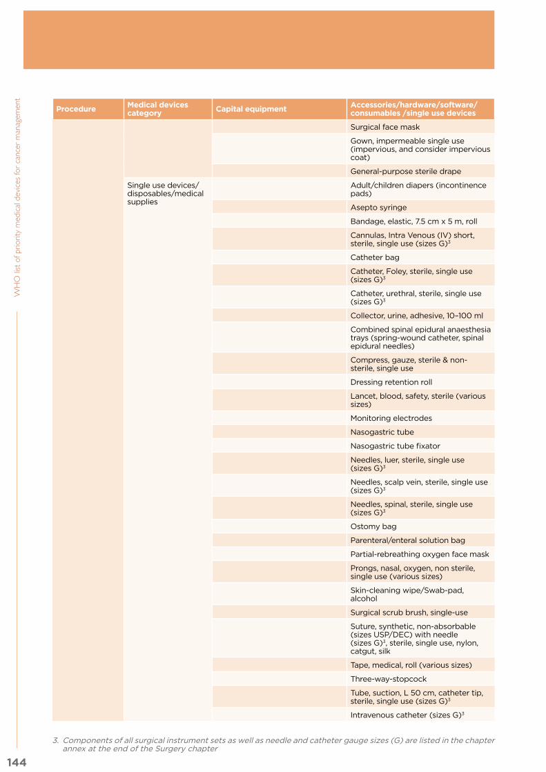
144
WHO
list o
f prio
rity m
edica
l dev
ices f
or ca
ncer
man
agem
ent
Procedure Medical devices category Capital equipment Accessories/hardware/software/
consumables /single use devices
Surgical face mask
Gown, impermeable single use (impervious, and consider impervious coat)
General-purpose sterile drape
Single use devices/disposables/medical supplies
Adult/children diapers (incontinence pads)
Asepto syringe
Bandage, elastic, 7.5 cm x 5 m, roll
Cannulas, Intra Venous (IV) short, sterile, single use (sizes G)3
Catheter bag
Catheter, Foley, sterile, single use (sizes G)3
Catheter, urethral, sterile, single use (sizes G)3
Collector, urine, adhesive, 10–100 ml
Combined spinal epidural anaesthesia trays (spring-wound catheter, spinal epidural needles)
Compress, gauze, sterile & non-sterile, single use
Dressing retention roll
Lancet, blood, safety, sterile (various sizes)
Monitoring electrodes
Nasogastric tube
Nasogastric tube fixator
Needles, luer, sterile, single use (sizes G)3
Needles, scalp vein, sterile, single use (sizes G)3
Needles, spinal, sterile, single use (sizes G)3
Ostomy bag
Parenteral/enteral solution bag
Partial-rebreathing oxygen face mask
Prongs, nasal, oxygen, non sterile, single use (various sizes)
Skin-cleaning wipe/Swab-pad, alcohol
Surgical scrub brush, single-use
Suture, synthetic, non-absorbable (sizes USP/DEC) with needle (sizes G)3, sterile, single use, nylon, catgut, silk
Tape, medical, roll (various sizes)
Three-way-stopcock
Tube, suction, L 50 cm, catheter tip, sterile, single use (sizes G)3
Intravenous catheter (sizes G)3
3. Components of all surgical instrument sets as well as needle and catheter gauge sizes (G) are listed in the chapter annex at the end of the Surgery chapter

145
7. Pallia
tive c
are
& e
nd
of life
care
Procedure Medical devices category Capital equipment Accessories/hardware/software/
consumables /single use devices
Syringes (various capacities)
Syringes, luer lock, sterile, single use (various capacities)
Syringes for drug preparation/formulation
Absorbent tipped applicator
Compression bandages (for Deep Vein Thrombosis)
Medication cups
Tracheostomy kit with adult-child T-tube and ties
Thoracentesis set
Paracentesis set
Solutions and reagents
Alcohol
Distilled water
Hand/body hygiene products
Isopropyl alcohol 70%
IV Solutions
Iodine povacrylex solution or similar Petroleum jelly
Other Arm/leg tourniquet
Basin, kidney, stainless steel/polypropylene
Bedpan, stainless steel/polypropylene
Bowl (dressing changes), stainless steel/polypropylene
Bowl (hygiene), stainless steel/polypropylene
Brush, hand, scrubbing, plastic
Receptacle, waste, stainless steel, pedal action
Sterile container sample
Urinal
Oxygen availability (Oxygen concentrator, Oxygen/gas pipeline, cylinder)
Pressure relief products (e.g. Cushions, mattresses

146
WHO
list o
f prio
rity m
edica
l dev
ices f
or ca
ncer
man
agem
ent 7.2.2 Specific medical devices for palliative care and end of life care by
cancer type
7.2.2.1 Breast
Procedure Medical devices category Capital equipment Accessories/hardware/software/
consumables/single use devices
Palliative care delivery
Single use devices/disposables/medical supplies
Dressing sets for malodours/fungating wounds (carbon pads/silver impregnated pads, compression bandages for lymphoedema etc.)
7.2.2.2 Colorectal
Procedure Medical devices category Capital equipment Accessories/hardware/software/
consumables/single use devices
Palliative care delivery (including colostomy)
Single use devices/disposables/medical supplies
Stoma/ostomy bags and adhesive
Gastrostomy material for skin medication and use
Devices to deliver enemas through stomas
7.2.2.3 Lung
Procedure Medical devices category
Capital equipment Accessories/hardware/software/consumables/single use devices
Palliative care delivery
Single use devices/disposables/medical supplies
Sputum containers
7.2.2.4 Leukaemia
Procedure Medical devices category Capital equipment Accessories/hardware/software/
consumables/single use devices
Palliative care delivery
Single use devices/disposables/medical supplies
Blood giving sets and cannulas
7.2.2.5 Cervical
Procedure Medical devices category Capital equipment Accessories/hardware/software/
consumables/single use devices
Palliative care delivery
Single use devices/disposables/medical supplies
Female sanitary products
Devices to manage genital fistulae
7.2.2.6 Prostate
Procedure Medical devices category Capital equipment Accessories/hardware/software/
consumables/single use devices
Palliative care delivery (Suprapubic catheterization)
Single use devices/disposables/medical supplies
Suprapubic catheter
In addition to all of the above-mentioned medical devices, it is important to have the following available in a
palliative care setting:
• Tools for assessment (approved and validated pain and other symptoms tools)
• Bed sets: Sheets, pillows, pillowcases, blankets/duvets, bed with protective barriers
• Patient’s hygiene tools: Soft toothbrush – not pink swabs for mouth care, hair combs and brushes, manicure and pedicure sets, towels of different sizes and colours, single use sponges, soap, shampoo, skin cream, incontinence pads, wipes, tissues

147
7. Pallia
tive c
are
& e
nd
of life
care
• Food provision utensils: dishes, plates, glasses, sipping cups, straws, cutlery, food processors and
modifying consistency agents for dysphagia
• Other furniture and devices: table with light, fan, radio/music device
• If relatives stay overnight option to provide a recliner chair and/or a sofa bed
• Aromatherapy
The palliative care unit should have access to X-Ray, CT scan and echographe and blood laboratory test
equipment, moreover the devices and furniture should be adapted for adults’ and children’s needs and proper
patient identification through ID tags or similar is recommended.
7.3 Other health system components
7.3.1 Human resources for palliative care and end of life care
Depending on national context including workforce availability and models, different occupations can fulfil
the roles that may be required for palliative care. A prerequisite is having the required competencies to
perform these roles and being officially recognized for it as per the scope of practice. The ISCO[1] codes are
added as illustrative examples based on current practice for further reference. This should not be interpreted
as guidance on occupations that should perform each role but rather indicative occupations that are currently
performing these roles in different country contexts.
The list below presents different occupations that could fulfil the roles and competencies required for
palliative care. These would depend on national context and local regulations as long as the specific staff has
the competency and it fits within their scope of practice . The ISCO2 codes are added for further reference.
Sample role Sample occupations (ISCO-08 codes)
Medical care Specialist Palliative Care Physician, , Clinical Oncologist, Specialist medical practitioner (2212)
Nursing care Specialist Palliative Care Nurse, Nursing professional (2221),Nursing associate professional (3221)
Patient navigator, support patient follow up during all cancer treatment
Social worker and counselling professionals (2635)
Social work including financial welfare and family support, bereavement support
Specialist Palliative Care Social Worker, social work and counselling professionals (2635)
Psychological assessment and treatment for patient and family, bereavement support
Psychologist (2634)
Nutritional advice and support Dietician and nutritionist (2265)
Spiritual and existential support/religious support Religious professional (2636)
Physiotherapy, rehabilitation Physical therapist, physiotherapist (2264)
Rehabilitation, diversional therapies Occupational therapist (2269)
Help with swallowing, speech Speech and language Therapist (2266)
Creative arts therapies Arts therapists , other health professionals(2269)
Massage, well-being support Massage therapist (3255)
Domestic/cleaning/ laundry/catering Domestic/cleaning/ laundry/catering, Cleaning personnel (9112)
Manager or unit and administrative support Health service manager (1342) and Clerical support workers (4412)
Management of medical devices Biomedical Engineer (2149)Biomedical technician , physical and engineering science technician (3119)
2 International Standard Classification of Occupations, http://www.ilo.org/public/english/bureau/stat/isco/docs/publication08.pdf

148
WHO
list o
f prio
rity m
edica
l dev
ices f
or ca
ncer
man
agem
ent Palliative care work occurs within a multidisciplinary team, and involves the following:
• Engagement with medical oncologists, radiotherapists and palliative care teams to use chemotherapy
and radiotherapy with palliative intent
• Engagement with surgical teams to deal with intestinal obstruction, and other emergencies; insert PEG tubes
• Engagement with neurologists, anaesthetists and pain specialists for pain management such as nerve
blocks and intrathecal procedures
• Engagement with urologists to insert or manage suprapubic catheters
7.3.2 Infrastructure
Layout distributionThe palliative care service should include the following rooms:
• treatment room for patients (private /semi private rooms)
• room for interdisciplinary meetings/family meetings
• family room with kitchen, dining room (quiet space)
• space for children to play
• double lockable cupboard with drawers and shelves for opioid storage and controlled medications
• rooms reserved for barrier nursing (HIV AIDS, MRSA, C Difficile)
• washing/bathing room (ideally a toilet and shower room for each patient)
• airborne infection isolation room(s) (if it is considered necessary by an infection control risk assessment).
The healthcare facility should have:
• a mortuary
• a prayer room with washing facilities (if not already in the facility add this space within the unit).
Electrical systemsThe room utility requirements include enough electrical sockets and other services. Emergency electrical
system should be considered in the treatment area for critical care devices and illumination. To ensure
continuity of services, stable power supply and backup systems should be installed.
Heating, ventilation and air conditioning systemsIf an airborne infection isolation room(s) is considered, it should comply with the architectural requirements
and have adequate ventilation.
Communication systemsThe following considerations should be taken into account:
• an intercom between patient rooms and nursing station
• a manually triggered alarm system allowing immediate notification of the staff
• internet access and telephone
• mobile phone access.
Special systems and other considerationsThe list of the team members should be visible to visitors.
The area shall be adapted for disabled access
It is mandatory to provide open visiting access 24/7 for family members.
When and where allowed, pet therapy

149
7. Pallia
tive c
are
& e
nd
of life
care
7.3.3 Quality management
The safe storage of opioids, controlled medicines and other substances must be ensured to comply with
country specific legislation and procedures.
Policies and procedures for infection control as well as for the reporting of risks, incidents and accidents to
patients, visitors and staff must be put in place.
Bibliography:
1) Sepúlveda C, Marlin A, Yoshida T, Ullrich A. Palliative care: the World Health Organization’s global
perspective. Journal of pain and symptom management. 2002 Aug 31;24(2):91-6.
2) The National Council for Hospices and Specialist Palliative Care Services, 2002
3) Radbruch L, Payne S. White Paper on standards and norms for hospice and palliative care in Europe:
part 1. European Journal of Palliative Care, 2009, 16 (6): 278-289.
4) Radbruch L, Payne S. White Paper on standards and norms for hospice and palliative care in Europe:
part 2. European Journal of Palliative Care, 2010, 17 (1): 22-33.
5) Hoegler, “Radiotherapy for palliation of symptoms in incurable cancer,” Curr Probl Cancer,
Volume 21, Issue 3 (1997).
6) Lutz et al., “Palliative Radiotherapy for Bone Metastases: An ASTRO Evidence-Based Guideline,”
International Journal of Radiation Oncology,Biology,Physics, Volume 79, Issue 4 (2011).
7) Skowronek, “Brachytherapy in the treatment of lung cancer – a valuable solution,” J Contemp
Brachytherapy, Volume 7, Issue 4 (2015).
8) Mallick et al., “Endobronchial brachytherapy for symptom palliation in non-small cell lung cancer-
Analysis of symptom response, endoscopic improvement and quality of life,” Lung Cancer, Volume 55,
Issue 3 (2007).

150
WHO
list o
f prio
rity m
edica
l dev
ices f
or ca
ncer
man
agem
ent The lists presented in this document include a selection of medical devices of different categories (from
single use devices to medical equipment and consumables) for selected interventions extracted from clinical
guidelines addressing the cancer management continuum of care, including prevention, diagnosis, therapy
and palliative care. The lists presented herein are nonexclusive, however serve as guidance for countries
seeking to develop or implement a management policy/strategy for cancer specific medical devices.
It is of utmost importance to consider the critical interdependence of these technologies. Best outcomes
in cancer cannot be achieved by investing in one set of technologies alone. Diagnostic methods must be
combined with therapeutic capability in order to have meaningful impact.
This chapter includes a general implementation strategy and additional considerations to reach pragmatic
decisions.
IV.I Approach for adapting the WHO list of priority medical devices for cancer management in each setting
As mentioned, it is important for each country to define an implementation plan that will take into account
country-specific needs, epidemiological situation, and availability of infrastructure, related human resources
and finances, and affordability to patients, among others.
These lists of priority medical devices for cancer management consider those devices best suited to the
management of the priority health problems defined herein (breast, cervical, colorectal, lung, leukaemia, and
prostate cancer), and the selected procedures/interventions for each clinical area. Therefore, it should be
noted that in order to select them and be able to use these technologies at country level, further steps are
required:
1. Perform a needs assessment of in-country resources for cancer management
Fig. 11. General Needs Assessment Process (1)
Generic standards and examples
Baseline data
Level of care
Clinical practice guidelines
Medical device lists
Epidemiological needs/disease prioritiesPopulation data (demography, catchment area, patient rate)
Service availability and accessibilityInfrastructure situation
Health technology/medical device situationHuman resources situation
Affordability to patients, reimbursement scheme
Analysing/interpretingOverall gap/
needBudgetary & HR situation
Prioritized need
IV. Implementation strategy

151
IV. Im
ple
men
tatio
n stra
teg
y
a. Firstly consider National Health Plan and any available Cancer Control Programme priorities, strategies
or objectives.
i. Review country data, epidemiology and registries
ii. Consider if indicators or evaluation criteria are already in place regarding resources and specifically
medical devices.
b. Define the scope of the implementation plan: national, regional, only specialty hospital or all health
facilities. Including: district hospitals, health centers and community health posts
i. It is important that all the levels of care are included in the considerations of the implementation plan,
from community health centres to specialized oncology hospitals, even if only one or many hospitals
are being equipped
ii. The network has to consider reference to higher levels of care.
c. Review the lists, unit by unit, with local specialists, and compare to your inventory or availability of
technologies to manage cancer. Please note the devices that are general and can serve other diseases
besides cancer and those specific only to cancer.
d. Compile information about resources available or not yet appropriate or sufficient on the following
topics:
i. Health facilities that could be used for cancer diagnosis or treatment in the scope of the study and
related referral services
ii. Medical devices available per clinical unit, particularly those specified in this document for:
• Medical imaging
• Nuclear medicine
• Laboratory and pathology
• Surgery
• Radiotherapy
• Systemic therapy
• Palliative care and end of life care
iii. Specialized human resources, particularly those mentioned in this document:
• Biomedical Engineers/Equipment Technicians
• Laboratory Technicians/Biomedical Laboratory Scientists
• Medical Physicists
• Oncologists
• Oncological Nurses
• Pathologists
• Radiologists
• Radiotherapists/Radiation Therapy Technologists
• Radiographers
• Surgical Oncologists
iv. National treatment guidelines/protocols or care pathways
v. Priority interventions if defined by an insurance package linked to universal health coverage and
reimbursement schemes.
vi. Target population
• Type of procedures, for type of patients
• Population distribution in the country
• Catchment areas for diagnosis,
• Support for patients transportation and stay in treatment centers.
vii. Funding allocation
1. Depending on the country or setting, this funding could be pre-established or will have to be
calculated depending on the technology required and then search for approval.
2. Funding to support the on-going interventions for diagnosis, treatment, monitoring and palliative care required.
3. Consider other sponsorships and national cancer programmes funds allocation to support
patients access and affordability.

152
WHO
list o
f prio
rity m
edica
l dev
ices f
or ca
ncer
man
agem
ent 2. Once the needs assessment is done and the gaps identified, select an appropriate methodology to prioritize
and select which medical devices would be most needed considering some of the following aspects:
a. Target population
b. Type of cancer
c. Equity, gender and ethical issues, if applicable (i.e. Mammography)
d. Equipment complementary to other diseases
e. Available budget for:
i. Infrastructure, to install equipment (e.g. bunkers for radiotherapy)
ii. Capital equipment and its life cycle operation, maintenance and consumables
iii. Procurement and supply of single use medical devices
iv. Training of health care workers in safe and appropriate use
v. Other medical devices, i.e., quality control and safety, complementary to the capital equipment.
f. Feasibility and organizational challenges, i.e., where to place the technology, where more resources are
available or where most target population is based.
Most of these considerations are part of health technology assessment and health technology management
studies, or certificate of need. Most of them should be done in a committee to make informed decisions based
on evidence, when and wherever available.
3. If a technology is approved for incorporation into the health care system, the following must be considered and defined, usually within a process of health technology management handled by personnel with relevant technical expertise (i.e. biomedical engineer):
a. Regulatory clearance: The regulatory approval process of the specific technology performed by the
national regulatory agency which can be nuclear, radiological in other Ministries or for medical devices in
the Ministry of Health, these have to be in agreement and continuous collaboration.
b. Technical specifications that are generic and comply with standards to guarantee the technical quality of
the device.
c. Bidding process:
i. In the case of radiotherapy, nuclear medicine or other diagnostic imaging equipment, the mechanical
guidelines of the infrastructure are to be considered
ii. Bids should include extended warranty, training for users, installation and spare parts
iii. Budget should include operating costs, including maintenance for the whole life cycle of technologies
(around 10 years)
d. Government importation requirements
e. Transportation, insurance, taxes, customs, delivery and installation
f. Reception of the equipment, verification of installation, and training of the users/health care workers by
the manufacturer
g. Supply chain of consumables
h. Verification of quality, safety and performance
i. Decommissioning and waste management procedures (2).
More guidance on each of the mentioned steps can be found in the following WHO publications: Needs assessment for medical devices: http://apps.who.int/iris/bitstream/10665/44562/1/9789241501385_eng.pdf
Procurement process resource guide: http://apps.who.int/iris/bitstream/10665/44563/1/9789241501378_eng.pdf
Medical device donations: consideration for solicitation and provision: http://apps.who.int/iris/bitstream/10665/44568/1/9789241501408_eng.pdf
Introduction to medical equipment inventory management: http://apps.who.int/iris/bitstream/10665/44561/1/9789241501392_eng.pdf
Medical equipment maintenance programme overview: http://apps.who.int/iris/bitstream/10665/44587/1/9789241501538_eng.pdf
Computerized maintenance management system: http://apps.who.int/iris/bitstream/10665/44567/1/9789241501415_eng.pdf

153
IV. Im
ple
men
tatio
n stra
teg
y
Related WHO websites:
Topic Website
Overview of Health technology management
http://www.who.int/medical_devices/management_use/en/
Technical specifications of medical devices
http://www.who.int/medical_devices/management_use/mde_tech_spec/en/
Donations of medical devices http://www.who.int/medical_devices/management_use/manage_donations/
Priority medical devices http://www.who.int/medical_devices/priority/en/
Further collaboration:Besides the guidelines and documents mentioned in each of the chapters, additional support to incorporate
technologies can be provided by international organizations, NGOs and professional organizations, some of
which are listed below:
Type of organization Entity Information
UN organizations Interagency Task force for NCD
http://www.who.int/ncds/un-task-force/en/
IAEA www.iaea.org
IARC http://www.who.int/ionizing_radiation/research/iarc/en/
UNICEF www.unicef.org
UNOPS www.unops.org
WHO http://www.who.int/medical_devices/en
NGOs in official relations with WHO
DITTA http://globalditta.org/
GMTA http://www.globalmedicaltechnologyalliance.org
HUMATEM http://www.humatem.org
HTAI http://www.htai.org
IAPO https://www.iapo.org.uk
IFBLS http://www.ifbls.org
IFCC http://www.ifcc.org
IFHE https://www.ifhe.org
IFMBE http://2016.ifmbe.org/
IOMP http://www.iomp.org
INCTR http://www.inctr.org
ISR http://www.isradiology.org
ISRRT http://www.isrrt.org/
IUA http://www.uia.archi/en
RAD AID http://www.rad-aid.org
WASPaLM www.waspalm.org
WFUMB www.wfumb.org
NGOs related to cancer in official relations with WHO
ESMO www.esmo.org
UICC www.uicc.org
Other important cancer organizations
AORTIC www.aortic-africa.org
ASCO www.asco.org
BHGI www.bhgi.info
NCCN www.nccn.org
NCI www.cancer.gov
SLACOM www.slacom.org
Other networks EuroScan www.euroscangroup.com/
Note: Please see Acronyms and Abbreviations for full organization names

154
WHO
list o
f prio
rity m
edica
l dev
ices f
or ca
ncer
man
agem
ent
IV.II Future activities
The report here presented is a first step towards defining medical devices required for many diseases
including cancer, but it has a limited scope and further work has to be done in the future, to support the best
implementation in countries, in the following areas:
1. WHO guidelines on cancer managementOnly the cervical cancer WHO guidelines are currently available and future work should encompass other
types of cancers, considering guidelines already available in middle income countries. Guidelines usually
describe medicines and pharmaceuticals required, but almost none specify the medical devices or the related
human resources needed to perform the interventions, which would be useful to have in the future.
2. PrioritizationA methodology is needed to prioritize and select medical devices after the needs assessment considerations
(integration with other methodologies such as the ones used for HTA). Most health technology assessment
analyses are for specific high cost technologies or for innovative ones, but no studies are found on the total
cost of radiotherapy units, palliative care units or pathology units in low or middle income countries, and
this would be very helpful for those Member States that would like to have more information, especially
considering feasibility, organizational issues, planning, etc.
The MCDA methodology used to developed the working tools in order to collect evidence and justification
from experts for inclusion on the list can be leveraged by member states by including contextual criteria
(such a size of population, country priorities) to prioritize devices based on their unique context.
3. Indicators, quantificationFuture WHO work will need to be undertaken to define and recommend the following global, regional and
local indicators:
• Number of pieces of equipment per inhabitant (e.g. radiotherapy system per million inhabitants)
• Size and type of equipment required depending on caseload and utilization rate (e.g. automatic versus
manual equipment, multiparameter physiological monitors, etc.)
• Consumables (medical supplies) per inhabitants, or
• Consumables per treatment, or
• Consumables per hospital
• Further studies on country situations need to be conducted, especially for low and middle income
countries to allow planning of resources and budget allocation.
4. Technical specifications for procurement Technical specifications need to be developed, or if developed, placed in one single template which will be
useful for the user, procurer, industry or donor agencies. Some of the UN agencies have their own, but they
still need to be harmonized and widely disseminated to provide the manufacturers with the market needs so
as to produce accordingly.
5. Appropriate affordable technologiesMore research and development for affordable technologies for cancer management are required. Currently
most technologies for cancer management are very expensive, complicated to use and require specialized
human resources (for example, point of care diagnostics for cancer would be a major innovation to support
early diagnosis).
a. Evidence and studies of the use of innovative technologies for cancer care in low resource settings,
especially where there are no specialized human resources
b. Assessment of the technologies (e.g. measurement of health outcomes, problems of the use of the
proposed technologies in different settings), as well as the cost of the packages of interventions when
using the technologies proposed in this publication.

155
IV. Im
ple
men
tatio
n stra
teg
y
IV.III Concluding Remarks
Seventy percent of cancer cases occur in low- and middle-income countries. Much work is still needed in order
to place the appropriate, good quality medical technology required to screen, diagnose, treat, monitor and
palliate cancer disease worldwide, and to develop the human resource competencies of specialized oncology,
pathology, nuclear medicine, radiation therapy, and other disciplines, particularly in low resource settings.
This publication was completed thanks to the very important collaboration from interdisciplinary experts
around the world, as well as supportive NGO organizations and individuals committed to cancer care.
It is absolutely indispensable to consider the interdependence of these technologies. The outcomes in
cancer cannot be achieved by investing in one set of technologies alone. Diagnostics must be combined
with therapeutic capability to have meaningful impact and vice versa. It is therefore hoped that these lists
of medical devices by clinical unit will assist policy makers, health care managers and technical experts, to
define the needs of each setting.
Medical devices run the substantial risk of being unused due to technology misalignment to deployment
settings (3). It is important to note that contextually relevant interventions and devices must be selected to
improve resource allocation in each country, depending on the local needs and resources available. Financing
models are an important topic that should be evaluated as well in order to deliver services within the health
care systems.
The lists presented in this publication should be adapted and reviewed according to local epidemiology,
national policies, regulatory frameworks, and available specialized health care workforce, infrastructure,
budget, and organizational structures.
It is therefore important to consider innovation, regulatory approval of the technologies, and the health
technology assessment process to enable informed decision making taking into consideration equity, social,
clinical and economic aspects as well as health technology management aspects. Such informed decision
making will ensure that the procurement, installation, training and safe use of technologies is done in the
most effective way, ensuring the well-being of the patient.
The selection of the technologies required from the lists presented in this publication is just the first step
of many to provide best care for cancer patients. These technologies require multidisciplinary expertise to
implement.
More information will be available from WHO and other agencies to support selection, procurement and best
use of these medical devices for all patients and specifically to target cancer management to those most in
need.
Bibliography
1) Needs Assessment for Medical Devices. WHO Medical Device Technical Series.
Available at: http://apps.who.int/medicinedocs/documents/s21562en/s21562en.pdf
2) Safe management of wastes from health-care activities. WHO (2014).
Available at: http://www.who.int/water_sanitation_health/publications/wastemanag/en/
3) Lilford RJ, Burn SL, Diaconu KD, Lilford P, Chilton PJ, Bion V, Cummins C, Manaseki-Holland S. An
approach to prioritization of medical devices in low-income countries: an example based on the Republic
of South Sudan. Cost effectiveness and resource allocation. 2015 Jan 10;13(1):1.

156
WHO
list o
f prio
rity m
edica
l dev
ices f
or ca
ncer
man
agem
ent Accessibility: refers to people’s ability to obtain and appropriately use good-quality health technologies
when they are needed.
Adverse event: any untoward medical occurrence in a subject whether it is device-related or not.
Affordability: in the context of this report it is defined as the extent to which the intended clients of a service can pay for it.
Appropriate(ness): refers to medical methods, procedures, techniques, and equipment that are scientifically valid, adapted to local needs, acceptable to both patient and health-care personnel, and that can be utilized and maintained with resources the community or country can afford.
Availability: when a medical device can be found on the medical device market.
Best practice: an examination of the methods by which optimal outcomes are achieved.
Care pathways: one mechanism of putting a protocol into operation. Care pathways determine locally agreed, multidisciplinary practice, based on guidelines and evidence (where available) for a specific patient group. They form all, or part of the clinical record, they document the care given, and they facilitate the evaluation of outcomes for quality improvement purposes.
Clinical evaluation: the assessment and analysis of clinical data pertaining to a medical device in order to verify the clinical safety and performance of the device.
Clinical evidence: the clinical data and the clinical evaluation report pertaining to a medical device.
Clinical guideline: systematically-developed statements to assist practitioner and patient decisions about appropriate health care for specific clinical circumstances. A clinical guideline is a tool to support clinical decision-making and it covers a specific clinical problem.
Consumables: liquids or supplies required for the use of the equipment but allowing only limited, or no, reuse.
Cost(s): (1) the value of the resources used in an activity; (2) the benefits sacrificed through a particular event or choice of action rather than another.
Cost-effectiveness analysis: analysis that involves the allocation of scarce resources among competing alternative uses, and the distribution of the products from these uses among the members of the society.
Effectiveness: a device is clinically effective when it produces the effect intended by the manufacturer relative to the medical condition for which it was created.
Efficacy: the ability to produce a desired or intended result, as linked to the performance of a device.
eHealth: the use of information and communication technologies (ICT) for health.
Equity in health: where people’s needs guide the distribution of resources and opportunities for well-being.
Gamma camera: medical device that detects gamma rays emitted from a person’s body after the administration of a radioactive compound, which produces images of the organ being investigated.
Gap: a disparity between health-care need and reality.
Global burden of disease (GBD): the WHO GBD project draws on a wide range of data sources to quantify global and regional effects of diseases, injuries and risk factors on population health.
Hazard: potential cause of harm.
Health care: any type of service provided by professionals or paraprofessionals with an impact on health status.
Health technology: the application of organized knowledge and skills in the form of devices, medicines, vaccines, procedures and systems developed to solve a health problem and improve quality of life. The term is used interchangeably with “health-care technology” (1).
Health technologies for the purpose of this project include medical devices, assistive devices (e.g. hearing aids, wheelchairs), e-health solutions (e.g. telemedicine) and related health technologies— excluding clinical
procedures, medicinal products, vaccines, biological therapeutic products, or tissue engineered medical products.
Glossary
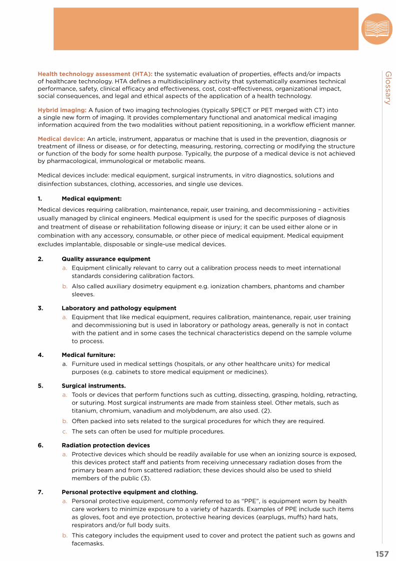
157
Glo
ssary
Health technology assessment (HTA): the systematic evaluation of properties, effects and/or impacts of healthcare technology. HTA defines a multidisciplinary activity that systematically examines technical performance, safety, clinical efficacy and effectiveness, cost, cost-effectiveness, organizational impact, social consequences, and legal and ethical aspects of the application of a health technology.
Hybrid imaging: A fusion of two imaging technologies (typically SPECT or PET merged with CT) into a single new form of imaging. It provides complementary functional and anatomical medical imaging information acquired from the two modalities without patient repositioning, in a workflow efficient manner.
Medical device: An article, instrument, apparatus or machine that is used in the prevention, diagnosis or treatment of illness or disease, or for detecting, measuring, restoring, correcting or modifying the structure or function of the body for some health purpose. Typically, the purpose of a medical device is not achieved by pharmacological, immunological or metabolic means.
Medical devices include: medical equipment, surgical instruments, in vitro diagnostics, solutions and
disinfection substances, clothing, accessories, and single use devices.
1. Medical equipment:
Medical devices requiring calibration, maintenance, repair, user training, and decommissioning – activities
usually managed by clinical engineers. Medical equipment is used for the specific purposes of diagnosis
and treatment of disease or rehabilitation following disease or injury; it can be used either alone or in
combination with any accessory, consumable, or other piece of medical equipment. Medical equipment
excludes implantable, disposable or single-use medical devices.
2. Quality assurance equipment
a. Equipment clinically relevant to carry out a calibration process needs to meet international standards considering calibration factors.
b. Also called auxiliary dosimetry equipment e.g. ionization chambers, phantoms and chamber sleeves.
3. Laboratory and pathology equipment
a. Equipment that like medical equipment, requires calibration, maintenance, repair, user training and decommissioning but is used in laboratory or pathology areas, generally is not in contact with the patient and in some cases the technical characteristics depend on the sample volume to process.
4. Medical furniture:
a. Furniture used in medical settings (hospitals, or any other healthcare units) for medical purposes (e.g. cabinets to store medical equipment or medicines).
5. Surgical instruments.
a. Tools or devices that perform functions such as cutting, dissecting, grasping, holding, retracting, or suturing. Most surgical instruments are made from stainless steel. Other metals, such as titanium, chromium, vanadium and molybdenum, are also used. (2).
b. Often packed into sets related to the surgical procedures for which they are required.
c. The sets can often be used for multiple procedures.
6. Radiation protection devices
a. Protective devices which should be readily available for use when an ionizing source is exposed, this devices protect staff and patients from receiving unnecessary radiation doses from the primary beam and from scattered radiation; these devices should also be used to shield members of the public (3).
7. Personal protective equipment and clothing.
a. Personal protective equipment, commonly referred to as “PPE”, is equipment worn by health care workers to minimize exposure to a variety of hazards. Examples of PPE include such items as gloves, foot and eye protection, protective hearing devices (earplugs, muffs) hard hats, respirators and/or full body suits.
b. This category includes the equipment used to cover and protect the patient such as gowns and facemasks.
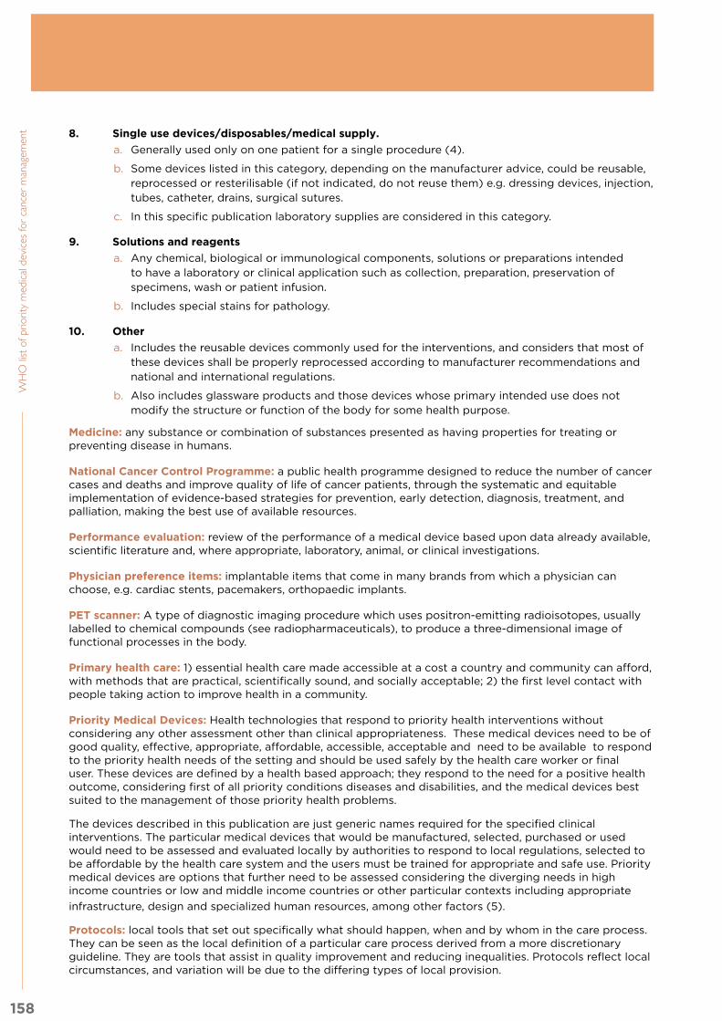
158
WHO
list o
f prio
rity m
edica
l dev
ices f
or ca
ncer
man
agem
ent 8. Single use devices/disposables/medical supply.
a. Generally used only on one patient for a single procedure (4).
b. Some devices listed in this category, depending on the manufacturer advice, could be reusable, reprocessed or resterilisable (if not indicated, do not reuse them) e.g. dressing devices, injection, tubes, catheter, drains, surgical sutures.
c. In this specific publication laboratory supplies are considered in this category.
9. Solutions and reagents
a. Any chemical, biological or immunological components, solutions or preparations intended to have a laboratory or clinical application such as collection, preparation, preservation of specimens, wash or patient infusion.
b. Includes special stains for pathology.
10. Other
a. Includes the reusable devices commonly used for the interventions, and considers that most of these devices shall be properly reprocessed according to manufacturer recommendations and national and international regulations.
b. Also includes glassware products and those devices whose primary intended use does not modify the structure or function of the body for some health purpose.
Medicine: any substance or combination of substances presented as having properties for treating or preventing disease in humans.
National Cancer Control Programme: a public health programme designed to reduce the number of cancer cases and deaths and improve quality of life of cancer patients, through the systematic and equitable implementation of evidence-based strategies for prevention, early detection, diagnosis, treatment, and palliation, making the best use of available resources.
Performance evaluation: review of the performance of a medical device based upon data already available, scientific literature and, where appropriate, laboratory, animal, or clinical investigations.
Physician preference items: implantable items that come in many brands from which a physician can choose, e.g. cardiac stents, pacemakers, orthopaedic implants.
PET scanner: A type of diagnostic imaging procedure which uses positron-emitting radioisotopes, usually labelled to chemical compounds (see radiopharmaceuticals), to produce a three-dimensional image of functional processes in the body.
Primary health care: 1) essential health care made accessible at a cost a country and community can afford, with methods that are practical, scientifically sound, and socially acceptable; 2) the first level contact with people taking action to improve health in a community.
Priority Medical Devices: Health technologies that respond to priority health interventions without considering any other assessment other than clinical appropriateness. These medical devices need to be of good quality, effective, appropriate, affordable, accessible, acceptable and need to be available to respond to the priority health needs of the setting and should be used safely by the health care worker or final user. These devices are defined by a health based approach; they respond to the need for a positive health outcome, considering first of all priority conditions diseases and disabilities, and the medical devices best suited to the management of those priority health problems.
The devices described in this publication are just generic names required for the specified clinical interventions. The particular medical devices that would be manufactured, selected, purchased or used would need to be assessed and evaluated locally by authorities to respond to local regulations, selected to be affordable by the health care system and the users must be trained for appropriate and safe use. Priority medical devices are options that further need to be assessed considering the diverging needs in high income countries or low and middle income countries or other particular contexts including appropriate
infrastructure, design and specialized human resources, among other factors (5).
Protocols: local tools that set out specifically what should happen, when and by whom in the care process. They can be seen as the local definition of a particular care process derived from a more discretionary guideline. They are tools that assist in quality improvement and reducing inequalities. Protocols reflect local circumstances, and variation will be due to the differing types of local provision.

159
Glo
ssary
Post-market surveillance: proactive collection of information on medical devices carried out by the manufacturers after those devices have reached the market.
Public health: a social and political concept aimed at improving health, prolonging life and improving the quality of life among whole populations through health promotion, disease prevention, and other forms of health intervention.
Radiopharmaceuticals: are radionuclides or radionuclide-labelled chemical compounds that are mainly used for the diagnosis and, to a limited but growing extent, for the therapy of diseases.
Research and development (R&D): creative work undertaken on a systematic basis in order to increase the stock of knowledge, including knowledge of humans, culture and society, and the use of this stock of knowledge to devise new items, applications, etc.
Risk: combination of the probability of occurrence of harm and the severity of that harm.
Secondary health care (see also primary/tertiary health care): specialized ambulatory medical services and commonplace hospital care (outpatient and inpatient services). Access is often via referral from primary health care services.
SPECT: An acronym that stands for single photon emission computed tomography, a nuclear medicine procedure in which a gamma camera rotates around the patient and takes pictures from many angles, which a computer then uses to form a tomographic (cross-sectional) image. The calculation process in SPECT is similar to that in CT (X-ray computed tomography) and in PET (positron emission tomography). It differentiates from PET in that it uses single-photon emitting radioisotopes, usually labelled to chemical compounds (see radiopharmaceuticals).
Telehealth: the use of electronic information and communication technologies to support long-distance clinical health care, patient and professional health-related education, public health, and health administration.
Telemedicine: the delivery of health care services through the use of information and communication technologies in a situation where the actors are not at the same location. The actors can either be two healthcare professionals (for example in teleradiology) or a health-care professional and a patient (for example in telemonitoring of patients with diabetes).
Tertiary health care (see also primary/secondary health care): refers to medical and related services of high complexity and usually high cost. Those referred from secondary care for diagnosis and treatment, which is not available in primary and secondary care. Tertiary care is generally only available at national or international referral centres.
Bibliography:
1) The WHA60.29 World Health Assembly resolution on health technologies, May 2007.
2) http://www.surgeryencyclopedia.com/St-Wr/Surgical-Instruments.html#ixzz3fMr5ZeVZ
3) http://apps.who.int/medicinedocs/documents/s15961e/s15961e.pdfhttp://apps.who.int/medicinedocs/
documents/s15961e/s15961e.pdf
4) http://www.fda.gov/MedicalDevices/DeviceRegulationandGuidance/ReprocessingofSingle-UseDevices/
ucm121465.htm
5) http://apps.who.int/iris/bitstream/10665/44407/1/9789241564045_eng.pdf
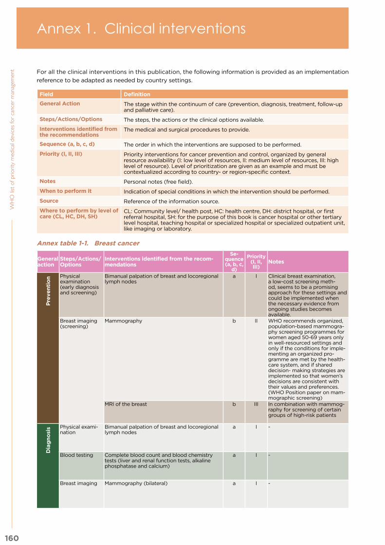
160
WHO
list o
f prio
rity m
edica
l dev
ices f
or ca
ncer
man
agem
ent For all the clinical interventions in this publication, the following information is provided as an implementation
reference to be adapted as needed by country settings.
Field Definition
General Action The stage within the continuum of care (prevention, diagnosis, treatment, follow-up and palliative care).
Steps/Actions/Options The steps, the actions or the clinical options available.
Interventions identified from the recommendations
The medical and surgical procedures to provide.
Sequence (a, b, c, d) The order in which the interventions are supposed to be performed.
Priority (I, II, III) Priority interventions for cancer prevention and control, organized by general resource availability (I: low level of resources, II: medium level of resources, III: high level of resource). Level of prioritization are given as an example and must be contextualized according to country- or region-specific context.
Notes Personal notes (free field).
When to perform it Indication of special conditions in which the intervention should be performed.
Source Reference of the information source.
Where to perform by level of care (CL, HC, DH, SH)
CL: Community level/ health post, HC: health centre, DH: district hospital, or first referral hospital, SH: for the purpose of this book is cancer hospital or other tertiary level hospital, teaching hospital or specialized hospital or specialized outpatient unit, like imaging or laboratory.
Annex table 1-1. Breast cancer
General action
Steps/Actions/Options
Interventions identified from the recom-mendations
Se-quence (a, b, c,
d)
Priority (I, II, III)
Notes General action When to perform it Source Source Source Source
Pre
ven
tio
n Physical examination (early diagnosis and screening)
Bimanual palpation of breast and locoregional lymph nodes
a I Clinical breast examination, a low-cost screening meth-od, seems to be a promising approach for these settings and could be implemented when the necessary evidence fromongoing studies becomes available.
Pre
ven
tio
n There is uncertainty about the appropriate age groups for screen-ing and the steps that should be taken by responsible authorities to commission and im-plement breast cancer screening programmes of appropriate quality (WHO).
WHO posi-tion paper on mammogra-phy screening (2014)
BHGI Guidelines for Inter-national Breast Health and Cancer Control–Implemen-tation. Cancer 2008;113(S8): i–ix, 2215–2371
Early and locally ad-vanced breast cancer. NICE clinical guideline 80 (2014)
-
Breast imaging (screening)
Mammography b II WHO recommends organized, population-based mammogra-phy screening programmes for women aged 50-69 years only in well-resourced settings and only if the conditions for imple-menting an organized pro-gramme are met by the health-care system, and if shared decision- making strategies are implemented so that women’s decisions are consistent with their values and preferences. (WHO Position paper on mam-mographic screening)
WHO posi-tion paper on mammogra-phy screening (2014)
BHGI Guidelines for Inter-national Breast Health and Cancer Control–Implemen-tation. Cancer 2008;113(S8): i–ix, 2215–2371
Early and locally ad-vanced breast cancer. NICE clinical guideline 80 (2014)
-
MRI of the breast b III In combination with mammog-raphy for screening of certain groups of high-risk patients
- - - NCCN. Breast Can-cer Screening and Diagnosis. Version 1.2014
Dia
gn
osi
s Physical exami-nation
Bimanual palpation of breast and locoregional lymph nodes
a I -D
iag
no
sis - - BHGI Guidelines for Inter-
national Breast Health and Cancer Control–Implemen-tation. Cancer 2008;113(S8): i–ix, 2215–2371
Early and locally ad-vanced breast cancer. NICE clinical guideline 80 (2014)
NCCN. Breast Can-cer Screening and Diagnosis. Version 1.2014
Blood testing Complete blood count and blood chemistry tests (liver and renal function tests, alkaline phosphatase and calcium)
a I - - - BHGI Guidelines for Inter-national Breast Health and Cancer Control–Implemen-tation. Cancer 2008;113(S8): i–ix, 2215–2371
- NCCN. Breast Can-cer Screening and Diagnosis. Version 1.2014
Breast imaging Mammography (bilateral) a I - - - BHGI Guidelines for Inter-national Breast Health and Cancer Control–Implemen-tation. Cancer 2008;113(S8): i–ix, 2215–2371
Early and locally ad-vanced breast cancer. NICE clinical guideline 80 (2014)
NCCN. Breast Can-cer Screening and Diagnosis. Version 1.2014
Annex 1. Clinical interventions

161
An
nex 1. C
linic
al in
terv
en
tion
s
For all the clinical interventions in this publication, the following information is provided as an implementation
reference to be adapted as needed by country settings.
Field Definition
General Action The stage within the continuum of care (prevention, diagnosis, treatment, follow-up and palliative care).
Steps/Actions/Options The steps, the actions or the clinical options available.
Interventions identified from the recommendations
The medical and surgical procedures to provide.
Sequence (a, b, c, d) The order in which the interventions are supposed to be performed.
Priority (I, II, III) Priority interventions for cancer prevention and control, organized by general resource availability (I: low level of resources, II: medium level of resources, III: high level of resource). Level of prioritization are given as an example and must be contextualized according to country- or region-specific context.
Notes Personal notes (free field).
When to perform it Indication of special conditions in which the intervention should be performed.
Source Reference of the information source.
Where to perform by level of care (CL, HC, DH, SH)
CL: Community level/ health post, HC: health centre, DH: district hospital, or first referral hospital, SH: for the purpose of this book is cancer hospital or other tertiary level hospital, teaching hospital or specialized hospital or specialized outpatient unit, like imaging or laboratory.
Annex table 1-1. Breast cancer
General action
Steps/Actions/Options
Interventions identified from the recom-mendations
Se-quence (a, b, c,
d)
Priority (I, II, III)
Notes General action When to perform it Source Source Source Source
Pre
ven
tio
n Physical examination (early diagnosis and screening)
Bimanual palpation of breast and locoregional lymph nodes
a I Clinical breast examination, a low-cost screening meth-od, seems to be a promising approach for these settings and could be implemented when the necessary evidence fromongoing studies becomes available.
Pre
ven
tio
n There is uncertainty about the appropriate age groups for screen-ing and the steps that should be taken by responsible authorities to commission and im-plement breast cancer screening programmes of appropriate quality (WHO).
WHO posi-tion paper on mammogra-phy screening (2014)
BHGI Guidelines for Inter-national Breast Health and Cancer Control–Implemen-tation. Cancer 2008;113(S8): i–ix, 2215–2371
Early and locally ad-vanced breast cancer. NICE clinical guideline 80 (2014)
-
Breast imaging (screening)
Mammography b II WHO recommends organized, population-based mammogra-phy screening programmes for women aged 50-69 years only in well-resourced settings and only if the conditions for imple-menting an organized pro-gramme are met by the health-care system, and if shared decision- making strategies are implemented so that women’s decisions are consistent with their values and preferences. (WHO Position paper on mam-mographic screening)
WHO posi-tion paper on mammogra-phy screening (2014)
BHGI Guidelines for Inter-national Breast Health and Cancer Control–Implemen-tation. Cancer 2008;113(S8): i–ix, 2215–2371
Early and locally ad-vanced breast cancer. NICE clinical guideline 80 (2014)
-
MRI of the breast b III In combination with mammog-raphy for screening of certain groups of high-risk patients
- - - NCCN. Breast Can-cer Screening and Diagnosis. Version 1.2014
Dia
gn
osi
s Physical exami-nation
Bimanual palpation of breast and locoregional lymph nodes
a I -
Dia
gn
osi
s - - BHGI Guidelines for Inter-national Breast Health and Cancer Control–Implemen-tation. Cancer 2008;113(S8): i–ix, 2215–2371
Early and locally ad-vanced breast cancer. NICE clinical guideline 80 (2014)
NCCN. Breast Can-cer Screening and Diagnosis. Version 1.2014
Blood testing Complete blood count and blood chemistry tests (liver and renal function tests, alkaline phosphatase and calcium)
a I - - - BHGI Guidelines for Inter-national Breast Health and Cancer Control–Implemen-tation. Cancer 2008;113(S8): i–ix, 2215–2371
- NCCN. Breast Can-cer Screening and Diagnosis. Version 1.2014
Breast imaging Mammography (bilateral) a I - - - BHGI Guidelines for Inter-national Breast Health and Cancer Control–Implemen-tation. Cancer 2008;113(S8): i–ix, 2215–2371
Early and locally ad-vanced breast cancer. NICE clinical guideline 80 (2014)
NCCN. Breast Can-cer Screening and Diagnosis. Version 1.2014
Annex table 1-1. Breast cancer 160
Annex table 1-2. Cervical cancer 169
Annex table 1-3. Colorectal cancer 185
Annex table 1-4. Leukaemia 192
Annex table 1-5. Lung cancer 200
Annex table 1-6. Prostate cancer 212
Prevention
Diagnosis
Treatment
Follow up
Palliative care
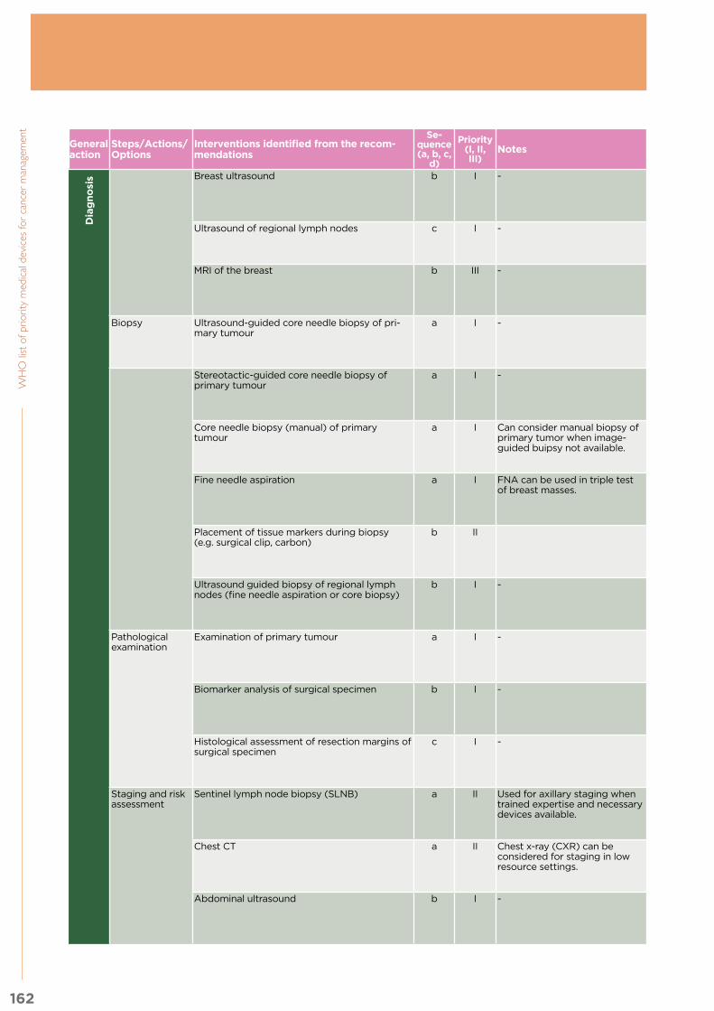
162
WHO
list o
f prio
rity m
edica
l dev
ices f
or ca
ncer
man
agem
ent
General action
Steps/Actions/Options
Interventions identified from the recom-mendations
Se-quence (a, b, c,
d)
Priority (I, II, III)
Notes General action When to perform it Source Source Source Source
Dia
gn
osi
s Breast ultrasound b I -
Dia
gn
osi
s May be in combination with mammography.
- BHGI Guidelines for Inter-national Breast Health and Cancer Control–Implemen-tation. Cancer 2008;113(S8): i–ix, 2215–2371
Early and locally ad-vanced breast cancer. NICE clinical guideline 80 (2014)
NCCN. Breast Can-cer Screening and Diagnosis. Version 1.2014
Ultrasound of regional lymph nodes c I - - - - Early and locally ad-vanced breast cancer. NICE clinical guideline 80 (2014)
-
MRI of the breast b III - Not routinely recom-mended. May be con-sidered in some cases and high-risk groups
BHGI Guidelines for Inter-national Breast Health and Cancer Control–Implemen-tation. Cancer 2008;113(S8): i–ix, 2215–2371
Early and locally ad-vanced breast cancer. NICE clinical guideline 80 (2014)
NCCN. Breast Can-cer Screening and Diagnosis. Version 1.2014
Biopsy Ultrasound-guided core needle biopsy of pri-mary tumour
a I - - BHGI Guidelines for Inter-national Breast Health and Cancer Control–Implemen-tation. Cancer 2008;113(S8): i–ix, 2215–2371
Early and locally ad-vanced breast cancer. NICE clinical guideline 80 (2014)
NCCN. Breast Can-cer Screening and Diagnosis. Version 1.2014
Stereotactic-guided core needle biopsy of primary tumour
a I - - BHGI Guidelines for Inter-national Breast Health and Cancer Control–Implemen-tation. Cancer 2008;113(S8): i–ix, 2215–2371
- -
Core needle biopsy (manual) of primary tumour
a I Can consider manual biopsy of primary tumor when image-guided buipsy not available.
If ultrasound- or stere-otactic-guided biopsy cannot be performed.
- BHGI Guidelines for Inter-national Breast Health and Cancer Control–Implemen-tation. Cancer 2008;113(S8): i–ix, 2215–2371
- -
Fine needle aspiration a I FNA can be used in triple test of breast masses.
If core needle biopsy cannot be performed.
- BHGI Guidelines for Inter-national Breast Health and Cancer Control–Implemen-tation. Cancer 2008;113(S8): i–ix, 2215–2371
Early and locally ad-vanced breast cancer. NICE clinical guideline 80 (2014)
NCCN. Breast Can-cer Screening and Diagnosis. Version 1.2014
Placement of tissue markers during biopsy (e.g. surgical clip, carbon)
b II If preoperative systemic therapy is planned.
- BHGI Guidelines for Inter-national Breast Health and Cancer Control–Implemen-tation. Cancer 2008;113(S8): i–ix, 2215–2371
- NCCN. Breast Can-cer Screening and Diagnosis. Version 1.2014
Ultrasound guided biopsy of regional lymph nodes (fine needle aspiration or core biopsy)
b I - If suspicious lymph nodes have been de-tected.
- BHGI Guidelines for Inter-national Breast Health and Cancer Control–Implemen-tation. Cancer 2008;113(S8): i–ix, 2215–2371
Early and locally ad-vanced breast cancer. NICE clinical guideline 80 (2014)
NCCN. Breast Can-cer Screening and Diagnosis. Version 1.2014
Pathological examination
Examination of primary tumour a I - - - BHGI Guidelines for Inter-national Breast Health and Cancer Control–Implemen-tation. Cancer 2008;113(S8): i–ix, 2215–2371
- NCCN. Breast Can-cer. Version 3.2014
Biomarker analysis of surgical specimen b I - - - BHGI Guidelines for Inter-national Breast Health and Cancer Control–Implemen-tation. Cancer 2008;113(S8): i–ix, 2215–2371
- NCCN. Breast Can-cer. Version 3.2014
Histological assessment of resection margins of surgical specimen
c I - - - BHGI Guidelines for Inter-national Breast Health and Cancer Control–Implemen-tation. Cancer 2008;113(S8): i–ix, 2215–2371
- NCCN. Breast Can-cer. Version 3.2014
Staging and risk assessment
Sentinel lymph node biopsy (SLNB) a II Used for axillary staging when trained expertise and necessary devices available.
- - BHGI Guidelines for Inter-national Breast Health and Cancer Control–Implemen-tation. Cancer 2008;113(S8): i–ix, 2215–2371
Early and locally ad-vanced breast cancer. NICE clinical guideline 80 (2014)
NCCN. Breast Can-cer. Version 3.2014
Chest CT a II Chest x-ray (CXR) can be considered for staging in low resource settings.
- - BHGI Guidelines for Inter-national Breast Health and Cancer Control–Implemen-tation. Cancer 2008;113(S8): i–ix, 2215–2371
- -
Abdominal ultrasound b I - When CT and MRI are not available.
- BHGI Guidelines for Inter-national Breast Health and Cancer Control–Implemen-tation. Cancer 2008;113(S8): i–ix, 2215–2371
- NCCN. Breast Can-cer. Version 3.2014
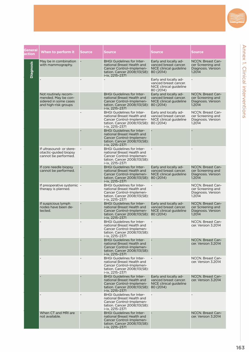
163
An
nex 1. C
linic
al in
terv
en
tion
s
General action
Steps/Actions/Options
Interventions identified from the recom-mendations
Se-quence (a, b, c,
d)
Priority (I, II, III)
Notes General action When to perform it Source Source Source Source
Dia
gn
osi
s Breast ultrasound b I -
Dia
gn
osi
s May be in combination with mammography.
- BHGI Guidelines for Inter-national Breast Health and Cancer Control–Implemen-tation. Cancer 2008;113(S8): i–ix, 2215–2371
Early and locally ad-vanced breast cancer. NICE clinical guideline 80 (2014)
NCCN. Breast Can-cer Screening and Diagnosis. Version 1.2014
Ultrasound of regional lymph nodes c I - - - - Early and locally ad-vanced breast cancer. NICE clinical guideline 80 (2014)
-
MRI of the breast b III - Not routinely recom-mended. May be con-sidered in some cases and high-risk groups
BHGI Guidelines for Inter-national Breast Health and Cancer Control–Implemen-tation. Cancer 2008;113(S8): i–ix, 2215–2371
Early and locally ad-vanced breast cancer. NICE clinical guideline 80 (2014)
NCCN. Breast Can-cer Screening and Diagnosis. Version 1.2014
Biopsy Ultrasound-guided core needle biopsy of pri-mary tumour
a I - - BHGI Guidelines for Inter-national Breast Health and Cancer Control–Implemen-tation. Cancer 2008;113(S8): i–ix, 2215–2371
Early and locally ad-vanced breast cancer. NICE clinical guideline 80 (2014)
NCCN. Breast Can-cer Screening and Diagnosis. Version 1.2014
Stereotactic-guided core needle biopsy of primary tumour
a I - - BHGI Guidelines for Inter-national Breast Health and Cancer Control–Implemen-tation. Cancer 2008;113(S8): i–ix, 2215–2371
- -
Core needle biopsy (manual) of primary tumour
a I Can consider manual biopsy of primary tumor when image-guided buipsy not available.
If ultrasound- or stere-otactic-guided biopsy cannot be performed.
- BHGI Guidelines for Inter-national Breast Health and Cancer Control–Implemen-tation. Cancer 2008;113(S8): i–ix, 2215–2371
- -
Fine needle aspiration a I FNA can be used in triple test of breast masses.
If core needle biopsy cannot be performed.
- BHGI Guidelines for Inter-national Breast Health and Cancer Control–Implemen-tation. Cancer 2008;113(S8): i–ix, 2215–2371
Early and locally ad-vanced breast cancer. NICE clinical guideline 80 (2014)
NCCN. Breast Can-cer Screening and Diagnosis. Version 1.2014
Placement of tissue markers during biopsy (e.g. surgical clip, carbon)
b II If preoperative systemic therapy is planned.
- BHGI Guidelines for Inter-national Breast Health and Cancer Control–Implemen-tation. Cancer 2008;113(S8): i–ix, 2215–2371
- NCCN. Breast Can-cer Screening and Diagnosis. Version 1.2014
Ultrasound guided biopsy of regional lymph nodes (fine needle aspiration or core biopsy)
b I - If suspicious lymph nodes have been de-tected.
- BHGI Guidelines for Inter-national Breast Health and Cancer Control–Implemen-tation. Cancer 2008;113(S8): i–ix, 2215–2371
Early and locally ad-vanced breast cancer. NICE clinical guideline 80 (2014)
NCCN. Breast Can-cer Screening and Diagnosis. Version 1.2014
Pathological examination
Examination of primary tumour a I - - - BHGI Guidelines for Inter-national Breast Health and Cancer Control–Implemen-tation. Cancer 2008;113(S8): i–ix, 2215–2371
- NCCN. Breast Can-cer. Version 3.2014
Biomarker analysis of surgical specimen b I - - - BHGI Guidelines for Inter-national Breast Health and Cancer Control–Implemen-tation. Cancer 2008;113(S8): i–ix, 2215–2371
- NCCN. Breast Can-cer. Version 3.2014
Histological assessment of resection margins of surgical specimen
c I - - - BHGI Guidelines for Inter-national Breast Health and Cancer Control–Implemen-tation. Cancer 2008;113(S8): i–ix, 2215–2371
- NCCN. Breast Can-cer. Version 3.2014
Staging and risk assessment
Sentinel lymph node biopsy (SLNB) a II Used for axillary staging when trained expertise and necessary devices available.
- - BHGI Guidelines for Inter-national Breast Health and Cancer Control–Implemen-tation. Cancer 2008;113(S8): i–ix, 2215–2371
Early and locally ad-vanced breast cancer. NICE clinical guideline 80 (2014)
NCCN. Breast Can-cer. Version 3.2014
Chest CT a II Chest x-ray (CXR) can be considered for staging in low resource settings.
- - BHGI Guidelines for Inter-national Breast Health and Cancer Control–Implemen-tation. Cancer 2008;113(S8): i–ix, 2215–2371
- -
Abdominal ultrasound b I - When CT and MRI are not available.
- BHGI Guidelines for Inter-national Breast Health and Cancer Control–Implemen-tation. Cancer 2008;113(S8): i–ix, 2215–2371
- NCCN. Breast Can-cer. Version 3.2014

164
WHO
list o
f prio
rity m
edica
l dev
ices f
or ca
ncer
man
agem
ent
General action
Steps/Actions/Options
Interventions identified from the recom-mendations
Se-quence (a, b, c,
d)
Priority (I, II, III)
Notes General action When to perform it Source Source Source Source
Dia
gn
osi
s CT scan (whole body) c II Staging with CT scan of chest and abdomen done according to symptoms and clinical stage.
Dia
gn
osi
s - - BHGI Guidelines for Inter-national Breast Health and Cancer Control–Implemen-tation. Cancer 2008;113(S8): i–ix, 2215–2371
Advanced breast cancer. NICE clinical guideline 81 (2014)
NCCN. Breast Can-cer. Version 3.2014
Bone scan (bone scintigraphy) d II X-ray of symptomatic bone sites can be considered in low-resourced settings.
- - BHGI Guidelines for Inter-national Breast Health and Cancer Control–Implemen-tation. Cancer 2008;113(S8): i–ix, 2215–2371
Advanced breast cancer. NICE clinical guideline 81 (2014)
NCCN. Breast Can-cer. Version 3.2014
MRI scan e III - - - BHGI Guidelines for Inter-national Breast Health and Cancer Control–Implemen-tation. Cancer 2008;113(S8): i–ix, 2215–2371
Advanced breast cancer. NICE clinical guideline 81 (2014)
NCCN. Breast Can-cer. Version 3.2014
Specimen radiography f I Specimen radiograph used to confirm appropriate resection in settings where lumpectomy performed and lesion identifiable or localized on mammogram.
- - BHGI Guidelines for Inter-national Breast Health and Cancer Control–Implemen-tation. Cancer 2008;113(S8): i–ix, 2215–2371
- NCCN. Breast Can-cer. Version 3.2014
PET/CT g III - Assessment with fluo-rodeoxyglucose (FDG) is considered optional (NCCN).
- BHGI Guidelines for Inter-national Breast Health and Cancer Control–Implemen-tation. Cancer 2008;113(S8): i–ix, 2215–2371
Advanced breast cancer. NICE clinical guideline 81 (2014)
NCCN. Breast Can-cer. Version 3.2014
SPECT with Technetium (99mTc) sestamibi h III - - - BHGI Guidelines for Inter-national Breast Health and Cancer Control–Implemen-tation. Cancer 2008;113(S8): i–ix, 2215–2371
- -
Tre
atm
en
t Mastectomy a I -
Tre
atm
en
t - - BHGI Guidelines for Inter-national Breast Health and Cancer Control–Implemen-tation. Cancer 2008;113(S8): i–ix, 2215–2371
Early and locally ad-vanced breast cancer. NICE clinical guideline 80 (2014)
NCCN. Breast Can-cer. Version 3.2014
Surgery Breast-conservation surgery (lumpectomy) a I Lumpectomy can be considered when radiotherapy is generally available for post-lumpectomy radiotherapy.
- - BHGI Guidelines for Inter-national Breast Health and Cancer Control–Implemen-tation. Cancer 2008;113(S8): i–ix, 2215–2371
Early and locally ad-vanced breast cancer. NICE clinical guideline 80 (2014)
NCCN. Breast Can-cer. Version 3.2014
Breast reconstruction b I Breast reconstruction can be considered using a variety of techniques with differing levels of complexity and resource requirements.
Should be proposed (immediate or delayed) after mastectomy.
- BHGI Guidelines for Inter-national Breast Health and Cancer Control–Implemen-tation. Cancer 2008;113(S8): i–ix, 2215–2371
Early and locally ad-vanced breast cancer. NICE clinical guideline 80 (2014)
NCCN. Breast Can-cer. Version 3.2014
Radiation ther-apy
Postoperative radiation therapy a I - Recommended after breast-conservation surgery. Recommend-ed after mastectomy if high-risk of local recurrence.
- BHGI Guidelines for Inter-national Breast Health and Cancer Control–Implemen-tation. Cancer 2008;113(S8): i–ix, 2215–2371
Early and locally ad-vanced breast cancer. NICE clinical guideline 80 (2014)
NCCN. Breast Can-cer. Version 3.2014
Adjuvant sys-temic treatment
Chemotherapy a I - Depending on the intrinsic phenotype determined by ER and HER2 assessment.
- BHGI Guidelines for Inter-national Breast Health and Cancer Control–Implemen-tation. Cancer 2008;113(S8): i–ix, 2215–2371
Early and locally ad-vanced breast cancer. NICE clinical guideline 80 (2014)
NCCN. Breast Can-cer. Version 3.2014
Endocrine therapy a I - Depending on the intrinsic phenotype determined by ER and HER2 assessment.
- BHGI Guidelines for Inter-national Breast Health and Cancer Control–Implemen-tation. Cancer 2008;113(S8): i–ix, 2215–2371
Early and locally ad-vanced breast cancer. NICE clinical guideline 80 (2014)
NCCN. Breast Can-cer. Version 3.2014
Biological therapy a I Biological or targeted therapies can be considered according to WHO List of Essential Medicines.
Depending on the intrinsic phenotype determined by ER and HER2 assessment.
- BHGI Guidelines for Inter-national Breast Health and Cancer Control–Implemen-tation. Cancer 2008;113(S8): i–ix, 2215–2371
Early and locally ad-vanced breast cancer. NICE clinical guideline 80 (2014)
NCCN. Breast Can-cer. Version 3.2014

165
An
nex 1. C
linic
al in
terv
en
tion
s
General action
Steps/Actions/Options
Interventions identified from the recom-mendations
Se-quence (a, b, c,
d)
Priority (I, II, III)
Notes General action When to perform it Source Source Source Source
Dia
gn
osi
s CT scan (whole body) c II Staging with CT scan of chest and abdomen done according to symptoms and clinical stage.
Dia
gn
osi
s - - BHGI Guidelines for Inter-national Breast Health and Cancer Control–Implemen-tation. Cancer 2008;113(S8): i–ix, 2215–2371
Advanced breast cancer. NICE clinical guideline 81 (2014)
NCCN. Breast Can-cer. Version 3.2014
Bone scan (bone scintigraphy) d II X-ray of symptomatic bone sites can be considered in low-resourced settings.
- - BHGI Guidelines for Inter-national Breast Health and Cancer Control–Implemen-tation. Cancer 2008;113(S8): i–ix, 2215–2371
Advanced breast cancer. NICE clinical guideline 81 (2014)
NCCN. Breast Can-cer. Version 3.2014
MRI scan e III - - - BHGI Guidelines for Inter-national Breast Health and Cancer Control–Implemen-tation. Cancer 2008;113(S8): i–ix, 2215–2371
Advanced breast cancer. NICE clinical guideline 81 (2014)
NCCN. Breast Can-cer. Version 3.2014
Specimen radiography f I Specimen radiograph used to confirm appropriate resection in settings where lumpectomy performed and lesion identifiable or localized on mammogram.
- - BHGI Guidelines for Inter-national Breast Health and Cancer Control–Implemen-tation. Cancer 2008;113(S8): i–ix, 2215–2371
- NCCN. Breast Can-cer. Version 3.2014
PET/CT g III - Assessment with fluo-rodeoxyglucose (FDG) is considered optional (NCCN).
- BHGI Guidelines for Inter-national Breast Health and Cancer Control–Implemen-tation. Cancer 2008;113(S8): i–ix, 2215–2371
Advanced breast cancer. NICE clinical guideline 81 (2014)
NCCN. Breast Can-cer. Version 3.2014
SPECT with Technetium (99mTc) sestamibi h III - - - BHGI Guidelines for Inter-national Breast Health and Cancer Control–Implemen-tation. Cancer 2008;113(S8): i–ix, 2215–2371
- -
Tre
atm
en
t Mastectomy a I -
Tre
atm
en
t - - BHGI Guidelines for Inter-national Breast Health and Cancer Control–Implemen-tation. Cancer 2008;113(S8): i–ix, 2215–2371
Early and locally ad-vanced breast cancer. NICE clinical guideline 80 (2014)
NCCN. Breast Can-cer. Version 3.2014
Surgery Breast-conservation surgery (lumpectomy) a I Lumpectomy can be considered when radiotherapy is generally available for post-lumpectomy radiotherapy.
- - BHGI Guidelines for Inter-national Breast Health and Cancer Control–Implemen-tation. Cancer 2008;113(S8): i–ix, 2215–2371
Early and locally ad-vanced breast cancer. NICE clinical guideline 80 (2014)
NCCN. Breast Can-cer. Version 3.2014
Breast reconstruction b I Breast reconstruction can be considered using a variety of techniques with differing levels of complexity and resource requirements.
Should be proposed (immediate or delayed) after mastectomy.
- BHGI Guidelines for Inter-national Breast Health and Cancer Control–Implemen-tation. Cancer 2008;113(S8): i–ix, 2215–2371
Early and locally ad-vanced breast cancer. NICE clinical guideline 80 (2014)
NCCN. Breast Can-cer. Version 3.2014
Radiation ther-apy
Postoperative radiation therapy a I - Recommended after breast-conservation surgery. Recommend-ed after mastectomy if high-risk of local recurrence.
- BHGI Guidelines for Inter-national Breast Health and Cancer Control–Implemen-tation. Cancer 2008;113(S8): i–ix, 2215–2371
Early and locally ad-vanced breast cancer. NICE clinical guideline 80 (2014)
NCCN. Breast Can-cer. Version 3.2014
Adjuvant sys-temic treatment
Chemotherapy a I - Depending on the intrinsic phenotype determined by ER and HER2 assessment.
- BHGI Guidelines for Inter-national Breast Health and Cancer Control–Implemen-tation. Cancer 2008;113(S8): i–ix, 2215–2371
Early and locally ad-vanced breast cancer. NICE clinical guideline 80 (2014)
NCCN. Breast Can-cer. Version 3.2014
Endocrine therapy a I - Depending on the intrinsic phenotype determined by ER and HER2 assessment.
- BHGI Guidelines for Inter-national Breast Health and Cancer Control–Implemen-tation. Cancer 2008;113(S8): i–ix, 2215–2371
Early and locally ad-vanced breast cancer. NICE clinical guideline 80 (2014)
NCCN. Breast Can-cer. Version 3.2014
Biological therapy a I Biological or targeted therapies can be considered according to WHO List of Essential Medicines.
Depending on the intrinsic phenotype determined by ER and HER2 assessment.
- BHGI Guidelines for Inter-national Breast Health and Cancer Control–Implemen-tation. Cancer 2008;113(S8): i–ix, 2215–2371
Early and locally ad-vanced breast cancer. NICE clinical guideline 80 (2014)
NCCN. Breast Can-cer. Version 3.2014

166
WHO
list o
f prio
rity m
edica
l dev
ices f
or ca
ncer
man
agem
ent
General action
Steps/Actions/Options
Interventions identified from the recom-mendations
Se-quence (a, b, c,
d)
Priority (I, II, III)
Notes General action When to perform it Source Source Source Source
Fo
llo
w u
p Imaging Mammography a I -
Fo
llo
w u
p - - - Early and locally ad-vanced breast cancer. NICE clinical guideline 80 (2014)
NCCN. Breast Can-cer. Version 3.2014
MRI scan b III - In case of recurrent disease.
- - - NCCN. Breast Can-cer. Version 3.2014
Bone density scan (bone densitometry) b I - Recommended for patients on aromatase inhibitor therapy.
- - Early and locally ad-vanced breast cancer. NICE clinical guideline 80 (2014)
NCCN. Breast Can-cer. Version 3.2014
Chest CT b III CT scan can be considered according to symptoms.
In case of recurrent disease for assessment of metastasis.
- - - NCCN. Breast Can-cer. Version 3.2014
Abdominal and/or pelvic CT b III CT scan can be considered according to symptoms.
In case of recurrent disease for assessment of metastasis.
- - - NCCN. Breast Can-cer. Version 3.2014
Brain MRI b III Brain MRI can be considered according to symptoms and resource availability.
In case of recurrent disease for assessment of metastasis.
- - - NCCN. Breast Can-cer. Version 3.2014
Blood testing Complete blood count and blood chemistry tests (liver and renal function tests, alkaline phosphatase and calcium)
b I - In case of recurrent disease.
- - - NCCN. Breast Can-cer. Version 3.2014
Physical examination
Gynaecological examination b I - For patients on tamox-ifen.
- - - NCCN. Breast Can-cer. Version 3.2014
Pallia
tive
ca
re
Palliative radiotherapy
Palliative breast irradiation a I -
Pallia
tive
ca
re
- - - - NCCN. Breast Can-cer. Version 3.2014
Palliative system-ic therapy
Biphosphonates therapy a I - - - - - NCCN. Breast Can-cer. Version 3.2014
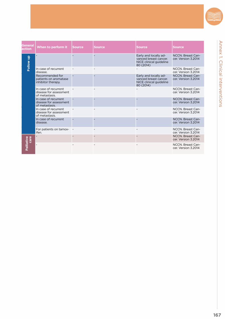
167
An
nex 1. C
linic
al in
terv
en
tion
s
General action
Steps/Actions/Options
Interventions identified from the recom-mendations
Se-quence (a, b, c,
d)
Priority (I, II, III)
Notes General action When to perform it Source Source Source Source
Fo
llo
w u
p Imaging Mammography a I -
Fo
llo
w u
p - - - Early and locally ad-vanced breast cancer. NICE clinical guideline 80 (2014)
NCCN. Breast Can-cer. Version 3.2014
MRI scan b III - In case of recurrent disease.
- - - NCCN. Breast Can-cer. Version 3.2014
Bone density scan (bone densitometry) b I - Recommended for patients on aromatase inhibitor therapy.
- - Early and locally ad-vanced breast cancer. NICE clinical guideline 80 (2014)
NCCN. Breast Can-cer. Version 3.2014
Chest CT b III CT scan can be considered according to symptoms.
In case of recurrent disease for assessment of metastasis.
- - - NCCN. Breast Can-cer. Version 3.2014
Abdominal and/or pelvic CT b III CT scan can be considered according to symptoms.
In case of recurrent disease for assessment of metastasis.
- - - NCCN. Breast Can-cer. Version 3.2014
Brain MRI b III Brain MRI can be considered according to symptoms and resource availability.
In case of recurrent disease for assessment of metastasis.
- - - NCCN. Breast Can-cer. Version 3.2014
Blood testing Complete blood count and blood chemistry tests (liver and renal function tests, alkaline phosphatase and calcium)
b I - In case of recurrent disease.
- - - NCCN. Breast Can-cer. Version 3.2014
Physical examination
Gynaecological examination b I - For patients on tamox-ifen.
- - - NCCN. Breast Can-cer. Version 3.2014
Pallia
tive
ca
re
Palliative radiotherapy
Palliative breast irradiation a I -
Pallia
tive
ca
re
- - - - NCCN. Breast Can-cer. Version 3.2014
Palliative system-ic therapy
Biphosphonates therapy a I - - - - - NCCN. Breast Can-cer. Version 3.2014
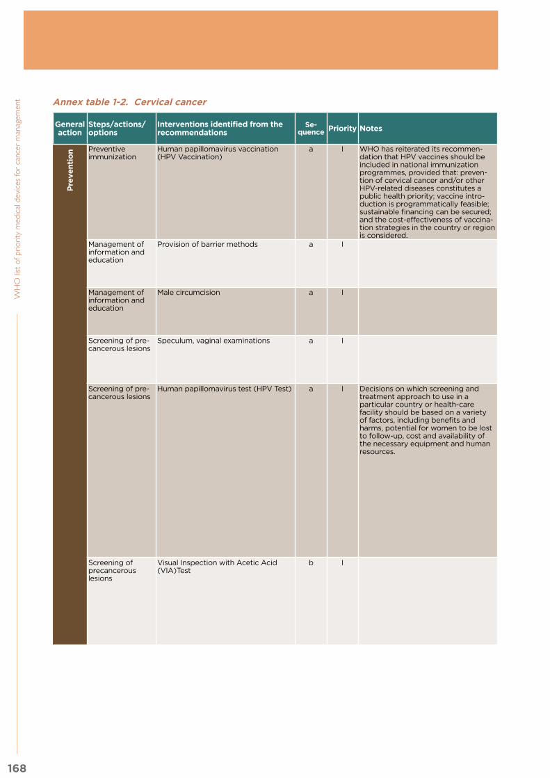
168
WHO
list o
f prio
rity m
edica
l dev
ices f
or ca
ncer
man
agem
ent Annex table 1-2. Cervical cancer
General action
Steps/actions/options
Interventions identified from the recommendations
Se-quence Priority Notes General
action When to perform it Source Support Services
Medical devices (without classification) CL HC DH SH
Pre
ven
tio
n Preventive immunization
Human papillomavirus vaccination (HPV Vaccination)
a I WHO has reiterated its recommen-dation that HPV vaccines should be included in national immunization programmes, provided that: preven-tion of cervical cancer and/or other HPV-related diseases constitutes a public health priority; vaccine intro-duction is programmatically feasible; sustainable financing can be secured; and the cost-effectiveness of vaccina-tion strategies in the country or region is considered.
Pre
ven
tio
n World Health Organi-zation (WHO). Human papillomavirus vaccines: WHO position paper, October 2014.
x
Management of information and education
Provision of barrier methods a I World Health Organiza-tion (WHO). Comprehen-sive Cervical Cancer Con-trol. A guide to essential practice Geneva: WHO; 2017
x x
Management of information and education
Male circumcision a I World Health Organiza-tion (WHO). Comprehen-sive Cervical Cancer Con-trol. A guide to essential practice Geneva: WHO; 2017
Screening of pre-cancerous lesions
Speculum, vaginal examinations a I World Health Organiza-tion (WHO). Comprehen-sive Cervical Cancer Con-trol. A guide to essential practice Geneva: WHO; 2017
x
Screening of pre-cancerous lesions
Human papillomavirus test (HPV Test) a I Decisions on which screening and treatment approach to use in a particular country or health-care facility should be based on a variety of factors, including benefits and harms, potential for women to be lost to follow-up, cost and availability of the necessary equipment and human resources.
A strategy of screening with HPV and treat with cryotherapy (Or leep when not eligible with cryotherapy) over a strategy of screen with VIA and over a strategy of screen with cytology followed by colposcopy (with or without biopsy) and over a strategy of screen with and HPV test followed by col-poscopy and treat with cryotherapy (Or leep when not eligible with cryotherapy) or either a strategy of HPV Test fol-lowed by VIA and treat with cryotherapy (Or leep when not eligible with cryotherapy)
World Health Organ-ization (WHO). WHO guidelines for screening and treatment of precan-cerous lesions for cervical cancer prevention. Ge-neva: WHO; 2013 World Health Organization (WHO). Comprehensive Cervical Cancer Control. A guide to essential prac-tice Geneva: WHO; 2014
Histology service
Disposable or high level disin-fected examination gloves, a small brush or soft swab swabs and a labelled vessel with pre-servative solution, Hystopatho-logical analysis
x x
Screening of precancerous lesions
Visual Inspection with Acetic Acid (VIA)Test
b I In resource constrained settings where screen-ing with an HPV test is not feasible and over a strategy of screen with cytology followed by colposcopy (with or without biopsy) and treat with cryotherapy (Or leep when not eligi-ble with cryotherapy)
World Health Organ-ization (WHO). WHO guidelines for screening and treatment of precan-cerous lesions for cervical cancer prevention. Gene-va: WHO; 2013
A bright light source to examine the cervix, an examination table covered by clean paper or cloth, a speculum, high-level disinfect-ed (need not be sterile), dispos-able or high-level disinfected examination gloves (need not be sterile, 3-5% acetic acid solution, large cotton swab
x

169
An
nex 1. C
linic
al in
terv
en
tion
s
Annex table 1-2. Cervical cancer
General action
Steps/actions/options
Interventions identified from the recommendations
Se-quence Priority Notes General
action When to perform it Source Support Services
Medical devices (without classification) CL HC DH SH
Pre
ven
tio
n Preventive immunization
Human papillomavirus vaccination (HPV Vaccination)
a I WHO has reiterated its recommen-dation that HPV vaccines should be included in national immunization programmes, provided that: preven-tion of cervical cancer and/or other HPV-related diseases constitutes a public health priority; vaccine intro-duction is programmatically feasible; sustainable financing can be secured; and the cost-effectiveness of vaccina-tion strategies in the country or region is considered.
Pre
ven
tio
n World Health Organi-zation (WHO). Human papillomavirus vaccines: WHO position paper, October 2014.
x
Management of information and education
Provision of barrier methods a I World Health Organiza-tion (WHO). Comprehen-sive Cervical Cancer Con-trol. A guide to essential practice Geneva: WHO; 2017
x x
Management of information and education
Male circumcision a I World Health Organiza-tion (WHO). Comprehen-sive Cervical Cancer Con-trol. A guide to essential practice Geneva: WHO; 2017
Screening of pre-cancerous lesions
Speculum, vaginal examinations a I World Health Organiza-tion (WHO). Comprehen-sive Cervical Cancer Con-trol. A guide to essential practice Geneva: WHO; 2017
x
Screening of pre-cancerous lesions
Human papillomavirus test (HPV Test) a I Decisions on which screening and treatment approach to use in a particular country or health-care facility should be based on a variety of factors, including benefits and harms, potential for women to be lost to follow-up, cost and availability of the necessary equipment and human resources.
A strategy of screening with HPV and treat with cryotherapy (Or leep when not eligible with cryotherapy) over a strategy of screen with VIA and over a strategy of screen with cytology followed by colposcopy (with or without biopsy) and over a strategy of screen with and HPV test followed by col-poscopy and treat with cryotherapy (Or leep when not eligible with cryotherapy) or either a strategy of HPV Test fol-lowed by VIA and treat with cryotherapy (Or leep when not eligible with cryotherapy)
World Health Organ-ization (WHO). WHO guidelines for screening and treatment of precan-cerous lesions for cervical cancer prevention. Ge-neva: WHO; 2013 World Health Organization (WHO). Comprehensive Cervical Cancer Control. A guide to essential prac-tice Geneva: WHO; 2014
Histology service
Disposable or high level disin-fected examination gloves, a small brush or soft swab swabs and a labelled vessel with pre-servative solution, Hystopatho-logical analysis
x x
Screening of precancerous lesions
Visual Inspection with Acetic Acid (VIA)Test
b I In resource constrained settings where screen-ing with an HPV test is not feasible and over a strategy of screen with cytology followed by colposcopy (with or without biopsy) and treat with cryotherapy (Or leep when not eligi-ble with cryotherapy)
World Health Organ-ization (WHO). WHO guidelines for screening and treatment of precan-cerous lesions for cervical cancer prevention. Gene-va: WHO; 2013
A bright light source to examine the cervix, an examination table covered by clean paper or cloth, a speculum, high-level disinfect-ed (need not be sterile), dispos-able or high-level disinfected examination gloves (need not be sterile, 3-5% acetic acid solution, large cotton swab
x

170
WHO
list o
f prio
rity m
edica
l dev
ices f
or ca
ncer
man
agem
ent
General action
Steps/actions/options
Interventions identified from the recommendations
Se-quence Priority Notes General
action When to perform it Source Support Services
Medical devices (without classification) CL HC DH SH
Pre
ven
tio
n Screening of precancerous lesions
Screen with cytology (Pap test) b I
Pre
ven
tio
n World Health Organ-ization (WHO). WHO guidelines for screening and treatment of precan-cerous lesions for cervical cancer prevention. Gene-va: WHO; 2013
Cytology services
A bright light source to examine the cervix, an examination table covered by clean paper or cloth, a speculum, high-level disinfect-ed (need not be sterile), dispos-able or high-level disinfected examination gloves (need not be sterile, An extended-tip wooden or plastic spatula or a brush for sampling (see Figure PS5.6.1), a glass slide with frosted edge, and fixative spray or solution, a small container of warm water to lubricate and warm the spec-ulum, 0.5% chlorine solution for decontaminating instruments and gloves, the use of a specially design devices that sample the area so the transformation zone is recommended, wooden or plastic spatula, moistened cotton swab or a brush -type endocervical sampler device (cytobrush)
x x
Screening of precancerous lesions
Screen with cytology (Liquid Based Cytology LBC)
b I World Health Organ-ization (WHO). WHO guidelines for screening and treatment of precan-cerous lesions for cervical cancer prevention. Gene-va: WHO; 2013
Cytology services
A bright light source to exam-ine the cervix, an examination table covered by clean paper or cloth, a speculum, high-level disinfected (need not be sterile), disposable or high-level disin-fected examination gloves (need not be sterile A tube containing a special preservative solution, a small container of warm water to lubricate and warm the spec-ulum, 0.5% chlorine solution for decontaminating instruments and gloves.
x x
Screening of precancerous lesions
Colposcopy (with or without biopsy) b I World Health Organ-ization (WHO). WHO guidelines for screening and treatment of precan-cerous lesions for cervical cancer prevention. Gene-va: WHO; 2013
Cytology services
A bright light source to exam-ine the cervix, an examination table covered by clean paper or cloth, a speculum, high-level disinfected (need not be sterile), disposable or high-level disin-fected examination gloves (need not be sterile, Colposcope, all necessary supplies for infection prevention because biopsies and/or endocervical curettage (ECC) are usually performed during colposcopy, you may also need 3–5% acetic acid; Monsel’s paste (see Annex 13); punch biopsy forceps; endocervical curette; ring forceps; cotton swabs; specimen bottles with 10% formalin
x
Screening of pre-cancerous lesions
Screen with cytology followed by colposcopy
b I World Health Organ-ization (WHO). WHO guidelines for screening and treatment of precan-cerous lesions for cervical cancer prevention. Gene-va: WHO; 2013
Cytology services
x
Screening of pre-cancerous lesions
HPV followed by colposcopy b I World Health Organ-ization (WHO). WHO guidelines for screening and treatment of precan-cerous lesions for cervical cancer prevention. Gene-va: WHO; 2013
Cytology services
x
Screening of pre-cancerous lesions
HPV followed by VIA b I World Health Organ-ization (WHO). WHO guidelines for screening and treatment of precan-cerous lesions for cervical cancer prevention. Gene-va: WHO; 2013
x

171
An
nex 1. C
linic
al in
terv
en
tion
s
General action
Steps/actions/options
Interventions identified from the recommendations
Se-quence Priority Notes General
action When to perform it Source Support Services
Medical devices (without classification) CL HC DH SH
Pre
ven
tio
n Screening of precancerous lesions
Screen with cytology (Pap test) b I
Pre
ven
tio
n World Health Organ-ization (WHO). WHO guidelines for screening and treatment of precan-cerous lesions for cervical cancer prevention. Gene-va: WHO; 2013
Cytology services
A bright light source to examine the cervix, an examination table covered by clean paper or cloth, a speculum, high-level disinfect-ed (need not be sterile), dispos-able or high-level disinfected examination gloves (need not be sterile, An extended-tip wooden or plastic spatula or a brush for sampling (see Figure PS5.6.1), a glass slide with frosted edge, and fixative spray or solution, a small container of warm water to lubricate and warm the spec-ulum, 0.5% chlorine solution for decontaminating instruments and gloves, the use of a specially design devices that sample the area so the transformation zone is recommended, wooden or plastic spatula, moistened cotton swab or a brush -type endocervical sampler device (cytobrush)
x x
Screening of precancerous lesions
Screen with cytology (Liquid Based Cytology LBC)
b I World Health Organ-ization (WHO). WHO guidelines for screening and treatment of precan-cerous lesions for cervical cancer prevention. Gene-va: WHO; 2013
Cytology services
A bright light source to exam-ine the cervix, an examination table covered by clean paper or cloth, a speculum, high-level disinfected (need not be sterile), disposable or high-level disin-fected examination gloves (need not be sterile A tube containing a special preservative solution, a small container of warm water to lubricate and warm the spec-ulum, 0.5% chlorine solution for decontaminating instruments and gloves.
x x
Screening of precancerous lesions
Colposcopy (with or without biopsy) b I World Health Organ-ization (WHO). WHO guidelines for screening and treatment of precan-cerous lesions for cervical cancer prevention. Gene-va: WHO; 2013
Cytology services
A bright light source to exam-ine the cervix, an examination table covered by clean paper or cloth, a speculum, high-level disinfected (need not be sterile), disposable or high-level disin-fected examination gloves (need not be sterile, Colposcope, all necessary supplies for infection prevention because biopsies and/or endocervical curettage (ECC) are usually performed during colposcopy, you may also need 3–5% acetic acid; Monsel’s paste (see Annex 13); punch biopsy forceps; endocervical curette; ring forceps; cotton swabs; specimen bottles with 10% formalin
x
Screening of pre-cancerous lesions
Screen with cytology followed by colposcopy
b I World Health Organ-ization (WHO). WHO guidelines for screening and treatment of precan-cerous lesions for cervical cancer prevention. Gene-va: WHO; 2013
Cytology services
x
Screening of pre-cancerous lesions
HPV followed by colposcopy b I World Health Organ-ization (WHO). WHO guidelines for screening and treatment of precan-cerous lesions for cervical cancer prevention. Gene-va: WHO; 2013
Cytology services
x
Screening of pre-cancerous lesions
HPV followed by VIA b I World Health Organ-ization (WHO). WHO guidelines for screening and treatment of precan-cerous lesions for cervical cancer prevention. Gene-va: WHO; 2013
x

172
WHO
list o
f prio
rity m
edica
l dev
ices f
or ca
ncer
man
agem
ent
General action
Steps/actions/options
Interventions identified from the recommendations
Se-quence Priority Notes General
action When to perform it Source Support Services
Medical devices (without classification) CL HC DH SH
Pre
ven
tio
n Treatment of pre-cancerous lesions
Cryotherapy c I
Pre
ven
tio
n World Health Organ-ization (WHO). WHO guidelines for screening and treatment of precan-cerous lesions for cervical cancer prevention. Gene-va: WHO; 2013
Histology service
Speculum, high-level disinfected (need not be sterile); dispos-able or high-level disinfected examination gloves (need not be sterile); cotton swabs for wiping the cervix; normal saline solu-tion; colposcope, if used in the particular venue; cryosurgery unit with adequate gas supply (carbon dioxide or nitrous oxide) and with the equipment components: 1)Probe or Cryo-probe 2) Trigger, 3)Handle grip (fibreglass), 4) Yoke, 5) Inlet of gas from cylinder 6) Tightening knob, 7) Pressure gauge show-ing cylinder pressure 8) Silencer (outlet) 9) Gas-conveying tube 10) Probe tip
x x
Treatment of pre-cancerous lesions
Large loop excision of transformation zone (LEEP/LLETZ)
c I World Health Organiza-tion (WHO). Comprehen-sive Cervical Cancer Con-trol. A guide to essential practice Geneva: WHO; 2013
Histology service
Reliable power supply, electro-surgical generator and electrode handle, colposcope, non-con-ducting speculum, preferably with side retractors, return electrode, wire electrodes of several sizes (see Figure PS5.11.1), coagulating/ball electrode, smoke evacuator, forceps, local anaesthetic: 1% or 2% lidocaine, with or without 1:100 000 epinephrin, 5-ml syringes with long 27-gauge needles, bottles with normal saline and with 5% acetic acid, Monsel’s paste (see Annex 13), large swabs, needles and suture material, specimen containers with 10% formalin, Different types and sizes of electrodes (a) ball electrode, (b) square loop electrode, (c) semi-circular loop electrode,
x
Treatment of pre-cancerous lesions
Cold knife conization (CKC) c I Recommends against their use in a screen and treat strategy
World Health Organiza-tion (WHO). Comprehen-sive Cervical Cancer Con-trol. A guide to essential practice Geneva: WHO; 2013
Histology service
x
Dia
gn
osi
s Definitive diagnosis of invasive cervical cancer in second care level
Pelvic examinations a I
Dia
gn
osi
s World Health Organiza-tion (WHO). Comprehen-sive Cervical Cancer Con-trol. A guide to essential practice Geneva: WHO; 2013
x x
Definitive diagnosis of invasive cervical cancer in second care level
Pregnancy test b I If appropriate for the patient before taking a biopsy
World Health Organiza-tion (WHO). Comprehen-sive Cervical Cancer Con-trol. A guide to essential practice Geneva: WHO; 2013
Clinical Labora-tory
x x
Definitive diagnosis of invasive cervical cancer in second care level
HIV Test c I If appropriate for the patient before taking a biopsy
World Health Organiza-tion (WHO). Comprehen-sive Cervical Cancer Con-trol. A guide to essential practice Geneva: WHO; 2013
Clinical Labora-tory
HIV Test x x
Definitive diagnosis of invasive cervical cancer in second care level
Biopsy d I World Health Organiza-tion (WHO). Comprehen-sive Cervical Cancer Con-trol. A guide to essential practice Geneva: WHO; 2013
Histology service
x x
Definitive diagnosis of invasive cervical cancer in second care level
Endocervical curettage ECC d I World Health Organiza-tion (WHO). Comprehen-sive Cervical Cancer Con-trol. A guide to essential practice Geneva: WHO; 2013
x x

173
An
nex 1. C
linic
al in
terv
en
tion
s
General action
Steps/actions/options
Interventions identified from the recommendations
Se-quence Priority Notes General
action When to perform it Source Support Services
Medical devices (without classification) CL HC DH SH
Pre
ven
tio
n Treatment of pre-cancerous lesions
Cryotherapy c I
Pre
ven
tio
n World Health Organ-ization (WHO). WHO guidelines for screening and treatment of precan-cerous lesions for cervical cancer prevention. Gene-va: WHO; 2013
Histology service
Speculum, high-level disinfected (need not be sterile); dispos-able or high-level disinfected examination gloves (need not be sterile); cotton swabs for wiping the cervix; normal saline solu-tion; colposcope, if used in the particular venue; cryosurgery unit with adequate gas supply (carbon dioxide or nitrous oxide) and with the equipment components: 1)Probe or Cryo-probe 2) Trigger, 3)Handle grip (fibreglass), 4) Yoke, 5) Inlet of gas from cylinder 6) Tightening knob, 7) Pressure gauge show-ing cylinder pressure 8) Silencer (outlet) 9) Gas-conveying tube 10) Probe tip
x x
Treatment of pre-cancerous lesions
Large loop excision of transformation zone (LEEP/LLETZ)
c I World Health Organiza-tion (WHO). Comprehen-sive Cervical Cancer Con-trol. A guide to essential practice Geneva: WHO; 2013
Histology service
Reliable power supply, electro-surgical generator and electrode handle, colposcope, non-con-ducting speculum, preferably with side retractors, return electrode, wire electrodes of several sizes (see Figure PS5.11.1), coagulating/ball electrode, smoke evacuator, forceps, local anaesthetic: 1% or 2% lidocaine, with or without 1:100 000 epinephrin, 5-ml syringes with long 27-gauge needles, bottles with normal saline and with 5% acetic acid, Monsel’s paste (see Annex 13), large swabs, needles and suture material, specimen containers with 10% formalin, Different types and sizes of electrodes (a) ball electrode, (b) square loop electrode, (c) semi-circular loop electrode,
x
Treatment of pre-cancerous lesions
Cold knife conization (CKC) c I Recommends against their use in a screen and treat strategy
World Health Organiza-tion (WHO). Comprehen-sive Cervical Cancer Con-trol. A guide to essential practice Geneva: WHO; 2013
Histology service
x
Dia
gn
osi
s Definitive diagnosis of invasive cervical cancer in second care level
Pelvic examinations a I
Dia
gn
osi
s World Health Organiza-tion (WHO). Comprehen-sive Cervical Cancer Con-trol. A guide to essential practice Geneva: WHO; 2013
x x
Definitive diagnosis of invasive cervical cancer in second care level
Pregnancy test b I If appropriate for the patient before taking a biopsy
World Health Organiza-tion (WHO). Comprehen-sive Cervical Cancer Con-trol. A guide to essential practice Geneva: WHO; 2013
Clinical Labora-tory
x x
Definitive diagnosis of invasive cervical cancer in second care level
HIV Test c I If appropriate for the patient before taking a biopsy
World Health Organiza-tion (WHO). Comprehen-sive Cervical Cancer Con-trol. A guide to essential practice Geneva: WHO; 2013
Clinical Labora-tory
HIV Test x x
Definitive diagnosis of invasive cervical cancer in second care level
Biopsy d I World Health Organiza-tion (WHO). Comprehen-sive Cervical Cancer Con-trol. A guide to essential practice Geneva: WHO; 2013
Histology service
x x
Definitive diagnosis of invasive cervical cancer in second care level
Endocervical curettage ECC d I World Health Organiza-tion (WHO). Comprehen-sive Cervical Cancer Con-trol. A guide to essential practice Geneva: WHO; 2013
x x

174
WHO
list o
f prio
rity m
edica
l dev
ices f
or ca
ncer
man
agem
ent
General action
Steps/actions/options
Interventions identified from the recommendations
Se-quence Priority Notes General
action When to perform it Source Support Services
Medical devices (without classification) CL HC DH SH
Dia
gn
osi
s Determine the cervical cancer staging
Speculum, vaginal and rectal exami-nations
a I
Dia
gn
osi
s Mandatory for staging May be the only avail-able tools for staging, based on this limited number of tests an experienced specialist will know the location of the tumour, whether it is growing outwards or inwards from the tissues of the cervix, its size, its extension to the tissues next to the uterus and the ligaments holding the uterus in place as well as to the pelvic walls. Involvement of the urinary bladder and rectum can also be determined. At most ter-tiary level facilities more advanced tests are also available and will be used to obtain a more detailed description of the disease which will assist in determining the best treatments availa-ble for the patient.
World Health Organiza-tion (WHO). Comprehen-sive Cervical Cancer Con-trol. A guide to essential practice Geneva: WHO; 2013
x x
Determine the cervical cancer staging
Intravenous pyelogram (IVP) b I Abdominal ultrasound can be used as an alternate staging investigation according to resource and expertise availability.
Mandatory for staging. At many centres CT or MR is now substituted for this evaluation.
World Health Organiza-tion (WHO). Comprehen-sive Cervical Cancer Con-trol. A guide to essential practice Geneva: WHO; 2013
x x
Determine the cervical cancer staging
Abdominal Ultrasound b I Mandatory for staging, it can be used because it says Intravenous pyelogram or abdominal ultrasound.
World Health Organiza-tion (WHO). Comprehen-sive Cervical Cancer Con-trol. A guide to essential practice Geneva: WHO; 2013
Ultrasound x x
Determine the cervical cancer staging
Cystoscopy (Test to examine the urinary system)
c I Used as a supplement for staging according to symptoms, resource availability and expertise.
Supplementary for staging FIGO May be the only avail-able tools for staging, based on this limited number of tests an experienced specialist will know the location of the tumour, whether it is growing outwards or inwards from the tissues of the cervix, its size, its extension to the tissues next to the uterus and the ligaments holding the uterus in place as well as to the pelvic walls. Involvement of the urinary bladder and rectum can also be determined. At most tertiary level facilities more advanced tests are also available and will be used to obtain a more detailed description of the disease which will assist in determining the best treatments availa-ble for the patient.
World Health Organiza-tion (WHO). Comprehen-sive Cervical Cancer Con-trol. A guide to essential practice Geneva: WHO; 2013
Cystoscopy x x

175
An
nex 1. C
linic
al in
terv
en
tion
s
General action
Steps/actions/options
Interventions identified from the recommendations
Se-quence Priority Notes General
action When to perform it Source Support Services
Medical devices (without classification) CL HC DH SH
Dia
gn
osi
s Determine the cervical cancer staging
Speculum, vaginal and rectal exami-nations
a I
Dia
gn
osi
s Mandatory for staging May be the only avail-able tools for staging, based on this limited number of tests an experienced specialist will know the location of the tumour, whether it is growing outwards or inwards from the tissues of the cervix, its size, its extension to the tissues next to the uterus and the ligaments holding the uterus in place as well as to the pelvic walls. Involvement of the urinary bladder and rectum can also be determined. At most ter-tiary level facilities more advanced tests are also available and will be used to obtain a more detailed description of the disease which will assist in determining the best treatments availa-ble for the patient.
World Health Organiza-tion (WHO). Comprehen-sive Cervical Cancer Con-trol. A guide to essential practice Geneva: WHO; 2013
x x
Determine the cervical cancer staging
Intravenous pyelogram (IVP) b I Abdominal ultrasound can be used as an alternate staging investigation according to resource and expertise availability.
Mandatory for staging. At many centres CT or MR is now substituted for this evaluation.
World Health Organiza-tion (WHO). Comprehen-sive Cervical Cancer Con-trol. A guide to essential practice Geneva: WHO; 2013
x x
Determine the cervical cancer staging
Abdominal Ultrasound b I Mandatory for staging, it can be used because it says Intravenous pyelogram or abdominal ultrasound.
World Health Organiza-tion (WHO). Comprehen-sive Cervical Cancer Con-trol. A guide to essential practice Geneva: WHO; 2013
Ultrasound x x
Determine the cervical cancer staging
Cystoscopy (Test to examine the urinary system)
c I Used as a supplement for staging according to symptoms, resource availability and expertise.
Supplementary for staging FIGO May be the only avail-able tools for staging, based on this limited number of tests an experienced specialist will know the location of the tumour, whether it is growing outwards or inwards from the tissues of the cervix, its size, its extension to the tissues next to the uterus and the ligaments holding the uterus in place as well as to the pelvic walls. Involvement of the urinary bladder and rectum can also be determined. At most tertiary level facilities more advanced tests are also available and will be used to obtain a more detailed description of the disease which will assist in determining the best treatments availa-ble for the patient.
World Health Organiza-tion (WHO). Comprehen-sive Cervical Cancer Con-trol. A guide to essential practice Geneva: WHO; 2013
Cystoscopy x x

176
WHO
list o
f prio
rity m
edica
l dev
ices f
or ca
ncer
man
agem
ent
General action
Steps/actions/options
Interventions identified from the recommendations
Se-quence Priority Notes General
action When to perform it Source Support Services
Medical devices (without classification) CL HC DH SH
Dia
gn
osi
s Determine the cervical cancer staging
Proctoscopy (Test to examine the intestinal system)
d I Used as a supplement for staging according to symptoms, resource availability and expertise.
Dia
gn
osi
s Supplementary for staging FIGO May be the only avail-able tools for staging, based on this limited number of tests an experienced specialist will know the location of the tumour, whether it is growing outwards or inwards from the tissues of the cervix, its size, its extension to the tissues next to the uterus and the ligaments holding the uterus in place as well as to the pelvic walls. Involvement of the urinary bladder and rectum can also be determined. At most tertiary level facilities more advanced tests are also available and will be used to obtain a more detailed description of the disease which will assist in determining the best treatments availa-ble for the patient.
World Health Organiza-tion (WHO). Comprehen-sive Cervical Cancer Con-trol. A guide to essential practice Geneva: WHO; 2013
Proctoscopy x x
Determine the cervical cancer staging
Endocervical curettage or smear ECC e I Supplementary for stag-ing FIGO
World Health Organiza-tion (WHO). Comprehen-sive Cervical Cancer Con-trol. A guide to essential practice Geneva: WHO; 2013
x x
Determine the cervical cancer staging
Chest X-Ray f I Supplementary for stag-ing FIGO
World Health Organiza-tion (WHO). Comprehen-sive Cervical Cancer Con-trol. A guide to essential practice Geneva: WHO; 2013
X-Ray System x x
Determine the cervical cancer staging
Skeletal X-Ray or bone scan (if bone pain)
g I Used as a supplement for staging according to symptoms, resource availability and expertise.
Supplementary for staging FIGO May be the only avail-able tools for staging, based on this limited number of tests an experienced specialist will know the location of the tumour, whether it is growing outwards or inwards from the tissues of the cervix, its size, its extension to the tissues next to the uterus and the ligaments holding the uterus in place as well as to the pelvic walls. Involvement of the urinary bladder and rectum can also be determined. At most tertiary level facilities more advanced tests are also available and will be used to obtain a more detailed description of the disease which will assist in determining the best treatments availa-ble for the patient.
World Health Organiza-tion (WHO). Comprehen-sive Cervical Cancer Con-trol. A guide to essential practice Geneva: WHO; 2013
X-Ray or bone scan x x
Determine the cervical cancer staging
Blood test (including full blood count and haemoglobin levels ) in addition to Pregnancy and HIV tests when appropriate.
h I The specialists at the hospital may conduct additional tests to further investigate the extent of the cancer.
World Health Organiza-tion (WHO). Comprehen-sive Cervical Cancer Con-trol. A guide to essential practice Geneva: WHO; 2013
Clinical Labora-tory
x x
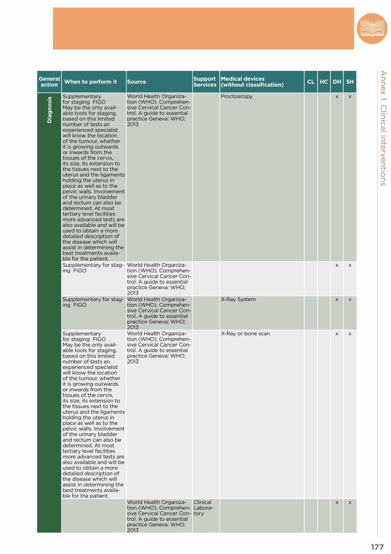
177
An
nex 1. C
linic
al in
terv
en
tion
s
General action
Steps/actions/options
Interventions identified from the recommendations
Se-quence Priority Notes General
action When to perform it Source Support Services
Medical devices (without classification) CL HC DH SH
Dia
gn
osi
s Determine the cervical cancer staging
Proctoscopy (Test to examine the intestinal system)
d I Used as a supplement for staging according to symptoms, resource availability and expertise.
Dia
gn
osi
s Supplementary for staging FIGO May be the only avail-able tools for staging, based on this limited number of tests an experienced specialist will know the location of the tumour, whether it is growing outwards or inwards from the tissues of the cervix, its size, its extension to the tissues next to the uterus and the ligaments holding the uterus in place as well as to the pelvic walls. Involvement of the urinary bladder and rectum can also be determined. At most tertiary level facilities more advanced tests are also available and will be used to obtain a more detailed description of the disease which will assist in determining the best treatments availa-ble for the patient.
World Health Organiza-tion (WHO). Comprehen-sive Cervical Cancer Con-trol. A guide to essential practice Geneva: WHO; 2013
Proctoscopy x x
Determine the cervical cancer staging
Endocervical curettage or smear ECC e I Supplementary for stag-ing FIGO
World Health Organiza-tion (WHO). Comprehen-sive Cervical Cancer Con-trol. A guide to essential practice Geneva: WHO; 2013
x x
Determine the cervical cancer staging
Chest X-Ray f I Supplementary for stag-ing FIGO
World Health Organiza-tion (WHO). Comprehen-sive Cervical Cancer Con-trol. A guide to essential practice Geneva: WHO; 2013
X-Ray System x x
Determine the cervical cancer staging
Skeletal X-Ray or bone scan (if bone pain)
g I Used as a supplement for staging according to symptoms, resource availability and expertise.
Supplementary for staging FIGO May be the only avail-able tools for staging, based on this limited number of tests an experienced specialist will know the location of the tumour, whether it is growing outwards or inwards from the tissues of the cervix, its size, its extension to the tissues next to the uterus and the ligaments holding the uterus in place as well as to the pelvic walls. Involvement of the urinary bladder and rectum can also be determined. At most tertiary level facilities more advanced tests are also available and will be used to obtain a more detailed description of the disease which will assist in determining the best treatments availa-ble for the patient.
World Health Organiza-tion (WHO). Comprehen-sive Cervical Cancer Con-trol. A guide to essential practice Geneva: WHO; 2013
X-Ray or bone scan x x
Determine the cervical cancer staging
Blood test (including full blood count and haemoglobin levels ) in addition to Pregnancy and HIV tests when appropriate.
h I The specialists at the hospital may conduct additional tests to further investigate the extent of the cancer.
World Health Organiza-tion (WHO). Comprehen-sive Cervical Cancer Con-trol. A guide to essential practice Geneva: WHO; 2013
Clinical Labora-tory
x x

178
WHO
list o
f prio
rity m
edica
l dev
ices f
or ca
ncer
man
agem
ent
General action
Steps/actions/options
Interventions identified from the recommendations
Se-quence Priority Notes General
action When to perform it Source Support Services
Medical devices (without classification) CL HC DH SH
Dia
gn
osi
s Determine the cervical cancer staging
Kidney and liver function tests; a I The specialists at the hospital may conduct additional tests to further investigate the extent of the cancer.
Dia
gn
osi
s World Health Organiza-tion (WHO). Comprehen-sive Cervical Cancer Con-trol. A guide to essential practice Geneva: WHO; 2013
x x
Determine the cervical cancer staging and follow up
CT a II Used for staging according to symptoms, resource availability and expertise. Useful to evaluate the tumour size, correlation with anatomic structures, metastases and nodal involvement.
Though not part of the clinical staging, eval-uation for regional or distant metastases using CT, MR or PET/CT may help guide treatment planning. Routine imag-ing is not recommended for patients who have completed primary therapy.
World Health Organiza-tion (WHO). Comprehen-sive Cervical Cancer Con-trol. A guide to essential practice Geneva: WHO; 2013
Imaging Services
x x
Determine the cervical cancer staging and follow up
Nuclear Magnetic Resonance a II Gives high resolution of soft tissues particularly for the cervix, parametrial invasion, bladder or rectal invasion, ureteral obstruction, lymph node enlargement.
World Health Organiza-tion (WHO). Comprehen-sive Cervical Cancer Con-trol. A guide to essential practice Geneva: WHO; 2013
Imaging Services
x
Determine the cervical cancer staging and follow up
PET/CT a III Allows for increased sensitivity in assessing lymphatic invasion.
World Health Organiza-tion (WHO). Comprehen-sive Cervical Cancer Con-trol. A guide to essential practice Geneva: WHO; 2013
Imaging Services
x
Tre
atm
en
t Treatment with Surgery (appropriate to the stage , based on the diagnostic)
Pelvic lymphadenectomy a I
Tre
atm
en
t World Health Organiza-tion (WHO). Comprehen-sive Cervical Cancer Con-trol. A guide to essential practice Geneva: WHO; 2013
Surgery x
Treatment with Surgery (appropriate to the stage, based on the diagnostic)
Cone Biopsy a I For Microinvasive cancers (those that are entirely contained within the cervical epithelium) can be treated with cone biopsy, particularly if retaining fertility is an issue.
World Health Organiza-tion (WHO). Comprehen-sive Cervical Cancer Con-trol. A guide to essential practice Geneva: WHO; 2013
Surgery x
Treatment with Surgery (appropriate to the stage, based on the diagnostic)
Simple Hysterectomy a I Indicated for the treat-ment of early microinva-sive cervical cancers in postmenopausal women and younger women who are not interested in preserving fertility.
World Health Organiza-tion (WHO). Comprehen-sive Cervical Cancer Con-trol. A guide to essential practice Geneva: WHO; 2013
Surgery x
Treatment with Surgery (appropriate to the stage, based on the diagnostic)
Radical Hysterectomy a I World Health Organiza-tion (WHO). Comprehen-sive Cervical Cancer Con-trol. A guide to essential practice Geneva: WHO; 2013
Surgery x
Treatment with Surgery (appropriate to the stage, based on the diagnostic)
Salvage Surgery a I When the patient has had primary surgery, but microscopic exam-ination of the removed tissue shows that the margin of normal tissue around the cancer is too thin or when the patient has undergone radiotherapy and/or chemotherapy, but early recurrences or incom-plete destruction of the cancer are noted on follow-up.
World Health Organiza-tion (WHO). Comprehen-sive Cervical Cancer Con-trol. A guide to essential practice Geneva: WHO; 2013
Surgery x

179
An
nex 1. C
linic
al in
terv
en
tion
s
General action
Steps/actions/options
Interventions identified from the recommendations
Se-quence Priority Notes General
action When to perform it Source Support Services
Medical devices (without classification) CL HC DH SH
Dia
gn
osi
s Determine the cervical cancer staging
Kidney and liver function tests; a I The specialists at the hospital may conduct additional tests to further investigate the extent of the cancer.
Dia
gn
osi
s World Health Organiza-tion (WHO). Comprehen-sive Cervical Cancer Con-trol. A guide to essential practice Geneva: WHO; 2013
x x
Determine the cervical cancer staging and follow up
CT a II Used for staging according to symptoms, resource availability and expertise. Useful to evaluate the tumour size, correlation with anatomic structures, metastases and nodal involvement.
Though not part of the clinical staging, eval-uation for regional or distant metastases using CT, MR or PET/CT may help guide treatment planning. Routine imag-ing is not recommended for patients who have completed primary therapy.
World Health Organiza-tion (WHO). Comprehen-sive Cervical Cancer Con-trol. A guide to essential practice Geneva: WHO; 2013
Imaging Services
x x
Determine the cervical cancer staging and follow up
Nuclear Magnetic Resonance a II Gives high resolution of soft tissues particularly for the cervix, parametrial invasion, bladder or rectal invasion, ureteral obstruction, lymph node enlargement.
World Health Organiza-tion (WHO). Comprehen-sive Cervical Cancer Con-trol. A guide to essential practice Geneva: WHO; 2013
Imaging Services
x
Determine the cervical cancer staging and follow up
PET/CT a III Allows for increased sensitivity in assessing lymphatic invasion.
World Health Organiza-tion (WHO). Comprehen-sive Cervical Cancer Con-trol. A guide to essential practice Geneva: WHO; 2013
Imaging Services
x
Tre
atm
en
t Treatment with Surgery (appropriate to the stage , based on the diagnostic)
Pelvic lymphadenectomy a I
Tre
atm
en
t World Health Organiza-tion (WHO). Comprehen-sive Cervical Cancer Con-trol. A guide to essential practice Geneva: WHO; 2013
Surgery x
Treatment with Surgery (appropriate to the stage, based on the diagnostic)
Cone Biopsy a I For Microinvasive cancers (those that are entirely contained within the cervical epithelium) can be treated with cone biopsy, particularly if retaining fertility is an issue.
World Health Organiza-tion (WHO). Comprehen-sive Cervical Cancer Con-trol. A guide to essential practice Geneva: WHO; 2013
Surgery x
Treatment with Surgery (appropriate to the stage, based on the diagnostic)
Simple Hysterectomy a I Indicated for the treat-ment of early microinva-sive cervical cancers in postmenopausal women and younger women who are not interested in preserving fertility.
World Health Organiza-tion (WHO). Comprehen-sive Cervical Cancer Con-trol. A guide to essential practice Geneva: WHO; 2013
Surgery x
Treatment with Surgery (appropriate to the stage, based on the diagnostic)
Radical Hysterectomy a I World Health Organiza-tion (WHO). Comprehen-sive Cervical Cancer Con-trol. A guide to essential practice Geneva: WHO; 2013
Surgery x
Treatment with Surgery (appropriate to the stage, based on the diagnostic)
Salvage Surgery a I When the patient has had primary surgery, but microscopic exam-ination of the removed tissue shows that the margin of normal tissue around the cancer is too thin or when the patient has undergone radiotherapy and/or chemotherapy, but early recurrences or incom-plete destruction of the cancer are noted on follow-up.
World Health Organiza-tion (WHO). Comprehen-sive Cervical Cancer Con-trol. A guide to essential practice Geneva: WHO; 2013
Surgery x

180
WHO
list o
f prio
rity m
edica
l dev
ices f
or ca
ncer
man
agem
ent
General action
Steps/actions/options
Interventions identified from the recommendations
Se-quence Priority Notes General
action When to perform it Source Support Services
Medical devices (without classification) CL HC DH SH
Tre
atm
en
t Treatment with Radiation (appropriate to the stage, based on the diagnostic)
External-beam radiotherapy, or tele-therapy.
a I
Tre
atm
en
t For women with cancer at stage IIA2 or greater. It may be offered to women with cancers greater than 4 cm in diameter confined to the cervix, and for cancers that have spread beyond the cervix. Primary radiotherapy intended to cure earlier cancers.
World Health Organiza-tion (WHO). Comprehen-sive Cervical Cancer Con-trol. A guide to essential practice Geneva: WHO; 2013
Radio-therapy
x
Treatment with Radiation (appropriate to the stage, based on the diagnostic)
High-dose-rate (HDR) brachytherapy a I For women with cancer at stage IIA2 or greater. It may be offered to women with cancers greater than 4 cm in diameter confined to the cervix, and for cancers that have spread beyond the cervix. Primary radiotherapy intended to cure earlier cancers.
World Health Organiza-tion (WHO). Comprehen-sive Cervical Cancer Con-trol. A guide to essential practice Geneva: WHO; 2013
Radio-therapy
x
Treatment with Radiation (appropriate to the stage, based on the diagnostic)
Radiation as adjunctive therapy a I World Health Organiza-tion (WHO). Comprehen-sive Cervical Cancer Con-trol. A guide to essential practice Geneva: WHO; 2013
Radio-therapy
x
Planning Radiotherapy
Computerized tomography (CT) scan a I CT scan or MRI of the abdomen and pelvis (to help plan radiotherapy); however, treatment can be planned in the absence of these procedures if they are not available, affordable or feasible.
World Health Organiza-tion (WHO). Comprehen-sive Cervical Cancer Con-trol. A guide to essential practice Geneva: WHO; 2013
Imaging Services
x
Planning Radiotherapy
Magnetic resonance imaging (MRI) of the abdomen and pelvis
a I CT scan or MRI of the abdomen and pelvis (to help plan radiotherapy); however, treatment can be planned in the absence of these procedures if they are not available, affordable or feasible.
CT or better yet mag-netic resonance imaging (MRI) of the abdomen and pelvis (to help plan radiotherapy); howev-er treatment can be planned in the absence of these procedures if they are not available, affordable or feasible.
World Health Organiza-tion (WHO). Comprehen-sive Cervical Cancer Con-trol. A guide to essential practice Geneva: WHO; 2013
Imaging Services
x
Treatment with Chemotherapy (appropriate to the stage , based on the diagnostic)
Chemotherapy a I Chemotherapy is rarely used alone as the prima-ry treatment for cervical cancer; rather; it is used in combination with radiotherapy and less often with surgery.
World Health Organiza-tion (WHO). Comprehen-sive Cervical Cancer Con-trol. A guide to essential practice Geneva: WHO; 2013
Chemo-therapy Services
x
Treatment with Chemotherapy (appropriate to the stage, based on the diagnostic)
Chemotherapy combined with radio-therapy
a I Chemotherapy is used first in women with very large and bulky tumours, to reduce the cancer size, and the followed by radiotherapy. Treatment is done in this sequence because cancer is shown to respond better to radiation when tumour is less bulky.
World Health Organiza-tion (WHO). Comprehen-sive Cervical Cancer Con-trol. A guide to essential practice Geneva: WHO; 2013
Chemo-therapy Services and Radi-otherapy
x
Treatment with Chemotherapy (appropriate to the stage, based on the diagnostic)
Chemotherapy combined with surgery a I World Health Organiza-tion (WHO). Comprehen-sive Cervical Cancer Con-trol. A guide to essential practice Geneva: WHO; 2013
Chemo-therapy Services and Sur-gery
x
Combined Treat-ments
Teletherapy plus brachytherapy a I World Health Organiza-tion (WHO). Comprehen-sive Cervical Cancer Con-trol. A guide to essential practice Geneva: WHO; 2013
Radio-therapy Services
x

181
An
nex 1. C
linic
al in
terv
en
tion
s
General action
Steps/actions/options
Interventions identified from the recommendations
Se-quence Priority Notes General
action When to perform it Source Support Services
Medical devices (without classification) CL HC DH SH
Tre
atm
en
t Treatment with Radiation (appropriate to the stage, based on the diagnostic)
External-beam radiotherapy, or tele-therapy.
a I
Tre
atm
en
t For women with cancer at stage IIA2 or greater. It may be offered to women with cancers greater than 4 cm in diameter confined to the cervix, and for cancers that have spread beyond the cervix. Primary radiotherapy intended to cure earlier cancers.
World Health Organiza-tion (WHO). Comprehen-sive Cervical Cancer Con-trol. A guide to essential practice Geneva: WHO; 2013
Radio-therapy
x
Treatment with Radiation (appropriate to the stage, based on the diagnostic)
High-dose-rate (HDR) brachytherapy a I For women with cancer at stage IIA2 or greater. It may be offered to women with cancers greater than 4 cm in diameter confined to the cervix, and for cancers that have spread beyond the cervix. Primary radiotherapy intended to cure earlier cancers.
World Health Organiza-tion (WHO). Comprehen-sive Cervical Cancer Con-trol. A guide to essential practice Geneva: WHO; 2013
Radio-therapy
x
Treatment with Radiation (appropriate to the stage, based on the diagnostic)
Radiation as adjunctive therapy a I World Health Organiza-tion (WHO). Comprehen-sive Cervical Cancer Con-trol. A guide to essential practice Geneva: WHO; 2013
Radio-therapy
x
Planning Radiotherapy
Computerized tomography (CT) scan a I CT scan or MRI of the abdomen and pelvis (to help plan radiotherapy); however, treatment can be planned in the absence of these procedures if they are not available, affordable or feasible.
World Health Organiza-tion (WHO). Comprehen-sive Cervical Cancer Con-trol. A guide to essential practice Geneva: WHO; 2013
Imaging Services
x
Planning Radiotherapy
Magnetic resonance imaging (MRI) of the abdomen and pelvis
a I CT scan or MRI of the abdomen and pelvis (to help plan radiotherapy); however, treatment can be planned in the absence of these procedures if they are not available, affordable or feasible.
CT or better yet mag-netic resonance imaging (MRI) of the abdomen and pelvis (to help plan radiotherapy); howev-er treatment can be planned in the absence of these procedures if they are not available, affordable or feasible.
World Health Organiza-tion (WHO). Comprehen-sive Cervical Cancer Con-trol. A guide to essential practice Geneva: WHO; 2013
Imaging Services
x
Treatment with Chemotherapy (appropriate to the stage , based on the diagnostic)
Chemotherapy a I Chemotherapy is rarely used alone as the prima-ry treatment for cervical cancer; rather; it is used in combination with radiotherapy and less often with surgery.
World Health Organiza-tion (WHO). Comprehen-sive Cervical Cancer Con-trol. A guide to essential practice Geneva: WHO; 2013
Chemo-therapy Services
x
Treatment with Chemotherapy (appropriate to the stage, based on the diagnostic)
Chemotherapy combined with radio-therapy
a I Chemotherapy is used first in women with very large and bulky tumours, to reduce the cancer size, and the followed by radiotherapy. Treatment is done in this sequence because cancer is shown to respond better to radiation when tumour is less bulky.
World Health Organiza-tion (WHO). Comprehen-sive Cervical Cancer Con-trol. A guide to essential practice Geneva: WHO; 2013
Chemo-therapy Services and Radi-otherapy
x
Treatment with Chemotherapy (appropriate to the stage, based on the diagnostic)
Chemotherapy combined with surgery a I World Health Organiza-tion (WHO). Comprehen-sive Cervical Cancer Con-trol. A guide to essential practice Geneva: WHO; 2013
Chemo-therapy Services and Sur-gery
x
Combined Treat-ments
Teletherapy plus brachytherapy a I World Health Organiza-tion (WHO). Comprehen-sive Cervical Cancer Con-trol. A guide to essential practice Geneva: WHO; 2013
Radio-therapy Services
x

182
WHO
list o
f prio
rity m
edica
l dev
ices f
or ca
ncer
man
agem
ent
General action
Steps/actions/options
Interventions identified from the recommendations
Se-quence Priority Notes General
action When to perform it Source Support Services
Medical devices (without classification) CL HC DH SH
Fo
llo
w u
p Special Managing cervical cancer in women living with HIV
Baseline CD4 count test a I
Fo
llo
w u
p World Health Organiza-tion (WHO). Comprehen-sive Cervical Cancer Con-trol. A guide to essential practice Geneva: WHO; 2013
Clinical Labora-tory
x
Follow up care Speculum, vaginal and rectal exami-nations
a I World Health Organiza-tion (WHO). Comprehen-sive Cervical Cancer Con-trol. A guide to essential practice Geneva: WHO; 2013
x x x
Full examination, including general systems and abdominal examination plus palpation of lymph nodes, with particular attention to the neck and groin;
a I World Health Organiza-tion (WHO). Comprehen-sive Cervical Cancer Con-trol. A guide to essential practice Geneva: WHO; 2013
x x x
Cytological smear of the vaginal vault for women
a I Who have only been treated with surgery
World Health Organiza-tion (WHO). Comprehen-sive Cervical Cancer Con-trol. A guide to essential practice Geneva: WHO; 2013
Histology service
x x
Blood test (including full blood count and haemoglobin levels )
a I World Health Organiza-tion (WHO). Comprehen-sive Cervical Cancer Con-trol. A guide to essential practice Geneva: WHO; 2013
Clinical Labora-tory
x x
Pallia
tive C
are
Palliative care Palliative surgery a I Is sometimes done in advanced can-cer to relieve obstruction of the bowel, or to treat fistulae (abnormal channels between the vagina and the urinary organs or rectum) that result from radiation or extension of the primary disease.
Pallia
tive C
are
World Health Organiza-tion (WHO). Comprehen-sive Cervical Cancer Con-trol. A guide to essential practice Geneva: WHO; 2013
Surgery Services
x
Radiation as palliative therapy a I World Health Organiza-tion (WHO). Comprehen-sive Cervical Cancer Con-trol. A guide to essential practice Geneva: WHO; 2013
Radio-therapy Services
x
Chemotherapy as palliative care a I Palliative chemotherapy is sometimes used, after careful consideration of the expected benefits versus the adverse side effects, to relieve symp-toms in women with widespread metastases to liver, lung and bone.
World Health Organiza-tion (WHO). Comprehen-sive Cervical Cancer Con-trol. A guide to essential practice Geneva: WHO; 2013
Chemo-therapy services
x
Nonmedical pain management (mas-sage, acupuncture)
a I World Health Organiza-tion (WHO). Comprehen-sive Cervical Cancer Con-trol. A guide to essential practice Geneva: WHO; 2013
x x
Home-based management of vaginal problems
a I World Health Organiza-tion (WHO). Comprehen-sive Cervical Cancer Con-trol. A guide to essential practice Geneva: WHO; 2013
x x
Treatment of cervical infections and pelvic inflammatory disease (PID)
a I World Health Organiza-tion (WHO). Comprehen-sive Cervical Cancer Con-trol. A guide to essential practice Geneva: WHO; 2013
x x
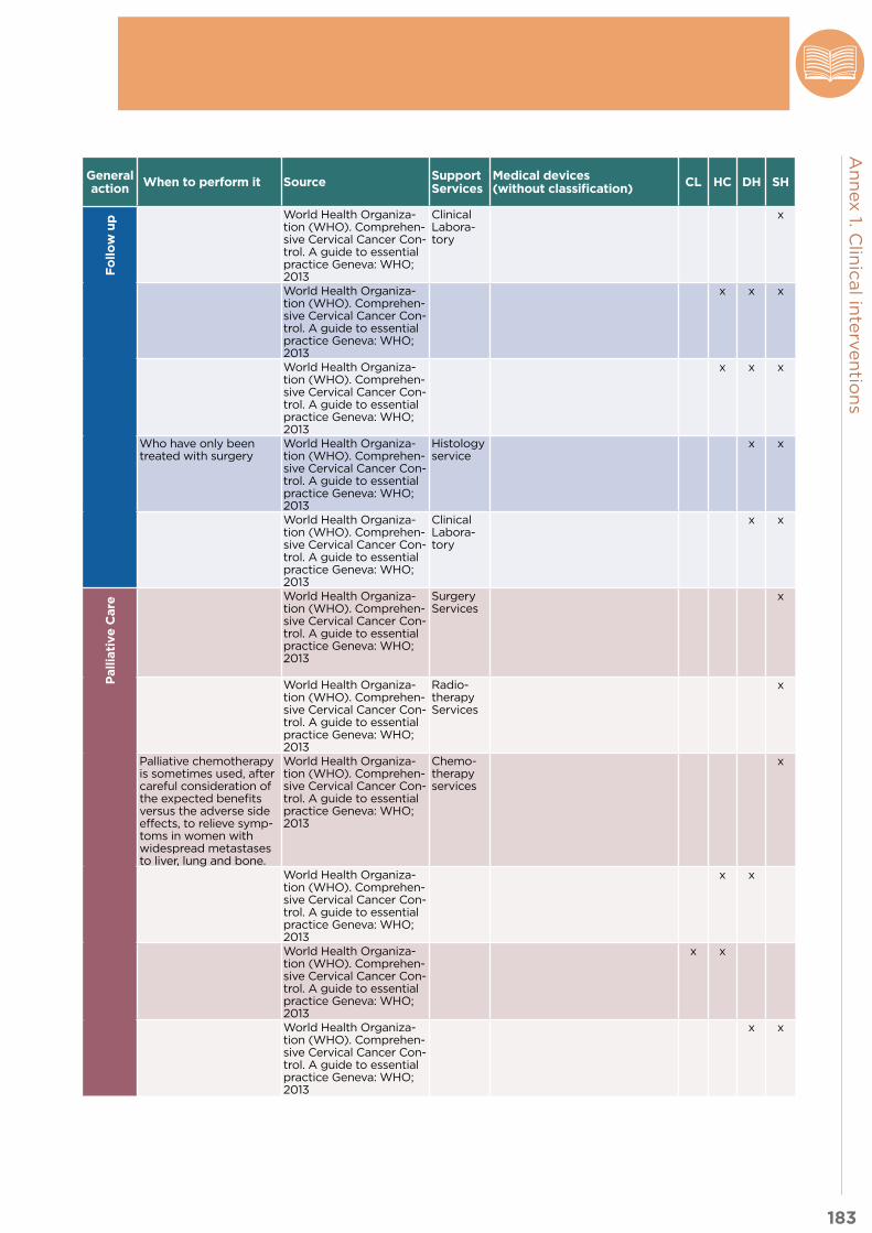
183
An
nex 1. C
linic
al in
terv
en
tion
s
General action
Steps/actions/options
Interventions identified from the recommendations
Se-quence Priority Notes General
action When to perform it Source Support Services
Medical devices (without classification) CL HC DH SH
Fo
llo
w u
p Special Managing cervical cancer in women living with HIV
Baseline CD4 count test a I
Fo
llo
w u
p World Health Organiza-tion (WHO). Comprehen-sive Cervical Cancer Con-trol. A guide to essential practice Geneva: WHO; 2013
Clinical Labora-tory
x
Follow up care Speculum, vaginal and rectal exami-nations
a I World Health Organiza-tion (WHO). Comprehen-sive Cervical Cancer Con-trol. A guide to essential practice Geneva: WHO; 2013
x x x
Full examination, including general systems and abdominal examination plus palpation of lymph nodes, with particular attention to the neck and groin;
a I World Health Organiza-tion (WHO). Comprehen-sive Cervical Cancer Con-trol. A guide to essential practice Geneva: WHO; 2013
x x x
Cytological smear of the vaginal vault for women
a I Who have only been treated with surgery
World Health Organiza-tion (WHO). Comprehen-sive Cervical Cancer Con-trol. A guide to essential practice Geneva: WHO; 2013
Histology service
x x
Blood test (including full blood count and haemoglobin levels )
a I World Health Organiza-tion (WHO). Comprehen-sive Cervical Cancer Con-trol. A guide to essential practice Geneva: WHO; 2013
Clinical Labora-tory
x x
Pallia
tive C
are
Palliative care Palliative surgery a I Is sometimes done in advanced can-cer to relieve obstruction of the bowel, or to treat fistulae (abnormal channels between the vagina and the urinary organs or rectum) that result from radiation or extension of the primary disease.
Pallia
tive C
are
World Health Organiza-tion (WHO). Comprehen-sive Cervical Cancer Con-trol. A guide to essential practice Geneva: WHO; 2013
Surgery Services
x
Radiation as palliative therapy a I World Health Organiza-tion (WHO). Comprehen-sive Cervical Cancer Con-trol. A guide to essential practice Geneva: WHO; 2013
Radio-therapy Services
x
Chemotherapy as palliative care a I Palliative chemotherapy is sometimes used, after careful consideration of the expected benefits versus the adverse side effects, to relieve symp-toms in women with widespread metastases to liver, lung and bone.
World Health Organiza-tion (WHO). Comprehen-sive Cervical Cancer Con-trol. A guide to essential practice Geneva: WHO; 2013
Chemo-therapy services
x
Nonmedical pain management (mas-sage, acupuncture)
a I World Health Organiza-tion (WHO). Comprehen-sive Cervical Cancer Con-trol. A guide to essential practice Geneva: WHO; 2013
x x
Home-based management of vaginal problems
a I World Health Organiza-tion (WHO). Comprehen-sive Cervical Cancer Con-trol. A guide to essential practice Geneva: WHO; 2013
x x
Treatment of cervical infections and pelvic inflammatory disease (PID)
a I World Health Organiza-tion (WHO). Comprehen-sive Cervical Cancer Con-trol. A guide to essential practice Geneva: WHO; 2013
x x

184
WHO
list o
f prio
rity m
edica
l dev
ices f
or ca
ncer
man
agem
ent Annex table 1-3. Colorectal cancer
General action
Steps/Actions/Options
Interventions identified from the recommendations
Se-quence (a, b, c, d)
Priority (I, II, III)
Notes General action When to perform it Source Source Source
Pre
ven
tio
n Dietary modification
Dietary counselling a I Increasing dietary fibre and reducing red and processed meat consumption (NCCN) and alcoholic drinks as well as regular phys-ical exercise for colon cancer prevention (EURECCA)
Pre
ven
tio
n - - NCCN. Colon Cancer. Version 2.2015 NCCN. Rectal Cancer. Version 2.2015
van de Velde CJ, Boelens PG, Borras JM, et al. EURECCA colorectal: multidisciplinary management: European consensus conference colon & rectum. Eur J Cancer. 2014 Jan;50(1):1.e1-1.e34.
Dia
gn
osi
s Stool-based testing (early diagnosis and screening)
Faecal immunochemical testing (FIT)
a II Colon + Rectal. WHO-IARC has not developed official recommendations for colorectal cancer screening at this time.
Dia
gn
osi
s As a second choice according to NCCN. No clear indications from EURECCA.
- NCCN. Colorectal Cancer Screening. Version 1.2014
van de Velde CJ, Boelens PG, Borras JM, et al. EURECCA colorectal: multidisciplinary management: European consensus conference colon & rectum. Eur J Cancer. 2014 Jan;50(1):1.e1-1.e34.
Guaiac faecal occult blood test (gFOBT)
a II Colon + Rectal. WHO-IARC has not developed official recommendations for colorectal cancer screening at this time.
As a second choice according to NCCN. No clear indications from EURECCA.
- NCCN. Colorectal Cancer Screening. Version 1.2014
van de Velde CJ, Boelens PG, Borras JM, et al. EURECCA colorectal: multidisciplinary management: European consensus conference colon & rectum. Eur J Cancer. 2014 Jan;50(1):1.e1-1.e34.
Endoscopy (screening)
Colonoscopy a II Colon + Rectal. WHO-IARC has not developed official recommendations for colorectal cancer screening at this time.
As a first choice accord-ing to NCCN. No clear indications from EUREC-CA.
- NCCN. Colorectal Cancer Screening. Version 1.2014
-
Flexible sigmoidoscopy a III Colon + Rectal. WHO-IARC has not developed official recommendations for colorectal cancer screening at this time.
As a third choice accord-ing to NCCN. No clear indications from EUREC-CA.
- NCCN. Colorectal Cancer Screening. Version 1.2014
van de Velde CJ, Boelens PG, Borras JM, et al. EURECCA colorectal: multidisciplinary management: European consensus conference colon & rectum. Eur J Cancer. 2014 Jan;50(1):1.e1-1.e34.
Physical exami-nation
Digital rectal examination (just if rectal cancer is suspected) and examination of abdomen, liver and lymph nodes.
a I Colon + Rectal - - NCCN. Colon Cancer. Version 2.2015 NCCN. Rectal Cancer. Version 2.2015
van de Velde CJ, Boelens PG, Borras JM, et al. EURECCA colorectal: multidisciplinary management: European consensus conference colon & rectum. Eur J Cancer. 2014 Jan;50(1):1.e1-1.e34.
Endoscopy (early diagnosis)
Colonoscopy a I Colon + Rectal For patients without ma-jor comorbidity
Colorectal cancer. NICE clinical guideline 131 (2014)
NCCN. Colon Cancer. Version 2.2015 NCCN. Rectal Cancer. Version 2.2015
van de Velde CJ, Boelens PG, Borras JM, et al. EURECCA colorectal: multidisciplinary management: European consensus conference colon & rectum. Eur J Cancer. 2014 Jan;50(1):1.e1-1.e34.
Flexible sigmoidoscopy a I Colon + Rectal For patients with major comorbidity followed by barium enema
Colorectal cancer. NICE clinical guideline 131 (2014)
- -
Rectoscopy a I Rectal - - - van de Velde CJ, Boelens PG, Borras JM, et al. EURECCA colorectal: multidisciplinary management: European consensus conference colon & rectum. Eur J Cancer. 2014 Jan;50(1):1.e1-1.e34.
Colorectal imaging
Barium enema b II Colon + Rectal After flexible sigmoidos-copy for patients with major comorbidity (NICE) As a third choice for colon cancer when colo-noscopy is not possible or contraindicated and CT colonography is not available (EURECCA)
Colorectal cancer. NICE clinical guideline 131 (2014)
- van de Velde CJ, Boelens PG, Borras JM, et al. EURECCA colorectal: multidisciplinary management: European consensus conference colon & rectum. Eur J Cancer. 2014 Jan;50(1):1.e1-1.e34.
CT colonography (early diagnosis or screening)
a II Colon + Rectal. WHO-IARC has not developed official recommendations for colorectal cancer screening at this time.
If the local radiology service can demonstrate competency in this tech-nique. Suspicious lesions need to be confirmed by colonoscopy with biopsy (NICE) As a second choice colon cancer di-agnostic test for patients with incomplete colonos-copy (EURECCA)
Colorectal cancer. NICE clinical guideline 131 (2014)
- van de Velde CJ, Boelens PG, Borras JM, et al. EURECCA colorectal: multidisciplinary management: European consensus conference colon & rectum. Eur J Cancer. 2014 Jan;50(1):1.e1-1.e34.
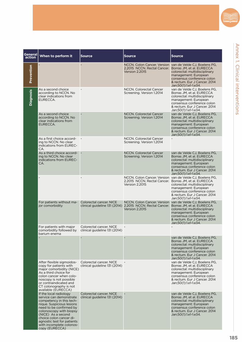
185
An
nex 1. C
linic
al in
terv
en
tion
s
Annex table 1-3. Colorectal cancer
General action
Steps/Actions/Options
Interventions identified from the recommendations
Se-quence (a, b, c, d)
Priority (I, II, III)
Notes General action When to perform it Source Source Source
Pre
ven
tio
n Dietary modification
Dietary counselling a I Increasing dietary fibre and reducing red and processed meat consumption (NCCN) and alcoholic drinks as well as regular phys-ical exercise for colon cancer prevention (EURECCA)
Pre
ven
tio
n - - NCCN. Colon Cancer. Version 2.2015 NCCN. Rectal Cancer. Version 2.2015
van de Velde CJ, Boelens PG, Borras JM, et al. EURECCA colorectal: multidisciplinary management: European consensus conference colon & rectum. Eur J Cancer. 2014 Jan;50(1):1.e1-1.e34.
Dia
gn
osi
s Stool-based testing (early diagnosis and screening)
Faecal immunochemical testing (FIT)
a II Colon + Rectal. WHO-IARC has not developed official recommendations for colorectal cancer screening at this time.
Dia
gn
osi
s As a second choice according to NCCN. No clear indications from EURECCA.
- NCCN. Colorectal Cancer Screening. Version 1.2014
van de Velde CJ, Boelens PG, Borras JM, et al. EURECCA colorectal: multidisciplinary management: European consensus conference colon & rectum. Eur J Cancer. 2014 Jan;50(1):1.e1-1.e34.
Guaiac faecal occult blood test (gFOBT)
a II Colon + Rectal. WHO-IARC has not developed official recommendations for colorectal cancer screening at this time.
As a second choice according to NCCN. No clear indications from EURECCA.
- NCCN. Colorectal Cancer Screening. Version 1.2014
van de Velde CJ, Boelens PG, Borras JM, et al. EURECCA colorectal: multidisciplinary management: European consensus conference colon & rectum. Eur J Cancer. 2014 Jan;50(1):1.e1-1.e34.
Endoscopy (screening)
Colonoscopy a II Colon + Rectal. WHO-IARC has not developed official recommendations for colorectal cancer screening at this time.
As a first choice accord-ing to NCCN. No clear indications from EUREC-CA.
- NCCN. Colorectal Cancer Screening. Version 1.2014
-
Flexible sigmoidoscopy a III Colon + Rectal. WHO-IARC has not developed official recommendations for colorectal cancer screening at this time.
As a third choice accord-ing to NCCN. No clear indications from EUREC-CA.
- NCCN. Colorectal Cancer Screening. Version 1.2014
van de Velde CJ, Boelens PG, Borras JM, et al. EURECCA colorectal: multidisciplinary management: European consensus conference colon & rectum. Eur J Cancer. 2014 Jan;50(1):1.e1-1.e34.
Physical exami-nation
Digital rectal examination (just if rectal cancer is suspected) and examination of abdomen, liver and lymph nodes.
a I Colon + Rectal - - NCCN. Colon Cancer. Version 2.2015 NCCN. Rectal Cancer. Version 2.2015
van de Velde CJ, Boelens PG, Borras JM, et al. EURECCA colorectal: multidisciplinary management: European consensus conference colon & rectum. Eur J Cancer. 2014 Jan;50(1):1.e1-1.e34.
Endoscopy (early diagnosis)
Colonoscopy a I Colon + Rectal For patients without ma-jor comorbidity
Colorectal cancer. NICE clinical guideline 131 (2014)
NCCN. Colon Cancer. Version 2.2015 NCCN. Rectal Cancer. Version 2.2015
van de Velde CJ, Boelens PG, Borras JM, et al. EURECCA colorectal: multidisciplinary management: European consensus conference colon & rectum. Eur J Cancer. 2014 Jan;50(1):1.e1-1.e34.
Flexible sigmoidoscopy a I Colon + Rectal For patients with major comorbidity followed by barium enema
Colorectal cancer. NICE clinical guideline 131 (2014)
- -
Rectoscopy a I Rectal - - - van de Velde CJ, Boelens PG, Borras JM, et al. EURECCA colorectal: multidisciplinary management: European consensus conference colon & rectum. Eur J Cancer. 2014 Jan;50(1):1.e1-1.e34.
Colorectal imaging
Barium enema b II Colon + Rectal After flexible sigmoidos-copy for patients with major comorbidity (NICE) As a third choice for colon cancer when colo-noscopy is not possible or contraindicated and CT colonography is not available (EURECCA)
Colorectal cancer. NICE clinical guideline 131 (2014)
- van de Velde CJ, Boelens PG, Borras JM, et al. EURECCA colorectal: multidisciplinary management: European consensus conference colon & rectum. Eur J Cancer. 2014 Jan;50(1):1.e1-1.e34.
CT colonography (early diagnosis or screening)
a II Colon + Rectal. WHO-IARC has not developed official recommendations for colorectal cancer screening at this time.
If the local radiology service can demonstrate competency in this tech-nique. Suspicious lesions need to be confirmed by colonoscopy with biopsy (NICE) As a second choice colon cancer di-agnostic test for patients with incomplete colonos-copy (EURECCA)
Colorectal cancer. NICE clinical guideline 131 (2014)
- van de Velde CJ, Boelens PG, Borras JM, et al. EURECCA colorectal: multidisciplinary management: European consensus conference colon & rectum. Eur J Cancer. 2014 Jan;50(1):1.e1-1.e34.
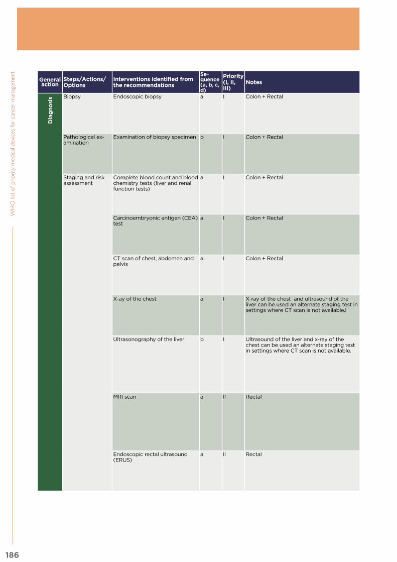
186
WHO
list o
f prio
rity m
edica
l dev
ices f
or ca
ncer
man
agem
ent
General action
Steps/Actions/Options
Interventions identified from the recommendations
Se-quence (a, b, c, d)
Priority (I, II, III)
Notes General action When to perform it Source Source Source
Dia
gn
osi
s Biopsy Endoscopic biopsy a I Colon + Rectal
Dia
gn
osi
s To perform histological analysis
Colorectal cancer. NICE clinical guideline 131 (2014)
NCCN. Colon Cancer. Version 2.2015 NCCN. Rectal Cancer. Version 2.2015
van de Velde CJ, Boelens PG, Borras JM, et al. EURECCA colorectal: multidisciplinary management: European consensus conference colon & rectum. Eur J Cancer. 2014 Jan;50(1):1.e1-1.e34.
Pathological ex-amination
Examination of biopsy specimen b I Colon + Rectal To confirm diagnosis of cancer
Colorectal cancer. NICE clinical guideline 131 (2014)
NCCN. Colon Cancer. Version 2.2015 NCCN. Rectal Cancer. Version 2.2015
van de Velde CJ, Boelens PG, Borras JM, et al. EURECCA colorectal: multidisciplinary management: European consensus conference colon & rectum. Eur J Cancer. 2014 Jan;50(1):1.e1-1.e34.
Staging and risk assessment
Complete blood count and blood chemistry tests (liver and renal function tests)
a I Colon + Rectal Recommended - NCCN. Colon Cancer. Version 2.2015 NCCN. Rectal Cancer. Version 2.2015
van de Velde CJ, Boelens PG, Borras JM, et al. EURECCA colorectal: multidisciplinary management: European consensus conference colon & rectum. Eur J Cancer. 2014 Jan;50(1):1.e1-1.e34.
Carcinoembryonic antigen (CEA) test
a I Colon + Rectal Recommended (NCCN) CEA can assist in staging and evaluation of progno-sis (EURECCA)
- NCCN. Colon Cancer. Version 2.2015 NCCN. Rectal Cancer. Version 2.2015
van de Velde CJ, Boelens PG, Borras JM, et al. EURECCA colorectal: multidisciplinary management: European consensus conference colon & rectum. Eur J Cancer. 2014 Jan;50(1):1.e1-1.e34.
CT scan of chest, abdomen and pelvis
a I Colon + Rectal Contrast-enhanced CT of the chest, abdomen and pelvis to estimate the stage of disease unless it is contraindicated (NICE) (NCCN) For patients with colon cancer (EURECCA)
Colorectal cancer. NICE clinical guideline 131 (2014)
NCCN. Colon Cancer. Version 2.2015 NCCN. Rectal Cancer. Version 2.2015
van de Velde CJ, Boelens PG, Borras JM, et al. EURECCA colorectal: multidisciplinary management: European consensus conference colon & rectum. Eur J Cancer. 2014 Jan;50(1):1.e1-1.e34.
X-ay of the chest a I X-ray of the chest and ultrasound of the liver can be used an alternate staging test in settings where CT scan is not available.l
For staging of distant metastases if CT scan cannot be performed (EURECCA)
- - van de Velde CJ, Boelens PG, Borras JM, et al. EURECCA colorectal: multidisciplinary management: European consensus conference colon & rectum. Eur J Cancer. 2014 Jan;50(1):1.e1-1.e34.
Ultrasonography of the liver b I Ultrasound of the liver and x-ray of the chest can be used an alternate staging test in settings where CT scan is not available.
When contrast-enhanced CT is not clinically appro-priate, not accessible or not acceptable to the per-son, contrast-enhanced ultrasound is recommend-ed (NICE) If abdominal CT is not possible in diagnosing liver metasta-ses (EURECCA)
Colorectal cancer. NICE clinical guideline 131 (2014)
- van de Velde CJ, Boelens PG, Borras JM, et al. EURECCA colorectal: multidisciplinary management: European consensus conference colon & rectum. Eur J Cancer. 2014 Jan;50(1):1.e1-1.e34.
MRI scan a II Rectal To assess the risk of local recurrence, as deter-mined by anticipated resection margin, tumour and lymph node staging in rectal cancer patients (NICE) First choice for rectal cancer; second choice for colon cancer (EURECCA) (NCCN)
Colorectal cancer. NICE clinical guideline 131 (2014)
NCCN. Rectal Cancer. Version 2.2015
van de Velde CJ, Boelens PG, Borras JM, et al. EURECCA colorectal: multidisciplinary management: European consensus conference colon & rectum. Eur J Cancer. 2014 Jan;50(1):1.e1-1.e34.
Endoscopic rectal ultrasound (ERUS)
a II Rectal As alternative to pelvic MRI.
Colorectal cancer. NICE clinical guideline 131 (2014)
NCCN. Rectal Cancer. Version 2.2015
van de Velde CJ, Boelens PG, Borras JM, et al. EURECCA colorectal: multidisciplinary management: European consensus conference colon & rectum. Eur J Cancer. 2014 Jan;50(1):1.e1-1.e34.
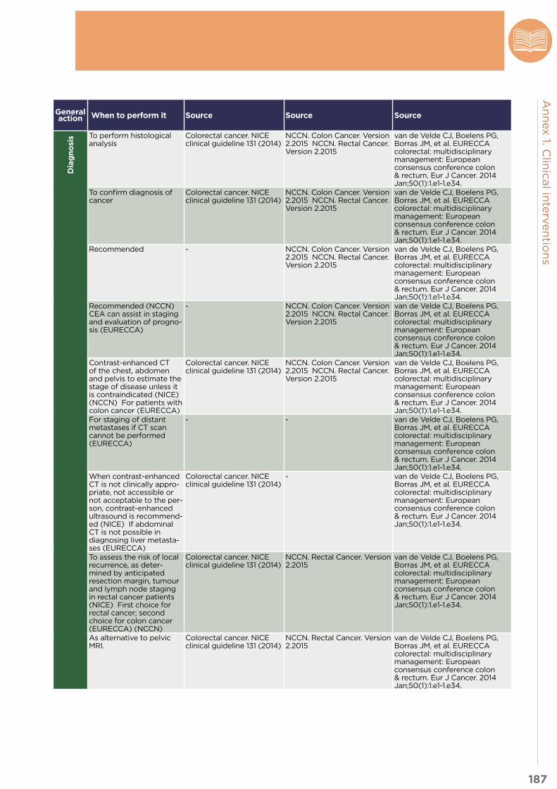
187
An
nex 1. C
linic
al in
terv
en
tion
s
General action
Steps/Actions/Options
Interventions identified from the recommendations
Se-quence (a, b, c, d)
Priority (I, II, III)
Notes General action When to perform it Source Source Source
Dia
gn
osi
s Biopsy Endoscopic biopsy a I Colon + Rectal
Dia
gn
osi
s To perform histological analysis
Colorectal cancer. NICE clinical guideline 131 (2014)
NCCN. Colon Cancer. Version 2.2015 NCCN. Rectal Cancer. Version 2.2015
van de Velde CJ, Boelens PG, Borras JM, et al. EURECCA colorectal: multidisciplinary management: European consensus conference colon & rectum. Eur J Cancer. 2014 Jan;50(1):1.e1-1.e34.
Pathological ex-amination
Examination of biopsy specimen b I Colon + Rectal To confirm diagnosis of cancer
Colorectal cancer. NICE clinical guideline 131 (2014)
NCCN. Colon Cancer. Version 2.2015 NCCN. Rectal Cancer. Version 2.2015
van de Velde CJ, Boelens PG, Borras JM, et al. EURECCA colorectal: multidisciplinary management: European consensus conference colon & rectum. Eur J Cancer. 2014 Jan;50(1):1.e1-1.e34.
Staging and risk assessment
Complete blood count and blood chemistry tests (liver and renal function tests)
a I Colon + Rectal Recommended - NCCN. Colon Cancer. Version 2.2015 NCCN. Rectal Cancer. Version 2.2015
van de Velde CJ, Boelens PG, Borras JM, et al. EURECCA colorectal: multidisciplinary management: European consensus conference colon & rectum. Eur J Cancer. 2014 Jan;50(1):1.e1-1.e34.
Carcinoembryonic antigen (CEA) test
a I Colon + Rectal Recommended (NCCN) CEA can assist in staging and evaluation of progno-sis (EURECCA)
- NCCN. Colon Cancer. Version 2.2015 NCCN. Rectal Cancer. Version 2.2015
van de Velde CJ, Boelens PG, Borras JM, et al. EURECCA colorectal: multidisciplinary management: European consensus conference colon & rectum. Eur J Cancer. 2014 Jan;50(1):1.e1-1.e34.
CT scan of chest, abdomen and pelvis
a I Colon + Rectal Contrast-enhanced CT of the chest, abdomen and pelvis to estimate the stage of disease unless it is contraindicated (NICE) (NCCN) For patients with colon cancer (EURECCA)
Colorectal cancer. NICE clinical guideline 131 (2014)
NCCN. Colon Cancer. Version 2.2015 NCCN. Rectal Cancer. Version 2.2015
van de Velde CJ, Boelens PG, Borras JM, et al. EURECCA colorectal: multidisciplinary management: European consensus conference colon & rectum. Eur J Cancer. 2014 Jan;50(1):1.e1-1.e34.
X-ay of the chest a I X-ray of the chest and ultrasound of the liver can be used an alternate staging test in settings where CT scan is not available.l
For staging of distant metastases if CT scan cannot be performed (EURECCA)
- - van de Velde CJ, Boelens PG, Borras JM, et al. EURECCA colorectal: multidisciplinary management: European consensus conference colon & rectum. Eur J Cancer. 2014 Jan;50(1):1.e1-1.e34.
Ultrasonography of the liver b I Ultrasound of the liver and x-ray of the chest can be used an alternate staging test in settings where CT scan is not available.
When contrast-enhanced CT is not clinically appro-priate, not accessible or not acceptable to the per-son, contrast-enhanced ultrasound is recommend-ed (NICE) If abdominal CT is not possible in diagnosing liver metasta-ses (EURECCA)
Colorectal cancer. NICE clinical guideline 131 (2014)
- van de Velde CJ, Boelens PG, Borras JM, et al. EURECCA colorectal: multidisciplinary management: European consensus conference colon & rectum. Eur J Cancer. 2014 Jan;50(1):1.e1-1.e34.
MRI scan a II Rectal To assess the risk of local recurrence, as deter-mined by anticipated resection margin, tumour and lymph node staging in rectal cancer patients (NICE) First choice for rectal cancer; second choice for colon cancer (EURECCA) (NCCN)
Colorectal cancer. NICE clinical guideline 131 (2014)
NCCN. Rectal Cancer. Version 2.2015
van de Velde CJ, Boelens PG, Borras JM, et al. EURECCA colorectal: multidisciplinary management: European consensus conference colon & rectum. Eur J Cancer. 2014 Jan;50(1):1.e1-1.e34.
Endoscopic rectal ultrasound (ERUS)
a II Rectal As alternative to pelvic MRI.
Colorectal cancer. NICE clinical guideline 131 (2014)
NCCN. Rectal Cancer. Version 2.2015
van de Velde CJ, Boelens PG, Borras JM, et al. EURECCA colorectal: multidisciplinary management: European consensus conference colon & rectum. Eur J Cancer. 2014 Jan;50(1):1.e1-1.e34.

188
WHO
list o
f prio
rity m
edica
l dev
ices f
or ca
ncer
man
agem
ent
General action
Steps/Actions/Options
Interventions identified from the recommendations
Se-quence (a, b, c, d)
Priority (I, II, III)
Notes General action When to perform it Source Source Source
Tre
atm
en
t Open excision/resection a I Colon + Rectal
Tre
atm
en
t - Colorectal cancer. NICE clinical guideline 131 (2014)
NCCN. Colon Cancer. Version 2.2015 NCCN. Rectal Cancer. Version 2.2016
van de Velde CJ, Boelens PG, Borras JM, et al. EURECCA colorectal: multidisciplinary management: European consensus conference colon & rectum. Eur J Cancer. 2014 Jan;50(1):1.e1-1.e34.
Surgery Laparoscopic excision/resection a II Colon + Rectal For early colon cancer, endoscopic techniques such as endoscopic mucosal resection (EMR), endoscopic submucosal dissection (ESD) or even endoscopic mucosal ablation (EMA) can be considered as alternatives to colectomy (EURECCA) Laparoscopic resection of rectal cancer is safe with similar oncological outcomes and short term advantages compared to open surgery (EURECCA) Laparoscopic resection of rectal cancer is preferred in the setting of a clinical trial (NCCN)
Colorectal cancer. NICE clinical guideline 131 (2014)
NCCN. Colon Cancer. Version 2.2015 NCCN. Rectal Cancer. Version 2.2015
van de Velde CJ, Boelens PG, Borras JM, et al. EURECCA colorectal: multidisciplinary management: European consensus conference colon & rectum. Eur J Cancer. 2014 Jan;50(1):1.e1-1.e34.
Stenting a III Colon + Rectal In selected cases. Colorectal cancer. NICE clinical guideline 131 (2014)
NCCN. Colon Cancer. Version 2.2015 NCCN. Rectal Cancer. Version 2.2015
van de Velde CJ, Boelens PG, Borras JM, et al. EURECCA colorectal: multidisciplinary management: European consensus conference colon & rectum. Eur J Cancer. 2014 Jan;50(1):1.e1-1.e34.
Radiation therapy External beam radiation therapy a I Rectal Recommended, depend-ing on the stage.
Colorectal cancer. NICE clinical guideline 131 (2014)
NCCN. Rectal Cancer. Version 2.2015
van de Velde CJ, Boelens PG, Borras JM, et al. EURECCA colorectal: multidisciplinary management: European consensus conference colon & rectum. Eur J Cancer. 2014 Jan;50(1):1.e1-1.e34.
Intraoperative radiation therapy (IORT)
a III Rectal For very close or positive margins after resections, an additional boost, es-pecially for patients with T4 or recurrent cancer (NCCN) To be considered for non-responders or in expected R1-R2 (EUREC-CA)
- NCCN. Rectal Cancer. Version 2.2015
van de Velde CJ, Boelens PG, Borras JM, et al. EURECCA colorectal: multidisciplinary management: European consensus conference colon & rectum. Eur J Cancer. 2014 Jan;50(1):1.e1-1.e34.
Brachytherapy a II Rectal If IORT is not available (NCCN) To be considered for non-responders or in expected R1-R2 (EUREC-CA)
- NCCN. Rectal Cancer. Version 2.2016
van de Velde CJ, Boelens PG, Borras JM, et al. EURECCA colorectal: multidisciplinary management: European consensus conference colon & rectum. Eur J Cancer. 2014 Jan;50(1):1.e1-1.e34.
Arterial embolisation a III Colon + Rectal In patients with large or multiple liver metastasis to shrink the affected liver lobes and induce growth of the later remnant liver (EURECCA)
- NCCN. Colon Cancer. Version 2.2015 NCCN. Rectal Cancer. Version 2.2015
van de Velde CJ, Boelens PG, Borras JM, et al. EURECCA colorectal: multidisciplinary management: European consensus conference colon & rectum. Eur J Cancer. 2014 Jan;50(1):1.e1-1.e34.
Systemic therapy Chemotherapy a I Colon + Rectal Recommended, with different indications, depending on stage
Colorectal cancer. NICE clinical guideline 131 (2014)
NCCN. Colon Cancer. Version 2.2015 NCCN. Rectal Cancer. Version 2.2016
van de Velde CJ, Boelens PG, Borras JM, et al. EURECCA colorectal: multidisciplinary management: European consensus conference colon & rectum. Eur J Cancer. 2014 Jan;50(1):1.e1-1.e34.
Monoclonal antibodies therapy a II Colon + Rectal. WHO List of Essential Medicines can be used to assist with selection of systemic therapy.
Recommended mainly in advanced stages
Colorectal cancer. NICE clinical guideline 131 (2014)
NCCN. Colon Cancer. Version 2.2015 NCCN. Rectal Cancer. Version 2.2016
-

189
An
nex 1. C
linic
al in
terv
en
tion
s
General action
Steps/Actions/Options
Interventions identified from the recommendations
Se-quence (a, b, c, d)
Priority (I, II, III)
Notes General action When to perform it Source Source Source
Tre
atm
en
t Open excision/resection a I Colon + Rectal
Tre
atm
en
t - Colorectal cancer. NICE clinical guideline 131 (2014)
NCCN. Colon Cancer. Version 2.2015 NCCN. Rectal Cancer. Version 2.2016
van de Velde CJ, Boelens PG, Borras JM, et al. EURECCA colorectal: multidisciplinary management: European consensus conference colon & rectum. Eur J Cancer. 2014 Jan;50(1):1.e1-1.e34.
Surgery Laparoscopic excision/resection a II Colon + Rectal For early colon cancer, endoscopic techniques such as endoscopic mucosal resection (EMR), endoscopic submucosal dissection (ESD) or even endoscopic mucosal ablation (EMA) can be considered as alternatives to colectomy (EURECCA) Laparoscopic resection of rectal cancer is safe with similar oncological outcomes and short term advantages compared to open surgery (EURECCA) Laparoscopic resection of rectal cancer is preferred in the setting of a clinical trial (NCCN)
Colorectal cancer. NICE clinical guideline 131 (2014)
NCCN. Colon Cancer. Version 2.2015 NCCN. Rectal Cancer. Version 2.2015
van de Velde CJ, Boelens PG, Borras JM, et al. EURECCA colorectal: multidisciplinary management: European consensus conference colon & rectum. Eur J Cancer. 2014 Jan;50(1):1.e1-1.e34.
Stenting a III Colon + Rectal In selected cases. Colorectal cancer. NICE clinical guideline 131 (2014)
NCCN. Colon Cancer. Version 2.2015 NCCN. Rectal Cancer. Version 2.2015
van de Velde CJ, Boelens PG, Borras JM, et al. EURECCA colorectal: multidisciplinary management: European consensus conference colon & rectum. Eur J Cancer. 2014 Jan;50(1):1.e1-1.e34.
Radiation therapy External beam radiation therapy a I Rectal Recommended, depend-ing on the stage.
Colorectal cancer. NICE clinical guideline 131 (2014)
NCCN. Rectal Cancer. Version 2.2015
van de Velde CJ, Boelens PG, Borras JM, et al. EURECCA colorectal: multidisciplinary management: European consensus conference colon & rectum. Eur J Cancer. 2014 Jan;50(1):1.e1-1.e34.
Intraoperative radiation therapy (IORT)
a III Rectal For very close or positive margins after resections, an additional boost, es-pecially for patients with T4 or recurrent cancer (NCCN) To be considered for non-responders or in expected R1-R2 (EUREC-CA)
- NCCN. Rectal Cancer. Version 2.2015
van de Velde CJ, Boelens PG, Borras JM, et al. EURECCA colorectal: multidisciplinary management: European consensus conference colon & rectum. Eur J Cancer. 2014 Jan;50(1):1.e1-1.e34.
Brachytherapy a II Rectal If IORT is not available (NCCN) To be considered for non-responders or in expected R1-R2 (EUREC-CA)
- NCCN. Rectal Cancer. Version 2.2016
van de Velde CJ, Boelens PG, Borras JM, et al. EURECCA colorectal: multidisciplinary management: European consensus conference colon & rectum. Eur J Cancer. 2014 Jan;50(1):1.e1-1.e34.
Arterial embolisation a III Colon + Rectal In patients with large or multiple liver metastasis to shrink the affected liver lobes and induce growth of the later remnant liver (EURECCA)
- NCCN. Colon Cancer. Version 2.2015 NCCN. Rectal Cancer. Version 2.2015
van de Velde CJ, Boelens PG, Borras JM, et al. EURECCA colorectal: multidisciplinary management: European consensus conference colon & rectum. Eur J Cancer. 2014 Jan;50(1):1.e1-1.e34.
Systemic therapy Chemotherapy a I Colon + Rectal Recommended, with different indications, depending on stage
Colorectal cancer. NICE clinical guideline 131 (2014)
NCCN. Colon Cancer. Version 2.2015 NCCN. Rectal Cancer. Version 2.2016
van de Velde CJ, Boelens PG, Borras JM, et al. EURECCA colorectal: multidisciplinary management: European consensus conference colon & rectum. Eur J Cancer. 2014 Jan;50(1):1.e1-1.e34.
Monoclonal antibodies therapy a II Colon + Rectal. WHO List of Essential Medicines can be used to assist with selection of systemic therapy.
Recommended mainly in advanced stages
Colorectal cancer. NICE clinical guideline 131 (2014)
NCCN. Colon Cancer. Version 2.2015 NCCN. Rectal Cancer. Version 2.2016
-

190
WHO
list o
f prio
rity m
edica
l dev
ices f
or ca
ncer
man
agem
ent
General action
Steps/Actions/Options
Interventions identified from the recommendations
Se-quence (a, b, c, d)
Priority (I, II, III)
Notes General action When to perform it Source Source Source
Fo
llo
w u
p Physical exami-nation
Digital rectal examination a I Rectal
Fo
llo
w u
p Recommended but with moderate consensus (EURECCA)
- NCCN. Rectal Cancer. Version 2.2016
van de Velde CJ, Boelens PG, Borras JM, et al. EURECCA colorectal: multidisciplinary management: European consensus conference colon & rectum. Eur J Cancer. 2014 Jan;50(1):1.e1-1.e34.
Examination of abdomen, liver and lymph nodes
a I Colon Physical examination is recommended as post-treatment surveil-lance (NCCN)
- NCCN. Colon Cancer. Version 2.2015
-
Blood testing Carcinoembryonic antigen (CEA) test
b I Colon + Rectal Recommended peri-odically with different frequency and duration
Colorectal cancer. NICE clinical guideline 131 (2014)
NCCN. Colon Cancer. Version 2.2015 NCCN. Rectal Cancer. Version 2.2015
van de Velde CJ, Boelens PG, Borras JM, et al. EURECCA colorectal: multidisciplinary management: European consensus conference colon & rectum. Eur J Cancer. 2014 Jan;50(1):1.e1-1.e34.
Endoscopy Sigmoidoscopy a I Rectal Recommended but with moderate consensus (EURECCA).
- - van de Velde CJ, Boelens PG, Borras JM, et al. EURECCA colorectal: multidisciplinary management: European consensus conference colon & rectum. Eur J Cancer. 2014 Jan;50(1):1.e1-1.e34.
Colonoscopy a I Colon Recommended peri-odically with different frequency and duration
Colorectal cancer. NICE clinical guideline 131 (2014)
NCCN. Colon Cancer. Version 2.2015 NCCN. Rectal Cancer. Version 2.2015
van de Velde CJ, Boelens PG, Borras JM, et al. EURECCA colorectal: multidisciplinary management: European consensus conference colon & rectum. Eur J Cancer. 2014 Jan;50(1):1.e1-1.e34.
Imaging CT scan a I Colon + Rectal. X-ray of chest and liver ultrasound can be considered in low resource settings where CT scan may not be available
CT scan of chest abdo-men and pelvis is rec-ommended periodically (NICE)
Colorectal cancer. NICE clinical guideline 131 (2014)
NCCN. Colon Cancer. Version 2.2015 NCCN. Rectal Cancer. Version 2.2015
van de Velde CJ, Boelens PG, Borras JM, et al. EURECCA colorectal: multidisciplinary management: European consensus conference colon & rectum. Eur J Cancer. 2014 Jan;50(1):1.e1-1.e34.
Pallia
tive C
are
Systemic therapy Palliative chemotherapy a I Colon + Rectal
Pallia
tive C
are
According to the disease characteristics and pa-tients’ preference regard-ing toxicity and efficacy
Colorectal cancer. NICE clinical guideline 131 (2014)
NCCN. Colon Cancer. Version 2.2015 NCCN. Rectal Cancer. Version 2.2015
van de Velde CJ, Boelens PG, Borras JM, et al. EURECCA colorectal: multidisciplinary management: European consensus conference colon & rectum. Eur J Cancer. 2014 Jan;50(1):1.e1-1.e34.
Surgery Palliative resection or diversion a I Colon + Rectal. Palliative stenting can be considered in select cases and according to resource availability.
Chemotherapy is mostly active in eliminating local tumour related symptoms (EURECCA)
Colorectal cancer. NICE clinical guideline 131 (2014)
NCCN. Colon Cancer. Version 2.2015 NCCN. Rectal Cancer. Version 2.2015
van de Velde CJ, Boelens PG, Borras JM, et al. EURECCA colorectal: multidisciplinary management: European consensus conference colon & rectum. Eur J Cancer. 2014 Jan;50(1):1.e1-1.e34.

191
An
nex 1. C
linic
al in
terv
en
tion
s
General action
Steps/Actions/Options
Interventions identified from the recommendations
Se-quence (a, b, c, d)
Priority (I, II, III)
Notes General action When to perform it Source Source Source
Fo
llo
w u
p Physical exami-nation
Digital rectal examination a I Rectal
Fo
llo
w u
p Recommended but with moderate consensus (EURECCA)
- NCCN. Rectal Cancer. Version 2.2016
van de Velde CJ, Boelens PG, Borras JM, et al. EURECCA colorectal: multidisciplinary management: European consensus conference colon & rectum. Eur J Cancer. 2014 Jan;50(1):1.e1-1.e34.
Examination of abdomen, liver and lymph nodes
a I Colon Physical examination is recommended as post-treatment surveil-lance (NCCN)
- NCCN. Colon Cancer. Version 2.2015
-
Blood testing Carcinoembryonic antigen (CEA) test
b I Colon + Rectal Recommended peri-odically with different frequency and duration
Colorectal cancer. NICE clinical guideline 131 (2014)
NCCN. Colon Cancer. Version 2.2015 NCCN. Rectal Cancer. Version 2.2015
van de Velde CJ, Boelens PG, Borras JM, et al. EURECCA colorectal: multidisciplinary management: European consensus conference colon & rectum. Eur J Cancer. 2014 Jan;50(1):1.e1-1.e34.
Endoscopy Sigmoidoscopy a I Rectal Recommended but with moderate consensus (EURECCA).
- - van de Velde CJ, Boelens PG, Borras JM, et al. EURECCA colorectal: multidisciplinary management: European consensus conference colon & rectum. Eur J Cancer. 2014 Jan;50(1):1.e1-1.e34.
Colonoscopy a I Colon Recommended peri-odically with different frequency and duration
Colorectal cancer. NICE clinical guideline 131 (2014)
NCCN. Colon Cancer. Version 2.2015 NCCN. Rectal Cancer. Version 2.2015
van de Velde CJ, Boelens PG, Borras JM, et al. EURECCA colorectal: multidisciplinary management: European consensus conference colon & rectum. Eur J Cancer. 2014 Jan;50(1):1.e1-1.e34.
Imaging CT scan a I Colon + Rectal. X-ray of chest and liver ultrasound can be considered in low resource settings where CT scan may not be available
CT scan of chest abdo-men and pelvis is rec-ommended periodically (NICE)
Colorectal cancer. NICE clinical guideline 131 (2014)
NCCN. Colon Cancer. Version 2.2015 NCCN. Rectal Cancer. Version 2.2015
van de Velde CJ, Boelens PG, Borras JM, et al. EURECCA colorectal: multidisciplinary management: European consensus conference colon & rectum. Eur J Cancer. 2014 Jan;50(1):1.e1-1.e34.
Pallia
tive C
are
Systemic therapy Palliative chemotherapy a I Colon + Rectal
Pallia
tive C
are
According to the disease characteristics and pa-tients’ preference regard-ing toxicity and efficacy
Colorectal cancer. NICE clinical guideline 131 (2014)
NCCN. Colon Cancer. Version 2.2015 NCCN. Rectal Cancer. Version 2.2015
van de Velde CJ, Boelens PG, Borras JM, et al. EURECCA colorectal: multidisciplinary management: European consensus conference colon & rectum. Eur J Cancer. 2014 Jan;50(1):1.e1-1.e34.
Surgery Palliative resection or diversion a I Colon + Rectal. Palliative stenting can be considered in select cases and according to resource availability.
Chemotherapy is mostly active in eliminating local tumour related symptoms (EURECCA)
Colorectal cancer. NICE clinical guideline 131 (2014)
NCCN. Colon Cancer. Version 2.2015 NCCN. Rectal Cancer. Version 2.2015
van de Velde CJ, Boelens PG, Borras JM, et al. EURECCA colorectal: multidisciplinary management: European consensus conference colon & rectum. Eur J Cancer. 2014 Jan;50(1):1.e1-1.e34.

192
WHO
list o
f prio
rity m
edica
l dev
ices f
or ca
ncer
man
agem
ent Annex table 1-4. Leukaemia
General action
Steps/Actions/Options
Interventions identified from the recommendations
Se-quence (a, b, c,
d)
Priority(I, II, III) Notes When to perform
itGeneral action Source Source Source Source Source Source
Dia
gn
osi
s Physical exami-nation
General assessment of symptoms a I - -
Dia
gn
osi
s NCCN. Acute Lymph-oblastic Leukemia. Version 2.2014
NCCN. Acute Myeloid Leukemia. Version 1.2015
NCCN. Chronic My-elogenous Leukemia. Version 1.2015
- Eichhorst B, Dreyling M, Robak T, et al., on behalf of the ESMO Guidelines Working Group. Chronic lymphocytic leukemia: ESMO Clinical Practice Guidelines for diagnosis, treatment and follow-up. Ann Oncol. 2011;22(6): vi50–vi54
Baccarani M, Pileri S, Steegmann JL, et al.; ESMO Guidelines Working Group. Chronic myeloid leukemia: ESMO Clinical Practice Guidelines for diagnosis, treatment and follow-up. Ann Oncol. 2012;23(7): vii72-7
Blood testing Complete blood count, platelets, dif-ferential, chemistry profile
a I - - NCCN. Acute Lymph-oblastic Leukemia. Version 2.2014
NCCN. Acute Myeloid Leukemia. Version 1.2015
NCCN. Chronic My-elogenous Leukemia. Version 1.2015
Fey MF, Buske C, on behalf of the ESMO Guidelines Working Group. Acute myeloblas-tic leukaemias in adult patients: ESMO Clinical Practice Guidelines for diagnosis, treatment and follow-up. Ann Oncol. 2013;24(6): vi138–vi143
Eichhorst B, Dreyling M, Robak T, et al., on behalf of the ESMO Guidelines Working Group. Chronic lymphocytic leukemia: ESMO Clinical Practice Guidelines for diagnosis, treatment and follow-up. Ann Oncol. 2011;22(6): vi50–vi54
Baccarani M, Pileri S, Steegmann JL, et al.; ESMO Guidelines Working Group. Chronic myeloid leukemia: ESMO Clinical Practice Guidelines for diagnosis, treatment and follow-up. Ann Oncol. 2012;23(7): vii72-7
Disseminated intravascular coagula-tion (DIC) panel: D-dimers, fibrinogen, prothrombin time (PT), partial throm-boplastin time (PTT);
b I Measurement for D-dimers only recommended for ALL
- NCCN. Acute Lymph-oblastic Leukemia. Version 2.2014
NCCN. Acute Myeloid Leukemia. Version 1.2015
- - - -
Tumour lysis syndrome panel: lactate dehydrogenase (LDH), uric acid, po-tassium (K), Calcium (Ca), Phosphore
c I - - NCCN. Acute Lymph-oblastic Leukemia. Version 2.2014
- - - Eichhorst B, Dreyling M, Robak T, et al., on behalf of the ESMO Guidelines Working Group. Chronic lymphocytic leukemia: ESMO Clinical Practice Guidelines for diagnosis, treatment and follow-up. Ann Oncol. 2011;22(6): vi50–vi54
-
Cardiac function assessment
Echocardiography a I - - NCCN. Acute Lymph-oblastic Leukemia. Version 2.2014
NCCN. Acute Myeloid Leukemia. Version 1.2015
NCCN. Chronic My-elogenous Leukemia. Version 1.2015
- - -
Radionuclide angiography b III - - - NCCN. Acute Myeloid Leukemia. Version 1.2015
- - - -
CT of the heart b III - - NCCN. Acute Lymph-oblastic Leukemia. Version 2.2014
- - - - -
Imaging X-Ray scan a I - To assess bones according to clinical symptoms.
NCCN. Acute Lymph-oblastic Leukemia. Version 2.2014
NCCN. Acute Myeloid Leukemia. Version 1.2015
- Fey MF, Buske C, on behalf of the ESMO Guidelines Working Group. Acute myeloblas-tic leukaemias in adult patients: ESMO Clinical Practice Guidelines for diagnosis, treatment and follow-up. Ann Oncol. 2013;24(6): vi138–vi143
- -
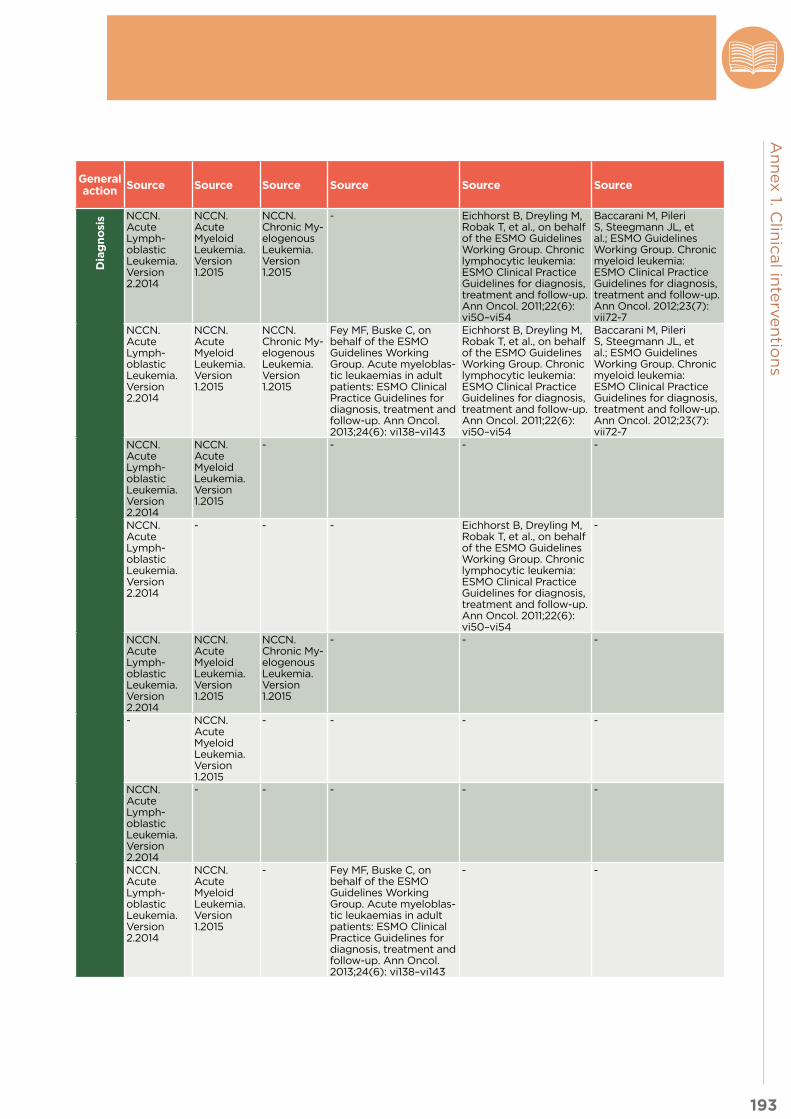
193
An
nex 1. C
linic
al in
terv
en
tion
s
Annex table 1-4. Leukaemia
General action
Steps/Actions/Options
Interventions identified from the recommendations
Se-quence (a, b, c,
d)
Priority(I, II, III) Notes When to perform
itGeneral action Source Source Source Source Source Source
Dia
gn
osi
s Physical exami-nation
General assessment of symptoms a I - -
Dia
gn
osi
s NCCN. Acute Lymph-oblastic Leukemia. Version 2.2014
NCCN. Acute Myeloid Leukemia. Version 1.2015
NCCN. Chronic My-elogenous Leukemia. Version 1.2015
- Eichhorst B, Dreyling M, Robak T, et al., on behalf of the ESMO Guidelines Working Group. Chronic lymphocytic leukemia: ESMO Clinical Practice Guidelines for diagnosis, treatment and follow-up. Ann Oncol. 2011;22(6): vi50–vi54
Baccarani M, Pileri S, Steegmann JL, et al.; ESMO Guidelines Working Group. Chronic myeloid leukemia: ESMO Clinical Practice Guidelines for diagnosis, treatment and follow-up. Ann Oncol. 2012;23(7): vii72-7
Blood testing Complete blood count, platelets, dif-ferential, chemistry profile
a I - - NCCN. Acute Lymph-oblastic Leukemia. Version 2.2014
NCCN. Acute Myeloid Leukemia. Version 1.2015
NCCN. Chronic My-elogenous Leukemia. Version 1.2015
Fey MF, Buske C, on behalf of the ESMO Guidelines Working Group. Acute myeloblas-tic leukaemias in adult patients: ESMO Clinical Practice Guidelines for diagnosis, treatment and follow-up. Ann Oncol. 2013;24(6): vi138–vi143
Eichhorst B, Dreyling M, Robak T, et al., on behalf of the ESMO Guidelines Working Group. Chronic lymphocytic leukemia: ESMO Clinical Practice Guidelines for diagnosis, treatment and follow-up. Ann Oncol. 2011;22(6): vi50–vi54
Baccarani M, Pileri S, Steegmann JL, et al.; ESMO Guidelines Working Group. Chronic myeloid leukemia: ESMO Clinical Practice Guidelines for diagnosis, treatment and follow-up. Ann Oncol. 2012;23(7): vii72-7
Disseminated intravascular coagula-tion (DIC) panel: D-dimers, fibrinogen, prothrombin time (PT), partial throm-boplastin time (PTT);
b I Measurement for D-dimers only recommended for ALL
- NCCN. Acute Lymph-oblastic Leukemia. Version 2.2014
NCCN. Acute Myeloid Leukemia. Version 1.2015
- - - -
Tumour lysis syndrome panel: lactate dehydrogenase (LDH), uric acid, po-tassium (K), Calcium (Ca), Phosphore
c I - - NCCN. Acute Lymph-oblastic Leukemia. Version 2.2014
- - - Eichhorst B, Dreyling M, Robak T, et al., on behalf of the ESMO Guidelines Working Group. Chronic lymphocytic leukemia: ESMO Clinical Practice Guidelines for diagnosis, treatment and follow-up. Ann Oncol. 2011;22(6): vi50–vi54
-
Cardiac function assessment
Echocardiography a I - - NCCN. Acute Lymph-oblastic Leukemia. Version 2.2014
NCCN. Acute Myeloid Leukemia. Version 1.2015
NCCN. Chronic My-elogenous Leukemia. Version 1.2015
- - -
Radionuclide angiography b III - - - NCCN. Acute Myeloid Leukemia. Version 1.2015
- - - -
CT of the heart b III - - NCCN. Acute Lymph-oblastic Leukemia. Version 2.2014
- - - - -
Imaging X-Ray scan a I - To assess bones according to clinical symptoms.
NCCN. Acute Lymph-oblastic Leukemia. Version 2.2014
NCCN. Acute Myeloid Leukemia. Version 1.2015
- Fey MF, Buske C, on behalf of the ESMO Guidelines Working Group. Acute myeloblas-tic leukaemias in adult patients: ESMO Clinical Practice Guidelines for diagnosis, treatment and follow-up. Ann Oncol. 2013;24(6): vi138–vi143
- -

194
WHO
list o
f prio
rity m
edica
l dev
ices f
or ca
ncer
man
agem
ent
General action
Steps/Actions/Options
Interventions identified from the recommendations
Se-quence (a, b, c,
d)
Priority(I, II, III) Notes When to perform
itGeneral action Source Source Source Source Source Source
Dia
gn
osi
s CT scan a I - In case of involvement of head, chest, mediastinal or other site and according to symptoms.
Dia
gn
osi
s NCCN. Acute Lymph-oblastic Leukemia. Version 2.2014
NCCN. Acute Myeloid Leukemia. Version 1.2015
- Fey MF, Buske C, on behalf of the ESMO Guidelines Working Group. Acute myeloblas-tic leukaemias in adult patients: ESMO Clinical Practice Guidelines for diagnosis, treatment and follow-up. Ann Oncol. 2013;24(6): vi138–vi143
- -
MRI scan a II - In case of involvement of head, chest, mediastinal or other site and according to symptoms
NCCN. Acute Lymph-oblastic Leukemia. Version 2.2014
NCCN. Acute Myeloid Leukemia. Version 1.2015
- - - -
Staging and risk assessment
Bone marrow aspirate a I - - NCCN. Acute Lymph-oblastic Leukemia. Version 2.2014
NCCN. Acute Myeloid Leukemia. Version 1.2015
NCCN. Chronic My-elogenous Leukemia. Version 1.2015
Fey MF, Buske C, on behalf of the ESMO Guidelines Working Group. Acute myeloblas-tic leukaemias in adult patients: ESMO Clinical Practice Guidelines for diagnosis, treatment and follow-up. Ann Oncol. 2013;24(6): vi138–vi143
- Baccarani M, Pileri S, Steegmann JL, et al.; ESMO Guidelines Working Group. Chronic myeloid leukemia: ESMO Clinical Practice Guidelines for diagnosis, treatment and follow-up. Ann Oncol. 2012;23(7): vii72-7
Bone marrow biopsy b I - - NCCN. Acute Lymph-oblastic Leukemia. Version 2.2014
NCCN. Acute Myeloid Leukemia. Version 1.2015
NCCN. Chronic My-elogenous Leukemia. Version 1.2015
Fey MF, Buske C, on behalf of the ESMO Guidelines Working Group. Acute myeloblas-tic leukaemias in adult patients: ESMO Clinical Practice Guidelines for diagnosis, treatment and follow-up. Ann Oncol. 2013;24(6): vi138–vi144
Eichhorst B, Dreyling M, Robak T, et al., on behalf of the ESMO Guidelines Working Group. Chronic lymphocytic leukemia: ESMO Clinical Practice Guidelines for diagnosis, treatment and follow-up. Ann Oncol. 2011;22(6): vi50–vi54
Baccarani M, Pileri S, Steegmann JL, et al.; ESMO Guidelines Working Group. Chronic myeloid leukemia: ESMO Clinical Practice Guidelines for diagnosis, treatment and follow-up. Ann Oncol. 2012;23(7): vii72-7
Comprehensive flow cytometric im-munophenotyping
c I - - NCCN. Acute Lymph-oblastic Leukemia. Version 2.2014
NCCN. Acute Myeloid Leukemia. Version 1.2015
NCCN. Chronic My-elogenous Leukemia. Version 1.2015
Fey MF, Buske C, on behalf of the ESMO Guidelines Working Group. Acute myeloblas-tic leukaemias in adult patients: ESMO Clinical Practice Guidelines for diagnosis, treatment and follow-up. Ann Oncol. 2013;24(6): vi138–vi145
Eichhorst B, Dreyling M, Robak T, et al., on behalf of the ESMO Guidelines Working Group. Chronic lymphocytic leukemia: ESMO Clinical Practice Guidelines for diagnosis, treatment and follow-up. Ann Oncol. 2011;22(6): vi50–vi54
-
Lumbar puncture d I - - NCCN. Acute Lymph-oblastic Leukemia. Version 2.2014
NCCN. Acute Myeloid Leukemia. Version 1.2015
- - - -
Genetic characterisation
Cytogenetics a I - Cytogenetics and FISH testing are important components of treatment for leukemia and can be considered according to resource availability.
NCCN. Acute Lymph-oblastic Leukemia. Version 2.2014
NCCN. Acute Myeloid Leukemia. Version 1.2015
NCCN. Chronic My-elogenous Leukemia. Version 1.2015
Fey MF, Buske C, on behalf of the ESMO Guidelines Working Group. Acute myeloblas-tic leukaemias in adult patients: ESMO Clinical Practice Guidelines for diagnosis, treatment and follow-up. Ann Oncol. 2013;24(6): vi138–vi145
- Baccarani M, Pileri S, Steegmann JL, et al.; ESMO Guidelines Working Group. Chronic myeloid leukemia: ESMO Clinical Practice Guidelines for diagnosis, treatment and follow-up. Ann Oncol. 2012;23(7): vii72-7
Fluorescence in situ hybridisation (FISH) testing
b I - Cytogenetics and FISH testing are important compo-nents of treatment for leu-kemia and can be considered accord-ing to resource availability.
NCCN. Acute Lymph-oblastic Leukemia. Version 2.2014
NCCN. Acute Myeloid Leukemia. Version 1.2015
NCCN. Chronic My-elogenous Leukemia. Version 1.2015
Fey MF, Buske C, on behalf of the ESMO Guidelines Working Group. Acute myeloblas-tic leukaemias in adult patients: ESMO Clinical Practice Guidelines for diagnosis, treatment and follow-up. Ann Oncol. 2013;24(6): vi138–vi145
Eichhorst B, Dreyling M, Robak T, et al., on behalf of the ESMO Guidelines Working Group. Chronic lymphocytic leukemia: ESMO Clinical Practice Guidelines for diagnosis, treatment and follow-up. Ann Oncol. 2011;22(6): vi50–vi54
-

195
An
nex 1. C
linic
al in
terv
en
tion
s
General action
Steps/Actions/Options
Interventions identified from the recommendations
Se-quence (a, b, c,
d)
Priority(I, II, III) Notes When to perform
itGeneral action Source Source Source Source Source Source
Dia
gn
osi
s CT scan a I - In case of involvement of head, chest, mediastinal or other site and according to symptoms.
Dia
gn
osi
s NCCN. Acute Lymph-oblastic Leukemia. Version 2.2014
NCCN. Acute Myeloid Leukemia. Version 1.2015
- Fey MF, Buske C, on behalf of the ESMO Guidelines Working Group. Acute myeloblas-tic leukaemias in adult patients: ESMO Clinical Practice Guidelines for diagnosis, treatment and follow-up. Ann Oncol. 2013;24(6): vi138–vi143
- -
MRI scan a II - In case of involvement of head, chest, mediastinal or other site and according to symptoms
NCCN. Acute Lymph-oblastic Leukemia. Version 2.2014
NCCN. Acute Myeloid Leukemia. Version 1.2015
- - - -
Staging and risk assessment
Bone marrow aspirate a I - - NCCN. Acute Lymph-oblastic Leukemia. Version 2.2014
NCCN. Acute Myeloid Leukemia. Version 1.2015
NCCN. Chronic My-elogenous Leukemia. Version 1.2015
Fey MF, Buske C, on behalf of the ESMO Guidelines Working Group. Acute myeloblas-tic leukaemias in adult patients: ESMO Clinical Practice Guidelines for diagnosis, treatment and follow-up. Ann Oncol. 2013;24(6): vi138–vi143
- Baccarani M, Pileri S, Steegmann JL, et al.; ESMO Guidelines Working Group. Chronic myeloid leukemia: ESMO Clinical Practice Guidelines for diagnosis, treatment and follow-up. Ann Oncol. 2012;23(7): vii72-7
Bone marrow biopsy b I - - NCCN. Acute Lymph-oblastic Leukemia. Version 2.2014
NCCN. Acute Myeloid Leukemia. Version 1.2015
NCCN. Chronic My-elogenous Leukemia. Version 1.2015
Fey MF, Buske C, on behalf of the ESMO Guidelines Working Group. Acute myeloblas-tic leukaemias in adult patients: ESMO Clinical Practice Guidelines for diagnosis, treatment and follow-up. Ann Oncol. 2013;24(6): vi138–vi144
Eichhorst B, Dreyling M, Robak T, et al., on behalf of the ESMO Guidelines Working Group. Chronic lymphocytic leukemia: ESMO Clinical Practice Guidelines for diagnosis, treatment and follow-up. Ann Oncol. 2011;22(6): vi50–vi54
Baccarani M, Pileri S, Steegmann JL, et al.; ESMO Guidelines Working Group. Chronic myeloid leukemia: ESMO Clinical Practice Guidelines for diagnosis, treatment and follow-up. Ann Oncol. 2012;23(7): vii72-7
Comprehensive flow cytometric im-munophenotyping
c I - - NCCN. Acute Lymph-oblastic Leukemia. Version 2.2014
NCCN. Acute Myeloid Leukemia. Version 1.2015
NCCN. Chronic My-elogenous Leukemia. Version 1.2015
Fey MF, Buske C, on behalf of the ESMO Guidelines Working Group. Acute myeloblas-tic leukaemias in adult patients: ESMO Clinical Practice Guidelines for diagnosis, treatment and follow-up. Ann Oncol. 2013;24(6): vi138–vi145
Eichhorst B, Dreyling M, Robak T, et al., on behalf of the ESMO Guidelines Working Group. Chronic lymphocytic leukemia: ESMO Clinical Practice Guidelines for diagnosis, treatment and follow-up. Ann Oncol. 2011;22(6): vi50–vi54
-
Lumbar puncture d I - - NCCN. Acute Lymph-oblastic Leukemia. Version 2.2014
NCCN. Acute Myeloid Leukemia. Version 1.2015
- - - -
Genetic characterisation
Cytogenetics a I - Cytogenetics and FISH testing are important components of treatment for leukemia and can be considered according to resource availability.
NCCN. Acute Lymph-oblastic Leukemia. Version 2.2014
NCCN. Acute Myeloid Leukemia. Version 1.2015
NCCN. Chronic My-elogenous Leukemia. Version 1.2015
Fey MF, Buske C, on behalf of the ESMO Guidelines Working Group. Acute myeloblas-tic leukaemias in adult patients: ESMO Clinical Practice Guidelines for diagnosis, treatment and follow-up. Ann Oncol. 2013;24(6): vi138–vi145
- Baccarani M, Pileri S, Steegmann JL, et al.; ESMO Guidelines Working Group. Chronic myeloid leukemia: ESMO Clinical Practice Guidelines for diagnosis, treatment and follow-up. Ann Oncol. 2012;23(7): vii72-7
Fluorescence in situ hybridisation (FISH) testing
b I - Cytogenetics and FISH testing are important compo-nents of treatment for leu-kemia and can be considered accord-ing to resource availability.
NCCN. Acute Lymph-oblastic Leukemia. Version 2.2014
NCCN. Acute Myeloid Leukemia. Version 1.2015
NCCN. Chronic My-elogenous Leukemia. Version 1.2015
Fey MF, Buske C, on behalf of the ESMO Guidelines Working Group. Acute myeloblas-tic leukaemias in adult patients: ESMO Clinical Practice Guidelines for diagnosis, treatment and follow-up. Ann Oncol. 2013;24(6): vi138–vi145
Eichhorst B, Dreyling M, Robak T, et al., on behalf of the ESMO Guidelines Working Group. Chronic lymphocytic leukemia: ESMO Clinical Practice Guidelines for diagnosis, treatment and follow-up. Ann Oncol. 2011;22(6): vi50–vi54
-
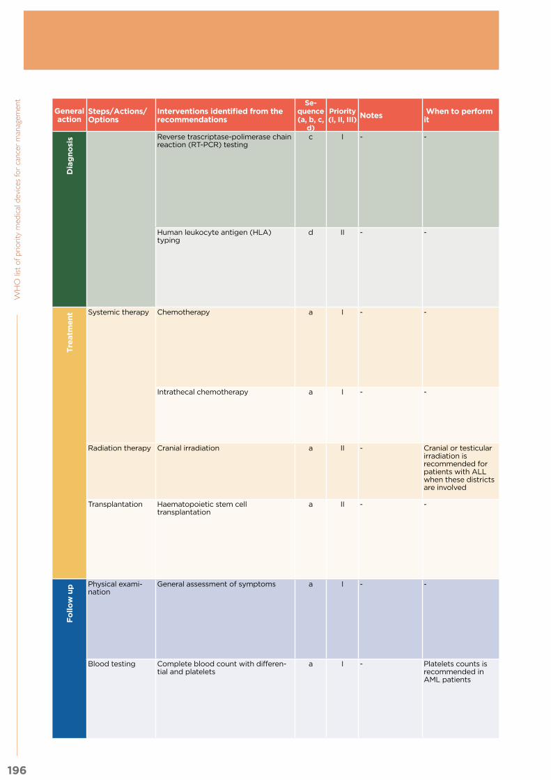
196
WHO
list o
f prio
rity m
edica
l dev
ices f
or ca
ncer
man
agem
ent
General action
Steps/Actions/Options
Interventions identified from the recommendations
Se-quence (a, b, c,
d)
Priority(I, II, III) Notes When to perform
itGeneral action Source Source Source Source Source Source
Dia
gn
osi
s Reverse trascriptase-polimerase chain reaction (RT-PCR) testing
c I - -
Dia
gn
osi
s NCCN. Acute Lymph-oblastic Leukemia. Version 2.2014
NCCN. Acute Myeloid Leukemia. Version 1.2015
NCCN. Chronic My-elogenous Leukemia. Version 1.2015
Fey MF, Buske C, on behalf of the ESMO Guidelines Working Group. Acute myeloblas-tic leukaemias in adult patients: ESMO Clinical Practice Guidelines for diagnosis, treatment and follow-up. Ann Oncol. 2013;24(6): vi138–vi145
Yeoh AE, Tan D, Li CK, Hori H, Tse E, Pui CH; Asian Oncology Summit 2013. Management of adult and paediatric acute lymphoblas-tic leukaemia in Asia: resource-stratified guidelines from the Asian Oncology Summit 2013. Lancet Oncol. 2013 Nov;14(12):e508-23.
Baccarani M, Pileri S, Steegmann JL, et al.; ESMO Guidelines Working Group. Chronic myeloid leukemia: ESMO Clinical Practice Guidelines for diagnosis, treatment and follow-up. Ann Oncol. 2012;23(7): vii72-7
Human leukocyte antigen (HLA) typing
d II - - NCCN. Acute Lymph-oblastic Leukemia. Version 2.2014
NCCN. Acute Myeloid Leukemia. Version 1.2015
NCCN. Chronic My-elogenous Leukemia. Version 1.2015
Fey MF, Buske C, on behalf of the ESMO Guidelines Working Group. Acute myeloblas-tic leukaemias in adult patients: ESMO Clinical Practice Guidelines for diagnosis, treatment and follow-up. Ann Oncol. 2013;24(6): vi138–vi145
Eichhorst B, Dreyling M, Robak T, et al., on behalf of the ESMO Guidelines Working Group. Chronic lymphocytic leukemia: ESMO Clinical Practice Guidelines for diagnosis, treatment and follow-up. Ann Oncol. 2011;22(6): vi50–vi54
-
Tre
atm
en
t Systemic therapy Chemotherapy a I - -
Tre
atm
en
t NCCN. Acute Lymph-oblastic Leukemia. Version 2.2014
NCCN. Acute Myeloid Leukemia. Version 1.2015
NCCN. Chronic My-elogenous Leukemia. Version 1.2015
Fey MF, Buske C, on behalf of the ESMO Guidelines Working Group. Acute myeloblas-tic leukaemias in adult patients: ESMO Clinical Practice Guidelines for diagnosis, treatment and follow-up. Ann Oncol. 2013;24(6): vi138–vi145
Eichhorst B, Dreyling M, Robak T, et al., on behalf of the ESMO Guidelines Working Group. Chronic lymphocytic leukemia: ESMO Clinical Practice Guidelines for diagnosis, treatment and follow-up. Ann Oncol. 2011;22(6): vi50–vi54
Baccarani M, Pileri S, Steegmann JL, et al.; ESMO Guidelines Working Group. Chronic myeloid leukemia: ESMO Clinical Practice Guidelines for diagnosis, treatment and follow-up. Ann Oncol. 2012;23(7): vii72-7
Intrathecal chemotherapy a I - - NCCN. Acute Lymph-oblastic Leukemia. Version 2.2014
NCCN. Acute Myeloid Leukemia. Version 1.2015
- - - -
Radiation therapy Cranial irradiation a II - Cranial or testicular irradiation is recommended for patients with ALL when these districts are involved
NCCN. Acute Lymph-oblastic Leukemia. Version 2.2014
NCCN. Acute Myeloid Leukemia. Version 1.2015
- - - -
Transplantation Haematopoietic stem cell transplantation
a II - - NCCN. Acute Lymph-oblastic Leukemia. Version 2.2014
NCCN. Acute Myeloid Leukemia. Version 1.2015
NCCN. Chronic My-elogenous Leukemia. Version 1.2015
Fey MF, Buske C, on behalf of the ESMO Guidelines Working Group. Acute myeloblas-tic leukaemias in adult patients: ESMO Clinical Practice Guidelines for diagnosis, treatment and follow-up. Ann Oncol. 2013;24(6): vi138–vi145
Eichhorst B, Dreyling M, Robak T, et al., on behalf of the ESMO Guidelines Working Group. Chronic lymphocytic leukemia: ESMO Clinical Practice Guidelines for diagnosis, treatment and follow-up. Ann Oncol. 2011;22(6): vi50–vi54
Baccarani M, Pileri S, Steegmann JL, et al.; ESMO Guidelines Working Group. Chronic myeloid leukemia: ESMO Clinical Practice Guidelines for diagnosis, treatment and follow-up. Ann Oncol. 2012;23(7): vii72-7
Fo
llo
w u
p Physical exami-nation
General assessment of symptoms a I - -
Fo
llo
w u
p NCCN. Acute Lymph-oblastic Leukemia. Version 2.2014
- - Eichhorst B, Dreyling M, Robak T, et al., on behalf of the ESMO Guidelines Working Group. Chronic lymphocytic leukemia: ESMO Clinical Practice Guidelines for diagnosis, treatment and follow-up. Ann Oncol. 2011;22(6): vi50–vi54
-
Blood testing Complete blood count with differen-tial and platelets
a I - Platelets counts is recommended in AML patients
NCCN. Acute Lymph-oblastic Leukemia. Version 2.2014
NCCN. Acute Myeloid Leukemia. Version 1.2015
- Fey MF, Buske C, on behalf of the ESMO Guidelines Working Group. Acute myeloblas-tic leukaemias in adult patients: ESMO Clinical Practice Guidelines for diagnosis, treatment and follow-up. Ann Oncol. 2013;24(6): vi138–vi145
Eichhorst B, Dreyling M, Robak T, et al., on behalf of the ESMO Guidelines Working Group. Chronic lymphocytic leukemia: ESMO Clinical Practice Guidelines for diagnosis, treatment and follow-up. Ann Oncol. 2011;22(6): vi50–vi54
Baccarani M, Pileri S, Steegmann JL, et al.; ESMO Guidelines Working Group. Chronic myeloid leukemia: ESMO Clinical Practice Guidelines for diagnosis, treatment and follow-up. Ann Oncol. 2012;23(7): vii72-7

197
An
nex 1. C
linic
al in
terv
en
tion
s
General action
Steps/Actions/Options
Interventions identified from the recommendations
Se-quence (a, b, c,
d)
Priority(I, II, III) Notes When to perform
itGeneral action Source Source Source Source Source Source
Dia
gn
osi
s Reverse trascriptase-polimerase chain reaction (RT-PCR) testing
c I - -
Dia
gn
osi
s NCCN. Acute Lymph-oblastic Leukemia. Version 2.2014
NCCN. Acute Myeloid Leukemia. Version 1.2015
NCCN. Chronic My-elogenous Leukemia. Version 1.2015
Fey MF, Buske C, on behalf of the ESMO Guidelines Working Group. Acute myeloblas-tic leukaemias in adult patients: ESMO Clinical Practice Guidelines for diagnosis, treatment and follow-up. Ann Oncol. 2013;24(6): vi138–vi145
Yeoh AE, Tan D, Li CK, Hori H, Tse E, Pui CH; Asian Oncology Summit 2013. Management of adult and paediatric acute lymphoblas-tic leukaemia in Asia: resource-stratified guidelines from the Asian Oncology Summit 2013. Lancet Oncol. 2013 Nov;14(12):e508-23.
Baccarani M, Pileri S, Steegmann JL, et al.; ESMO Guidelines Working Group. Chronic myeloid leukemia: ESMO Clinical Practice Guidelines for diagnosis, treatment and follow-up. Ann Oncol. 2012;23(7): vii72-7
Human leukocyte antigen (HLA) typing
d II - - NCCN. Acute Lymph-oblastic Leukemia. Version 2.2014
NCCN. Acute Myeloid Leukemia. Version 1.2015
NCCN. Chronic My-elogenous Leukemia. Version 1.2015
Fey MF, Buske C, on behalf of the ESMO Guidelines Working Group. Acute myeloblas-tic leukaemias in adult patients: ESMO Clinical Practice Guidelines for diagnosis, treatment and follow-up. Ann Oncol. 2013;24(6): vi138–vi145
Eichhorst B, Dreyling M, Robak T, et al., on behalf of the ESMO Guidelines Working Group. Chronic lymphocytic leukemia: ESMO Clinical Practice Guidelines for diagnosis, treatment and follow-up. Ann Oncol. 2011;22(6): vi50–vi54
-
Tre
atm
en
t Systemic therapy Chemotherapy a I - -
Tre
atm
en
t NCCN. Acute Lymph-oblastic Leukemia. Version 2.2014
NCCN. Acute Myeloid Leukemia. Version 1.2015
NCCN. Chronic My-elogenous Leukemia. Version 1.2015
Fey MF, Buske C, on behalf of the ESMO Guidelines Working Group. Acute myeloblas-tic leukaemias in adult patients: ESMO Clinical Practice Guidelines for diagnosis, treatment and follow-up. Ann Oncol. 2013;24(6): vi138–vi145
Eichhorst B, Dreyling M, Robak T, et al., on behalf of the ESMO Guidelines Working Group. Chronic lymphocytic leukemia: ESMO Clinical Practice Guidelines for diagnosis, treatment and follow-up. Ann Oncol. 2011;22(6): vi50–vi54
Baccarani M, Pileri S, Steegmann JL, et al.; ESMO Guidelines Working Group. Chronic myeloid leukemia: ESMO Clinical Practice Guidelines for diagnosis, treatment and follow-up. Ann Oncol. 2012;23(7): vii72-7
Intrathecal chemotherapy a I - - NCCN. Acute Lymph-oblastic Leukemia. Version 2.2014
NCCN. Acute Myeloid Leukemia. Version 1.2015
- - - -
Radiation therapy Cranial irradiation a II - Cranial or testicular irradiation is recommended for patients with ALL when these districts are involved
NCCN. Acute Lymph-oblastic Leukemia. Version 2.2014
NCCN. Acute Myeloid Leukemia. Version 1.2015
- - - -
Transplantation Haematopoietic stem cell transplantation
a II - - NCCN. Acute Lymph-oblastic Leukemia. Version 2.2014
NCCN. Acute Myeloid Leukemia. Version 1.2015
NCCN. Chronic My-elogenous Leukemia. Version 1.2015
Fey MF, Buske C, on behalf of the ESMO Guidelines Working Group. Acute myeloblas-tic leukaemias in adult patients: ESMO Clinical Practice Guidelines for diagnosis, treatment and follow-up. Ann Oncol. 2013;24(6): vi138–vi145
Eichhorst B, Dreyling M, Robak T, et al., on behalf of the ESMO Guidelines Working Group. Chronic lymphocytic leukemia: ESMO Clinical Practice Guidelines for diagnosis, treatment and follow-up. Ann Oncol. 2011;22(6): vi50–vi54
Baccarani M, Pileri S, Steegmann JL, et al.; ESMO Guidelines Working Group. Chronic myeloid leukemia: ESMO Clinical Practice Guidelines for diagnosis, treatment and follow-up. Ann Oncol. 2012;23(7): vii72-7
Fo
llo
w u
p Physical exami-nation
General assessment of symptoms a I - -
Fo
llo
w u
p NCCN. Acute Lymph-oblastic Leukemia. Version 2.2014
- - Eichhorst B, Dreyling M, Robak T, et al., on behalf of the ESMO Guidelines Working Group. Chronic lymphocytic leukemia: ESMO Clinical Practice Guidelines for diagnosis, treatment and follow-up. Ann Oncol. 2011;22(6): vi50–vi54
-
Blood testing Complete blood count with differen-tial and platelets
a I - Platelets counts is recommended in AML patients
NCCN. Acute Lymph-oblastic Leukemia. Version 2.2014
NCCN. Acute Myeloid Leukemia. Version 1.2015
- Fey MF, Buske C, on behalf of the ESMO Guidelines Working Group. Acute myeloblas-tic leukaemias in adult patients: ESMO Clinical Practice Guidelines for diagnosis, treatment and follow-up. Ann Oncol. 2013;24(6): vi138–vi145
Eichhorst B, Dreyling M, Robak T, et al., on behalf of the ESMO Guidelines Working Group. Chronic lymphocytic leukemia: ESMO Clinical Practice Guidelines for diagnosis, treatment and follow-up. Ann Oncol. 2011;22(6): vi50–vi54
Baccarani M, Pileri S, Steegmann JL, et al.; ESMO Guidelines Working Group. Chronic myeloid leukemia: ESMO Clinical Practice Guidelines for diagnosis, treatment and follow-up. Ann Oncol. 2012;23(7): vii72-7

198
WHO
list o
f prio
rity m
edica
l dev
ices f
or ca
ncer
man
agem
ent
General action
Steps/Actions/Options
Interventions identified from the recommendations
Se-quence (a, b, c,
d)
Priority(I, II, III) Notes When to perform
itGeneral action Source Source Source Source Source Source
Fo
llo
w u
p Liver function test b I - -
Fo
llo
w u
p NCCN. Acute Lymph-oblastic Leukemia. Version 2.2014
- - Fey MF, Buske C, on behalf of the ESMO Guidelines Working Group. Acute myeloblas-tic leukaemias in adult patients: ESMO Clinical Practice Guidelines for diagnosis, treatment and follow-up. Ann Oncol. 2013;24(6): vi138–vi145
- -
Biopsy Bone marrow aspirate a I - - NCCN. Acute Lymph-oblastic Leukemia. Version 2.2014
NCCN. Acute Myeloid Leukemia. Version 1.2015
- Fey MF, Buske C, on behalf of the ESMO Guidelines Working Group. Acute myeloblas-tic leukaemias in adult patients: ESMO Clinical Practice Guidelines for diagnosis, treatment and follow-up. Ann Oncol. 2013;24(6): vi138–vi145
- Baccarani M, Pileri S, Steegmann JL, et al.; ESMO Guidelines Working Group. Chronic myeloid leukemia: ESMO Clinical Practice Guidelines for diagnosis, treatment and follow-up. Ann Oncol. 2012;23(7): vii72-7
Cerebrospinal fluid aspirate b I - - NCCN. Acute Lymph-oblastic Leukemia. Version 2.2014
- - - - -
Bone marrow biopsy c I - - - NCCN. Acute Myeloid Leukemia. Version 1.2015
- Fey MF, Buske C, on behalf of the ESMO Guidelines Working Group. Acute myeloblas-tic leukaemias in adult patients: ESMO Clinical Practice Guidelines for diagnosis, treatment and follow-up. Ann Oncol. 2013;24(6): vi138–vi145
- Baccarani M, Pileri S, Steegmann JL, et al.; ESMO Guidelines Working Group. Chronic myeloid leukemia: ESMO Clinical Practice Guidelines for diagnosis, treatment and follow-up. Ann Oncol. 2012;23(7): vii72-7
Cardiac function assessment
Echocardiography a I - - NCCN. Acute Lymph-oblastic Leukemia. Version 2.2014
- - Fey MF, Buske C, on behalf of the ESMO Guidelines Working Group. Acute myeloblas-tic leukaemias in adult patients: ESMO Clinical Practice Guidelines for diagnosis, treatment and follow-up. Ann Oncol. 2013;24(6): vi138–vi145
- -
Genetic character-isation
Fluorescence in situ hybridisation (FISH) testing
a I - - NCCN. Acute Lymph-oblastic Leukemia. Version 2.2014
- - - - -
Flow cytometry b I - - NCCN. Acute Lymph-oblastic Leukemia. Version 2.2014
- - - Eichhorst B, Dreyling M, Robak T, et al., on behalf of the ESMO Guidelines Working Group. Chronic lymphocytic leukemia: ESMO Clinical Practice Guidelines for diagnosis, treatment and follow-up. Ann Oncol. 2011;22(6): vi50–vi54
-
Quantitative reverse trascriptase-poli-merase chain reaction (QPCR) testing
c I - - - - NCCN. Chronic My-elogenous Leukemia. Version 1.2015
- - Baccarani M, Pileri S, Steegmann JL, et al.; ESMO Guidelines Working Group. Chronic myeloid leukemia: ESMO Clinical Practice Guidelines for diagnosis, treatment and follow-up. Ann Oncol. 2012;23(7): vii72-7
Palli-
ati
ve
C
are
- - - - - -
Palli-
ati
ve
C
are
- - - - -
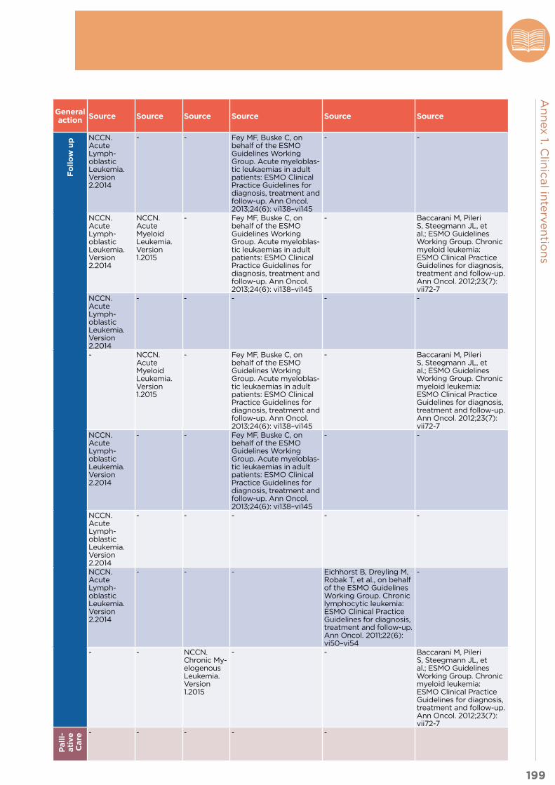
199
An
nex 1. C
linic
al in
terv
en
tion
s
General action
Steps/Actions/Options
Interventions identified from the recommendations
Se-quence (a, b, c,
d)
Priority(I, II, III) Notes When to perform
itGeneral action Source Source Source Source Source Source
Fo
llo
w u
p Liver function test b I - -
Fo
llo
w u
p NCCN. Acute Lymph-oblastic Leukemia. Version 2.2014
- - Fey MF, Buske C, on behalf of the ESMO Guidelines Working Group. Acute myeloblas-tic leukaemias in adult patients: ESMO Clinical Practice Guidelines for diagnosis, treatment and follow-up. Ann Oncol. 2013;24(6): vi138–vi145
- -
Biopsy Bone marrow aspirate a I - - NCCN. Acute Lymph-oblastic Leukemia. Version 2.2014
NCCN. Acute Myeloid Leukemia. Version 1.2015
- Fey MF, Buske C, on behalf of the ESMO Guidelines Working Group. Acute myeloblas-tic leukaemias in adult patients: ESMO Clinical Practice Guidelines for diagnosis, treatment and follow-up. Ann Oncol. 2013;24(6): vi138–vi145
- Baccarani M, Pileri S, Steegmann JL, et al.; ESMO Guidelines Working Group. Chronic myeloid leukemia: ESMO Clinical Practice Guidelines for diagnosis, treatment and follow-up. Ann Oncol. 2012;23(7): vii72-7
Cerebrospinal fluid aspirate b I - - NCCN. Acute Lymph-oblastic Leukemia. Version 2.2014
- - - - -
Bone marrow biopsy c I - - - NCCN. Acute Myeloid Leukemia. Version 1.2015
- Fey MF, Buske C, on behalf of the ESMO Guidelines Working Group. Acute myeloblas-tic leukaemias in adult patients: ESMO Clinical Practice Guidelines for diagnosis, treatment and follow-up. Ann Oncol. 2013;24(6): vi138–vi145
- Baccarani M, Pileri S, Steegmann JL, et al.; ESMO Guidelines Working Group. Chronic myeloid leukemia: ESMO Clinical Practice Guidelines for diagnosis, treatment and follow-up. Ann Oncol. 2012;23(7): vii72-7
Cardiac function assessment
Echocardiography a I - - NCCN. Acute Lymph-oblastic Leukemia. Version 2.2014
- - Fey MF, Buske C, on behalf of the ESMO Guidelines Working Group. Acute myeloblas-tic leukaemias in adult patients: ESMO Clinical Practice Guidelines for diagnosis, treatment and follow-up. Ann Oncol. 2013;24(6): vi138–vi145
- -
Genetic character-isation
Fluorescence in situ hybridisation (FISH) testing
a I - - NCCN. Acute Lymph-oblastic Leukemia. Version 2.2014
- - - - -
Flow cytometry b I - - NCCN. Acute Lymph-oblastic Leukemia. Version 2.2014
- - - Eichhorst B, Dreyling M, Robak T, et al., on behalf of the ESMO Guidelines Working Group. Chronic lymphocytic leukemia: ESMO Clinical Practice Guidelines for diagnosis, treatment and follow-up. Ann Oncol. 2011;22(6): vi50–vi54
-
Quantitative reverse trascriptase-poli-merase chain reaction (QPCR) testing
c I - - - - NCCN. Chronic My-elogenous Leukemia. Version 1.2015
- - Baccarani M, Pileri S, Steegmann JL, et al.; ESMO Guidelines Working Group. Chronic myeloid leukemia: ESMO Clinical Practice Guidelines for diagnosis, treatment and follow-up. Ann Oncol. 2012;23(7): vii72-7
Palli-
ati
ve
C
are
- - - - - -
Palli-
ati
ve
C
are
- - - - -

200
WHO
list o
f prio
rity m
edica
l dev
ices f
or ca
ncer
man
agem
ent Annex table 1-5. Lung cancer
General action
Steps/Actions/Options
Interventions identified from the recommendations
Se-quence (a, b, c,
d)
Priority (I, II, III)
NotesGeneral action When to perform it Source Source Source Source
Pre
ven
tio
n Smoking cessation Smoking cessation counselling a I -
Pre
ven
tio
n - Lung cancer. NICE clinical guideline 121 (2011)
NCCN. Non-small cell lung cancer. Version 3.2015
Vansteenkiste J, De Ruysscher D, Eberhardt WE, Lim E, Senan S, Felip E, Peters S; ESMO Guide-lines Working Group. Early and locally advanced non-small-cell lung cancer (NSCLC): ESMO Clinical Practice Guidelines for diagnosis, treatment and follow-up. Ann Oncol. 2013;24(6):vi89-98.
-
Dia
gn
osi
s Lung imaging (screening)
CT scan of the chest a - WHO has not reviewed low-dose CT as a modality for cancer screening at this time because of insufficient data.
Dia
gn
osi
s For selected and high-risk smokers or former heavy smokers according to NCCN and ESMO.
- NCCN. Lung Cancer Screening. Version 1.2015
Vansteenkiste J, De Ruysscher D, Eberhardt WE, Lim E, Senan S, Felip E, Peters S; ESMO Guide-lines Working Group. Early and locally advanced non-small-cell lung cancer (NSCLC): ESMO Clinical Practice Guidelines for diagnosis, treatment and follow-up. Ann Oncol. 2013;24(6):vi89-98.
-
Physical examination
Assessment of symptoms that may suggest lung cancer (check breathing, lymph nodes, etc.)
a I - - Lung cancer. NICE clinical guideline 121 (2011)
NCCN. Non-small cell lung cancer. Version 3.2015
Vansteenkiste J, De Ruysscher D, Eberhardt WE, Lim E, Senan S, Felip E, Peters S; ESMO Guide-lines Working Group. Early and locally advanced non-small-cell lung cancer (NSCLC): ESMO Clinical Practice Guidelines for diagnosis, treatment and follow-up. Ann Oncol. 2013;24(6):vi89-98.
-
Lung imaging (early diagnosis)
Chest X-Ray a I - When lung cancer is suspected at physical examination.
Lung cancer. NICE clinical guideline 121 (2011)
- - -
CT scan of the chest b I Contrast-enhanced CT scan of the chest is recommended by NICE.
When lung cancer is highly suspected from X-Ray results.
Lung cancer. NICE clinical guideline 121 (2011)
NCCN. Non-small cell lung cancer. Version 3.2015
-
Pulmonary function test
Spirometry a I - - Lung cancer. NICE clinical guideline 121 (2011)
- Vansteenkiste J, De Ruysscher D, Eberhardt WE, Lim E, Senan S, Felip E, Peters S; ESMO Guide-lines Working Group. Early and locally advanced non-small-cell lung cancer (NSCLC): ESMO Clinical Practice Guidelines for diagnosis, treatment and follow-up. Ann Oncol. 2013;24(6):vi89-98.
-
Staging and risk assessment
Haematology, renal and hepatic func-tion, and bone biochemistry tests
a I - - - - - Reck M, Popat S, Rein-muth N, De Ruysscher D, Kerr KM, Peters S; ESMO Guidelines Work-ing Group. Metastatic non-small-cell lung cancer (NSCLC): ESMO Clinical Practice Guidelines for diagnosis, treatment and follow-up. Ann Oncol. 2014;25(3):iii27-39
Abdominal ultrasound c I Abdominal ultrasound can be considered as an alternate staging test in setting where CT scan may not be available.
When abnormal-look-ing areas in the liver that are noticed.
Lung cancer. NICE clinical guideline 121 (2011)
- - -

201
An
nex 1. C
linic
al in
terv
en
tion
s
Annex table 1-5. Lung cancer
General action
Steps/Actions/Options
Interventions identified from the recommendations
Se-quence (a, b, c,
d)
Priority (I, II, III)
NotesGeneral action When to perform it Source Source Source Source
Pre
ven
tio
n Smoking cessation Smoking cessation counselling a I -
Pre
ven
tio
n - Lung cancer. NICE clinical guideline 121 (2011)
NCCN. Non-small cell lung cancer. Version 3.2015
Vansteenkiste J, De Ruysscher D, Eberhardt WE, Lim E, Senan S, Felip E, Peters S; ESMO Guide-lines Working Group. Early and locally advanced non-small-cell lung cancer (NSCLC): ESMO Clinical Practice Guidelines for diagnosis, treatment and follow-up. Ann Oncol. 2013;24(6):vi89-98.
-
Dia
gn
osi
s Lung imaging (screening)
CT scan of the chest a - WHO has not reviewed low-dose CT as a modality for cancer screening at this time because of insufficient data.
Dia
gn
osi
s For selected and high-risk smokers or former heavy smokers according to NCCN and ESMO.
- NCCN. Lung Cancer Screening. Version 1.2015
Vansteenkiste J, De Ruysscher D, Eberhardt WE, Lim E, Senan S, Felip E, Peters S; ESMO Guide-lines Working Group. Early and locally advanced non-small-cell lung cancer (NSCLC): ESMO Clinical Practice Guidelines for diagnosis, treatment and follow-up. Ann Oncol. 2013;24(6):vi89-98.
-
Physical examination
Assessment of symptoms that may suggest lung cancer (check breathing, lymph nodes, etc.)
a I - - Lung cancer. NICE clinical guideline 121 (2011)
NCCN. Non-small cell lung cancer. Version 3.2015
Vansteenkiste J, De Ruysscher D, Eberhardt WE, Lim E, Senan S, Felip E, Peters S; ESMO Guide-lines Working Group. Early and locally advanced non-small-cell lung cancer (NSCLC): ESMO Clinical Practice Guidelines for diagnosis, treatment and follow-up. Ann Oncol. 2013;24(6):vi89-98.
-
Lung imaging (early diagnosis)
Chest X-Ray a I - When lung cancer is suspected at physical examination.
Lung cancer. NICE clinical guideline 121 (2011)
- - -
CT scan of the chest b I Contrast-enhanced CT scan of the chest is recommended by NICE.
When lung cancer is highly suspected from X-Ray results.
Lung cancer. NICE clinical guideline 121 (2011)
NCCN. Non-small cell lung cancer. Version 3.2015
-
Pulmonary function test
Spirometry a I - - Lung cancer. NICE clinical guideline 121 (2011)
- Vansteenkiste J, De Ruysscher D, Eberhardt WE, Lim E, Senan S, Felip E, Peters S; ESMO Guide-lines Working Group. Early and locally advanced non-small-cell lung cancer (NSCLC): ESMO Clinical Practice Guidelines for diagnosis, treatment and follow-up. Ann Oncol. 2013;24(6):vi89-98.
-
Staging and risk assessment
Haematology, renal and hepatic func-tion, and bone biochemistry tests
a I - - - - - Reck M, Popat S, Rein-muth N, De Ruysscher D, Kerr KM, Peters S; ESMO Guidelines Work-ing Group. Metastatic non-small-cell lung cancer (NSCLC): ESMO Clinical Practice Guidelines for diagnosis, treatment and follow-up. Ann Oncol. 2014;25(3):iii27-39
Abdominal ultrasound c I Abdominal ultrasound can be considered as an alternate staging test in setting where CT scan may not be available.
When abnormal-look-ing areas in the liver that are noticed.
Lung cancer. NICE clinical guideline 121 (2011)
- - -
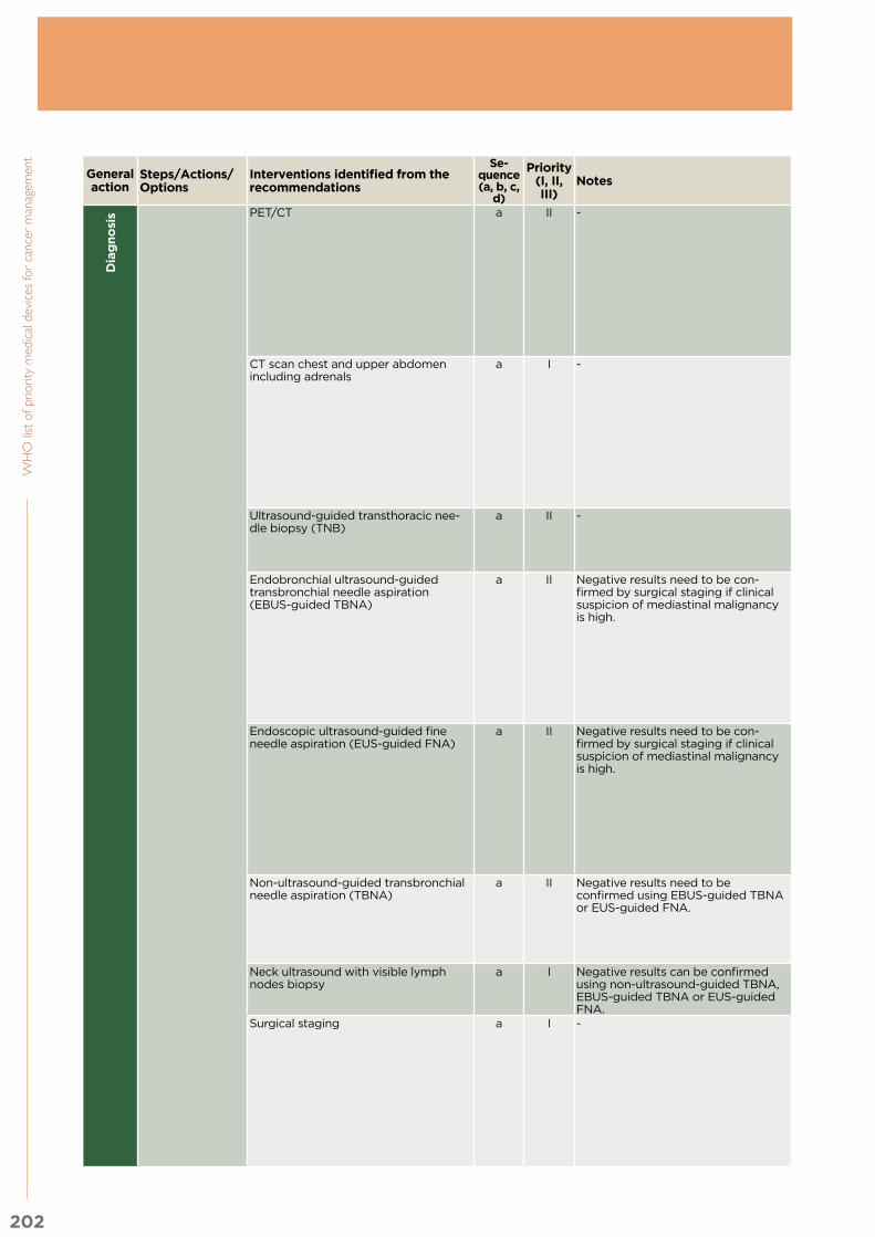
202
WHO
list o
f prio
rity m
edica
l dev
ices f
or ca
ncer
man
agem
ent
General action
Steps/Actions/Options
Interventions identified from the recommendations
Se-quence (a, b, c,
d)
Priority (I, II, III)
NotesGeneral action When to perform it Source Source Source Source
Dia
gn
osi
s PET/CT a II -
Dia
gn
osi
s First choice after CT showing a low probability of medi-astinal malignancy for patients who are potentially suitable for treatment with curative intent.
Lung cancer. NICE clinical guideline 121 (2011)
NCCN. Non-small cell lung cancer. Version 3.2015
Vansteenkiste J, De Ruysscher D, Eberhardt WE, Lim E, Senan S, Felip E, Peters S; ESMO Guide-lines Working Group. Early and locally advanced non-small-cell lung cancer (NSCLC): ESMO Clinical Practice Guidelines for diagnosis, treatment and follow-up. Ann Oncol. 2013;24(6):vi89-98.
Reck M, Popat S, Rein-muth N, De Ruysscher D, Kerr KM, Peters S; ESMO Guidelines Work-ing Group. Metastatic non-small-cell lung cancer (NSCLC): ESMO Clinical Practice Guidelines for diagnosis, treatment and follow-up. Ann Oncol. 2014;25(3):iii27-39
CT scan chest and upper abdomen including adrenals
a I - To patients with pe-ripheral lung lesions when treatment can be planned on the basis of this test.
Lung cancer. NICE clinical guideline 121 (2011)
NCCN. Non-small cell lung cancer. Version 3.2015
Vansteenkiste J, De Ruysscher D, Eberhardt WE, Lim E, Senan S, Felip E, Peters S; ESMO Guide-lines Working Group. Early and locally advanced non-small-cell lung cancer (NSCLC): ESMO Clinical Practice Guidelines for diagnosis, treatment and follow-up. Ann Oncol. 2013;24(6):vi89-98.
Reck M, Popat S, Rein-muth N, De Ruysscher D, Kerr KM, Peters S; ESMO Guidelines Work-ing Group. Metastatic non-small-cell lung cancer (NSCLC): ESMO Clinical Practice Guidelines for diagnosis, treatment and follow-up. Ann Oncol. 2014;25(3):iii27-39
Ultrasound-guided transthoracic nee-dle biopsy (TNB)
a II - To patients with pe-ripheral lung lesions when treatment can be planned on the basis of this test.
Lung cancer. NICE clinical guideline 121 (2011)
NCCN. Non-small cell lung cancer. Version 3.2015
- -
Endobronchial ultrasound-guided transbronchial needle aspiration (EBUS-guided TBNA)
a II Negative results need to be con-firmed by surgical staging if clinical suspicion of mediastinal malignancy is high.
As the first test for patients with an inter-mediate probability of mediastinal malignan-cy who are potentially suitable for treatment with curative intent.
Lung cancer. NICE clinical guideline 121 (2011)
NCCN. Non-small cell lung cancer. Version 3.2015
Vansteenkiste J, De Ruysscher D, Eberhardt WE, Lim E, Senan S, Felip E, Peters S; ESMO Guide-lines Working Group. Early and locally advanced non-small-cell lung cancer (NSCLC): ESMO Clinical Practice Guidelines for diagnosis, treatment and follow-up. Ann Oncol. 2013;24(6):vi89-98.
-
Endoscopic ultrasound-guided fine needle aspiration (EUS-guided FNA)
a II Negative results need to be con-firmed by surgical staging if clinical suspicion of mediastinal malignancy is high.
As the first test for patients with an inter-mediate probability of mediastinal malignan-cy who are potentially suitable for treatment with curative intent.
Lung cancer. NICE clinical guideline 121 (2011)
NCCN. Non-small cell lung cancer. Version 3.2015
Vansteenkiste J, De Ruysscher D, Eberhardt WE, Lim E, Senan S, Felip E, Peters S; ESMO Guide-lines Working Group. Early and locally advanced non-small-cell lung cancer (NSCLC): ESMO Clinical Practice Guidelines for diagnosis, treatment and follow-up. Ann Oncol. 2013;24(6):vi89-98.
-
Non-ultrasound-guided transbronchial needle aspiration (TBNA)
a II Negative results need to be confirmed using EBUS-guided TBNA or EUS-guided FNA.
As the first test for patients with an inter-mediate probability of mediastinal malignan-cy who are potentially suitable for treatment with curative intent.
Lung cancer. NICE clinical guideline 121 (2011)
NCCN. Non-small cell lung cancer. Version 3.2015
- -
Neck ultrasound with visible lymph nodes biopsy
a I Negative results can be confirmed using non-ultrasound-guided TBNA, EBUS-guided TBNA or EUS-guided FNA.
To patients with a high probability of mediastinal malig-nancy.
Lung cancer. NICE clinical guideline 121 (2011)
- - -
Surgical staging a I - To confirm negative results obtained by non-ultra-sound-guided TBNA, EBUS-guided TBNA or EUS-guided FNA (if clinical suspicion of mediastinal malignan-cy is high).
Lung cancer. NICE clinical guideline 121 (2011)
NCCN. Non-small cell lung cancer. Version 3.2015
Vansteenkiste J, De Ruysscher D, Eberhardt WE, Lim E, Senan S, Felip E, Peters S; ESMO Guide-lines Working Group. Early and locally advanced non-small-cell lung cancer (NSCLC): ESMO Clinical Practice Guidelines for diagnosis, treatment and follow-up. Ann Oncol. 2013;24(6):vi89-98.
Reck M, Popat S, Rein-muth N, De Ruysscher D, Kerr KM, Peters S; ESMO Guidelines Work-ing Group. Metastatic non-small-cell lung cancer (NSCLC): ESMO Clinical Practice Guidelines for diagnosis, treatment and follow-up. Ann Oncol. 2014;25(3):iii27-39

203
An
nex 1. C
linic
al in
terv
en
tion
s
General action
Steps/Actions/Options
Interventions identified from the recommendations
Se-quence (a, b, c,
d)
Priority (I, II, III)
NotesGeneral action When to perform it Source Source Source Source
Dia
gn
osi
s PET/CT a II -
Dia
gn
osi
s First choice after CT showing a low probability of medi-astinal malignancy for patients who are potentially suitable for treatment with curative intent.
Lung cancer. NICE clinical guideline 121 (2011)
NCCN. Non-small cell lung cancer. Version 3.2015
Vansteenkiste J, De Ruysscher D, Eberhardt WE, Lim E, Senan S, Felip E, Peters S; ESMO Guide-lines Working Group. Early and locally advanced non-small-cell lung cancer (NSCLC): ESMO Clinical Practice Guidelines for diagnosis, treatment and follow-up. Ann Oncol. 2013;24(6):vi89-98.
Reck M, Popat S, Rein-muth N, De Ruysscher D, Kerr KM, Peters S; ESMO Guidelines Work-ing Group. Metastatic non-small-cell lung cancer (NSCLC): ESMO Clinical Practice Guidelines for diagnosis, treatment and follow-up. Ann Oncol. 2014;25(3):iii27-39
CT scan chest and upper abdomen including adrenals
a I - To patients with pe-ripheral lung lesions when treatment can be planned on the basis of this test.
Lung cancer. NICE clinical guideline 121 (2011)
NCCN. Non-small cell lung cancer. Version 3.2015
Vansteenkiste J, De Ruysscher D, Eberhardt WE, Lim E, Senan S, Felip E, Peters S; ESMO Guide-lines Working Group. Early and locally advanced non-small-cell lung cancer (NSCLC): ESMO Clinical Practice Guidelines for diagnosis, treatment and follow-up. Ann Oncol. 2013;24(6):vi89-98.
Reck M, Popat S, Rein-muth N, De Ruysscher D, Kerr KM, Peters S; ESMO Guidelines Work-ing Group. Metastatic non-small-cell lung cancer (NSCLC): ESMO Clinical Practice Guidelines for diagnosis, treatment and follow-up. Ann Oncol. 2014;25(3):iii27-39
Ultrasound-guided transthoracic nee-dle biopsy (TNB)
a II - To patients with pe-ripheral lung lesions when treatment can be planned on the basis of this test.
Lung cancer. NICE clinical guideline 121 (2011)
NCCN. Non-small cell lung cancer. Version 3.2015
- -
Endobronchial ultrasound-guided transbronchial needle aspiration (EBUS-guided TBNA)
a II Negative results need to be con-firmed by surgical staging if clinical suspicion of mediastinal malignancy is high.
As the first test for patients with an inter-mediate probability of mediastinal malignan-cy who are potentially suitable for treatment with curative intent.
Lung cancer. NICE clinical guideline 121 (2011)
NCCN. Non-small cell lung cancer. Version 3.2015
Vansteenkiste J, De Ruysscher D, Eberhardt WE, Lim E, Senan S, Felip E, Peters S; ESMO Guide-lines Working Group. Early and locally advanced non-small-cell lung cancer (NSCLC): ESMO Clinical Practice Guidelines for diagnosis, treatment and follow-up. Ann Oncol. 2013;24(6):vi89-98.
-
Endoscopic ultrasound-guided fine needle aspiration (EUS-guided FNA)
a II Negative results need to be con-firmed by surgical staging if clinical suspicion of mediastinal malignancy is high.
As the first test for patients with an inter-mediate probability of mediastinal malignan-cy who are potentially suitable for treatment with curative intent.
Lung cancer. NICE clinical guideline 121 (2011)
NCCN. Non-small cell lung cancer. Version 3.2015
Vansteenkiste J, De Ruysscher D, Eberhardt WE, Lim E, Senan S, Felip E, Peters S; ESMO Guide-lines Working Group. Early and locally advanced non-small-cell lung cancer (NSCLC): ESMO Clinical Practice Guidelines for diagnosis, treatment and follow-up. Ann Oncol. 2013;24(6):vi89-98.
-
Non-ultrasound-guided transbronchial needle aspiration (TBNA)
a II Negative results need to be confirmed using EBUS-guided TBNA or EUS-guided FNA.
As the first test for patients with an inter-mediate probability of mediastinal malignan-cy who are potentially suitable for treatment with curative intent.
Lung cancer. NICE clinical guideline 121 (2011)
NCCN. Non-small cell lung cancer. Version 3.2015
- -
Neck ultrasound with visible lymph nodes biopsy
a I Negative results can be confirmed using non-ultrasound-guided TBNA, EBUS-guided TBNA or EUS-guided FNA.
To patients with a high probability of mediastinal malig-nancy.
Lung cancer. NICE clinical guideline 121 (2011)
- - -
Surgical staging a I - To confirm negative results obtained by non-ultra-sound-guided TBNA, EBUS-guided TBNA or EUS-guided FNA (if clinical suspicion of mediastinal malignan-cy is high).
Lung cancer. NICE clinical guideline 121 (2011)
NCCN. Non-small cell lung cancer. Version 3.2015
Vansteenkiste J, De Ruysscher D, Eberhardt WE, Lim E, Senan S, Felip E, Peters S; ESMO Guide-lines Working Group. Early and locally advanced non-small-cell lung cancer (NSCLC): ESMO Clinical Practice Guidelines for diagnosis, treatment and follow-up. Ann Oncol. 2013;24(6):vi89-98.
Reck M, Popat S, Rein-muth N, De Ruysscher D, Kerr KM, Peters S; ESMO Guidelines Work-ing Group. Metastatic non-small-cell lung cancer (NSCLC): ESMO Clinical Practice Guidelines for diagnosis, treatment and follow-up. Ann Oncol. 2014;25(3):iii27-39

204
WHO
list o
f prio
rity m
edica
l dev
ices f
or ca
ncer
man
agem
ent
General action
Steps/Actions/Options
Interventions identified from the recommendations
Se-quence (a, b, c,
d)
Priority (I, II, III)
NotesGeneral action When to perform it Source Source Source Source
Dia
gn
osi
s MRI of the head a II -
Dia
gn
osi
s To patients with features suggestive of intracranial pathology.
Lung cancer. NICE clinical guideline 121 (2011)
NCCN. Non-small cell lung cancer. Version 3.2015
- Reck M, Popat S, Rein-muth N, De Ruysscher D, Kerr KM, Peters S; ESMO Guidelines Work-ing Group. Metastatic non-small-cell lung cancer (NSCLC): ESMO Clinical Practice Guidelines for diagnosis, treatment and follow-up. Ann Oncol. 2014;25(3):iii27-39
CT of the head a II Needs to be followed by MRI of the head if normal.
To patients with features suggestive of intracranial pathology.
Lung cancer. NICE clinical guideline 121 (2011)
NCCN. Non-small cell lung cancer. Version 3.2015
- Reck M, Popat S, Rein-muth N, De Ruysscher D, Kerr KM, Peters S; ESMO Guidelines Work-ing Group. Metastatic non-small-cell lung cancer (NSCLC): ESMO Clinical Practice Guidelines for diagnosis, treatment and follow-up. Ann Oncol. 2014;25(3):iii27-39
Localised X-Ray exams a I Negative or inconclusive results can be confirmed by bone scan or MRI as resources permit.
To patients with localised signs or symptoms of bone metastasis.
Lung cancer. NICE clinical guideline 121 (2011)
- - Reck M, Popat S, Rein-muth N, De Ruysscher D, Kerr KM, Peters S; ESMO Guidelines Work-ing Group. Metastatic non-small-cell lung cancer (NSCLC): ESMO Clinical Practice Guidelines for diagnosis, treatment and follow-up. Ann Oncol. 2014;25(3):iii27-39
Bone scan b II - To confirm negative or inconclusive results from X-Ray exams.
Lung cancer. NICE clinical guideline 121 (2011)
NCCN. Non-small cell lung cancer. Version 3.2015
- Reck M, Popat S, Rein-muth N, De Ruysscher D, Kerr KM, Peters S; ESMO Guidelines Work-ing Group. Metastatic non-small-cell lung cancer (NSCLC): ESMO Clinical Practice Guidelines for diagnosis, treatment and follow-up. Ann Oncol. 2014;25(3):iii27-39
Epidermal growth factor receptor ty-rosine kinase (EGFR-TK) mutation test
a II EGFR-TK mutation testing done according to availability and accesibility of targetted therapy. WHO List of Essential Medicines can be used to assist with selection of systemic therapy.
To identify patients who might benefit from treatment with EGFR–TK inhibitors rather than chemo-therapy.
Lung cancer. NICE clinical guideline 121 (2011)
NCCN. Non-small cell lung cancer. Version 3.2015
- Reck M, Popat S, Rein-muth N, De Ruysscher D, Kerr KM, Peters S; ESMO Guidelines Work-ing Group. Metastatic non-small-cell lung cancer (NSCLC): ESMO Clinical Practice Guidelines for diagnosis, treatment and follow-up. Ann Oncol. 2014;25(3):iii27-39

205
An
nex 1. C
linic
al in
terv
en
tion
s
General action
Steps/Actions/Options
Interventions identified from the recommendations
Se-quence (a, b, c,
d)
Priority (I, II, III)
NotesGeneral action When to perform it Source Source Source Source
Dia
gn
osi
s MRI of the head a II -
Dia
gn
osi
s To patients with features suggestive of intracranial pathology.
Lung cancer. NICE clinical guideline 121 (2011)
NCCN. Non-small cell lung cancer. Version 3.2015
- Reck M, Popat S, Rein-muth N, De Ruysscher D, Kerr KM, Peters S; ESMO Guidelines Work-ing Group. Metastatic non-small-cell lung cancer (NSCLC): ESMO Clinical Practice Guidelines for diagnosis, treatment and follow-up. Ann Oncol. 2014;25(3):iii27-39
CT of the head a II Needs to be followed by MRI of the head if normal.
To patients with features suggestive of intracranial pathology.
Lung cancer. NICE clinical guideline 121 (2011)
NCCN. Non-small cell lung cancer. Version 3.2015
- Reck M, Popat S, Rein-muth N, De Ruysscher D, Kerr KM, Peters S; ESMO Guidelines Work-ing Group. Metastatic non-small-cell lung cancer (NSCLC): ESMO Clinical Practice Guidelines for diagnosis, treatment and follow-up. Ann Oncol. 2014;25(3):iii27-39
Localised X-Ray exams a I Negative or inconclusive results can be confirmed by bone scan or MRI as resources permit.
To patients with localised signs or symptoms of bone metastasis.
Lung cancer. NICE clinical guideline 121 (2011)
- - Reck M, Popat S, Rein-muth N, De Ruysscher D, Kerr KM, Peters S; ESMO Guidelines Work-ing Group. Metastatic non-small-cell lung cancer (NSCLC): ESMO Clinical Practice Guidelines for diagnosis, treatment and follow-up. Ann Oncol. 2014;25(3):iii27-39
Bone scan b II - To confirm negative or inconclusive results from X-Ray exams.
Lung cancer. NICE clinical guideline 121 (2011)
NCCN. Non-small cell lung cancer. Version 3.2015
- Reck M, Popat S, Rein-muth N, De Ruysscher D, Kerr KM, Peters S; ESMO Guidelines Work-ing Group. Metastatic non-small-cell lung cancer (NSCLC): ESMO Clinical Practice Guidelines for diagnosis, treatment and follow-up. Ann Oncol. 2014;25(3):iii27-39
Epidermal growth factor receptor ty-rosine kinase (EGFR-TK) mutation test
a II EGFR-TK mutation testing done according to availability and accesibility of targetted therapy. WHO List of Essential Medicines can be used to assist with selection of systemic therapy.
To identify patients who might benefit from treatment with EGFR–TK inhibitors rather than chemo-therapy.
Lung cancer. NICE clinical guideline 121 (2011)
NCCN. Non-small cell lung cancer. Version 3.2015
- Reck M, Popat S, Rein-muth N, De Ruysscher D, Kerr KM, Peters S; ESMO Guidelines Work-ing Group. Metastatic non-small-cell lung cancer (NSCLC): ESMO Clinical Practice Guidelines for diagnosis, treatment and follow-up. Ann Oncol. 2014;25(3):iii27-39
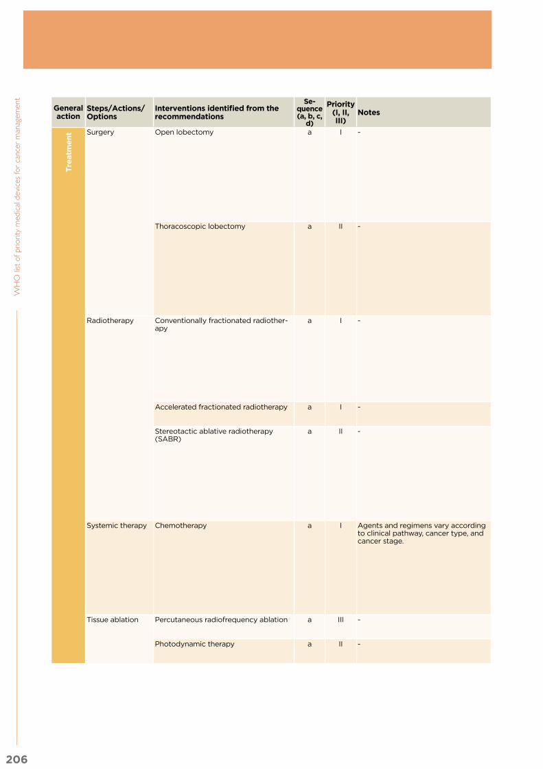
206
WHO
list o
f prio
rity m
edica
l dev
ices f
or ca
ncer
man
agem
ent
General action
Steps/Actions/Options
Interventions identified from the recommendations
Se-quence (a, b, c,
d)
Priority (I, II, III)
NotesGeneral action When to perform it Source Source Source Source
Tre
atm
en
t Surgery Open lobectomy a I -
Tre
atm
en
t Treatment of first choice.
Lung cancer. NICE clinical guideline 121 (2011)
NCCN. Non-small cell lung cancer. Version 3.2015
Vansteenkiste J, De Ruysscher D, Eberhardt WE, Lim E, Senan S, Felip E, Peters S; ESMO Guide-lines Working Group. Early and locally advanced non-small-cell lung cancer (NSCLC): ESMO Clinical Practice Guidelines for diagnosis, treatment and follow-up. Ann Oncol. 2013;24(6):vi89-98.
-
Thoracoscopic lobectomy a II - Treatment of first choice.
Lung cancer. NICE clinical guideline 121 (2011)
NCCN. Non-small cell lung cancer. Version 3.2015
Vansteenkiste J, De Ruysscher D, Eberhardt WE, Lim E, Senan S, Felip E, Peters S; ESMO Guide-lines Working Group. Early and locally advanced non-small-cell lung cancer (NSCLC): ESMO Clinical Practice Guidelines for diagnosis, treatment and follow-up. Ann Oncol. 2013;24(6):vi89-98.
-
Radiotherapy Conventionally fractionated radiother-apy
a I - When CHART is not available.
Lung cancer. NICE clinical guideline 121 (2011)
NCCN. Non-small cell lung cancer. Version 3.2015
- Reck M, Popat S, Rein-muth N, De Ruysscher D, Kerr KM, Peters S; ESMO Guidelines Work-ing Group. Metastatic non-small-cell lung cancer (NSCLC): ESMO Clinical Practice Guidelines for diagnosis, treatment and follow-up. Ann Oncol. 2014;25(3):iii27-39
Accelerated fractionated radiotherapy a I - For patients who are not suitable for surgery.
Lung cancer. NICE clinical guideline 121 (2011)
- - -
Stereotactic ablative radiotherapy (SABR)
a II - First choice for non-surgical treat-ment of stage I NSCLC
- NCCN. Non-small cell lung cancer. Version 3.2015
Vansteenkiste J, De Ruysscher D, Eberhardt WE, Lim E, Senan S, Felip E, Peters S; ESMO Guide-lines Working Group. Early and locally advanced non-small-cell lung cancer (NSCLC): ESMO Clinical Practice Guidelines for diagnosis, treatment and follow-up. Ann Oncol. 2013;24(6):vi89-98.
Reck M, Popat S, Rein-muth N, De Ruysscher D, Kerr KM, Peters S; ESMO Guidelines Work-ing Group. Metastatic non-small-cell lung cancer (NSCLC): ESMO Clinical Practice Guidelines for diagnosis, treatment and follow-up. Ann Oncol. 2014;25(3):iii27-39
Systemic therapy Chemotherapy a I Agents and regimens vary according to clinical pathway, cancer type, and cancer stage.
To be performed after surgery (adju-vant chemotherapy), after radiotherapy (chemoradiotherapy) or for advanced and metastatic cancer.
Lung cancer. NICE clinical guideline 121 (2011)
NCCN. Non-small cell lung cancer. Version 3.2015
Vansteenkiste J, De Ruysscher D, Eberhardt WE, Lim E, Senan S, Felip E, Peters S; ESMO Guide-lines Working Group. Early and locally advanced non-small-cell lung cancer (NSCLC): ESMO Clinical Practice Guidelines for diagnosis, treatment and follow-up. Ann Oncol. 2013;24(6):vi89-98.
Reck M, Popat S, Rein-muth N, De Ruysscher D, Kerr KM, Peters S; ESMO Guidelines Work-ing Group. Metastatic non-small-cell lung cancer (NSCLC): ESMO Clinical Practice Guidelines for diagnosis, treatment and follow-up. Ann Oncol. 2014;25(3):iii27-39
Tissue ablation Percutaneous radiofrequency ablation a III - - Lung cancer. NICE clinical guideline 121 (2011)
NCCN. Non-small cell lung cancer. Version 3.2015
- -
Photodynamic therapy a II - For localised inoper-able endobronchial cancer.
Lung cancer. NICE clinical guideline 121 (2011)
NCCN. Non-small cell lung cancer. Version 3.2015
- -

207
An
nex 1. C
linic
al in
terv
en
tion
s
General action
Steps/Actions/Options
Interventions identified from the recommendations
Se-quence (a, b, c,
d)
Priority (I, II, III)
NotesGeneral action When to perform it Source Source Source Source
Tre
atm
en
t Surgery Open lobectomy a I -
Tre
atm
en
t Treatment of first choice.
Lung cancer. NICE clinical guideline 121 (2011)
NCCN. Non-small cell lung cancer. Version 3.2015
Vansteenkiste J, De Ruysscher D, Eberhardt WE, Lim E, Senan S, Felip E, Peters S; ESMO Guide-lines Working Group. Early and locally advanced non-small-cell lung cancer (NSCLC): ESMO Clinical Practice Guidelines for diagnosis, treatment and follow-up. Ann Oncol. 2013;24(6):vi89-98.
-
Thoracoscopic lobectomy a II - Treatment of first choice.
Lung cancer. NICE clinical guideline 121 (2011)
NCCN. Non-small cell lung cancer. Version 3.2015
Vansteenkiste J, De Ruysscher D, Eberhardt WE, Lim E, Senan S, Felip E, Peters S; ESMO Guide-lines Working Group. Early and locally advanced non-small-cell lung cancer (NSCLC): ESMO Clinical Practice Guidelines for diagnosis, treatment and follow-up. Ann Oncol. 2013;24(6):vi89-98.
-
Radiotherapy Conventionally fractionated radiother-apy
a I - When CHART is not available.
Lung cancer. NICE clinical guideline 121 (2011)
NCCN. Non-small cell lung cancer. Version 3.2015
- Reck M, Popat S, Rein-muth N, De Ruysscher D, Kerr KM, Peters S; ESMO Guidelines Work-ing Group. Metastatic non-small-cell lung cancer (NSCLC): ESMO Clinical Practice Guidelines for diagnosis, treatment and follow-up. Ann Oncol. 2014;25(3):iii27-39
Accelerated fractionated radiotherapy a I - For patients who are not suitable for surgery.
Lung cancer. NICE clinical guideline 121 (2011)
- - -
Stereotactic ablative radiotherapy (SABR)
a II - First choice for non-surgical treat-ment of stage I NSCLC
- NCCN. Non-small cell lung cancer. Version 3.2015
Vansteenkiste J, De Ruysscher D, Eberhardt WE, Lim E, Senan S, Felip E, Peters S; ESMO Guide-lines Working Group. Early and locally advanced non-small-cell lung cancer (NSCLC): ESMO Clinical Practice Guidelines for diagnosis, treatment and follow-up. Ann Oncol. 2013;24(6):vi89-98.
Reck M, Popat S, Rein-muth N, De Ruysscher D, Kerr KM, Peters S; ESMO Guidelines Work-ing Group. Metastatic non-small-cell lung cancer (NSCLC): ESMO Clinical Practice Guidelines for diagnosis, treatment and follow-up. Ann Oncol. 2014;25(3):iii27-39
Systemic therapy Chemotherapy a I Agents and regimens vary according to clinical pathway, cancer type, and cancer stage.
To be performed after surgery (adju-vant chemotherapy), after radiotherapy (chemoradiotherapy) or for advanced and metastatic cancer.
Lung cancer. NICE clinical guideline 121 (2011)
NCCN. Non-small cell lung cancer. Version 3.2015
Vansteenkiste J, De Ruysscher D, Eberhardt WE, Lim E, Senan S, Felip E, Peters S; ESMO Guide-lines Working Group. Early and locally advanced non-small-cell lung cancer (NSCLC): ESMO Clinical Practice Guidelines for diagnosis, treatment and follow-up. Ann Oncol. 2013;24(6):vi89-98.
Reck M, Popat S, Rein-muth N, De Ruysscher D, Kerr KM, Peters S; ESMO Guidelines Work-ing Group. Metastatic non-small-cell lung cancer (NSCLC): ESMO Clinical Practice Guidelines for diagnosis, treatment and follow-up. Ann Oncol. 2014;25(3):iii27-39
Tissue ablation Percutaneous radiofrequency ablation a III - - Lung cancer. NICE clinical guideline 121 (2011)
NCCN. Non-small cell lung cancer. Version 3.2015
- -
Photodynamic therapy a II - For localised inoper-able endobronchial cancer.
Lung cancer. NICE clinical guideline 121 (2011)
NCCN. Non-small cell lung cancer. Version 3.2015
- -

208
WHO
list o
f prio
rity m
edica
l dev
ices f
or ca
ncer
man
agem
ent
General action
Steps/Actions/Options
Interventions identified from the recommendations
Se-quence (a, b, c,
d)
Priority (I, II, III)
NotesGeneral action When to perform it Source Source Source Source
Fo
llo
w u
p Physical exami-nation
Assessment of symptoms that may suggest lung cancer
a I -
Fo
llo
w u
p Recommended peri-odically.
Lung cancer. NICE clinical guideline 121 (2011)
NCCN. Non-small cell lung cancer. Version 3.2015
Vansteenkiste J, De Ruysscher D, Eberhardt WE, Lim E, Senan S, Felip E, Peters S; ESMO Guide-lines Working Group. Early and locally advanced non-small-cell lung cancer (NSCLC): ESMO Clinical Practice Guidelines for diagnosis, treatment and follow-up. Ann Oncol. 2013;24(6):vi89-98.
Reck M, Popat S, Rein-muth N, De Ruysscher D, Kerr KM, Peters S; ESMO Guidelines Work-ing Group. Metastatic non-small-cell lung cancer (NSCLC): ESMO Clinical Practice Guidelines for diagnosis, treatment and follow-up. Ann Oncol. 2014;25(3):iii27-39
Lung imaging CT scan of the chest a I With or without contrast. Recommended peri-odically.
- NCCN. Non-small cell lung cancer. Version 3.2015
Vansteenkiste J, De Ruysscher D, Eberhardt WE, Lim E, Senan S, Felip E, Peters S; ESMO Guide-lines Working Group. Early and locally advanced non-small-cell lung cancer (NSCLC): ESMO Clinical Practice Guidelines for diagnosis, treatment and follow-up. Ann Oncol. 2013;24(6):vi89-98.
Reck M, Popat S, Rein-muth N, De Ruysscher D, Kerr KM, Peters S; ESMO Guidelines Work-ing Group. Metastatic non-small-cell lung cancer (NSCLC): ESMO Clinical Practice Guidelines for diagnosis, treatment and follow-up. Ann Oncol. 2014;25(3):iii27-39
Chest X-Ray a I - Recommended peri-odically.
- - Vansteenkiste J, De Ruysscher D, Eberhardt WE, Lim E, Senan S, Felip E, Peters S; ESMO Guide-lines Working Group. Early and locally advanced non-small-cell lung cancer (NSCLC): ESMO Clinical Practice Guidelines for diagnosis, treatment and follow-up. Ann Oncol. 2013;24(6):vi89-98.
Reck M, Popat S, Rein-muth N, De Ruysscher D, Kerr KM, Peters S; ESMO Guidelines Work-ing Group. Metastatic non-small-cell lung cancer (NSCLC): ESMO Clinical Practice Guidelines for diagnosis, treatment and follow-up. Ann Oncol. 2014;25(3):iii27-39
Pallia
tive C
are
Palliative cancer treatment
Radiotherapy a I -
Pallia
tive C
are
- Lung cancer. NICE clinical guideline 121 (2011)
NCCN. Non-small cell lung cancer. Version 3.2015
- Reck M, Popat S, Rein-muth N, De Ruysscher D, Kerr KM, Peters S; ESMO Guidelines Work-ing Group. Metastatic non-small-cell lung cancer (NSCLC): ESMO Clinical Practice Guidelines for diagnosis, treatment and follow-up. Ann Oncol. 2014;25(3):iii27-39
Management of pleural effusions
Pleural aspiration or drainage a I - - Lung cancer. NICE clinical guideline 121 (2011)
- - Reck M, Popat S, Rein-muth N, De Ruysscher D, Kerr KM, Peters S; ESMO Guidelines Work-ing Group. Metastatic non-small-cell lung cancer (NSCLC): ESMO Clinical Practice Guidelines for diagnosis, treatment and follow-up. Ann Oncol. 2014;25(3):iii27-39
Pleurodesis a I - - - - - Reck M, Popat S, Rein-muth N, De Ruysscher D, Kerr KM, Peters S; ESMO Guidelines Work-ing Group. Metastatic non-small-cell lung cancer (NSCLC): ESMO Clinical Practice Guidelines for diagnosis, treatment and follow-up. Ann Oncol. 2014;25(3):iii27-39
Management of endobronchial obstructions
External beam radiotherapy a I - - Lung cancer. NICE clinical guideline 121 (2011)
NCCN. Non-small cell lung cancer. Version 3.2015
- -

209
An
nex 1. C
linic
al in
terv
en
tion
s
General action
Steps/Actions/Options
Interventions identified from the recommendations
Se-quence (a, b, c,
d)
Priority (I, II, III)
NotesGeneral action When to perform it Source Source Source Source
Fo
llo
w u
p Physical exami-nation
Assessment of symptoms that may suggest lung cancer
a I -
Fo
llo
w u
p Recommended peri-odically.
Lung cancer. NICE clinical guideline 121 (2011)
NCCN. Non-small cell lung cancer. Version 3.2015
Vansteenkiste J, De Ruysscher D, Eberhardt WE, Lim E, Senan S, Felip E, Peters S; ESMO Guide-lines Working Group. Early and locally advanced non-small-cell lung cancer (NSCLC): ESMO Clinical Practice Guidelines for diagnosis, treatment and follow-up. Ann Oncol. 2013;24(6):vi89-98.
Reck M, Popat S, Rein-muth N, De Ruysscher D, Kerr KM, Peters S; ESMO Guidelines Work-ing Group. Metastatic non-small-cell lung cancer (NSCLC): ESMO Clinical Practice Guidelines for diagnosis, treatment and follow-up. Ann Oncol. 2014;25(3):iii27-39
Lung imaging CT scan of the chest a I With or without contrast. Recommended peri-odically.
- NCCN. Non-small cell lung cancer. Version 3.2015
Vansteenkiste J, De Ruysscher D, Eberhardt WE, Lim E, Senan S, Felip E, Peters S; ESMO Guide-lines Working Group. Early and locally advanced non-small-cell lung cancer (NSCLC): ESMO Clinical Practice Guidelines for diagnosis, treatment and follow-up. Ann Oncol. 2013;24(6):vi89-98.
Reck M, Popat S, Rein-muth N, De Ruysscher D, Kerr KM, Peters S; ESMO Guidelines Work-ing Group. Metastatic non-small-cell lung cancer (NSCLC): ESMO Clinical Practice Guidelines for diagnosis, treatment and follow-up. Ann Oncol. 2014;25(3):iii27-39
Chest X-Ray a I - Recommended peri-odically.
- - Vansteenkiste J, De Ruysscher D, Eberhardt WE, Lim E, Senan S, Felip E, Peters S; ESMO Guide-lines Working Group. Early and locally advanced non-small-cell lung cancer (NSCLC): ESMO Clinical Practice Guidelines for diagnosis, treatment and follow-up. Ann Oncol. 2013;24(6):vi89-98.
Reck M, Popat S, Rein-muth N, De Ruysscher D, Kerr KM, Peters S; ESMO Guidelines Work-ing Group. Metastatic non-small-cell lung cancer (NSCLC): ESMO Clinical Practice Guidelines for diagnosis, treatment and follow-up. Ann Oncol. 2014;25(3):iii27-39
Pallia
tive C
are
Palliative cancer treatment
Radiotherapy a I -
Pallia
tive C
are
- Lung cancer. NICE clinical guideline 121 (2011)
NCCN. Non-small cell lung cancer. Version 3.2015
- Reck M, Popat S, Rein-muth N, De Ruysscher D, Kerr KM, Peters S; ESMO Guidelines Work-ing Group. Metastatic non-small-cell lung cancer (NSCLC): ESMO Clinical Practice Guidelines for diagnosis, treatment and follow-up. Ann Oncol. 2014;25(3):iii27-39
Management of pleural effusions
Pleural aspiration or drainage a I - - Lung cancer. NICE clinical guideline 121 (2011)
- - Reck M, Popat S, Rein-muth N, De Ruysscher D, Kerr KM, Peters S; ESMO Guidelines Work-ing Group. Metastatic non-small-cell lung cancer (NSCLC): ESMO Clinical Practice Guidelines for diagnosis, treatment and follow-up. Ann Oncol. 2014;25(3):iii27-39
Pleurodesis a I - - - - - Reck M, Popat S, Rein-muth N, De Ruysscher D, Kerr KM, Peters S; ESMO Guidelines Work-ing Group. Metastatic non-small-cell lung cancer (NSCLC): ESMO Clinical Practice Guidelines for diagnosis, treatment and follow-up. Ann Oncol. 2014;25(3):iii27-39
Management of endobronchial obstructions
External beam radiotherapy a I - - Lung cancer. NICE clinical guideline 121 (2011)
NCCN. Non-small cell lung cancer. Version 3.2015
- -

210
WHO
list o
f prio
rity m
edica
l dev
ices f
or ca
ncer
man
agem
ent
General action
Steps/Actions/Options
Interventions identified from the recommendations
Se-quence (a, b, c,
d)
Priority (I, II, III)
NotesGeneral action When to perform it Source Source Source Source
Pallia
tive C
are
Endobronchial debulking a II -
Pallia
tive C
are
- Lung cancer. NICE clinical guideline 121 (2011)
- - Reck M, Popat S, Rein-muth N, De Ruysscher D, Kerr KM, Peters S; ESMO Guidelines Work-ing Group. Metastatic non-small-cell lung cancer (NSCLC): ESMO Clinical Practice Guidelines for diagnosis, treatment and follow-up. Ann Oncol. 2014;25(3):iii27-39
Endobronchial stenting a II - - Lung cancer. NICE clinical guideline 121 (2011)
NCCN. Non-small cell lung cancer. Version 3.2015
- Reck M, Popat S, Rein-muth N, De Ruysscher D, Kerr KM, Peters S; ESMO Guidelines Work-ing Group. Metastatic non-small-cell lung cancer (NSCLC): ESMO Clinical Practice Guidelines for diagnosis, treatment and follow-up. Ann Oncol. 2014;25(3):iii27-39
Management of symptomatic brain metastases
Palliative whole-brain radiotherapy a II - - Lung cancer. NICE clinical guideline 121 (2011)
NCCN. Non-small cell lung cancer. Version 3.2015
- Reck M, Popat S, Rein-muth N, De Ruysscher D, Kerr KM, Peters S; ESMO Guidelines Work-ing Group. Metastatic non-small-cell lung cancer (NSCLC): ESMO Clinical Practice Guidelines for diagnosis, treatment and follow-up. Ann Oncol. 2014;25(3):iii27-39
Management of superior vena cava obstructions
Chemotherapy a II - - Lung cancer. NICE clinical guideline 121 (2011)
NCCN. Non-small cell lung cancer. Version 3.2015
- -
Radiotherapy a II - - Lung cancer. NICE clinical guideline 121 (2011)
NCCN. Non-small cell lung cancer. Version 3.2015
- -
Stenting a II - - Lung cancer. NICE clinical guideline 121 (2011)
NCCN. Non-small cell lung cancer. Version 3.2015
- Reck M, Popat S, Rein-muth N, De Ruysscher D, Kerr KM, Peters S; ESMO Guidelines Work-ing Group. Metastatic non-small-cell lung cancer (NSCLC): ESMO Clinical Practice Guidelines for diagnosis, treatment and follow-up. Ann Oncol. 2014;25(3):iii27-39
Management of bone metastases
Radiotherapy a II - - Lung cancer. NICE clinical guideline 121 (2011)
NCCN. Non-small cell lung cancer. Version 3.2015
- Reck M, Popat S, Rein-muth N, De Ruysscher D, Kerr KM, Peters S; ESMO Guidelines Work-ing Group. Metastatic non-small-cell lung cancer (NSCLC): ESMO Clinical Practice Guidelines for diagnosis, treatment and follow-up. Ann Oncol. 2014;25(3):iii27-39
Managing treat-ment-resistant recurrent ascites
Peritoneal catheter drainage a II Palliative peritoneal drainage can be considered.
- Lung cancer. NICE clinical guideline 121 (2011)
- - -

211
An
nex 1. C
linic
al in
terv
en
tion
s
General action
Steps/Actions/Options
Interventions identified from the recommendations
Se-quence (a, b, c,
d)
Priority (I, II, III)
NotesGeneral action When to perform it Source Source Source Source
Pallia
tive C
are
Endobronchial debulking a II -
Pallia
tive C
are
- Lung cancer. NICE clinical guideline 121 (2011)
- - Reck M, Popat S, Rein-muth N, De Ruysscher D, Kerr KM, Peters S; ESMO Guidelines Work-ing Group. Metastatic non-small-cell lung cancer (NSCLC): ESMO Clinical Practice Guidelines for diagnosis, treatment and follow-up. Ann Oncol. 2014;25(3):iii27-39
Endobronchial stenting a II - - Lung cancer. NICE clinical guideline 121 (2011)
NCCN. Non-small cell lung cancer. Version 3.2015
- Reck M, Popat S, Rein-muth N, De Ruysscher D, Kerr KM, Peters S; ESMO Guidelines Work-ing Group. Metastatic non-small-cell lung cancer (NSCLC): ESMO Clinical Practice Guidelines for diagnosis, treatment and follow-up. Ann Oncol. 2014;25(3):iii27-39
Management of symptomatic brain metastases
Palliative whole-brain radiotherapy a II - - Lung cancer. NICE clinical guideline 121 (2011)
NCCN. Non-small cell lung cancer. Version 3.2015
- Reck M, Popat S, Rein-muth N, De Ruysscher D, Kerr KM, Peters S; ESMO Guidelines Work-ing Group. Metastatic non-small-cell lung cancer (NSCLC): ESMO Clinical Practice Guidelines for diagnosis, treatment and follow-up. Ann Oncol. 2014;25(3):iii27-39
Management of superior vena cava obstructions
Chemotherapy a II - - Lung cancer. NICE clinical guideline 121 (2011)
NCCN. Non-small cell lung cancer. Version 3.2015
- -
Radiotherapy a II - - Lung cancer. NICE clinical guideline 121 (2011)
NCCN. Non-small cell lung cancer. Version 3.2015
- -
Stenting a II - - Lung cancer. NICE clinical guideline 121 (2011)
NCCN. Non-small cell lung cancer. Version 3.2015
- Reck M, Popat S, Rein-muth N, De Ruysscher D, Kerr KM, Peters S; ESMO Guidelines Work-ing Group. Metastatic non-small-cell lung cancer (NSCLC): ESMO Clinical Practice Guidelines for diagnosis, treatment and follow-up. Ann Oncol. 2014;25(3):iii27-39
Management of bone metastases
Radiotherapy a II - - Lung cancer. NICE clinical guideline 121 (2011)
NCCN. Non-small cell lung cancer. Version 3.2015
- Reck M, Popat S, Rein-muth N, De Ruysscher D, Kerr KM, Peters S; ESMO Guidelines Work-ing Group. Metastatic non-small-cell lung cancer (NSCLC): ESMO Clinical Practice Guidelines for diagnosis, treatment and follow-up. Ann Oncol. 2014;25(3):iii27-39
Managing treat-ment-resistant recurrent ascites
Peritoneal catheter drainage a II Palliative peritoneal drainage can be considered.
- Lung cancer. NICE clinical guideline 121 (2011)
- - -

212
WHO
list o
f prio
rity m
edica
l dev
ices f
or ca
ncer
man
agem
ent Annex table 1-6. Prostate cancer
General action
Steps/Actions/Options
Interventions identified from the recommendations
Se-quence (a, b, c,
d)
Priority(I, II, III)
NotesGeneral action When to perform it Source Source Source
Dia
gn
osi
s Blood testing Prostate-specific antigen (PSA) test a I WHO-IARC does not have an official position on prostate cancer screening at this time. Consideration must be made for resource availability, potential benefits and potential harms” D
iag
no
sis Frequency and age
groups are still under debate.
- NCCN. Prostate Cancer Early Detection. Version 1.2014
Horwich A, Parker C, de Reijke T, Kataja V; ESMO Guidelines Working Group. Prostate cancer: ESMO Clinical Practice Guidelines for diagnosis, treatment and follow-up. Ann Oncol. 2013;24(6):vi106-14.
Blood testing Prostate-specific antigen (PSA) test a I - Prostate cancer. NICE clinical guideline 175 (2014)
NCCN. Prostate Cancer Early Detection. Version 1.2014
Horwich A, Parker C, de Reijke T, Kataja V; ESMO Guidelines Working Group. Prostate cancer: ESMO Clinical Practice Guidelines for diagnosis, treatment and follow-up. Ann Oncol. 2013;24(6):vi106-14.
Urine testing PCA3 test b III - In case of elevated PSA and negative initial biopsies, to determine whetherre-biopsies are indicated (ESMO) (NCCN)
- NCCN. Prostate Cancer Early Detection. Version 1.2014
Horwich A, Parker C, de Reijke T, Kataja V; ESMO Guidelines Working Group. Prostate cancer: ESMO Clinical Practice Guidelines for diagnosis, treatment and follow-up. Ann Oncol. 2013;24(6):vi106-14.
Physical examination
Digital rectal examination b I - Prostate cancer. NICE clinical guideline 175 (2014)
NCCN. Prostate Cancer Early Detection. Version 1.2014
Horwich A, Parker C, de Reijke T, Kataja V; ESMO Guidelines Working Group. Prostate cancer: ESMO Clinical Practice Guidelines for diagnosis, treatment and follow-up. Ann Oncol. 2013;24(6):vi106-14.
Biopsy TRUS-guided prostate biopsy a II - . Prostate cancer. NICE clinical guideline 175 (2014)
NCCN. Prostate Cancer Early Detection. Version 1.2014
Horwich A, Parker C, de Reijke T, Kataja V; ESMO Guidelines Working Group. Prostate cancer: ESMO Clinical Practice Guidelines for diagnosis, treatment and follow-up. Ann Oncol. 2013;24(6):vi106-14.
Transperineal template biopsy a III - In patients with suspected prostate cancer who have had negative or equivocal results from other biopsy methods (NICE)
At present not recommended (NCCN)
Prostate cancer. NICE clinical guideline 175 (2014)
- -
Pathological examination
Examination of biopsy specimen b I - Recommended. Prostate cancer. NICE clinical guideline 175 (2014)
NCCN. Prostate Cancer Early Detection. Version 1.2014
Horwich A, Parker C, de Reijke T, Kataja V; ESMO Guidelines Working Group. Prostate cancer: ESMO Clinical Practice Guidelines for diagnosis, treatment and follow-up. Ann Oncol. 2013;24(6):vi106-14.
Pelvic imaging MRI scan a II Pelvic MRI or CT can be considered for diagnostic and staging purposes according to clinical context and resource availability.
Consider multiparametric MRI for men with a negative biopsy to determine whether another biopsy is needed (NICE)
Prostate cancer. NICE clinical guideline 175 (2014)
NCCN. Prostate Cancer Early Detection. Version 1.2014
Horwich A, Parker C, de Reijke T, Kataja V; ESMO Guidelines Working Group. Prostate cancer: ESMO Clinical Practice Guidelines for diagnosis, treatment and follow-up. Ann Oncol. 2013;24(6):vi106-14.
Staging and Risk Assessment
Digital rectal examination a I - Recommended for low-, intermediate- or high-risk patients.
- NCCN. Prostate Cancer. Version 1.2015
Horwich A, Parker C, de Reijke T, Kataja V; ESMO Guidelines Working Group. Prostate cancer: ESMO Clinical Practice Guidelines for diagnosis, treatment and follow-up. Ann Oncol. 2013;24(6):vi106-14.
Endorectal MRI b III - To increase resolution of MRI in the pelvis
- NCCN. Prostate Cancer. Version 1.2015
-
Bone scintigraphy b II X-ray and CT scan can also be considered for staging according to clinical context and resource availability.
For high-risk disease, bone scintigraphyshould be carried out and an MRI of the pelvis should be considered (ESMO)
Prostate cancer. NICE clinical guideline 175 (2014)
NCCN. Prostate Cancer. Version 1.2015
Horwich A, Parker C, de Reijke T, Kataja V; ESMO Guidelines Working Group. Prostate cancer: ESMO Clinical Practice Guidelines for diagnosis, treatment and follow-up. Ann Oncol. 2013;24(6):vi106-14.

213
An
nex 1. C
linic
al in
terv
en
tion
s
Annex table 1-6. Prostate cancer
General action
Steps/Actions/Options
Interventions identified from the recommendations
Se-quence (a, b, c,
d)
Priority(I, II, III)
NotesGeneral action When to perform it Source Source Source
Dia
gn
osi
s Blood testing Prostate-specific antigen (PSA) test a I WHO-IARC does not have an official position on prostate cancer screening at this time. Consideration must be made for resource availability, potential benefits and potential harms” D
iag
no
sis Frequency and age
groups are still under debate.
- NCCN. Prostate Cancer Early Detection. Version 1.2014
Horwich A, Parker C, de Reijke T, Kataja V; ESMO Guidelines Working Group. Prostate cancer: ESMO Clinical Practice Guidelines for diagnosis, treatment and follow-up. Ann Oncol. 2013;24(6):vi106-14.
Blood testing Prostate-specific antigen (PSA) test a I - Prostate cancer. NICE clinical guideline 175 (2014)
NCCN. Prostate Cancer Early Detection. Version 1.2014
Horwich A, Parker C, de Reijke T, Kataja V; ESMO Guidelines Working Group. Prostate cancer: ESMO Clinical Practice Guidelines for diagnosis, treatment and follow-up. Ann Oncol. 2013;24(6):vi106-14.
Urine testing PCA3 test b III - In case of elevated PSA and negative initial biopsies, to determine whetherre-biopsies are indicated (ESMO) (NCCN)
- NCCN. Prostate Cancer Early Detection. Version 1.2014
Horwich A, Parker C, de Reijke T, Kataja V; ESMO Guidelines Working Group. Prostate cancer: ESMO Clinical Practice Guidelines for diagnosis, treatment and follow-up. Ann Oncol. 2013;24(6):vi106-14.
Physical examination
Digital rectal examination b I - Prostate cancer. NICE clinical guideline 175 (2014)
NCCN. Prostate Cancer Early Detection. Version 1.2014
Horwich A, Parker C, de Reijke T, Kataja V; ESMO Guidelines Working Group. Prostate cancer: ESMO Clinical Practice Guidelines for diagnosis, treatment and follow-up. Ann Oncol. 2013;24(6):vi106-14.
Biopsy TRUS-guided prostate biopsy a II - . Prostate cancer. NICE clinical guideline 175 (2014)
NCCN. Prostate Cancer Early Detection. Version 1.2014
Horwich A, Parker C, de Reijke T, Kataja V; ESMO Guidelines Working Group. Prostate cancer: ESMO Clinical Practice Guidelines for diagnosis, treatment and follow-up. Ann Oncol. 2013;24(6):vi106-14.
Transperineal template biopsy a III - In patients with suspected prostate cancer who have had negative or equivocal results from other biopsy methods (NICE)
At present not recommended (NCCN)
Prostate cancer. NICE clinical guideline 175 (2014)
- -
Pathological examination
Examination of biopsy specimen b I - Recommended. Prostate cancer. NICE clinical guideline 175 (2014)
NCCN. Prostate Cancer Early Detection. Version 1.2014
Horwich A, Parker C, de Reijke T, Kataja V; ESMO Guidelines Working Group. Prostate cancer: ESMO Clinical Practice Guidelines for diagnosis, treatment and follow-up. Ann Oncol. 2013;24(6):vi106-14.
Pelvic imaging MRI scan a II Pelvic MRI or CT can be considered for diagnostic and staging purposes according to clinical context and resource availability.
Consider multiparametric MRI for men with a negative biopsy to determine whether another biopsy is needed (NICE)
Prostate cancer. NICE clinical guideline 175 (2014)
NCCN. Prostate Cancer Early Detection. Version 1.2014
Horwich A, Parker C, de Reijke T, Kataja V; ESMO Guidelines Working Group. Prostate cancer: ESMO Clinical Practice Guidelines for diagnosis, treatment and follow-up. Ann Oncol. 2013;24(6):vi106-14.
Staging and Risk Assessment
Digital rectal examination a I - Recommended for low-, intermediate- or high-risk patients.
- NCCN. Prostate Cancer. Version 1.2015
Horwich A, Parker C, de Reijke T, Kataja V; ESMO Guidelines Working Group. Prostate cancer: ESMO Clinical Practice Guidelines for diagnosis, treatment and follow-up. Ann Oncol. 2013;24(6):vi106-14.
Endorectal MRI b III - To increase resolution of MRI in the pelvis
- NCCN. Prostate Cancer. Version 1.2015
-
Bone scintigraphy b II X-ray and CT scan can also be considered for staging according to clinical context and resource availability.
For high-risk disease, bone scintigraphyshould be carried out and an MRI of the pelvis should be considered (ESMO)
Prostate cancer. NICE clinical guideline 175 (2014)
NCCN. Prostate Cancer. Version 1.2015
Horwich A, Parker C, de Reijke T, Kataja V; ESMO Guidelines Working Group. Prostate cancer: ESMO Clinical Practice Guidelines for diagnosis, treatment and follow-up. Ann Oncol. 2013;24(6):vi106-14.

214
WHO
list o
f prio
rity m
edica
l dev
ices f
or ca
ncer
man
agem
ent
General action
Steps/Actions/Options
Interventions identified from the recommendations
Se-quence (a, b, c,
d)
Priority(I, II, III)
NotesGeneral action When to perform it Source Source Source
Tre
atm
en
t Surgery Radical prostatectomy a I -
Tre
atm
en
t Optimal treatment of prostate cancer requires assessment of risk and depends on: patient age, expected patient survival, TNM, PSA level, Gleason score, symptoms, patient's preferences. Treatment options can be offered as stand-alone or in combination
Prostate cancer. NICE clinical guideline 175 (2014)
NCCN. Prostate Cancer. Version 1.2015
Horwich A, Parker C, de Reijke T, Kataja V; ESMO Guidelines Working Group. Prostate cancer: ESMO Clinical Practice Guidelines for diagnosis, treatment and follow-up. Ann Oncol. 2013;24(6):vi106-14.
Pelvic lymph node dissection a I - - NCCN. Prostate Cancer. Version 1.2015
Horwich A, Parker C, de Reijke T, Kataja V; ESMO Guidelines Working Group. Prostate cancer: ESMO Clinical Practice Guidelines for diagnosis, treatment and follow-up. Ann Oncol. 2013;24(6):vi106-14.
Bilateral orchiectomy a I - Prostate cancer. NICE clinical guideline 175 (2014)
NCCN. Prostate Cancer. Version 1.2015
Horwich A, Parker C, de Reijke T, Kataja V; ESMO Guidelines Working Group. Prostate cancer: ESMO Clinical Practice Guidelines for diagnosis, treatment and follow-up. Ann Oncol. 2013;24(6):vi106-14.
Radiation therapy Brachytherapy a I - Prostate cancer. NICE clinical guideline 175 (2014)
NCCN. Prostate Cancer. Version 1.2015
Horwich A, Parker C, de Reijke T, Kataja V; ESMO Guidelines Working Group. Prostate cancer: ESMO Clinical Practice Guidelines for diagnosis, treatment and follow-up. Ann Oncol. 2013;24(6):vi106-14.
External beam radiation therapy a I - Prostate cancer. NICE clinical guideline 175 (2014)
NCCN. Prostate Cancer. Version 1.2015
Horwich A, Parker C, de Reijke T, Kataja V; ESMO Guidelines Working Group. Prostate cancer: ESMO Clinical Practice Guidelines for diagnosis, treatment and follow-up. Ann Oncol. 2013;24(6):vi106-14.
Radiopharmaceutical therapy a II Radiopharmaceutical therapy can be used in setting of metastases or bulky nodal disease according to clinical context and resource availability.
- NCCN. Prostate Cancer. Version 1.2015
Horwich A, Parker C, de Reijke T, Kataja V; ESMO Guidelines Working Group. Prostate cancer: ESMO Clinical Practice Guidelines for diagnosis, treatment and follow-up. Ann Oncol. 2013;24(6):vi106-14.
Systemic treatment
Hormonal therapy a I - Prostate cancer. NICE clinical guideline 175 (2014)
NCCN. Prostate Cancer. Version 1.2015
Horwich A, Parker C, de Reijke T, Kataja V; ESMO Guidelines Working Group. Prostate cancer: ESMO Clinical Practice Guidelines for diagnosis, treatment and follow-up. Ann Oncol. 2013;24(6):vi106-14.
Chemotherapy, immunotherapy and targetted therapy
a I WHO List of Essential Medicines can be used to assist with selection of systemic therapy.
Prostate cancer. NICE clinical guideline 175 (2014)
NCCN. Prostate Cancer. Version 1.2015
Horwich A, Parker C, de Reijke T, Kataja V; ESMO Guidelines Working Group. Prostate cancer: ESMO Clinical Practice Guidelines for diagnosis, treatment and follow-up. Ann Oncol. 2013;24(6):vi106-14.
Fo
llo
w u
p Blood testing PSA test a II -F
ollo
w u
p Recommended routinely
Prostate cancer. NICE clinical guideline 175 (2014)
NCCN. Prostate Cancer. Version 1.2015
Horwich A, Parker C, de Reijke T, Kataja V; ESMO Guidelines Working Group. Prostate cancer: ESMO Clinical Practice Guidelines for diagnosis, treatment and follow-up. Ann Oncol. 2013;24(6):vi106-14.
Physical examination
Digital rectal examination a I - Not always required Prostate cancer. NICE clinical guideline 175 (2014)
NCCN. Prostate Cancer. Version 1.2015
Horwich A, Parker C, de Reijke T, Kataja V; ESMO Guidelines Working Group. Prostate cancer: ESMO Clinical Practice Guidelines for diagnosis, treatment and follow-up. Ann Oncol. 2013;24(6):vi106-14.
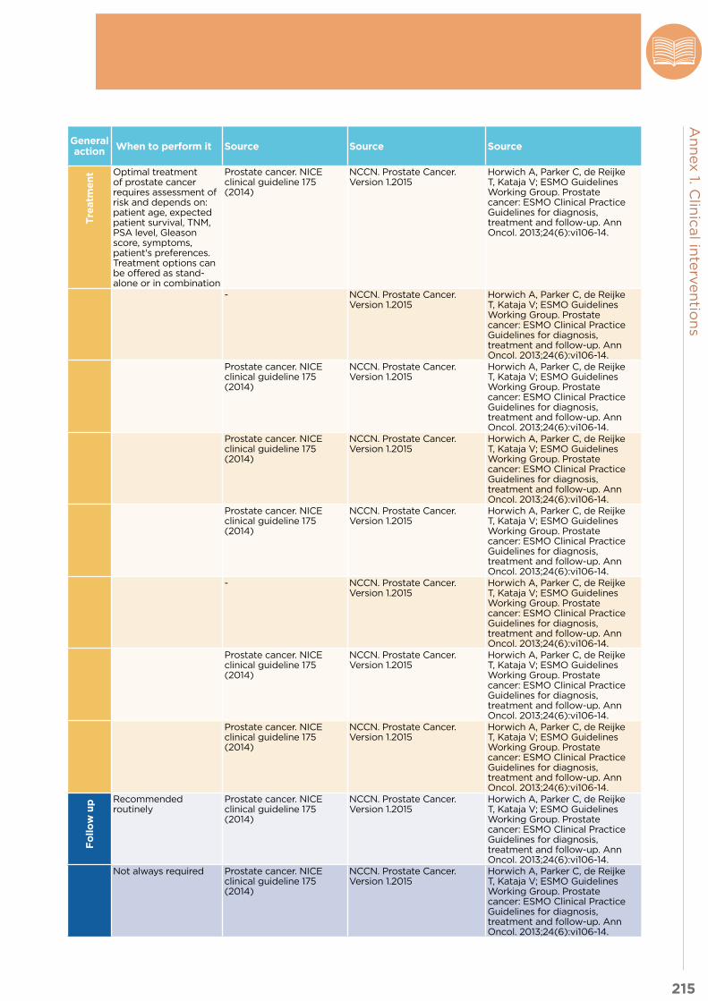
215
An
nex 1. C
linic
al in
terv
en
tion
s
General action
Steps/Actions/Options
Interventions identified from the recommendations
Se-quence (a, b, c,
d)
Priority(I, II, III)
NotesGeneral action When to perform it Source Source Source
Tre
atm
en
t Surgery Radical prostatectomy a I -
Tre
atm
en
t Optimal treatment of prostate cancer requires assessment of risk and depends on: patient age, expected patient survival, TNM, PSA level, Gleason score, symptoms, patient's preferences. Treatment options can be offered as stand-alone or in combination
Prostate cancer. NICE clinical guideline 175 (2014)
NCCN. Prostate Cancer. Version 1.2015
Horwich A, Parker C, de Reijke T, Kataja V; ESMO Guidelines Working Group. Prostate cancer: ESMO Clinical Practice Guidelines for diagnosis, treatment and follow-up. Ann Oncol. 2013;24(6):vi106-14.
Pelvic lymph node dissection a I - - NCCN. Prostate Cancer. Version 1.2015
Horwich A, Parker C, de Reijke T, Kataja V; ESMO Guidelines Working Group. Prostate cancer: ESMO Clinical Practice Guidelines for diagnosis, treatment and follow-up. Ann Oncol. 2013;24(6):vi106-14.
Bilateral orchiectomy a I - Prostate cancer. NICE clinical guideline 175 (2014)
NCCN. Prostate Cancer. Version 1.2015
Horwich A, Parker C, de Reijke T, Kataja V; ESMO Guidelines Working Group. Prostate cancer: ESMO Clinical Practice Guidelines for diagnosis, treatment and follow-up. Ann Oncol. 2013;24(6):vi106-14.
Radiation therapy Brachytherapy a I - Prostate cancer. NICE clinical guideline 175 (2014)
NCCN. Prostate Cancer. Version 1.2015
Horwich A, Parker C, de Reijke T, Kataja V; ESMO Guidelines Working Group. Prostate cancer: ESMO Clinical Practice Guidelines for diagnosis, treatment and follow-up. Ann Oncol. 2013;24(6):vi106-14.
External beam radiation therapy a I - Prostate cancer. NICE clinical guideline 175 (2014)
NCCN. Prostate Cancer. Version 1.2015
Horwich A, Parker C, de Reijke T, Kataja V; ESMO Guidelines Working Group. Prostate cancer: ESMO Clinical Practice Guidelines for diagnosis, treatment and follow-up. Ann Oncol. 2013;24(6):vi106-14.
Radiopharmaceutical therapy a II Radiopharmaceutical therapy can be used in setting of metastases or bulky nodal disease according to clinical context and resource availability.
- NCCN. Prostate Cancer. Version 1.2015
Horwich A, Parker C, de Reijke T, Kataja V; ESMO Guidelines Working Group. Prostate cancer: ESMO Clinical Practice Guidelines for diagnosis, treatment and follow-up. Ann Oncol. 2013;24(6):vi106-14.
Systemic treatment
Hormonal therapy a I - Prostate cancer. NICE clinical guideline 175 (2014)
NCCN. Prostate Cancer. Version 1.2015
Horwich A, Parker C, de Reijke T, Kataja V; ESMO Guidelines Working Group. Prostate cancer: ESMO Clinical Practice Guidelines for diagnosis, treatment and follow-up. Ann Oncol. 2013;24(6):vi106-14.
Chemotherapy, immunotherapy and targetted therapy
a I WHO List of Essential Medicines can be used to assist with selection of systemic therapy.
Prostate cancer. NICE clinical guideline 175 (2014)
NCCN. Prostate Cancer. Version 1.2015
Horwich A, Parker C, de Reijke T, Kataja V; ESMO Guidelines Working Group. Prostate cancer: ESMO Clinical Practice Guidelines for diagnosis, treatment and follow-up. Ann Oncol. 2013;24(6):vi106-14.
Fo
llo
w u
p Blood testing PSA test a II -
Fo
llo
w u
p Recommended routinely
Prostate cancer. NICE clinical guideline 175 (2014)
NCCN. Prostate Cancer. Version 1.2015
Horwich A, Parker C, de Reijke T, Kataja V; ESMO Guidelines Working Group. Prostate cancer: ESMO Clinical Practice Guidelines for diagnosis, treatment and follow-up. Ann Oncol. 2013;24(6):vi106-14.
Physical examination
Digital rectal examination a I - Not always required Prostate cancer. NICE clinical guideline 175 (2014)
NCCN. Prostate Cancer. Version 1.2015
Horwich A, Parker C, de Reijke T, Kataja V; ESMO Guidelines Working Group. Prostate cancer: ESMO Clinical Practice Guidelines for diagnosis, treatment and follow-up. Ann Oncol. 2013;24(6):vi106-14.

216
WHO
list o
f prio
rity m
edica
l dev
ices f
or ca
ncer
man
agem
ent
General action
Steps/Actions/Options
Interventions identified from the recommendations
Se-quence (a, b, c,
d)
Priority(I, II, III)
NotesGeneral action When to perform it Source Source Source
Biopsy (sample collection)
TRUS-guided prostate biopsy b I - Not always required - NCCN. Prostate Cancer. Version 1.2015
Horwich A, Parker C, de Reijke T, Kataja V; ESMO Guidelines Working Group. Prostate cancer: ESMO Clinical Practice Guidelines for diagnosis, treatment and follow-up. Ann Oncol. 2013;24(6):vi106-14.
cardiovascular examination
Assessment for cardiovascular disease
a I - Recommended for patients receiving androgen deprivation therapy.
- NCCN. Prostate Cancer. Version 1.2015
-
Blood testing Assessment for diabetes a I - Recommended for patients receiving androgen deprivation therapy.
- NCCN. Prostate Cancer. Version 1.2015
-
Gastroenterologi-cal examination
Assessment of chronic bowel symptoms
a I - Recommended for patients received radiation therapy
- - Horwich A, Parker C, de Reijke T, Kataja V; ESMO Guidelines Working Group. Prostate cancer: ESMO Clinical Practice Guidelines for diagnosis, treatment and follow-up. Ann Oncol. 2013;24(6):vi106-14.
Bone assessment Assessment of osteoporosis symptoms
a I - Recommended for patients on long-term androgen deprivation therapy
- NCCN. Prostate Cancer. Version 1.2015
Horwich A, Parker C, de Reijke T, Kataja V; ESMO Guidelines Working Group. Prostate cancer: ESMO Clinical Practice Guidelines for diagnosis, treatment and follow-up. Ann Oncol. 2013;24(6):vi106-14.
Pallia
tive
C
are
Systemic treatment
Palliative androgen deprivation therapy and hormonal manipulation
a I -
Pallia
tive
C
are
Palliative approaches are offered within some approaches (e.g. observation) and tumour stages (e.g. metastatic castration-resistant prostate cancer).
Prostate cancer. NICE clinical guideline 175 (2014)
NCCN. Prostate Cancer. Version 1.2015
Horwich A, Parker C, de Reijke T, Kataja V; ESMO Guidelines Working Group. Prostate cancer: ESMO Clinical Practice Guidelines for diagnosis, treatment and follow-up. Ann Oncol. 2013;24(6):vi106-14.
Palliative chemotherapy a I - Prostate cancer. NICE clinical guideline 175 (2014)
NCCN. Prostate Cancer. Version 1.2015
Horwich A, Parker C, de Reijke T, Kataja V; ESMO Guidelines Working Group. Prostate cancer: ESMO Clinical Practice Guidelines for diagnosis, treatment and follow-up. Ann Oncol. 2013;24(6):vi106-14.
Palliative immunotherapy a III Clinical trials are ongoing to assess impact of immunotherapy in metastaic prostate cancer. WHO List of Essential Medicines can be used for reference.
- NCCN. Prostate Cancer. Version 1.2015
Horwich A, Parker C, de Reijke T, Kataja V; ESMO Guidelines Working Group. Prostate cancer: ESMO Clinical Practice Guidelines for diagnosis, treatment and follow-up. Ann Oncol. 2013;24(6):vi106-14.
Radiation therapy Palliative radiation therapy a I - Prostate cancer. NICE clinical guideline 175 (2014)
NCCN. Prostate Cancer. Version 1.2015
Horwich A, Parker C, de Reijke T, Kataja V; ESMO Guidelines Working Group. Prostate cancer: ESMO Clinical Practice Guidelines for diagnosis, treatment and follow-up. Ann Oncol. 2013;24(6):vi106-14.
Radiopharmaceutical therapy a II - - NCCN. Prostate Cancer. Version 1.2015
-
Management of obstructive uropathy
Percutaneous nephrostomy a II Prostate cancer. NICE clinical guideline 175 (2014)
- -
Insertion of a double J stent a II Prostate cancer. NICE clinical guideline 175 (2014)
- -
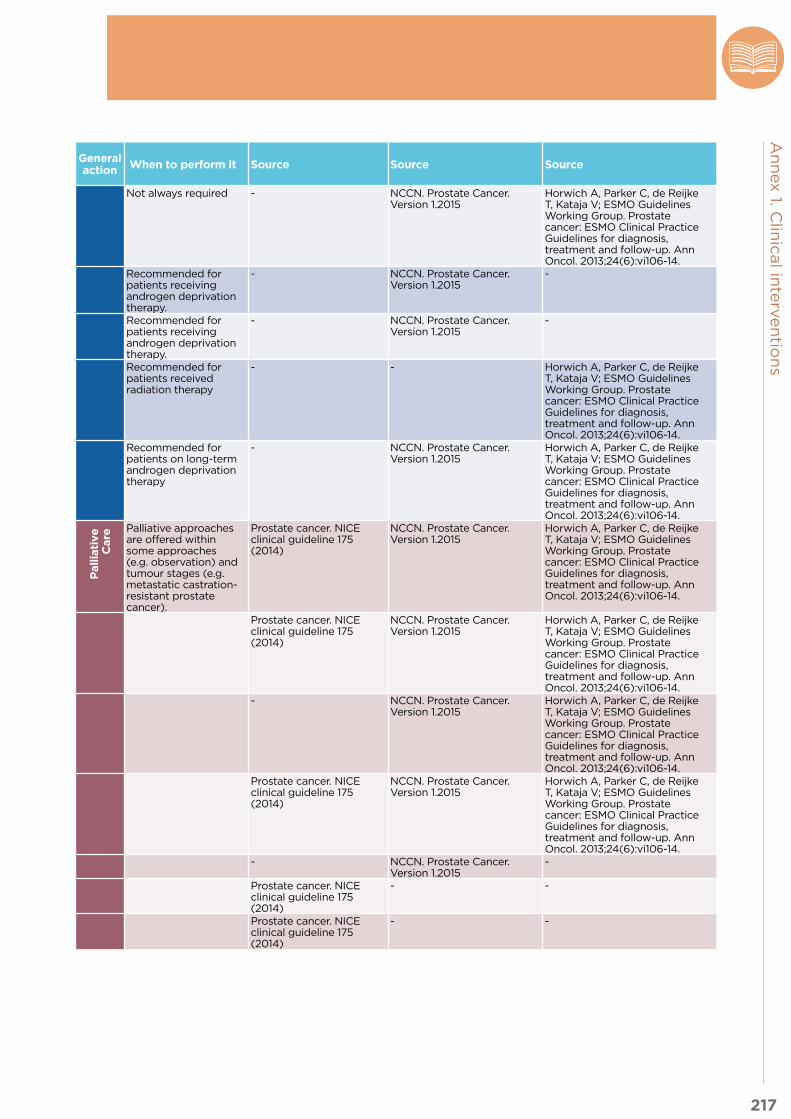
217
An
nex 1. C
linic
al in
terv
en
tion
s
General action
Steps/Actions/Options
Interventions identified from the recommendations
Se-quence (a, b, c,
d)
Priority(I, II, III)
NotesGeneral action When to perform it Source Source Source
Biopsy (sample collection)
TRUS-guided prostate biopsy b I - Not always required - NCCN. Prostate Cancer. Version 1.2015
Horwich A, Parker C, de Reijke T, Kataja V; ESMO Guidelines Working Group. Prostate cancer: ESMO Clinical Practice Guidelines for diagnosis, treatment and follow-up. Ann Oncol. 2013;24(6):vi106-14.
cardiovascular examination
Assessment for cardiovascular disease
a I - Recommended for patients receiving androgen deprivation therapy.
- NCCN. Prostate Cancer. Version 1.2015
-
Blood testing Assessment for diabetes a I - Recommended for patients receiving androgen deprivation therapy.
- NCCN. Prostate Cancer. Version 1.2015
-
Gastroenterologi-cal examination
Assessment of chronic bowel symptoms
a I - Recommended for patients received radiation therapy
- - Horwich A, Parker C, de Reijke T, Kataja V; ESMO Guidelines Working Group. Prostate cancer: ESMO Clinical Practice Guidelines for diagnosis, treatment and follow-up. Ann Oncol. 2013;24(6):vi106-14.
Bone assessment Assessment of osteoporosis symptoms
a I - Recommended for patients on long-term androgen deprivation therapy
- NCCN. Prostate Cancer. Version 1.2015
Horwich A, Parker C, de Reijke T, Kataja V; ESMO Guidelines Working Group. Prostate cancer: ESMO Clinical Practice Guidelines for diagnosis, treatment and follow-up. Ann Oncol. 2013;24(6):vi106-14.
Pallia
tive
C
are
Systemic treatment
Palliative androgen deprivation therapy and hormonal manipulation
a I -
Pallia
tive
C
are
Palliative approaches are offered within some approaches (e.g. observation) and tumour stages (e.g. metastatic castration-resistant prostate cancer).
Prostate cancer. NICE clinical guideline 175 (2014)
NCCN. Prostate Cancer. Version 1.2015
Horwich A, Parker C, de Reijke T, Kataja V; ESMO Guidelines Working Group. Prostate cancer: ESMO Clinical Practice Guidelines for diagnosis, treatment and follow-up. Ann Oncol. 2013;24(6):vi106-14.
Palliative chemotherapy a I - Prostate cancer. NICE clinical guideline 175 (2014)
NCCN. Prostate Cancer. Version 1.2015
Horwich A, Parker C, de Reijke T, Kataja V; ESMO Guidelines Working Group. Prostate cancer: ESMO Clinical Practice Guidelines for diagnosis, treatment and follow-up. Ann Oncol. 2013;24(6):vi106-14.
Palliative immunotherapy a III Clinical trials are ongoing to assess impact of immunotherapy in metastaic prostate cancer. WHO List of Essential Medicines can be used for reference.
- NCCN. Prostate Cancer. Version 1.2015
Horwich A, Parker C, de Reijke T, Kataja V; ESMO Guidelines Working Group. Prostate cancer: ESMO Clinical Practice Guidelines for diagnosis, treatment and follow-up. Ann Oncol. 2013;24(6):vi106-14.
Radiation therapy Palliative radiation therapy a I - Prostate cancer. NICE clinical guideline 175 (2014)
NCCN. Prostate Cancer. Version 1.2015
Horwich A, Parker C, de Reijke T, Kataja V; ESMO Guidelines Working Group. Prostate cancer: ESMO Clinical Practice Guidelines for diagnosis, treatment and follow-up. Ann Oncol. 2013;24(6):vi106-14.
Radiopharmaceutical therapy a II - - NCCN. Prostate Cancer. Version 1.2015
-
Management of obstructive uropathy
Percutaneous nephrostomy a II Prostate cancer. NICE clinical guideline 175 (2014)
- -
Insertion of a double J stent a II Prostate cancer. NICE clinical guideline 175 (2014)
- -
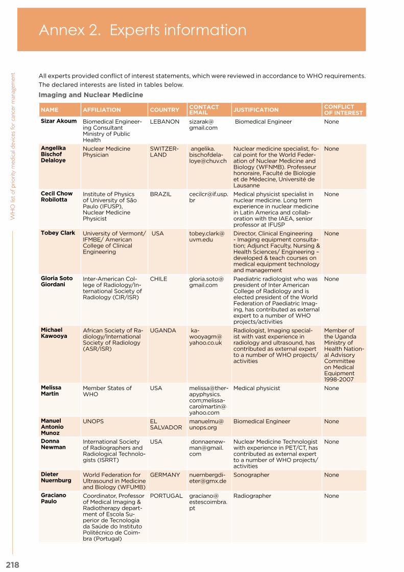
218
WHO
list o
f prio
rity m
edica
l dev
ices f
or ca
ncer
man
agem
ent
NAME AFFILIATION COUNTRY CONTACT EMAIL JUSTIFICATION CONFLICT
OF INTEREST
Sizar Akoum Biomedical Engineer-ing ConsultantMinistry of Public Health
LEBANON [email protected]
Biomedical Engineer None
Angelika Bischof Delaloye
Nuclear Medicine Physician
SWITZER-LAND
Nuclear medicine specialist, fo-cal point for the World Feder-ation of Nuclear Medicine and Biology (WFNMB). Professeur honoraire, Faculté de Biologie et de Médecine, Université de Lausanne
None
Cecil ChowRobilotta
Institute of Physics of University of São Paulo (IFUSP),Nuclear Medicine Physicist
BRAZIL [email protected]
Medical physicist specialist in nuclear medicine. Long term experience in nuclear medicine in Latin America and collab-oration with the IAEA, senior professor at IFUSP
None
Tobey Clark University of Vermont/IFMBE/ American College of Clinical Engineering
Director, Clinical Engineering - Imaging equipment consulta-tion; Adjunct Faculty, Nursing & Health Sciences/ Engineering – developed & teach courses on medical equipment technology and management
None
Gloria Soto Giordani
Inter-American Col-lege of Radiology/In-ternational Society of Radiology (CIR/ISR)
CHILE [email protected]
Paediatric radiologist who was president of Inter American College of Radiology and is elected president of the World Federation of Paediatric Imag-ing, has contributed as external expert to a number of WHO projects/activities
None
Michael Kawooya
African Society of Ra-diology/International Society of Radiology (ASR/ISR)
UGANDA [email protected]
Radiologist, Imaging special-ist with vast experience in radiology and ultrasound, has contributed as external expert to a number of WHO projects/activities
Member of the Uganda Ministry of Health Nation-al Advisory Committee on Medical Equipment 1998-2007
Melissa Martin
Member States of WHO
USA [email protected];[email protected]
Medical physicist None
Manuel Antonio Munoz
UNOPS EL SALVADOR
Biomedical Engineer None
Donna Newman
International Society of Radiographers and Radiological Technolo-gists (ISRRT)
Nuclear Medicine Technologist with experience in PET/CT, has contributed as external expert to a number of WHO projects/activities
None
Dieter Nuernburg
World Federation for Ultrasound in Medicine and Biology (WFUMB)
GERMANY [email protected]
Sonographer None
Graciano Paulo
Coordinator, Professor of Medical Imaging & Radiotherapy depart-ment of Escola Su-perior de Tecnologia da Saúde do Instituto Politécnico de Coim-bra (Portugal)
PORTUGAL [email protected]
Radiographer None
Annex 2. Experts information
All experts provided conflict of interest statements, which were reviewed in accordance to WHO requirements.
The declared interests are listed in tables below.
Imaging and Nuclear Medicine

219
An
nex 2
. Exp
erts in
form
atio
n
NAME AFFILIATION COUNTRY CONTACT EMAIL JUSTIFICATION CONFLICT
OF INTEREST
Madan Rehani
International Organ-ization for Medical Physics (IOMP), Har-vard Medical School, Massachusetts General Hospital and Duke University
Medical physicist None
Shuvro H Roy-Choudhury
FRCS (London), FRCR (London), CCST (UK), FCIRSE, EBIR (Inter-ventional Radiologist)
INDIA [email protected]
Interventional radiologist, LMIC None
Sat-chithanan-tham Somanesan
Nuclear Medicine, PhysicistSingapore General Hospital
SINGAPORE [email protected]
Nuclear medicine physicistLong term experience in nuclear medicine in Asia and collaboration with the IAEA
None
Rathan Sub-ramaniam
Nuclear Radiology Prof.Associate Professor in the Division of Nuclear Medicine, Russell H Morgan Department of Radiology and Radiological Sciences at the Johns Hopkins University School of Medicine, Baltimore, MD
Nuclear medicine specialistLong term experience in estab-lishing nuclear medicine and diagnostic imaging facilities
None
Surgery
NAME AFFILIATION COUNTRY CONTACT EMAIL JUSTIFICATION CONFLICT
OF INTEREST
Nicholas Adjabu
Deputy director, clinical engineering department, Ghana health service
GHANA [email protected]
Medical Practitioner and Biomedical Engineer with extensive experience in health technology management especially in Ghana and other developing countries. Has been a health technology consult-ant in WHO-AFRO. Also the Deputy Director for Clinical Engineering Department of the Ghana Health Service.
None
Benjamin O. Anderson
MD, Professor of Surgery and Global Health Medicine, Uni-versity of Washington, Seattle, Washington, USA
Chair and Director, Breast Health Global Initiative (BHGI), Fred Hutchinson Cancer Re-search Center, Seattle, Wash-ington, USA
Unrestricted educational grant support from Pfizer and Roche
Ainhoa Costas-Chavarri
MD, MPH, FACS Gen-eral Surgeon, Human Resources for Health Rwanda, Boston Chil-dren’s Hospital/Har-vard Medical School
RWANDA [email protected]
Extensive experience in Rwan-da and Haiti; Very experienced on the ground with breast cancer in very low-income countries.
None.
Serigne Gueye
Professor of Urology at University Cheikh Anta DIOP, Dakar, Senegal and Chair of Urology and Androl-ogy, Grand Yoff Gen-eral Hospital. Adjunct Dean for Research at School of Medicine St Christopher-Iba Mar Diop, University El Hadj Ibrahima Niasse, Dakar, Senegal
SENEGAL [email protected]
Professor of urology in Senegal None
Tom Judd IFMBE USA [email protected]
Biomedical engineer None

220
WHO
list o
f prio
rity m
edica
l dev
ices f
or ca
ncer
man
agem
ent
NAME AFFILIATION COUNTRY CONTACT EMAIL JUSTIFICATION CONFLICT
OF INTEREST
Guy Maddern Professor of Surgery, University of Adelaide
AUSTRALIA [email protected]
Surgical technology assess-ment
None
C.S. Pramesh Professor and Chief, Thoracic Surgery, Department of Surgi-cal Oncology at Tata Memorial Hospital
INDIA [email protected]
Professor of Surgery Research support from Ethicon Endo-surgery
Judith Shamian
International Council of Nurses
CANADA [email protected]
Nursing specialist None
Richard Sullivan
Director , Institute of Cancer Policy and King’s Health Partners Comprehensive Can-cer Centre
Onco-urologist. Expertise in global cancer policy, imple-mentation science and health services research. Led Lancet Oncology Commission on Global Cancer Surgery. Mem-ber of Global Surgery 2030 team
None
Audrey Tsunoda
MD, PhD (oncology)Hospital Erasto Gaert-ner and Instituto de Oncologia do Paraná
BRAZIL [email protected]
Surgical Oncologist - Gynaeco-logic Oncology DepartmentMedical Director at IRCAD Latin America, for minimally invasive surgery
Received educational support for lectures, from Roche, in 2015
David Watters
Deakin University, Australia. Professor of Surgery at Deakin Uni-versity and Director of Surgery at Univer-sity Hospital Geelong; President, Royal Australasian College of Surgeons
AUSTRALIA [email protected]
Professor of Surgery None
Cheng-Har Yip
Department of Surgery, Faculty of Medicine, University of Malaya
MALAYSIA [email protected]
Nearly 30 years' experience working as a general surgeon, paediatric surgeon and now a breast surgeon in a low resource setting. Co-author in the chapter on Surgical Ser-vices for Cancer Care in DCP3 Cancer Volume.
None
Pathology
NAME AFILIATION AND DEGREE COUNTRY CONTACT
EMAIL JUSTIFICATION CONFLICT OF INTEREST
Jagdish Butany
MBBS, MS, FRCPC CANADA [email protected]
Consultant Pathologist and substantial experience in labo-ratory services planning
None
Kirti Chadha M.D.(Pathology), PDDC (Oncopatholo-gy & Oncohematopa-thology)
INDIA [email protected]
Hematopathologist, Histopa-thologist/Oncopathologist, and Oncohematopathologist in LMIC
None
Patrick Fitzgibbons
ASCO and CAP/Ex-pert to review medical devices for cancer care
Pathologist None
John Flanigan
Senior Adviser for NCDs
USA Internist and Emergency Physician
Employed by the United States Gov-ernment
Ken Fleming Senior adviser for Pathology, Centre for Global Health NCI. MB ChB, DPhil FRCPath
A pathologist with many years of experience. Dean of the Ox-ford University Medical School and recently finished a term as global health lead for the Royal College of Pathologists U.K.
Owns a consultancy business. Currently con-tracted by the United States Government

221
An
nex 2
. Exp
erts in
form
atio
n
NAME AFFILIATION COUNTRY CONTACT EMAIL JUSTIFICATION CONFLICT
OF INTEREST
Paolo Lago IFMBE-CEDDirector Clinical Engi-neering Dept.San Matteo Hospital - Pavia
ITALY [email protected]
Biomedical Engineer None
Héctor A Maldonado-Martínez
M.D. (Pathology), MSc, Ph.D. Head of the Pa-thology Department, Insituto Nacional de Cancerologia (Nation-al Cancer Institute), México
MEXICO [email protected], [email protected]
Pathologist Consultant and receives research sup-port from Ro-che-Ventana
Lai-Meng Looi
MBBS BSc FRANZCR. Liverpool and Macarthur Cancer Therapy Centres, Ingham Institute for Applied Medical Research, University of New South Wales, Western Sydney University
MALAYSIA [email protected]
Anatomical pathologist with long practice experience, including pathology laboratory planning and capacity build-ing in low & middle income countries. Past-President of WASPaLM and Co-Chair of the InterAcademy Partnership for Health.
None
Danny A. Milner, Jr.
M.D., MSc(Epi) US [email protected]
Pathologist Extensive experi-ence in Rwanda and Haiti. Very experienced on the ground in very low-income countries with successful implementation.
None
James Pepoon
Administrative Direc-tor, Clinical Operations & Planning, Anatomic Pathology - Brigham and Women’s Hospital, Boston, MA. Appoint-ed Advisor, Global Cancer Diagnostic Outreach - Partners in Health, Inc., Bos-ton, MA. Appointed Advisor, Global Cancer Diagnostic Outreach - Partners in Health, Inc., Boston, MA
US [email protected]. [email protected]
Pathology Administrator of Anatomic Pathology clinical and research operations with extensive technical back-ground. Substantial experience in global healthcare diagnostic outreach including procure-ment, construction, operations and human resource needs. Primary projects in Rwanda and Haiti. Very experienced on the ground in very low income countries with successful im-plementation.
None
Lawrence N Shulman
M.D.; Director, Center for Global Cancer Medicine. University of Pennsylvania
WHO Advisory committee None
Michael L. Wilson
M.D; Editor of the AJCPProfessor of Pathol-ogy,University of Colorado African Strategies for Advancing Pathology group
Substantial experience working in LMIC and in building capaci-ty for Pathology and Laborato-ry Services
Received research support from NCI, USA for a non-profit organiza-tion African Strategies for Advancing Pathology
Ms Gabrielle Wolff
BSc, MLT; Dept of Pathology, Toronto General Hospital
CANADA [email protected]
Pathology technician None
Radiotherapy
NAME AFILIATION AND DEGREE COUNTRY CONTACT
EMAIL JUSTIFICATION CONFLICT OF INTEREST
Mary Coffey Radiation Therapist (RTT), Adjunct Associate Professor
IRELAND [email protected]
Radiation therapy advisor to IAEA
None
Yadin David IFMBE/Clinical Engi-neer
Biomedical engineer None

222
WHO
list o
f prio
rity m
edica
l dev
ices f
or ca
ncer
man
agem
ent
NAME AFFILIATION COUNTRY CONTACT EMAIL JUSTIFICATION CONFLICT
OF INTEREST
Ahmed Elzawawy
MD, PHD EGYPT [email protected]
Radiation and Medical on-cologist; Professor of Clinical Oncology, Egypt and Chair of three Departments of Clini-cal and Radiation Oncology, Port Said and Ismailia, Egypt; President of International Campaign for Establishment and Development of Oncology Centers (ICEDOC); President of AORTIC; Director of SEMCO
None
Surbhi Grover
MD Botswana, USA
Radiation oncologist Extensive experience on the ground in Botswana
None
Geoffrey Ibbott
International Organ-ization for Medical Physics (IOMP)
Medical Physicist None
Adela Poitevin
MD MEXICO [email protected]
Radiation oncologist None
Sandra Rocha
IFMBE/Clinical Engi-neer
MEXICO [email protected]
Biomedical engineer None
Gustavo Sarria
Professor PERU [email protected]
Radiation oncologist with long term experience in radiotherapy in Latin America and collaboration with the IAEA
None
Scott Triedman
MD USA [email protected]
Radiation oncologist Extensive experience planning radiation implementation in Kenya and Rwanda Extensive planning with considerable detail all that is needed for radiation imple-mentation – external beam and brachytherapy
None
Jacob Van Dyk
DSc. Professor Emer-itus, Western Univer-sity, London, Ontario, Canada. Former head of Physics and Engineering, Lon-don Regional Cancer Program, London, Ontario, Canada.
CANADA [email protected]
Medical Physicist. Chairman of UICC/GTFRCC Work Group 2 which determined infrastruc-ture requirements (facili-ties, equipment, personnel). Worked as consultant in the Dosimetry and Medical Radi-ation Physics Section at the IAEA from 2009-2011.
JVD has a license agree-mentfor devel-opment of QUASARquality assur-ance phan-toms forradiation ther-apy treatmentplanning, sold commerciallythroughout the world; JVD has received honoraria and travel support for being a consultant and lecturer for the IAEA.
Mei Ling Yap MBBS BSc FRANZCR. Liverpool and Macar-thur Cancer Therapy Centres, Ingham Institute for Applied Medical Research, University of New South Wales, Western Sydney University
AUSTRALIA [email protected]
Radiation oncologist. UICC Global Task force on Radiother-apy for Cancer Control, Lancet Commission Paper on global radiotherapy, co-chair RANZCR Asia-Pacific Radiation Oncolo-gy Special Interest Group and GlobalRT.org
None

223
An
nex 2
. Exp
erts in
form
atio
n
NAME AFFILIATION COUNTRY CONTACT EMAIL JUSTIFICATION CONFLICT
OF INTEREST
Systemic Therapy
NAME AFILIATION AND DEGREE COUNTRY CONTACT
EMAIL JUSTIFICATION CONFLICT OF INTEREST
Lori Buswell RN, NP. Dana-Farber Cancer Institute
Oncology nurse. Extensive experience in Rwanda and Haiti developing in-country edu-cation and capacity building for oncology nurses including chemotherapy mixing and ad-ministration competency.
None
Eduardo Cazap
President, Latin Ameri-can and Caribbean Society of Medical Oncology (SLACOM)
ARGENTINA [email protected]
Medical oncologist Both, Latin America, Europe, Middle East, Global Past President of UICC, Expert in Global cancer control and Global health
Honoraria, Consultant or Adviso-ry: Bayer; Bristol-Myers Squibb; Ro-che, Frese-nius, Pfizer; Research Funding: Paid to Institution: Poniard Phar-maceuticals; Daiichi Sankyo Pharma; Breast Cancer Research Foundation (BCRF); Leadership Position (No honoraria). SLACOM, SIS.
Laura Cedro Dana Farber Cancer Institute/Operations Manager, IDS Phar-macy
US [email protected], [email protected]
Oncology pharmacist None
Ahmed Elzawawy
MD, PHD EGYPT [email protected]
Medical oncologist, Professor of Clinical Oncology, Egypt & Chairs of three Departments of Clinical and Radiation Oncol-ogy, Port Said and Ismailia, Egypt , President of ICEDOC (International Campaign for Establishment and Develop-ment of Oncology Centers) www.icedoc.net & President of AORTIC & Director of SEMCO
None
Alexandru Eniu
MD, PhD;Cancer Institute “Ion Chiricuta”Department of Breast TumorsHead of the Day Hos-pital Unit
ROMANIA [email protected]
Medical oncologist None
Sidnei Epelman
M.D. Director INCTR (International Network for Cancer Treatment and Research)
BRAZIL [email protected]
Paediatric oncologist None
Temidayo Fadelu
M.D. USA, RWANDA, NIGERIA
Medical oncologist Extensive on the ground experience in Rwanda
None
Rosa Giuliani M.D. ESMO Public Policy Committee
ITALY [email protected]
WHO Advisory committee None
Brendon Kearney
MBBS FRACP FRAC-MA, Site Clinical Direc-tor, RAHHaematology
AUSTRALIA [email protected]
Haematologist Site Clinical Director, RAHHaematology
None

224
WHO
list o
f prio
rity m
edica
l dev
ices f
or ca
ncer
man
agem
ent
NAME AFFILIATION COUNTRY CONTACT EMAIL JUSTIFICATION CONFLICT
OF INTEREST
Lisa Morrissey
MPH MSN RN; Da-na-Farber/Boston Children’s Cancer and Blood Disorder Center
Nurse Manager, Inpatient Haematology/Oncology, Boston Children’s Hospital
None
Verna Vanderpuye
FWACS FGCP; National Center for Radiotherapy, Korlebu Teaching Hospital, Accra
GHANA [email protected]
Radiation/Medical oncologist. Vast experience in managing cancer patients in Africa.
None
Palliative Care
NAME AFILIATION AND DEGREE COUNTRY CONTACT
EMAIL JUSTIFICATION CONFLICT OF INTEREST
Claudia Gamondi
Oncology Institute of Southern SwitzerlandPalliative Care Depart-ment
SWITZER-LAND
Clinical director and Palliative medicine specialist
None
Philip Larkin Professor of Clinical Nursing [Palliative Care], Chair, All-Ireland Institute of Hospice and Palliative Care, UCD School of Nurs-ing, Midwifery and Health Systems and Our Lady's Hospice & Care Services, Head of Subject for Children's Nursing, UCD College of Health Sciences
IRELAND [email protected]
Palliative care nurse specialist None
Sheila Payne Emeritus ProfessorInternational Obser-vatory on End of Life Care, Lancaster Uni-versity & Member of WHO ad-hoc technical advisory group on palliative care
Palliative care specialist Palliative care health psycholo-gist and nurse
None
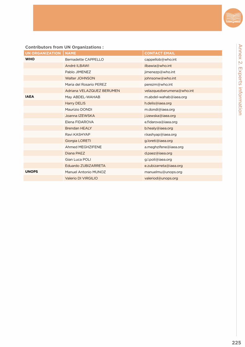
225
An
nex 2
. Exp
erts in
form
atio
n
Contributors from UN Organizations :
UN ORGANIZATION NAME CONTACT EMAIL
WHO Bernadette CAPPELLO [email protected]
André ILBAWI [email protected]
Pablo JIMENEZ [email protected]
Walter JOHNSON [email protected]
Maria del Rosario PEREZ [email protected]
Adriana VELAZQUEZ BERUMEN [email protected]
IAEA May ABDEL-WAHAB [email protected]
Harry DELIS [email protected]
Maurizio DONDI [email protected]
Joanna IZEWSKA [email protected]
Elena FIDAROVA [email protected]
Brendan HEALY [email protected]
Ravi KASHYAP [email protected]
Giorgia LORETI [email protected]
Ahmed MEGHZIFENE [email protected]
Diana PAEZ [email protected]
Gian Luca POLI [email protected]
Eduardo ZUBIZARRETA [email protected]
UNOPS Manuel Antonio MUNOZ [email protected]
Valerio DI VIRGILIO [email protected]
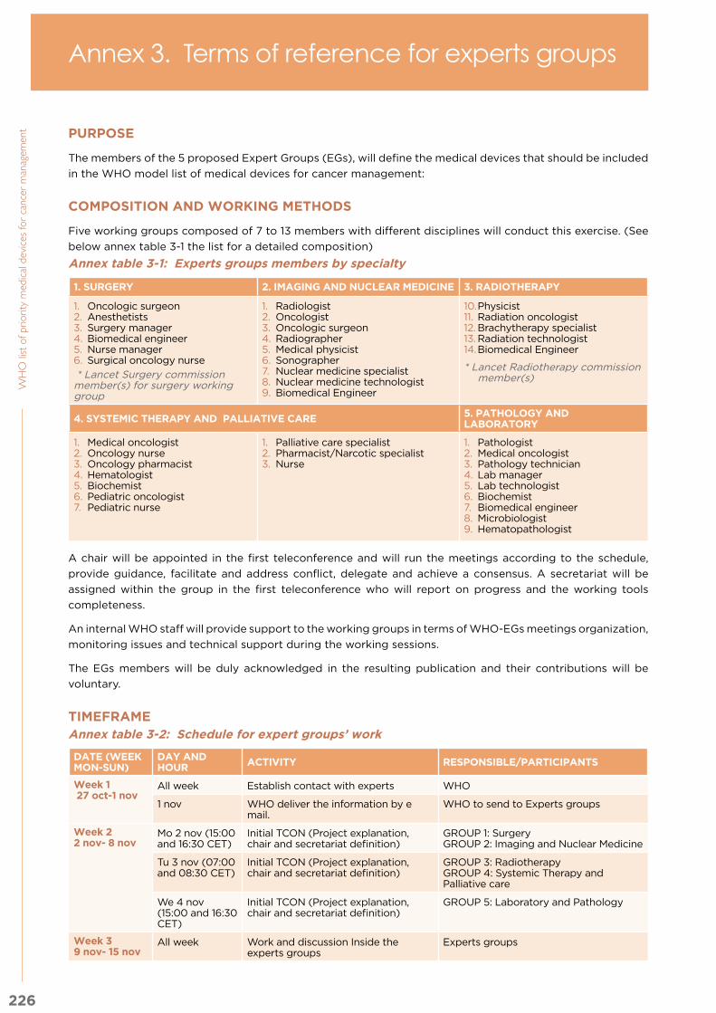
226
WHO
list o
f prio
rity m
edica
l dev
ices f
or ca
ncer
man
agem
ent PURPOSE
The members of the 5 proposed Expert Groups (EGs), will define the medical devices that should be included
in the WHO model list of medical devices for cancer management:
COMPOSITION AND WORKING METHODS
Five working groups composed of 7 to 13 members with different disciplines will conduct this exercise. (See
below annex table 3-1 the list for a detailed composition)
Annex table 3-1: Experts groups members by specialty
1. SURGERY 2. IMAGING AND NUCLEAR MEDICINE 3. RADIOTHERAPY
1. Oncologic surgeon 2. Anesthetists 3. Surgery manager4. Biomedical engineer5. Nurse manager6. Surgical oncology nurse
* Lancet Surgery commission member(s) for surgery working group
1. Radiologist2. Oncologist 3. Oncologic surgeon4. Radiographer5. Medical physicist6. Sonographer7. Nuclear medicine specialist 8. Nuclear medicine technologist9. Biomedical Engineer
10. Physicist11. Radiation oncologist12. Brachytherapy specialist 13. Radiation technologist 14. Biomedical Engineer
* Lancet Radiotherapy commission member(s)
4. SYSTEMIC THERAPY AND PALLIATIVE CARE 5. PATHOLOGY AND LABORATORY
1. Medical oncologist2. Oncology nurse3. Oncology pharmacist 4. Hematologist 5. Biochemist6. Pediatric oncologist7. Pediatric nurse
1. Palliative care specialist 2. Pharmacist/Narcotic specialist 3. Nurse
1. Pathologist 2. Medical oncologist3. Pathology technician 4. Lab manager5. Lab technologist 6. Biochemist7. Biomedical engineer8. Microbiologist9. Hematopathologist
A chair will be appointed in the first teleconference and will run the meetings according to the schedule,
provide guidance, facilitate and address conflict, delegate and achieve a consensus. A secretariat will be
assigned within the group in the first teleconference who will report on progress and the working tools
completeness.
An internal WHO staff will provide support to the working groups in terms of WHO-EGs meetings organization,
monitoring issues and technical support during the working sessions.
The EGs members will be duly acknowledged in the resulting publication and their contributions will be
voluntary.
TIMEFRAMEAnnex table 3-2: Schedule for expert groups’ work
DATE (WEEK MON-SUN)
DAY AND HOUR ACTIVITY RESPONSIBLE/PARTICIPANTS
Week 1 27 oct-1 nov
All week Establish contact with experts WHO
1 nov WHO deliver the information by e mail.
WHO to send to Experts groups
Week 22 nov- 8 nov
Mo 2 nov (15:00 and 16:30 CET)
Initial TCON (Project explanation, chair and secretariat definition)
GROUP 1: Surgery GROUP 2: Imaging and Nuclear Medicine
Tu 3 nov (07:00 and 08:30 CET)
Initial TCON (Project explanation, chair and secretariat definition)
GROUP 3: Radiotherapy GROUP 4: Systemic Therapy and Palliative care
We 4 nov (15:00 and 16:30 CET)
Initial TCON (Project explanation, chair and secretariat definition)
GROUP 5: Laboratory and Pathology
Week 39 nov- 15 nov
All week Work and discussion Inside the experts groups
Experts groups
Annex 3. Terms of reference for experts groups

227
An
nex 3
. Term
s of re
fere
nce fo
r exp
erts g
rou
ps
DATE (WEEK MON-SUN)
DAY AND HOUR ACTIVITY RESPONSIBLE/PARTICIPANTS
Week 416 nov- 22 nov
Mo 16 nov (15:00 and 16:30 CET)
Monitoring TCON GROUP 1: Surgery GROUP 2: Imaging and Nuclear Medicine
Tu 17 nov (07:00 and 08:30 CET)
Monitoring TCON GROUP 3: Radiotherapy GROUP 4: Systemic Therapy and Palliative care
We 18 nov (15:00 and 16:30 CET)
Monitoring TCON GROUP 5: Laboratory and Pathology
Week 523 nov- 29 nov
All week Work and discussion Inside the experts groups
Experts groups
Week 630 nov- 06 dic
Mo 30 nov Deliver to WHO completed documents
GROUP 1: Surgery GROUP 2: Imaging and Nuclear Medicine
Tu 1 dec Deliver to WHO completed documents
GROUP 3: Radiotherapy GROUP 4: Systemic Therapy and Palliative care
We 2 dec Deliver to WHO completed documents
GROUP 5: Laboratory and Pathology
Fr 4 dec (14:00 CET)
Advisory Committee teleconference to review the outcome of the working groups
Advisory committee and WHO
Dec-Jan 2016 Compile information for final publication
WHO
COMMUNICATION
The communication between members will be held by teleconferences and emails; moreover, the documents
for working sessions will be published on a Collaborative Project Website. The secretariat and chair will report
back to WHO at least 1 day previous to the teleconference by e mail concerning the main achievements,
comments and the meeting agenda.
The EGs should be available to answer their e-mail and be actively involved in the teleconferences. The group
has the authority to define their own meeting schedule but it is anticipated that the groups will meet remotely
at least 2 times with WHO HQ staff.
GOVERNANCE
The EGs need to decide what is basic when more than one alternative exists and the devices will be rated
based upon Multi-criteria decision analysis MCDA, a recommendation will be given whether to include the
device or not. The chair should establish consensus regarding discrepancies within the group.
The project will be supervised by WHO headquaters staff.
ACTIVITIES AND DELIVERABLES OF EXPERTS GROUPS:
The EGs will: 1) review the general basic medical devices per service using Working tools 1, 2) identify the
basic services, functions/interventions and specific basic medical devices in order to be included in to the
WHO Model List of basic medical devices of cancer management using Working tool 2., 3) the EGs will use
the Multi-criteria decision analysis (MCDA) tool to complement the selection and prioritization exercise in
case of contentious options (or if further information is deemed useful by the EGs to justify inclusion in the
list), using Working tool 3 and 4). Develop special notes per type of working group.
The above mentioned activities will be detailed below.
1. Reviewing all the general basic medical devices per services using Working tool 1 considering:
a. Pertinence:
i. Are the enounced medical devices considered basic? If so, please highlight with yellow in the list provided of general medical devices whether the devices is listed in the basic column or not.

228
WHO
list o
f prio
rity m
edica
l dev
ices f
or ca
ncer
man
agem
ent b. Completeness:
i. Are any basic medical devices missing from this list? If so, please ADD at the end of the list.
c. Relevance:
i. Are all listed basic medical devices relevant to this list? Please DELETE the device you deem not relevant
d. Classification:
i. Are the devices classified accordingly: Medical and QA equipment, instruments, high cost consumables and software, medical furniture, PPE, Clothing, Disposables, Solutions and Reagents and Utensils? If not specify the adequate category in brackets next to the device.
Refer to annex figure 3-3.
2. Identify the basic services, functions/interventions and specific basic medical devices required for
cancer management using the Working tool 2.
Review the information presented in the working tool 2 about Category, Basic services, Basic functions/
interventions/procedures (by line), Type of Cancer, Basic Specific Medical Devices Devices considering
a. Pertinence:
i. Is it a BASIC service (column B “Service”)? Is the category (column A “category” matching? If so, please leave the information and continue if not highlight with orange) the entire line and all the interventions and devices associated.
ii. Is it a BASIC/Indispensable intervention (column C “Function/Intervention”)? If so
iii. Is the name and type of cancer adequate for the intervention? If so, please leave the entire line and continue or modify as pertinent.
iv. Are all the enounced medical devices considered as specific basic devices (column F) If so, please leave the devices in the table.
b. Completeness:
i. Are there any basic services or interventions missing from this list? If so, please ADD at the end of the list.
ii. Are any specific basic devices missing from this list? If so, please ADD in medical devices column.
c. Relevance:
i. Are all listed basic services and interventions relevant to this list? Please DELETE the entire line you deem not relevant.
ii. Are all listed basic devices relevant to this list? Please DELETE the device you deem not relevant.
Fill in the working tool 2 with the information required.
a. Fill in the column “Expected outcome”.
b. Fill in the column ”Contentious option” (Column e and G) in the following cases:
i. In case of doubt as to whether or not a function or a device should be included in this list.
ii. If the working group considers that further information is needed to justify inclusion in the list.
c. Fill in the Key & contextual considerations (Column H)
d. Identify the level of health care system for each intervention/function (Column G)
Refer to annex table 3-4 for working tool definitions and instructions.
Refer to the example in annex table 3-5.
3. Use the Multi-criteria decision analysis (MCDA) tool in case of contentious options (or if further
information is deemed useful by the EGs to justify inclusion in the list), using Working tool 3
The MCDA tool allows for the selection and inclusion of optimal devices by accounting for the outcomes
(effectiveness, safety, patient dignity and perspective), the type of benefit (therapeutic benefit [e.g. cure
vs. symptom alleviation]; applicability to several diseases and several cancers), as well as implementation
aspects (ease of use, ease of training, ability to reach remote communities), economic considerations and
positive consequences on healthcare resource utilization.
WHO acknowledges that some criteria need to be considered by member states (e.g., size of population,
budget associated) however they are beyond the purpose of this exercise. Furthermore, given the methodology
adopted for elaborating the list based on clinical guidelines, the criteria “Expert consensus” was omitted from
the working tool.
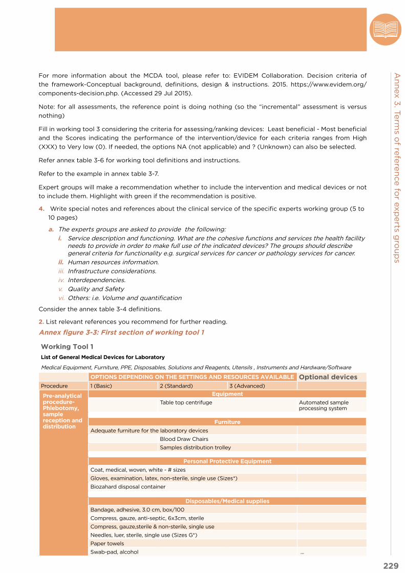
229
An
nex 3
. Term
s of re
fere
nce fo
r exp
erts g
rou
ps
For more information about the MCDA tool, please refer to: EVIDEM Collaboration. Decision criteria of
the framework-Conceptual background, definitions, design & instructions. 2015. https://www.evidem.org/
components-decision.php. (Accessed 29 Jul 2015).
Note: for all assessments, the reference point is doing nothing (so the “incremental” assessment is versus
nothing)
Fill in working tool 3 considering the criteria for assessing/ranking devices: Least beneficial - Most beneficial
and the Scores indicating the performance of the intervention/device for each criteria ranges from High
(XXX) to Very low (0). If needed, the options NA (not applicable) and ? (Unknown) can also be selected.
Refer annex table 3-6 for working tool definitions and instructions.
Refer to the example in annex table 3-7.
Expert groups will make a recommendation whether to include the intervention and medical devices or not
to include them. Highlight with green if the recommendation is positive.
4. Write special notes and references about the clinical service of the specific experts working group (5 to
10 pages)
a. The experts groups are asked to provide the following:
i. Service description and functioning. What are the cohesive functions and services the health facility needs to provide in order to make full use of the indicated devices? The groups should describe general criteria for functionality e.g. surgical services for cancer or pathology services for cancer.
ii. Human resources information.
iii. Infrastructure considerations.
iv. Interdependencies.
v. Quality and Safety
vi. Others: i.e. Volume and quantification
Consider the annex table 3-4 definitions.
2. List relevant references you recommend for further reading.
Annex figure 3-3: First section of working tool 1
Working Tool 1
List of General Medical Devices for Laboratory
Medical Equipment, Furniture, PPE, Disposables, Solutions and Reagents, Utensils , Instruments and Hardware/Software
OPTIONS DEPENDING ON THE SETTINGS AND RESOURCES AVAILABLE Optional devicesProcedure 1 (Basic) 2 (Standard) 3 (Advanced)
Pre-analytical procedure- Phlebotomy, sample reception and distribution
Equipment
Table top centrifuge Automated sample processing system
Furniture
Adequate furniture for the laboratory devices
Blood Draw Chairs
Samples distribution trolley
Personal Protective Equipment
Coat, medical, woven, white - # sizes
Gloves, examination, latex, non-sterile, single use (Sizes*)
Biozahard disposal container
Disposables/Medical supplies
Bandage, adhesive, 3.0 cm, box/100
Compress, gauze, anti-septic, 6x3cm, sterile
Compress, gauze,sterile & non-sterile, single use
Needles, luer, sterile, single use (Sizes G*)
Paper towels
Swab-pad, alcohol ...

230
WHO
list o
f prio
rity m
edica
l dev
ices f
or ca
ncer
man
agem
ent Annex table 3-4: Instruction tool for working tool 2 and definitions.
CRITERIA DEFINITION
Category Intended purpose of functions/interventions: Investigational (Diagnostic/Monitoring) and Therapeutic, commonly the category will apply to the devices associated as intended use.
Basic services Fundamental services absolutely necessary for any cancer health care system to function, attainable with limited financial means and modest infrastructure. i.e. Diagnostic imaging, surgery, histopathology.
Basic interventions/function
Fundamental interventions absolutely necessary for any cancer health care system to function attainable with limited financial means and modest infrastructure.
Expected outcome Mention the expected outcome of the intervention if applicable. Therapeutic outcome (e.g., removal of tumour) or diagnostic/investigational outcome of the intervention defined if applicable.
Type of Cancer: Define the cancer type(s) applicable to the intervention/function. B/Breast cancer, CR/Colorectal Cancer, P/Prostate cancer, L/Leukaemia, CU Cervical Cancer, L/Lung Cancer.
Specific Basic Medical Devices:
Describe the specific basic medical devices needed to carry out the intervention safely. These devices are components of cancer care (although not necessarily specific to cancer). They are basic (indispensable to provide cancer care), and affordable (low range of overall costs of acquisition and maintenance). This includes medical equipment, instruments, disposables, devices that cannot be included in the basic general devices because they are intervention specific and probably cancer specific. For specific examples, please see provided tool.
Contentious op-tion
Describe whether the working group had a discrepancy or considered it is worthwhile to detail further information about function/interventions or devices.
Interdependen-cies:
Device to be considered only if pre-requisite intervention/device has been used (e.g., diagnostic before radiation). Interpendencies may also occur with other services.
Human resources requirement
Indicate whether specialized skills are needed. What human resources should be available?
Infrastructure requirements
Indicate whether special infrastructure is needed including utilities (e.g. AC, voltage, power, water, IT, confinement, waste management, temperature control)
Key equipment associated (not stand-alone):
Indicate if other devices are necessary to operate the specific device or to achieve the intended function.
Planning, quality assurance and safety considerations:
Indicate need for planning and quality assessment. How can and should the devices (and their output) be quality assured? What is necessary in order to make safe use of devices?
Other special considerations:
Please specify if there are special considerations to account for, for example, regarding selection and procurement or use of the medical equipment for that service. List any particular issues planners should bear in mind.
Level of Healh care system
Community Level (CL)This level includes individuals and organizations; community-based, faith-based and other nongovernmental organizations; and community and home-based palliative care services. Also included are health posts, which are usually staffed by an auxiliary nurse or community health worker.Health Centre – Primary Care Level (HC)A health centre is a primary care facility with trained staff and regular working hours. Maternity and minimal laboratory services may be available. District Hospital – Secondary Care Level (DH)Typically, a hospital at this level provides general medical, paediatric and maternity services, general surgery, inpatient and outpatient care, and specialized care in some specific areas. Patients may be referred to this level from health centres and private practitioners in the district. Laboratory services may include cytology and histopathology.Central Or Referral Hospital – Tertiary Care Level (RH)Tertiary care hospitals provide specialized care for complex cases andacutely ill patients, including surgery, radiotherapy and multiple outpatientand inpatient services. General medical, acute and chronic care clinicsare offered. The most complete public-sector diagnostic and referencelaboratory services are available with pathologists and cytotechnologists,radiology and diagnostic imaging.

231
An
nex 3
. Term
s of re
fere
nce fo
r exp
erts g
rou
ps
Annex table 3-5: Working tool 2 (Examples are included below for illustration only. For full working tools, see www.who.int/medical_devices for supplementary annex.)
A: Category
B: Basic Service
C: Basic Function or Inter-vention
D. Expected Outcomes
E Type of Cancer
E: Contentious Option (function) Y/N
F: Basic Specific Medical devices
G: Contentious Option (de-vice) Y/N
H: K
ey &
co
nte
xtu
al co
nsi
dera
tio
ns
(see d
efi
nit
ion
s &
gu
idan
ce f
or
belo
w)
Co
mp
lete
as
ap
plica
ble
C: Level
of
Healt
h
Care
Interdependencies
HR requirements
Infrastructure re-quirements
Key Associated devices
QA & management
Other
Invest
i-g
ati
onal
Imag
ing
Mam
mo
-g
rap
hy
BN
Sta
-ti
onary
m
am
mo
g
rap
hic
X
-Ray
syst
em
NN
A1. R
ad
i-o
log
ist
2.R
ad
iog
ra-
pher
Shie
lded
ro
om
Co
m-
pre
ssio
n
devic
es
for
ad
dit
ional
pro
jecti
ons
Rad
iati
on
sh
ield
ing
ap
ron,
Rad
iati
on
sh
ield
ing
ap
ron r
ack,
Do
sim
ete
r
NA
DH
,RH
Invest
i-g
ati
onal
NA
Pap
ani-
co
lau
C
CU
NC
olp
o-
sco
pe
YP
ath
olo
gy
Lab
fo
r b
iop
sy e
x-am
inati
on
N1A
HN
AN
AN
AN
A H
C, D
H,
RH
Thera
-p
euti
cR
ad
io-
thera
py
Exte
rnal
Rad
io-
thera
py
B C
U,
YTe
leth
er-
ap
y u
nit
YC
om
put-
eri
zed
tr
eatm
ent
pla
nnin
g
syst
em
s
1. P
hysi
cis
t 2. R
ad
iati
on
o
nco
log
ist
3. R
TT
(R
ad
iati
on
T
hera
pis
t)
Ad
eq
uate
N
etw
ork
In-
frast
ructu
re
and
sto
rag
e
cap
acit
y
Resu
scit
a-
tio
n t
rolle
y
eq
uip
ped
,-C
lose
d-
cir
cuit
te
levis
ion
sy
stem
, A
dvanced
V
ital S
igns
Mo
nit
or
RE
FE
R-
EN
CE
TO
T
HE
RA
DI-
OT
HE
RA
PY
G
EN
ER
AL
D
EV
ICE
S
NA
DH
,RH
HR
: h
um
an
reso
urc
es;
QA
: q
ualit
y a
ssess
men
t

232
WHO
list o
f prio
rity m
edica
l dev
ices f
or ca
ncer
man
agem
ent Annex table 3-6: Criteria and scoring instruction for MCDA methodology for working tool 3
DOMAIN
Cluster of criteria based on MCDA principle of meaningful clustering
CRITERIACriteria representing a contribution to the value of the intervention/device; criteria included are justified by ethical positions for healthcare decision making (adapted from EVIDEM (www.evidem.org)
DEFINITION
Outcomes Efficacy/effectiveness A broad notion of effectiveness is used that is adapted to the type of device:For devices performing a therapeutic function, assessment of effectiveness is based on therapeutic outcomes.For devices performing a diagnostic function, assessment of effectiveness is based on sensitivity, specificity and negative/positive predictive valueFor other types of devices, specify what is considered (e.g., pump type features) Note: If effectiveness varies across cancer types, indicate a range, e.g. X–XXX, and specify as footnote below table OR create a row for each type of cancer (e.g., XXX for breast cancer, 0 for leukaemia)
Safety Includes both: risk of patient adverse events with correct operation and risk of errors in operating the device resulting in patient adverse events
Note: If safety varies across cancer types, indicate a range, e.g. X–XXX, and specify as footnote below table OR create a row for each type of cancer (e.g., XXX for breast cancer, 0 for leukaemia)
Patient reported outcomes (PROs)
Quality of patient experience with the device; includes notion of convenience, invasiveness, impact on dignity, autonomy, and tolerability by patient of the application of the device
Note: If PROs vary across cancer types, indicate a range, e.g. X–XXX, and specify as footnote below table OR create a row for each type of cancer (e.g., XXX for breast cancer, 0 for leukaemia)
Type of benefit Type of therapeutic benefit (e.g. cure)
Only for devices which fulfill therapeutic functions (cure is high and symptom relief is low)Note: If type of benefit varies across cancer types, indicate a range, e.g. X–XXX, and specify as footnote below table OR create a row for each type of cancer (e.g., XXX for breast cancer, 0 for leukaemia)
Applicability across disease areas
Device can be used for multiple diseases
Applicability across cancers
Device can be used for multiple types of cancer
Implementation Ease of use Broad notion of ease of use from the healthcare worker perspective (includes also safety considerations for worker)
Ease of training Knowledge and training necessary to apply the device
Ability to reach remote communities
Device easily portable, easy to use with telemedicine
Economics Affordability of device Cost of purchase/acquisition and operation/administration
Affordability of device maintenance & replacement
Cost of maintenance (repair) & replacement (taking into account the lifespan of the device)
Positive consequences on healthcare resource utilization
Limited need of physician visits, hospitalization, monitoring etc.
Quality of evidence
Quality of evidence Extent to which evidence on the intervention is valid with respect to scientific standards. This includes consideration of uncertainty (eg conflicting results/interpretation, limited date). Common sense might be deemed as sufficient evidence.

233
An
nex 3
. Term
s of re
fere
nce fo
r exp
erts g
rou
ps
Annex table 3-7: Working tool 3 (Examples are included below for illustration only. For full working tools, see www.who.int/medical_devices for supplementary annex.) Expert group: Surgery
A: F
un
ctio
n/
Inte
rven
tio
n
B
Co
nte
nti
ou
s O
pti
on
C: S
peci
fic
Med
ical
Devic
es
D:
Co
nte
nti
ou
s O
pti
on
Y
es/
No
as
E: V
alu
e C
rite
ria (s
ee d
efi
nit
ion
s an
d g
uid
an
ce f
or
sco
rin
g in
tab
le a
bo
ve)
Co
mp
lete
as
ap
plica
ble
Effectiveness*
Safety*
PROs*
Therapeutic ben-efit*
Multi-disease
Multi-cancer
Ease of use
Ease of training
Remote commu-nities
Affordability –device
Affordability – maintenance / replacement
HC resource con-sequences
Quality of evi-dence
Co
lono
sco
py
YE
S (
see
ab
ove)
Co
lono
sco
pe
YE
Sxxx
xxx
xxx
NA
xx
xx
xx
xx
xx
xx
Med
iast
ino
s-co
py
YE
S (
see
ab
ove)
Oth
er
devic
eY
ES
xx
xxx
xxx
NA
xx
xx
xx
xxx
xxx
xxx
xxx
PR
Os:
pati
en
t-re
po
rted
ou
tco
mes;
HC
: h
ealt
hcare

234
WHO
list o
f prio
rity m
edica
l dev
ices f
or ca
ncer
man
agem
ent
MEDICAL EQUIPMENT
Anaesthesia unit, mobile, basic
Aneroid sphygmomanometer
Biopsy gun
Blood glucometer
Bronchoscope
Cerebrospinal fluid manometer
Colposcope
Computed Tomography (CT) System (multi-slice)
Computerized treatment planning systems (three-dimensional)
Contrast medium injection system
Conventional simulator with digital imaging systems
Cryosurgery unit
Defibrillator
Diagnostic spirometer
Elastomeric pump
Electrocardiography system
Electrosurgical unit
Emergency cart with medicines
Endoscope sterilization/ disinfection support set
Endoscope washer/disinfector
Endoscopy tower system
Examination light
Fixed examination/treatment light
Flexible sigmoidoscope
Floor scale with stadiometer
Gamma camera system
Gamma probes (for intraoperative use)
General physical examination set (Ophthalmoscope, Otoscope, Lamp)
General ultrasound colour Doppler imaging system
General-purpose digital radiography system
General-purpose suction system, vacuum
Gynaecological examination/treatment table
Hammer for neurological examination
Infant scale
Infusion pump
Infusion pump for enteral nutrition
Linear Accelerator (LINAC) 3D Conformal therapy
Magnetic Resonance Imaging (MRI) System (1.5 T )
Mammographic stereotactic biopsy system
Mammographic X-ray system
Mobile diagnostic X-ray digital system (C-arm)
MR-safe stethoscope and sphygmomanometer
MRI compatible anaesthesia machine
MRI compatible biopsy procedure equipment
MRI compatible infusion pump
MRI compatible patient physiologic monitoring system
MRI compatible resuscitation trolley equipped with medicines and defibrillator
Nebulizer
Non-heated respiratory humidifier
Operating light, light source (lamp & flashlight)
Oxygen humidifier with flowmeter
Oxygen therapy flowmeter, dialtype
Patient positioning/tracking system
PET/CT system
Proctoscope
Pulse oximeter
Radionuclide generator
Rectal irrigation system
Remote-afterloading brachytherapy system, at least 12 channels
Respirator
Resuscitation trolley, equipped with medicines and defibrillator with laryngoscope
Resuscitator bag valve and mask (adult and paediatric)
Rigid cystoscope
Rigid sigmoidoscope
Rotary saw (optional)
Single-patient physiologic monitoring system for ECG, Capnography, SpO2, blood pressure
Stadiometer (wall mounted)
Stepping unit and stabilizer
Stethoscope, adult, binaural and paediatric
Suction and irrigation device
Syringe pump
Thermometer, clinical, digital 32–43°C
Thin layer chromatography scanner
Tympanic thermometer
Ultrasonic washer
ultrasound imaging system specific for TRUS
Ultrasound unit with biplanar transducer
Universal operating table
Universal operating table radiotransparent
Vibratory (reciprocating) saw
Video-colonoscope
Weighing scale, range 0 –150 Kg
Annex 4. List of priority medical devices for cancer management, by categories
Capital equipment

235
An
nex 4
. List o
f prio
rity m
ed
ical d
evic
es fo
r can
cer m
an
ag
em
en
t, by c
ate
go
ries
LABORATORY AND PATHOLOGY EQUIPMENT
Analyser Laboratory Haematology, Manual or Automated
Basic laboratory mixer/Laboratory shaker vortex
Camera (photo microscopy)
Cassette printer (optional)
Centrifuge
Centrifuge, micro - haematocrit
Class-II biological safety cabinet
Clinical chemistry Analyser
Coagulation analyser, manual or automated
Cover slipper for slides
Cryostat for intraoperative frozen sections
Cytocentrifuge
Distillation unit, 2 L/h, with tank
Flow cytometer
Freezer, laboratory
Gravity-convection laboratory oven
Grossing table, simple with cutting board and exhaust
High performance liquid chromatography
Hot plate, with stirrer
Humid chamber
Hygrometer
iFOB immunochemical analyzer, automated
Immunoassay analyser, automated
Immunohistochemistry (IHC)/In situ hybridization (ISH) staining platform, semi-automated
Immunostaining centre
Incubator, 30 L, up to 80° C
Macro digital imaging for pathology (optional)
Magnetic stirrer plate
Magnifying glass
Mechanical balance
Microscope, binocular
Microtome
Microwave oven
Negative Pressure Fume Hood
Organ balance
Oven
pH meter
Pharmacy refrigerator
Positive Pressure Laminar Flow cabinet for radiopharmaceutical preparation
Professional grossing bench with sink (exhaust system)
Refrigerator
Refrigerator/freezer, laboratory
Rotator, agglutination test
Scale, digital, 1500 g/0.1 g
Scale, precision, digital, 500 g/0.01 g
Shaker, orbital
Slide label printer (Optional)
Slide trays or plastic slide carriers for transport
Spectrophotometer, ultraviolet/ visible
Stainer
Staining station/Automated slide stainer
Sterilizer steam autoclave, 24 L
Table Lamp
Table top centrifuge
Thermometer, glass, min/max -20°C/100°C
Thermometer, min/max -30°C/60°C
Timer, 60 min, mechanical
Timer, digital
Tissue embedding unit or station
Tissue processor
Water bath thermometer
Water bath, 7 L
QUALITY ASSURANCE DEVICES
Array of diodes or ion chambers for routine quality assurance checks
Barometer
Calibrated radioactive reference sources for quality control of activity measuring systems
CT phantom and CT quality control devices
Diagnostic ultrasound phantom
Dose calibrator
EPID image quality phantom
Film dosimetry system
In-vivo dosimetry system
Large volume ionization chamber
Long lived reference source for checking the stability of the welltype ionization chamber
Measurement marker clip
Measurement marker wire
MRI system quality assurance device
Phantom for daily mechanical and light field checks on teletherapy unit
Plastic slab phantom with holes for ionization chambers for beam output verification
QA Phantom for brachytherapy
QA phantom for CT positioning lasers evaluation
QA/QC phantoms and accessories for Gamma Camera system
QA/QC phantoms and accessories for PET /CT System
Radiation field analyser to measure isodose distributions

236
WHO
list o
f prio
rity m
edica
l dev
ices f
or ca
ncer
man
agem
ent
Radioactive source for checking the stability of ionization chambers
Survey meter
Thermometer calibrated at a standards laboratory
Water phantom for calibration
Waterproof cylindrical ionization chambers
Waterproof plane-parallel ionization chamber
Well-type ionization chamber or an isotope calibrator with source holding inserts calibrated at a standards laboratory
SURGICAL INSTRUMENTS
Abdominal Hysterectomy set
Aspiration tray
Basic Colon Surgery set
Basic Rectal Surgery set
Basic Surgery set/ Minor tray
Biopsy forceps
Blunt obturator
Bone marrow aspiration set
Catheter placement set
Cervical punch biopsy forceps
Cervix conization set
Cheron forceps
Chest Aspirations set
Chest tube insertion kit
Chest tube set
Clamp, test tubes
Cricothyroidotomy set
Cystoscope sheath
Cystoscopy biopsy forceps
Dressing set
Endo-GIA stapler
Endocervical curette
Endoclip-applicator
Endoscopic hemoclip
Endotracheal tube
Examination/suturing, vaginal/cervical set
Excisional breast biopsy set
Forcep, artery
Forceps
Forceps tissue-long
Forceps, dressing, 155 mm, straight
General-purpose surgical scissors, reusable
Gynaecologic biopsy set
Gynecological and Kidney set
Hysterometer
Internally-anchored endotherapy retractor
Laparoscope holder
Laparoscopic biopsy forceps
Laparoscopic dissection spatula
Laparoscopic electrosurgical blunt dissector
Laparoscopic grasper
Laparoscopic grasping forceps
Laparoscopic irrigation/aspiration cannula
Laparoscopic multi-instrument access port
Laparoscopic needle holder
Laparoscopic swab forceps
Laparotomy ring
Laparotomy set
Laryngoscope handle with Macintosh blades
Lobectomy and segmental lung set
Long needle holders
Lumbar Puncture set, Adult
Lumbar Puncture set, Paediatrics
Manual expandable cervical dilator
Mastectomy set
McGill forceps adult and paediatric
Measurement ruler
Metzenbaum scissors
Needle holders
Pleural Biopsy set
Polypectomy snare
Prostatectomy set
Punch, Dry Blood Spot (DBS), 3.0 mm
Retractors (various sizes)
Ring forceps
Scalpel with blades
Scissors
Spatula, stainless steel (various sizes)
Suture set
Suture set
Thoracentesis set
Thoracotomy set
Tracheostomy set
Trocars with safety sheath (multiple sizes)
Urology set
Uterine forceps
Vaginal hysterectomy set
Vaginal sidewall retractors
Vaginal speculum, Non conducting preferably with side retractors
Vaginal speculum, reusable
Wire oval snare

237
An
nex 4
. List o
f prio
rity m
ed
ical d
evic
es fo
r can
cer m
an
ag
em
en
t, by c
ate
go
ries
LABORATORY AND PATHOLOGY EQUIPMENT
Bag, disposable for biohazardous waste
Bags for contaminated disposable supplies
Containers for hazardous waste (solutions and others)
Faecal occult blood test (FOBT) rapid test kit (slides and applicator sticks)
Graduated pipettes
HPV DNA Test
Micropipettes (different microliters)
Microplate, ELISA, 96 U-well
Pasteur Pipette
Pipette, digital, 10–100 ul
Pipette, digital, 100–1000 ul
Pipette, digital, 2–20 u
Pipette, digital, 20–200 ul
Pipette, digital, 8 channel, 20-200 ul
Pipette, digital, 8 channel, 5–50 ul
Pipette, filler, wheel-run, set/2
Pipette, repeating, 5 volume
Pipette, stand, 4 positions
Pipettes, blood graduated, 0.05 ml
Rack, drying glass & plastic ware
Rack, staining slides
Rack, test tubes, 24 positions
Rack, tubes, 0.5/2.0/5.0 ml, 24 positions
Safety box for used syringes/needles
Serological pipette
Staining rack
PERSONAL PROTECTIVE EQUIPMENT AND CLOTHING
Apron impermeable
Clogs, plastic (various sizes)
Coat, medical, woven, white (various sizes)
Disposable apron or heavy duty plastic apron (washable, reusable)
Drawsheet, plastic, approx. 90x180 cm
Examination/treatment table cover
Eye protective wear
Face masks N95
General-purpose sterile drape
Glasses, safety, regular size
Gloves, examination, non-sterile,single use (various sizes)
Gloves, nitrile, powder-free, nonsterile, single use
Gown, impermeable single use
Gown, patient
Heat-resistant gloves
Mask, surgical, non-woven
Medical scrubs for healthcare workers or similar
Non-conductive shoe cover
Operating room gown, reusable
Plastic face shield
Surgical cap for patients and healthcare worker
Surgical face mask
RADIATION PROTECTION/MONITORING DEVICES
Area monitoring devices (area survey meter)
Area radiation monitor (audible alarm)
Breast shielding
Clamps for the source management
Dosimeter, personal
Dummy sources
Emergency container and emergency source handling instruments
Geiger-Müller counter radiation survey meter and measuring probe
Gonadal shielding
Ion chamber survey meter
Patient radiation shielding
Portable radiation protection barriers
Radiation shielding apron
Radiation shielding apron rack
Radiation shielding gloves
Radiation shielding goggles
Radiation shielding headwear
Radioactive waste storage
Ring dosimeter
Source handling instruments and accessories
Source loading and cutting devices
Source storage and transport containers within the department
Thyroid shielding
Single use, accessories, software, consumables, protection devices and reagents

238
WHO
list o
f prio
rity m
edica
l dev
ices f
or ca
ncer
man
agem
ent
SINGLE USE DEVICES/DISPOSABLES/ MEDICAL SUPPLIES
Absorbent tipped applicator
Adult/children diapers (incontinence pads)
Anaesthesia breathing circuit
Asepto syringe
Bag, biohazard, 20 L
Bag, re-sealable, plastic
Bags for contaminated supplies
Bandage, adhesive, 3.0 cm, 100/box
Bandage, elastic, 7.5 cm x 5 m, roll
Biopsy needle
Blood collection tube, neonatal cord blood, sterile
Blood giving sets and cannulas
Cannulas, Intra Venous (IV) short, sterile, single use (sizes G)
Cassettes
Catheter (18 gauge or larger)
Catheter bag
Catheter, Foley, sterile, single use (sizes G)
Catheter, urethral, sterile, single use (sizes G)
Cervical aspiration catheter
Cervical cytology brush or cervical cytology scraper
Chest tube
Chest tubes drainage
Closed system drug transfer devices
Closed-wound drainage reservoir system with closed wound drain connector
Collection tube/sterile plastic tubes
Collector, urine, adhesive, 10–100 ml
Combined spinal epidural anaesthesia trays (spring-wound catheter, spinal epidural needles)
Compress gauze, sterile & non-sterile, single use
Compress, gauze, antiseptic, 6x3 cm, sterile
Compression bandages (for Deep Vein Thrombosis)
Container, sample, 50 ml
Cotton wool, 500 g, roll, non-sterile
Cover glass, slides
Cover glass/cover slips
Cryptographic/urethrographic catheter, female
Devices to deliver enemas through stomas
Devices to manage genital fistulae
Drainage bag including gravitational IV tube or connect to suction system
Dressing retention roll
Dressing sets for malodours/ fungating wounds (carbon pads/ silver impregnated pads, etc.)
Dressing strip, adhesive, diameter 3.0 cm, sterile
Elastic bandage
Endotracheal tubes adult and paediatric
Envelope, packing, 27x36 cm
Epidural catheter
Examination table paper cover
Female sanitary products
Fiducial markers/soft tissue markers
Film, sealing, flexible, 10 cm x 38 m, roll
Filter Discs
Filter needles
Filter Venting devices
First aid gauze/bandage
Flexible silicon catheter with needle
Gastrostomy material for skin medication and use
Gauze strip antimicrobial
Gelfoam (for plugged biopsy)
General-purpose sterile drape
Grey bottle, sterile universal specimen bottles
Hemoclips/clip cartridge or similar for cardiothoracic surgery
Hypodermic needles: gauge 25 G, 23 G, 21 G
ICD set / Thoracic tube insertion set
Infusion giving set, sterile, single use
Infusion set, sterile, single use
Inoculation loop, plastic, sterile
Instrument/equipment drape, single-use, non-sterile
Intravenous catheters
Intubating bougies, adult and paediatric
IV burettes
IV catheters
IV infusor bags/sets
Kato-Katz, kit, stool sample preparation
Lancet, 2 mm, safety, sterile
Lancet, blood, safety, sterile (various sizes)
Laryngeal mask airways
Lead sharps container according to isotope energy used
Markers, fine point, permanent black, for glassware and slides
Mask and tubing for oxygen
Medication cups
Micro-vial tubes

239
An
nex 4
. List o
f prio
rity m
ed
ical d
evic
es fo
r can
cer m
an
ag
em
en
t, by c
ate
go
ries
Monitor card, humidity, passive/cumulative
Monitoring electrodes
Nasogastric tube
Nasogastric tube fixator
Needle cradle
Needle holder, vacuum tubes, sterile
Needle, vacuum tube, 20 G/ 22 G, sterile
Needles, luer, sterile, single use (sizes G)
Needles, scalp vein, sterile, single use (sizes G)
Needles, spinal, sterile, single use (sizes G)
Needles, sterile, single use: • 20–24G (for fine needle aspiration)• 11–14 G (for bone biopsy)• 16–20G (for other tissue biopsy)
Non-implantable needle guide
Non-sterile coupling gel
Operating room laundry bag
Organ bag
Oropharyngeal airway (adult size)
Ostomy bag
Paediatric foley catheters and nasogastric tubes
Paediatric spinal needles
Paper towels
Paper, dry blood spot
Paper, exam table
Paper, filter
Paper, lens
Paper, pH indicator 2.0 to 9.0
Paper, weighing
Paracentesis set
Parafilm paper
Parenteral/enteral solution bag
Partial-rebreathing oxygen face mask
Pipette, repeat, tip 2.5/5.0 ml, 10/25 ml
Pipette, tip, barrier, 200 ul / 1000 ul, sterile
Pipette, tip, blue, 100–1000 ul
Pipette, tip, white, 2–20 ul
Pipette, tip, yellow, 10–100 ul / 20–200 ul
Pipette, transfer, 3 ml, non-sterile
Pipette, transfer, 3 ml, sterile
Pneumonectomy pleurevacs only for Pneumonectomy
Prongs, nasal, oxygen, non sterile, single use (various sizes)
Rack, drying DBS cards, 10 positions
Radiographic or radiochromic film
Rectal probe or rectal catheter
Reservoir, reagent, 60 ml
Safety straps
Scalp vein intravenous administration set
Sclerotherapy endoscopic needles
Sealant, compound
Secondary set with drip chamber
Septo syringe
Sheet, absorbent, bench, 50x40 cm
Skin-cleansing wipe
Skin-cover adhesive strip
Slide, microscope
Slide, microscope, frosted
Specimen container
Spinal anaesthesia needle, single-use
Sponges
Sputum containers
Staplers (linear and thoracotomy) with staples reloads
Sterile culture tube
Sterile sample container
Sterile ultrasound coupling gel
Stoma/ostomy bags and adhesive
Suprapubic catheter
Surgery table, padded accessories for patient positioning or similar
Surgical clip
Surgical scrub brush, single-use
Surgical scrub sponge
Suture, synthetic, non-absorbable (sizes USP/DEC) with needle (sizes G), sterile, single use, nylon, catgut, silk
Sutures
Swab-pad, alcohol
Swab, cotton-tip, tube, sterile
Syringes (various capacities)
Syringes for drug preparation/ formulation
Syringes for the biopsy and holder (optional)
Syringes with needles (disposable)
Syringes, autodisable (AD), (various capacities)
Syringes, luer, sterile, single use (various capacities)
T-tube and ties
Tape, medical, roll (various sizes)
Three-way-stopcock
Tongue depressor, single use (wooden or plastic spatula)
Transparent film dressings
Transparent film dressings with a gel pad
Tube containing EDTA anticoagulant
Tube suction, Yankauer, 270 mm/Yankauer suction tips

240
WHO
list o
f prio
rity m
edica
l dev
ices f
or ca
ncer
man
agem
ent
Tube, push cap, 0.2 ml, PCR, sterile
Tube, push cap, 5.0 ml, non-sterile
Tube, screw cap, 0.2 ml / 0.5 ml / 2.0 ml / 5.0 ml, non-sterile
Tube, screw cap, 0.2 ml / 0.5 ml / 2.0 ml / 5.0 ml, sterile
Tube, screw cap, conic, 15/50 ml, non-sterile
Tube, suction, L 50 cm, catheter tip, sterile, single use (sizes G)
Tube, vacuum, EDTA, 2 ml / 4 ml / 6 ml, sterile
Tube, vacuum, Ethylene Diamine
Tube, vacuum, plain/dry, sterile (various capacities)
Tube, vacuum, serum, 4 ml / 6 ml, sterile
Ureteral catheter connector and other connectors as required
Urinary catheter (Foley)
Urine drain bag
Urological irrigation kit
Veress needle (optional, for transperitoneal access only)
Vortex, test tube
Wire localization needle ( e.g. Kopan’s Needle 21G, 20G)
Wooden or plastic applicator sticks
SOLUTIONS AND REAGENTS
Acetic acid solution
Acetone, bottle
Acid Alcohol 1%
Alcohol
Alcohol isopropyl 70%
Alcoholic Ammonia 1%
Alkaline detergent solution
Ammonium hydroxide/Ammonia
Amplification Kit for IHC staining platform
Antibody detection kit for IHC staining platform
Antibody diluent
Antibody recovery solution
Antimicrobial solution
Antiseptic skin cleansing agent
Aqueous antibacterial solution/ Aqueous cleaning and decontaminating solutions
Blocking protein solution
Bluing Reagent
Bromine solution
Buffer, tablets, pH 7.2, box
Chlorhexidine
Chlorine solution
Contrast medium, injectable
Contrast medium, oral
Cytology stain kit
Detection system based on polymers
Diamine
Diethyl ether, bottle
Dispenser with pre kit for automated staining platform
Distilled water
Endoscope cleaning kit
Enzymatic detergent, test strip to measure the action of enzyme
Enzyme solutions
Eosin
Ethanol
Ethanol, denatured, bottle
Ethyl alcohol
Fixative spray or solution for pap smear (if slides are used)
Formaldehyde, 10%, 10 ml, ampoule
Formalin 10%, or tissue fixation reagents
Gelatin Titanium dioxide (E171) Indigo carmine solution
Gentian violet, solution, bottle
Giemsa Stain
Glutaraldehyde 3.4% (Cidex, Maxicide, Wavicide)
Glycerol, bottle
Haematoxylin
Hand/body hygiene products
Harris’s Haematoxylin
Hydrochloric acid, 40%, bottle
Hydrogen peroxide
Immunoassay analyser reagent sample diluent
Immunoassay analyser reagents
Immunohistochemistry (IHC)/In situ hybridization (ISH) staining platform reagents
Indian ink, black, bottle
Ink (for surgical margins)
Iodine povacrylex
Iodine preparation cleansing agent
Isopropyl alcohol
IV solution
KI starch solution
Lubricating jelly (K-Y)
Lugol iodine, bottle

241
An
nex 4
. List o
f prio
rity m
ed
ical d
evic
es fo
r can
cer m
an
ag
em
en
t, by c
ate
go
ries
May Grunewald stain (BDH)
Methanol, bottle
Methylene blue, bottle
Monsel’s paste
Mounting medium
MRI contrast medium, injectable
Neutral Buffered Formalin 10%
Nitric acid
Oil, immersion, bottle
Orange Gelb-6
Oxidase test
Oxygenated water
Paraffin
Peracetic acid disinfectant anticorrosion additive (for endoscopes or similar)
Peroxidase
Petroleum gel, paraffin, bottle
Phosphate Buffer (pH 6.8)
Pipettes cleaning solution
Potassium iodide
Preservative solutions
Primary antibodies
Rubber cement
Saline solution
Sclerodesis agent (e.g. talc)
Secondary antibodies
Silica gel (desiccant for DBS), pouch
Sodium bicarbonate
Sodium chloride, powder, bottle
Sodium hypochlorite solution
Sodium hypochlorite, tablets
Sodium persulfate
Stain, Field A, solution
Stain, Field B, solution
Stain, Giemsa, solution
Stain, Gram, set
Stain, May-Grunwald Giemsa, set
Stain, Ziehl–Neelsen, solution, bottle
Substrate, adamantyl phosphate oxetane
Sugar fermentation tests
Sulphuric acid, 95%
Test, Nickerson or sabouraud medium, kit
Test, potassium hydroxide KOH, preparation
Transurethral-instrument lubricant
Trichloroacetic acid, crystals, bottle
Wash solution
Washing buffer
Xylene, bottle
OTHER (glassware, utensils, etc.)
Acetate sheets
Arm/leg tourniquet
Basic sedation equipment
Basin, kidney, polypropylene
Beaker, glass, 100 ml / 250 ml
Bedpan
Bolus
Bottle reagent
Bottle, amber, dropper, 30 ml
Bottle, amber, screw cap, 100 ml /250 ml / 1000 ml
Bottle, plastic, 1 L
Bowl, polypropylene
Bowl/Emesis bowl
Box, refill, pipette tips, empty
Box, specimen transport, 2 L/4 L
Box, storage 0.5/2/5 ml tubes, 100 positions
Box, storage, 100 slides
Brush, bottles and flasks (various sizes)
Brush, hand, scrubbing, plastic
Brush, test tubes (various sizes)
Container for specimen immersion
Container for warm water
Coplin jar
Cover plates
Cover slipper - Shandon cover plate
Cylinder, measuring, glass, 10 ml/ 100 ml / 500 ml/ 100 ml
Cytotoxic waste receptacle
Drill
Dropper bottles
Funnel, glass
Funnel, plastic
Glassware Beaker
Hand/body hygiene products
Hazardous drug spill kits
Hot water bath for thermoplastic immobilization system
Hot wire cutter

242
WHO
list o
f prio
rity m
edica
l dev
ices f
or ca
ncer
man
agem
ent
Jar, Coplin, staining
Knife for specimens
Label or pen for labelling specimen containers
Label, biohazard, adhesive, 3x4 cm
Label, self-adhesive, different sizes
Label, self-adhesive, freezer
Labels for the drugs identification
Magnetic metal detector
Manual brachytherapy source, temporary placement
Marker pen, cryoware
Marker pen, glassware
Marker, diamond
Markers, fine point, permanent black, for glassware
Mould materials (Foam blocks/Styrofoam/Polystyrene, Thermoplastic, vacuum bag or similar as needed)
MRI compatible oxygen canisters
Oxygen/gas pipeline, cylinder
Petri dish, glass, with lid
Pot for cerrobend cadmium free low melting point alloy
Pressure cooker
Pressure relief products (e.g. Cushions, mattresses)
Receptacle, waste, stainless steel,pedal action
Rod, glass
Rubber hammer
Ruler
Sample distribution container
Skin marker pen
Smoke evacuator
Specimen cup
Sponge bowl
Thermic containers
Urinal
Warming cabinet for contrast media
Wash bottle, 250 ml
Waste disposal container
SOFTWARE
Laboratory Information System (LIS)
Picture Archiving Communication System (PACS)
Radiology Information System (RIS)


WHO list of priority medical devices for cancer
management
ISBN 978 92 4 156546 2
WHO Medical device technical series
For more information , please contact:World Health OrganizationDepartment of Essential Medicines and Health ProductsMedical Devices20 Avenue Appia,CH 1211, Geneva 27Switzerland
Tel: (+41)22 791 1239
Email: [email protected]://www.who.int/medical_devices