what is pathology? - uniba.sk...• if no buffered solution is available, dilute formaldehyde with...
TRANSCRIPT

1
Introduction. Basic histological techniques.
Methods in pathology (physical methods of investigation).
General medicine
Pavol Janega
Svetoslav Štvrtina
Peter Michalka
Institute of Pathological Anatomy Faculty of Medicine, Comenius University in Bratislava
and
University Hospital Bratislava
Prof. MUDr. Ľudovít Danihel, CSc.
What is pathology?
• Processing of biological material
any tissue removed from the living or dead body
biopsy autopsy
• Determining the diagnosis and disease / causes of death
• Determining the nature and causes of the disease
• The effect on the choice of an adequate therapeutic approach
THE ROLE OF PATHOLOGY
COLLECTION OF BIOLOGICAL MATERIAL
BIOPSY (COLLECTION) CYTOLOGY (COLLECTION) BIOPSY CYTOLOGY
• organs and tissues
• excisions
• core-cut (core needle)
• Invasive method
• Representative (larger sample of tissue, we see
relationships between cells)
• Financially more expensive (surgery, anesthesia, ...)
• Hospitalization of the patient (often needed)
exfoliative
• smear cytology (brush, spatula, ...)
• cytology of body fluids (urine, pleural effusion,
ascites, liquor, ...)
• lavage (bronchi, ...)
intervention:
• aspiration with a fine needle (FNAC)
• Non-invasive (minimally invasive) method
• Screening, not always representative
(dependence on collection, fewer tissues,
missing cell relationships)
• Cheap and affordable method
• Hospitalization is not necessary

2
BIOPSY
BIOLOGICAL MATERIAL
• Processing and subsequent material evaluation•with the least possible damage to the structure of the
cells and tissues
•to prevent cellular degradation and autolytic process
• Immediately after the collection of tissue samples it is
necessary to fix them
•kill microorganisms and fix the cells
•avoid autolysis and microbial damage
• Accurate labeling
• Fast delivery to the laboratory for further processing
COVER LETTER
Source of important information
subject of examination and
localization
clinical course, therapy, irradiation, diagnosis
FIXATION
BIOLOGICAL MATERIAL
1. Fixation associated with loss of biological function of structures- chemical, physical methods
2. Fixation without loss of biological function of structures
- freezing
3
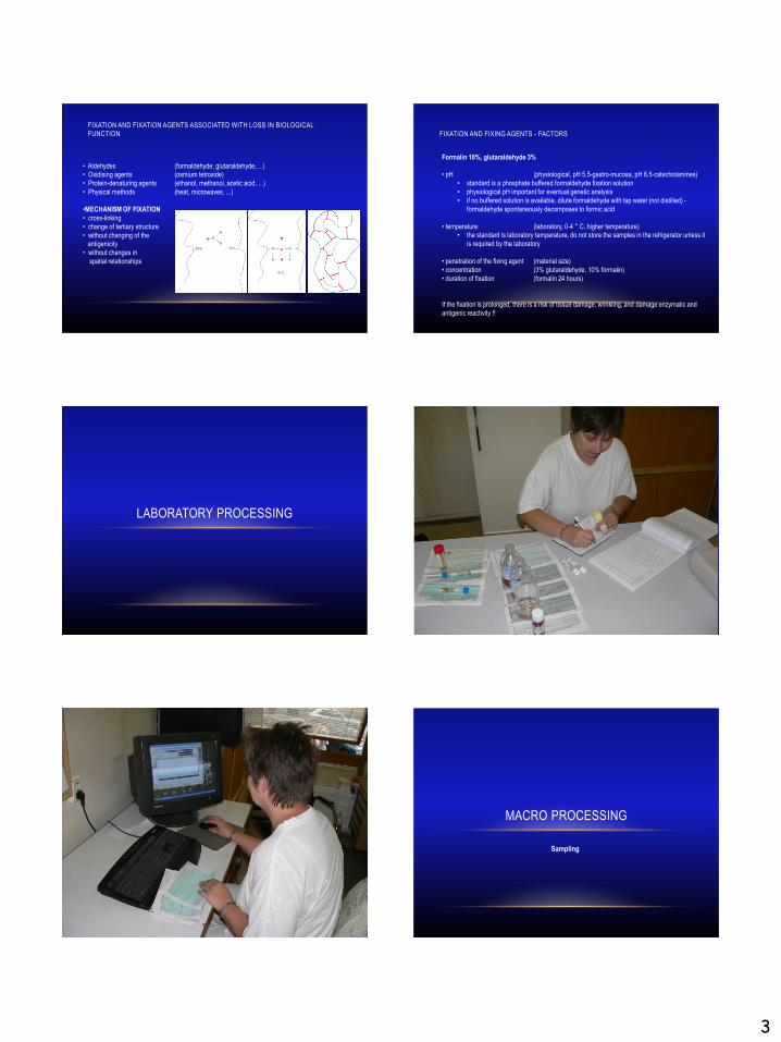
FIXATION AND FIXATION AGENTS ASSOCIATED WITH LOSS IN BIOLOGICAL FUNCTION
• Aldehydes (formaldehyde, glutaraldehyde, ...)• Oxidising agents (osmium tetroxide)
• Protein-denaturing agents (ethanol, methanol, acetic acid, ...)
• Physical methods (heat, microwaves, ...)
•MECHANISM OF FIXATION
• cross-linking
• change of tertiary structure
• without changing of the antigenicity
• without changes in
spatial relationships
FIXATION AND FIXING AGENTS - FACTORS
Formalin 10%, glutaraldehyde 3%
• pH (physiological, pH 5.5-gastro-mucosa, pH 6.5-catecholamines)
• standard is a phosphate buffered formaldehyde fixation solution
• physiological pH important for eventual genetic analysis
• if no buffered solution is available, dilute formaldehyde with tap water (not distilled) -
formaldehyde spontaneously decomposes to formic acid
• temperature (laboratory, 0-4 ° C, higher temperature)• the standard is laboratory temperature, do not store the samples in the refrigerator unless it
is required by the laboratory
• penetration of the fixing agent (material size)
• concentration (3% glutaraldehyde, 10% formalin)
• duration of fixation (formalin 24 hours)
If the fixation is prolonged, there is a risk of tissue damage, wrinkling, and damage enzymatic and
antigenic reactivity !!
LABORATORY PROCESSING
MACRO PROCESSING
Sampling

4
PROCESSING
TISSUE PROCESSING
Aim: it is necessary to put the tissue into a solid medium which allows its subsequent cutting
If necessary, the tissue must be pre-treated (eg decalcification)
EMBEDDING MEDIA
•Paraffin cheap, solid, melting point 40-70 ° C, fast settingadditives (gum, beeswax, ...)
•Resin electron microscopy, thin sections, high resolution microscopy, bones
•Agar small fragments of tissue
•Gelatin frozen cuts
Tissue in fixative (10% formalin)
Tissue impregnated with embedding medium

5
Tissue in fixative (10% formalin)
Tissue impregnated with embedding medium
Dehydration - removal of the fixation solution(ethanol, methanol, acetone, isopropyl alcohol, ...)
in increasing concentration .70% .. 90% .. 100%
Impregnation - embedding medium, vacuum technology
Tissue in fixative (10% formalin)
Tissue impregnated with embedding medium
Dehydration - removal of the fixation solution(ethanol, methanol, acetone, isopropyl alcohol, ...)
in increasing concentration .70% .. 90% .. 100%
Impregnation - embedding medium, vacuum technology
Cleaning - the removal of the dehydrating solutionsubstances well miscible with the embedding medium
(xylene, toluene, chloroform, ...)
DEHYDRATION AND CLEANING10% formalin
70% alcohol
95% alcohol
100% alcohol
100% alcohol
100% alcohol
100% alcohol + Xylene
xylene
xylene
paraffin
paraffin

6
FROZEN SECTIONS PROCESSING
• Freezing as a form of "fixation" of fresh tissue
• Condition: Speed (slow freezing – i.c. ice crystals formation and cell destruction)
• Fast diagnosis (lack of time for other fixation)
• Enzyme histochemistry when enzymes are labile
• Some immunofluorescence methods

7
CYTOLOGY
1846 V.D. Lambl
“Urinary bladder cancer cells found in urine”
1936 J. Trapl
“Women estrus evaluated in vaginal cells”
HISTORY OF CLINICAL CYTODIAGNOSIS
1943 G.N. Papanikolaou
“Diagnosis of the uterus
cancer by the vaginal smear”
after 1950 - development of non-gynecological cytology diagnostics
lung, oral cavity, urinary bladder, GIT
- later fine needle aspiration cytodiagnostics
breast, parenchymal organs, body cavities ...
HISTORY OF CLINICAL CYTODIAGNOSIS CYTOLOGY
• The advantages of cytology are:
• Speed of diagnostics
• Opportunity to take and evaluate samples from hard-to-reach locations
• Economic aspect
• The disadvantages of cytology are:
• The impossibility of accurate classification and typing of tumors
• Impossible to consider an invasion
• Possibility of false negativity and false positivity
Today, cytology is used to diagnose tumors of all organ systems.
Cytology makes it possible to carry out screening tests at vulnerable and risky populations with low financial costs.
41/69
BASIC STAINING TECHNIQUES
BASIC PRINCIPLES OF STAINING
• Acidofilia:
Staining with acidic dyes
Eosin, Azocarmine, Aniline Blue, Carbofucin
Substrates: Cytoplasm, intercellular substances, ...
• Basophilia:
Staining with alkaline dyes:
Methyl blue, Toluidine, Hematoxylin
Substrates: Chromatin, Ribozymes, ...
• Metal impregnation
Methods by Golgi and Cajal
Silver and Gold Salts
Neurons and glial cells

8
BASIC STAINING METHODS
HEMATOXYLIN AND EOSIN MALLORY HEMATOXYLIN
RETICULIN LIPIDS – FAT TISSUE (HE)

9
OIL RED PERIODIC ACID SCHIFF
ALCIAN BLUE GRAM STAINING
STAINING BK (ZIEHL–NEELSEN)
HISTOCHEMICAL METHODS
10
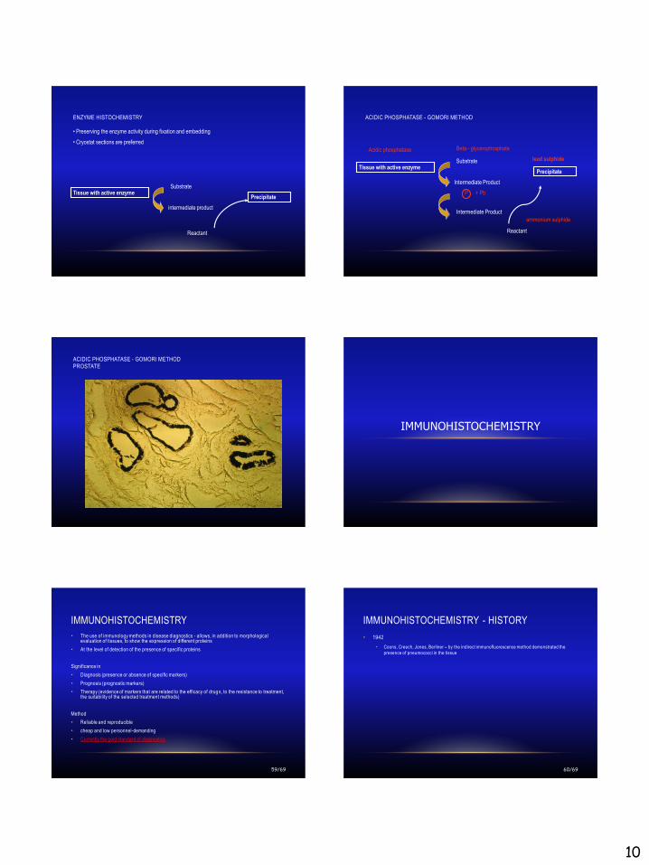
ENZYME HISTOCHEMISTRY
• Preserving the enzyme activity during fixation and embedding
• Cryostat sections are preferred
Tissue with active enzymeSubstrate
intermediate product
Precipitate
Reactant
ACIDIC PHOSPHATASE - GOMORI METHOD
Tissue with active enzymeSubstrate
Intermediate Product
Precipitate
Reactant
Acidic phosphatase Beta - glycerophosphate
P + Pb
Intermediate Product
ammonium sulphide
lead sulphide
ACIDIC PHOSPHATASE - GOMORI METHODPROSTATE
IMMUNOHISTOCHEMISTRY
IMMUNOHISTOCHEMISTRY
59/69
• The use of immunology methods in disease diagnostics - allows, in addition to morphological evaluation of tissues, to show the expression of different proteins
• At the level of detection of the presence of specific proteins
Significance in
• Diagnosis (presence or absence of specific markers)
• Prognosis (prognostic markers)
• Therapy (evidence of markers that are related to the efficacy of drug s, to the resistance to treatment, the suitability of the selected treatment methods)
Method
• Reliable and reproducible
• cheap and low personnel-demanding
• Currently the gold standard of diagnostics
IMMUNOHISTOCHEMISTRY - HISTORY
60/69
• 1942
• Coons, Creech, Jones, Berliner – by the indirect immunofluorescence method demonstrated the
presence of pneumococci in the tissue

11
FLUORESCENCE METHOD IHC
Bullous pemphigoid - IgG antibodies against BM
marked secondary
antibody
primary antibody
antigen
ENZYME METHOD IHC
Soluble chromogen
Insoluble precipitate
enzyme
(horseradish peroxidase)
SCC – AE1/3, anti-cytokeratin antibodies
marked secondary
antibody
primary antibody
antigen
IMMUNOHISTOCHEMISTRY
• Diagnostic markers
• Therapeutic markers
• Prognostic markers
cytokeratins
• Protein filaments present in epithelial
cells
• I. Acidic cytokeratins, 9-20
• II: Basic cytokeratins, 1-8
Vimentin
• Intermediate filaments (57 kD) present in
mesenchymal cells
Neuroendocrine markers:
Chromogranin, Synaptophysin
• A group of acid glycoproteins located in neurosecretive granules
• Present in neuroendocrine tumors
Proliferation Markers:
Ki67
• Protein expressed by cells in late G1, S, G2 and M phases. Not present in the G0
phase
• The sign of cell growth and division
66/69
70 year old female patient
The patient hospitalized with symptoms of sepsis, pain in the abdomen. Multi-organ
failure due to probable acute cholecystitis. Hypostatic bronchopneumonia of lung.
CONCLUSION: Multi-organ failure in sepsis.

12
lungs68/69
colon
cytokeratins chromogranin
CK7 CK20
cytokeratins chromogranin
CK7 CK20
Adenocarcinoma of the colon

13
75 year old patient
Small prominent structure on the skin temporally. Suspected basal cell carcinoma.
74/69
75/69MelanA S100
vimentin cytokeratins
diagnostic markers76/69
MelanA S100
vimentin cytokeratins
diagnostic markers
metastasis of malignant melanoma
77/69
60 year old female patient
Metrorrhagia in menopause. Last menstruation: 2014
cytokeratins vimentin

14
ER PR
Ki67
Endometrioid uterine adenocarcinoma
FIGO Grade 2
ER + 90%
PR + 50%
prognostic and
therapeutic markers
ELEKTRON AND RADIOACTIVE
MICROSCOPIC METHODS
ELEKTRON AND RADIOACTIVE MICROSCOPIC METHODS
scanning electron microscopy
transmission electron microscopy
SIMS analysis
EDAX analysis
GENETIC METHODS
MOLECULAR PATHOLOGY
THE IMPORTANCE OF MOLECULAR MEDICINE IN PATHOLOGY
• Oncology - in the human genome there are described several types of gene changes that lead to oncogenic transformation
• oncogens, tumor suppressor genes, „mutator“ genes
• gene, chromosome and genomic mutations
• Current WHO cancer classification often requires genetic testing
• Personalization of medicine / treatment
• The ability to differentiate familial cases from sporadic
• Detection of carriers - genetic consultation, disease prevention, early diagnosis
• Estimation of response to therapy, choice of appropriate therapy (pharmacogenomics)
• Estimation of disease prognosis
• Follow-up of donor cells in transplants
• Detection of minimal residual disease
• Gene therapy? - the music of the future