what causes gray meat in the atlantic sea scallop...
TRANSCRIPT

What Causes Gray Meat in the Atlantic Sea Scallop Placopecten magellanicus in Georges Bank Closed Areas?
Scallop RSA Project : NA12NMF4540036
Kevin D.E. Stokesbury, Susan D. Inglis
Collaborators: Arni Kristmundsson, Mark A. Freeman

What are “Gray Meat” Scallops ?
Discolored: Brown Gray, small, loosely bound adductor muscle vs. White, compact adductor muscle
Clinical Signs:

History of Occurrence
Since 1936: Periodic reports of scallops with reduced, darkened (gray) adductor muscles: Stevenson (1936) described gray, stingy meat in scallops from the Bay of Fundy
o related the condition to senescence. Medcof (1949) described darkened meat color in scallops off Digby, Nova Scotia
o attributed to chronic infestation of older scallops in high densities by boring sponges (Clinoa sp.) Gulka et al. (1983) associated with a mass mortality in 1979-80 in Narragansett Bay, Rhode Island
o Unidentified prokaryotic infestation Stokesbury et al. (2007) reported gray meat in scallops during a mass mortality in NLCA in 2004-2005.
o Attributed to synergistic effect of senescence and parasitism by shell borers, and prokaryotic infection.
Inglis and Stokesbury (2012) reported gray meat as incidental finding in laboratory experiment looking at effect of ration on shell carbonate 13C signature.

Impact on Fishery • Associated with reduced harvestable biomass and mass mortality events
• Commonly observed in rotational management areas on Georges Bank
following periods of fishing closures
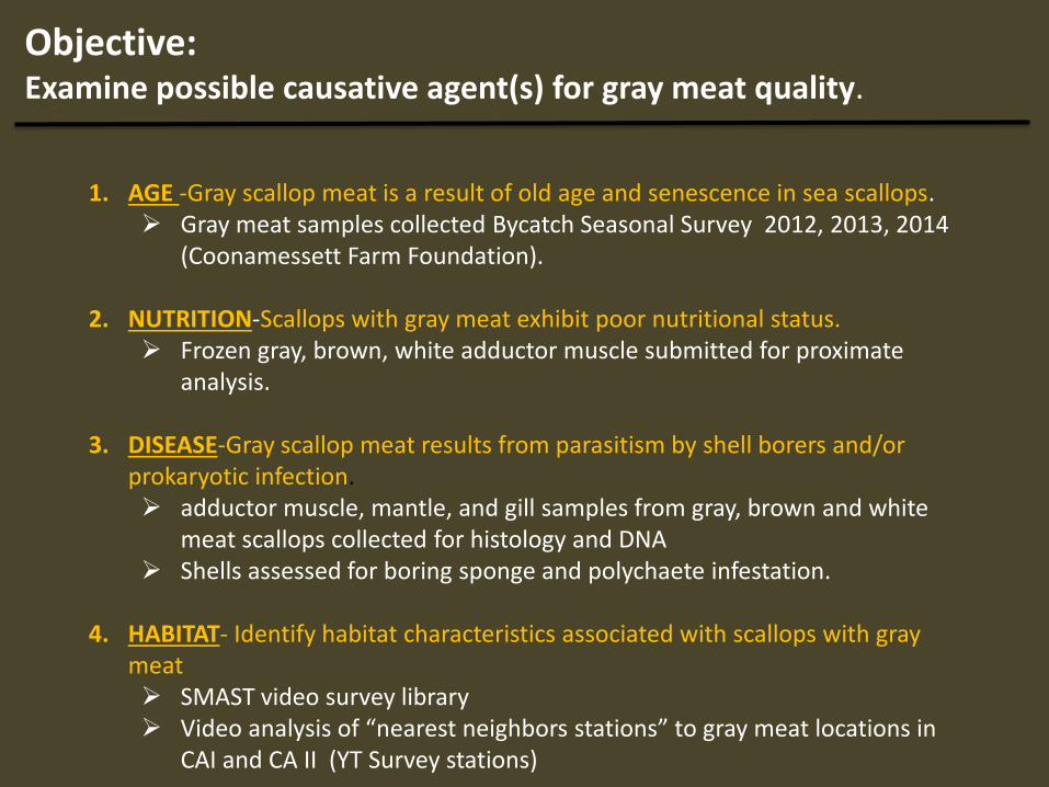
Objective: Examine possible causative agent(s) for gray meat quality.
1. AGE -Gray scallop meat is a result of old age and senescence in sea scallops.
Gray meat samples collected Bycatch Seasonal Survey 2012, 2013, 2014 (Coonamessett Farm Foundation).
2. NUTRITION-Scallops with gray meat exhibit poor nutritional status. Frozen gray, brown, white adductor muscle submitted for proximate
analysis.
3. DISEASE-Gray scallop meat results from parasitism by shell borers and/or prokaryotic infection. adductor muscle, mantle, and gill samples from gray, brown and white
meat scallops collected for histology and DNA Shells assessed for boring sponge and polychaete infestation.
4. HABITAT- Identify habitat characteristics associated with scallops with gray
meat SMAST video survey library Video analysis of “nearest neighbors stations” to gray meat locations in
CAI and CA II (YT Survey stations)

Methods: Scallops with white, brown and gray meat color were collected from the rotational management areas on Georges Bank and analyzed for: 1. Shell height and meat color (n=613)
• Age 2. Reproductive stage (n=395)
• Senescence 3. Shell height : meat weight (n=395)
• Condition/Nutrition 4. Adductor muscle composition (n=88)
• Condition/Nutrition 5. Histopathology and molecular analysis (n=80)
• Disease
Laboratories: Kennebec River Biosciences Dept. of Fish Diseases at the Institute of Pathology, University of Iceland

Sample Collection
Seasonal Bycatch Survey Stations (Coonamessett Farm Foundation Scallop RSA NA13NMF4540011) in Georges Bank where randomly selected scallop samples were collected for shell height (mm): meat weight (g) and meat quality and histopathology (circled stations).
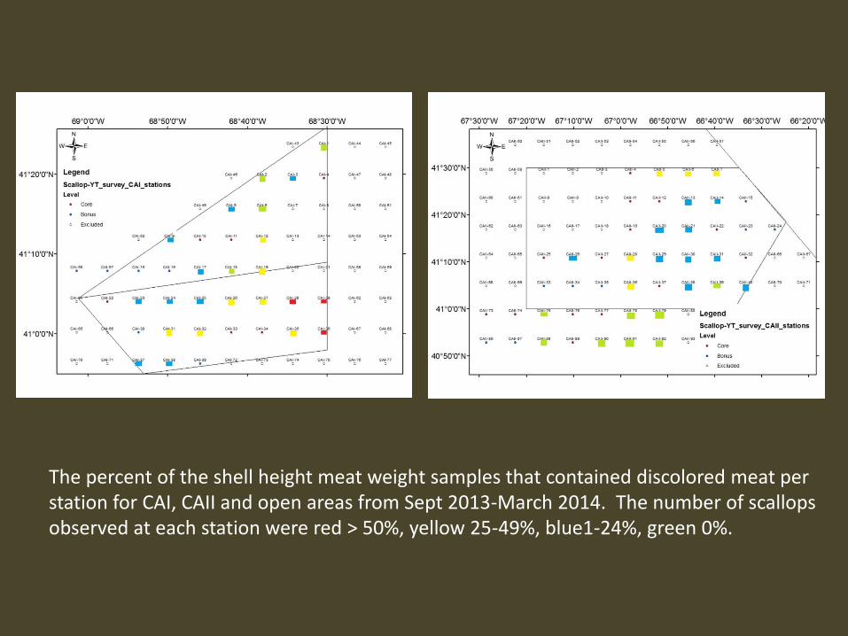
The percent of the shell height meat weight samples that contained discolored meat per station for CAI, CAII and open areas from Sept 2013-March 2014. The number of scallops observed at each station were red > 50%, yellow 25-49%, blue1-24%, green 0%.
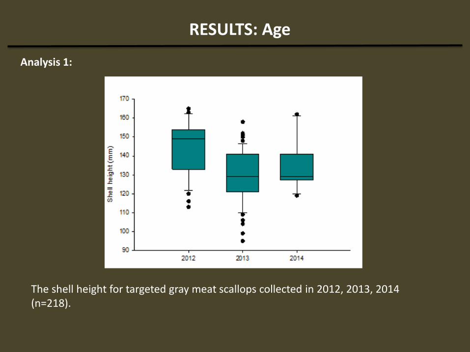
RESULTS: Age
Analysis 1:
The shell height for targeted gray meat scallops collected in 2012, 2013, 2014 (n=218).

RESULTS: Age
Analysis 2:
Pearson's Chi-squared test
Sample
area
X2 df p-value
CA1 209.7 195 0.22
CA2 152.5 130 0.09
Open 85.0 70 0.10
Pearson's Chi-squared test with simulated p-value did not change significance
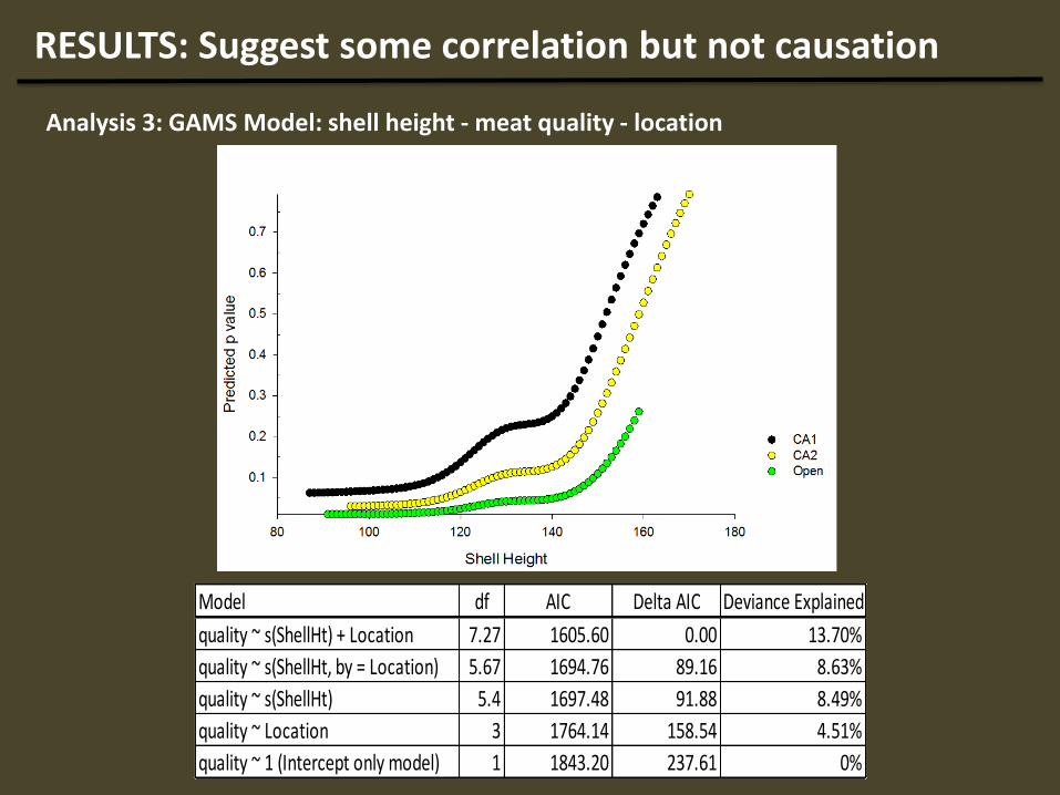
RESULTS: Suggest some correlation but not causation
Analysis 3: GAMS Model: shell height - meat quality - location
Model df AIC Delta AIC Deviance Explained
quality ~ s(ShellHt) + Location 7.27 1605.60 0.00 13.70%
quality ~ s(ShellHt, by = Location) 5.67 1694.76 89.16 8.63%
quality ~ s(ShellHt) 5.4 1697.48 91.88 8.49%
quality ~ Location 3 1764.14 158.54 4.51%
quality ~ 1 (Intercept only model) 1 1843.20 237.61 0%
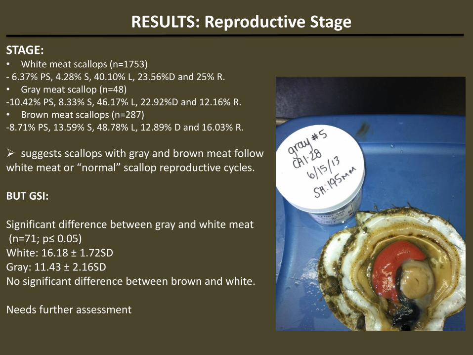
RESULTS: Reproductive Stage
STAGE: • White meat scallops (n=1753) - 6.37% PS, 4.28% S, 40.10% L, 23.56%D and 25% R. • Gray meat scallop (n=48) -10.42% PS, 8.33% S, 46.17% L, 22.92%D and 12.16% R. • Brown meat scallops (n=287) -8.71% PS, 13.59% S, 48.78% L, 12.89% D and 16.03% R.
suggests scallops with gray and brown meat follow white meat or “normal” scallop reproductive cycles. BUT GSI: Significant difference between gray and white meat (n=71; p≤ 0.05) White: 16.18 ± 1.72SD Gray: 11.43 ± 2.16SD No significant difference between brown and white. Needs further assessment
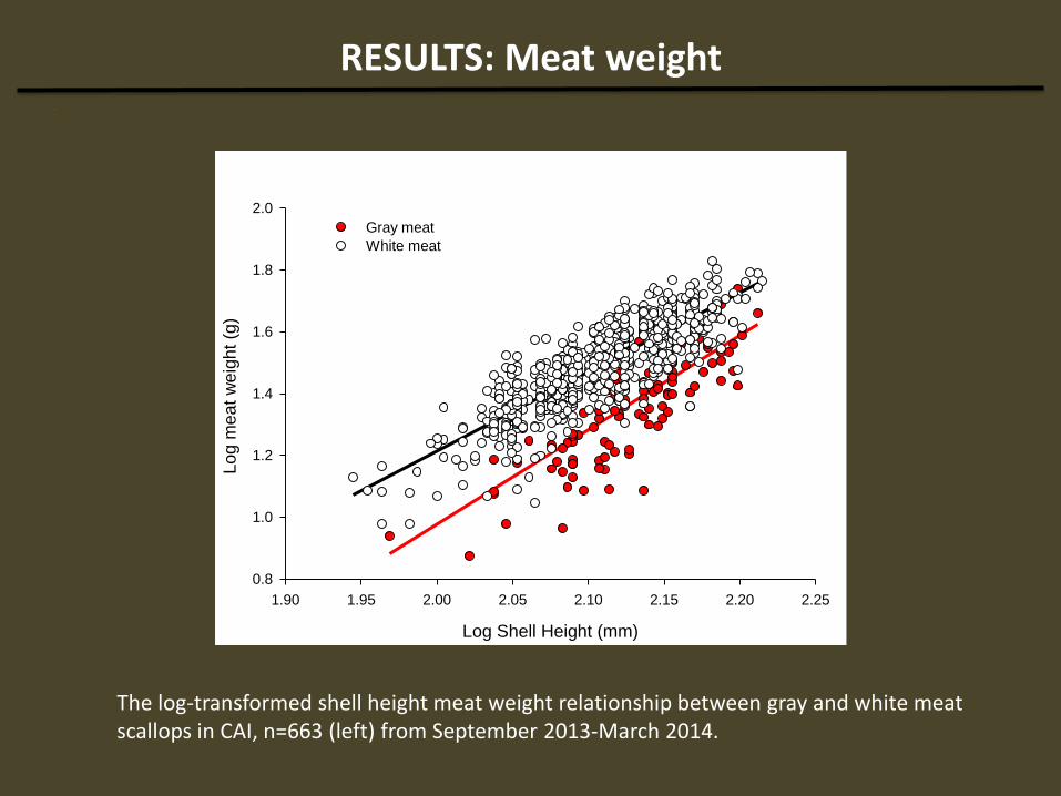
RESULTS: Meat weight
The log-transformed shell height meat weight relationship between gray and white meat scallops in CAI, n=663 (left) from September 2013-March 2014.
Log Shell Height (mm)
1.90 1.95 2.00 2.05 2.10 2.15 2.20 2.25
Log m
eat
weig
ht
(g)
0.8
1.0
1.2
1.4
1.6
1.8
2.0
Gray meat
White meat
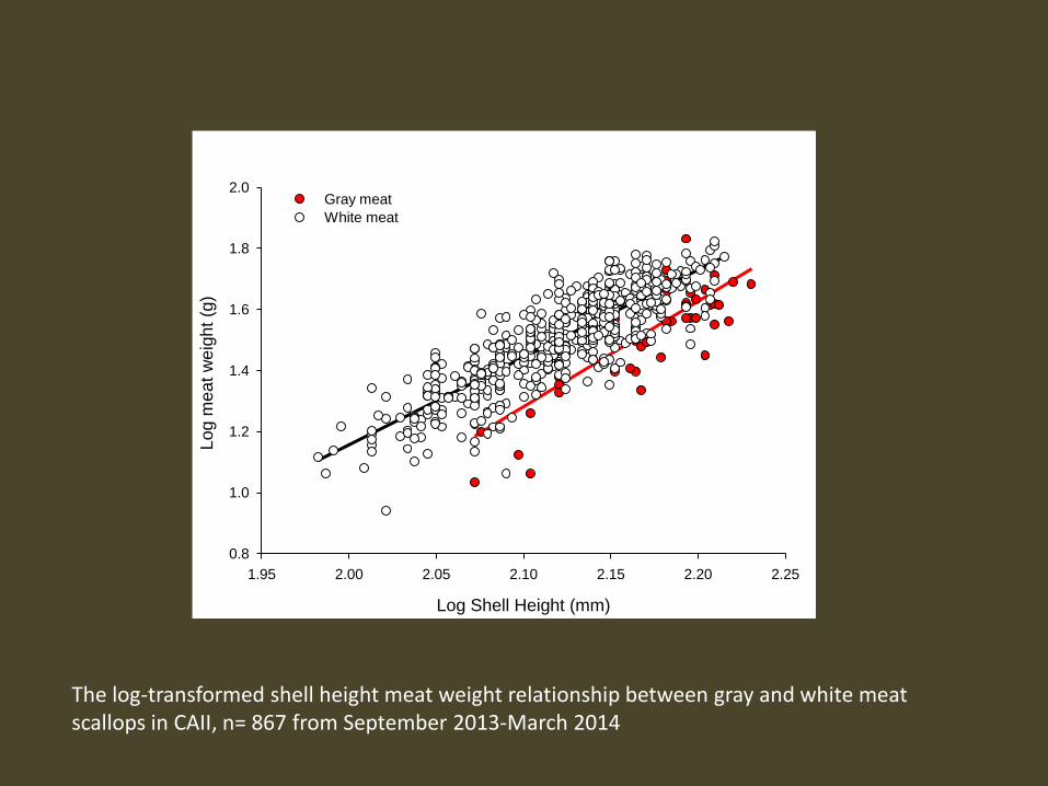
The log-transformed shell height meat weight relationship between gray and white meat scallops in CAII, n= 867 from September 2013-March 2014
Log Shell Height (mm)
1.95 2.00 2.05 2.10 2.15 2.20 2.25
Log m
eat
weig
ht
(g)
0.8
1.0
1.2
1.4
1.6
1.8
2.0Gray meat
White meat
RESULTS: Meat weight
CAI Estimate Std. Error t value Pr(>|t|)
Gray
(Intercept) -4.3815 0.8419 -5.205 6.56E-06 ***
SH 2.674 0.3978 6.722 5.17E-08 ***
White
(Intercept) -3.90039 0.1339 -29.13 <2e-16 ***
SH 2.55677 0.06353 40.24 <2e-16 ***
CAII
Gray
(Intercept) -5.8278 0.8484 -6.869 1.17E-08 ***
SH 3.3891 0.3913 8.66 2.25E-11 ***
White
(Intercept) -4.54568 0.15217 -29.87 <2e-16 ***
SH 2.8523 0.07168 39.79 <2e-16 ***
Sig Significance codes: 0 ‘***’ 0.001 ‘**’ 0.01 ‘*’ 0.05 ‘.’ 0.1 ‘ ’
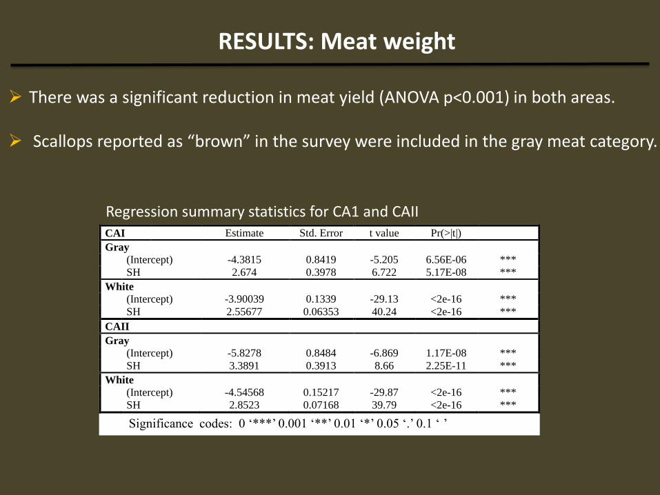
Regression summary statistics for CA1 and CAII
There was a significant reduction in meat yield (ANOVA p<0.001) in both areas. Scallops reported as “brown” in the survey were included in the gray meat category.

RESULTS: Muscle Composition
Map of sample collection locations for proximate analyses. The red shading represents samples from the general area.

RESULTS: Muscle Composition
moi
stur
e:pr
otei
n ra
tio
0
2
4
6
8
10
12
14
16
18
20
white meat
brown meat
gray meat
The moisture : protein ratio (mean percent wet weight ± SD) found in white, brown and gray meat scallops. In Atlantic sea scallops a ratio of 4.0-4.9 is considered “normal” by the FDA and USDA.
RESULTS: Muscle Composition
Analysis White
(n=33)
Brown
(n=26)
Gray
(n=29)
Moisture 77.86 ± 2.56 80.82 ± 2.33 90.33 ± 3.05
Protein 17.68 ± 1.68 14.81 ± 2.57 6.97 ± 1.01
Carbohydrate 2.56 ± 0.87 0.62 ± 0.92 0.08 ± 0.76
Ash 2.90 ± 0.24 3.67 ± 0.14 2.77 ± 0.14
Lipid 0.08 ± 0.02 0.08 ± 0.01 0.03 ± 0.01
M:P 4.40 ± 0.89 6.46 ± 1.36 12.96 ± 4.2
1
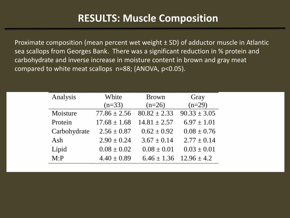
Proximate composition (mean percent wet weight ± SD) of adductor muscle in Atlantic sea scallops from Georges Bank. There was a significant reduction in % protein and carbohydrate and inverse increase in moisture content in brown and gray meat compared to white meat scallops n=88; (ANOVA, p<0.05).

RESULTS: Disease
Infection by newly identified apicomplexan parasite causing severe histopathology in muscle tissue
IDENTICAL rDNA sequence to a new genus and species of apicomplexan parasite observed in 3 other scallop species: 1. Iceland scallop, Chlamys islandica in Icelandic waters (associated with stock collapse ) 2. Queen scallop Aequipecten opercularis, the Faroe Islands (high rates of natural
mortality) 3. King scallop Pecten maximus, West Coast of Scotland (parasite observed in low levels)
Reproduced from Eiriksson et al 2000

From Kristmundsson et al 2011

Name of apicomplexan Host Class/species Infected organs
Bivalve clam
Pseudoklossia pelseneeri1 Donax sp. Kidney
Tellina sp.
P. glomerata2 Tapes floridas
Tapes virginius
Kidney
P. (Merocystis) Tellina tenuis Ovary
Unnamed5 Protothaca staminae Kidney Margolisiella kabatai6
Protothaca staminae Kidney Unnamed7
Tridacna crocea Hemolymph
Bivalve cockle
Pseudoklossia sp.8 Cerastoderma edule Kidney
Bivalve Scallop
P. pectinis9 Pecten maximus Kidney
Many species10 Argopecten irradians Kidney and other organs
Pseudoklossia sp.11 Argopecten irradians Kidney and other organs
P. pectinis-like12 Argopecten irradians Kidney
Unnamed13 Argopecten irradians Kidney and other organs
Margolisiella islandica14 Chlamys islandica Heart
Bivalve mussel
P. semiluna15 Mylilus spp. Kidney
Gastropoda
P. haliotis*,16 Haliotis spp. Kidney
Polyplacophora
P. patellae*,17 Acanthochites Intestine
fasciularis hepatopancreas
P. chitonis*,17
From Kristmundsson et al 2011
Comparison of apicomplexan species described from molluscs
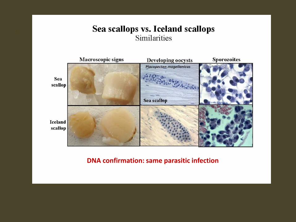
DNA confirmation: same parasitic infection

All apicomplexan stages (both sexual and asexual stages): free sporozoites, trophozoites, meronts, merozoites were found in Placopecten adductor muscle samples
General Apicomplexan Life Cycle Life cycle of this newly identified parasite is not fully
understood

Histopathology
Apicomplexan found in all muscular tissues •Intracellular in muscular tissues
•Free in the extracellular space • Inside hemocytes Adductor muscle most heavily infected tissue Observe hemocyte neoplasia
• “Gray scallops” most heavily infected • Some white scallops lightly infected
Effect of Parasite on Muscle Tissue
Causes severe histopathological changes in adductor muscle as well as other muscular tissues
Focal or disseminated necrosis Observe hyalinization and myoliquefaction of muscle tissues
General sequence of changes observed in muscle tissues :
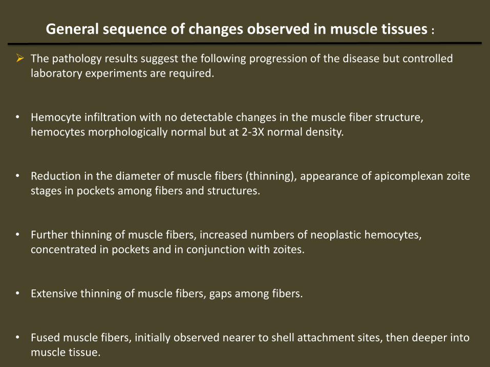
The pathology results suggest the following progression of the disease but controlled laboratory experiments are required.
• Hemocyte infiltration with no detectable changes in the muscle fiber structure,
hemocytes morphologically normal but at 2-3X normal density.
• Reduction in the diameter of muscle fibers (thinning), appearance of apicomplexan zoite stages in pockets among fibers and structures.
• Further thinning of muscle fibers, increased numbers of neoplastic hemocytes, concentrated in pockets and in conjunction with zoites.
• Extensive thinning of muscle fibers, gaps among fibers.
• Fused muscle fibers, initially observed nearer to shell attachment sites, then deeper into muscle tissue.
Adductor Muscle Color Muscle
Degeneration
Hemocyte
Neoplasia
Apicomplexon
Infection
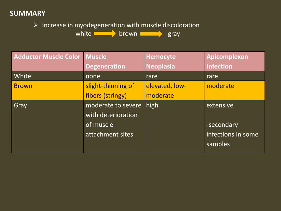
White none rare rare
Brown slight-thinning of
fibers (stringy)
elevated, low-
moderate
moderate
Gray moderate to severe
with deterioration
of muscle
attachment sites
high extensive
-secondary
infections in some
samples
SUMMARY
Increase in myodegeneration with muscle discoloration white brown gray

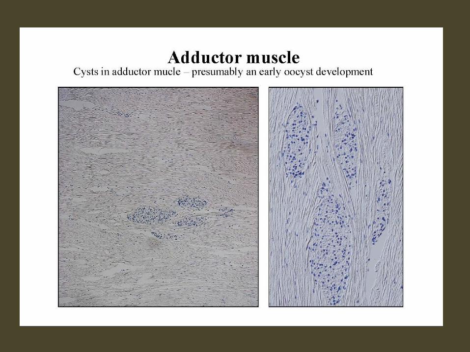


Adductor muscle A cluster of sporozoites associated with muscular degeneration

Muscular necrosis associated with a cluster of sporozoites in adductor muscle


Transmission of Infection
All known apicomplexan stages observed in scallop host Suggests no intermediate host - monoxenous life cycle DIRECT TRANSMISSION
• Pseudofeces? • Decomposition of infected tissue?
Site of entrance via the oral feeding route or gills? Possible autoinfection
Need verification through controlled infection transmission studies.
IMPORTANT AS SCALLOP FISHERMEN SCHUCK AND DISCARD TISSUE AT SEA
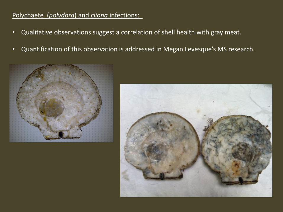
Polychaete (polydora) and cliona infections: • Qualitative observations suggest a correlation of shell health with gray meat.
• Quantification of this observation is addressed in Megan Levesque’s MS research.

RESULTS: Habitat
Gray meat stations were characterized by • a predominantly sand substrate with shell debris. • very few clappers observed • Sea stars were the most abundant predator in gray meat areas.
Percent occurrence of substrate in locations where gray meat were found in Closed Area 1 (CAI) and Closed Area II (CAII) in 2011 and 2012 (n=13 stations and 52 quadrats).
This analysis is being expanded and continued in the 2014 RSA project entitled “Tracking the Occurrence of Gray Meat in Atlantic Sea Scallops, Placopecten magellanicus” and the Saltonstall Kennedy Grant “Combining Fishermen's Knowledge with Oceanographic and Economic Models to Locate and Predict Gray Meat Outbreaks in Atlantic Sea Scallops”
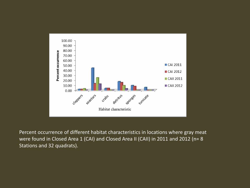
Percent occurrence of different habitat characteristics in locations where gray meat were found in Closed Area 1 (CAI) and Closed Area II (CAII) in 2011 and 2012 (n= 8 Stations and 32 quadrats).
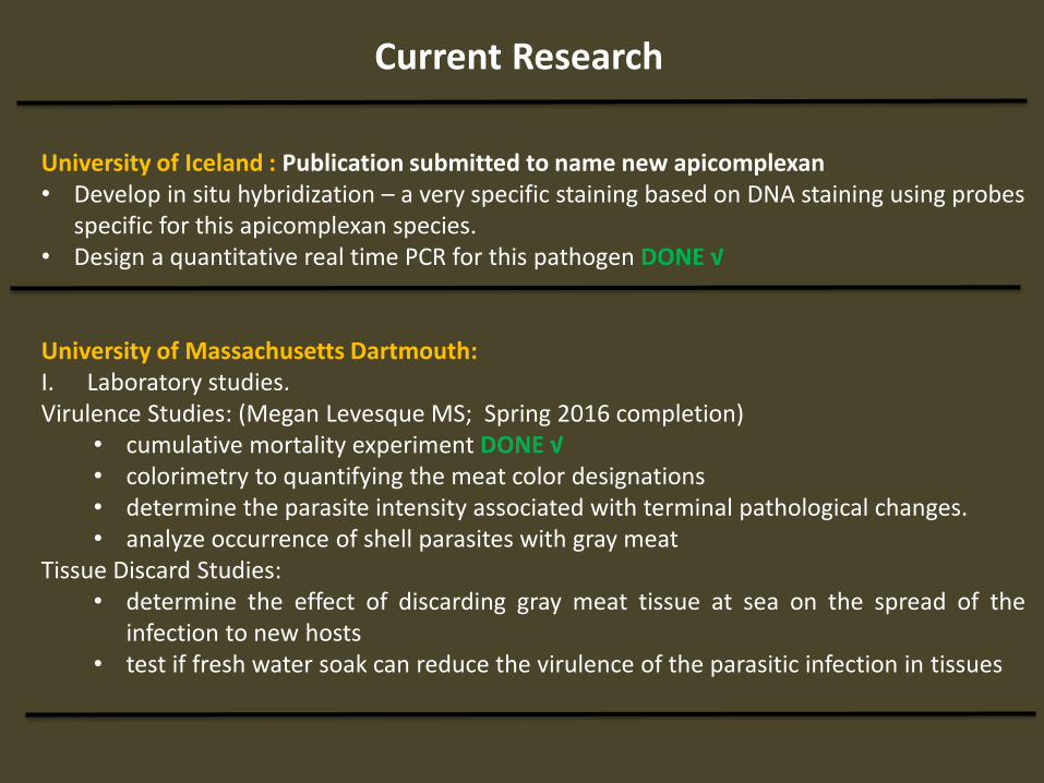
Current Research
University of Iceland : Publication submitted to name new apicomplexan • Develop in situ hybridization – a very specific staining based on DNA staining using probes
specific for this apicomplexan species. • Design a quantitative real time PCR for this pathogen DONE √ University of Massachusetts Dartmouth: I. Laboratory studies. Virulence Studies: (Megan Levesque MS; Spring 2016 completion)
• cumulative mortality experiment DONE √ • colorimetry to quantifying the meat color designations • determine the parasite intensity associated with terminal pathological changes. • analyze occurrence of shell parasites with gray meat
Tissue Discard Studies: • determine the effect of discarding gray meat tissue at sea on the spread of the
infection to new hosts • test if fresh water soak can reduce the virulence of the parasitic infection in tissues
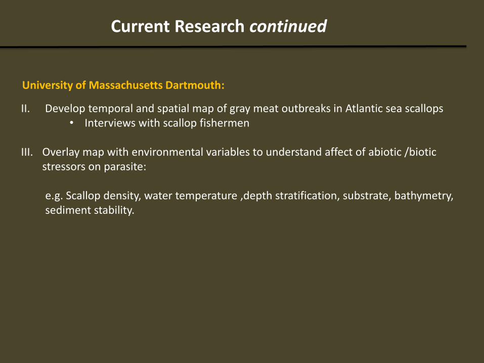
II. Develop temporal and spatial map of gray meat outbreaks in Atlantic sea scallops • Interviews with scallop fishermen
III. Overlay map with environmental variables to understand affect of abiotic /biotic
stressors on parasite: e.g. Scallop density, water temperature ,depth stratification, substrate, bathymetry, sediment stability.
University of Massachusetts Dartmouth:
Current Research continued

IV. Determine range and prevalence of the parasitic infection in Atlantic sea scallops: Canadian waters -Georges Bank Parasite confirmed Gulf of Maine-Inshore Parasite confirmed
Ryan Burt. Alaska Dept. Fish and Game
Note: • Samples from weathervane scallop
Patinopecten caurinus to be tested for apicomplexan

Summary
Gray meat caused by newly identified apicomplexan parasite that
targets all muscle tissue, but concentrates in adductor muscle.
Same parasite found in Iceland, Queen and King scallops with differing stock consequences.
Likely direct transmission of parasite between hosts
possible high impact on fishery. Infection transmission trials required.
Age, nutritional stress as well as secondary infections and shell parasites (cliona and polydora) may be covariates reducing overall fitness.
Combination of physiological and site specific environmental conditions that supports the proliferation and transmission of this parasite likely responsible for “gray meat” outbreaks.
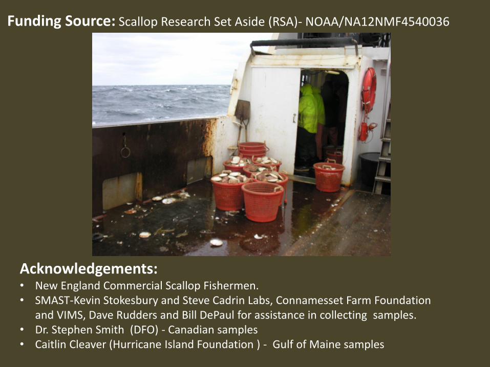
Acknowledgements: • New England Commercial Scallop Fishermen. • SMAST-Kevin Stokesbury and Steve Cadrin Labs, Connamesset Farm Foundation
and VIMS, Dave Rudders and Bill DePaul for assistance in collecting samples. • Dr. Stephen Smith (DFO) - Canadian samples • Caitlin Cleaver (Hurricane Island Foundation ) - Gulf of Maine samples
Funding Source: Scallop Research Set Aside (RSA)- NOAA/NA12NMF4540036

1. Tracking the Occurrence of Gray Meat in Atlantic Sea Scallops, Placopecten magellanicus Scallop RSA : NA14NMF4540080
End Date: April 2016 Kevin D.E. Stokesbury, Susan D. Inglis, Daniel Georgianna
Support for MS Student- Megan Levesque Collaborators- Arni Kristmundsson, Mark A. Freeman: University of Iceland
Kevin St. Martin: Rutgers University
2. Combining Fishermen's Knowledge with Oceanographic and Economic Models to Locate, Evaluate, and Predict Gray Meat Outbreaks in Atlantic Sea Scallops
Saltonstall Kennedy: NOAA-NMFS-FHQ-2015-2004246 End Date: September 2017
Daniel Georgianna, Susan Inglis, Gavin Fay, Kevin Stokesbury: SMAST Kevin St. Martin: Rutgers University Min-Yang Lee: Northeast Fisheries Science Center, NOAA
Collaborators- Arni Kristmundsson, Mark A. Freeman: University of Iceland
Current Funding

Can Infected Scallops Recover? Megan Levesque
Photo: Megan Levesque, 2015
Pilot Study: Nonlethal method to categorize meat color and quality • MgCl₂ Dead Sea Works

Photo: Megan Levesque, 2015
Summary Laboratory Experiment: Tanks: White meat -Full Ration - Half Ration Gray/Brown meat - Full Ration - Half Ration Diet: Full Ration = 2.5 L cultured phytoplankton/day Half Ration= 1.25 L cultured phytoplankton/day
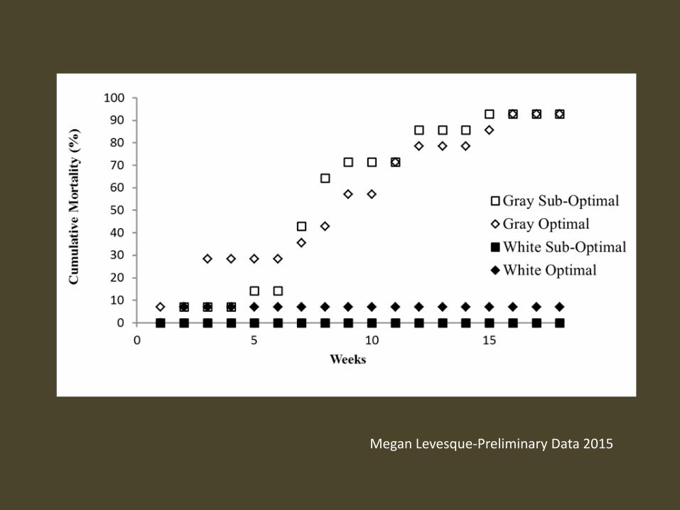
Megan Levesque-Preliminary Data 2015

Tracking Gray Meat Using Fisherman’s Knowledge
Conducted Captain’s Workshop August 2014 to develop interview questions and protocols
28 interviews completed to date (goal 40 interviews)
Fishermen were asked questions about gray meat attributes:
Temporality of gray meat occurrence Causes/environmental conditions Spatiality of gray meat areas (i.e. small or large areas) Severity of problem Changes in fishing practices due to gray meat Management practices—causes and solutions Direction for scientists
Attribute Information provided about each location

Preliminary Results:
• 116 individual gray meat locations assessed so far
0
10
20
30
40
50
60
Recent (2011-2014) Access area (2000-2010)
Closed areas (1994-1999)
Open access (pre-1994)
Poin
t co
un
t
Preliminary number of results for gray meat locations reported per period

QUESTIONS