sea scallop research noaa grant number: noaa...
TRANSCRIPT

Final Report
Sea Scallop Research
NOAA Grant Number: NOAA/NA12NMF4540036
Award Date: 2/10/2012
Start Date: 5/01/2012
End Date: 4/30/2013
Extended End Date: 4/30/2014
Project Title: What Causes Gray Meat in the Atlantic Sea Scallop
Placopecten magellanicus in Georges Bank Closed Areas?
Principal Investigator: Kevin D. E. Stokesbury, Ph.D. and Susan D. Inglis
Address: School for Marine Science and Technology,
University of Massachusetts Dartmouth,
200 Mill Road Suite 325
Fairhaven, MA, 02719
Phone: (508) 910-6373
Fax: (508) 910-6374
Email: [email protected]
Amount: We were granted (40,323 lbs) from the Open Area ($379,843) for research and
compensation.
2
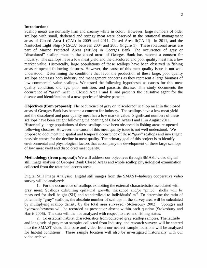
Introduction: Scallop meats are normally firm and creamy white in color. However, large numbers of older
scallops with small, darkened and stringy meat were observed in the rotational management
areas of Closed Area I (CAI) in 2009 and 2011, Closed Area II(CA II) in 2011, and the
Nantucket Light Ship (NLSCA) between 2004 and 2005 (Figure 1). These rotational areas are
part of Marine Protected Areas (MPAs) in Georges Bank. The occurrence of gray or
“discolored” scallop meat in the closed areas of Georges Bank has become a concern for
industry. The scallops have a low meat yield and the discolored and poor quality meat has a low
market value. Historically, large populations of these scallops have been observed in fishing
areas re-opened following closures. However, the cause of this meat quality issue is not well
understood. Determining the conditions that favor the production of these large, poor quality
scallops addresses both industry and management concerns as they represent a large biomass of
low commercial value scallops. We tested the following hypotheses as causes for this meat
quality condition; old age, poor nutrition, and parasitic disease. This study documents the
occurrence of “gray” meat in Closed Area I and II and presents the causative agent for the
disease and identification of a new species of bivalve parasite.
Objectives (from proposal): The occurrence of gray or “discolored” scallop meat in the closed
areas of Georges Bank has become a concern for industry. The scallops have a low meat yield
and the discolored and poor quality meat has a low market value. Significant numbers of these
scallops have been caught following the opening of Closed Areas I and II in August 2011.
Historically, large populations of these scallops have been observed in fishing areas re-opened
following closures. However, the cause of this meat quality issue is not well understood. We
propose to document the spatial and temporal occurrence of these “gray” scallops and investigate
possible causes for the decline in meat quality. The primary goal of this project is to identify
environmental and physiological factors that accompany the development of these large scallops
of low meat yield and discolored meat quality.
Methodology (from proposal): We will address our objectives through SMAST video digital
still image analysis of Georges Bank Closed Areas and whole scallop physiological examination
collected from the rotational access areas.
Digital Still Image Analysis: Digital still images from the SMAST–Industry cooperative video
survey will be analyzed:
1. For the occurrence of scallops exhibiting the external characteristics associated with
gray meat. Scallops exhibiting epifaunal growth, thickened and/or “pitted” shells will be
measured for shell height (SH) and standardized to individuals’ m-2
. To determine the ratio of
potentially “gray” scallops, the absolute number of scallops in the survey area will be calculated
by multiplying scallop density by the total area surveyed (Stokesbury 2002). Sponges and
hydrozoa/bryozoa will be recorded as present or absent within each quadrat (Stokesbury and
Harris 2006). The data will then be analyzed with respect to area and fishing status.
2. To establish habitat characteristics from collected gray scallop samples. The latitude
and longitude of gray meat samples collected from Industry, and research surveys will be entered
into the SMAST video data base and video from our nearest sample locations will be analyzed
for habitat conditions. These sample location will also be investigated historically with our
video archive.
3
Whole Scallop Examination: In cooperation with commercial scallop fishing vessels, and
research surveys, whole scallop samples, grossly representing gray meat scallops, will be
collected from Georges Bank rotational areas and donated to the project. The date, location,
depth and general observations on the condition of the catch and other organisms found in the
dredge will be recorded.
Each scallop will be externally examined and photographed for evidence of shell boring
activity. The shell thickness and height will be recorded and the scallop dissected. Following the
methodology of Sarro and Stokesbury (2009), the shell height/meat weight and gonadal somatic
index will be calculated. The meat will be analyzed for proximate composition (lipid, protein,
water and ash content) using Official Methods of Analysis (1990) to determine the nutritional
status of the animal. Reproductive tissues will be prepared to determine the presence of
senescence. The meat will also be prepared for histological examination for myodegenerative
changes in the muscle indicating both nutritional stress and possible proykaryotic infection. The
top and ventral shells will be examined by radiography for extent and type of shell boring
activity and a sub sampled aged using δ 18
O carbonate analysis.
Time Line: Samples were collected from Industry during the spring and summer 2012 and from
the Yellowtail Bycatch survey of Georges Bank from May 2012 through March 2013 for
analysis.
Figure 1: Map of Georges Bank Closed Areas showing Closed Area I (CAI), Closed Area II
(CAII) and Nantucket Lightship (NLCA). The areas shaded in light blue represent the rotational
management access areas.

4
Methods:
Whole Scallop Examination for Causative Agent:
Scallop samples, frozen whole for gray meat analysis were collected from Georges Bank
Closed Areas I and II, the Northern Edge. These samples were obtained opportunistically from
commercial scallop fishermen and research survey trips including the Scallop RSA survey of the
Northern Edge by the Virginia Institute of Marine Sciences. We also collected samples monthly
from the Scallop RSA Project-Yellowtail Bycatch Survey of Georges Bank (12-SCA-06) starting
May 2012.
These monthly samples included:
A random sample of thirty scallops with shell height (SH) > 145mm frozen whole from
stations in Closed Area 1 South (CA1 S), Closed Area 1 North(CA1 N) and Closed Area
II (CA2) (10 from each area). These samples were dissected while frozen, meat quality
(color) recorded and the adductor muscle shipped to the New Jersey Feed Laboratory for
proximate analysis to determine the water, lipid, ash, protein and carbohydrate content of
each tissue to test the nutrition hypothesis.
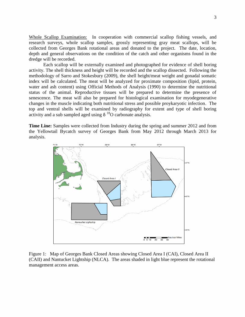
Twenty samples from confirmed gray meat scallops for pathological and histological
analysis (Figure 2). Each adductor muscle and gonad was preserved in formalin and the
corresponding shell labeled for subsequent analysis (Figure 8B). The shells from these
samples were treated in a fresh water bath to collect polychaetes present in the shell.
Tissue and polychaete samples were sent to the Kennebec River Biosciences Laboratory
in Maine and Dept. of Fish Diseases at the Institute of Pathology, University of Iceland
the University of Iceland Fish Pathology Laboratory for identification and analyses to test
the disease hypothesis and provide shell height data for the age hypothesis.
Data collected from stations selected for shell height/meat weight analysis for the
Yellowtail Bycatch Survey were also analyzed for meat quality in 2013 (Figure 3). A
bushel of randomly selected scallops from each tow in designated stations were collected
and processed for shell height (mm): meat weight (g) and meat quality. This data set
was used to map the occurrence of gray meat scallops in Closed Area I and II and
document the shell height frequency associated with the discolored meat to test the age
hypothesis.
5
Figure 2. Stations in Georges Bank where gray meat samples were collected for
histopathology (circled stations).
Figure 3. The YT Bycatch Survey Stations (Coonamessett Farm Foundation) in the Georges
Bank Closed Area I and Closed Area II study area. These stations were sampled monthly for
shell height:meat weight (n=12 samples per assigned station). Meat quality was added to the
analysis in 2013.

6
Digital Still Image Analysis:
The latitude and longitude of gray meat samples collected from Industry and research
surveys were entered into a “nearest neighbor” query for the SMAST video data base which
identified video survey results for the “gray meat” locations. Video footage was analyzed
following Stokesbury 2002, and Stokesbury and Harris 2006. The number of clappers and mega
fauna representing potential predators such as sea stars, crabs, sponges and tunicates were
identified.
Substrate sediments were visually identified following the Wentworth particle grade scale
from the video images, where the sediment particle size categories were based on a doubling or
halving of the fixed reference point of 1 mm; sand = 0.0625 to 2.0 mm, gravel = 2.0 to 256.0 mm
and boulders > 256.0 mm (Lincoln et al. 1992). Gravel was divided into two categories,
granule/pebble = 2.0 to 64.0 mm and cobble = 64.0 to 256.0 mm (Lincoln et al. 1992). Shell
debris and detritus were also identified.
The habitat characterization of the gray meat areas was determined by the percent
absence: presence of each of these sediment and mega fauna categories.
Results:
Digital Still Image Analysis:
Results from the video analysis of “nearest neighbors” stations to gray meat locations in
Closed Area I and II for 2011 and 2012 are presented in Figures 4 and 5. This analysis is being
expanded and continued the 2014 RSA project entitled “Tracking the Occurrence of Gray
Meat in Atlantic Sea Scallops, Placopecten magellanicus”. Subsequent results that include
physical characteristics of gray meat habitat including scallop density and bottom water
temperature will be presented in the new RSA project reports.
Gray meat stations were characterized by a predominantly sand substrate with shell
debris. There were very few clappers observed that would indicate natural mortality events. Sea
stars were the most abundant predator in gray meat areas.
Figure 4. Percent occurrence of substrate in locations where gray meat were found in Closed
Area 1 (CAI) and Closed Area II (CAII) in 2011 and 2012 (n=13 stations and 52 quadrats).

7
Figure 5. Percent occurrence of different habitat characteristics in locations where gray meat
were found in Closed Area 1 (CAI) and Closed Area II (CAII) in 2011 and 2012 (n= 8 Stations
and 32 quadrats).
Distribution
Result for the distribution of gray meat scallops in Georges Bank Closed Area I and
Closed Area II are presented in Figures 6 and 7. Gray meat scallops are found throughout
Closed Area I with the greatest occurrence in the southeast portion where over 50% of the
scallops sampled had discolored meat. This is consistent with reports from fishermen. Although
not as concentrated as in Closed Area I, gray meat scallops are also found in high concentrations
(25-49%) throughout Closed Area II. From this study the condition does not seem to have
moved into open area south of Closed Area II yet.
Gray meat scallops (n=71) were also found in “pockets” in the Nantucket Lightship
access area during a fall and spring survey conducted for the RSA Project - Combined high-
resolution video survey and biological sampling using a modified sled dredge of the sea scallop
resource in Nantucket Lightship Access area (Figure 8A). .

8
Figure 6. The percent of the sample (bushel) that contained gray meat per station for Closed Area
I Sept 2013-March 2014 surveys > 50%, 25-49%, 1-24%, 0%.
Figure 7. The percent of the sample (bushel) that contained gray meat per station for Closed Area
II and Open Areas Sept 2013-March 2014 surveys > 50%, 25-49%, 1-24%, 0%.

9
Figure 8. Examples of gray meat scallops from the Nantucket Lightship access area (A) and
Closed Area 1 (B).
Age:
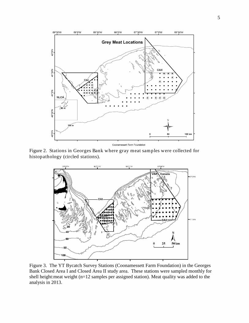
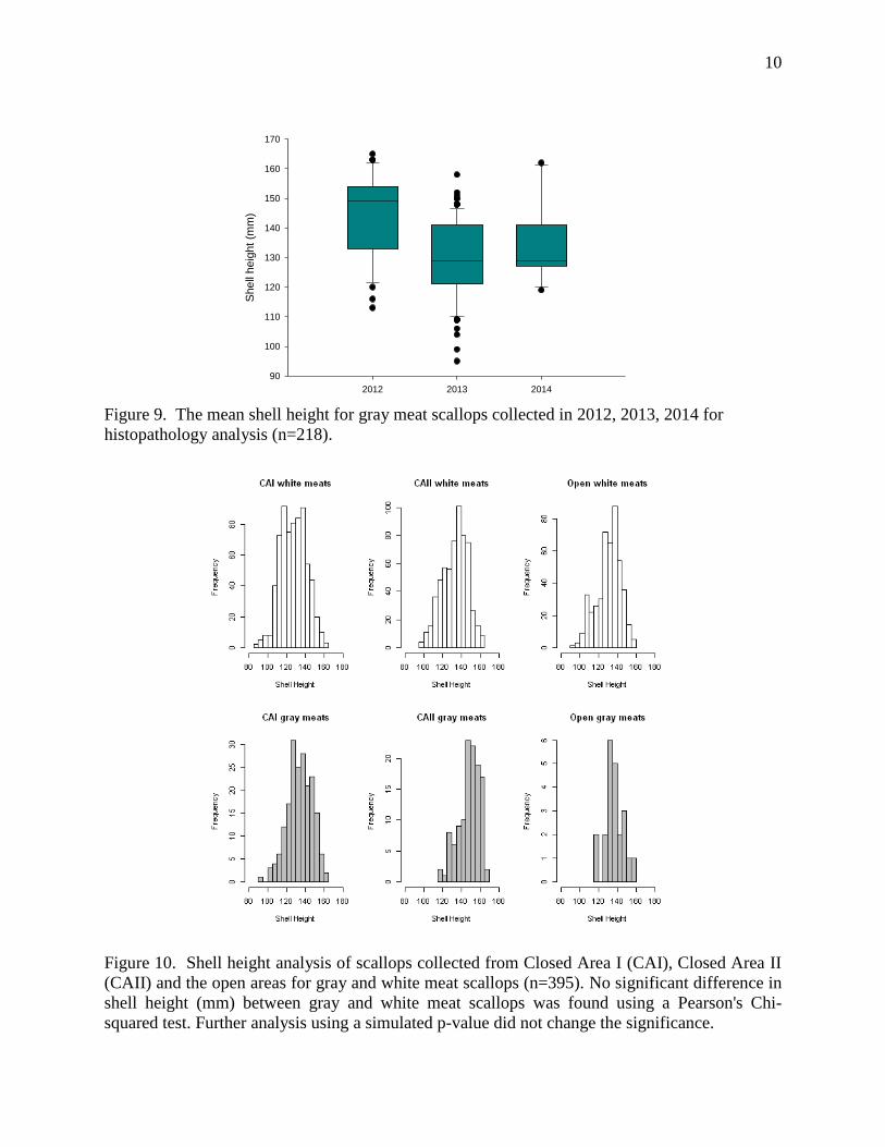
Results from shell height analysis of scallop samples collected specifically for pathology
and histology (n=218) did not show a significant difference in shell height (mm) with gray meat
occurrence (Figure 9). These findings were confirmed in the random samples collected for shell
height meat weight analysis (Figure 10; n=395). A Pearson’s chi-squared test found no
significant difference between the shell height (mm) of white and gray meat scallops in Closed
Area 1, Closed Area II and Open Area of Georges Bank. Running the test with a simulated p
value did not change the significance result. Scallop age is associated with shell height. Thus,
these finding suggest that the gray meat condition is not directly caused by old age.
Pearson's Chi-squared test:
CA1: X-squared = 209.6667, df = 195, p-value = 0.2241
CA11: X-squared = 152.5278, df = 130, p-value = 0.08621
Open Area: X-squared = 85, df = 70, p-value = 0.107
We were unable to use stable isotope δ18
O analysis to age the gray meat scallops we
collected due to the poor condition of the shell. Stable isotope analysis of the shell carbonate is
compromised by contamination by boring organism tissue and debris. The gray meat scallops
often exhibited extensive polychaete infestation of the shell. The University of Michigan Stable
Isotope Laboratory is working on a process to extract the contamination from the shell carbonate
samples but these results are not available at this time.
A
B
10
2012 2013 2014
Shell
heig
ht
(mm
)
90
100
110
120
130
140
150
160
170
Figure 9. The mean shell height for gray meat scallops collected in 2012, 2013, 2014 for
histopathology analysis (n=218).
Figure 10. Shell height analysis of scallops collected from Closed Area I (CAI), Closed Area II
(CAII) and the open areas for gray and white meat scallops (n=395). No significant difference in
shell height (mm) between gray and white meat scallops was found using a Pearson's Chi-
squared test. Further analysis using a simulated p-value did not change the significance.

11
Nutrition:
Samples were tested at the New Jersey Feed Laboratory for percent fat, carbohydrate,
protein, moisture and ash in the muscle tissue using standard analytical methods AOAC (1990).
The samples were categorized as white, brown, or gray adductor meat color. There was a
significant decrease the protein content of scallops with discolored meat, and the darker the meat
color (brown to gray) the greater the decrease in protein content. There was also a reduction in
carbohydrate and lipid levels in gray meat scallops (Figure 11). Lipid levels were low in all meat
samples which is consistent with scallops that preferentially use carbohydrate for energy
(Shumway and Parsons, 2012).
In 1992, the USA Food and Drug Administration (FDA) determined that scallops with a
total moisture content of 80% or less, if not subjected to processing conditions utilizing
excessive water and/or phosphate treatment, could be labeled simply as “scallops”.
Moisture:protein ratios are used as indicators of conditions representing “water-added” when
marketing scallop meats and are carefully monitored by the FDA and US Department of
Agriculture (USDA). In Atlantic sea scallops the moisture:protein ratio is considered to be 4.0
to 4.9 (Lampilla, 1993). Thus, a ratio about 5.1 would indicate a product with added water or
“altered”. Scallops with gray meat have a significantly higher moisture:protein ratio of 12.9
(±4.2), with brown meat scallops at 6.46 (±1.36) (Figure 12). These results generally indicate
nutritional stress in individuals due to either a reduction in energy intake or assimilation, or an
extra metabolic burden. However, both of these conditions can also be caused by a disease
process.
Figure 11. Proximate analysis of scallop adductor muscle (mean ± SD) found a significant
reduction in % protein and carbohydrate and increase in moisture content in gray meat scallops
n=68; (ANOVA: p>0.05).
Moisture Lipid Protein Ash Carbohydrate
% t
issu
e c
om
po
sitio
n
0
20
40
60
80
100
white meat
brown meat
gray meat

12
Figure 12. The moisture:protein ratio found in white, brown and gray meat scallops. In Atlantic
sea scallops a ratio of 4.0-4.9 is considered “normal” by the FDA and USDA.
A controlled laboratory experiment conducted to determine the effects of food
availability on growth and shell isotopic signatures in Atlantic sea scallops resulted in a finding
of gray meat scallops in the reduced ration group. This study maintained scallops from Georges
Bank Closed Area 1 of approximately 100 mm shell height in a controlled laboratory setting on
either a full or reduced ration of a commercial Shellfish diet (Reed ShellFish Diet). There was a
significant difference in meat quality between the experimental groups, with the reduced ration
groups presenting with small, soft, and in some instances discolored meat. We observed five
gray meat scallops out of twenty in this reduced ration group. These scallops did not show any
signs of boring parasite infection in the shells. The appearance of gray meat in this experiment
was an unexpected result, and suggested that poor nutrition may be a physiological factor or
stressor in scallops exhibiting gray meat (Figure 13).
Figure 13. Examples of meat quality observations in the diet studies; A. Gray meat in reduced
ration group, B. White meat from full ration group.
mois
ture
:pro
tein
ratio
0
2
4
6
8
10
12
14
16
18
20
white meat
brown meat
gray meat
A B

13
Disease:
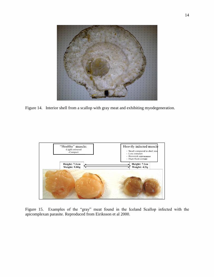
Results from the shell analysis show a high density of polychaete, Polydora species
activity (Figure 14). The level of infestation was moderate to high among the majority of the
shells from large scallops (shell height > 150 mm) with gray meat. Burrow holes were present on
both valves, but at a higher density on the left valves. In the more advanced stages of infestation,
polychaete burrows had penetrated the scallop shell and formed lesions on the interior surface.
These areas and adductor muscle attachment sites were marked by conchiolin deposition.
Conchiolin is secreted by the mantle in response to shell damage. Interior shell morphology
suggested possible partial deterioration of muscle attachment sites in some of the samples. Mud
blisters were also visible along the interior shell wall in more advanced infections, observed in
approximately 30% of samples.
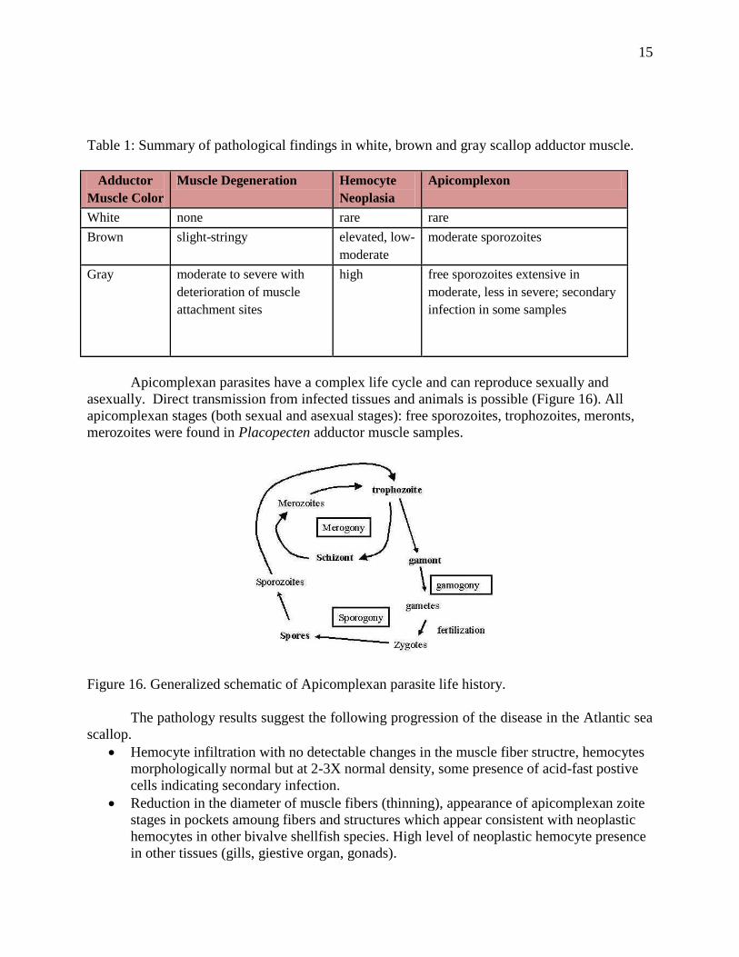
Histological analysis of the adductor muscle (n=85) exhibited degeneration of muscle
fibers (myodegeneration ) in all gray meat samples at ‘moderate to extensive’ level (per Gulka et
al. 1983) and the presence of a previously unknown apicomplexan parasite. This parasite is
similar to an apicomplexan parasite identified in the Iceland Scallop Chlamys islandica and was
the cause of a mass mortality event in the year 2000 (Figure 15; Eiriksson et al 2010,
Kristmundsson et al 2011). Research conducted by Dr. Arni Kristmundsson with the
Department of Fish Diseases at the Institute of Pathology, University of Iceland and Dr. Mark
Frement with the Institute of Biological Science at the University of Malaya in Malaysia
identified this new apicomplexan species (parasite), which caused visible gross pathology and
extensive muscular necrosis or myodegeneraion in the Iceland scallop. In the case of the Iceland
scallop, high summer sea bottom temperatures were thought to have made the scallops
susceptible to this parasitic infection (Eiriksson et al 2013). This parasite is non zoonotic and
only affects bivalves. It has also been found in the queen scallop, Aequpecten opercularis, L and
king scallop, Pecten maximus.
In the Atlantic sea scallop increased muscle discoloration, from white to brown to gray,
was associated with increased muscle degeneration and parasite intensity. The parasite was
found in high intensity in all gray meat samples, in moderate intensity in brown meat scallops
and rarely in white meat scallops (Table 1).
14
Figure 14. Interior shell from a scallop with gray meat and exhibiting myodegeneration.
Figure 15. Examples of the “gray” meat found in the Iceland Scallop infected with the
apicomplexan parasite. Reproduced from Eiriksson et al 2000.
15
Table 1: Summary of pathological findings in white, brown and gray scallop adductor muscle.
Adductor
Muscle Color
Muscle Degeneration Hemocyte
Neoplasia
Apicomplexon
White none rare rare
Brown slight-stringy elevated, low-
moderate
moderate sporozoites
Gray moderate to severe with
deterioration of muscle
attachment sites
high free sporozoites extensive in
moderate, less in severe; secondary
infection in some samples
Apicomplexan parasites have a complex life cycle and can reproduce sexually and
asexually. Direct transmission from infected tissues and animals is possible (Figure 16). All
apicomplexan stages (both sexual and asexual stages): free sporozoites, trophozoites, meronts,
merozoites were found in Placopecten adductor muscle samples.
Figure 16. Generalized schematic of Apicomplexan parasite life history.
The pathology results suggest the following progression of the disease in the Atlantic sea
scallop.
Hemocyte infiltration with no detectable changes in the muscle fiber structre, hemocytes
morphologically normal but at 2-3X normal density, some presence of acid-fast postive
cells indicating secondary infection.
Reduction in the diameter of muscle fibers (thinning), appearance of apicomplexan zoite
stages in pockets amoung fibers and structures which appear consistent with neoplastic
hemocytes in other bivalve shellfish species. High level of neoplastic hemocyte presence
in other tissues (gills, giestive organ, gonads).

16
Further thinning of muscle fibers, increased numbers of neoplastic hemocytes,
concentrated in pockets and in conjucntion with zoites.
Extensive thinning of muscle fibers, gaps among fibers with debris, greater observation
of other structures such as acid-fast cells and other potential apicomplaxan stages.
Fused muscle fibers, initally observed nearer to shell attachment sites, then deeper into
muslc tissue, decreased hemocyte number and near absence in fused areas.
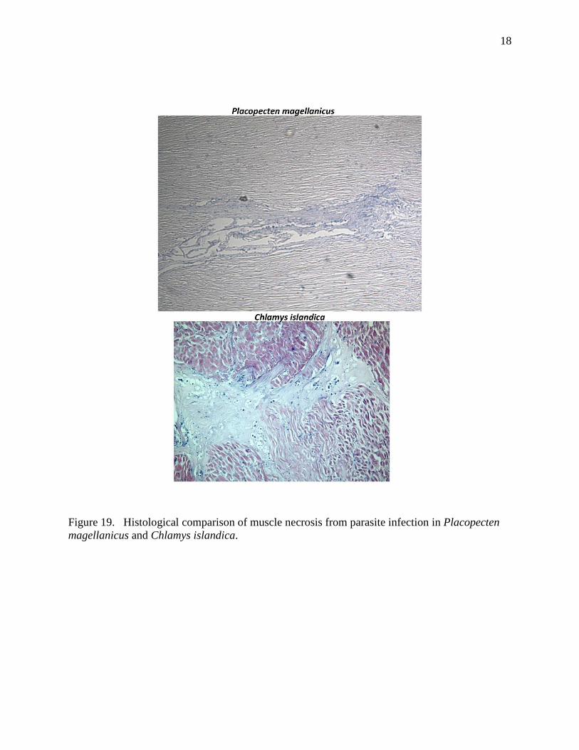
A histological comparison of the parasite infecting Placopecten magellanicus and
Chlamys islandica found the morphology of sporozoites and effect on muscle tissue to be
identical (Figures 18, 19, 20). DNA results on the apicomplexan parasite found in the Atlantic
sea scallop indicate it is conspecific with the parasite found in the Iceland, Queen, and King
scallop. This has now been recognized as a new species of bivalve parasite.

17
Figure 18. Histological comparison of parasite infecting Placopecten magellanicus and
Chlamys islandica. The morphology of sporozoites appears identical.
18
Figure 19. Histological comparison of muscle necrosis from parasite infection in Placopecten
magellanicus and Chlamys islandica.

19
Figure 20. Zoite stages of parasite within adductor muscle fibers.
Results from this study indicate that gray meat in the Atlantic sea scallop is caused by an
apicomplexan parasite infection. Age, nutritional stress, as well as secondary infections and
shell parasites such as Polydora sp. may be covariates of this condition by reducing the fitness of
the scallop and thus the scallop’s ability to control the parasite infection and subsequent disease
process. Therefore, a combination of physiological and site specific environmental conditions
that supports the proliferation and transmission of this parasite is likely responsible for the
outbreaks of “gray meat” in Georges Bank scallop stocks. Unfortunately, we currently do not
understand the interactions between these variables well enough to be able to predict these
events. We hope to continue and expand this research to address this question.

20
Outreach and Publications:
Susan Inglis presented the results of this project to the Scallop Plan Development Team (PDT)
on April 8 2014, Woods Hole, MA. These data have also been presented at the SMAST
Fishermen’s Steering Committee meetings and to the public at the 2012 High School Marine
Science Symposium in New Bedford, MA.
Publications:
A paper entitled “ The apicomplexan parasite, parasarcocystis pectinis, and gray meat in
the Atlantic sea scallop Placopecten magellanicus” is in preparation for the Journal: Diseases of
Aquatic Organisms.
Conferences/Seminars:
1. Inglis, S.D., Kristmundsson, A and K.D.E. Stokesbury. What Causes Gray Meat in the
Atlantic Sea Scallop Placopecten magellanicus in Georges Bank Closed Areas? Northeast
Fisheries Science Center-Invertebrate Subcommittee. Sea Scallop Stock Assessment
Meeting (SARC-59). Woods Hole, MA. March 17-21.
2. Inglis, S.D. What Causes Gray Meat in the Atlantic Sea Scallop Placopecten magellanicus in
Georges Bank Closed Areas? Department of Fisheries and Ocean Sciences Seminar Series.
University of Massachusetts, Dartmouth. Fairhaven, MA. April 30 2014.
3. Inglis, S.D. and K.D.E. Stokesbury. What Causes Gray Meat in the Atlantic Sea Scallop
Placopecten magellanicus in Georges Bank Closed Areas? 44th
American Fisheries Society
Annual Meeting, Quebec City, Quebec. Poster Presentation. August 18-22. 2014.
4. Kristmundsson, A, Inglis, S.D., Stokesbury, K.D.E. and M.A. Freeman. Apicomplexan
infection of the Atlantic sea scallop Placopecten magellanicus. Seventh International
Symposium on Aquatic Animal Health, Portland , Oregon , August 31-September 4th
2014.
Literature Cited
Association of Official Analytical Chemists. 1990. Official methods of analysis. 15th
edition, Vol. 2, Arlington, AOAC. 1298 pp.
Eiriksson,H., Thorarinsdottir,G.G., Jonasson,J.P.,and A. Kristmundsson. 2010. Increase in
natural mortality of the Iceland Scallop (Chlamys Islandica) in West Iceland and collapse
of the fishery in the early 2000s. ICES CM: 20.
Gulka, G., Change, P.W., and K.A. Marti. 1983. Prokaryotic infection associated with a mass
mortality of the sea scallop, Placopecten magellanicus. J Fish Dis 6:355-364.
Kristmundsson, A., Helgason, S., Bambir, S.H., Eydal, M., and M.A. Freeman. 2011. Previously
unknown apicomplexan species infecting the Iceland scallop Chlamys Islandica (Muller,
1776), a queen scallop, Aequpecten opercularis, L and king scallop, Pecten maximus,L. J.
Invertebr. Pathol., 108. 147-155.
21
Lincoln, R.J., G.A. Boxshall, and P.F. Clark. 1992. A dictionary of ecology, evolution and
systematics, Vol. Cambridge University Press, Cambridge.
Naidu KS. 1970. Reproduction and breeding cycle of the giant scallop Placopecten magellanicus
(Gmelin) in Port au Port Bay, Newfoundland. Canadian Journal of Zoology, 48(5): 1003-
1012.
Sarro, C. and K.D.E. Stokesbury. 2009. Spatial and temporal variation in the shell height/meat
weight relationship of the sea scallop (Placopecten magellanicus) in the Georges Bank
fishery. J Shellfish Res 28:1-7
Shumway, S.E and J. Parsons. 2012. Scallops: Biology, Ecology and Aquaculture:
Biology, Ecology and Aquaculture.
Stokesbury, K. D. E. 2002. Estimation of sea scallop abundance in closed areas of Georges
Bank, USA. Transactions of the American Fisheries Society 131: 1081-1092.
Stokesbury, K.D.E., and B.P. Harris. 2006. Impact of limited short-term sea scallop fishery on
epibenthic community of Georges Bank closed areas. Mar Ecol Prog Ser 307: 85-100.