what can flies tell us about copper homeostasis?
TRANSCRIPT
1346 Metallomics, 2013, 5, 1346--1356 This journal is c The Royal Society of Chemistry 2013
Cite this: Metallomics,2013,5, 1346
What can flies tell us about copper homeostasis?
Adam Southon,a Richard Burkeb and James Camakaris*a
Copper (Cu) is an essential redox active metal that is potentially toxic in excess. Multicellular organisms
acquire Cu from the diet and must regulate uptake, storage, distribution and export of Cu at both the
cellular and organismal levels. Systemic Cu deficiency can be fatal, as seen in Menkes disease patients.
Conversely Cu toxicity occurs in patients with Wilson disease. Cu dyshomeostasis has also been
implicated in neurodegenerative disorders such as Alzheimer’s disease. Over the last decade, the fly
Drosophila melanogaster has become an important model organism for the elucidation of eukaryotic
Cu regulatory mechanisms. Gene discovery approaches with Drosophila have identified novel genes
with conserved protein functions relevant to Cu homeostasis in humans. This review focuses on our
current understanding of Cu uptake, distribution and export in Drosophila and the implications for
mammals.
Cu is essential, yet potentially toxic
In aerobic organisms, copper can exist in both cupric (Cu(II))and cuprous (Cu(I)) states and this redox property is essentialfor life.1,2 Cu is utilized by numerous cupro-enzymes involved
in diverse cellular processes including energy production (cyto-chrome-c oxidase), pigmentation (tyrosinase), antioxidantdefence (superoxide dismutase 1 (SOD1)) and connective tissueformation (lysyl oxidase).1,3 Cu levels must therefore be main-tained to ensure optimal cupro-enzyme activity. However, theredox properties of Cu can also be detrimental. The oxidation ofCu(I) to Cu(II) can catalyse the formation of hydroxyl radicals(�OH) from the decomposition of hydrogen peroxide (H2O2) ina process known as the Fenton reaction.4 Hydroxyl radicalsare highly reactive and can damage cell membranes, proteinsand DNA.2,5 Furthermore, oxidative stress can activate the
a Department of Genetics, University of Melbourne, Parkville, Australia.
E-mail: [email protected]; Fax: +61 3 8344 5139;
Tel: +61 3 8344 5138b School of Biological Sciences, Monash University, Clayton, Australia.
E-mail: [email protected]; Fax: +61 3 9905 5613; Tel: +61 3 9905 9531
Adam Southon
Adam Southon obtained his PhDfrom the department of genetics atthe University of Melbourne in2010. His doctoral and post-doctoral studies focused on identi-fying and characterizing novelgenes involved in the regulationof copper homeostasis. This workprimarily utilized the modelorganism Drosophila melano-gaster. He is now working at theMelbourne Brain Centre at theUniversity of Melbourne, usingboth mammalian model systems
and Caenorhabditis elegans, to elucidate the link between metaldyshomeostasis and Alzheimer’s disease with a focus on ionophore-based therapeutics.
James Camakaris
James Camakaris was awardeda PhD from the University ofMelbourne, Australia. After post-doctoral positions at the Walterand Eliza Hall Institute ofMedical Research and the MurdochChildren’s Research Institute, hewas appointed as lecturer andthen Professor in the Depart-ment of Genetics, University ofMelbourne. Since 2011 hislaboratory has been located inthe Melbourne Brain Centre atthe University of Melbourne. His
research has utilized a range of model organisms and biochemical,genetic, and cell biology approaches to understand copperhomeostasis and related diseases. His current focus is onunderstanding the ‘‘inter-play’’ between copper and Alzheimer’sdisease.
Received 3rd April 2013,Accepted 16th July 2013
DOI: 10.1039/c3mt00105a
www.rsc.org/metallomics
Metallomics
MINIREVIEW
Publ
ishe
d on
16
July
201
3. D
ownl
oade
d by
Ast
on U
nive
rsity
on
16/0
1/20
14 0
4:58
:46.
View Article OnlineView Journal | View Issue
This journal is c The Royal Society of Chemistry 2013 Metallomics, 2013, 5, 1346--1356 1347
transcription factors NF-kB, AP-1 and p53 that regulate genesinvolved in DNA repair, immunity, cell proliferation and apop-tosis.6 SOD1 catalyses the dismutation of �OH to H2O2 andtherefore has a primary role in preventing oxidative stress.7
Several enzymes can eliminate H2O2 including catalase andglutathione peroxidase.7 Oxidative stress is also prevented bythe regulation of Cu uptake, sequestration and export to pre-vent accumulation of free Cu. The amount of unbound Cu isestimated to be less than one molecule per cell.8
Studies with baker’s yeast (Saccharomyces cerevisiae) andvarious mammalian model systems have identified fundamen-tally important Cu regulatory proteins, and these are largelyconserved in eukaryotes (recently reviewed).9,10 Cu enters thecell via the high affinity uptake protein, Cu transporter 1 (Ctr1),and may be sequestered in the cytosol by metallothionein (MT)or glutathione (GSH). The Cu chaperones Atox1, CCS and Cox17deliver Cu to the secretory pathway, SOD1 and the mitochon-dria, respectively. The P-type ATPases ATP7A and ATP7B trans-port Cu to cupro-enzymes in the secretory pathway such astyrosinase and ceruloplasmin. Under elevated Cu conditions,ATP7A and ATP7B can translocate from the trans-Golgi network(TGN) towards the cell surface and facilitate Cu transport acrossepithelial and endothelial membranes. However, there is stillmuch we do not understand about how Cu levels are sensedand regulated and how Cu dyshomeostasis contributes todisease. As yet uncharacterized proteins are likely to be impor-tant in these processes.
At the organismal level dietary Cu enters the circulation intwo distinct phases (recently reviewed).11,12 Cu is initiallyabsorbed in the small intestine and exported into the circula-tion where it is bound by serum proteins. Cu is then taken upby the liver, incorporated into ceruloplasmin and exported intothe circulation for uptake by peripheral tissues. The absence ofceruloplasmin in patients with the autosomal recessive diseaseaceruloplasminemia,13,14 and mice lacking ceruloplasmin,15,16
causes iron accumulation in multiple tissues as a result of theloss of ferroxidase activity of this enzyme. Although hepaticcopper levels are elevated in this disease, systemic copperdistribution and uptake is largely unaffected, demonstratingthis cupro-enzyme is not essential for organismal copperhomeostasis.
The consequences of systemic Cu deficiency are evident inpatients with Menkes disease, which is caused by mutations inthe MNK (ATP7A) gene (recently reviewed).3,17 ATP7A is requiredfor Cu transport from intestinal enterocytes into the blood-stream as well as Cu transport across the blood brain barrier.Cupro-enzyme deficiencies cause a range of symptoms includ-ing neurological degeneration, connective tissue defects,hypothermia, anaemia, ataxia and kinky hair, and can resultin death in early childhood. Conversely, systemic Cu excessoccurs in patients with Wilson disease, which is caused bymutations in the WND (ATP7B) gene (recently reviewed).18,19
ATP7B is required for hepatic transport Cu into the bile forexcretion. Wilson disease patients suffer from Cu toxicosisdue to Cu accumulation in the liver and peripheral tissuessuch as the kidneys and brain. Systemic Cu homeostasis is also
perturbed in patients with MEDNIK syndrome (mental retarda-tion, enteropathy, deafness, peripheral neuropathy, ichthyosis,keratodermia), a rare disease caused by mutations in the AP1S1gene that encodes the s1A subunit of the adaptor protein 1 (AP-1)complex.20 AP-1 is involved in clathrin-mediated vesiculartrafficking.21 The increased retention of ATP7A at the plasmamembrane (PM) of cultured fibroblasts derived from patientswith MEDNIK syndrome illustrates how defects in a specificcomponent of a vesicular trafficking pathway can disturb Cuhomeostasis.20
Cu, as well as zinc (Zn) and iron (Fe), are implicated inAlzheimer’s disease (AD) and other neurodegenerative disor-ders (recently reviewed).22,23 AD is characterized by intracellularneurofibrillary tangles caused by hyper-phosphorylation of thetau protein, extracellular plaques caused by the aggregationof the amyloid beta (Ab) peptide, and neuronal loss. Ab isproduced from the amyloid precursor protein (APP) followingseveral secretase-mediated cleavage steps involving proteinsincluding Presenilin 1 and Presenilin 2.24 APP is a conservedcupro-protein capable of reducing Cu(II) to Cu(I).25 While theimportance of metals in the pathogenesis of AD remains to befully elucidated, increasing intracellular copper bioavailabilitycan inhibit Ab aggregation and tau phosphorylation.26 Impor-tantly, 8-hydroxy quinoline-based drugs, which behave as Cuand Zn ionophores, have shown therapeutic benefits in ADpatients and mouse models of AD.27–31
Understanding how Cu levels are sensed and regulated atthe cellular, tissue and organismal levels is essential if we are toelucidate the complex relationship between Cu dyshomeostasisand health. While mice, and to a lesser extent rats, have beenthe primary multicellular model organisms in this field, researchin the last decade has also made use of nematode worms(Caenorhabditis elegans),32 zebrafish (Danio rerio)33 and vinegarflies (Drosophila melanogaster). Orthologues of the major eukar-yotic Cu regulatory genes are present in the genomes of theseorganisms. Each offers some advantages when compared tomammalian models, particularly with respect to forward andreverse genetics approaches that aim to identify and character-ize novel regulatory proteins. This review focuses on whatwe have learnt about regulation of Cu homeostasis fromDrosophila.
Drosophila melanogaster, a model organism
Drosophila melanogaster has been a primary genetic modelorganism for a century, largely due to a short generation time,high fecundity and the simplicity and cost effectiveness ofmaintenance.34 The genetic tools available for Drosophila areexceptional. As well as the traditional forward genetic screensusing EMS or P-transposable element insertions and positionalcloning,35 thousands of mutant Drosophila lines are availablefrom various stock centres accessible through FlyBase (http://flybase.bio.indiana.edu). The Gal4-UAS system can be used toexpress transgenes in specific tissues and this system has nowbeen extended to knocking down genes with UAS-RNAi con-structs.36,37 Over 22 000 UAS-RNAi Drosophila lines are available
Minireview Metallomics
Publ
ishe
d on
16
July
201
3. D
ownl
oade
d by
Ast
on U
nive
rsity
on
16/0
1/20
14 0
4:58
:46.
View Article Online
1348 Metallomics, 2013, 5, 1346--1356 This journal is c The Royal Society of Chemistry 2013
at The Vienna Drosophila RNAi Centre (VDRC), targeting over85 percent of the genome.38
These in vivo systems can be complemented with in vitrostudies using cultured cells. Embryonically-derived S2 cells arethe most well characterized39 and have been used most exten-sively with respect to Cu research.40–52 Continuous cell lineshave also been established from the central nervous system andother tissues.39 A significant advantage of cultured Drosophilacells is the ability to knockdown gene expression by simplyadding large dsRNA fragments to the growth media, withoutthe need for more costly transfection of small interfering RNAioligomers.53 Researchers have capitalized on this to conducthigh throughput RNAi screens to identify genes involved inseveral pathways including cell morphology,54 viability,55 cyto-kinesis56 and signalling.57 A similar approach could be used toidentify novel genes that effect Cu tolerance, cupro-enzymeactivity or Cu levels.
Although insects and vertebrates diverged from a commonancestor over 700 million years ago, approximately 39 percentof human genes have orthologues in Drosophila.58 Importantly60–75 percent of human disease-causing genes have ortho-logues in this organism.59,60 Developmental and cellular pro-cesses are highly conserved, and Drosophila is an invaluabletool for human cancer research.61,62 Drosophila is also used as amodel for AD and other neurodegenerative disorders poten-tially associated with Cu dyshomeostasis such as Parkinson’sdisease, Prion diseases and amyotrophic lateral sclerosis(recently reviewed).63,64 Neuronal expression of APP or Abproduces a range of AD-related phenotypes in Drosophila.65–67
Importantly, phenotypic improvements are seen when raisingthese flies on Cu-limited media68,69 or when treated with the Cuionophore clioquinol.70 Neuronally-derived Drosophila cellsoverexpressing human APP or Ab, could provide additionalsupport for the Drosophila AD model.
The digestive system of Drosophila
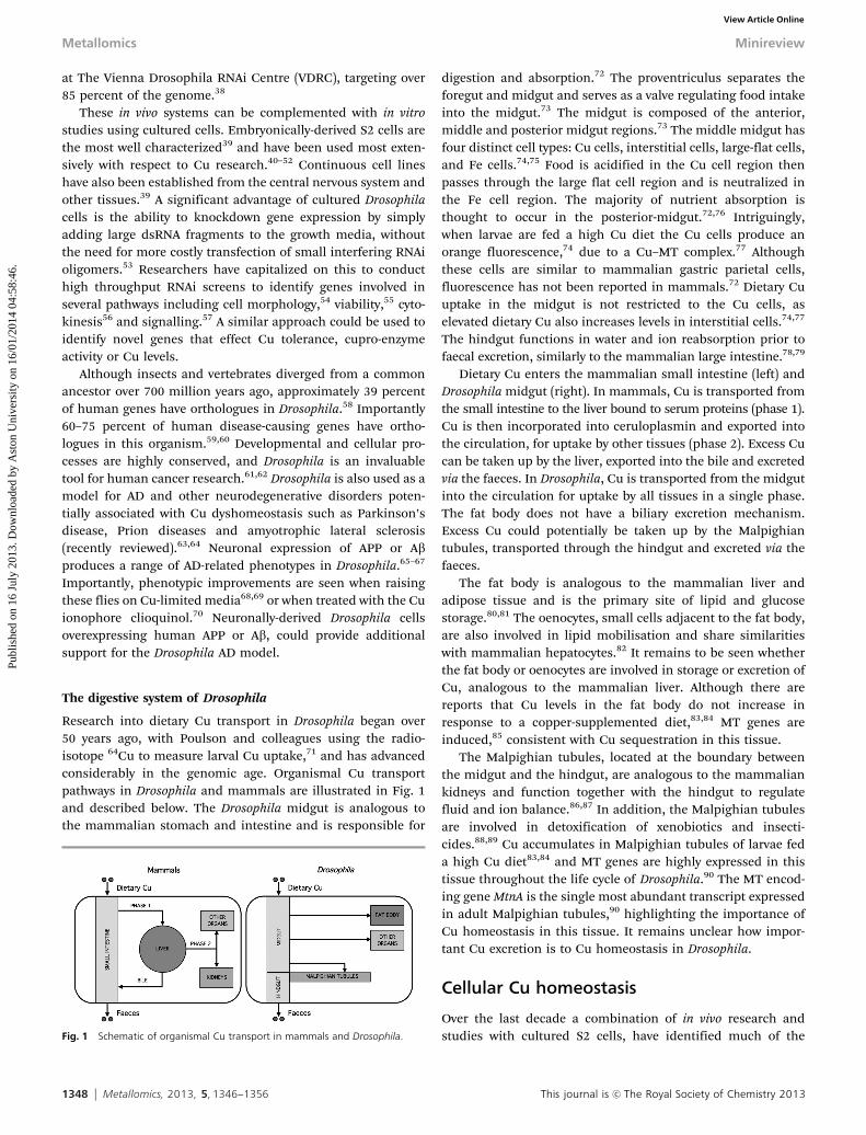
Research into dietary Cu transport in Drosophila began over50 years ago, with Poulson and colleagues using the radio-isotope 64Cu to measure larval Cu uptake,71 and has advancedconsiderably in the genomic age. Organismal Cu transportpathways in Drosophila and mammals are illustrated in Fig. 1and described below. The Drosophila midgut is analogous tothe mammalian stomach and intestine and is responsible for
digestion and absorption.72 The proventriculus separates theforegut and midgut and serves as a valve regulating food intakeinto the midgut.73 The midgut is composed of the anterior,middle and posterior midgut regions.73 The middle midgut hasfour distinct cell types: Cu cells, interstitial cells, large-flat cells,and Fe cells.74,75 Food is acidified in the Cu cell region thenpasses through the large flat cell region and is neutralized inthe Fe cell region. The majority of nutrient absorption isthought to occur in the posterior-midgut.72,76 Intriguingly,when larvae are fed a high Cu diet the Cu cells produce anorange fluorescence,74 due to a Cu–MT complex.77 Althoughthese cells are similar to mammalian gastric parietal cells,fluorescence has not been reported in mammals.72 Dietary Cuuptake in the midgut is not restricted to the Cu cells, aselevated dietary Cu also increases levels in interstitial cells.74,77
The hindgut functions in water and ion reabsorption prior tofaecal excretion, similarly to the mammalian large intestine.78,79
Dietary Cu enters the mammalian small intestine (left) andDrosophila midgut (right). In mammals, Cu is transported fromthe small intestine to the liver bound to serum proteins (phase 1).Cu is then incorporated into ceruloplasmin and exported intothe circulation, for uptake by other tissues (phase 2). Excess Cucan be taken up by the liver, exported into the bile and excretedvia the faeces. In Drosophila, Cu is transported from the midgutinto the circulation for uptake by all tissues in a single phase.The fat body does not have a biliary excretion mechanism.Excess Cu could potentially be taken up by the Malpighiantubules, transported through the hindgut and excreted via thefaeces.
The fat body is analogous to the mammalian liver andadipose tissue and is the primary site of lipid and glucosestorage.80,81 The oenocytes, small cells adjacent to the fat body,are also involved in lipid mobilisation and share similaritieswith mammalian hepatocytes.82 It remains to be seen whetherthe fat body or oenocytes are involved in storage or excretion ofCu, analogous to the mammalian liver. Although there arereports that Cu levels in the fat body do not increase inresponse to a copper-supplemented diet,83,84 MT genes areinduced,85 consistent with Cu sequestration in this tissue.
The Malpighian tubules, located at the boundary betweenthe midgut and the hindgut, are analogous to the mammaliankidneys and function together with the hindgut to regulatefluid and ion balance.86,87 In addition, the Malpighian tubulesare involved in detoxification of xenobiotics and insecti-cides.88,89 Cu accumulates in Malpighian tubules of larvae feda high Cu diet83,84 and MT genes are highly expressed in thistissue throughout the life cycle of Drosophila.90 The MT encod-ing gene MtnA is the single most abundant transcript expressedin adult Malpighian tubules,90 highlighting the importance ofCu homeostasis in this tissue. It remains unclear how impor-tant Cu excretion is to Cu homeostasis in Drosophila.
Cellular Cu homeostasis
Over the last decade a combination of in vivo research andstudies with cultured S2 cells, have identified much of theFig. 1 Schematic of organismal Cu transport in mammals and Drosophila.
Metallomics Minireview
Publ
ishe
d on
16
July
201
3. D
ownl
oade
d by
Ast
on U
nive
rsity
on
16/0
1/20
14 0
4:58
:46.
View Article Online
This journal is c The Royal Society of Chemistry 2013 Metallomics, 2013, 5, 1346--1356 1349
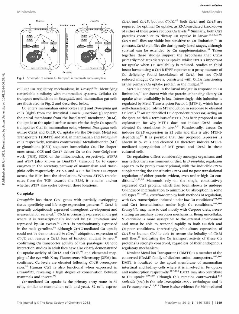
cellular Cu regulatory mechanisms in Drosophila, identifyingremarkable similarity with mammalian systems. Cellular Cutransport mechanisms in Drosophila and mammalian gut cellsare illustrated in Fig. 2 and described below.
Cu enters mammalian enterocytes (left) and Drosophila gutcells (right) from the intestinal lumen. Junctions (J) separatethe apical membrane from the basolateral membrane (BLM).Cu uptake at the apical surface occurs via the single Cu specifictransporter Ctr1 in mammalian cells, whereas Drosophila cellsutilize Ctr1A and Ctr1B. Cu uptake via the Divalent Metal ionTransporters 1 (DMT1) and Mvl, in mammalian and Drosophilacells respectively, remains controversial. Metallothionein (MT)or glutathione (GSH) sequester intracellular Cu. The chaper-ones Atox1, CCS and Cox17 deliver Cu to the trans-Golgi net-work (TGN), SOD1 or the mitochondria, respectively. ATP7Aand ATP7 (also known as DmATP7) transport Cu to cupro-enzymes in the secretory pathway of mammalian and Droso-phila cells respectively. ATP7A and ATP7 facilitate Cu exportacross the BLM into the circulation. Whereas ATP7A translo-cates from the TGN towards the BLM, it remains unclearwhether ATP7 also cycles between these locations.
Cu uptake
Drosophila has three Ctr1 genes with partially overlappingtissue specificity and life stage expression patterns.52 Ctr1A isgenerally ubiquitously expressed throughout development andis essential for survival.51 Ctr1B is primarily expressed in the gutwhere it is transcriptionally induced by Cu limitation andrepressed by Cu excess.52 Ctr1C is predominantly expressedin the male germline.91 Although Ctr1C-mediated Cu uptakecould not be demonstrated in vitro,52 ubiquitous expression ofCtr1C can rescue a Ctr1A loss of function mutant in vivo,92
confirming Cu transporter activity of this paralogue. Geneticinteraction studies in adult flies have also clearly demonstratedCu uptake activity of Ctr1A and Ctr1B,93 and elemental map-ping of the eye with X-ray Fluorescence Microscopy (XFM) hasconfirmed Cu levels are elevated following Ctr1B overexpres-sion.94 Human Ctr1 is also functional when expressed inDrosophila, revealing a high degree of conservation betweenmammals and insects.92
Ctr-mediated Cu uptake is the primary entry route in S2cells, similar to mammalian cells and yeast. S2 cells express
Ctr1A and Ctr1B, but not Ctr1C.47 Both Ctr1A and Ctr1B arerequired for optimal Cu uptake, as RNAi-mediated knockdownof either of these genes reduces Cu levels.47 Similarly, both Ctr1proteins contribute to dietary Cu uptake in larvae.51,52,93,95
Ctr1B null flies are viable but sensitive to Cu limitation.52 Incontrast, Ctr1A null flies die during early larval stages, althoughsurvival can be extended by Cu supplementation.51 Takentogether these studies support the hypothesis that Ctr1Aprimarily mediates dietary Cu uptake, whilst Ctr1B is importantfor uptake when Cu availability is reduced. Studies in thirdinstar larvae using a Ctr1B-EYFP reporter as a proxy measure ofCu deficiency found knockdown of Ctr1A, but not Ctr1Breduced midgut Cu levels, consistent with Ctr1A functioningas the primary Cu uptake protein in the midgut.93
Ctr1B is upregulated in the larval midgut in response to Culimitation,52 consistent with the protein enhancing dietary Cuuptake when availability is low. Interestingly, this induction isregulated by Metal Transcription Factor 1 (MTF-1), which has awell-characterized role in MT induction in response to elevatedCu levels.96 An unidentified Cu-dependent repressor, acting onthe cysteine-rich C-terminus of MTF-1, has been proposed as anexplanation for why MTF-1 does not induce Ctr1B underelevated Cu conditions in vivo.9,41 Paradoxically, excess Cuinduces Ctr1B expression in S2 cells and this is also MTF-1-dependent.47 It is possible that this proposed repressor isabsent in S2 cells and elevated Cu therefore induces MTF-1-mediated upregulation of MT genes and Ctr1B in thesecultured cells.
Ctr regulation differs considerably amongst organisms andmay reflect their environment or diet. In Drosophila, regulationappears to be purely transcriptional, with the inducible Ctr1Bsupplementing the constitutive Ctr1A and no post-translationalregulation of either protein evident, even under high Cu con-ditions.51,52,95 Mammals rely on the single, constitutivelyexpressed Ctr1 protein, which has been shown to undergoCu-induced internalization to minimize Cu absorption in somecell types.97–100 S. cerevisiae employ both methods of regulation,with Ctr1 transcription induced under low Cu conditions101,102
and Ctr1 internalization under high Cu conditions.103,104
Drosophila may have to deal mostly with Cu-poor diets, neces-sitating an auxiliary absorption mechanism. Being unicellular,S. cerevisiae is more susceptible to the external environmentand must be able to respond rapidly to both Cu-rich andCu-poor conditions. Interestingly, ubiquitous expression ofCtr1B or human Ctr1 is able to rescue the lethality of Ctr1Anull flies,92 indicating the Cu transport activity of these Ctrproteins is strongly conserved, regardless of their endogenousregulatory mechanism.
Divalent Metal ion Transporter 1 (DMT1) is a member of theconserved NRAMP family of divalent cation transporters.105,106
DMT1 is localized to the apical membrane of mammalianintestinal and kidney cells where it is involved in Fe uptakeand reabsorption respectively.107,108 DMT1 may also contributeto Cu uptake,109,110 although this remains controversial.111
Malvolio (Mvl) is the sole Drosophila DMT1 orthologue and isan Fe transporter.112,113 There is also evidence for Mvl-mediated
Fig. 2 Schematic of cellular Cu transport in mammals and Drosophila.
Minireview Metallomics
Publ
ishe
d on
16
July
201
3. D
ownl
oade
d by
Ast
on U
nive
rsity
on
16/0
1/20
14 0
4:58
:46.
View Article Online
1350 Metallomics, 2013, 5, 1346--1356 This journal is c The Royal Society of Chemistry 2013
Cu uptake in S2 cells and in vivo.48 While systemic Cu levels arenot reduced by impaired Mvl function in Drosophila,48,112 expres-sion of the Cu-responsive MTNB-EYFP reporter in the anteriormidgut is reduced, indicative of decreased Cu levels in the earlydigestive system.48 Reduced Mvl activity causes both S2 cellsand female flies to be sensitive to Cu limitation, suggesting thisnon-specific Cu uptake protein is functionally important.48
Nevertheless, Ctr1A and Ctr1B proteins are responsible for themajority of dietary Cu uptake in Drosophila.
Recent discoveries with Drosophila have highlighted theimportance of vesicular trafficking pathways in regulating Cuuptake in both Drosophila and mammalian systems. Syntaxin 5(Syx5) heterozygous flies are tolerant to excess Cu as a resultof reduced dietary Cu uptake.44,85 Syx5 is a SNARE (solubleN-ethylmaleimide-sensitive factor attachment protein receptor)protein involved in vesicle fusion to target membranes.114
Knockdown of Syx5 in both S2 and mammalian cells reducesCu accumulation44 indicating a conserved role of this vesicletrafficking mechanism in maintaining Cu homeostasis. A com-parable reduction in Cu accumulation was also seen in thesecells following knockdown of ADP-Ribosylation Factor 1 (ARF1),49
which is important for the formation of COPI coated vesicles thatfunction in retrograde and anterograde trafficking.21 Biotinyla-tion studies in mammalian cells indicate that knockdown ofARF149 and Syx544 reduces the amount of Ctr1 at the PM, andthis could mediate the reduction in Cu uptake. These results,together with the recent link between MEDNIK syndrome andAP-1,20 suggest Cu homeostasis is particularly sensitive to per-turbation in components of particular vesicular traffickingpathways.
Presenilin (PSN) knockdown in the Drosophila midgut pro-duces a similar phenotype to the Syx5 heterozygote flies, withreduced dietary Cu uptake and increased Cu tolerance.115
Presenilins are evolutionarily conserved components of thegamma secretase complex that functions in intramembraneproteolysis of many substrates including APP.24 Studies in miceand cultured mammalian cells have shown that the ortho-logous PSEN1 and PSEN2 proteins are important for uptakeof Cu and also Zn.116 An effect of Presenilins on Ctr1 localisa-tion, analogous to the effect of Syx5 and ARF1,115,116 is possible.SOD1 activity is impaired by the reduction in PSN levels in bothmammalian and Drosophila models, indicative of a functionalCu deficiency.115,116 Given the involvement of Presenilins in ADpathogenesis,24 these results provide additional support for theassociation between Cu dyshomeostasis and AD.
Cu sequestration and distribution
MT-mediated Cu sequestration has been well studied in Droso-phila. Five MT genes have been identified in Drosophila, desig-nated MtnA, MtnB, MtnC, MtnD and MtnE.40,117–119 MtnC, MtnDand MtnE are likely to have evolved following duplications ofthe MtnB gene.117,120 MTF-1 encodes the Drosophila transcrip-tion factor able to induce expression of these MT genes inresponse to Cu and other metals,40,117,121 similar to mamma-lian systems.122–124 MtnA, MtnB and MtnD are expressed in S2cells and upregulated following Cu exposure in a time-dependent
and concentration-dependent manner.47 Cu tolerance in S2cells was significantly reduced following RNAi knockdownof MTF-1, illustrating the importance of Cu sequestration inthese cells.
MTF-1 knockout flies are viable, but sensitive to Cuexcess.117 Comprehensive in vivo analysis of individual MTgenes demonstrate each is expressed in the larval midgut andinducible by Cu, with MtnA and MtnB performing the primaryCu sequestration role.40,125,126 Paradoxically MTF-1 null flies arealso sensitive to Cu limitation as MTF-1 also acts to upregulateCtr1B under these conditions.96 MTF-1 also induces upregula-tion of ATP7 in larval midgut in response to elevated Cu127 andoverexpression of MTF-1 prolongs survival of flies with RNAiknockdown of ATP7 in the gut.128 Drosophila MTF-1 is thereforethe master regulator of gene expression under conditions ofboth Cu limitation and excess. MTF-1-dependent expression ofmammalian Ctr1, ATP7A or ATP7B genes has not been reported.The mechanisms by which target genes are induced in responseto different metals is becoming clearer, and may involvespecific metal response elements (MRE)46 as well as MTF-1regulatory domains within the cysteine clusters (residues 547–565 and 642–766) and phosphorylation within the Zn fingerdomain (Tyr118, Ser126, Thr127 and Ser160).41,43
Cu chaperones have been less well studied in Drosophila andgenerally assumed to deliver Cu to specific proteins similarly toyeast and mammals.9 The Drosophila genome contains ortho-logues of Atox1, CCS, Sco1/2 and Cox17, all of which areexpressed in S2 cells.47 In S. cerevisiae Cu transport to cyto-chrome-c oxidase in the mitochondria involves the chaperonesSco1/2 and Cox17. While Cox17 remains uncharacterized inDrosophila, the Sco orthologue has recently been shown to beessential for cytochrome-c oxidase assembly and function inDrosophila similarly to other eukaryotes.129 RNAi knockdown ofSco in the adult thorax also cause a mild thoracic cleft, loss ofscutellum and thin bristles.93 While these phenotypes cannotbe directly attributed to a mitochondrial Cu defect, pigmenta-tion was unaffected indicating Sco may be involved in Cudistribution to cellular compartments other than the secretorypathway.93 As with yeast and mammalian systems, CCS deliversCu to SOD1 in Drosophila although a CCS-independent pathwayof SOD1 activation also exists and may play a minor role whenin the absence of CCS.130
Atox1 null Drosophila accumulate Cu in the midgut and aresensitive to dietary Cu-limitation.131 Cu levels are also elevatedin the heads of Atox1 loss of function mutants.69 These resultsare consistent with impaired ATP7-mediated Cu efflux, similarto that reported in cells derived from Atox1 null mice.132 It isunclear if loss of Drosophila Atox1 also impairs Cu transport tocupro-enzymes in the secretory pathway, as has been demon-strated in mice.132 Also unclear is whether Drosophila Ctrproteins deliver Cu directly to Atox1 as has been demonstratedin vitro with the orthologous yeast proteins.133 If Ctr-mediatedtransport is dependent on a chaperone such as Atox1 removingCu directly from the protein, then Cu chaperones may have amore important role in regulating Cu uptake than currentlyappreciated. In support of this hypothesis, co-overexpression of
Metallomics Minireview
Publ
ishe
d on
16
July
201
3. D
ownl
oade
d by
Ast
on U
nive
rsity
on
16/0
1/20
14 0
4:58
:46.
View Article Online
This journal is c The Royal Society of Chemistry 2013 Metallomics, 2013, 5, 1346--1356 1351
the human orthologues of CCS and Ctr1 in the Drosophila eyeproduces a more severe rough-eye phenotype than either genealone,92 consistent with a synergistic effect.
Cu export
Organisms such as yeast, insects and nematodes utilize one Cutransporting P-type ATPase whereas a gene duplication duringchordate evolution means birds, fish and mammals havetwo.134 The Drosophila orthologue, designated ATP7, showsstrong sequence similarity to mammalian ATP7A and ATP7B,and is expressed in all tissues tested to date.50,85,135 ATP7functions similarly to ATP7A, transporting Cu from the gut tothe circulatory system and delivering Cu to cupro-enzymes inperipheral tissues.85,93,127 ATP7 is required for delivery of Cu tocupro-enzymes in the secretory pathway including tyrosinase,which is required for pigmentation,85,93 and peptidylglycine-a-hydroxylating mono-oxygenase, which is required for neuro-peptide amidation.136 Genetic interaction studies in adult flieshave clearly demonstrated Cu efflux activity of ATP793,127 andelemental mapping of the adult fly eye with XFM confirmed areduction in Cu levels following ATP7 overexpression.94 ADrosophila model of classical Menkes disease has been estab-lished, where absence of functional ATP7 causes reduced Cutransport from the gut, decreased cupro-enzyme activity andearly lethality.85 RNAi knockdown of ATP7 in the gut produces amilder phenotype with some flies surviving until adulthood.128
Similarly, DmAPT7 knockdown causes Cu to accumulate in S2cells.47
Cu-induced trafficking of ATP7A and ATP7B from the TGNtowards the cell surface is central to their ability to transport Cuacross the PM of mammalian cells. While an analogous mecha-nism in Drosophila is an attractive hypothesis, Cu-inducedtrafficking of ATP7 has not been demonstrated. Biotinylationstudies identified ATP7 at the PM of S2 cells and neuronally-derived Bm3-c2 cells, although the majority remained at theTGN irrespective of Cu status.50 During embryogenesis endo-genous ATP7 is localized to the PM, and this localisationbecomes progressively more cytoplasmic as the embryo devel-ops.85 Ectopically-expressed FLAG-tagged ATP7 could be seen ateither the PM or dispersed throughout the cytoplasm of thelarval midgut,85 whereas GFP-tagged ATP7 has been observedpredominantly on the basolateral PM of the midgut, with noobvious shift in localization seen in either high or low Cuconditions.127 Localisation motifs may be active in differentcells or during different developmental stages. Hormonal reg-ulation of ATP7 also remains a possibility, as the localisation ofATP7A and ATP7B are influenced by hormones such as insulinand oestrogen, independently of intracellular Cu status incultured placental and mammary cells.137,138
The transcriptional regulation of ATP7 in the midgut oflarvae raised on Cu-supplemented media127 is not unprece-dented, as upregulation of ATP7A has also been reported in theintestine of infant rats fed on Cu-supplemented food.139 Inter-estingly, heart-specific knockout of Ctr1 in mice leads toincreased ATP7A expression in enterocytes and hepatocytes,suggestive of a mechanism to increase circulating Cu levels in
response to localized Cu deficiency.140 This study also foundcardiomyopathy in flies following Ctr1A knockdown, althoughthere was no report of whether ATP7 expression in the midgutwas similarly affected.140
P-type ATPases from mammals (ATP7A and ATP7B) andS. cerevisiae (Ccc2p) undergo kinase-mediated phosphorylation,and these post-translational modifications regulate proteinlocalisation and function (recently reviewed).134 Many of theseresidues are conserved in ATP7 and similar phosphorylation-mediated regulation is plausible.50
The C-terminal di-leucine motifs and class one PDZdomains, important for trafficking and basolateral localisationof ATP7A134 are conserved in ATP7 and in the orthologousproteins in twelve different Drosophila species.50 In contrast,the N-terminal FAFDNVGYE apical targeting motif present inATP7B was not present in these Drosophila P-type ATPaseorthologues. ATP7 therefore more closely resembles ATP7A thenATP7B. Indeed, when expressed in mammalian cells DmAPT7was able to compensate for the loss of ATP7A, delivering Cu totyrosinase in the secretory pathway and also transporting Cuout of the cell.50 The reversible Cu-dependent trafficking ofATP7 to the basolateral membrane of cultured kidney cellsdemonstrates the di-leucine and PDZ motifs can be recognizedby trafficking mechanisms in these cells. In addition to copper-dependent trafficking, constitutive trafficking and recycling ofATP7A has also been reported in several mammalian cell linesindependently of Cu status.141–143 If ATP7 constitutivelyrecycles between the TGN and the PM, then the putativedi-leucine and PDZ domains may be required for this. Endo-genous ATP7A144 and ATP7B145 load Cu into vesicles destinedfor the PM, rather than completely translocating to the PM.If these cultured cells use a similar efflux mechanism, thiscould explain why so little ATP7 is localized at the PM. AlthoughCu-dependent translocation of ATP7 has not been seen inDrosophila or the two cultured cell lines tested to date, thepossibility that this occurs in other cell types or under differentconditions cannot be ruled out. A ATP7 specific antibody will berequired for determination of the copper-dependent localisa-tion of endogenous ATP7 in vivo, if this issue is to be resolved.
Conclusions and future directions
Cellular Cu uptake, sequestration, distribution and effluxmechanisms are highly conserved between Drosophila andmammals. At the organismal level, Cu transport within theDrosophila midgut is comparable to that in mammalian enter-ocytes, while mammalian renal and hepatic systems are lesswell conserved in Drosophila. The fat body and oenocytes arethe closest equivalents to the mammalian hepatic system, yetDrosophila do not have a comparable biliary Cu excretionmechanism. The possibility that the Malpighian tubules takeup excess Cu from the hemolymph, and that this Cu is excretedvia the hindgut warrants investigation. The generation ofestablished cell lines from these Drosophila tissues would helpto elucidate cellular transport mechanisms. Cu in the hemolymphis presumably protein-bound, but this has not been demonstrated.
Minireview Metallomics
Publ
ishe
d on
16
July
201
3. D
ownl
oade
d by
Ast
on U
nive
rsity
on
16/0
1/20
14 0
4:58
:46.
View Article Online
1352 Metallomics, 2013, 5, 1346--1356 This journal is c The Royal Society of Chemistry 2013
In mammals, circulatory Cu is predominantly bound to cerulo-plasmin with some also bound to albumin, a2-macroglobulinand smaller peptides and amino acids.11,12 In Drosophila, thereare four members of the multicopper oxidase (MCO) family andMCO3 has ferroxidase activity similar to ceruloplasmin.112,146
While MCO3 is highly expressed in the fat body, the protein hasnot been detected in the hemolymph.147,148 Nevertheless, thepossibility that an MCO orthologue contributes to Cu homeo-stasis in the hemolymph warrants further investigation.
One of the most intriguing discoveries with Drosophila, hasbeen the MTF-1 dependent regulation of not only MT geneexpression, but also genes involved in Cu uptake (Ctr1B) andefflux (ATP7), implicating MTF-1 as the key regulator of systemicCu homeostasis. It will be interesting to see whether the hypothe-tical MTF-1 repressor is identified in Drosophila. Although Droso-phila downregulate midgut Ctr1B expression in response to a highCu diet, this is not sufficient to prevent excess Cu accumulation.95
Rearing flies on a high Cu diet increases the Cu stores of theirprogeny, leading Balamurugan and colleagues95 to suggest Droso-phila risk excessive Cu accumulation for the potential benefit ofovercoming a period of Cu scarcity. This supports the hypothesisthat Drosophila evolved under conditions of Cu scarcity ratherthan Cu excess,127 which is also consistent with the absence ofCu-induced internalization of Ctr1A or Ctr1B. Although organis-mal Cu levels are remarkably similar between different Drosophilaspecies raised on standard laboratory media,149 Cu-tolerant fliescan be selected within two years by dietary manipulation,150
demonstrating the adaptive potential for Cu homeostasis inDrosophila melanogaster. As techniques for high throughput geneexpression and sequence analysis continue to become more afford-able, the genes responsible for adaptation to Cu-supplementationcould realistically be identified in Drosophila.
The identification of Syx5 has provided an important proofof principal that genetic screens in Drosophila can identifygenes of relevance to mammalian systems.44,45 The potentialfor additional novel Cu regulatory genes to be identified andcharacterized within Drosophila and cultured Drosophila cellsremains. There are dozens of Drosophila mutants that are eithertolerant or sensitive to elevated Cu levels for which the gene orgenes responsible are yet to be identified.45 There are alsohundreds of genes that have responded transcriptionally toaltered Cu homeostasis.45,151 Two promising approaches toidentify novel Cu regulatory genes are RNAi-based screensutilising the GAL4-UAS system and Genome-Wide AssociationStudies (GWAS). The Drosophila melanogaster Genetic ReferencePanel (DGRP) consists of 162 fly strains, each with their genomefully sequenced, which can be used for rapid GWAS.152 TheseDGRP strains contain over four million genetic variants andhave successfully been used to identify candidate genes forstarvation resistance and other traits.152 A similar GWAS couldidentify candidate genes that affect a quantitative phenotypesuch as Cu levels using inductively coupled plasma massspectrometry (ICP-MS). Larval or adult survival could also beassessed to identify Cu-tolerant or Cu-sensitive Drosophila.
Virtually all Drosophila genes have corresponding RNAi linescommercially available and a genome wide RNAi screen has
successfully identified genes involved in notch signalling.153
A similar genome wide screen could be conducted for Cuhomeostasis genes, or a smaller subset could be selected basedon previously identified candidates. Alternatively, RNAi linescould target genes involved in specific biological processes,such as protein and vesicular trafficking pathways. An RNAi-based screen of Zn and Fe homeostasis genes might alsoidentify interactions between the regulation of Cu and thesemetals. GAL4 drivers could be used to knockdown genesubiquitously or within specific tissues such as the midgut.Moreover, a sensitized screen could be used to identify genesthat enhance or suppress an established Cu tolerance orsensitivity phenotype. For example, overexpression of Ctr1Aor Ctr1B with the ubiquitous actin-GAL4 has been shown tocauses lethality that can be partially rescued by raising flies onCu-limited media.92 An RNAi screen could be conducted withthese flies raised on normal food to identify candidate genesthat rescue this Cu sensitivity. RNAi-based modifier screenscould be conducted for other Cu-related phenotypes such ascuticle hypopigmentation, which is induced by ATP7 over-expression with the Pannier-GAL4 driver,93 or rough-eyes,which are induced by Ctr1B overexpression with the GMR-GAL4 driver.92
The regulation of Cu homeostasis is linked with that ofother metals such as Zn and Fe, which is perhaps best illu-strated by the ferroxidase activity of the cupro-protein cerulo-plasmin154 and the antioxidant SOD1, which requires Cu andZn.7 In addition, the Fe transporter DMT1 can potentiallytransport Zn, Cu and other divalent cations109,110 and the Zntransporter Zip4 may have Cu transport activity.155 If we are tounderstand the complex relationship between metal dys-homeostasis and diseases such as AD, experimental approachesthat interrogate multiple metals will be of benefit. SynchrotronXFM is one such technique, and this has already been usedwith Drosophila, to characterize subtle metal changes withinthe brain and other tissues in response to genetic manipulationof Cu transporters.94 The integration of XFM with confocalimmuno-fluorescence microscopy will allow quantification anddistribution analysis of metallo-proteins at the cellular level.Laser Ablation-ICP-MS can also quantify metal distribution andthis has been used to create a three dimensional map of Cu, Znand Fe distribution in the mouse brain.156 Liquid Chromato-graphy-ICP-MS can quantify metal levels of individual proteinsand has been used to assess the metallo-protein profile ofDrosophila larvae in response to Cu supplementation (unpub-lished observations).
The use of multiple model systems including Drosophila, hasadvanced our understanding of eukaryotic Cu homeostasis,and future studies will continue to elucidate the complexrelationship between Cu dyshomeostasis and health.
Acknowledgements
This work was supported by in part by grants from theAustralian Research Council, The National Health and MedicalResearch Council, the International Copper Association and
Metallomics Minireview
Publ
ishe
d on
16
July
201
3. D
ownl
oade
d by
Ast
on U
nive
rsity
on
16/0
1/20
14 0
4:58
:46.
View Article Online
This journal is c The Royal Society of Chemistry 2013 Metallomics, 2013, 5, 1346--1356 1353
the Australian Institute of Nuclear Science and Engineering.A. S. and R. B. were recipients the J. N. Peters Bequest Fellow-ship from the University of Melbourne.
Notes and references
1 M. M. Pena, J. Lee and D. J. Thiele, J. Nutr., 1999, 129,1251–1260.
2 M. Valko, H. Morris and M. T. Cronin, Curr. Med. Chem.,2005, 12, 1161–1208.
3 Z. Tumer and L. B. Moller, Eur. J. Hum. Genet., 2009, 18,1–8.
4 P. Wardman and L. P. Candeias, Radiat. Res., 1996, 145,523–531.
5 B. Halliwell and J. M. Gutteridge, Biochem. J., 1984, 219,1–14.
6 J. E. Klaunig and L. M. Kamendulis, Annu. Rev. Pharmacol.Toxicol., 2004, 44, 239–267.
7 I. Fridovich, J. Exp. Biol., 1998, 201, 1203–1209.8 T. D. Rae, P. J. Schmidt, R. A. Pufahl, V. C. Culotta and
T. V. O’Halloran, Science, 1999, 284, 805–808.9 K. Balamurugan and W. Schaffner, Biochim. Biophys. Acta,
2006, 1763, 737–746.10 T. Nevitt, H. Ohrvik and D. J. Thiele, Biochim. Biophys. Acta,
2012, 1823, 1580–1593.11 M. C. Linder, L. Wooten, P. Cerveza, S. Cotton, R. Shulze
and N. Lomeli, Am. J. Clin. Nutr., 1998, 67, 965S–971S.12 P. V. van den Berghe and L. W. Klomp, Nutr. Rev., 2009, 67,
658–672.13 Z. L. Harris, Y. Takahashi, H. Miyajima, M. Serizawa,
R. T. MacGillivray and J. D. Gitlin, Proc. Natl. Acad. Sci.U. S. A., 1995, 92, 2539–2543.
14 H. Miyajima, Y. Nishimura, K. Mizoguchi, M. Sakamoto,T. Shimizu and N. Honda, Neurology, 1987, 37, 761–767.
15 Z. L. Harris, A. P. Durley, T. K. Man and J. D. Gitlin, Proc.Natl. Acad. Sci. U. S. A., 1999, 96, 10812–10817.
16 L. A. Meyer, A. P. Durley, J. R. Prohaska and Z. L. Harris,J. Biol. Chem., 2001, 276, 36857–36861.
17 H. Kodama, Y. Murata and M. Kobayashi, Pediatr. Int.,1999, 41, 423–429.
18 C. M. Mak and C. W. Lam, Crit. Rev. Clin. Lab. Sci., 2008,45, 263–290.
19 E. A. Roberts and M. L. Schilsky, Hepatology, 2008, 47,2089–2111.
20 D. Martinelli, L. Travaglini, C. Drouin, I. Ceballos-Picot,T. Rizza, E. Bertini, R. Carrozzo, S. Petrini, P. de Lonlay,M. El Hachem, L. Hubert, A. Montpetit, G. Torre andC. Dionisi-Vici, Brain, 2013, 136, 872–881, DOI: 10.1093/brain/awt012.
21 J. S. Bonifacino and B. S. Glick, Cell, 2004, 116, 153–166.22 M. A. Greenough, J. Camakaris and A. I. Bush, J. Biol. Chem.,
2011, 286, 9776–9786, DOI: 10.1074/jbc.M110.163964.23 S. Ayton, P. Lei and A. I. Bush, Free Radicals Biol. Med.,
2012, DOI: 10.1016/j.freeradbiomed.2012.10.558.24 K. S. Vetrivel, Y. W. Zhang, H. Xu and G. Thinakaran, Mol.
Neurodegener., 2006, 1, 4.
25 A. Simons, T. Ruppert, C. Schmidt, A. Schlicksupp,R. Pipkorn, J. Reed, C. L. Masters, A. R. White,R. Cappai, K. Beyreuther, T. A. Bayer and G. Multhaup,Biochemistry, 2002, 41, 9310–9320.
26 P. J. Crouch, L. W. Hung, P. A. Adlard, M. Cortes, V. Lal,G. Filiz, K. A. Perez, M. Nurjono, A. Caragounis, T. Du,K. Laughton, I. Volitakis, A. I. Bush, Q. X. Li, C. L. Masters,R. Cappai, R. A. Cherny, P. S. Donnelly, A. R. White andK. J. Barnham, Proc. Natl. Acad. Sci. U. S. A., 2009, 106,381–386.
27 P. A. Adlard, L. Bica, A. R. White, M. Nurjono, G. Filiz,P. J. Crouch, P. S. Donnelly, R. Cappai, D. I. Finkelstein andA. I. Bush, PLoS One, 2011, 6, e17669.
28 R. A. Cherny, C. S. Atwood, M. E. Xilinas, D. N. Gray,W. D. Jones, C. A. McLean, K. J. Barnham, I. Volitakis,F. W. Fraser, Y. Kim, X. Huang, L. E. Goldstein, R. D. Moir,J. T. Lim, K. Beyreuther, H. Zheng, R. E. Tanzi,C. L. Masters and A. I. Bush, Neuron, 2001, 30, 665–676.
29 N. G. Faux, C. W. Ritchie, A. Gunn, A. Rembach,A. Tsatsanis, J. Bedo, J. Harrison, L. Lannfelt,K. Blennow, H. Zetterberg, M. Ingelsson, C. L. Masters,R. E. Tanzi, J. L. Cummings, C. M. Herd and A. I. Bush,J. Alzheimer’s Dis., 2010, 20, 509–516.
30 C. Grossi, S. Francese, A. Casini, M. C. Rosi, I. Luccarini,A. Fiorentini, C. Gabbiani, L. Messori, G. Moneti andF. Casamenti, J. Alzheimer’s Dis., 2009, 17, 423–440.
31 C. W. Ritchie, A. I. Bush, A. Mackinnon, S. Macfarlane,M. Mastwyk, L. MacGregor, L. Kiers, R. Cherny, Q. X. Li,A. Tammer, D. Carrington, C. Mavros, I. Volitakis,M. Xilinas, D. Ames, S. Davis, K. Beyreuther, R. E. Tanziand C. L. Masters, Arch. Neurol., 2003, 60, 1685–1691.
32 E. J. Martinez-Finley and M. Aschner, J. Toxicol., 2011,2011, 895236.
33 P. P. Hernandez and M. L. Allende, Am. J. Clin. Nutr., 2008,88, 835S–839S.
34 G. M. Rubin and E. B. Lewis, Science, 2000, 287, 2216–2218.35 D. St Johnston, Nat. Rev. Genet., 2002, 3, 176–188.36 J. B. Duffy, Genesis, 2002, 34, 1–15.37 A. H. Brand and N. Perrimon, Development, 1993, 118,
401–415.38 G. Dietzl, D. Chen, F. Schnorrer, K. C. Su, Y. Barinova,
M. Fellner, B. Gasser, K. Kinsey, S. Oppel, S. Scheiblauer,A. Couto, V. Marra, K. Keleman and B. J. Dickson, Nature,2007, 448, 151–156.
39 B. Baum and L. Cherbas, Methods Mol. Biol., 2008, 420,391–424.
40 L. Atanesyan, V. Gunther, S. E. Celniker, O. Georgiev andW. Schaffner, JBIC, J. Biol. Inorg. Chem., 2011, 16,1047–1056.
41 V. Gunther, D. Waldvogel, M. Nosswitz, O. Georgiev andW. Schaffner, Int. J. Biochem. Cell Biol., 2012, 44, 404–411.
42 F. Liu, Y. Chen, B. Yang, J. Wang, Q. Peng, Q. Shao, X. Li,B. T. Beerntsen, Y. Xu, J. Li, X. Q. Yu and E. Ling, Dev.Comp. Immunol., 2012, 36, 619–628.
43 S. K. Marr, K. L. Pennington and M. T. Marr, Biochim.Biophys. Acta, 2012, 1819, 902–912.
Minireview Metallomics
Publ
ishe
d on
16
July
201
3. D
ownl
oade
d by
Ast
on U
nive
rsity
on
16/0
1/20
14 0
4:58
:46.
View Article Online
1354 Metallomics, 2013, 5, 1346--1356 This journal is c The Royal Society of Chemistry 2013
44 M. Norgate, A. Southon, M. Greenough, M. Cater,A. Farlow, P. Batterham, A. I. Bush, V. N. Subramaniam,R. Burke and J. Camakaris, PLoS One, 2010, 5, e14303.
45 M. Norgate, A. Southon, S. Zou, M. Zhan, Y. Sun,P. Batterham and J. Camakaris, Biometals, 2007, 20,683–697.
46 H. I. Sims, G. W. Chirn and M. T. Marr, 2nd, Proc. Natl.Acad. Sci. U. S. A., 2012, 109, 16516–16521.
47 A. Southon, R. Burke, M. Norgate, P. Batterham andJ. Camakaris, Biochem. J., 2004, 383, 303–309.
48 A. Southon, A. Farlow, M. Norgate, R. Burke andJ. Camakaris, J. Exp. Biol., 2008, 211, 709–716.
49 A. Southon, M. Greenough, Y. H. Hung, M. Norgate,R. Burke and J. Camakaris, Int. J. Biochem. Cell Biol.,2011, 43, 146–153.
50 A. Southon, N. Palstra, N. Veldhuis, A. Gaeth, C. Robin,R. Burke and J. Camakaris, Biometals, 2010, 23, 681–694.
51 M. L. Turski and D. J. Thiele, J. Biol. Chem., 2007, 282,24017–24026.
52 H. Zhou, K. M. Cadigan and D. J. Thiele, J. Biol. Chem.,2003, 278, 48210–48218.
53 C. A. Worby, N. Simonson-Leff and J. E. Dixon, Sci. STKE,2001, 2001, PL1.
54 A. A. Kiger, B. Baum, S. Jones, M. R. Jones, A. Coulson,C. Echeverri and N. Perrimon, J. Biol., 2003, 2, 27.
55 M. Boutros, A. A. Kiger, S. Armknecht, K. Kerr, M. Hild,B. Koch, S. A. Haas, R. Paro and N. Perrimon, Science, 2004,303, 832–835.
56 U. S. Eggert, A. A. Kiger, C. Richter, Z. E. Perlman,N. Perrimon, T. J. Mitchison and C. M. Field, PLoS Biol.,2004, 2, e379.
57 L. Lum, S. Yao, B. Mozer, A. Rovescalli, D. Von Kessler,M. Nirenberg and P. A. Beachy, Science, 2003, 299,2039–2045.
58 D. G. Gilbert, Nucleic Acids Res., 2002, 30, 145–148.59 E. Bier, Nat. Rev. Genet., 2005, 6, 9–23.60 G. M. Rubin, M. D. Yandell, J. R. Wortman, G. L. G. Miklos,
C. R. Nelson, I. K. Hariharan, M. E. Fortini, P. W. Li,R. Apweiler, W. Fleischmann, J. M. Cherry, S. Henikoff,M. P. Skupski, S. Misra, M. Ashburner, E. Birney,M. S. Boguski, T. Brody, P. Brokstein, S. E. Celniker,S. A. Chervitz, D. Coates, A. Cravchik, A. Gabrielian,R. F. Galle, W. M. Gelbart, R. A. George, L. S. Goldstein,F. Gong, P. Guan, N. L. Harris, B. A. Hay, R. A. Hoskins,J. Li, Z. Li, R. O. Hynes, S. J. Jones, P. M. Kuehl, B. Lemaitre,J. T. Littleton, D. K. Morrison, C. Mungall, P. H. O’Farrell,O. K. Pickeral, C. Shue, L. B. Vosshall, J. Zhang, Q. Zhao,X. H. Zheng and S. Lewis, Science, 2000, 287, 2204–2215.
61 M. Vidal and R. L. Cagan, Curr. Opin. Genet. Dev., 2006, 16,10–16.
62 A. M. Brumby and H. E. Richardson, Nat. Rev. Cancer, 2005,5, 626–639.
63 A. Jeibmann and W. Paulus, Int. J. Mol. Sci., 2009, 10,407–440.
64 B. Lu and H. Vogel, Annu. Rev. Pathol.: Mech. Dis., 2009, 4,315–342.
65 I. Greeve, D. Kretzschmar, J. A. Tschape, A. Beyn,C. Brellinger, M. Schweizer, R. M. Nitsch andR. Reifegerste, J. Neurosci., 2004, 24, 3899–3906.
66 K. Iijima, H. P. Liu, A. S. Chiang, S. A. Hearn, M. Konsolakiand Y. Zhong, Proc. Natl. Acad. Sci. U. S. A., 2004, 101,6623–6628.
67 G. Merdes, P. Soba, A. Loewer, M. V. Bilic, K. Beyreutherand R. Paro, EMBO J., 2004, 23, 4082–4095.
68 H. Hua, L. Munter, A. Harmeier, O. Georgiev, G. Multhaupand W. Schaffner, Biol. Chem., 2011, 392, 919–926.
69 R. Sanokawa-Akakura, W. Cao, K. Allan, K. Patel,A. Ganesh, G. Heiman, R. Burke, F. W. Kemp,J. D. Bogden, J. Camakaris, R. B. Birge and M. Konsolaki,PLoS One, 2010, 5, e8626.
70 T. Rival, R. M. Page, D. S. Chandraratna, T. J. Sendall,E. Ryder, B. Liu, H. Lewis, T. Rosahl, R. Hider,L. M. Camargo, M. S. Shearman, D. C. Crowther andD. A. Lomas, Eur. J. Neurosci., 2009, 29, 1335–1347.
71 D. F. Poulson, V. T. Bowen, R. M. Hilse and A. C. Rubinson,Zoology, 1952, 38, 912–921.
72 R. R. Dubreuil, Int. J. Biochem. Cell Biol., 2004, 36, 745–752.73 H. Nakagoshi, Dev., Growth Differ., 2005, 47, 383–392.74 B. K. Filshie, D. F. Poulson and D. F. Waterhouse, Tissue
Cell, 1971, 3, 77–102.75 R. Tanaka, Y. Takase, M. Kanachi, R. Enomoto-Katayama,
T. Shirai and H. Nakagoshi, Dev. Biol., 2007, 304, 53–61.76 R. R. Dubreuil, J. Frankel, P. Wang, J. Howrylak, M. Kappil
and T. A. Grushko, Dev. Biol., 1998, 194, 1–11.77 M. McNulty, M. Puljung, G. Jefford and R. R. Dubreuil, Cell
Tissue Res., 2001, 304, 383–389.78 R. Murakami and Y. Shiotsuki, J. Morphol., 2001, 248,
144–150.79 J. A. Lengyel and D. D. Iwaki, Dev. Biol., 2002, 243, 1–19.80 L. Sondergaard, Trends Genet., 1993, 9, 193.81 Y. Liu, H. Liu, S. Liu, S. Wang, R. J. Jiang and S. Li, Arch.
Insect Biochem. Physiol., 2009, 71, 16–30.82 E. Gutierrez, D. Wiggins, B. Fielding and A. P. Gould,
Nature, 2007, 445, 275–280.83 D. Marchal-Segault, C. Briancon, S. Halpern, P. Fragu and
G. Lauge, Biol. Cell, 1990, 70, 129–132.84 R. M. Schofield, J. H. Postlethwait and H. W. Lefevre,
J. Exp. Biol., 1997, 200, 3235–3243.85 M. Norgate, E. Lee, A. Southon, A. Farlow, P. Batterham,
J. Camakaris and R. Burke, Mol. Biol. Cell, 2006, 17,475–484.
86 M. J. O’Donnell and S. H. Maddrell, J. Exp. Biol., 1995, 198,1647–1653.
87 R. Cagan, Curr. Opin. Nephrol. Hypertens., 2003, 12, 11–17.88 H. Chung, T. Sztal, S. Pasricha, M. Sridhar, P. Batterham
and P. J. Daborn, Proc. Natl. Acad. Sci. U. S. A., 2009, 106,5731–5736.
89 J. Yang, C. McCart, D. J. Woods, S. Terhzaz,K. G. Greenwood, R. H. ffrench-Constant and J. A. Dow,Physiol. Genomics, 2007, 30, 223–231.
90 J. Wang, L. Kean, J. Yang, A. K. Allan, S. A. Davies,P. Herzyk and J. A. Dow, Genome Biol., 2004, 5, R69.
Metallomics Minireview
Publ
ishe
d on
16
July
201
3. D
ownl
oade
d by
Ast
on U
nive
rsity
on
16/0
1/20
14 0
4:58
:46.
View Article Online

This journal is c The Royal Society of Chemistry 2013 Metallomics, 2013, 5, 1346--1356 1355
91 D. Steiger, M. Fetchko, A. Vardanyan, L. Atanesyan,K. Steiner, M. L. Turski, D. J. Thiele, O. Georgiev andW. Schaffner, J. Biol. Chem., 2010, 285, 17089–17097.
92 H. Hua, O. Georgiev, W. Schaffner and D. Steiger, JBIC, J.Biol. Inorg. Chem., 2010, 15, 107–113.
93 T. Binks, J. C. Lye, J. Camakaris and R. Burke, JBIC, J. Biol.Inorg. Chem., 2010, 15, 621–628.
94 J. C. Lye, J. E. Hwang, D. Paterson, M. D. de Jonge,D. L. Howard and R. Burke, PLoS One, 2011, 6, e26867.
95 K. Balamurugan, D. Egli, H. Hua, R. Rajaram,G. Seisenbacher, O. Georgiev and W. Schaffner, EMBO J.,2007, 26, 1035–1044.
96 A. Selvaraj, K. Balamurugan, H. Yepiskoposyan, H. Zhou,D. Egli, O. Georgiev, D. J. Thiele and W. Schaffner, GenesDev., 2005, 19, 891–896.
97 Y. Guo, K. Smith, J. Lee, D. J. Thiele and M. J. Petris, J. Biol.Chem., 2004, 279, 17428–17433.
98 M. J. Petris, K. Smith, J. Lee and D. J. Thiele, J. Biol. Chem.,2003, 278, 9639–9646.
99 Y. Nose, L. K. Wood, B. E. Kim, J. R. Prohaska, R. S. Fry,J. W. Spears and D. J. Thiele, J. Biol. Chem., 2010, 285,32385–32392.
100 S. A. Molloy and J. H. Kaplan, J. Biol. Chem., 2009, 284,29704–29713.
101 A. Dancis, D. Haile, D. S. Yuan and R. D. Klausner, J. Biol.Chem., 1994, 269, 25660–25667.
102 S. A. Knight, S. Labbe, L. F. Kwon, D. J. Kosman andD. J. Thiele, Genes Dev., 1996, 10, 1917–1929.
103 J. Liu, A. Sitaram and C. G. Burd, Traffic, 2007, 8,1375–1384.
104 C. E. Ooi, E. Rabinovich, A. Dancis, J. S. Bonifacino andR. D. Klausner, EMBO J., 1996, 15, 3515–3523.
105 A. Cohen, H. Nelson and N. Nelson, J. Biol. Chem., 2000,275, 33388–33394.
106 H. Gunshin, B. Mackenzie, U. V. Berger, Y. Gunshin,M. F. Romero, W. F. Boron, S. Nussberger, J. L. Gollanand M. A. Hediger, Nature, 1997, 388, 482–488.
107 F. Canonne-Hergaux and P. Gros, Kidney Int., 2002, 62,147–156.
108 M. Knopfel, L. Zhao and M. D. Garrick, Biochemistry, 2005,44, 3454–3465.
109 M. Arredondo, P. Munoz, C. V. Mura and M. T. Nunez, Am.J. Physiol.: Cell Physiol., 2003, 284, C1525–C1530.
110 A. Espinoza, S. Le Blanc, M. Olivares, F. Pizarro, M. Ruz andM. Arredondo, Biol. Trace Elem. Res., 2012, 146, 281–286,DOI: 10.1007/s12011-011-9243-2.
111 A. C. Illing, A. Shawki, C. L. Cunningham andB. Mackenzie, J. Biol. Chem., 2012, 287, 30485–30496.
112 L. Bettedi, M. F. Aslam, J. Szular, K. Mandilaras andF. Missirlis, J. Exp. Biol., 2011, 214, 971–978.
113 S. Orgad, H. Nelson, D. Segal and N. Nelson, J. Exp. Biol.,1998, 201, 115–120.
114 W. Hong, Biochim. Biophys. Acta, 2005, 1744, 493–517.115 A. Southon, M. A. Greenough, G. Ganio, A. I. Bush,
R. Burke and J. Camakaris, PLoS One, 2013, 8, e62811,DOI: 10.1371/journal.pone.0062811.
116 M. A. Greenough, I. Volitakis, Q. X. Li, K. Laughton,G. Evin, M. Ho, A. H. Dalziel, J. Camakaris and A. I. Bush,J. Biol. Chem., 2011, 286, 9776–9786.
117 D. Egli, A. Selvaraj, H. Yepiskoposyan, B. Zhang, E. Hafen,O. Georgiev and W. Schaffner, EMBO J., 2003, 22, 100–108.
118 D. Lastowski-Perry, E. Otto and G. Maroni, J. Biol. Chem.,1985, 260, 1527–1530.
119 R. Mokdad, A. Debec and M. Wegnez, Proc. Natl. Acad. Sci.U. S. A., 1987, 84, 2658–2662.
120 S. Perez-Rafael, A. Kurz, M. Guirola, M. Capdevila,O. Palacios and S. Atrian, Metallomics, 2012, 4, 342–349.
121 B. Zhang, D. Egli, O. Georgiev and W. Schaffner, Mol. Cell.Biol., 2001, 21, 4505–4514.
122 K. Balamurugan, D. Egli, A. Selvaraj, B. Zhang, O. Georgievand W. Schaffner, Biol. Chem., 2004, 385, 597–603.
123 K. Ghoshal and S. T. Jacob, Prog. Nucleic Acid Res. Mol.Biol., 2001, 66, 357–384.
124 L. Tapia, M. Gonzalez-Aguero, M. F. Cisternas, M. Suazo,V. Cambiazo, R. Uauy and M. Gonzalez, Biochem. J., 2003,378, 617–624.
125 D. Egli, J. Domenech, A. Selvaraj, K. Balamurugan, H. Hua,M. Capdevila, O. Georgiev, W. Schaffner and S. Atrian,Genes Cells, 2006, 11, 647–658.
126 D. Egli, H. Yepiskoposyan, A. Selvaraj, K. Balamurugan,R. Rajaram, A. Simons, G. Multhaup, S. Mettler,A. Vardanyan, O. Georgiev and W. Schaffner, Mol. Cell.Biol., 2006, 26, 2286–2296.
127 R. Burke, E. Commons and J. Camakaris, Int. J. Biochem.Cell Biol., 2008, 40, 1850–1860.
128 S. Bahadorani, P. Bahadorani, E. Marcon, D. W. Walkerand A. J. Hilliker, Dis. Models & Mech., 2010, 3, 84–91.
129 D. Porcelli, M. Oliva, S. Duchi, D. Latorre, V. Cavaliere,P. Barsanti, G. Villani, G. Gargiulo and C. Caggese,Mitochondrion, 2010, 10, 433–448.
130 K. Kirby, L. T. Jensen, J. Binnington, A. J. Hilliker, J. Ulloa,V. C. Culotta and J. P. Phillips, J. Biol. Chem., 2008, 283,35393–35401.
131 H. Hua, V. Gunther, O. Georgiev and W. Schaffner,Biometals, 2011, 24, 445–453.
132 I. Hamza, A. Faisst, J. Prohaska, J. Chen, P. Gruss and J. D.Gitlin, Proc. Natl. Acad. Sci. U. S. A., 2001, 98, 6848–6852.
133 Z. Xiao and A. G. Wedd, Chem. Commun., 2002, 588–589.134 N. A. Veldhuis, A. P. Gaeth, R. B. Pearson, K. Gabriel and
J. Camakaris, Biometals, 2009, 22, 177–190.135 V. R. Chintapalli, J. Wang and J. A. Dow, Nat. Genet., 2007,
39, 715–720.136 A. Sellami, C. Wegener and J. A. Veenstra, FEBS Lett., 2012,
586, 3633–3638.137 M. L. Ackland, E. J. Cornish, J. A. Paynter, A. Grimes,
A. Michalczyk and J. F. Mercer, Biochem. J., 1997, 328(Pt 1),237–243.
138 B. Hardman, A. Michalczyk, M. Greenough, J. Camakaris,J. F. Mercer and M. L. Ackland, Biochem. J., 2007, 402,241–250.
139 K. A. Bauerly, S. L. Kelleher and B. Lonnerdal, Am. J. Physiol.:Gastrointest. Liver Physiol., 2005, 288, G1007–G1014.
Minireview Metallomics
Publ
ishe
d on
16
July
201
3. D
ownl
oade
d by
Ast
on U
nive
rsity
on
16/0
1/20
14 0
4:58
:46.
View Article Online

1356 Metallomics, 2013, 5, 1346--1356 This journal is c The Royal Society of Chemistry 2013
140 B. E. Kim, M. L. Turski, Y. Nose, M. Casad, H. A. Rockmanand D. J. Thiele, Cell Metab., 2010, 11, 353–363.
141 C. Cobbold, S. Ponnambalam, M. J. Francis and A. P.Monaco, Hum. Mol. Genet., 2002, 11, 2855–2866.
142 M. Greenough, L. Pase, I. Voskoboinik, M. J. Petris, A. W.O’Brien and J. Camakaris, Am. J. Physiol.: Cell Physiol.,2004, 287, C1463–C1471.
143 M. J. Petris and J. F. Mercer, Hum. Mol. Genet., 1999, 8, 2107–2115.144 L. Nyasae, R. Bustos, L. Braiterman, B. Eipper and
A. Hubbard, Am. J. Physiol.: Gastrointest. Liver Physiol.,2007, 292, G1181–G1194.
145 S. Lutsenko, N. L. Barnes, M. Y. Bartee and O. Y. Dmitriev,Physiol. Rev., 2007, 87, 1011–1046.
146 M. Lang, C. L. Braun, M. R. Kanost and M. J. Gorman, Proc.Natl. Acad. Sci. U. S. A., 2012, 109, 13337–13342.
147 M. Guedes Sde, R. Vitorino, K. Tomer, M. R. Domingues,A. J. Correia, F. Amado and P. Domingues, Biochem.Biophys. Res. Commun., 2003, 312, 545–554.
148 E. Vierstraete, A. Cerstiaens, G. Baggerman, G. Van denBergh, A. De Loof and L. Schoofs, Biochem. Biophys. Res.Commun., 2003, 304, 831–838.
149 M. Sadraie and F. Missirlis, Biometals, 2011, 24, 679–686.150 B. Wallace, J. Hered., 1982, 73, 35–42.
151 H. Yepiskoposyan, D. Egli, T. Fergestad, A. Selvaraj,C. Treiber, G. Multhaup, O. Georgiev and W. Schaffner,Nucleic Acids Res., 2006, 34, 4866–4877.
152 T. F. Mackay, S. Richards, E. A. Stone, A. Barbadilla, J. F.Ayroles, D. Zhu, S. Casillas, Y. Han, M. M. Magwire, J. M.Cridland, M. F. Richardson, R. R. Anholt, M. Barron, C. Bess,K. P. Blankenburg, M. A. Carbone, D. Castellano, L. Chaboub,L. Duncan, Z. Harris, M. Javaid, J. C. Jayaseelan, S. N.Jhangiani, K. W. Jordan, F. Lara, F. Lawrence, S. L. Lee,P. Librado, R. S. Linheiro, R. F. Lyman, A. J. Mackey,M. Munidasa, D. M. Muzny, L. Nazareth, I. Newsham,L. Perales, L. L. Pu, C. Qu, M. Ramia, J. G. Reid, S. M.Rollmann, J. Rozas, N. Saada, L. Turlapati, K. C. Worley, Y. Q.Wu, A. Yamamoto, Y. Zhu, C. M. Bergman, K. R. Thornton,D. Mittelman and R. A. Gibbs, Nature, 2012, 482, 173–178.
153 J. L. Mummery-Widmer, M. Yamazaki, T. Stoeger,M. Novatchkova, S. Bhalerao, D. Chen, G. Dietzl, B. J.Dickson and J. A. Knoblich, Nature, 2009, 458, 987–992.
154 S. Kono, Curr. Drug Targets, 2012, 13, 1190–1199.155 S. Antala and R. E. Dempski, Biochemistry, 2012, 51, 963–973.156 D. J. Hare, J. K. Lee, A. D. Beavis, A. van Gramberg,
J. George, P. A. Adlard, D. I. Finkelstein and P. A. Doble,Anal. Chem., 2012, 84, 3990–3997.
Metallomics Minireview
Publ
ishe
d on
16
July
201
3. D
ownl
oade
d by
Ast
on U
nive
rsity
on
16/0
1/20
14 0
4:58
:46.
View Article Online