weeksella virosa type strain (9751t)
TRANSCRIPT

Standards in Genomic Sciences (2011) 4:81-90 DOI:10.4056/sigs.1603927
The Genomic Standards Consortium
Complete genome sequence of Weeksella virosa type strain (9751T)
Elke Lang1, Hazuki Teshima2,3, Susan Lucas2, Alla Lapidus2, Nancy Hammon2, Shweta Deshpande2, Matt Nolan2, Jan-Fang Cheng2, Sam Pitluck2, Konstantinos Liolios2, Ioanna Pagani2, Natalia Mikhailova2, Natalia Ivanova2, Konstantinos Mavromatis2, Amrita Pati2, Roxane Tapia2,3, Cliff Han2,3, Lynne Goodwin2,3, Amy Chen4, Krishna Palaniappan4, Miriam Land2,5, Loren Hauser2,5, Yun-Juan Chang2,5, Cynthia D. Jeffries2,5, Evelyne-Marie Brambilla1, Markus Kopitz1, Manfred Rohde6, Markus Göker1, Brian J. Tindall1, John C. Detter2,3, Tanja Woyke2, James Bristow2, Jonathan A. Eisen2,7, Victor Markowitz4, Philip Hugenholtz2,8, Hans-Peter Klenk1, and Nikos C. Kyrpides2*
1 DSMZ - German Collection of Microorganisms and Cell Cultures GmbH, Braunschweig, Germany
2 DOE Joint Genome Institute, Walnut Creek, California, USA 3 Los Alamos National Laboratory, Bioscience Division, Los Alamos, New Mexico USA 4 Biological Data Management and Technology Center, Lawrence Berkeley National
Laboratory, Berkeley, California, USA 5 Lawrence Livermore National Laboratory, Livermore, California, USA 6 HZI – Helmholtz Centre for Infection Research, Braunschweig, Germany 7 University of California Davis Genome Center, Davis, California, USA 8 Australian Centre for Ecogenomics, School of Chemistry and Molecular Biosciences, The
University of Queensland, Brisbane, Australia
*Corresponding author: Nikos C. Kyrpides
Keywords: strictly aerobic, slimy, Gram-negative, lyses proteins, inhabitant of mucosa, Fla-vobacteriaceae, GEBA
Weeksella virosa Holmes et al. 1987 is the sole member and type species of the genus Week-sella which belongs to the family Flavobacteriaceae of the phylum Bacteroidetes. Twenty-nine isolates, collected from clinical specimens provided the basis for the taxon description. While the species seems to be a saprophyte of the mucous membranes of healthy man and warm-blooded animals a causal relationship with disease has been reported in a few in-stances. Except for the ability to produce indole and to hydrolyze Tween and proteins such as casein and gelatin, this aerobic, non-motile, non-pigmented bacterial species is metabolically inert in most traditional biochemical tests. The 2,272,954 bp long genome with its 2,105 pro-tein-coding and 76 RNA genes consists of one circular chromosome and is a part of the Ge-nomic Encyclopedia of Bacteria and Archaea project.
Introduction Strain 9751T (= DSM 16922 = NCTC 11634 = JCM 21250) is the type strain of Weeksella virosa, which is the sole member and type species of the genus Weeksella [1,2]. The generic name was giv-en in honor of O.B. Weeks for his contributions to the taxonomy of the genus Flavobacterium. The species epithet is derived from the Latin word ‘virosa’ meaning ‘slimy’, referring to the colony appearance of the species [1]. W. virosa strain 9751T was isolated from a clinical specimen of urine and described by Holmes and coworkers in
1986 [1]. These authors collected 29 strains from clinical samples, mostly obtained in the USA but also in other nations and continents, as the basis for their species description. Most isolates came from genitourinary tract samples, predominantly from women. Since then, strains of W. virosa were detected by cultural methods in the oral cavity [3], the genitourinary tract [4,5] of man, in clinical specimens of pigs [6], the urine of a cow with bladder carcinoma [7], and in the midgut of a Bra-zilian dipteran [8]. Weeksella-like strains were
Weeksella virosa type strain (9751T)
82 Standards in Genomic Sciences
also found in food samples [9] and environmental samples, however, the latter matched the genus description but could not be assigned to the spe-cies virosa thus suggesting that they are repre-sentatives of additional species within the genus that are not yet described [10,11]. About 2% of healthy women carry W. virosa on their vaginal mucosa [4]. Two cases of peritonitis have been described as being caused by W. virosa [12,13]. Here we present a summary classification and a set of features for W. virosa 9751T, together with the description of the complete genomic sequenc-ing and annotation.
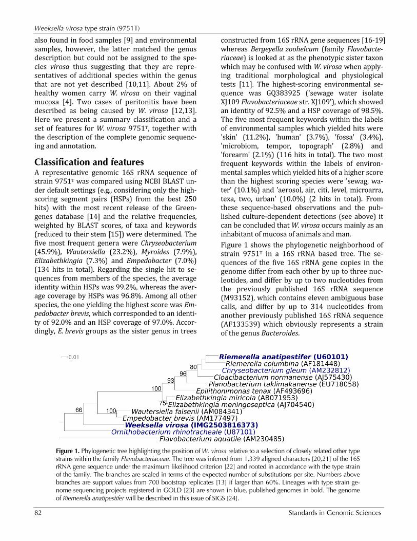
Classification and features A representative genomic 16S rRNA sequence of strain 9751T was compared using NCBI BLAST un-der default settings (e.g., considering only the high-scoring segment pairs (HSPs) from the best 250 hits) with the most recent release of the Green-genes database [14] and the relative frequencies, weighted by BLAST scores, of taxa and keywords (reduced to their stem [15]) were determined. The five most frequent genera were Chryseobacterium (45.9%), Wautersiella (23.2%), Myroides (7.9%), Elizabethkingia (7.3%) and Empedobacter (7.0%) (134 hits in total). Regarding the single hit to se-quences from members of the species, the average identity within HSPs was 99.2%, whereas the aver-age coverage by HSPs was 96.8%. Among all other species, the one yielding the highest score was Em-pedobacter brevis, which corresponded to an identi-ty of 92.0% and an HSP coverage of 97.0%. Accor-dingly, E. brevis groups as the sister genus in trees
constructed from 16S rRNA gene sequences [16-19] whereas Bergeyella zoohelcum (family Flavobacte-riaceae) is looked at as the phenotypic sister taxon which may be confused with W. virosa when apply-ing traditional morphological and physiological tests [11]. The highest-scoring environmental se-quence was GQ383925 ('sewage water isolate XJ109 Flavobacteriaceae str. XJ109'), which showed an identity of 92.5% and a HSP coverage of 98.5%. The five most frequent keywords within the labels of environmental samples which yielded hits were 'skin' (11.2%), 'human' (3.7%), 'fossa' (3.4%), 'microbiom, tempor, topograph' (2.8%) and 'forearm' (2.1%) (116 hits in total). The two most frequent keywords within the labels of environ-mental samples which yielded hits of a higher score than the highest scoring species were 'sewag, wa-ter' (10.1%) and 'aerosol, air, citi, level, microarra, texa, two, urban' (10.0%) (2 hits in total). From these sequence-based observations and the pub-lished culture-dependent detections (see above) it can be concluded that W. virosa occurs mainly as an inhabitant of mucosa of animals and man. Figure 1 shows the phylogenetic neighborhood of strain 9751T in a 16S rRNA based tree. The se-quences of the five 16S rRNA gene copies in the genome differ from each other by up to three nuc-leotides, and differ by up to two nucleotides from the previously published 16S rRNA sequence (M93152), which contains eleven ambiguous base calls, and differ by up to 314 nucleotides from another previously published 16S rRNA sequence (AF133539) which obviously represents a strain of the genus Bacteroides.
Figure 1. Phylogenetic tree highlighting the position of W. virosa relative to a selection of closely related other type strains within the family Flavobacteriaceae. The tree was inferred from 1,339 aligned characters [20,21] of the 16S rRNA gene sequence under the maximum likelihood criterion [22] and rooted in accordance with the type strain of the family. The branches are scaled in terms of the expected number of substitutions per site. Numbers above branches are support values from 700 bootstrap replicates [13] if larger than 60%. Lineages with type strain ge-nome sequencing projects registered in GOLD [23] are shown in blue, published genomes in bold. The genome of Riemerella anatipestifer will be described in this issue of SIGS [24].
Lang et al.
http://standardsingenomics.org 83
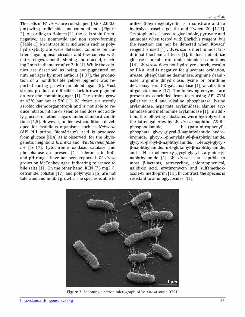
The cells of W. virosa are rod-shaped (0.6 × 2.0-3.0 µm) with parallel sides and rounded ends (Figure 2). According to Holmes [1], the cells stain Gram-negative, are nonmotile and non spore-forming (Table 1). No intracellular inclusions such as poly-hydroxybutyrate were detected. Colonies on nu-trient agar appear circular and low convex with entire edges, smooth, shining and mucoid, reach-ing 2mm in diameter after 24h [1]. While the colo-nies are described as being non-pigmented on nutrient agar by most authors [1,37], the produc-tion of a nondiffusible yellow pigment was re-ported during growth on blood agar [5]. Most strains produce a diffusible dark brown pigment on tyrosine-containing agar [1]. The strains grow at 42°C but not at 5°C [1]. W. virosa is a strictly aerobic chemoorganotroph and is not able to re-duce nitrate, nitrite or selenite and does not acidi-fy glucose or other sugars under standard condi-tions [1,5]. However, under test conditions devel-oped for fastidious organisms such as Neisseria (API NH strips, Biomérieux), acid is produced from glucose [IDA] as is observed for the phylo-genetic neighbors E. brevis and Wautersiella false-nii [16,17]. Cytochrome oxidase, catalase and phosphatase are present [1]. Tolerance to NaCl and pH ranges have not been reported. W. virosa grows on McConkey agar, indicating tolerance to bile salts [1] . On the other hand, KCN (75 mg l-1), cetrimide, colistin [17], and polymyxin [5] are not tolerated and inhibit growth. The species is able to
utilize β-hydroxybutyrate as a substrate and to hydrolyze casein, gelatin and Tween 20 [1,17]. Tryptophan is cleaved to give indole, pyruvate and ammonia when tested with Ehrlich’s reagent, but the reaction can not be detected when Kovacs’ reagent is used [1] . W. virosa is inert in most tra-ditional biochemical tests [1], it does not utilize glucose as a substrate under standard conditions [16]. W. virosa does not hydrolyze starch, esculin or DNA, and is negative for gluconate oxidation, urease, phenylalanine deaminase, arginine deami-nase, arginine dihydrolase, lysine or ornithine decarboxylase, β-D-galactosidase [1], alkalization of galacturonate [17]. The following enzymes are present as concluded from tests using API ZYM galleries: acid and alkaline phosphatase, lysine arylamidase, aspartate arylamidase, alanine ary-lamidase and methionine arylamidase [1]. In addi-tion, the following substrates were hydrolyzed in the latter galleries by W. virosa: naphthol-AS-BI-phosphodiamide, bis-(para-nitrophenyl)-phosphate, glycyl-glycyl-β-naphthylamide hydro-bromide, glycyl-L-phenylalanyl-β-naphthylamide, glycyl-L-prolyl-β-naphthylamide, L-leucyl-glycyl-β-naphthylamide, α-L-glutamyl-β-naphthylamide, and N-carbobenzoxy-glycyl-glycyl-L-arginine-β-naphthylamide [1]. W. virosa is susceptible to most β-lactams, tetracycline, chloramphenicol, nalidixic acid, erythromycin and sulfamethox-azole-trimethoprim [11]. In contrast, the species is resistant to aminoglycosides [11].
Figure 2. Scanning electron micrograph of W. virosa strain 9751T
Weeksella virosa type strain (9751T)
84 Standards in Genomic Sciences
Table 1. Classification and general features of W. virosa 9751T according to the MIGS recommendations [25]. MIGS ID Property Term Evidence code
Current classification
Domain Bacteria TAS [26] Phylum Bacteroidetes TAS [27] Class ‘Flavobacteria’ TAS [28] Order ‘Flavobacteriales’ TAS [29] Family Flavobacteriaceae TAS [30-33] Genus Weeksella TAS [1,34] Species Weeksella virosa TAS [1,34] Type strain 9751 (= CL345/78) TAS [1,34]
Gram stain negative TAS [1] Cell shape rod-shaped TAS [1] Motility non-motile TAS [1] Sporulation none TAS [1] Temperature range 10°C or less up to 42°C TAS [1] Optimum temperature not reported NAS Salinity not reported NAS MIGS-22 Oxygen requirement aerobic TAS [1] Carbon source proteins TAS [1] Energy source chemoorganotroph TAS [1] MIGS-6 Habitat human urogenital tract TAS [1,4] MIGS-15 Biotic relationship saprophyte TAS [1,11] MIGS-14 Pathogenicity none NAS Biosafety level 1 TAS [35] Isolation human urine TAS [1] MIGS-4 Geographic location North Carolina, USA TAS [1] MIGS-5 Sample collection time 1986 or before TAS [1] MIGS-4.1 Latitude not reported NAS MIGS-4.2 Longitude not reported NAS MIGS-4.3 Depth not reported NAS MIGS-4.4 Altitude not reported NAS
Evidence codes - IDA: Inferred from Direct Assay (first time in publication); TAS: Traceable Author Statement (i.e., a direct report exists in the literature); NAS: Non-traceable Author Statement (i.e., not directly observed for the living, isolated sample, but based on a general-ly accepted property for the species, or anecdotal evidence). These evidence codes are from of the Gene Ontology project [36]. If the evidence code is IDA, then the property was di-rectly observed by one of the authors or an expert mentioned in the acknowledgements.
Chemotaxonomy The major respiratory quinone of W. virosa is me-naquinone 6 and the major polyamine is homos-permidine, as is the case for all members of the family Flavobacteriaceae [11,38-40]. No sphingo-phospholipids were detected [1]. The polar lipids of W. virosa have not yet been described. The ma-jor whole-cell fatty acids of W. virosa are iso-C15:0 (46%), iso-C15:02-OH (10%), iso-C17:1ω12t (8%) and iso-C17:03-OH (7%) as described for CDC group IIf, the preliminary name given to these strains prior to being formally named W. virosa [41]. A comparison of the patterns of W. virosa and ‘W.
zoohelcum’ obtained at that time [41] with more recently published patterns of B. zoohelcum and E. brevis and phylogenetic neighbors [17,19] seems to cast doubts on the comparability of these early patterns. They are the only ones listing the pres-ence of high amounts of iso-C15:02-OH and iso-C17:1ω12t, which are not listed for phylogenetically related genera later on [19]. However, iso-C15:02-OH and isomers of iso-heptadecene are included in the summed features of the Microbial Identifica-tion System applied in many recent analyses in-cluding [17,19].

Lang et al.
http://standardsingenomics.org 85
Genome sequencing and annotation Genome project history This organism was selected for sequencing on the basis of its phylogenetic position [42], and is part of the Genomic Encyclopedia of Bacteria and Arc-haea project [40]. The genome project is deposited in the Genomes OnLine Database [23] and the
complete genome sequence is deposited in Gen-Bank. Sequencing, finishing and annotation were performed by the DOE Joint Genome Institute (JGI). A summary of the project information is shown in Table 2.
Table 2. Genome sequencing project information MIGS ID Property Term
MIGS-31 Finishing quality Finished
MIGS-28 Libraries used Tree genomic libraries: one 454 pyrosequence standard library, one 454 PE library (17 kb insert size), one Illumina library
MIGS-29 Sequencing platforms Illumina GAii, 454 GS FLX Titanium MIGS-31.2 Sequencing coverage 2,107.5 × Illumina; 64.3 × pyrosequence
MIGS-30 Assemblers Newbler version 2.5-internal-10Apr08-1-threads, Velvet version 0.7.63, phrap
MIGS-32 Gene calling method Prodigal 1.4, GenePRIMP INSDC ID CP02455 Genbank Date of Release February 15, 2011 GOLD ID Gc01619 NCBI project ID 50581 Database: IMG-GEBA 2503754024 MIGS-13 Source material identifier DSM 16922 Project relevance Tree of Life, GEBA
Growth conditions and DNA isolation W. virosa 9751T, DSM 16922, was grown on DSMZ medium 220 (Caso Agar) [37] at 30°C. DNA was isolated from 0.5-1 g of cell paste using Master-Pure Gram-positive DNA purification kit (Epicen-tre MGP04100) following the standard protocol as recommended by the manufacturer, with modifi-cation st/DL for cell lysis as described in Wu et al. [43]. DNA is available through the DNA Bank Net-work [44,45].
Genome sequencing and assembly The genome was sequenced using a combination of Illumina and 454 sequencing platforms. All gen-eral aspects of library construction and sequenc-ing can be found at the JGI website [46]. Pyrose-quencing reads were assembled using the Newb-ler assembler version 2.5-internal-10Apr08-1-threads (Roche). The initial Newbler assembly consisting of 27 contigs in one scaffold was con-verted into a phrap assembly by making fake reads from the consensus, to collect the read pairs in the 454 paired end library. Illumina GAii se-quencing data (4,788 Mb) was assembled with Velvet [47] and the consensus sequences were
shredded into 1.5 kb overlapped fake reads and assembled together with the 454 data. The 454 draft assembly was based on 131.6 Mb 454 draft data and all of the 454 paired end data. Newbler parameters are -consed -a 50 -l 350 -g -m -ml 20. The Phred/Phrap/Consed software package [48] was used for sequence assembly and quality as-sessment in the subsequent finishing process. Af-ter the shotgun stage, reads were assembled with parallel phrap (High Performance Software, LLC). Possible mis-assemblies were corrected with ga-pResolution, Dupfinisher [49], or sequencing cloned bridging PCR fragments with subcloning or transposon bombing (Epicentre Biotechnologies, Madison, WI). Gaps between contigs were closed by editing in Consed, by PCR and by Bubble PCR primer walks (J.-F.Chang, unpublished). A total of 60 additional reactions were necessary to close gaps and to raise the quality of the finished se-quence. Illumina reads were also used to correct potential base errors and increase consensus qual-ity using a software Polisher developed at JGI [50]. The error rate of the completed genome sequence is less than one in 100,000. Together, the combi-

Weeksella virosa type strain (9751T)
86 Standards in Genomic Sciences
nation of the Illumina and 454 sequencing plat-forms provided 2,171.8 × coverage of the genome. The final assembly contained 384,925 pyrose-quence and 63,008,730 Illumina reads.
Genome annotation Genes were identified using Prodigal [51] as part of the Oak Ridge National Laboratory genome an-notation pipeline, followed by a round of manual curation using the JGI GenePRIMP pipeline [52]. The predicted CDSs were translated and used to search the National Center for Biotechnology In-formation (NCBI) nonredundant database, UniProt, TIGR-Fam, Pfam, PRIAM, KEGG, COG, and InterPro databases. Additional gene prediction analysis and
functional annotation was performed within the Integrated Microbial Genomes - Expert Review (IMG-ER) platform [53].
Genome properties The genome consists of a 2,272,954 bp long chro-mosome with a GC content of 35.9% (Figure 3 and Table 3). Of the 2,181 genes predicted, 2,105 were protein-coding genes, and 76 RNAs; 56 pseudo-genes were also identified. The majority of the protein-coding genes (65.5%) were assigned with a putative function while the remaining ones were annotated as hypothetical proteins. The distribu-tion of genes into COGs functional categories is presented in Table 4.
Figure 3. Graphical circular map of the chromosome. From outside to the center: Genes on forward strand (color by COG categories), Genes on reverse strand (color by COG categories), RNA genes (tRNAs green, rRNAs red, other RNAs black), GC content, GC skew.
Lang et al.
http://standardsingenomics.org 87
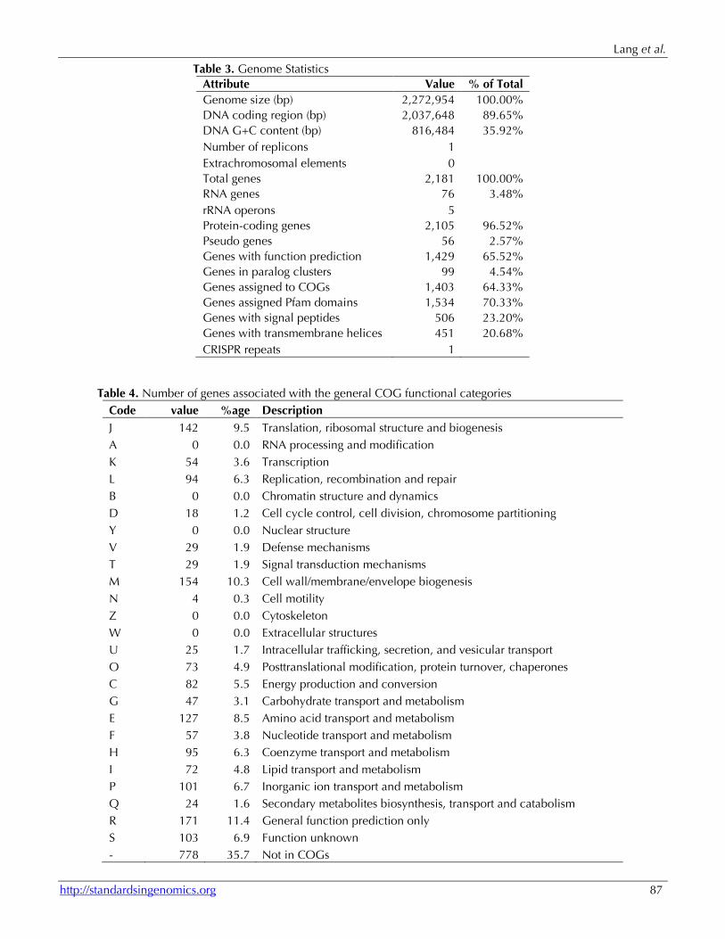
Table 3. Genome Statistics Attribute Value % of Total Genome size (bp) 2,272,954 100.00% DNA coding region (bp) 2,037,648 89.65% DNA G+C content (bp) 816,484 35.92% Number of replicons 1 Extrachromosomal elements 0 Total genes 2,181 100.00% RNA genes 76 3.48% rRNA operons 5 Protein-coding genes 2,105 96.52% Pseudo genes 56 2.57% Genes with function prediction 1,429 65.52% Genes in paralog clusters 99 4.54% Genes assigned to COGs 1,403 64.33% Genes assigned Pfam domains 1,534 70.33% Genes with signal peptides 506 23.20% Genes with transmembrane helices 451 20.68% CRISPR repeats 1
Table 4. Number of genes associated with the general COG functional categories Code value %age Description J 142 9.5 Translation, ribosomal structure and biogenesis A 0 0.0 RNA processing and modification K 54 3.6 Transcription L 94 6.3 Replication, recombination and repair B 0 0.0 Chromatin structure and dynamics D 18 1.2 Cell cycle control, cell division, chromosome partitioning Y 0 0.0 Nuclear structure V 29 1.9 Defense mechanisms T 29 1.9 Signal transduction mechanisms M 154 10.3 Cell wall/membrane/envelope biogenesis N 4 0.3 Cell motility Z 0 0.0 Cytoskeleton W 0 0.0 Extracellular structures U 25 1.7 Intracellular trafficking, secretion, and vesicular transport O 73 4.9 Posttranslational modification, protein turnover, chaperones C 82 5.5 Energy production and conversion G 47 3.1 Carbohydrate transport and metabolism E 127 8.5 Amino acid transport and metabolism F 57 3.8 Nucleotide transport and metabolism H 95 6.3 Coenzyme transport and metabolism I 72 4.8 Lipid transport and metabolism P 101 6.7 Inorganic ion transport and metabolism Q 24 1.6 Secondary metabolites biosynthesis, transport and catabolism R 171 11.4 General function prediction only S 103 6.9 Function unknown - 778 35.7 Not in COGs

Weeksella virosa type strain (9751T)
88 Standards in Genomic Sciences
Acknowledgements This work was performed under the auspices of the US Department of Energy Office of Science, Biological and Environmental Research Program, and by the Universi-ty of California, Lawrence Berkeley National Laboratory under contract No. DE-AC02-05CH11231, Lawrence Livermore National Laboratory under Contract No. DE-
AC52-07NA27344, and Los Alamos National Laborato-ry under contract No. DE-AC02-06NA25396, UT-Battelle and Oak Ridge National Laboratory under con-tract DE-AC05-00OR22725, as well as German Re-search Foundation (DFG) INST 599/1-2.
References 1. Holmes B, Steigerwaldt AG, Weaver RE, Brenner
DJ. Weeksella virosa gen. nov., sp. nov. (formerly group IIf) found in human clinical specimens. Syst Appl Microbiol 1986; 8:185-190.
2. Garrity G. NamesforLife. BrowserTool takes ex-pertise out of the database and puts it right in the browser. Microbiol Today 2010; 37:9.
3. Leung WK, Jin LJ, Yam WC, Samaranayake LP. Oral colonization of aerobic and facultatively anaerobic gram-negative rods and cocci in irra-diated, dentate, xerostomic individuals. Oral Mi-crobiol Immunol 2001; 16:1-9. PubMed doi:10.1034/j.1399-302x.2001.160101.x
4. Mardy C, Holmes B. Incidence of vaginal Week-sella virosa (formerly group IIf). J Clin Pathol 1988; 41:211-214. PubMed doi:10.1136/jcp.41.2.211
5. Reina J, Gil J, Salva F, Gomez J, Alomar P. Micro-biological characteristics of Weeksella virosa (formerly CDC group IIf) isolated from the human genitourinary tract. J Clin Microbiol 1990; 28:2357-2359. PubMed
6. Vela AI, García N, Latre MV, Casamayor A, Sán-chez-Porro C, Briones V, Ventosa A, Domínguez L, Fernández-Garayzábal JF. Aerococcus suis sp. nov., isolated from clinical specimens from swine. Int J Syst Evol Microbiol 2007; 57:1291-1294. PubMed doi:10.1099/ijs.0.64537-0
7. Brun R, Urraro C, Medaglia C, Russo V, Borzac-chiello G, Roperto F, Roperto S. Lymphoepitheli-oma-like carcinoma of the urinary bladder in a cow associated with bovine papillomavirus type-2. J Comp Pathol 2008; 139:121-125. Published on-line August 3, 2008. PubMed doi:10.1016/j.jcpa.2008.06.002
8. Gouveia C, Asensi MD, Zahner V, Rangel EF, Oliveira SM. Study on the bacterial midgut mi-crobiota associated to different Brazilian popula-tions of Lutzomyia longipalpis (Lutz & Neiva) (Diptera: Psychodidae). Neotrop Entomol 2008; 37:597-601. PubMed doi:10.1590/S1519-566X2008000500016
9. Botha WC, Jooste PJ, Hugo CJ. The incidence of Weeksella- and Bergeyella-like bacteria in the
food environment. J Appl Microbiol 1998; 84:349-356. PubMed doi:10.1046/j.1365-2672.1998.00346.x
10. Botha WC, Jooste PJ, Britz TJ. The taxonomic relationship of certain environmental flavobacte-ria to the genus Weeksella. J Appl Bacteriol 1989; 67:551-559. PubMed
11. Hugo CJ, Bruun B, Jooste PJ. The genera Bergeyel-la and Weeksella. In: The Prokaryotes, vol. 7: Pro-teobacteria: Delta, Epsilon Subclass. Dworkin M, Falkow S, Rosenberg E, Schleifer K-H and Stacke-brandt E (eds). 2006; 523-538.
12. Boixeda D, de Luis DA, Meseguer MA, Aller R, Martin de Argila C. Lopez Sanroman. A case of spontaneous peritonitis caused by Weeksella viro-sa. Eur J Gastroenterol Hepatol 1998; 10:897-898. PubMed doi:10.1097/00042737-199810000-00016
13. Pattengale ND, Alipour M, Bininda-Emonds ORP, Moret BME, Stamatakis A. How many bootstrap replicates are necessary? Lect Notes Comput Sci 2009; 5541:184-200. doi:10.1007/978-3-642-02008-7_13
14. DeSantis TZ, Hugenholtz P, Larsen N, Rojas M, Brodie EL, Keller K, Huber T, Dalevi D, Hu P, Andersen GL. Greengenes, a Chimera-Checked 16S rRNA Gene Database and Workbench Com-patible with ARB. Appl Environ Microbiol 2006; 72:5069-5072. PubMed doi:10.1128/AEM.03006-05
15. Porter MF. An algorithm for suffix stripping. Pro-gram: electronic library and information systems 1980; 14:130-137.
16. Bernardet JF, Nakagawa Y. An introduction to the family Flavobacteriaceae. In: The Prokaryotes, vol. 7: Proteobacteria: Delta, Epsilon Subclass. Dwor-kin M, Falkow S, Rosenberg E, Schleifer K-H and Stackebrandt E (eds). 2006; 455-480.
17. Kämpfer P, Avesani V, Janssens M, Charlier J, De Baere T, Vaneechoutte M. Description of Wauter-siella falsenii gen. nov., sp. nov., to accommodate clinical isolates phenotypically resembling mem-bers of the genera Chryseobacterium and Empe-

Lang et al.
http://standardsingenomics.org 89
dobacter. Int J Syst Evol Microbiol 2006; 56:2323-2329. PubMed doi:10.1099/ijs.0.64393-0
18. O'Sullivan LA, Rinna J, Humphreys G, Weightman AJ, Fry JC. Culturable phylogenetic diversity of the phylum 'Bacteroidetes' from river epilithon and coastal water and description of novel members of the family Flavobacteriaceae: Epilithonimonas tenax gen. nov., sp. nov. and Per-sicivirga xylanidelens gen.nov., sp. nov. Int J Syst Evol Microbiol 2006; 56:169-180. PubMed doi:10.1099/ijs.0.63941-0
19. Hugo CJ, Segers P, Hoste B, Vancanneyt M, Ker-sters K. Chryseobacterium joostei sp. nov., iso-lated from the dairy environment. Int J Syst Evol Microbiol 2003; 53:771-777. PubMed doi:10.1099/ijs.0.02232-0
20. Castresana J. Selection of conserved blocks from multiple alignments for their use in phylogenetic analysis. Mol Biol Evol 2000; 17:540-552. PubMed
21. Lee C, Grasso C, Sharlow MF. Multiple sequence alignment using partial order graphs. Bioinformat-ics 2002; 18:452-464. PubMed doi:10.1093/bioinformatics/18.3.452
22. Stamatakis A, Hoover P, Rougemont J. A rapid bootstrap algorithm for the RAxML Web servers. Syst Biol 2008; 57:758-771. PubMed doi:10.1080/10635150802429642
23. Liolios K, Chen IM, Mavromatis K, Tavernarakis N, Hugenholtz P, Markowitz VM, Kyrpides NC. The Genomes On Line Database (GOLD) in 2009: sta-tus of genomic and metagenomic projects and their associated metadata. Nucleic Acids Res 2010; 38:D346-D354. PubMed doi:10.1093/nar/gkp848
24. Han C, Goodwin L, Pitluck S, Liolios L, Pagani I, Ivanova N, Mikhailova N, Pati A, Chen A, Palia-nappan K, et al. Complete genome sequence of Riemerella anatipestifer type strain (BrunerT). Stand Genomic Sci 2011; 4(2).
25. Field D, Garrity G, Gray T, Morrison N, Selengut J, Sterk P, Tatusova T, Thomson N, Allen MJ, Angi-uoli SV, et al. The minimum information about a genome sequence (MIGS) specification. Nat Bio-technol 2008; 26:541-547. PubMed doi:10.1038/nbt1360
26. Woese CR, Kandler O, Wheelis ML. Towards a natural system of organisms: proposal for the do-mains Archaea, Bacteria, and Eucarya. Proc Natl Acad Sci USA 1990; 87:4576-4579. PubMed doi:10.1073/pnas.87.12.4576
27. Garrity GM, Holt JG. The Road Map to the Ma-nual. In: Garrity GM, Boone DR, Castenholz RW (eds), Bergey's Manual of Systematic Bacteriology, Second Edition, Volume 1, Springer, New York, 2001, p. 119-169.
28. Ludwig W, Euzeby J, Whitman WG. Draft tax-onomic outline of the Bacteroidetes, Planctomy-cetes, Chlamydiae, Spirochaetes, Fibrobacteres, Fusobacteria, Acidobacteria, Verrucomicrobia, Dictyoglomi, and Gemmatimonadetes. http://www.bergeys.org/outlines/Bergeys_Vol_4_Outline.pdf. Taxonomic Outline 2008
29. Garrity GM, Holt JG. 2001. Taxonomic outline of the Archaea and Bacteria, p. 155-166. In G. M. Garrity, D. R. Boone, and R. W. Castenholz (ed.), Bergey's Manual of Systematic Bacteriology, 2nd ed, vol. 1. Springer, New York.
30. Validation of the publication of new names and new combinations previously effectively pub-lished outside the IJSB. List No. 41. Int J Syst Bac-teriol 1992; 42:327-328. doi:10.1099/00207713-42-2-327
31. Reichenbach H. Order 1. Cytophagales Leadbet-ter 1974, 99AL. In: Holt JG (ed), Bergey's Manual of Systematic Bacteriology, First Edition, Volume 3, The Williams and Wilkins Co., Baltimore, 1989, p. 2011-2013.
32. Bernardet JF, Segers P, Vancanneyt M, Berthe F, Kersters K, Vandamme P. Cutting a Gordian knot: emended classification and description of the ge-nus Flavobacterium, emended description of the family Flavobacteriaceae, and proposal of Flavo-bacterium hydatis nom. nov. (Basonym, Cytopha-ga aquatilis Strohl and Tait 1978). Int J Syst Bacte-riol 1996; 46:128-148. doi:10.1099/00207713-46-1-128
33. Bernardet JF, Nakagawa Y, Holmes B. Proposed minimal standards for describing new taxa of the family Flavobacteriaceae, and emended descrip-tion of the family. Int J Syst Evol Microbiol 2002; 52:1049-1070. PubMed doi:10.1099/ijs.0.02136-0
34. Validation list No° 23. Int J Syst Bacteriol 1987; 37:179-180. doi:10.1099/00207713-37-2-179
35. Classification of bacteria and archaea in risk groups. http://www.baua.de TRBA 466.
36. Ashburner M, Ball CA, Blake JA, Botstein D, But-ler H, Cherry JM, Davis AP, Dolinski K, Dwight SS, Eppig JT, et al. Gene Ontology: tool for the unifi-cation of biology. Nat Genet 2000; 25:25-29. PubMed doi:10.1038/75556

Weeksella virosa type strain (9751T)
90 Standards in Genomic Sciences
37. List of growth media used at DSMZ: http://www.dsmz.de/microorganisms/media_list.php.
38. Vandamme P, Bernardet JF, Segers P, Kersters K, Holmes B. New perspectives in the classification of the flavobacteria: description of Chryseobacte-rium gen. nov., Bergeyella gen. nov., and Empe-dobacter nom. rev. Int J Syst Bacteriol 1994; 44:827-831. doi:10.1099/00207713-44-4-827
39. Bernardet JF, Nakagawa Y, Holmes B. Proposed minimal standards for describing new taxa of the family Flavobacteriaceae and emended descrip-tion of the family. Int J Syst Evol Microbiol 2002; 52:1049-1070. PubMed doi:10.1099/ijs.0.02136-0
40. Hamana K, Nakagawa Y. Polyamine distribution profiles in the eighteen genera phylogenetically located within the Flavobacterium-Flexibacter-Cytophaga complex. Microbios 2001; 106:7-17. PubMed
41. Hollis DG, Daneshvar MI, Moss CW, Baker CN. Phenotypic characteristics, fatty acid composition, and isoprenoid quinone content of CDC group IIg bacteria. J Clin Microbiol 1995; 33:762-764. PubMed
42. Klenk HP, Göker M. En route to a genome-based classification of Archaea and Bacteria? Syst Appl Microbiol 2010; 33:175-182. PubMed doi:10.1016/j.syapm.2010.03.003
43. Wu D, Hugenholtz P, Mavromatis K, Pukall R, Dalin E, Ivanova NN, Kunin V, Goodwin L, Wu M, Tindall BJ, et al. A phylogeny-driven genomic encyclopaedia of Bacteria and Archaea. Nature 2009; 462:1056-1060. PubMed doi:10.1038/nature08656
44. Gemeinholzer B, Dröge G, Zetzsche H, Haszpru-nar G, Klenk HP, Güntsch A, Berendsohn WG, Wägele JW. The DNA Bank Network: the start from a German initiative. Biopreservation and Biobanking (In press).
45. DNA bank Network. http://www.dnabank-network.org
46. DOE Joint Genome Institute. http://www.jgi.doe.gov
47. Zerbino DR, Birney E. Velvet: algorithms for de novo short read assembly using de Bruijn graphs. Genome Res 2008; 18:821-829. PubMed doi:10.1101/gr.074492.107
48. Phrap and Phred for Windows. MacOS, Linux, and Unix. http://www.phrap.com
49. Han C, Chain P. 2006. Finishing repeat regions automatically with Dupfinisher. Proceeding of the 2006 international conference on bioinformatics & computational biology. Edited by Hamid R. Arabnia & Homayoun Valafar, CSREA Press. June 26-29, 2006: 141-146.
50. Lapidus A, LaButti K, Foster B, Lowry S, Trong S, Goltsman E. POLISHER: An effective tool for us-ing ultra short reads in microbial genome assem-bly and finishing. AGBT, Marco Island, FL, 2008.
51. Hyatt D, Chen GL, LoCascio PF, Land ML, Lari-mer FW, Hauser LJ. Prodigal: prokaryotic gene recognition and translation initiation site identifi-cation. BMC Bioinformatics 2010; 11:119. PubMed doi:10.1186/1471-2105-11-119
52. Pati A, Ivanova NN, Mikhailova N, Ovchinnikova G, Hooper SD, Lykidis A, Kyrpides NC. Gene-PRIMP: a gene prediction improvement pipeline for prokaryotic genomes. Nat Methods 2010; 7:455-457. PubMed doi:10.1038/nmeth.1457
53. Markowitz VM, Ivanova NN, Chen IMA, Chu K, Kyrpides NC. IMG ER: a system for microbial ge-nome annotation expert review and curation. Bio-informatics 2009; 25:2271-2278. PubMed doi:10.1093/bioinformatics/btp393
54. Faber MD, del Busto R, Cruz C, Mezger E. Re-sponse of Weeksella virosa peritonitis to imipe-nem/cilastin. Adv Perit Dial 1991; 7:133-134. PubMed