wearing of soft contact lenses
TRANSCRIPT
British Journal ofOphthalmology, 1983, 67, 733-736
Biomicroscopy of papillae associated withwearing of soft contact lensesDONALD R. KORB,' JACK V. GREINER,2 VICTOR M. FINNEMORE,'AND MATHEA R. ALLANSMITH3
From '80 Boylston Street, Boston, Massachusetts; the Departments of Ophthalmology,2University ofIllinois Eye and Ear Infirmary, Chicago, and 3Harvard Medical School, Boston;the 2 3Department ofCornea Research, Eye Research Institute ofRetina Foundation, Boston; and the2Department ofPathology, Chicago College of Osteopathic Medicine, Chicago, USA
SUMMARY We studied the topographical, macrostructural, and fluorescein staining characteristicsof papillary changes of the upper tarsal conjunctiva associated with the wearing of hydrophilic (soft)contact lenses. Fifty soft contact lens wearers with elevated conjunctival papillae >03 mm indiameter were studied. Topographic characteristics recorded were distribution and number ofpapillae; macrostructural characteristics recorded were diameter and morphology of papillae. Theinformation collected included age of patient, duration of lens wear, average daily time of wearinglens, presence or absence of itching or mucus, refractive status, and atopic history. Papillae werefound in most cases in the conjunctival zone adjacent to the tarsal fold and were never found in thezone adjacent to the eyelid margin without also occurring in the intervening zone. The diameter ofthe papillae ranged from >03 mm to 2-0 mm. The number of papillae per eyelid ranged from 4 toover 100. The apices of the papillae were frequently flattened, and these flattened surfacesfrequently stained with fluorescein. The vascular supply of individual papillae was observed toradiate from a vessel occupying the central core of each papilla.
Clinically observable changes in the upper tarsalconjunctiva have been reported in the wearers ofboth hard and soft contact lenses.'2 The macro-structural characteristics of the papillary changes ofthe upper tarsal conjunctiva associated with thewearing of polymethylmethacrylate (PMMA) contactlenses have been established.34 We questionedwhether there are biomicroscopically observablecharacteristics that would permit differentiationbetween hard and soft contact lens papillary con-junctivitis based on the distribution, morphology,and staining characteristics of papillae, and thedegree of hyperaemia.The purpose of the present study was to determine
the characteristics of changes of the upper tarsalconjunctiva associated with the wearing of softcontact lenses, and to compare and contrast thesecharacteristics with those changes associated with thewearing of hard contact lenses.4
Correspondence to Mathea R. Allansmith, MD, 20 Staniford St,Boston, MA 02114, USA.
Subjects and methods
Subject selection. Fifty subjects with elevated con-junctival papillae >0 3 mm in diameter associatedwith hydrophilic (soft) contact lens wearing wereselected for study. Since standardised eyelid exam-inations had not been performed at regular intervalson all subjects, neither the onset nor the duration ofthis condition could be determined. Each subject'sage, duration of lens wear, average daily lens wearingtime, symptoms of itching and/or mucus, refractivestatus, and atopic history were obtained. Subjectswore their contact lenses for at least 2 hours beforeexamination. Since we observed that subjects whohad not worn their lenses for several days presented amarked decrease in conj-1nctival hyperaemia and thenumber of fluorescein-stained papillae, we selectedonly those subjects who were able to wear lenses aminimum of 4 hours.Method of examination. The upper eyelid was
everted, and the tarsal conjunctival surface wasexamined with the biomicroscope under white light
733
group.bmj.com on April 4, 2018 - Published by http://bjo.bmj.com/Downloaded from
Donald R. Korb, Jack V. Greiner, Victor M. Finnemore, and Mathea R. Allansmith
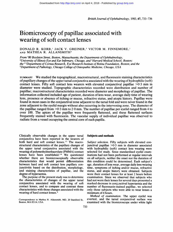
Fig. 1 Diagram illustrating the three zones ofthe tarsalconjunctiva (3).
and 16x magnification. The lid was returned to thenormal position and 2 drops of sterile 2% sodiumfluorescein were instilled into the lower cul-de-sac.Subjects were instructed to blink completely 6 times todistribute the fluorescein over the tarsal conjunctivalsurface. The upper eyelid was then re-everted, andbiomicroscopic examination was repeated with cobaltblue light.
Papillae were considered elevated only when theywere observed, with a slit-beam and lOx magnifica-tion, to protrude from the surrounding conjunctivasurface. The distribution, number, diameter,morphology, presence of absence of apical staining,and the vascular pattern of the elevated papillae werenoted. To describe the distribution of papillae theconjunctiva over the tarsal plate was divided into 3zones of equal width (Fig. 1). Papillary diameter wasmeasured with the micrometer on the slit apparatusof the Haag-Streit 900 slit-lamp. The upper tarsalconjunctiva was photographed first with white lightand then with cobalt blue light after fluoresceininstillation. Papillae or follicles occurring on thejunctional conjunctiva (Fig. 1) were not considered tobe abnormal, as they are frequently present in healthypersons.
Hyperaemia of the upper tarsal conjunctiva was
graded on a scale of zero to +3: zero represented theabsence of hyperaemia; grades 1, 2, and 3 thecategories of mild, moderate, and severe respectively.
Results
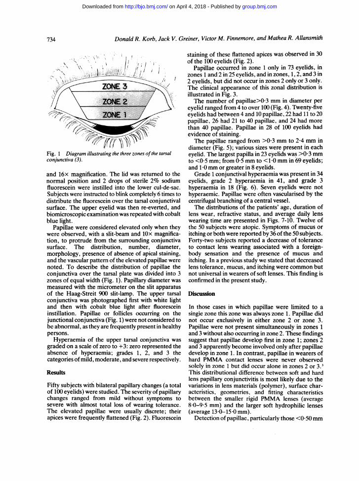
Fifty subjects with bilateral papillary changes (a totalof 100 eyelids) were studied. The severity of papillarychanges ranged from mild without symptoms tosevere with almost total loss of wearing tolerance.The elevated papillae were usually discrete; theirapices were frequently flattened (Fig. 2). Fluorescein
staining of these flattened apices was observed in 30of the 100 eyelids (Fig. 2).
Papillae occurred in zone 1 only in 73 eyelids, inzones 1 and 2 in 25 eyelids, and in zones, 1, 2, and 3 in2 eyelids, but did not occur in zones 2 only or 3 only.The clinical appearance of this zonal distribution isillustrated in Fig. 3.The number of papillae>0 3 mm in diameter per
eyelid ranged from 4 to over 100 (Fig. 4). Twenty-fiveeyelids had between 4 and 10 papillae, 22 had 11 to 20papillae, 26 had 21 to 40 papillae, and 24 had morethan 40 papillae. Papillae in 28 of 100 eyelids hadevidence of staining.The papillae ranged from >0 3 mm to 2-4 mm in
diameter (Fig. 5); various sizes were present in eacheyelid. The largest papilla in 23 eyelids was >0 3 mmto <0 5 mm; from 0-5 mm to <1 0 mm in 69 eyelids;and 1 0 mm or greater in 8 eyelids.Grade 1 conjunctival hyperaemia was present in 34
eyelids, grade 2 hyperaemia in 41, and grade 3hyperaemia in 18 (Fig. 6). Seven eyelids were nothyperaemic. Papillae were often vascularised by thecentrifugal branching of a central vessel.The distributions of the patients' age, duration of
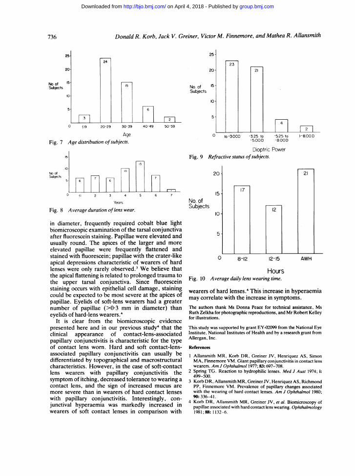
lens wear, refractive status, and average daily lenswearing time are presented in Figs. 7-10. Twelve ofthe 50 subjects were atopic. Symptoms of mucus oritching or both were reported by 36 of the 50 subjects.Forty-two subjects reported a decrease of toleranceto contact lens wearing associated with a foreign-body sensation and the presence of mucus anditching. In a previous study we stated that decreasedlens tolerance, mucus, and itching were common butnot universal in wearers of soft lenses. This finding isconfirmed in the present study.
Discussion
In those cases in which papillae were limited to asingie zone this zone was always zone 1. Papillae didnot occur exclusively in either zone 2 or zone 3.Papillae were not present simultaneously in zones 1and 3 without also occurring in zone 2. These findingssuggest that papillae develop first in zone 1; zones 2and 3 apparently become involved only after papillaedevelop in zone 1. In contrast, papillae in wearers ofhard PMMA contact lenses were never observedsolely in zone 1 but did occur alone in zones 2 or 3.3This distributional difference between soft and hardlens papillary conjunctivitis is most likely due to thevariations in lens materials (polymer), surface char-acteristics, geometries, and fitting characteristicsbetween the smaller rigid PMMA lenses (average8-0-9-5 mm) and the larger soft hydrophilic lenses(average 13 0-15 0 mm).
Detection of papillae, particularly those <0 50mm
734
group.bmj.com on April 4, 2018 - Published by http://bjo.bmj.com/Downloaded from

735Biomnicroscopy ofcontact lens conjunctivitis
IFig. 2 (roww) attarrenedpaptllaein zone 5 1.2and ?photographed (left) with robalt hblue lightafter fluorescein instillaition, (ccnt re) white lightafter trriFation of fluorescein. and (right)0llttenedpapillae in zones 1 and2.Fig. 3 (row 2) Zonal distribution of contactlens-associated papillae in the upper rarsalconjuncti va. l eft: Papillae zone I (niY.Cent.rePapillae in zones I and 2 on lY. Ri,ght Papillaein all 3 zones.Fig. 4 (row 3) Left: Upper tarsal conjuntiwawith less than 15 papillae confined to zones Iand 2. Right: Upper tarsal conjunctiva withmore than 100 papillaje in zones I and 2 withpartial invasion ofzone 3.Fig. 5 (rows 4 and 5) Row 4: Papillae rangingfrom >-003 to <0-5 tmm7 in diameter: (leftpalpillae extending from nasal junctionalconjunctiva to zones and 2 phorographed withwhite light and (right) .subsequentlyphotograiphed with cobalt blue lfght afterfluorescein instillaition. Row 5: left: Papillae inall.1 zones photographed with cobalt bluie fightafter fluorescein instillation. Papillae ranginjgfrom 0-S to > -0 mm in diameter. Centre:Papillae photographed with white light. Right:Papillae photographed with c obalt blue light|fterfluorescein instillation.Fig. 6 (row 6) Lipper tarsal conjunctiv'a with(left)grade I hvperaemia. (centre) grade 2hvperaernia (right) grade 3 hvperaemia.
group.bmj.com on April 4, 2018 - Published by http://bjo.bmj.com/Downloaded from
Donald R. Korb, Jack V. Greiner, Victor M. Finnemore, and Mathea R. Allansmith
25
20
No. ofSubjects
15
10o
5.
3
24
25
20
15 No. ofSubjects
15.
10
6
5.F2-F0 (19 20-29 30-39 40-49 50-59
AgeFig. 7 Age distribution ofsubjects.
15
13
10.~~~~~~~~~~~~1No of 3Subjects 6
7
0 (I 2 3 4 5 6 7
Years
Fig. 8 A verage duration of lens wear.
in diameter, frequently required cobalt blue lightbiomicroscopic examination of the tarsal conjunctivaafter fluorescein staining. Papillae were elevated andusually round. The apices of the larger and moreelevated papillae were frequently flattened andstained with fluorescein; papillae with the crater-likeapical depressions characteristic of wearers of hardlenses were only rarely observed.3 We believe thatthe apical flattening is related to prolonged trauma tothe upper tarsal conjunctiva. Since fluoresceinstaining occurs with epithelial cell damage, stainingcould be expected to be most severe at the apices ofpapillae. Eyelids of soft-lens wearers had a greaternumber of papillae (>0.3 mm in diameter) thaneyelids of hard-lens wearers.4
It is clear from the biomicroscopic evidencepresented here and in our previous study4 that theclinical appearance of contact-lens-associatedpapillary conjunctivitis is characteristic for the typeof contact lens worn. Hard and soft contact-lens-associated papillary conjunctivitis can usually bedifferentiated by topographical and macrostructuralcharacteristics. However, in the case of soft-contactlens wearers with papillary conjunctivitis thesymptom of itching, decreased tolerance to wearing acontact lens, and the sign of increased mucus aremore severe than in wearers of hard contact lenseswith papillary conjunctivitis. Interestingly, con-junctival hyperaemia was markedly increased inwearers of soft contact lenses in comparison with
2321
4
0 to-300D -3.25 to -5.25 to-5.00 D -8.00D
Dioptric PowerFig. 9 Refractive status ofsubjects.
20
No. ofSubjects
15-
10
5.
17
0 8-12
12
12-15
>-8.00D
21
AWH
HoursFig. 10 Average daily lens wearing time.
wearers of hard lenses.4 This increase in hyperaemiamay correlate with the increase in symptoms.The authors thank Ms Donna Peace for technical assistance, MsRuth Zelkha for photographic reproductions, and Mr Robert Kelleyfor illustrations.
This study was supported by grant EY-02099 from the National EyeInstitute, National Institutes of Health and by a research grant fromAllergan, Inc.
References
1 Allansmith MR, Korb DR, Greiner JV, Henriquez AS, SimonMA, Finnemore VM. Giant papillary conjunctivitis in contact lenswearers. Am J Ophthalmol 1977; 83: 697-708.
2 Spring TG. Reaction to hydrophilic lenses. Med J Aust 1974; i:499-500.
3 Korb DR, Allansmith MR, Greiner JV, Henriquez AS, RichmondPP, Finnemore VM. Prevalence of papillary changes associatedwith the wearing of hard contact lenses. Am J Ophthalmol 1980;90: 336-41.
4 Korb DR, Allansmith MR, Greiner JV, et al. Biomicroscopy ofpapillae associated with hard contact lens wearing. Ophthalmology1981;88: 1132-6.
736
group.bmj.com on April 4, 2018 - Published by http://bjo.bmj.com/Downloaded from
with wearing of soft contact lenses.Biomicroscopy of papillae associated
D R Korb, J V Greiner, V M Finnemore and M R Allansmith
doi: 10.1136/bjo.67.11.7331983 67: 733-736 Br J Ophthalmol
http://bjo.bmj.com/content/67/11/733Updated information and services can be found at:
These include:
serviceEmail alerting
Sign up in the box at the top right corner of the online article. Receive free email alerts when new articles cite this article.
Notes
http://group.bmj.com/group/rights-licensing/permissionsTo request permissions go to:
http://journals.bmj.com/cgi/reprintformTo order reprints go to:
http://group.bmj.com/subscribe/To subscribe to BMJ go to:
group.bmj.com on April 4, 2018 - Published by http://bjo.bmj.com/Downloaded from