waters glycan analysis by uplc-fl-ms€¦ · waters glycan analysis by uplc-fl-ms ... structure...
TRANSCRIPT
©2009 Waters Corporation | COMPANY CONFIDENTIAL ©2012 Waters Corporation
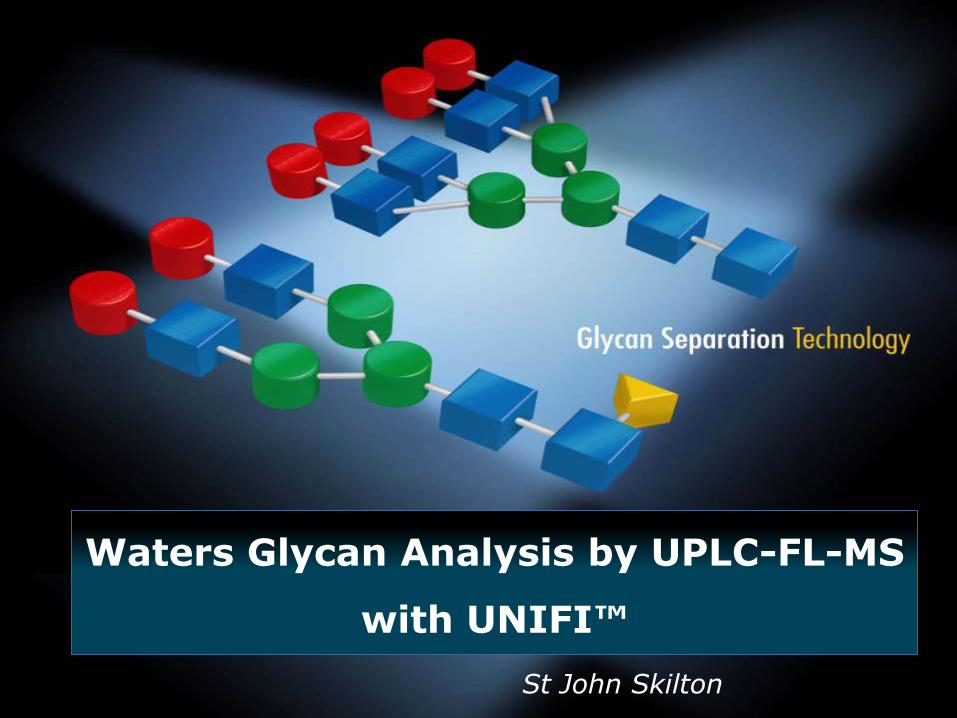
St John Skilton
Waters Glycan Analysis by UPLC-FL-MS
with UNIFI™

©2009 Waters Corporation | COMPANY CONFIDENTIAL ©2012 Waters Corporation
How Do you Explain it to the FDA?

©2009 Waters Corporation | COMPANY CONFIDENTIAL ©2012 Waters Corporation
How Do you Explain it to the FDA? In its constituent parts…

©2009 Waters Corporation | COMPANY CONFIDENTIAL ©2012 Waters Corporation
Workflow for Routine Intact Protein and Peptide Map Analysis by LC/MS
Prepare Standards & Samples
Data Acquisition & Processing
Data Management
Create Analysis Method and Sample Sequence
Reports Compiled, Reviewed & Signed
©2009 Waters Corporation | COMPANY CONFIDENTIAL ©2012 Waters Corporation
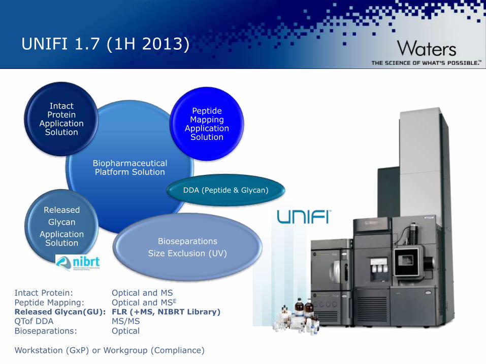
UNIFI 1.7 (1H 2013)
Biopharmaceutical Platform Solution
Intact Protein
Application Solution
Peptide Mapping
Application Solution
Bioseparations
Size Exclusion (UV)
Released
Glycan
Application Solution
DDA (Peptide & Glycan)
Intact Protein: Optical and MS Peptide Mapping: Optical and MSE
Released Glycan(GU): FLR (+MS, NIBRT Library)
QTof DDA MS/MS Bioseparations: Optical Workstation (GxP) or Workgroup (Compliance)

©2009 Waters Corporation | COMPANY CONFIDENTIAL ©2012 Waters Corporation
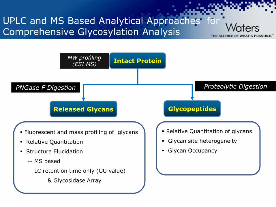
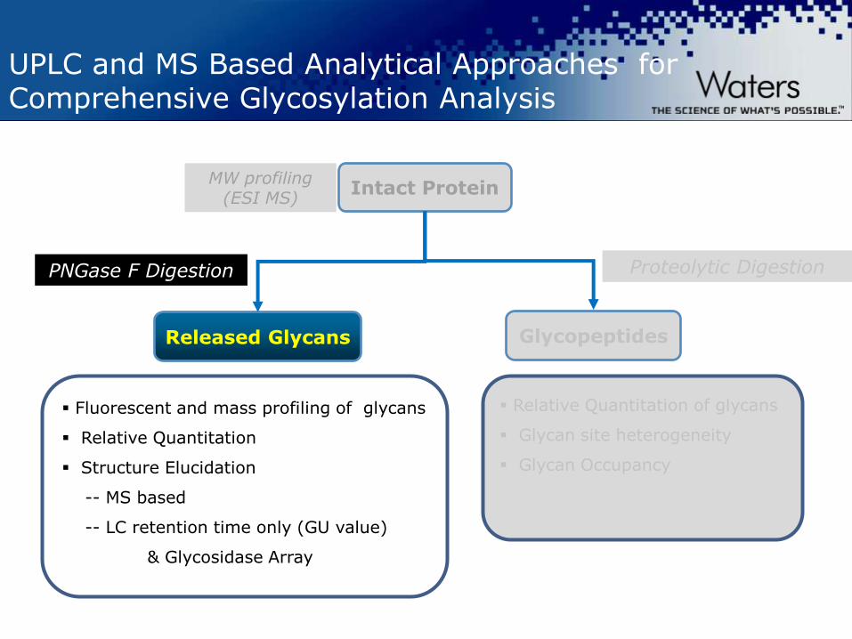
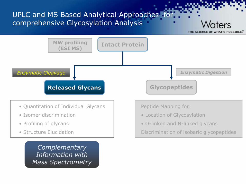
UPLC and MS Based Analytical Approaches for comprehensive Glycosylation Analysis
Export RT and
Import GU
values
Export MS and
MS/MS Spectra
for Structure
©2009 Waters Corporation | COMPANY CONFIDENTIAL ©2012 Waters Corporation
UPLC and MS Based Analytical Approaches for Comprehensive Glycosylation Analysis
Intact Protein
Glycopeptides Released Glycans
MW profiling (ESI MS)
Proteolytic Digestion PNGase F Digestion
Fluorescent and mass profiling of glycans
Relative Quantitation
Structure Elucidation
-- MS based
-- LC retention time only (GU value)
& Glycosidase Array
Relative Quantitation of glycans
Glycan site heterogeneity
Glycan Occupancy
©2009 Waters Corporation | COMPANY CONFIDENTIAL ©2012 Waters Corporation
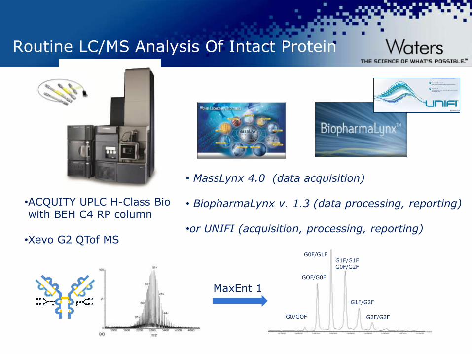
Routine LC/MS Analysis Of Intact Protein
•ACQUITY UPLC H-Class Bio with BEH C4 RP column •Xevo G2 QTof MS
• MassLynx 4.0 (data acquisition) • BiopharmaLynx v. 1.3 (data processing, reporting) •or UNIFI (acquisition, processing, reporting)
GOF/G0F
G0F/G1F G1F/G1F G0F/G2F
G1F/G2F
G0/GOF G2F/G2F
MaxEnt 1
©2009 Waters Corporation | COMPANY CONFIDENTIAL ©2012 Waters Corporation
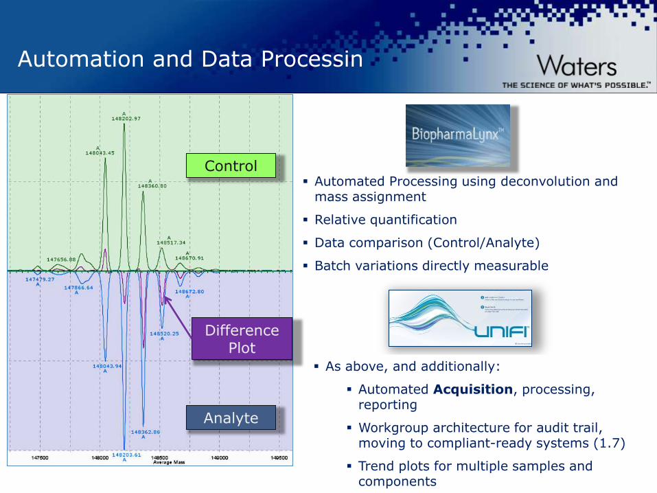
Automation and Data Processin
Automated Processing using deconvolution and mass assignment
Relative quantification
Data comparison (Control/Analyte)
Batch variations directly measurable
Control
As above, and additionally:
Automated Acquisition, processing, reporting
Workgroup architecture for audit trail, moving to compliant-ready systems (1.7)
Trend plots for multiple samples and components
Analyte
Difference Plot

©2009 Waters Corporation | COMPANY CONFIDENTIAL ©2012 Waters Corporation
Batch 1
MaxEnt1 Deconvoluted Spectra of Intact IgG1 from three Batches Processed by BiopharmaLynx
Batch 2
Batch 3
(M
an
5)2
(G1F)2 G0F/G2F
Man
5/
Man
6
(G
2F)2
(G
0F)2
G0
F/
G1
F
G1
F/
G2
F
G0
/G
0F

©2009 Waters Corporation | COMPANY CONFIDENTIAL ©2012 Waters Corporation
LC/MS Analysis of HC (Reduced IgG)
Heavy chain (HC) and light chain (LC) fragments generated by partial reduction of monoclonal antibody with DTT.
2 2 +
LC (~25 kD) HC (~50 kD)

©2009 Waters Corporation | COMPANY CONFIDENTIAL ©2012 Waters Corporation
Total Ion Chromatogram of reduced IgG1 antibody
Column: ACQUITY UPLC BEH C4, 1.7 µm, 2.1 x 50 mm
Gradient: 28-32% B in 12 min A: 0.02% TFA in water B: 0.018% TFA in ACN
LC HC
+34 Charge State
HC Mass Spectrum

©2009 Waters Corporation | COMPANY CONFIDENTIAL ©2012 Waters Corporation
MaxEnt1 deconvoluted spectra of IgG HC (~50kDa) generated from three different batches of Trastuzumab
Batch 1
Batch 2
Batch 3
G0
F-G
N
G0
G0
-GN
Man
5
G 1
G1
F
Man
6
G2
G2
F
G2
FS
1
G0
F
G1
F-G
N
Reduction of the antibody into monomeric HC improves isoform resolution allowing additional isoform identification and quantitation of the individual glycoform.
HC
A : Trastuzumab HC
A : Trastuzumab HC
A : Trastuzumab HC
Man
5

©2009 Waters Corporation | COMPANY CONFIDENTIAL ©2012 Waters Corporation
Intact Erythropoietin
LC/MS chromatogram
32 Glycoforms were accounted for

©2009 Waters Corporation | COMPANY CONFIDENTIAL ©2012 Waters Corporation
Comparison of Glycoform Quantitation by MS with 2AB labelled methodology – IgG1
Glycoform quantitation for reduced species by LC/MS can be compared to quantification of species by 2-AB labelled methods by UPLC with FL detection

©2009 Waters Corporation | COMPANY CONFIDENTIAL ©2012 Waters Corporation
Automated comparative analysis – intact protein analysis
Protein modifications identified
Deconvoluted annotated mass spectra
Chromatograms for reference and analyte

©2009 Waters Corporation | COMPANY CONFIDENTIAL ©2012 Waters Corporation
Tools for sample comparisons
Innovator
“Biosimilar”
©2009 Waters Corporation | COMPANY CONFIDENTIAL ©2012 Waters Corporation
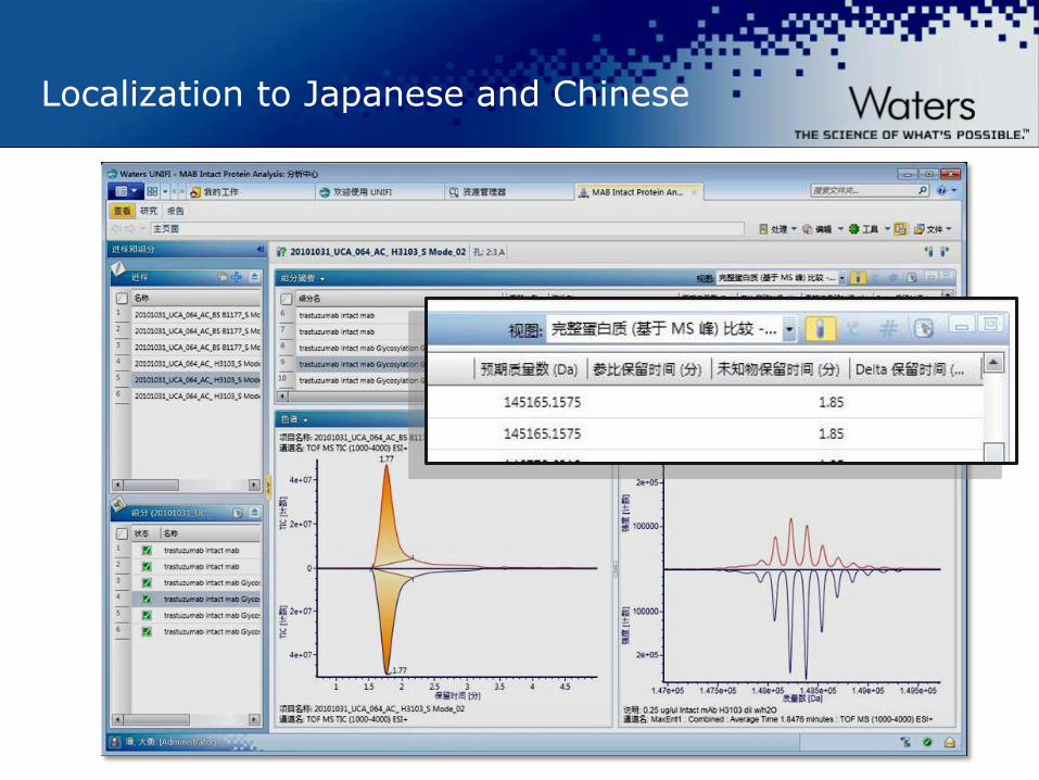
Localization to Japanese and Chinese

©2009 Waters Corporation | COMPANY CONFIDENTIAL ©2012 Waters Corporation

©2009 Waters Corporation | COMPANY CONFIDENTIAL ©2012 Waters Corporation

©2009 Waters Corporation | COMPANY CONFIDENTIAL ©2012 Waters Corporation
Investigating Intact Mass Results
Peak 14.81 min
Peak 15.51 min

©2009 Waters Corporation | COMPANY CONFIDENTIAL ©2012 Waters Corporation
UNIFI™ Intact Mass Reporting Tools
©2009 Waters Corporation | COMPANY CONFIDENTIAL ©2012 Waters Corporation
UPLC and MS Based Analytical Approaches for Comprehensive Glycosylation Analysis
Intact Protein
Glycopeptides Released Glycans
MW profiling (ESI MS)
Proteolytic Digestion PNGase F Digestion
Fluorescent and mass profiling of glycans
Relative Quantitation
Structure Elucidation
-- MS based
-- LC retention time only (GU value)
& Glycosidase Array
Relative Quantitation of glycans
Glycan site heterogeneity
Glycan Occupancy
©2009 Waters Corporation | COMPANY CONFIDENTIAL ©2012 Waters Corporation
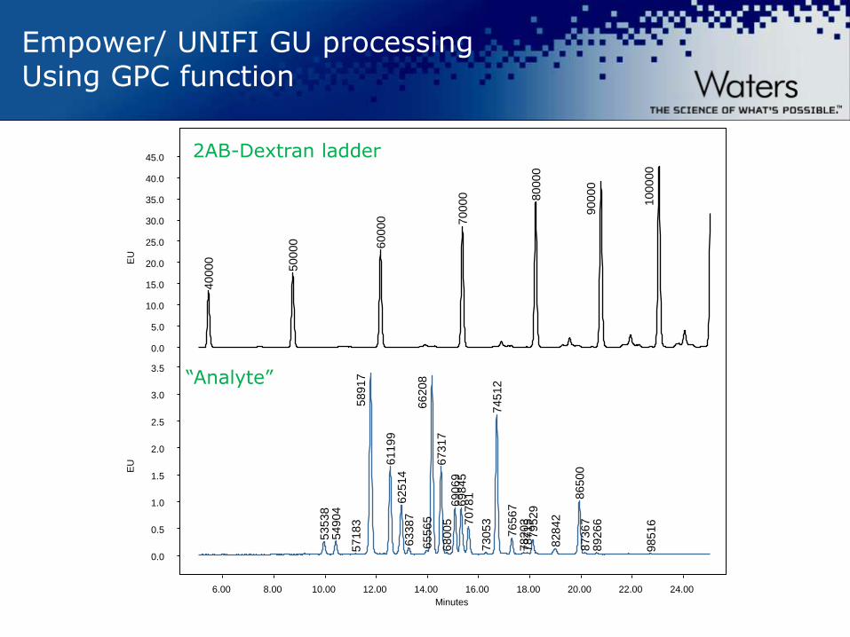
Reminder: How Are Glucose Unit (GU)
Values Generated?
The elution times of glycans are expressed in glucose
units (GU) by reference to a dextran ladder.
Each individual glycan structure has a GU value that is
related to the number, linkage of its monosaccharides.
GU values can be used to predict structures.
The use of arrays of exoglycosidase enzymes in
combination with UPLC-HILIC profiling and Glycobase
enables the individual monosaccharides and linkages to
be determined.
©2009 Waters Corporation | COMPANY CONFIDENTIAL ©2012 Waters Corporation
Empower/ UNIFI GU processing Using GPC function
Minutes
40000
50000
60000
70000
80000
90000
100000
53538
54904
57183
58917
61199
62514
63387
65565
66208
67317
68005
69069
69845
70781
73053
74512
76567
78203
78715
79529
82842
86500
87367
89266
98516
EU
0.0
5.0
10.0
15.0
20.0
25.0
30.0
35.0
40.0
45.0
EU
0.0
0.5
1.0
1.5
2.0
2.5
3.0
3.5
6.00 8.00 10.00 12.00 14.00 16.00 18.00 20.00 22.00 24.00
2AB-Dextran ladder
“Analyte”

©2009 Waters Corporation | COMPANY CONFIDENTIAL ©2012 Waters Corporation
A) B)
C)
UNIFI
Glycan FLR
workflow
Retention Time Database
A) 2AB-Dextran ladder B) Generate Calibration curve C) Apply the curve to an
“unknown” sample
Glycan FLR Analytical Workflow In UNIFI

©2009 Waters Corporation | COMPANY CONFIDENTIAL ©2012 Waters Corporation
UNIFI is able to Acquire Glycan UPLC/FLR/QTOF MS data
2AB-labeled Etanercept N-Glycans
FLR
MS
Determine GU Values for Glycobase™

©2009 Waters Corporation | COMPANY CONFIDENTIAL ©2012 Waters Corporation
109 Glycans were Identified in an EPO Protein

©2009 Waters Corporation | COMPANY CONFIDENTIAL ©2012 Waters Corporation
Glycan Search inside Glycobase Database - IgG

©2009 Waters Corporation | COMPANY CONFIDENTIAL ©2012 Waters Corporation
A Review Panel in UNIFI After Glycan FLR Data Processing
©2009 Waters Corporation | COMPANY CONFIDENTIAL ©2012 Waters Corporation
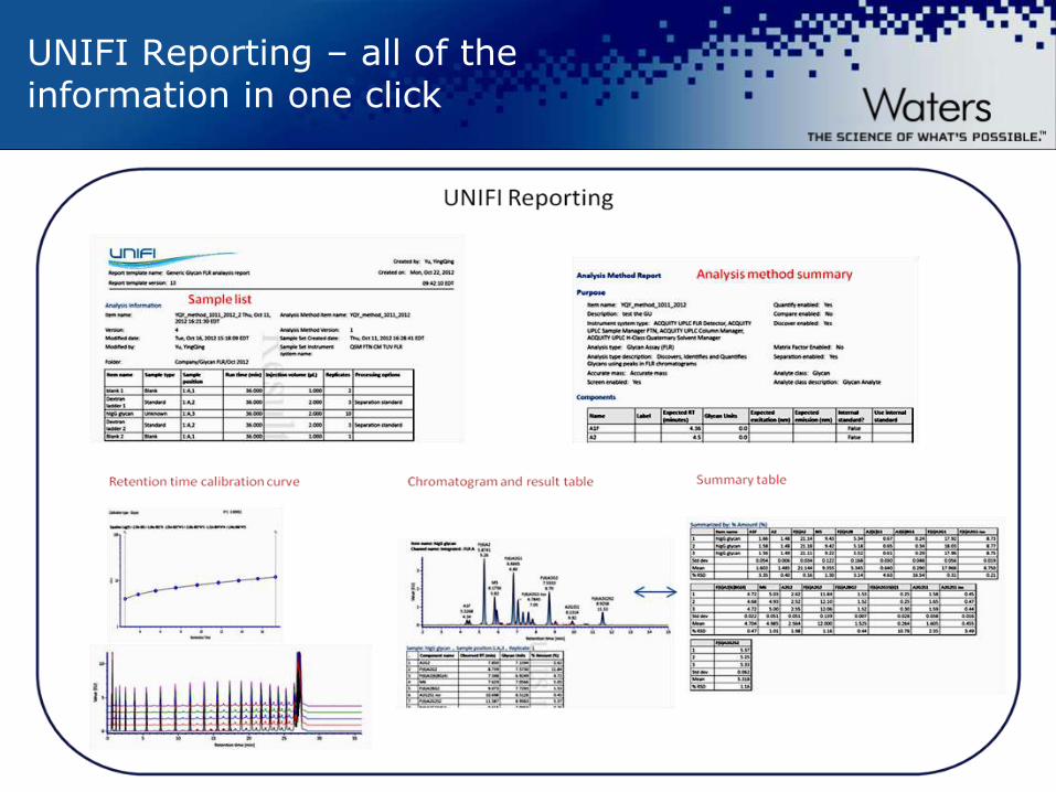
UNIFI Reporting – all of the information in one click
©2009 Waters Corporation | COMPANY CONFIDENTIAL ©2012 Waters Corporation
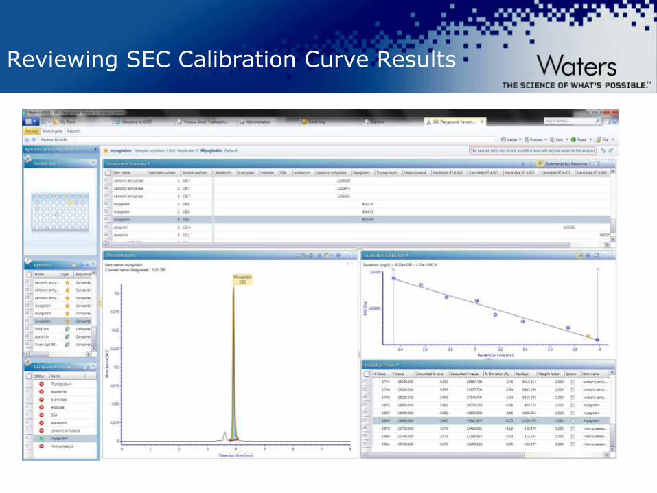
Reviewing SEC Calibration Curve Results

©2009 Waters Corporation | COMPANY CONFIDENTIAL ©2012 Waters Corporation
Product Portfolio
www.waters.com/biopharm www.waters.com/glycan Download or order our New biopharmceutical Application Notebook at www.waters.com/biopharmbook

©2009 Waters Corporation | COMPANY CONFIDENTIAL ©2012 Waters Corporation
Conclusions
Released glycan FLR analysis workflows are a recent
addition to the UNIFI Biopharmaceutical Platform Solution.
The UNIFI application workflow enables a scientist in
regulated or unregulated laboratory environments to
— acquire,
— process
— and report qualitative and quantitative glycan profiling
information,
— automatically

©2009 Waters Corporation | COMPANY CONFIDENTIAL ©2012 Waters Corporation
St John Skilton
Taking Glycan Analysis Further
IMS, ETD and TAP
©2009 Waters Corporation | COMPANY CONFIDENTIAL ©2012 Waters Corporation
UPLC and MS Based Analytical Approaches for comprehensive Glycosylation Analysis
• Quantitation of Individual Glycans
• Isomer discrimination
• Profiling of glycans
• Structure Elucidation
Intact Protein
Glycopeptides Released Glycans
MW profiling (ESI MS)
Enzymatic Digestion
Peptide Mapping for:
• Location of Glycosylation
• O-linked and N-linked glycans
Discrimination of isobaric glycopeptides
Enzymatic Cleavage
Complementary Information with
Mass Spectrometry
©2009 Waters Corporation | COMPANY CONFIDENTIAL ©2012 Waters Corporation
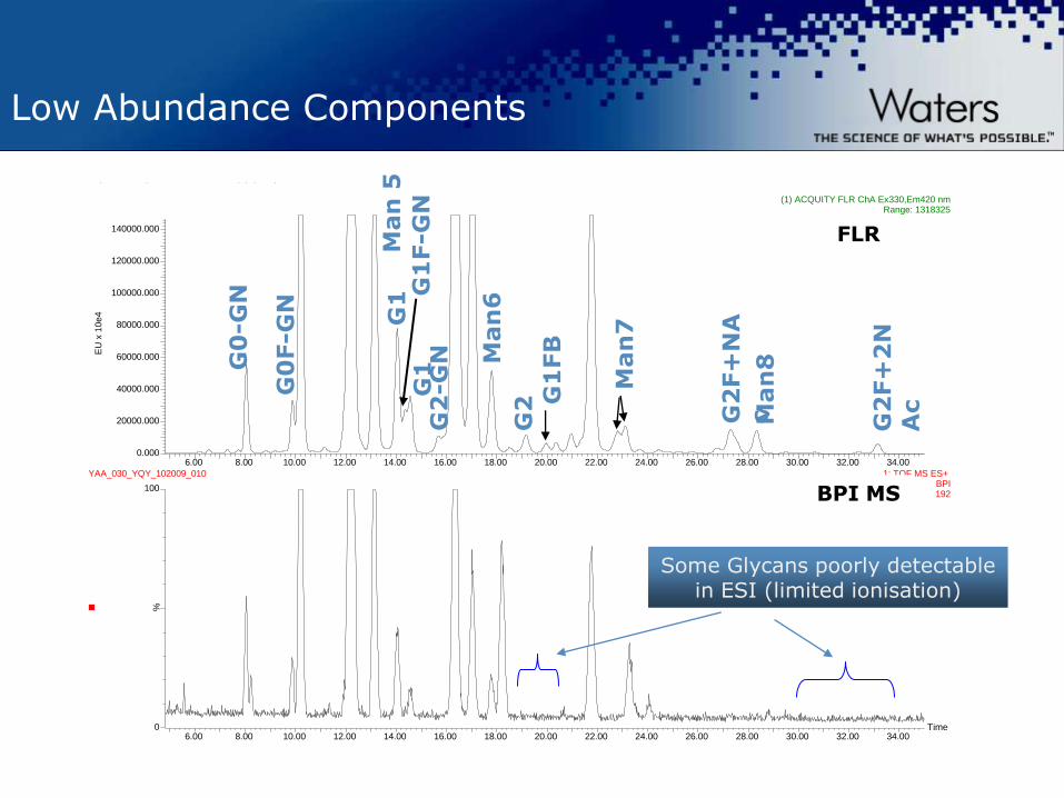
Low Abundance Components
2ab Herceptin B 2033, 2.5 ug p/ul, batch 3
Time6.00 8.00 10.00 12.00 14.00 16.00 18.00 20.00 22.00 24.00 26.00 28.00 30.00 32.00 34.00
%
0
100
6.00 8.00 10.00 12.00 14.00 16.00 18.00 20.00 22.00 24.00 26.00 28.00 30.00 32.00 34.00
EU
x 1
0e4
0.000
20000.000
40000.000
60000.000
80000.000
100000.000
120000.000
140000.000
YAA_030_YQY_102009_010 (1) ACQUITY FLR ChA Ex330,Em420 nmRange: 1318325
YAA_030_YQY_102009_010 1: TOF MS ES+ BPI192
FLR
BPI MS
G0
F-G
N
G0
-GN
G1
F-G
N
G2
-GN
Man
5
G2
Man
7
Man
6
G2
F+
NA
c
Man
8
G2
F+
2N
Ac G1
FB
G1
G
1
Some Glycans poorly detectable in ESI (limited ionisation)
©2009 Waters Corporation | COMPANY CONFIDENTIAL ©2012 Waters Corporation
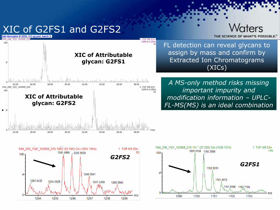
XIC of G2FS1 and G2FS2 2ab Herceptin B 2033, 2.5 ug p/ul, batch 3
Time24.00 26.00 28.00 30.00 32.00 34.00 36.00 38.00
%
0
100
24.00 26.00 28.00 30.00 32.00 34.00 36.00 38.00
%
0
100
YAA_030_YQY_102009_010 1: TOF MS ES+ 1099.9 0.15Da
40
YAA_030_YQY_102009_010 1: TOF MS ES+ 1245.5 0.15Da
24
XIC of Attributable glycan: G2FS2
XIC of Attributable glycan: G2FS1
G2FS1 2+
G2FS2 2+
FL detection can reveal glycans to assign by mass and confirm by Extracted Ion Chromatograms
(XICs)
A MS-only method risks missing important impurity and
modification information – UPLC-FL-MS(MS) is an ideal combination

©2009 Waters Corporation | COMPANY CONFIDENTIAL ©2012 Waters Corporation
DDA/MSMS of G2FS1 (M+2H)2+
MS/MS Spectra Deconvoluted by MaxEnt 3

©2009 Waters Corporation | COMPANY CONFIDENTIAL ©2012 Waters Corporation
UPLC/HILIC/MS of Glycopeptides

©2009 Waters Corporation | COMPANY CONFIDENTIAL ©2012 Waters Corporation
HILIC UPLC/MS for Glycopeptide Mapping
6 x magnified
4 x magnified
0 5 10 15 20 25 30 35 40 45
minutes
GLYCOPEPTIDES
MS BPI
UV 280 nm
A
B
*
Tryptic mAb IgG peptides.
BEH Glycan column -2.1 x 150 mm
Mobile phase A -10 mM Ammonium Formate
Mobile phase B -100% Acetonitrile
Gradient -90 – 50% B in 45 min.
Column temp. 40 °C
Non glycopeptides Glycosylated
MS trace (BPI)
UV trace (280nm)

©2009 Waters Corporation | COMPANY CONFIDENTIAL ©2012 Waters Corporation
Glycopeptides from Bovine Fetuin
0 5 10 15 20 25 30 35
minutes
MS BPI
A
B
C
MS BPI
XIC MS 657.24
non-glycopeptides
O-linked
glycopeptides
N-linked
glycopeptides
12
non-glycopeptides
34
56
78
1
2
34
5
6
78 9
9
2
3
4
5
6
78
9
1
1012
11
13
14
15
16
10 1211
13
14
1516
17
17
1819
+ H+
PNGase F treated
Individual Glycopeptide MS ion count was used for quantitation.
Oxonium ion

©2009 Waters Corporation | COMPANY CONFIDENTIAL ©2012 Waters Corporation
Glycopeptide Quantitation (IgG)
Rel. quantitation of glycopep. % Rel. quantitation of glycopep. %
UV 280 nm RSD % XIC MS RSD %
G0 6.3 ± 0.3 4.6 G0 6.1 0.1 1.4
G0F 35.7 ± 0.2 0.5 G0F 38.3 0.8 2.1
G1 a 3.1 ± 0.1 4.4 G1 a 2.4 0.2 10.3
G1 b 0.8 ± 0.1 14.4 G1 b 1.1 0.1 9.5
Man5 - - - Man5 1.1 0.0 2.8
G1F a 34.4 ± 0.1 0.2 G1F a 31.2 0.6 1.9
G1F b 11.2 ± 0.0 0.2 G1F b 11.6 0.2 1.6
G2F 8.5 ± 0.2 2.0 G2F 8.2 0.3 4.0
EEQYNSTYR

©2009 Waters Corporation | COMPANY CONFIDENTIAL ©2012 Waters Corporation
Bovine Fetuin Glycosylation Site Heterogeneity
5 glycosylation sites, 34 glycopeptides were identified.
Gilar et al, Analytical Biochemistry 417 (2011) 80–88
©2009 Waters Corporation | COMPANY CONFIDENTIAL ©2012 Waters Corporation
Asp
24
+ FA
4G
4S1
,FA
4G
4S1
Lac
3
Asp
24
+ FA
4G
4S2
Lac
2,F
A4
G4
S4 L
ac1
Asp
24
+FA
4G
4S1
Lac
3,
Asp
2
4+F
A4
G4
S3 L
ac2
, A
sp2
4+F
A4
G4
S4, A
sp
24
+FA
4G
4S2
Lac
2,
Asp
24
+FA
4G
4S1
Lac
3
A
sp 2
4 F
4G
4S4
Asp
24
+FA
4G
4S2
Lac
2,
Asp
24
+FA
4G
4S1
Lac2
A
sp2
4+F
A4
G4
S4 L
ac1
Asp
24
F4
G4
S4
Asp
24
+FA
4G
4S1
Lac
3
Asp
24
+ FA
4G
4S1
Lac
1
Asp
38
+ FA
4G
4S2
Asp
38
+ FA
4G
4S1
, FA
4G
4S2
Asp
38
+ FA
4G
4S1
iso
mer
s
Asp
38
+ FA
4G
4S1
Asp
24
+ FA
4G
4S2
Lac
2
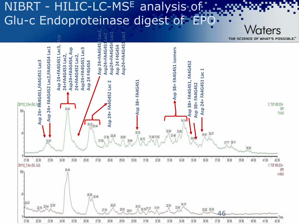
NIBRT - HILIC-LC-MSE analysis of Glu-c Endoproteinase digest of EPO
46

©2009 Waters Corporation | COMPANY CONFIDENTIAL ©2012 Waters Corporation
Informatics Tool for Glycan Site Heterogeneity
oxonium ion annotation
carbohydrate annotation
MSE elevated energy fragment ion spectrum
©2009 Waters Corporation | COMPANY CONFIDENTIAL ©2012 Waters Corporation
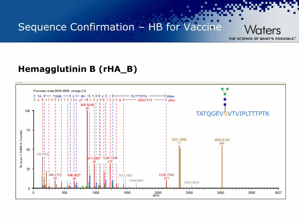
Sequence Confirmation – HB for Vaccine
Hemagglutinin B (rHA_B)
TATQGEVNVTVIPLTTTPTK

©2009 Waters Corporation | COMPANY CONFIDENTIAL ©2012 Waters Corporation
Other QTOF MS Tools for Glyco Site Heterogeneity ETD
Z2
Z4
Z5
C6
Precursor ion (3+)
C7
C8
C9
Charge reduced 2+
Z6
Z7
Z8 Z9
C10
C11
C13
EAISPPDAASAAPLR O-linked Glycopepide from EPO
©2009 Waters Corporation | COMPANY CONFIDENTIAL ©2012 Waters Corporation
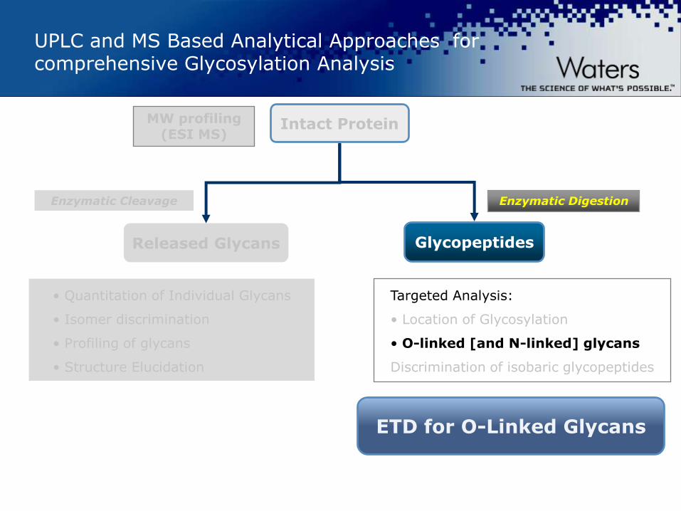
UPLC and MS Based Analytical Approaches for comprehensive Glycosylation Analysis
• Quantitation of Individual Glycans
• Isomer discrimination
• Profiling of glycans
• Structure Elucidation
Intact Protein
Glycopeptides Released Glycans
MW profiling (ESI MS)
Enzymatic Digestion Enzymatic Cleavage
ETD for O-Linked Glycans
Targeted Analysis:
• Location of Glycosylation
• O-linked [and N-linked] glycans
Discrimination of isobaric glycopeptides

©2009 Waters Corporation | COMPANY CONFIDENTIAL ©2012 Waters Corporation

©2009 Waters Corporation | COMPANY CONFIDENTIAL ©2012 Waters Corporation
Glycosylation Site Characterization
Scott McLuckey
ETD CID
©2009 Waters Corporation | COMPANY CONFIDENTIAL ©2012 Waters Corporation
ETD spectrum of N-linked Glycopeptide
m/z
300 400 500 600 700 800 900 1000 1100 1200 1300 1400 1500
TOF MSMS 740.30ES+
3.46e4986.81
510.30
277.13423.26
322.20
404.23
740.37
567.30
1480.21
1225.57
1197.07
1115.06
1297.12
1269.59
1401.661298.13
1401.15
1298.631343.14
Precursor ion
[M+4H] 4+
chargereduced
[M+3H]3+
chargereduced
[M+2H]2+
z3c2z2 c3
z4
c4z5
2+
z62+
z72+
z82+
c62+
c72+
c82+
c52+
E E Q Y N S T Y R
Waters Application Note: 720004281en “Electron Transfer Dissociation of N-linked Glycopeptides from an Recombinant mAb Using Synapt G2-S HDMS

©2009 Waters Corporation | COMPANY CONFIDENTIAL ©2012 Waters Corporation
ETD of O-linked Glycopeptide - 7 linkage sites are assigned
ETD spectrum

©2009 Waters Corporation | COMPANY CONFIDENTIAL ©2012 Waters Corporation
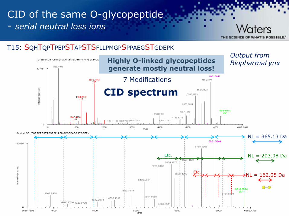
CID of the same O-glycopeptide - serial neutral loss ions
T15: SQHTQPTPEPSTAPSTSFLLPMGPSPPAEGSTGDEPK
7 Modifications
Output from BiopharmaLynx
CID spectrum
Highly O-linked glycopeptides generate mostly neutral loss!
©2009 Waters Corporation | COMPANY CONFIDENTIAL ©2012 Waters Corporation
CID of the same O-glycopeptide - serial neutral loss ions
T15: SQHTQPTPEPSTAPSTSFLLPMGPSPPAEGSTGDEPK
7 Modifications
NL = 365.13 Da
Output from BiopharmaLynx
CID spectrum
NL = 203.08 Da
NL = 162.05 Da
Highly O-linked glycopeptides generate mostly neutral loss!
Etc.
Etc.

©2009 Waters Corporation | COMPANY CONFIDENTIAL ©2012 Waters Corporation
UPLC and MS Based Analytical Approaches for comprehensive Glycosylation Analysis
• Quantitation of Individual Glycans
• Isomer discrimination
• Profiling of glycans
• Structure Elucidation
Intact Protein
Glycopeptides Released Glycans
MW profiling (ESI MS)
Enzymatic Digestion Enzymatic Cleavage
Peptide Mapping Technique: “TAP” fragmentation [SYNAPT™] for simultaneous
peptide sequence and glycan confirmation
Untargeted Peptide Mapping for:
• Location of Glycosylation
• O-linked and N-linked glycans
Discrimination of isobaric glycopeptides

©2009 Waters Corporation | COMPANY CONFIDENTIAL ©2012 Waters Corporation
Innovative Techniques for Sequencing Glycopeptides
Glycopeptides can be ‘mined’ from the LC/ MSE data
Unique architecture of SYNAPTTM used to simultaneously supply
multiple sets of information
—Glycan information from low-energy CID
—Peptide backbone sequence confirmed from high-energy CID
—Information is obtained simultaneously
—O- and N-linked glycans can be identified
© 2009 American Society for Mass Spectrometry.
Published by Elsevier Inc.
doi:10.1016/j.jasms.2009.07.017
Rapid Commun. Mass Spectrom. 2008; 22: 29–40;
(www.interscience.wiley.com) DOI: 10.1002/rcm.3330
©2009 Waters Corporation | COMPANY CONFIDENTIAL ©2012 Waters Corporation
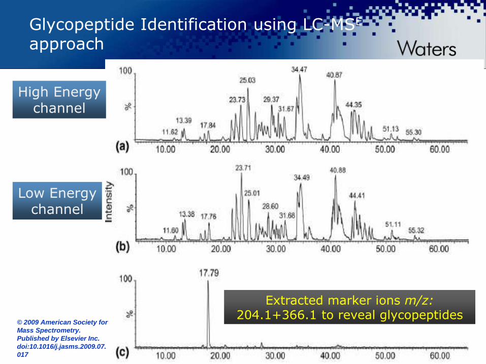
Glycopeptide Identification using LC-MSE approach
Low Energy channel
High Energy channel
Extracted marker ions m/z: 204.1+366.1 to reveal glycopeptides
© 2009 American Society for
Mass Spectrometry.
Published by Elsevier Inc.
doi:10.1016/j.jasms.2009.07.
017

©2009 Waters Corporation | COMPANY CONFIDENTIAL ©2012 Waters Corporation
Enhanced Structural Characterisation: Time Aligned Parallel Fragmentation (TAP)
Drift time
Product ions separated by IMS
m/z
Precursor ion fragmented
Drift time
m/z
Precursor and products share same drift time
Ions from Quad region
Refragmentation in TRANSFER Cell –
higher energy to break peptide bonds
Fragmentation in TRAP Cell – LOW energy to cleave Glycans

©2009 Waters Corporation | COMPANY CONFIDENTIAL ©2012 Waters Corporation
Glycan Fingerprint from Trap Cell Fragmentation
Low energy Fragmentation in
the Trap cell cleaves glycans
Glycan assignment according to accurate masses of precursors and fragments

©2009 Waters Corporation | COMPANY CONFIDENTIAL ©2012 Waters Corporation
P H+
EEQYNSTYR
Peptide Sequence and Glycosylation Site From Transfer Cell Fragmentation
High energy Fragmentation in the Transfer
cell cleaves peptide bonds
Peptide Sequence Confirmation using characteristic b and y ions

©2009 Waters Corporation | COMPANY CONFIDENTIAL ©2012 Waters Corporation
TAP fragmentation on the first stage to obtain O-linked glycan sequence
Pep
Pep
Pep
MH+
(EAISPPDAASAAPLR)
G1 data
Example of workflow applied to EPO: showing selected glycans
©2009 Waters Corporation | COMPANY CONFIDENTIAL ©2012 Waters Corporation
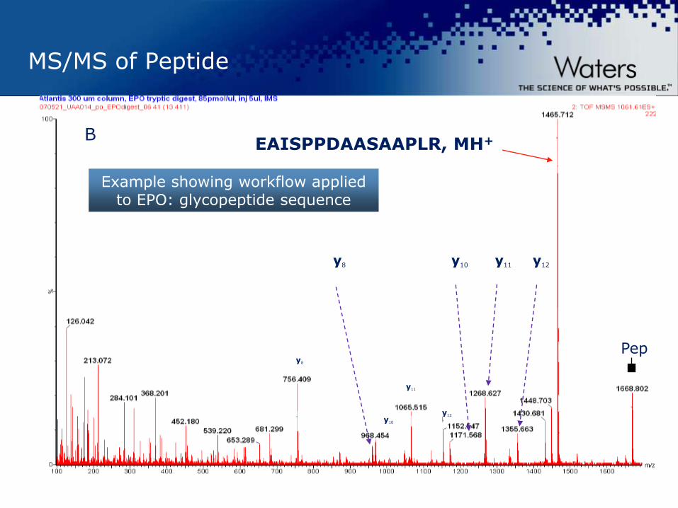
MS/MS of Peptide
B
y8
y10
EAISPPDAASAAPLR, MH+
y11 y12 y10 y8
y11
Pep
y12
Example showing workflow applied to EPO: glycopeptide sequence

©2009 Waters Corporation | COMPANY CONFIDENTIAL ©2012 Waters Corporation
UPLC and MS Based Analytical Approaches for comprehensive Glycosylation Analysis
• Quantitation of Individual Glycans
• Isomer discrimination
• Profiling of glycans
• Structure Elucidation
Intact Protein
Glycopeptides Released Glycans
MW profiling (ESI MS)
Enzymatic Digestion
Peptide Mapping for:
• Location of Glycosylation
• O-linked and N-linked glycans
Discrimination of isobaric glycopeptides
Enzymatic Cleavage
IMS of Glycopeptides in an LC/MSE run
©2009 Waters Corporation | COMPANY CONFIDENTIAL ©2012 Waters Corporation
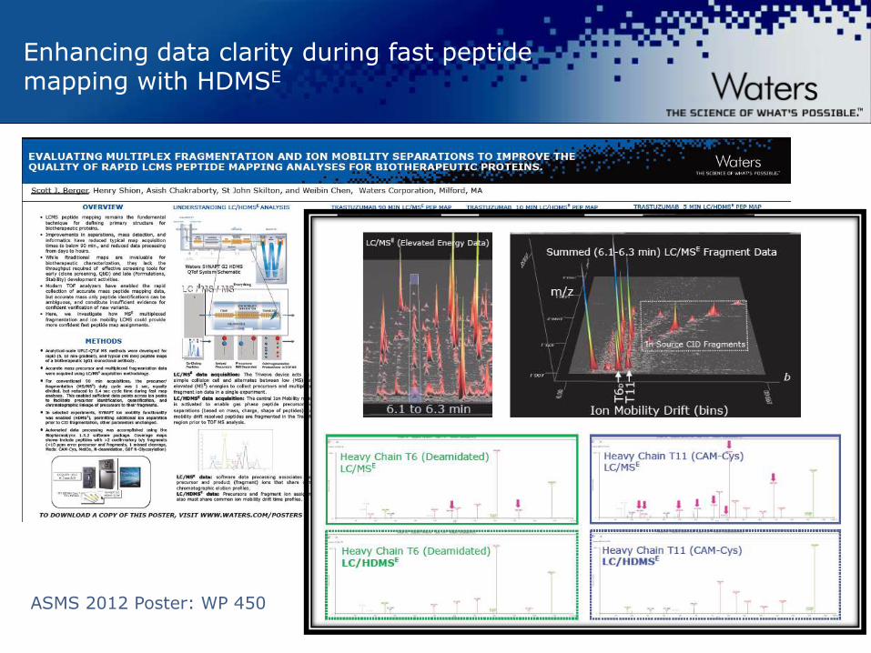
Enhancing data clarity during fast peptide mapping with HDMSE
ASMS 2012 Poster: WP 450

©2009 Waters Corporation | COMPANY CONFIDENTIAL ©2012 Waters Corporation
Chromatogram from LC/MS run of Bovine Fetuin
Extracted Chromatogram HexNAc Oxonium Ion
Elevated Energy
TIC Chromatogram Low Energy
(X-Axis in Scans)
Craig Dorschel et al, ASMS 2010 Oral presentation
©2009 Waters Corporation | COMPANY CONFIDENTIAL ©2012 Waters Corporation
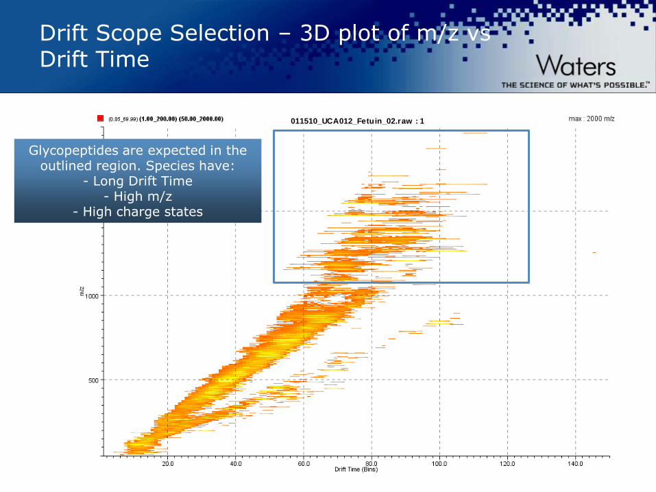
Drift Scope Selection – 3D plot of m/z vs Drift Time
011510_UCA012_Fetuin_02.raw : 1
Glycopeptides are expected in the outlined region. Species have:
- Long Drift Time - High m/z
- High charge states
©2009 Waters Corporation | COMPANY CONFIDENTIAL ©2012 Waters Corporation
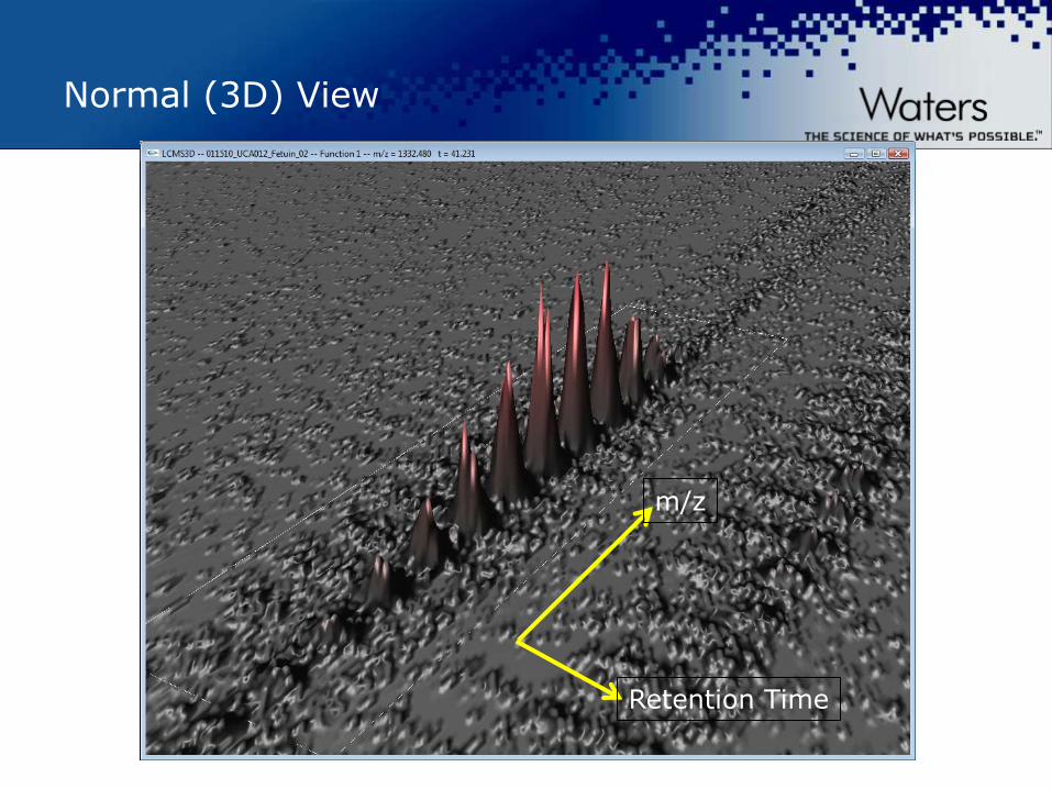
Normal (3D) View
m/z
Retention Time
©2009 Waters Corporation | COMPANY CONFIDENTIAL ©2012 Waters Corporation
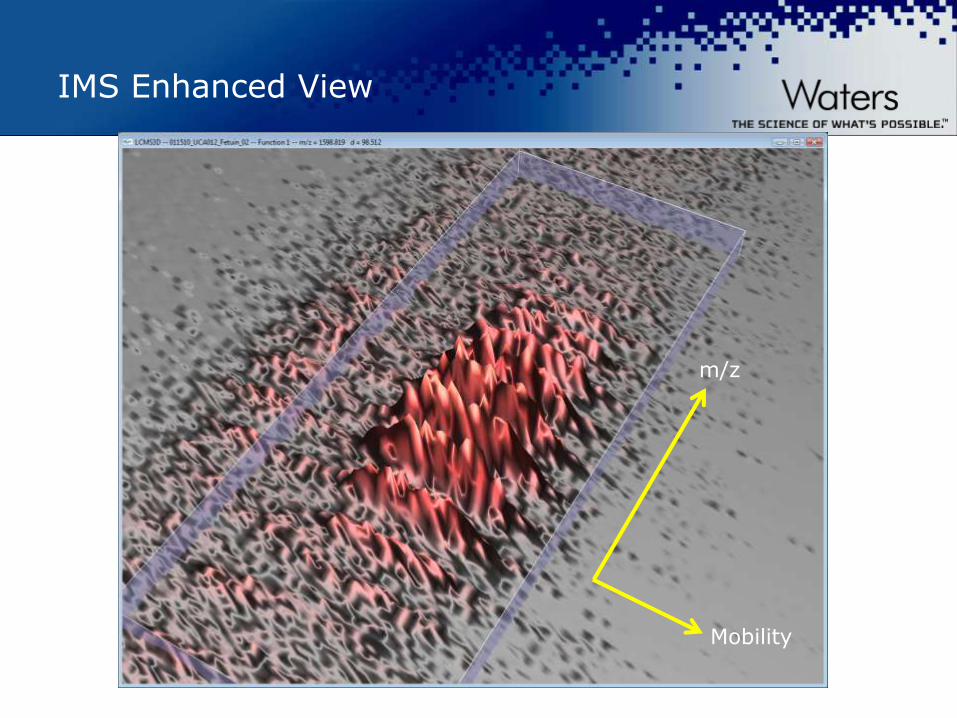
IMS Enhanced View
m/z
Mobility

©2009 Waters Corporation | COMPANY CONFIDENTIAL ©2012 Waters Corporation
Investigation of the mobility space clearly illustrates two isobaric forms of the O-linked glycopeptide
Delta RT 0.005 minutes
Delta RT 300 msec Delta m
/z 1
.4 a
mu
Delta m
/z 1
.4
am
u
Delta Drift 12
©2009 Waters Corporation | COMPANY CONFIDENTIAL ©2012 Waters Corporation
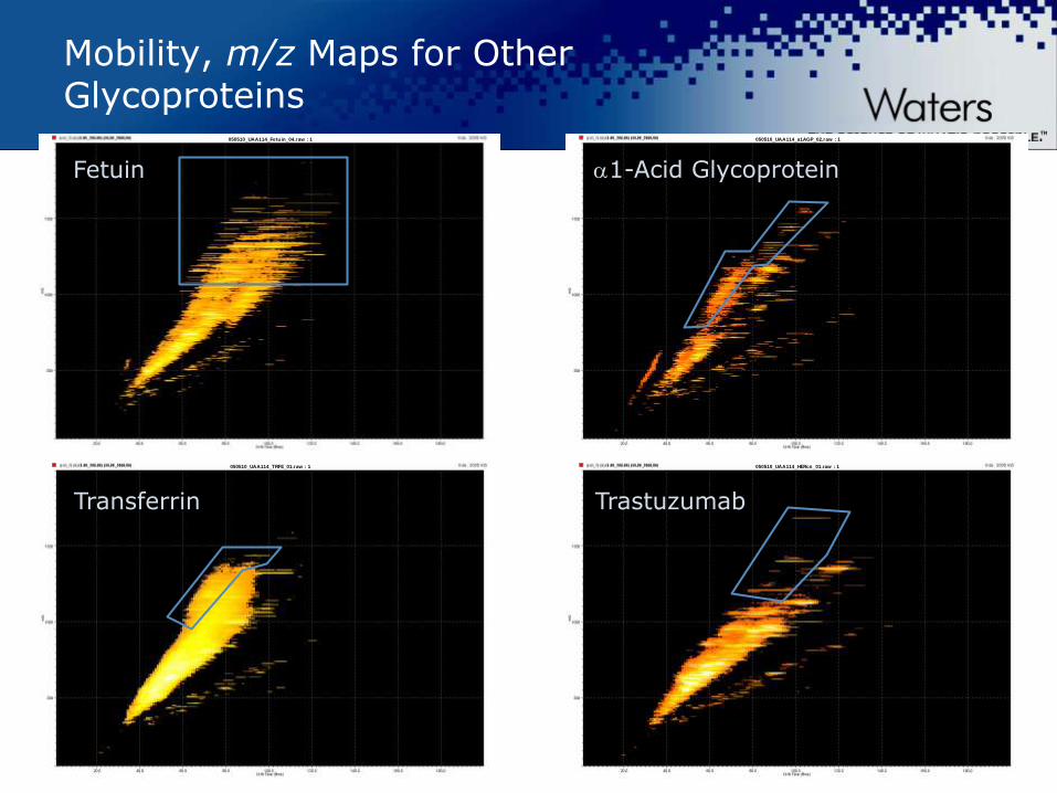
Mobility, m/z Maps for Other Glycoproteins
050510_UAA114_HERcn_01.raw : 1
050510_UAA114_a1AGP_02.raw : 1050510_UAA114_Fetuin_04.raw : 1
050510_UAA114_TRFE_01.raw : 1
Fetuin 1-Acid Glycoprotein
Transferrin Trastuzumab

©2009 Waters Corporation | COMPANY CONFIDENTIAL ©2012 Waters Corporation
Summary
Intact Protein Glycoprotein Profiling
Using BiopharmaLynx Bioinformatics
tool
—Batch data processing
—Data comparison (batch to batch,
Innovator/Biosilimar)
FLR labeling Glycan Analysis
—BEH Glycan Column chemistry gives
higher resolution and faster
chromatographic separation
—Exoglycosidic digestion + GU
assignment enable full glycan structure
characterization
UPLC/FLR/Qtof MS Platform offers
—Glycan profiling using FLR chromatogram
—Accurate mass confirmation
—MSMS
— ETD of glycopeptides (N-, O-linked)
gives linkage site information
UNIFI – platform solution for
biopharmaceuticals

©2009 Waters Corporation | COMPANY CONFIDENTIAL ©2012 Waters Corporation
It is child’s play…
http://www.ted.com/speakers/ursus_wehrli.html http://de.wikipedia.org/wiki/Ursus_Wehrli
©2009 Waters Corporation | COMPANY CONFIDENTIAL ©2012 Waters Corporation
It is child’s play… with the right tools!
http://www.ted.com/speakers/ursus_wehrli.html http://de.wikipedia.org/wiki/Ursus_Wehrli

©2009 Waters Corporation | COMPANY CONFIDENTIAL ©2012 Waters Corporation
AcknowledgementS
Waters Corporation
Ying Qing Yu
Joomi Ahn
Eoin Cosgrave
Asish Chakraborty
Stephane Houel
Jonathan Williams
Scott Berger
Beth Gildea
Vern Tisdale
Jennifer Fournier
Craig Dorschel
Weibin Chen
NIBRT (Ireland)
Pauline Rudd
Mark Hilliard
Giorgio Carta
Jonathan Bones

©2009 Waters Corporation | COMPANY CONFIDENTIAL ©2012 Waters Corporation

©2009 Waters Corporation | COMPANY CONFIDENTIAL ©2012 Waters Corporation
2013 Waters Biopharmaceutical Application Notebook
www.waters.com/ biopharmbook
©2009 Waters Corporation | COMPANY CONFIDENTIAL ©2012 Waters Corporation