water metabolism and disorders of water · pdf filewater metabolism and disorders of water...
TRANSCRIPT

1
WATER METABOLISM AND
DISORDERS OF WATER BALANCE
“If there is magic on this planet, it is contained in water.”- Loren Eiseley.
Rena Ramesh, DNB Pediatrics Resident,
Dr.Mehta’s Children Hospital, Chennai.
1. INTRODUCTION
The first vertebrates evolved in seawater and this origin is reflected in the physiology of all vertebrates. Water forms two-thirds of every vertebrate’s body reflecting the constitution of mother earth. Millions of years later, terrestrial forms emerged crawling out on dry land, challenged with the problem of fluid conservation which had to be balanced with fluid elimination. Various mechanisms are involved in facing this challenge of avoiding gaining or losing too much water.
Water balance means proper volume of total body water (TBW) and appropriate distribution in both intracellular (ICF) and extracellular compartments (ECF). This also includes maintenance of serum osmolality. To maintain osmotic balance, the extracellular compartment must be able to take in water from its environment or excrete excess water into it. This has to occur along with
1. Introduction. 2. Can water in the body be measured? 3. Fluid compartments. 4. Water metabolism.
4.1. Vasopressin. 4.2. Thirst. 4.3. The kidneys.
5. Integration of normal water balance mechanisms. 6. When the equilibrium is disturbed… 7. Disorders of water balance: excess and deficit.
7.1. Hyponatremia. 7.2. Hypernatremia.
8. Disorders of sodium balance: excess and deficit. 8.1. Hypovolemia. 8.2. Edema.
9. Conclusion.

2
the exchange of inorganic ions in order to maintain homeostasis. Such exchange of water and electrolytes between the body and the external environment takes place through specialized epithelial cells, and in most vertebrates, through a filtration process in the kidneys.
Animals have evolved a variety of mechanisms to cope with the problems of water balance. Osmo-regulatory organs like excretory tubules in worms and malphigian tubules in insects have evolved to become kidneys. In the chain of evolution, freshwater fishes are the first to have kidneys. Depending on the tonicity of the body fluid to their surrounding water, marine fishes had mechanisms for balance, such as maintaining a high blood urea which prevented water loss by making their blood approximately isotonic to the surrounding sea. Amphibians, the first terrestrial vertebrates, compensated for their sodium loss by actively transporting sodium across their skin from the surrounding water. Mammals and birds are the only vertebrates to produce urine which is hyperosmolar than their body fluids, which allows them to excrete waste products in a small volume of water, so that more water can be retained in the body.
In humans, water loss which occurs through various routes, is usually a continuous process. Water intake, however, is an intermittent process. The normal physiology of water balance allows water deficit to build over time which is corrected intermittently by thirst-induced intake of water. However, not all water intake depends on this mechanism. It is influenced by various behavioural and social factors, which results in water intake unrelated to deficit. This becomes a problem only when it exceeds the excretory capacity of kidneys. In such overload situations, the thirst mechanisms are not triggered.
2. CAN WATER IN THE BODY BE MEASURED?
Osmosis is the diffusion of water through a membrane from a solution with higher solute concentration. As the solute concentration of a solution determines its osmotic behaviour, the total moles of a solute per kilogram of water is expressed as the osmolality of the solution. It is an indirect indicator of the “concentration” of water. Solutions may be iso-osmotic, hypo or hyper-osmotic.
In clinical practice, solute concentrations are measured per litre of solvent which is referred to as osmolarity. But no correction is required as these both correspond closely at solute concentration of body fluids. The osmometer measures osmolality.
Tonicity or effective osmolality, is a measure of the movement of water across a semi-permeable membrane. If two solutions of unequal osmolality are separated by a membrane which is permeable to water but not the solutes in solution, the water will move down its concentration gradient (from the hypo-osmolar to the hyperosmolar solution). In this case, the hypo-osmolar solution is called hypotonic which has a higher osmotic pressure. The osmotic pressure of a solution is a measure of its tendency to take in water by osmosis. A cell when

3
placed in a hypertonic solution which has a higher osmotic pressure than the cell cytoplasm, will lose water to the surrounding solution and shrink. A cell placed in a hypotonic solution will gain water and expand. A perfect example in daily life is shrinking size of the mango pickle and rice puffing up when boiled in water.
However if solutes can cross the membrane, they will diffuse down their concentration gradient and equalize the activity of water on both sides of the membrane. In this case, the solutions will be isotonic and there will be no net movement of water across the membrane. Solutes like alcohol and urea permeate the cells freely and therefore increase osmolality but they have no effect on the tonicity. Since they do not induce the shift of water on either side of the cell they do not have any influences on the water balance. In contrast, accumulation of impermeable solutes, such as sodium and mannitol, in the extracellular compartment increases the tonicity and causes the movement of water from the cells.
To calculate osmolality:
Fig 1.Tonicity of solutionssolutionsand their influence on cell size.

4
Osmolality (mOsm/kg of H2O) = 2 x Sodium + Glucose/18 + BUN/2.8
(The values 18, 2.8 represent the conversion of mg/dL into mmol/L)
A discrepancy between the measured and calculated osmolality, also known as osmolar gap, can occur due to the presence of a solute that is unaccounted for, the most common being ingestion of a low-molecular weight toxin (such as alcohol, methanol and ethylene glycol).It is apractical clue for the presence of poisoning due these substances when we encounter a child with unexplained acidosis and osmolar gap in the background of poisoning,
3. FLUID COMPARTMENTS
Fig 2.Composition of body fluids.

5
The volume of different fluid compartments is determined by the content of major solute in each compartment. Sodium is the major extracellular solute. Presumably, water is in equilibrium among all fluid compartments and if the osmolality changes in any of the compartments, water will redistribute among the compartments until osmolality equalizes. Two major systems balance the sodium content – the “affector” and “effector” systems. The affector (or sensing) system monitors the sodium content through the baroreceptors. If the sodium content is abnormal, the effector system is involved which includes rennin-angiotensin-aldosterone (RAAS), catecholamines and vasopressin systems.
4. WATER METABOLISM
Water metabolism represents a balance between the intake and excretion of water. Each side of this balance equation consists of “regulated” and “unregulated” components, the magnitude of which depends on various physiological and pathophysiological states.
Water intake
Unregulated component Regulated component
Water excretion
Unregulated component Regulated component
Newborn 6 months 1 year Older Total body water 75 70 60 55 ICF 40 40 40 40 ECF 35 30 20 15 – 20
Intrinsic water content of ingested food, consumption of beverages for palatability/ secondary effects/ social or morbid reasons.(Unregulated means not warranted by thirst).
Fluids conserved in response to a perceived sensation of thirst.
Insensible water losses.
Obligate amount of water the kidneys must excrete in order to eliminate body metabolism solutes.
Renal excretion of free water in excess of the obligate amount necessary to excrete metabolic solutes.
Table 1: Composition of total body water in different ages ( in percentage).

6
The disturbances that result from unregulated water losses or gains are compensated for by the regulated components which maintain the water balance.
The normal plasma osmolality is 286-294 mOsm/kg. In order to maintain this narrow range of plasma osmolality, within which most cells and biological systems tend to work best, three major systems are required to work properly:
1. Normal vasopressin (anti-diuretic hormone- ADH) production and release. 2. Normal thirst and water intake. 3. A kidney that normally responds to vasopressin
4.1. VASOPRESSIN
Arginine Vasopressin (AVP) is synthesized as a prohormone in the highly specialized magnocellular nuclei of hypothalamus, supra-optic and paraventricular nuclei. After enzymatic cleavage, the hormone is transported to the posterior pituitary where it is stored in neuro-secretory granules until specific stimuli cause its secretion into the blood stream.
Osmoreceptors
Located in the anteromedial hypothalamus
Small increase in osmolality of 1-2% (2-5 mOsm/kg H2O)
will cause release
Decrease in serum osmolality will result in cessation of AVP
release
Non-osmotic stimuli
Pressure or stretch-sensitive receptors located in the left atrium or large arteries
of the chest- a reduction in effective circulatory volume will be sensed by these receptors which will send signals through the vagus and glosssopharyngeal nerves
and cause AVP release.
Other non-osmotic stimuli: anaesthetics, medications, nausea,
vomiting.
Regulators of AVP release

7
Other non-osmotic stimuli such as surgery, stress of hospitalization are the major areas of conflict leading to hospitalization associatedhyponatremia.
AVP circulates unbound and is rapidly degraded by vasopressinases in liver and kidney (t1/2 = 20-30 minutes). Vasopressin acts on V1, V2, V3 and oxytocin-type receptors.
V1 – vasculature, myometrium,platelets. V2 – distal tubule and collecting duct of nephrons. V3 – pituitary
V2 receptor antagonists are now utilised in SIADH.
4.2. THIRST Thirst is the drive to consume water to replace urinary and obligate water losses such as sweating and breathing. The thirst centre is separately located in the hypothalamus but is in close association with magnocellular neurons and osmoreceptors. Stimuli for thirst:
Increase in effective osmolality of ECF causing intracellular dehydration. Intravascular hypovolemia caused by losses of ECF.
The osmolar threshold for thirst has been considered to be approximately 5mOsm/kg above the threshold for vasopressin release.It is interesting to note that a leak in the pot is sealed first before adding water by thirst. Upon drinking, the sensation of thirst is quenched almost immediately.
4.3. THE KIDNEYS The kidneys can produce urine with osmolality between 40 and 1200 mOsm/kg, allowing a wide range of water intake. The average osmolar excretion needed to maintain osmolar balance is 600 mOsm per day. The osmolality of cortex is roughly the same as plasma, whereas the medullary interstitium has a concentration of more than four times than that of its surrounding fluid, which must be both generated and maintained. This is done by the countercurrent multiplier and countercurrent exchange mechanisms in the kidney. Finally, a urine which is maximally diluted reaches the collecting duct. This urine can remain diluted or concentrated depending on the presence or absence of AVP. If vasopressin is present, water will be returned to circulation resulting in a small amount of concentrated urine.

8
Fig3B. AQP-2 is endocytosed and internally degraded. AQP-3 and AQP-4, constitutively expressed on the basolateral membrane, allow water regress from the cell.
Fig 3A. Increased production of cAMP (mediated through V2R) activates protein kinase A, which in turn phosphorylates stored AQP-containing vesicles and targets them to the apical membrane, increasing water permeability
(Ref: Fig5 in John Danziger, Mark L.Zeidel.OsmoticHomeostasis.Clin J Am SocNephrol 2014,1-9.)

9
5. INTEGRATION OF NORMAL WATER BALANCE MECHANISMS. 6. WHEN THE EQUILIBRIUM IS DISTURBED… Disorders of water and sodium homeostasis go hand-in-hand as the extracellular fluid volume is regulated by regulating body sodium content (volume regulation) and plasma sodium concentration is regulated by changes in water content (osmoregulation). Table 2: Osmoregulation Vs Volume regulation. Osmoregulation Volume regulation
What is sensed Plasma osmolality
Arterial filling
Plasma osmolality 286-294 mOsm/kg
Decrease Increase
Suppression of thirst
Suppression of AVP release
Stimulation of thirst
Stimulation of AVP release
Dilute urine Concentrated urine
Plasma osmolality 286-294 mOsm/kg

10
Sensors Hypothalamic osmoreceptors Carotid sinus Afferent arteriole Atria
Effectors AVP Thirst
Sympathetic nervous system RAAS Atrial natriuretic peptide/Brain natriuretic peptide AVP
What is affected Urine osmolality H2O intake
Urinary sodium excretion
7. DISORDERS OF WATER BALANCE : EXCESS AND DEFICIT. 7.1. HYPONATREMIA Hyponatremia is defined as serum or plasma sodium <135 mEq/L. Whenever the serum sodium is low, sodium cannot move from the ICF to ECF to correct the sodium deficit. Instead, water moves from the ECF to the ICF leading to cell volume expansion. In a water-tight compartment like brain, cell swelling and cerebral edema can cause serious neurological complications which depend on the severity of hyponatremia and the rate of fall of serum sodium.Neurons have been given the power of self-adaptation. They can alter the cell size by intrusion or extrusion of electrolytes, idiogenicosmoles and osmotically active substances, which prevents further increase or decrease in cell size. This adaptation takes about 48 hours and likewise the dissipation of adaptive changes also takes 48 hours . This is the reason why serum sodium levels should be corrected not exceeding the rate of 12mEq/L per day to give time for the dissipation of adaptive mechanisms. If hyponatremia is corrected rapidly, the adaptation will not occur at the same speed and because the ICF is relatively hypotonic, movement of water occurs in a reverse direction from the ICF to ECF causing cellular dehydration and leading toosmotic demyelination syndrome, which was earlier called as central pontinemyelinosis.

11
Fig 4. Pathophysiology of hyponatremia

12
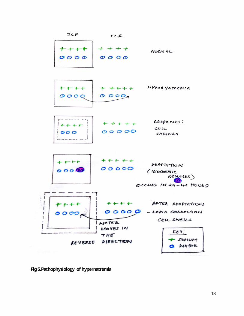
7.2. HYPERNATREMIA A sodium concentration >145mEq/L is defined as hypernatremia.Increase in sodium content in ECF leads to movement of water from ICF to ECF. This results in shrinkage of cells or cellular dehydration. This may lead to altered sensorium and sometimes in extreme conditions tearing of blood vessels and intracranial bleed. Neurons have the inherent capacity to prevent cell shrinking. They produce osmotically active substances called “idiogenicosmoles”. This protective mechanism may take a few hours to evolve as well as resolve. Hence treatment guidelines prescribe gradual reduction or elevation of serum sodium at not more than 12 mEq per day. If there is rapid reduction of sodium in the presence of undisposed idiogenicosmoles, it causes reversal of movement of water intothe cells from ECF, leading to the development of cerebral edema during therapy.
False True
Improper sampling Hyperglycemia, Mannitol Therapy, Hyperlipidemia, HyperproteinemiaHyponatremia
Hypovolemic (dehydrated)
Euvolemic (no dehydration, no edema)
Hypervolemic (edematous)
Replace water and sodium
Water restriction Restriction of water and sodium Treat the underlying cause- dialysis in renal failure. Cautious use of diuretics in other situations.
Hyponatremia
SYMPTOMATIC.: Emergency measures, 3 % Saline 5 mL/kg over 30-60 min
Asymptomatic
13
Fig 5.Pathophysiology of hypernatremia

14
Hypernatremia presents clinically as predominant neurological symptoms such as lethargy, restlessness, high- pitched cry, convulsions and loss of consciousness, features of intracranial bleed. Shock occurs late as ECF hyperosmolality causes water to shift from ICF to ECF and plasma volume is maintained till dehydration is more than 10 %. Doughy consistency of skin is particularly characteristic. A history regarding the dilution of ORS is important. Hyperglycaemia and/or hypocalcemia are common, unexplained findings in hypernatremia.
Table 3: Causes of hypernatremia.
Correction of Hypernatremia
Volume replacement if there is ECF volume contraction.
Water Deficit 1. Estimate TBW: 50–60% body weight (kg) depending on body composition 2. Calculate free-water deficit: [(Na+ – 140)/140] × TBW 3. Administer deficit maintaining a fall not more than 12 mEq/day. Ongoing water losses
A. Hypovolemichypernatremia: (Low total body sodium with water deficits > sodium deficits)
Extra-renal losses of water> sodium(Urine Na low)
Renal losses of water> sodium(Urine Na high)
Vomiting, diarrhoea Sweating Osmotic cathartics (lactulose)
Osmotic diuresis (mannitol, glucose) Diuretic phase of acute tubular necrosis Chronic renal disease, obstructive uropathies
B. Free water deficits with relative hypernatremia (Total body sodium normal)
Extra-renal losses of free water (Urine Na low)
Renal losses of free water (Urine Na low)
High insensible losses from skin: fever, burnsRadiant warmers, phototherapy
Diabetes Insipidus (DI): Central, nephrogenic
C. Increased total body sodium with no water deficits : Euvolemic hypernatremia (Urine Na high)
Excess sodium administration ( bicarbonate, 3% NaCl)Improperly diluted formula

15
4. Calculate free-water clearance, CeH2O: CeH2O = V [1- (UNa + UK)/SNa] where V is urinary volume, UNa is urinary [Na+], UKis urinary [K+], and SNa isserum [Na+]. Insensible Losses 5. ~10 mL/kg per day: less if ventilated, more if febrile. Total 6. Add components to determine H2O deficit and ongoing H2O loss; correct theH2O deficit and replace daily H2O loss. Salt(sodium) and water are the longest living partners in this universe. They always influence each other positively. When alone they are troublesome but together they created the essence of life in this world. Hence justice would be done only if disorders of sodium balance are included, which would otherwise be incomplete. 8. DISORDERS OF SODIUM BALANCE: DEFICIT AND EXCESS
8.1. HYPOVOLEMIA AND DEHYDRATION.
Hypovolemia refers to any condition in which the ECF volume is reduced and when severe can lead to hypotension or shock. Hypovolemia is usually induced by salt and water losses that are not replaced (eg, vomiting, diarrhea, diuretic therapy, bleeding, or third-space sequestration). In contrast, primary water loss, due to insensible loss by evaporation from the skin and respiratory tract or due to increased urinary water (diabetes insipidus) does not usually lead to hypovolemia because the associated rise in plasma tonicity stimulates thirst, leading to replacement of most of the lost water.
True hypovolemia due to fluid losses should be distinguished from decreased tissue perfusion in heart failure and cirrhosis in which cardiac dysfunction and systemic vasodilation, respectively, are the major hemodynamic abnormalities.
The plasma sodium concentration in hypovolemic patients may be normal, low (most often due to hypovolemia-induced release of ADH, which limits urinary water excretion), or high (if water

16
intake is impaired). The effect on the plasma sodium concentration depends upon both the composition of the fluid that is lost and fluid intake.
Hypovolemia should not be considered synonymous with dehydration. Hypovolemic losses usually refer to losses of salt and water from the ECF, whereas dehydration is primarily due to water loss from ECF, often resulting in hypernatremia, although these patients are also hypovolemic.
Fig 6. Hemodynamic responses induced by the sympathetic nervous system after effective circulatingvolume depletion.

17
The degree of dehydration is assessed and the deficit is corrected using normal saline or ringer lactate. This will correct bothdehydration and hyponatremia. For example, a one year oldweighing 10 kgs who is moderately dehydrated: Table 4. Three phase dehydration correction
Table 5.Two phase dehydration correction 0-1 hour 1-24 hours Rapid restoration of intravascular volume (20 ml/kg) 200 ml over one hour (NS/RL)
Deficit correction+ Maintenance 500+1000ml = 1500ml over 1-24 hours G5 ½ NS+ K 20 mEq/L 0-8 hours: 750 ml 9-24 hours: 750ml
8.1. EDEMA.
Edema is a manifestation of sodium excess and an expanded ECF volume. Movement of fluid out of the vascular space into the interstitium is most often mediated by an increase in capillary hydraulic pressure.
Tissue perfusion is variable in these disorders, depending upon the cause of edema:
●If due to renal failure or glomerulonephritis, tissue perfusion may be increased if cardiac function is intact.
●If due to heart failure or cirrhosis, tissue perfusion is often reduced due to decreased cardiac function and vasodilation, respectively.
0-1 hour 1-6 hours 7-24 hours
Rapid restoration of intravascular volume (20mL/kg) 200 ml over one hour (NS/RL)
Deficit correction: Rest (50 ml/kg) 500ml over 5 hours (NS/RL).
Maintenance 1000ml (100ml/kg/day) G5 ½ NS with K 20mEq/L + replacement of ongoing losses

18
●If due to the nephrotic syndrome, tissue perfusion may be reduced due to hypoalbuminemia or increased due to primary renal sodium retention.
The sodium retention in edematous patients is not associated with hypernatremia since a proportionate amount of water is retained. However, hyponatremia can occur if there is a concurrent reduction in the ability to excrete water. As an example, hyponatremia is common in patients with heart failure and cirrhosis because the reduction in tissue perfusion increases the secretion of ADH, thereby limiting the excretion of ingested water. In these disorders, the severity of hyponatremia is directly related to the severity of the underlying disease and is therefore a predictor of an adverse prognosis.
9. CONCLUSION
Water is essential for life, and maintaining hydration is important for physical and mental performance. Abnormalities of plasma sodium reflects imbalance of water homeostasis. In clinical practice, disorders of sodium and water balance can present as any type of critical derangements such as shock, effortless tachypnea, altered consciousness or seizures. Optimizing and restoring the water and sodium balance is an important component of all pediatric emergencies. Hence this has to be considered as a differential diagnosis in every sick child. Commonest medicine used in all levels of health care are intravenous fluids making it mandatory to have adequate knowledge about various intravenous fluid preparations. Identification and judicious management of disorders of water balance is important to reduce the morbidity and mortality in children.
References:
1. Leonard G.Feld, Aaron Friedman, Susan F.Massengill. Disorders of Water Homeostasis. In: Fluid and Electrolytes in Pediatrics, A Comprehensive Handbook, 1stEdn, Eds, Leonard G.Feld, Frederick J.Kaskel, Humana Press, London, 2010; pp 3- 46.
2. Roberto Gordillo, Juhi Kumar, Robert P.Woroniecki. Disorders of Sodium Homeostasis. In: Fluid and Electrolytes in Pediatrics, A Comprehensive Handbook, 1stEdn, Eds, Leonard G.Feld, Frederick J.Kaskel, Humana Press, London, 2010; pp 47-66.
3. Joseph G.Verbalis. Disorders of body water homeostasis. Best Practice and Research Clinical Endocrinology and Metabolism 2003; 17(4) : 471-503.
4. John Danziger, Mark L.Zeidel. Osmotic Homeostasis.Clin J Am SocNephrol 2014, 1-9.

19
5. Theresa R.Harring, Nathan S.Deal, Dick C.Kuo. Disorders of Sodium and Water Balance.Emerg Med Clin N Am 2014; 32: 379-401.
6. Thangavelu S, Ratnakumari TL. Practical approach to electrolyte disturbances. Indian Journal of Practical Pediatrics 2014; 16(2): 5-13.
7. Karen E.Yeates, Michael Singer, A.Ross Morton. Salt and water: a simple approach to hyponatremia. Canadian Medical Association Journal 2004; 170 (3): 365-369.
8. Ramsay DJ. Water : Distribution between Compartments and its Relationship to Thirst. In: Thirst: Physiological and Psychological Aspects, Edn, Ramsay DJ, London, 1991; pp 23-34.
9. F.JohnGennari. Serum Osmolality Uses and Limitations. The New England Journal of Medicine 1984; 310 (2): 102-105.
10. Pamela J.Fall. Hyponatremia and hypernatremia. Postgraduate Medicine 2000;107(5): 75-82.