anemias and iron metabolism disorders
TRANSCRIPT

ANEMIAS AND IRON METABOLISM DISORDERSGenetic testing for hereditary anemias and
iron metabolism disorders
PORTFOLIO 2021

3
Anemias and Iron Metabolism Global Panel [291 genes] PAG 04
Hereditary Hemolytic Anemia Panel [115 genes] PAG 06
Alpha-Thalassemia [2 genes]
Beta-Thalassemia [1 gene]
Sickle Cell Disease [1 gene]
Spherocytosis/Elliptocytosis Panel [7 genes]
Red Blood Cell Enzymopathies Panel [2 genes]
Aplastic Anemia Panel [91 genes] PAG 09
Fanconi Anemia Panel [22 genes]
Dyskeratosis Congenita Panel [16 genes]
Diamond-Blackfan Anemia Panel [29 genes]
Dyserythropoietic Anemia Panel [9 genes] PAG 14
Sideroblastic Anemia Panel [12 genes] PAG 15
Megaloblastic Anemia Panel [18 genes] PAG 16
Erythrocytosis Panel [11 genes] PAG 17
Porphyria Panel [10 genes] PAG 18
Hemochromatosis and Iron Metabolism Disorders Panel [33 genes] PAG 20
Hemochromatosis Panel [7 genes] PAG 22
Anemias
Hemochromatosis and Iron Metabolism Disorders
Services
Other genetic tests

5
Our anemias and iron metabolism global panel includes all genes of the other panels listed in this portfolio: hereditary hemolytic anemia, aplastic anemia, familial erythrocytosis, sideroblastic anemia, megaloblastic anemia, and dyserythropoietic anemia. It also includes genes associated with porphyrias, hemochromatosis, and other iron metabolism disorders. These are a group of heterogeneous diseases where genetic testing can prove valuable in differential diagnosis. It can also be of use when multigenic etiology is suspected.
It includes priority genes—genes that have been clearly associated with the development of these diseases—secondary genes—genes where the level of evidence is lower—and candidate genes, which are genes that may be of interest in research projects.
Anemias and Iron Metabolism
Anemias and Iron Metabolism Global Panel [291 genes]
Exome. Turnaround time: 5 weeks
ABCB6ABCB7ABCG8ACDACTBADAADA2ADAMTS13ADARAK1AK2ALADALAS2ALDOAAMNANGPT1ANK1AP3B1ATP11CATP7BB2MBMP2BMP6BPGMBRCA1BRCA2BRIP1BTNL2C15orf41C3CASKCASP10CD19
CD27CD3GCD40LGCD46CD59CD81CDAN1CFBCFHCFHR1CFHR3CFICIITACLPBCOL4A1COX4I2CPCPOXCR2CSF2RACSF3RCTC1CTLA4CTSCCUBNCXCR2CXCR4DCLRE1BDGKEDHFRDKC1DNAJC21DNASE1
DNM2EFL1EGLN1EIF2AK3ELANEEPAS1EPB41EPB42EPOEPORERCC4FANCAFANCBFANCCFANCD2FANCEFANCFFANCGFANCIFANCLFANCMFASFASLGFCGR2AFCGR2BFECHFOXP3FTCDFTH1FTLFUT1G6PC3G6PD
GALTGATA1GATA2GCLCGFI1GIFGINS1GLRX5GP1BAGPIGPX1GSRGSSGYPCHAMPHAX1HBA1HBA2HBBHEPHHFEHJVHK1HLA-DRB1HMBSHMOX1HOXA11HPHSPA9HYOU1ICOSIFNGIFNGR2
IL2RAIL2RBIRAK4JAGN1JAK2KCNN4KDM6AKIF23KLF1KMT2DKRASLAMTOR2LARS2LATLCATLMBRD1LPIN2LRBALYSTMAD2L2MKL1MMACHCMMADHCMS4A1MTHFD1MTRMTRRNBNNFKB1NFKB2NHLRC2NHP2NLRP1
NOP10NRASNT5C3APALB2PARNPFKMPGK1PGM3PHGDHPIEZO1PIGAPIGMPIGTPKLRPNPPPOXPRDX1PRF1PRKCDPTPN22PUS1RAB27ARAC2RAD51RAD51CRAG1RAG2RASGRP1RFWD3RFX5RFXANKRFXAPRHAGRMRP
RPL5RPL9RPL11RPL15RPL17RPL18RPL19RPL26RPL27RPL31RPL35RPL35ARPS10RPS15RPS15ARPS17RPS19RPS20RPS24RPS26RPS27RPS27ARPS28RPS29RPS7HPRT1RTEL1RUNX1SAMD9SAMD9LSBDSSEC23BSH2B3
SLC11A2SLC19A2SLC25A38SLC2A1SLC37A4SLC46A1SLC4A1SLC40A1SLX4SMARCD2SPTA1SPTBSRP54SRP72STAT1STAT3STAT4STEAP3STIM1STK4STN1TAZTCIRG1TCN2TERCTERTTFTFR2THBDTINF2TMPRSS6TNFAIP3TNFRSF13B
TNFRSF13CTNFRSF4TP53TPI1TPMTTREX1TRNT1TSR2TTC7AUBE2TUMPSURODUROSUSB1VHLVPS13BVPS45WASWDR1WFS1WIPF1WRAP53XIAPXRCC2YARS2ZAP70
[291 genes]
Indication for genetic testing: • This global panel is indicated if exhaustive testing for hereditary anemia and iron metabolism disorders is needed,
and particularly in cases where overlapping phenotypes are suspected or in cases where diagnosis is unclear.

7
Hereditary hemolytic anemias can be divided into three groups:
· Hemoglobinopathies
· Membranopathies
· Erythroenzymopathy
Hemoglobinopathies are associated with genetic alterations at the alpha (HBA1 and HBA2) and beta (HBB) loci, which determine the structure of adult hemoglobin. Different combinations of genotypes in these genes produce a wide spectrum of hemoglobinopathies, such as thalassemia, sickle cell disease, and other rare hemoglobinopathies. The majority of these diseases can be diagnosed with a number of standard clinical tests; however, genetic testing plays a major role in confirming etiology, clinical management, family screening, and reproductive genetic counseling for affected families.
The second group are membranopathies, which include spherocytosis and hereditary elliptocytosis. Genetic testing is recommended for confirming the etiology and ruling out possible differential diagnoses. Genotype-phe-notype correlations also indicate that variants in specific genes would produce more severe anemia or possibly a greater need for splenectomy, as could be seen in patients with hereditary spherocytosis carrying variants in ANK1.
Erythroenzymopathies are a group of diseases caused by either G6PD or phosphoglycerate kinase (PK) deficiency. These diseases are diagnosed when reduced enzymatic activity is detected, followed by molecular confirmation. As is the case in all hemolytic anemias, genetic counseling and early detection of at-risk individuals is key for the clinical management of these diseases.
In cases where hematological examination allows for identifying a specific phenotype, a particular method for laboratory investigation may be requested.
Hereditary Hemolytic Anemia [115 genes]
Indication for genetic testing: • Extended genetic testing using the Hereditary Hemolytic Anemia Panel is indicated for etiological confirmation.• Genetic testing is useful for family screening and is a valuable tool when planning a family (reproductive genetic
counseling). • Prenatal and preimplantation genetic diagnosis can be applied in the event causal variants are identified. • In cases where hematological examination allows for identifying a specific phenotype, a particular method for
laboratory investigation may be requested.
Yield of genetic testing:Various studies have evaluated the probability of detecting a causal variant in cohorts of patients with hereditary hemolytic anemia. In studies that included patients with different types of chronic hemolytic anemia, molecular diagnosis was established in 45-70% of the cases.
Hereditary Hemolytic Anemia Panel [115 genes]ABCB6ABCG8ADAADAMTS13ADARAK1ALADALDOAANK1ATP11CATP7BBPGMBTNL2
C3CASKCASP10CD19CD3GCD40LGCD46CD59CD81CFBCFHCFHR1CFHR3
CFICIITACOL4A1CPOXCR2CTLA4DGKEDNASE1EPB41EPB42FASFASLGFCGR2A
FCGR2BFECHFOXP3G6PDGALTGATA1GCLCGP1BAGPIGPX1GSRGSSGYPC
HBA1HBA2HBBHK1HLA-DRB1HMOX1ICOSIL2RAIL2RBKCNN4KDM6AKMT2DKRAS
LATLCATLRBAMS4A1NBNNFKB1NFKB2NHLRC2NLRP1NRASNT5C3APFKMPGK1
PGM3PIEZO1PIGAPIGMPIGTPKLRPNPPRKCDPTPN22RAG1RAG2RASGRP1RFX5
RFXANKRFXAPRHAGSLC2A1SLC4A1SPTA1SPTBSTAT1STAT3STAT4STIM1THBDTNFAIP3
TNFRSF13BTNFRSF13CTNFRSF4TPI1TREX1TTC7AURODUROSWASWIPF1ZAP70
Hemoglobinopathies tests and methods:
Alpha-Thalassemia. HBA1 and HBA2 (2 genes)
Alpha-Thalassemia. MLPA HBA1 and HBA2
Beta-Thalassemia. MLPA HBB
Beta-Thalassemia. HBB (1 gene)
Sickle cell disease. HBB (1 gene)
SANGER
MLPA
MLPA
SANGER
SANGER
Exome. Turnaround time: 5 weeks

9
Membranopathies tests and methods:
Erythroenzymopathies tests and methods:
Spherocytosis/elliptocytosis panel (7 genes)
Red blood cell enzymopathies panel (2 genes)
Glucose-6-phosphate dehydrogenase deficiency. G6PD (1 gene)
EXOME
EXOME
SANGER
Aplastic anemia is a disorder that is characterized by pancytopenia in peripheral blood (decrease in the number of red blood cells, lymphocytes, and platelets) as a result of a decrease in hematopoietic precursors in the bone marrow. Anemia is accompanied by leukopenia and thrombocytopenia, and, in 15% of cases, it develops from an inherited disorder.
This panel is an exhaustive test for germinal variants associated with the development of aplastic anemia. The majority of germline genetic disorders that cause bone marrow failure are characterized by a high predisposition for developing hematologic neoplasia. Early genetic diagnosis allows for proper follow-up and individualized therapy.
The main genetic causes of aplastic anemia are Fanconi anemia, congenital dyserythropoietic anemia, and dyskera-tosis congenita. Among others, Diamond-Blackfan anemia, both syndromic and non-syndromic forms, has also been reported in this context, although less frequently.
Aplastic Anemia[91 genes]
Indication for genetic testing: • This genetic study is recommended for etiological confirmation of germline mutations.• The extended study should be considered in cases with borderline clinical diagnosis or with suspicion of phenotypic
overlap, in which the possibility of multiple etiologies exists.• It is a key tool for family screening and if a family is planned (reproductive genetic counseling) in the presence of
germline variants.
Yield of genetic testing:The yield of the genetic study via NGS in patients with germline cause is still undetermined.
• How will next generation sequencing (NGS) improve the diagnosis of congenital hemolytic anemia? Bianchi P, Vercellati C, Fermo E.Ann Transl Med. 2020 Mar;8(6):268
• Risinger M, Emberesh M, Kalfa TA. Rare Hereditary Hemolytic Anemias: Diagnostic Approach and Considerations in Management. Hematol Oncol Clin North Am. 2019 Jun;33(3):373-392
• Shefer Averbuch N et al. Targeted next generation sequencing for the diagnosis of patients with rare congenital anemias. Eur J Haematol. 2018 Sep;101(3):297-304
• Russo R et al. Multi-gene panel testing improves diagnosis and management of patients with hereditary anemias. J Hematol. 2018 May;93(5):672-682

11
• Li Wang & Hong Liu. Pathogenesis of aplastic anemia, Hematology 2019, 24:1, 559-566.• Shallis RM, Ahmad R, Zeidan AM. Aplastic anemia: Etiology, molecular pathogenesis, and emerging concepts. Eur J Haematol. 2018
Dec;101(6):711-720.• Shimamura A. Aplastic anemia and clonal evolution: germ line and somatic genetics. Hematology Am Soc Hematol Educ Program. 2016 Dec
2;2016(1):74-82.• Mori M et al. Pathogenic mutations identified by a multimodality approach in 117 Japanese Fanconi anemia patients. Haematologica. 2019
Oct;104(10):1962-1973. • Solomon PJ et al. A case report and literature review of Fanconi Anemia (FA) diagnosed by genetic testing. Ital J Pediatr. 2015 May 8;41:38. • Alter BP, Kupfer G. Fanconi anemia. GeneReviews™ [Internet]. Seattle (W A): University of Washington, Seattle; 1993-2013
Aplastic Anemia Panel [91 genes]ACD ADA2 ALAS2 BRCA1 BRCA2 BRIP1 C15orf41 CDAN1 CD27 COX4I2 CTC1
DCLRE1B DKC1 DNAJC21 EFL1 ELANE EPO ERCC4 FANCA FANCB FANCC FANCD2
FANCE FANCF FANCG FANCI FANCL FANCM GATA1 GATA2 GFI1 HOXA11 IFNG
KLF1 KIF23 LPIN2 MAD2L2 NBN NHP2 NOP10 PALB2 PARN PIGA PRF1
RAD51 RAD51C RFWD3 RPL11 RPL15 RPL17 RPL18 RPL19 RPL26 RPL27 RPL31
RPL35 RPL35A RPL5 RPL9 RPS10 RPS15 RPS15A RPS17 RPS19 RPS20 RPS24
RPS26 RPS27 RPS27A RPS28 RPS29 RPS7 RTEL1 SAMD9 SAMD9L SBDS SEC23B
SLX4 SRP54 SRP72 STN1 TCIRG1 TERC TERT TINF2 TSR2 UBE2T USB1
WRAP53 XIAP XRCC2
Fanconi anemia is a syndrome associated with chromosome instability caused by genetic variants in proteins involved in DNA repair. It is the most frequent cause of aplastic anemia, which is also characterized by congenital malformations and predisposition to cancer, mainly acute leukemia, myelodysplastic syndromes, and solid tumors. Up to 30% of patients do not present congenital anomalies that raise the suspicion of the disease until it debuts with some type of cancer, making the molecular diagnosis essential in cases where suspicion exists.
Up to 22 genes associated with the development of the disease have been reported, and each one of them repre-sents a complementation group of this syndrome, of which three represent 70-80% of the described cases. The clinical relevance of correct and early diagnosis is in implementing adequate therapy and cancer prevention and management.
Fanconi Anemia [22 genes]
Indication for genetic testing: • The genetic study is important for disease diagnosis, prognosis, and progression. • The detection of causal variants allows for family screening and genetic counseling; it even may be used for
guidance on the possibility of prenatal or preimplantation diagnosis.
Yield of genetic testing:The probability of identifying a causal genetic variant in the study for Fanconi anemia via NGS is 70-80%, approximately.
Fanconi Anemia Panel [22 genes]BRCA1BRCA2BRIP1
ERCC4FANCAFANCB
FANCCFANCD2FANCE
FANCFFANCGFANCI
FANCLFANCMMAD2L2
PALB2RAD51RAD51C
RFWD3SLX4
UBE2TXRCC2
Exome. Turnaround time: 5 weeks
NGS. Turnaround time: 5 weeks

13
• Ulirsch JC et al. The Genetic Landscape of Diamond-Blackfan Anemia. Am J Hum Genet. 2018 Dec 6;103(6):930-947.• Da Costa L et al. Molecular approaches to diagnose Diamond-Blackfan anemia: The EuroDBA experience. Eur J Med Genet. 2018 Nov;61(11):664-673. • Da Costa L, Narla A, Mohandas N. An update on the pathogenesis and diagnosis of Diamond-Blackfan anemia. F1000Res. 2018 Aug 29;7:F1000
Faculty Rev-1350.
Diamond-Blackfan anemia is an inherited bone marrow failure syndrome characterized by early onset, red blood cell aplasia, and normal levels of leukocytes and platelets. It is caused mainly by genes that encode ribosomal subunits 40S and 60S (RP genes) and inherited as an autosomal dominant trait; nevertheless, Diamond-Blackfan anemia due to alterations in GATA1 and TSR2 is X-linked. Mutations in the RPS19 gene are the most frequent (25%), and mutations in GATA1 associated with severe anemia are not very frequent.
This bone marrow failure tends to be accompanied by congenital anomalies and confers a predisposition for the development of hematologic tumors, such as myelodysplastic syndrome (MDS) and acute myeloid leukemia (AML), as well as solid tumors, for example, osteosarcoma and Hodgkin lymphoma.
Its diagnostic sign is macrocytic anemia with erythroblastopenia; however, definitive diagnosis is made after identi-fying a pathogenic variant in some of the genes related to the disease.
Diamond-Blackfan Anemia[29 genes]
Indication for genetic testing: • Genetic testing is important for confirmation of the diagnosis and reproductive genetic counseling.
Yield of genetic testing:Molecular diagnosis in cohorts with Diamond-Blackfan anemia is established in 70-80% of cases.
Diamond-Blackfan Anemia Panel [29 genes]ADA2 EPO GATA1 RPL11
RPL15 RPL17 RPL18 RPL19
RPL26 RPL27 RPL31 RPL35
RPL35A RPL5 RPL9 RPS10
RPS15 RPS15A RPS17 RPS19
RPS20 RPS24 RPS26 RPS27
RPS27A RPS28 RPS29 RPS7
TSR2
Exome. Turnaround time: 5 weeks
• Ballew BJ, Savage SA. Updates on the biology and management of dyskeratosis congenita and related telomere biology disorders. Expert Rev Hematol. 2013;6(3):327-337. doi:10.1586/ehm.13.23
• Dokal I, Vulliamy T, Mason P, Bessler M. Clinical utility gene card for: Dyskeratosis congenita - update 2015. Eur J Hum Genet. 2015 Apr;23(4).
Dyskeratosis congenita (or Zinsser-Cole-Engman syndrome) is the second most prevalent cause of congenital aplastic anemia. It is associated with variants in genes that alter the telomerase enzyme, which is a crucial enzyme for the length control of telomeres. It is inherited in an autosomal dominant (genes TERC, TERT, TINF2, ACD, and RTEL1) or recessive (genes TERT, ACD, NHP2, NOP10, WRAP53, RTEL1, and PARN) manner, or X-linked (the DKC1 gene), which is the most prevalent form.
Clinical manifestations are variable, and they make accurate clinical diagnosis more difficult. The phenotype ranges from the classic triad of mucocutaneous alteration (leukoplakia, idiopathic trachyonychia, and reticulate pigmentary disorder) to bone marrow insufficiency, immunodeficiency, pulmonary fibrosis, or hepatic cirrhosis. Predisposition to neoplasia is presented, particularly at leukoplakia sites and those that have great susceptibility to infections.
Dyskeratosis Congenita [16 genes]
Indication for genetic testing: • Genetic testing is important for etiological confirmation, family screening, and accurate genetic counseling, since
different inheritance patterns exist.
Yield of genetic testing:Molecular diagnosis in cohorts of patients with dyskeratosis congenita is established in 60% of cases. When X-linked inheritance is suspected, the yield of the genetic study can be greater.
Dyskeratosis Congenita Panel [16 genes]ACD CTC1
DCLRE1B DKC1
NHP2 NOP10
PARN RTEL1
SAMD9 SAMD9L
STN1 TERC
TERT TINF2
USB1 WRAP53
NGS. Turnaround time: 5 weeks

15
• Hamada M et al. Whole-exome analysis to detect congenital hemolytic anemia mimicking congenital dyserythropoietic anemia. Int J Hematol. 2018 Sep;108(3):306-311.
• Moreno-Carralero MI et al. Clinical and genetic features of congenital dyserythropoietic anemia (CDA). Eur J Haematol. 2018 Sep;101(3):368-378.• Iolascon A, Esposito MR, Russo R.Clinical aspects and pathogenesis of congenital dyserythropoietic anemias: from morphology to molecular approach.
Haematologica. 2012 Dec;97(12):1786-94.
There are five types of congenital dyserythropoietic anemias, which all present inadequate erythropoiesis and parti-cular morphological alterations in erythroblasts (dyserythropoiesis).
Symptoms are, in part, similar to hemolytic anemias, such as jaundice, splenomegaly, anemia, and iron overload, in spite of marked increase in erythroid activity in bone marrow. Diagnosis is established during infancy and in young adults, and there are even cases diagnosed in the first days of life.
Nine genes are used to test for the cause of this disease and differential diagnosis, with different inheritance patterns being described (autosomal dominant, recessive, and X-linked). However, there are cases in which causal variants have not been identified. Reproductive genetic counseling is possible when a pathogenic variant is identified.
Dyserythropoietic Anemia [9 genes]
Indication for genetic testing: • The genetic study allows for establishing the etiological diagnosis and adequate management.• In the case of reproductive genetic counseling in which the risk of recurrence is related to etiology (inheritance
pattern).• Prenatal and preimplantation genetic diagnosis can be applied in the event causal variants are identified.
Yield of genetic testing:Molecular diagnosis in cohorts of patients with dyskeratosis congenita is established in 50% of cases.
Dyserythropoietic Anemia Panel [9 genes]ALAS2 C15orf41 CDAN1 COX4I2 GATA1 KIF23 KLF1 LPIN2 SEC23B
• Shefer Averbuch N et al. Targeted next generation sequencing for the diagnosis of patients with rare congenital anemias. Eur J Haematol. 2018 Sep;101(3):297-304
• Ashorobi D, Chhabra A. Sideroblastic Anemia. 2020 Aug 16. In: StatPearls [Internet]. Treasure Island (FL): StatPearls Publishing; 2020 Jan–.• Fouquet C et al. Genotype/phenotype correlations of childhood-onset congenital sideroblastic anaemia in a European cohort. Br J Haematol. 2019
Nov;187(4):530-542.• An WB et al. Clinical features and gene mutation spectrum in children with sideroblastic anemia. Zhongguo Dang Dai Er Ke Za Zhi. 2019
Oct;21(10):1016-1021.
Sideroblastic anemia is a disorder characterized by the presence of ring sideroblasts, a type of erythroblasts in which iron is accumulated in the form of perinuclear rings in the mitochondria of precursor cells of neurons and red blood cells. In peripheral blood, red blood cells with Pappenheimer bodies (iron particles) are observed.
This disease can manifest itself affecting only the circulatory system, this being the most frequent form caused by variants in the ALAS2 gene (X-linked inheritance), followed by mutations in the SLC25A38 gene (autosomal recessive inheritance). It has been described that polymorphisms in HFE along with a pathogenic variant in ALAS2 can produce a more severe form of X-linked anemia, leading to more serious iron overload. Sideroblastic anemia can be caused by variants in other genes associated with syndromic forms, especially mitochondrial ones.
The genetic study allows for the etiological confirmation, clinical management, and accurate genetic counseling.
Sideroblastic Anemia [12 genes]
Indication for genetic testing: • The genetic study allows for establishing the etiological diagnosis and adequate management.• In the case of reproductive genetic counseling in which the risk of recurrence is related to different etiologies
(inheritance pattern).• Prenatal and preimplantation genetic diagnosis can be applied in the event causal variants are identified.
Yield of genetic testing:The probability of detecting a causal variant in a patient with suspected hereditary sideroblastic anemia is between 50-70%.
Sideroblastic Anemia Panel [12 genes]ABCB7ALAS2
GLRX5HFE
HSPA9LARS2
PUS1 SLC19A2 SLC25A38 STEAP3 TRNT1 YARS2NGS. Turnaround time: 5 weeks
Exome. Turnaround time: 5 weeks

17
• Marcé-Grau A et al. Genetic defects of thiamine transport and metabolism: A review of clinical phenotypes, genetics, and functional studies. J Inherit Metab Dis. 2019 Jul;42(4):581-597.
• Lu H et al. Thiamine-responsive megaloblastic anemia or Rogers syndrome: A literature review. Rev Med Interne. 2019 Jan;40(1):20-27.
Megaloblastic anemia is a disease caused mainly by vitamin B12 and folic acid deficiency, vitamins that are necessary for DNA synthesis and nuclear maturation. Insufficiencies of said vitamins modify the production of cells in the bone marrow (ineffective erythropoiesis), causing asynchrony in the cell's maturation (the maturation of the nucleus is delayed with regards to the cytoplasm), ultimately resulting in larger cells at the end of maturation. It affects the three hematopoietic cell lines, but red blood cells show a higher degree of morphological alterations.
Mutations in the AMN, GIF, CUBN, DHFR, and MTHFD1 genes are most frequent. Thiamine-responsive megaloblastic anemia, associated with variants in the SLC19A2 gene, is also included in this group; carriers of these variants can manifest sensorineural hearing loss and/or diabetes.
Megaloblastic Anemia[18 genes]
Indication for genetic testing: • Molecular testing is used as a diagnostic method to plan the patient's adequate treatment and follow-up.
Yield of genetic testing:The yield of genetic testing in cohorts of patients with megaloblastic anemia is yet to be established. The majority of cases reported to date have been isolated cases or families.
Megaloblastic Anemia Panel [18 genes]AMNCUBN
DHFRFTCD
GIFHPRT1
LMBRD1MMACHC
MMADHCMTHFD1
MTRMTRR
PHGDHPRDX1
SLC19A2SLC46A1
UMPSWFS1
• Bento C. Genetic basis of congenital erythrocytosis. Int J Lab Hematol. 2018 May;40 Suppl 1:62-67. • McMullin MF. Congenital erythrocytosis. Int J Lab Hematol. 2016 May;38 Suppl 1:59-65.• Bento C, et al. Genetic basis of congenital erythrocytosis: mutation update and online databases. Hum Mutat. 2014 Jan;35(1):15-26.
Familial, or congenital, erythrocytosis is characterized by a pathological increase in red cell mass, which leads to hemoglobin and hematocrit levels greater than normal for sex and height. It is caused by dysregulated erythropoiesis.
Hereditary cases are caused by inherited defects in the function of proteins involved in the transport of oxygen, erythropoietin (Epo) receptor regulation, or the hypoxic response pathway. Unlike polycythemia vera, familial erythro-cytosis is not accompanied by leukocytosis or thrombocytosis, and the disorder does not progress to leukemia.
Erythrocytosis [11 genes]
Indication for genetic testing: • Genetic testing to confirm the diagnosis of congenital erythrocytosis and establish adequate management.• Molecular biology helps in the diagnosis of congenital erythrocytosis to differentiate congenital from acquired
causes.• In the case of reproductive genetic counseling in which the risk of recurrence is related to etiology (inheritance
pattern).• Prenatal and preimplantation genetic diagnosis can be applied in the event causal variants are identified.
Yield of genetic testing:It is a rare disease, and 30% of cases are caused by variants in some of the genes reported in the literature.
Erythrocytosis Panel [11 genes]BPGM EGLN1
EPAS1 EPO
EPOR JAK2 HBA1 HBA2 HBB SH2B3 VHL
Exome. Turnaround time: 5 weeks Exome. Turnaround time: 5 weeks

19
• Brenden Chen et al. International Porphyria Molecular Diagnostic Collaborative: an evidence-based database of verified pathogenic and benign variants for the porphyrias. Genet Med. Genet Med. 2019 Nov; 21(11): 2605–2613.
• Bonkovsky HL et al. Acute Porphyrias in the USA: Features of 108 Subjects from Porphyria Consortium. Am J Med. 2014 Dec; 127(12): 1233–1241.• Besur S et al. Clinically Important Features of Porphyrin and Heme Metabolism and the Porphyrias. Metabolites. 2014 Dec; 4(4): 977–1006.
Hereditary disorders cause defects in the function of eight enzymes involved in heme biosynthesis. Genetic anomaly in any of them may result in a buildup of porphyrin or heme precursors. Porphyrin buildup causes skin erosion, light sensitivity, abdominal pain, and other symptoms depending on the enzyme deficiency.
Depending on the location of the buildup, porphyria can be classified into erythropoietic and hepatic. In the erythro-poietic form, overproduction occurs in erythrocytes, the clinical picture is cutaneous, red blood cell levels are low, and the spleen is enlarged.
In the hepatic form, buildup is observed predominantly in hepatocytes, the clinical presentation is acute, the liver is working incorrectly, and it is associated with a greater risk of liver cancer.
PCT, associated with mutations in the UROD gene, is the most common porphyria. Mutations in the HFE gene increase the risk of developing this type of porphyria, probably due to increased iron absorption.
AIP is the most frequent form of acute porphyria. It is associated with mutations in the HMBS gene, and its prevalence in Spain is greater than the European average. In patients with confirmed biochemical diagnosis in whom no patho-genic variant has been identified using another technique, it is recommendable that the study be complemented by MLPA. This technique can detect exonic deletions/duplications and can be used in family screening to identify asymp-tomatic variant carriers.
Porphyria[10 genes]
Indication for genetic testing: • Genetic testing can confirm the clinical diagnosis of porphyria, type of the disease, and appropriate clinical
management.• In the case of reproductive genetic counseling in which the risk of recurrence is related to etiology (inheritance
pattern).• Prenatal and preimplantation genetic diagnosis can be applied in the event causal variants are identified.
Yield of genetic testing:This test identifies the causal variant in up to 95% of patients with a clinical diagnosis of porphyria.
Porphyria Panel [10 genes]
Acute porphyria (AP) AD/AR ALAD CPOX HMBS PPOX
Acute intermittent porphyria (AIP) AD HMBS
Variegate porphyria (VP) AD PPOX
Hereditary coproporphyria (HCP) AD CPOX
Porphyria cutanea tarda (PCT) AD/AR UROD, HFE
Congenital erythropoietic porphyria (CEP) AR UROS, GATA1
Erythropoietic protoporphyria (EPP) AD/AR FECH
Erythropoietic protoporphyria (EPP) X-linked ALAS2
Type of porphyria Inheritance Genes
ALADALAS2
CPOXFECH
GATA1HFE
HMBSPPOX
UROD UROS
Other tests and methods:
Acute intermittent porphyria. MLPA HMBS MLPA
Exome. Turnaround time: 5 weeks

21
This panel is used for an extended analysis of genes associated with hereditary hemochromatosis and other genes involved in iron metabolism. Hemochromatosis is an autosomal recessive iron metabolism disorder characterized by the gradual accumulation of non-transferrin-bound iron and excessive iron accumulation in parenchymal cells. The most common form of hemochromatosis is related to variants in the HFE gene.
Other more severe and less common forms are caused by inactivating mutations in HJV (hemojuvelin), HAMP (hepcidin), and TFR2 (transferrin receptor 2). Mutations in SLC40A1 (ferroportin) that cause hepcidin resistance produce a biochemical phenotype very similar to hemochromatosis, but it is inherited in an autosomal dominant manner. Aceruloplasminemia and atransferrinemia are other hereditary iron overload disorders, caused by cerulo-plasmin and transferrin deficiency, respectively.
Because of the very rare presentation of many hereditary iron overload disorders, a great number of patients with non-HFE hemochromatosis may remain undiagnosed, which is why they would greatly benefit from this kind of test.
Hemochromatosis and Iron Metabolism
Hemochromatosis and Iron Metabolism Disorders Panel [33 genes]ALAS2ABCB7BMP2BMP6
B2MCDAN1COX4I2CP
C15orf41FTH1FTLGATA1
GLRX5HAMPHEPHHFE
HJVHSPA9KIF23KLF1
LARS2LPIN2PUS1SEC23B
SLC11A2SLC19A2SLC25A38SLC40A1
STEAP3TFTFR2TRNT1
YARS2
[33 genes]
Indication for genetic testing: • This test is recommended when exhaustive hemochromatosis and iron metabolism disorder testing is intended.• As it enables selecting the most appropriate treatment for each patient, molecular diagnosis helps in the clinical
management of each patient.• Genetic testing is fundamental for offering genetic counseling and screening other family member, because
different inheritance patterns have been observed.
Yield of genetic testing:The probability that the study identifies a variant causing hereditary hemochromatosis type 1 is 80%. Extended genetic testing can be used to obtain maximum diagnostic yield, with an estimated yield greater than 90%, for patients with suspected iron metabolism disorder.
• Rametta R, Meroni M, Dongiovanni P. From Environment to Genome and Back: A Lesson from HFE Mutations. Int J Mol Sci. 2020 May 15;21(10):3505.• Brissot P et al. Pathophysiology and classification of iron overload diseases; update 2018. Transfus Clin Biol. 2019 Feb;26(1):80-88.• Palmer WC et al. Diagnosis and Management of Genetic Iron Overload Disorders. J Gen Intern Med. 2018 Dec;33(12):2230-2236. • Wallace DF, Subramaniam VN. The global prevalence of HFE and non-HFE hemochromatosis estimated from analysis of next-generation sequencing
data. Genet Med. 2016 Jun;18(6):618-26.
Exome. Turnaround time: 5 weeks
23
Hereditary hemochromatosis (HH) is caused by a genetic alteration that results in greater than normal iron absorption in the digestive tract. As a consequence, iron starts building up in different organs and system, such as in the liver, heart, endocrine glands, skin, and joints, which may produce functional deficiency.
HH is the most common genetic disorder in the Caucasian population, with a prevalence of 4.5 cases per 1,000 indivi-duals (many asymptomatic). It is caused by mutations in genes that encode proteins involved in the regulation of iron homeostasis, and it can be classified into 5 types based on age at onset, genetic basis, and inheritance pattern. Types 1 (classic hemochromatosis), 2, and 3 are inherited in an autosomal recessive manner, and HH type 4 and 5 in autosomal dominant.
Hereditary hemochromatosis type 1 is the most common, and it is caused mainly by the p.Cys282Tyr polymorphism in the HFE gene. It predisposes to disease development in homozygosity, but penetrance is incomplete, which is why not all individuals with this variant develop hereditary hemochromatosis. In compound heterozygosity with p.His63Asp, it has been originally associated with HH1; nevertheless, it is currently thought that when tissue iron overload is present, there probably are also other factors (genetic or environmental) associated with the clinical picture. Rare variants in HFE possibly involved in the development of hemochromatosis that can be detected only if the whole gene is sequenced have been reported.
Hereditary Hemochromatosis
Hereditary Hemochromatosis
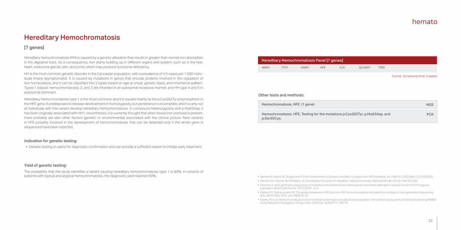
[7 genes]
Indication for genetic testing: • Genetic testing is useful for diagnostic confirmation and can provide a sufficient reason to initiate early treatment.
Yield of genetic testing:The probability that the study identifies a variant causing hereditary hemochromatosis type 1 is 80%. In cohorts of patients with typical and atypical hemochromatosis, the diagnostic yield reached 90%.
Hereditary Hemochromatosis Panel [7 genes]BMP2 FTH1 HAMP HFE HJV SLC40A1 TFR2
• Rametta R, Meroni M, Dongiovanni P. From Environment to Genome and Back: A Lesson from HFE Mutations. Int J Mol Sci. 2020 May 15;21(10):3505.• Gerhard GS, Paynton BV, DiStefano JK. Identification of Genes for Hereditary Hemochromatosis. Methods Mol Biol 2018;1706:353-365• Faria R et al. Next-generation sequencing of hereditary hemochromatosis-related genes: Novel likely pathogenic variants found in the Portuguese
population. Blood Cells Mol Dis. 2016 Oct;61:10-5.• Wallace DF, Subramaniam VN. The global prevalence of HFE and non-HFE hemochromatosis estimated from analysis of next-generation sequencing
data. Genet Med. 2016 Jun;18(6):618-26. • Adams PC et al. Hemochromatosis and iron-overload screening in a racially diverse population. Hemochromatosis and Iron Overload Screening (HEIRS)
Study Research Investigators. N Engl J Med. 2005 Apr 28;352(17):1769-78.
Other tests and methods:
Hemochromatosis. HFE (1 gene)
Hemochromatosis. HFE. Testing for the mutations p.Cys282Tyr, p.His63Asp, and p.Ser65Cys.
NGS
PCR
Exome. Turnaround time: 5 weeks

25
Other genetic tests
Gene sequencing
Exome sequencing
SNP array
Variant segregation/Family studies
NextGenDx® massive sequencing (NGS)
MLPA testing
Array CGH
We offer you the possibility of requesting the study of any gene or genes that you consider to be of interest and that are not included in the current portfolio.For more information, contact the sales representative of your area send us an email at [email protected]
Gene sequencing
Individual gene sequencing and interpretation service. Depending on its size and on the regions of interest, we can offer an approach based on Sanger sequencing or on NGS (enrichment using amplicons or hybridization probes). The NGS-based approach allows detecting copy number variations (CNVs).
NextGenDx® massive sequencing (NGS)
Next Generation Sequencing (NGS), or massive sequencing, is a term used to describe a group of newly developed technologies able to perform massive DNA sequencing. This means that millions of small DNA fragments can be sequenced at once, generating a vast amount of data. These data can add up to gigabytes of information, equivalent to 1,000 millions of DNA base pairs. In comparison, formerly used methods could only sequence one DNA fragment at a time, generating between 500 and 1,000 DNA base pairs in a single reaction.
NextGenDx® is indicated when a specific group of genes needs to be analyzed at the highest levels of diagnostic accuracy. It is aimed at:
• Monogenic diseases or diseases associated with a small number or large genes.
• Multigenic or genetically heterogeneous diseases with complex differential diagnosis.
Exome sequencing
NGS service based on sequencing the coding portion of the human genome. It is a versatile tool that enables the simultaneous testing of a large amount of genes, and it is particularly useful in those cases where clinical presentation does not allow selecting a specific clinical panel or in pathologies with a very wide range of candidate genes, e.g. epilepsy. The exome service allows for TARGETED (predefined) testing of a group of candidate genes or for CLINICAL (open) analysis, where an ad hoc test is performed based on the clinical features of each particular case. The most complex cases may benefit from TRIO- or FAMILY-BASED exome testing, which jointly analyze the exomes of several family members, taking into account the status of each studied individual (affected or healthy) and the suspected inheritance pattern.
Real-time PCR (Q-PCR)

27
MLPA testing
Semiquantitative technique that is widely applied in molecular genetic laboratories and that allows diagnosing patho-logies caused by copy number variations and, in some cases, by alterations in DNA methylation. A wide variety of commercial kits are available to test individual genes, gene panels related to specific pathologies, or large chromo-somal regions involved in microdeletion/microduplication syndromes. HIC offers MLPA services based on MRC-Ho-lland kits.
SNP array
They include more than 290 microdeletion/microduplication syndromes. Array analysis allows detecting copy number gains or losses throughout the whole genetic material of the patient. Within the field of cardiology, it is considered a first-line test for patients with congenital heart disease associated with other malformations, particularly intellectual disability, autism spectrum disorders, and/or multiple congenital malforma-tions. SNP array testing can detect copy number variations (CNVs) throughout the whole genetic material and allows confirming or ruling out microdeletion/microduplication syndromes, such as deletion 22q11 (velocardiofacial syndrome), deletion 7q11 (Williams syndrome), etc.
Indication for genetic testing. It is considered a first-line test in postnatal analysis for multiple non-specific congenital abnormalities and/or mental retardation/intellectual disability.
Among its advantages are the possibility of testing DNA from virtually any tissue, including non-cultured tissue; the detection of citogenetic abnormalities that cannot be detected by conventional tests; the identification of breakage points in chromosomal rearrangements, and the detection of loss of heterozygosity (SNP array only).
However, this technique also has some limitations. One of them is that it cannot detect balanced chromosomal rearran-gements (balanced translocations or inversions); however, it can determine whether rearrangements show losses or gains at breakage points. Likewise, it cannot detect low-level mosaicism, triploidy and other levels of polyploidy, or some aneuploidies such as XYY. CNVs from genomic regions are not covered by the platform. Moreover, the level of detection depends on study density. It does not allow detecting point mutations or gene expression, nor does it allow for methylation analysis. It also shows some limitations in the case of trisomies secondary to translocations (trisomies 13 and 21).
Array CGH
It is also known as molecular karyotyping, and its main advantage over classic karyotyping is its high sensitivity, which allows detecting structural variants that go unnoticed in conventional karyotyping. CGH array technology allows detecting losses and gains of genetic material and unbalanced rearrangements throughout the whole genome of an individual.
Postnatal CGX 108K is specifically designed for genetic diagnosis. Its mean resolution is 100 kb over the whole genome, and high resolution is 20 kb for regions of interest of the genome (regions with direct association between copy number variations and a described pathology or syndrome).
Array 37K is specifically designed for prenatal diagnosis and allows detecting genetic and chromosomal alterations with a single test. Its resolution is 10 times greater than that of conventional karyotyping and 50 times greater in critical regions for the main syndromes. Without substantially decreasing resolution in regions of interest, CGX 37K shows low coverage levels in the rest of the genome in order to minimize diagnostic uncertainty.
Variant segregation/Family studies
Sanger sequencing studies on carriers of variants that have been previously described in the family.
Real-time PCR (Q-PCR)
Real-time polymerase chain reaction (Q-PCR) is a DNA fragment amplification technique used to genotype a short genomic sequence or test for a specific genetic variant. In hematology, it is used for the detection of thrombophilia-related polymorphisms or the intron 22 inversion in F8 (hemophilia type A).

29
Sequencing Panels
Reference Test TATANEMIAS
S-202008684 Anemias and Iron Metabolism Global Panel [291 genes] 5 weeks
S-202008685 Hereditary Hemolytic Anemia Panel [115 genes] 5 weeks
S-202008700 Alfa-Thalassemia. HBA1 and HBA2 [2 genes] 5 weeks
S-202009340 Alfa-Thalassemia. MLPA HBA1 and HBA2 4 weeks
S-202008701 Beta-Thalassemia. HBB [1 gene] 5 weeks
S-202009339 Beta-Thalassemia. MLPA HBB 4 weeks
S-202008702 Sickle cell disease. HBB [1 gene] 5 weeks
S-202008690 Spherocytosis/Elliptocytosis Panel [7 genes] 5 weeks
S-202008689 Red Blood Cell Enzymopathies Panel [2 genes] 5 weeks
S-202008815 Glucose-6-phosphate dehydrogenase deficiency. G6PD [1 gene] 5 weeks
S-202008691 Aplastic Anemia Panel [91 genes] 5 weeks
S-201805629 Fanconi Anemia Panel [22 genes] 5 weeks
S-202008692 Diamond-Blackfan Anemia [29 genes] 5 weeks
S-202008261 Dyskeratosis Congenita Panel [16 genes] 5 weeks
S-202008697 Dyserythropoietic Anemia Panel [9 genes] 5 weeks
S-202008695 Sideroblastic Anemia Panel [12 genes] 5 weeks
S-202008694 Megaloblastic Anemia Panel [18 genes] 5 weeks
S-202008693 Erythrocytosis [11 genes] 5 weeks
S-202008696 Porphyria Panel [10 genes] 5 weeks
S-202009408 Acute intermittent porphyria. MLPA HMBS 4 weeks
HEMOCHROMATOSIS AND IRON METABOLISM DISORDERSS-202008698 Hemochromatosis and Iron Metabolism Disorders Panel [33 genes] 5 weeks
S-202008699 Hemochromatosis Panel [7 genes] 5 weeks
S-202009515 Hemochromatosis. HFE [1 gene] 5 weeks
S-202008844 Hemochromatosis. HFE. Testing for the mutations p.Cys282Tyr, p.His63Asp, and p.Ser65Cys 2 weeks
Other genetic tests
Consult the price and turnaround time with the team at [email protected]
We offer you the possibility of requesting the study of any gene or genes that you consider to be of interest and that are not included in the current portfolio.
For more information, contact the sales representative of your area send us an email at [email protected]

31
PRE-TEST AND POST-TEST COUNSELLING
Our studies include the possibility of pre-test and post-test counselling
For more information, please contact your sales representative

33
The quality assurance and management system of the Health in Code group combines the most rigorous management system standards (ISO 9001:2015) with excellence in performance and technical competence of a leading clinical diagnostic laboratory (authorized health center) (ISO 15189:2013 and CLIA-88) and efficient and respectful environmental management (ISO 14001:2015).
In addition, at Health in Code, we are members of the EMQN network (European Molecular Genetics Quality Network, United Kingdom) and GenQA Genomics Quality Assessment, United Kingdom), participating periodically in rigorous intercomparison tests (EQA Schemes) and obtaining highly satisfactory results that support our quality in both technical execution and clinical interpretation.
Our quality assurance has been recognized and positively evaluated by the College of American Pathologists (CAP, USA) for the detection of variants using NGS sequencing (CAP # 8280234-01).
The Health in Code services are pursuant to Spanish law on the protection of personal data (Organic Law 15/1999, of December 13, on the Protection of Personal Data, LOPD) and, therefore, to European regulations on data protection, in particular to the provisions of Regulation (EU) 2016/679 of the European Parliament and of the Council of 27 April 2016.
Our genetic diagnostic laboratories are accredited to the UNE-EN ISO 15189 standard by the Spanish national accreditation entity (ENAC) and the International Laboratory Accreditation Cooperation (ILAC). This accredi-tation represents the highest quality standard applicable to clinical laboratories at the international level.
The scope of UNE-EN ISO 15189 of Health in Code laboratories combines state-of-the-art massive parallel sequencing (custom NGS panels, targeted and whole exome sequencing) with reference techniques (gold standard) in genetics—Sanger sequencing, MLPA, dPCR, and array CGH—constituting a pioneering laboratory in obtaining a flexible scope that can be extended to all its genetic diagnostic services.
It should be noted that our laboratory's analytical developments, such testing for copy number variation (CNV), or structural variants, using a NGS depth coverage technique and mtDNA sequencing through the amplification of the complete mitochondrial genome and NGS, have been accredited to the ISO 15189 standard among first in Spain.
To achieve this, we have employed our custom-developed software for genetic diagnosis and analysis certified to the ISO 13485:2016 standard that also has the CE-IVD mark.
Additionally, Health in Code has received the prestigious CLIA certification (Clinical Laboratory Improvement Amendments) granted by the Centers for Medicare & Medical Services (CMS) of the US Federal Government, which authorizes it to perform high-complexity genetic testing (CLIA ID number 99D2153048), being one of the select group of 66 clinical laboratories worldwide that can process samples from the US outside of their territories.
Accreditations and quality assurance

+34 881 600 003 I [email protected] I www.healthincode.com For more information, please contact your sales representative