von hippel-lindau disease lancet vhl.pdf · von hippel-lindau disease is an autosomal dominant...
TRANSCRIPT

For personal use. Only reproduce with permission from The Lancet Publishing Group.
SEMINAR
von Hippel-Lindau disease is an autosomal dominantneoplasia syndrome that results from a germlinemutation in the VHL gene.1-6 Germline mutations in theVHL gene lead to the development of several benign ormalignant tumours, and cysts in many organ systems.Affected individuals might develop CNS lesionsincluding cerebellar, spinal cord, brainstem, nerve root,and supratentorial haemangioblastomas, as well asretinal haemangioblastomas and endolymphatic sactumours (table 1)5,7–17 (figure 1). Visceral features of thedisorder include renal cysts and carcinomas,phaeochromocytomas, pancreatic cysts andneuroendocrine tumours, as well as epididymal andbroad ligament cystadenomas (table 1, figure 1).
von Hippel-Lindau is not rare (incidence is roughlyone in 36 000 livebirths),18,19 and has over 90%penetrance by 65 years of age.20 Before comprehensivescreening surveys became routine, median survival ofpatients with the disease was less than 50 years of age.The main causes of death were complications linked torenal cell carcinomas and CNS haemangioblas-tomas.7–9,20,21 Improved surveillance, earlier diagnosis oflesions by modern imaging and laboratory studies (panel 1), improvements in treatment, and increasedknowledge of this disease have improved prognosis andreduced the complications related to these tumours.
Because of the progressive, diverse nature, and highfrequency of multiple neoplasms in various organsystems, the management of the tumour types iscomplicated by the presence of others. A multispecialityteam is needed for the optimum assessment andtreatment of these patients. Comprehensive serialscreening and routine scheduled follow-up are essentialfor proper care (panel 1).22 We review the molecularevents underlying tumour formation in von Hippel-
Lancet 2003; 361: 2059–67
Surgical Neurology Branch, National Institute of NeurologicalDisorders and Stroke (R R Lonser MD, E H Oldfield MD); GeneticEpidemiology Branch, Division of Cancer Epidemiology and Genetics,National Cancer Institute (G M Glenn MD); Urologic Oncology Branch,National Cancer Institute (M Walther MD, W M Linehan MD); ClinicalTrials Branch, National Eye Institute (E Y Chew MD); and SurgeryBranch, National Cancer Institute (S K Libutti MD); National Institutesof Health, Bethesda, Maryland, USA
Correspondence to: Dr Russell R Lonser, Surgical Neurology Branch,National Institute of Neurological Disorders and Stroke, NationalInstitutes of Health, Building 10, Room 5D37, Bethesda, MD 20892–1414, USA(e-mail: [email protected])
Lindau disease, discuss the salient clinical, laboratory,pathological, and radiographical findings, and examinecurrent treatment options for lesions associated with thedisease.
Molecular geneticsVHL is a tumour suppressor gene on the short arm ofchromosome 3 (3p25–26).23 Most people with thedisorder inherit a germline mutation of the gene from theaffected parent, and a normal (wild type) gene from theunaffected parent. According to Knudson’s two-hithypothesis of tumorigenesis, initiation of tumourformation arises when both VHL alleles areinactivated.24,25 Germline mutations of VHL are presentin all the cells of affected individuals who inherit thegenetic trait. However, only those cells that (1) undergoa deletion or mutation of the remaining wild type allele,and (2) are constituents of susceptible target organs
von Hippel-Lindau disease
Russell R Lonser, Gladys M Glenn, McClellan Walther, Emily Y Chew, Steven K Libutti, W Marston Linehan, Edward H Oldfield
Seminar
THE LANCET • Vol 361 • June 14, 2003 • www.thelancet.com 2059
von Hippel-Lindau disease is a heritable multisystem cancer syndrome that is associated with a germline mutation of theVHL tumour suppressor gene on the short arm of chromosome 3. This disorder is not rare (about one in 36 000 livebirths)and is inherited as a highly penetrant autosomal dominant trait (ie, with a high individual risk of disease). Affectedindividuals are at risk of developing various benign and malignant tumours of the central nervous system, kidneys, adrenalglands, pancreas, and reproductive adnexal organs. Because of the complexities associated with management of thevarious types of tumours in this disease, treatment is multidisciplinary. We present an overview of the clinical aspects,management, and treatment options for von Hippel-Lindau disease.
Mean (range) age Frequency in of onset (years) patients (%)
CNSRetinal haemangioblastomas 25 (1–67) 25–60%Endolymphatic sac tumours 22 (12–50) 10%Craniospinal haemangioblastomas
Cerebellum 33 (9–78) 44–72%Brainstem 32 (12–46) 10–25%Spinal cord 33 (12–66) 13–50%Lumbosacral nerve roots Unknown (..) <1%Supratentorial Unknown (..) <1%
VisceralRenal cell carcinoma or cysts 39 (16–67) 25–60%Phaeochromocytomas 30 (5–58) 10–20%Pancreatic tumour or cyst 36 (5–70) 35–70%Epididymal cystadenoma Unknown (..) 25–60%Broad ligament cystadenoma Unknown (16–46) Unknown
See references 5,7–17.
Table 1: Frequency of lesions and age at onset of von Hippel-Lindau disease lesions
Search strategy
We searched PubMed database using the keyword von Hippel-Lindau, combined with the terms central nervoussystem, haemangioblastoma, pancreatic neuroendocrinetumour, pheochromocytoma, renal cell carcinoma, ortreatment. We focused mainly on manuscripts publishedduring the past 10 years, but have also referenced papersfrom before that time. Relevant articles that were not identifiedby this search were also referenced.

For personal use. Only reproduce with permission from The Lancet Publishing Group.
(CNS, kidneys, adrenal glands, pancreas, andreproductive adnexal organs) develop tumours. Someinvestigators have shown somatic inactivation of theVHL gene in sporadically occurring CNShaemangioblastomas,26–29 and renal cell carcinomas.30–33
The VHL gene has three exons that encode the VHLprotein.23 VHL is a tumour suppressor protein that islocalised in the nucleus or cytoplasm, the extent to whichbeing dependent on cell density.34,35 The protein forms a complex with other proteins including elongin B,elongin C, and Cullin 2 (CUL2), to form the VCB-CUL2 complex (figure 2).36,37 This protein complexdetermines ubiquitin-dependent proteolysis of largecellular proteins. When normal oxygen levels arepresent, the VCB-CUL2 complex binds to, and targets,the � subunits of hypoxia-inducible factors (HIF) 1 and 2 for ubiquitin-mediated degradation of protein (figure 2).38
Abnormal or absent VHL protein function (as in vonHippel-Lindau syndrome) might disrupt tumour-suppression indirectly through HIF-mediated effects (figure 2) or directly through VHL-mediated effects, orboth. Many of the tumour suppressive effects of theprotein could result from the degradation of HIF. Innormal circumstances, HIF can coordinate the cell’sresponse to hypoxia. Through transcriptional regulation,HIF enhances glucose uptake and increases expressionof angiogenic, growth, and mitogenic factors includingvascular endothelial growth factor (VEGF), plateletderived growth factor � polypeptide (PDGF�),erythropoietin (which can cause polycythaemia thatoccasionally arises in von Hippel-Lindau), andtransforming growth factor � (TGF�) (figure 2).6,39–41
Subsequently, disruption of VHL-protein-mediateddegradation of HIF could contribute to tumour formationthrough multiple mechanisms (figure 2). If VHL functionwere absent or abnormal, HIF could stimulateangiogenesis, which is critical for persistence of tumoursassociated with the disorder. HIF-mediated angiogenesiscould result from increased levels of VEGF or PDGF�, orboth, which are known to be important for proliferation ofendothelial cells and pericytes, respectively (figure 2).
This link might explain the highly vascular nature oftumours associated with von Hippel-Lindau disease,especially haemangioblastomas and renal cell carcinomas.Moreover, high vascular permeability of the tumourvessels, resulting from increased VEGF levels, might alsounderlie the peritumoural oedema and cysts generallypresent in this disorder.10 Another potential mechanism ofHIF-mediated carcinogenesis is overproduction of TGF�.Besides being a potent mitogenic factor (especially forrenal epithelium), raised TGF� can stimulate cellularoverexpression of epidermal growth factor receptors(receptors for TGF�), creating an autocrine loop.6,40,42–46
Other possible mechanisms of tumorigenesis caused byabsent or abnormal VHL protein, independent of HIF,include disruption of normal cell cycle, increasedangiogenesis, and abnormalities in the extracellularmatrix. The inability to leave the cell cycle (ie, to enterGo) is seen in cells without VHL protein.43 This eventmight take place early in tumorigenesis. Furthermore,mutations of the protein itself could increase VEGFexpression through incorrect transcriptional and post-translational regulation.47,48 These mutations mightaugment the angiogenic effects mediated by HIF andfurther increase tumour vessel permeability. Finally,although cells without VHL protein can secretefibronectin, they cannot properly assemble a fibronectinextracellular matrix, which could contribute tocarcinogenesis.42 Overall, HIF-mediated, direct VHL-protein-mediated, and unknown effects of abnormal orabsent VHL protein probably interact to induce formationof the various tumours in this disease.
Clinical diagnosisDiagnosis of von Hippel-Lindau disease is often based onclinical criteria. Patients with a family history, and a CNShaemangioblastoma (including retinal haemangio-blastomas), phaeochromocytoma, or clear cell renalcarcinoma are diagnosed with the disease. Those with norelevant family history must have two or more CNShaemangioblastomas, or one CNS haemangioblastomaand a visceral tumour (with the exception of epididymaland renal cysts, which are frequent in the generalpopulation) to meet the diagnostic criteria.9,49,50
Specific correlations of genotype and phenotype haveemerged in affected families. Several familial phenotypesof von Hippel-Lindau disease are now recognised,providing useful information to screen and counselaffected individuals (panel 2). Type 1 families have agreatly reduced risk of phaeochromocytomas, but candevelop all the other tumour types generally associatedwith the disease. Type 2 families have phaeo-chromocytomas, but have either a low-risk (type 2A) orhigh-risk (type 2B) for renal cell carcinomas. Type 2Cfamilies have phaeochromocytomas only, with no otherneoplastic findings of VHL (panel 2).6,51–56
Genetic testing Advances in genetic testing for the disease includequalitative and quantitative Southern blotting, which hasbeen added to DNA sequence analysis. This improved
SEMINAR
2060 THE LANCET • Vol 361 • June 14, 2003 • www.thelancet.com
Retina
Cerebellum
BrainstemCentral nervous system
Visceralorgans
Endolymphatic sacSpinal cord
Kidneys
Epididymis (male) orbroad ligament
(female)
Adrenalglands
Pancreas
Figure 1: Affected organs

For personal use. Only reproduce with permission from The Lancet Publishing Group.
testing has increased the detection rate of DNAmutations in peripheral blood leucocytes from 75% tonearly 100%.57 In 1996, there were more than 137distinct intragenic germline mutations reported inaffected families in North America, Europe, andJapan.51 Mutation types included missense, non-sense,microdeletion, insertion, deletion, and splice site.51
Known mutations of von Hippel-Lindau disease arenow stored online (http://www.umd.necker.fr/). Sincegenetic testing detects mutations in nearly 100% ofdocumented affected families, serial clinical surveillancestudies are recommended for family members withmutations.
A diagnostic challenge arises in de novo cases (ie, thefirst affected member of a family) of von Hippel-Lindaudisease. These cases arise in as many as 20% ofkindreds.58 The initial mutation in a de novo case mightresult in disease mosaicism (ie, some, but not all, tissuescarry the new disease mutation). Thus, such patientsmight have clinical signs of the disease, but test negativegenetically, because the VHL mutation is not carried inall peripheral leucocytes. The earlier the new mutationarises in embryogenesis, the more numerous and variedthe types of cells that will carry the mutation.Mosaicism can occur as a mutated gene in somatictissue only, in germ tissue only, or in both. The risks toa carrier of a new mutation and to their offspring arevery different in these three circumstances.
Central nervous system lesions CNS haemangioblastomas (excluding retina)General featuresHaemangioblastomas of the CNS are the most commontumour in von Hippel-Lindau disease, affecting 60–80% ofall patients.11,21,59 The average age of presentation for CNShaemangioblastomas is 33 years (table 1).11 These tumoursare benign, but are a major cause of morbidity. They ariseanywhere along the craniospinal axis and are oftenassociated with oedema or cysts (associated cysts occur with 30–80% of haemangioblastomas), or both. CNShaemangioblastomas are generally seen in the spinal cord and cerebellum, followed by the brainstem,lumbosacral nerve roots, and supratentorial region (table 2).
Clinical, imaging, and histological findingsSymptoms related to haemangioblastomas depend ontumour location and size, and the presence of associatedoedema or cysts.11 We reviewed the natural history of CNShaemangioblastomas (665 in 160 consecutive patients) asdefined by serial MRI.11 As expected, the tumour volumeneeded to produce symptoms indicated the spatial capacitywherever the tumour is developing. Smaller lesions in thespinal cord and brainstem were needed to producesymptoms than in the cerebellum. Symptomatic tumoursgrew ten times faster than asymptomatic tumours; whereascysts grew 3·2-fold and 1·7-fold faster (symptomatic vsasymptomatic) than the haemangioblastoma associatedwith them. These tumours often have several periods oftumour growth separated by periods of arrested growth;many untreated tumours remain the same size for years.Table 2 shows the most common signs and symptomsassociated with haemangioblastomas in various regions ofthe CNS.
Haemangioblastomas of the craniospinal axis are bestassessed by contrast-enhanced T1-weighted MRI (panel 1).These tumours can be easily identified and quantified byradiography. T2-weighted or FLAIR (fluid-attenuatedinversion recovery) MRI is used to quantify peritumouraloedema and cysts (panel 1). Arteriography typically showsintense persistent staining of the haemangioblastoma,arteriovenous shunting, and early draining veins.
Haemangioblastomas are well defined, thinly encap-sulated tumours that appear bright red at operation.Histologically, they consist of a rich vascular plexus that issurrounded by polygonal stromal cells (figure 3). Thestromal cell is neoplastic in origin.60,61
Treatment Most haemangioblastomas of thecraniospinal axis can be safely andcompletely excised by surgery.10,12,62
Haemangioblastomas in the CNS oftengrow at several sites simultaneously,new lesions can arise with time, and thegrowth pattern of these tumours can beirregular and unpredictable, thereforeresection is deferred until the onset ofsymptoms to avoid unnecessarysurgery.10–12
Preoperative embolisation is done atsome centres to reduce tumourvascularity before resection. Generally,we do not use embolisation, since thesetumours can be excised with minimumblood loss if the basic tenets ofcircumferential microsurgical excisionare followed.10,12,62
SEMINAR
THE LANCET • Vol 361 • June 14, 2003 • www.thelancet.com 2061
Panel 1: Recommended intervals for screening inat-risk individualsTest Start age (frequency)Ophthalmoscopy Infancy (yearly)Plasma or 24 h urinary 2 years of age (yearly and whencatecholamines and blood pressure is raised)metanephrinesMRI of craniospinal axis* 11 years of age (yearly)CT and MRI of internal Onset of symptoms (hearing loss, auditory canals* tinnitus, vertigo, or unexplained
difficulties of balance)Ultrasound of abdomen 8 years of age (yearly; MRI as
clinically indicated)CT of abdomen* 18 years of age or earlier if
clinically indicated (yearly)Audiological function tests When clinically indicatedAdapted from reference 22. *Imaging techniques that are generallyrecommended before and after contrast infusion.
Hypoxia Normoxia
Ubiquitin-mediateddegradation
Increased Glut-1Increased VEGF
Increased PDGF�Increased erythropoietin
Increased TGF�
HIF
VHL
Elongin C
Elongin B
VCB-CUL2 complex
CUL2
Figure 2: Interaction of VHL protein with other proteins including elongin B, elongin C,and CUL2, to form the VCB-CUL2 complexGlut-1=glucose transporter 1.

For personal use. Only reproduce with permission from The Lancet Publishing Group.
Because of the potential morbidity associated withresection of multiple craniospinal haemangioblastomas invon Hippel-Lindau disease, stereotactic radiation therapyhas been used instead.63–68 Small haemangioblastomas (<3 cm diameter), and those not associated with cystsmight respond safely to radiation therapy.63–68 However,studies with longer assessment and more patients thanthose done so far, are needed to establish effectiveness andpotential long-term effects of this treatment.
Retinal hemangioblastomasGeneral featuresRetinal haemangioblastomas (retinal angiomas) are one ofthe most common tumours in von Hippel-Lindau disease,and are seen in as many as 60% of patients (table 1).13,14,50
They arise in the periphery, or on or near the optic disc, orboth. They are often multifocal and bilateral (about 50%).The mean age of patients at presentation is 25 years, but 5% of retinal angiomas present in patients younger than10 years of age.
Clinical, imaging, and histological findingsRetinal angiomas are symptomless in the initial stages, andare detectable only by examination of the dilated eye.Despite being asymptomatic, they can lead to part or totalloss of vision. Peripheral tumours cause visual symptomsthrough their growth, increased vascular permeability(which leads to accumulation of subretinal fluid), and thedevelopment of hard exudates at the macula. Tumours onthe optic disc behave similarly. Both peripheral and centralangiomas can cause exudative and tractional retinaldetachment as they enlarge.
Ophthalmoscopy with pharmacological dilation of the irisallows identification of most retinal tumours (figure 4).Fluorescein angiography is used to assess macular functionassociated with peripheral and optic nerve lesions.Screening examinations with dilated fundoscopy should bedone at least once a year starting at age 1 year (panel 1).Ophthalmologists specialising in vitreoretinal diseaseshould provide the ophthalmological management ofpatients with von Hippel-Lindau disease.
Retinal haemangioblastomas are grossly and histologi-cally identical to craniospinal haemangioblastomas.69
Treatment Early diagnosis and treatment can prevent visual loss orblindness. Most peripheral retinal tumours respond to laserphotocoagulation or cryotherapy.70 Vitrectomy should beconsidered for patients with substantial tractional detach-
ment of the retina with a large fibrovascular component.Because of the damage that some treatments including laserphotocoagulation can cause, tumours on the optic discshould be monitored without treatment. Enucleation mightbe necessary for irreversible glaucoma in which severe painis associated with end-stage ocular angiomatosis.
Various radiotherapy treatments have been applied tocases of severely affected retinas that did not respond tousual methods, but the usefulness of these approaches andtheir role in management of retinal haemangioblastomasneeds to be defined. Anti-VEGF treatment has beenreported to restore visual function in a patient with atumour in the optic nerve head.71 This experimentalapproach might provide hope for patients withhaemangioblastomas that are not amenable to currenttreatments.
Endolymphatic sac tumoursGeneral featuresThese tumours are rare in the general population, but arefrequently associated with von Hippel-Lindau disease.72
MRI revealed evidence of these tumours in 11% of patients(15 tumours in 13 of 121 patients assessed; the mean age ofpresentation was 22 years) (table 1). von Hippel-Lindau isthe only condition associated with bilateral endolymphaticsac tumours.
Clinical, imaging, and histological findingsPatients in whom there is evidence of these tumours fromimaging present with part or complete hearing loss(100%), tinnitus (77%), a sense of dysequilibrium (62%),and facial paresis (8%).72 The frequency of microscopictumours might be higher than that estimated by imaging,because patients often have vestibulocochlear symptomswith no CT or MRI evidence of such a tumour.72
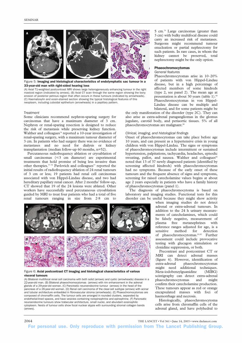
Radiological examination and diagnosis of endo-lymphatic sac tumours include precontrast and post-contrast CT and MRI of the internal auditory canals(figure 5). In CT images, the tumours are generallyisointense with brain parenchyma, but can have focal areasof low and high attenuation. CT of the temporal boneswith large tumours shows a destructive or expansive lesioncentred in the endolymphatic duct (in between the sigmoidsinus and internal auditory canal). MRI shows eitherhomogeneous or heterogeneous intensity after precontrastT1-weighted imaging. Postcontrast T1-weighted MRI canalso show homogeneous or variable patterns of patchyenhancement. Audiograms are used to supplementradiographical data, and to document the presence orprogression of hearing loss (panel 1).
Endolymphatic sac tumours are highly vascular, andoften erode or expand the surrounding temporal bone.
SEMINAR
2062 THE LANCET • Vol 361 • June 14, 2003 • www.thelancet.com
Panel 2: Genotype-phenotype classifications infamilies with von Hippel-Lindau disease*
Clinical characteristicsType 1 Retinal haemangioblastomas
CNS haemangioblastomasRenal cell carcinomaPancreatic neoplasms and cysts
Type 2A PhaeochromocytomasRetinal haemangioblastomasCNS haemangioblastomas
Type 2B PhaeochromocytomasRetinal haemangioblastomasCNS haemangioblastomasRenal cell carcinomasPancreatic neoplasms and cysts
Type 2C Phaeochromocytoma only
*Endolymphatic sac tumours and cystadenomas of the epididymis and broadligament have not been assigned to specific von Hippel-Lindau types.
Frequency of Signs and symptoms (%)*haemangio-blastomas in nervous system*
Cerebellum 37% Gait ataxia (64%), dysmetria (64%), headaches (12%), diplopia (8%), vertigo (8%), emesis (8%)
Brainstem 10% Hypaesthesia (55%), gait ataxia (22%), dysphagia (22%), hyper-reflexia (22%), headaches (11%), dysmetria (11%)
Spinal cord 50% Hypaesthesia (83%), weakness (65%), gait ataxia (65%), hyper-reflexia (52%), pain (17%), incontinence (14%)
*Frequency of signs and symptoms of patients who were undergoing resection.See reference 11.
Table 2: Signs and symptoms associated withhaemangioblastomas

For personal use. Only reproduce with permission from The Lancet Publishing Group.
Histologically, they form papillary cystic regions filled withproteinaceous material (figure 5). Because of theiradenomatous appearance and associated bone erosion,they were previously referred to as low-gradeadenocarcinomas despite the absence of malignantfeatures.
TreatmentSurgery is curative for completely excised tumours, and thepreoperative level of hearing is usually preserved. Decisionsabout the timing of surgical treatment must allow for (1)the slow, but variable, growth of these tumours; (2)preoperative hearing level; (3) severity of vestibularsymptoms; (4) possibility of hearing loss or facial nerveinjury as a result of surgery; and (5) the possibility ofbilateral tumours. The role of radiation therapy in thetreatment of these tumours is unclear.
Visceral lesionsRenal cell carcinomas and renal cystsGeneral featuresRenal cell carcinomas are the major malignant neoplasm invon Hippel-Lindau disease and the primary cause ofinherited renal cancer. These tumours are seen in 24–45%of patients, and adding renal cysts increases the finding ofrenal lesions to 60% (table 1).20,22 The mean age atpresentation is 39 years (table 1). Although small renaltumours in this disease tend to be low grade and minimally
invasive,73 their rate of growth varies widely.74 Renal lesions are often multiple and bilateral. Walther andcolleagues75 estimated that 600 microscopic tumours and 1100 microscopic clear-cell-lined cysts might bepresent in the kidneys of some 37-year-old patients. Results of an investigation of 228 renal lesions in 28 patients, followed for at least 1 year, showed thattransition from a cyst to a solid lesion was rare.76 However,complex cystic and solid lesions can contain neoplastictissue that frequently enlarges.73,76
Clinical, imaging, and histological findingsRenal cell carcinomas often remain asymptomatic for longintervals. Thus, serial imaging of the kidneys is useful for early diagnosis. Occasionally, the moreadvanced cases with these neoplasms can present withhaematuria, flank pain, or a flank mass. Renal cysts in vonHippel-Lindau disease are typically asymptomatic andseldom need treatment. However, complex cysts needmonitoring, as they often harbour solid components ofrenal cell carcinoma. Because of the frequent absence ofearly clinical symptoms and the importance of earlydetection, diagnosis during presymptomatic screening hasthe potential to enhance overall outcome.
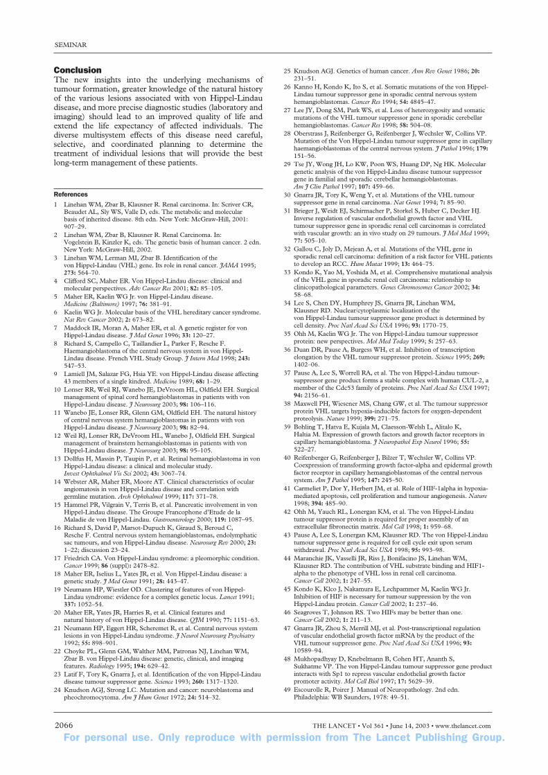
Contrast-enhanced abdominal CT is the standard fordetection of renal involvement in the disease (figure 6). CTallows detection and quantification by size and number ofrenal cell carcinomas and cysts, allowing serial monitoring
of individual lesions. Imaging is usuallyrecommended in 3–5 mm sections, andbefore and after intravenous injection ofcontrast media. Precontrast andpostcontrast MRI is an alternativemethod of detection for patients whohave reduced renal function.
These carcinomas are yellow ororange and are encapsulated. They can be solid or a mixture of solid andcystic in appearance. Histologically,they are always of the clear-cell subtype (figure 6), and small carcinomas tend to be low grade.73 Treatment recom-mendations can depend on tumour size.
SEMINAR
THE LANCET • Vol 361 • June 14, 2003 • www.thelancet.com 2063
A B C
A B C
C5
C6
D
*
Figure 3: MRI and histological features of CNS haemangioblastomas(A) Axial T1-weighted contrast-enhanced MRI of a cerebellar haemangioblastoma (arrow) with an associated cyst (homogeneous associated dark region) ina 40-year-old woman. (B) Mid-sagittal T1-weighted postcontrast MRI of medullary haemangioblastoma (arrow) with associated brainstem oedema (asterisk)in a 12-year-old girl. (C) Mid-sagittal postcontrast T1-weighted MRI of the spinal cord of a 50-year-old man. The haemangioblastoma is located in theposterior portion of the spinal cord at C5 and C6 (arrow), and is associated with a large syrinx (dark intraspinal region extending rostral and caudal to thelesion). (D) Haematoxylin and eosin staining of a haemangioblastoma showing the lipid-laden stromal cells (arrows) distributed within a capillary network(arrowheads).
Figure 4: Ophthalmoscopic view of retinal haemangioblastomas(A) Peripheral retinal haemangioblastoma (arrow) with an enlarged vessel (arrowheads) in a 22-year-old woman. (B) Peripheral retinal haemangioblastoma (arrow) with fibrous changes, and hard exudates and retinal oedema in the surrounding region in a 24-year-old man. (C) Retinalhaemangioblastoma (arrow) on the optic nerve head with yellow retinal hard exudates below it in a32-year-old man.
For personal use. Only reproduce with permission from The Lancet Publishing Group.
TreatmentSome clinicians recommend nephron-sparing surgery forcarcinomas that have a maximum diameter of 3 cm.Nephron or renal-sparing resection is designed to reducethe risk of metastasis while preserving kidney function.Walther and colleagues74 reported a 10-year investigation ofrenal-sparing surgery, with a maximum tumour diameter of3 cm. In patients who had surgery there was no evidence ofmetastases and no need for dialysis or kidneytransplantation (median follow-up 60 months, n=52).
Percutaneous radiofrequency ablation or cryoablation ofsmall carcinomas (�3 cm diameter) are experimentaltreatments that hold promise of being less invasive thanother therapies.77–79 Pavlovich and colleagues77 reported theinitial results of radiofrequency ablation of 24 renal tumoursof 3 cm or less; 19 patients had renal cell carcinomasassociated with von Hippel-Lindau disease, and two hadhereditary papillary renal cancer. After 2 months’ follow-up,CT showed that 19 of the 24 lesions were ablated. Otherworkers have successfully used percutaneous cryoablationguided by MRI to treat four patients who had a total of fiverenal tumours ranging in size from 2·8 cm to
5 cm.79 Large carcinomas (greater than 3 cm) with bulky multifocal disease couldcarry an increased risk of metastases.Surgeons might recommend tumourenucleation or partial nephrectomy forsuch patients. In rare cases, in whom thekidney cannot be preserved, totalnephrectomy might be the only option.
PhaeochromocytomasGeneral featuresPhaeochromocytomas arise in 10–20%of patients with von Hippel-Lindaudisease, but in a high percentage ofaffected members of some kindreds (type 2, see panel 2). The mean age atpresentation is about 30 years (table 1).80
Phaeochromocytomas in von Hippel-Lindau disease can be multiple andbilateral, and for some patients might be
the only manifestation of the disorder (type 2C). They canalso arise as extra-adrenal paragangliomas in the glomusjugulare, carotid body, and periaortic tissues. 5% of allphaeochromocytomas are malignant.80
Clinical, imaging, and histological findingsOnset of phaeochromocytomas can take place before age 10 years, and can present as a hypertensive crisis in youngchildren with von Hippel-Lindau. The signs or symptomsof phaeochromocytomas include intermittent or sustainedhypertension, palpitations, tachycardia, headaches, episodicsweating, pallor, and nausea. Walther and colleagues80
noted that 13 of 37 newly diagnosed patients (identified byscreening affected kindreds) with phaeochromocytomashad no symptoms. Because of the early onset of thesetumours and the frequent absence of signs and symptoms,screening for raised catecholamine values begins at aboutage 2 years especially in patients who have a family historyof phaeochromocytomas (panel 1).
The diagnosis of phaeochromocytoma is based onlaboratory and imaging studies. Functional tests for thisdisorder can be useful because they might show activity
when imaging studies do not detectadrenal or extra-adrenal tumours. Inaddition to the 24 h urinary measure-ments of catecholamines, which couldbe falsely negative, measurement ofplasma free metanephrines withreference ranges adjusted for age, is asensitive method for detection of phaeochromocytomas.81,82 Furtherassessment could include provocativetesting with glucagon stimulation orclonidine suppression, or both.
Precontrast and postcontrast CT orMRI can detect adrenal masses (figure 6). However, identification ofextra-adrenal phaeochromocytomasmight need additional techniques.Meta-iodobenzylguanidine (MIBG)scintigraphy can detect extra-adrenalphaeochromocytomas and mightconfirm their catecholamine production.These tumours appear as red or orangeencapsulated masses with foci ofhaemorrhage and necrosis.
Histologically, phaeochromocytomacells arise from chromaffin cells of theadrenal gland, and have polyhedral to
SEMINAR
2064 THE LANCET • Vol 361 • June 14, 2003 • www.thelancet.com
A B CC
Figure 5: Imaging and histological characteristics of endolymphatic sac tumour in a33-year-old man with right-sided hearing loss(A) Axial T1-weighted postcontrast MRI shows large heterogeneously enhancing tumour in the rightmastoid region (indicated by arrows). (B) Axial CT scan through the same region showing the bonyerosion of posterior petrous region that often occurs in these tumours (indicated by arrowheads). (C) Haematoxylin and eosin-stained section showing the typical histological features of thisneoplasm, including cuboidal epithelium (arrowheads) in a papillary pattern.
Figure 6: Axial postcontrast CT imaging and histological characteristics of variousvisceral tumours(A) Bilateral multifocal renal cell carcinoma with both solid (arrows) and cystic (arrowheads) disease in a22-year-old man. (B) Bilateral phaeochromocytomas (arrows) with rim enhancement in the adrenalglands of a 29-year-old woman. (C) Pancreatic neuroendocrine tumour (arrows) in the head of thepancreas of a 26-year-old woman. (D) Renal cell carcinoma of the clear-cell subtype (arrows) with acinarand tubular architecture embedded in fibrovascular stroma (arrowheads). (E) Phaeochromocytomas arecomposed of chromaffin cells. The tumour cells are arranged in rounded clusters, separated byendothelial-lined spaces, and have vesicles containing norepinephrine and epinephrine. (F) Pancreaticneuroendocrine tumours show trabecular architecture, small nuclei, and abundant eosinophiliccytoplasm. Nests of tumour cells show focal nuclear atypia with surrounding stromal collagen bands(arrows).

For personal use. Only reproduce with permission from The Lancet Publishing Group.
fusiform morphology with vesicular nuclei, and granular,amphophilic, or basophilic cytoplasm (figure 6).
TreatmentFor ideal perioperative management of patients with vonHippel-Lindau disease, an up-to-date assessment of thepatient’s overall condition should be done before they havesurgery or go into labour. However, preoperative screeningis especially important for detection of hidden phaeo-chromocytomas because of the potential risk of a hyper-tensive crisis perioperatively. Treatment of phaeochromocytomas is most often by surgical resection(preferably laparoscopically), and is increasingly done aspart adrenalectomy or enucleation to preserve adrenalfunction.83 Before surgery, pharmacological control with acombination of � blockade and metyrosine blockade isoften needed. Indications for surgery can include tumourswith abnormal function, Meta-iodobenzylguanidineuptake, or tumour size greater than 3·5 cm.80 In patientswith von Hippel-Lindau disease and phaeochromo-cytomas, early intervention with cortical-sparing adrenalsurgery results in low recurrence rates and long-termcorticosteroid independence.84
Pancreatic neuroendocrine tumours and cystsGeneral featuresPancreatic neuroendocrine tumours arise in 8–17% ofpatients with von Hippel-Lindau disease.15,85,86 Pancreaticcysts and serous cystadenomas occur with a prevalence of17–56%.85,87 Overall, 35–70% of patients have a pancreaticneuroendocrine tumour, cyst, or cystadenoma.87–89
Distinguishing between a benign multicystic cystadenomaand a pancreatic neuroendocrine tumour can be difficult.The mean age at presentation for neuroendocrine tumoursis 35 years, and for pancreatic cysts is 37 years.
Clinical, imaging, and histological findingsPancreatic cysts in this disease are generally asymptomatic,and do not need treatment. Most of these tumours arenon-functional. Thus, they are often clinically silent androutine periodic imaging of asymptomatic individuals isimportant for diagnosis of von Hippel-Lindau disease.
A pancreatic neuroendocrine tumour can be seen as an enhancing mass in postcontrast CT imaging done during the arterial (early) phase (later phase imagingmight not clearly distinguish the tumour from surroundinghealthy tissue) (figure 6, F). Once this tumour is identifiedwith CT, pancreatic MRI can be used to confirm thediagnosis. Additional studies, including endoscopicultrasound exploration in conjunction with somatostatinreceptor scintigraphy, could be useful for diagnosis.Pancreatic neuroendocrine tumours are encapsulated andwell circumscribed.
Histologically, they are formed from pancreatic isletsand have been historically termed islet cell tumours.Although they are clinically non-functional, withimmunohistochemistry they stain positive for pancreaticand gastrointestinal hormones (figure 6, F).90
TreatmentTreatment is by surgical resection, and the specificapproach is determined by the location and size of thetumour. Tumours detected during imaging ofasymptomatic periods, and resected on the basis of sizehave been successfully managed with no development ofmetastasis. Libutti and colleagues85,90 recommend thefollowing criteria for resection of these lesions: (1) noevidence of metastatic disease; (2) tumour size greater than3 cm in the body or tail, or greater than 2 cm in the head of
the pancreas; or (3) the patient undergoing laparotomy forother lesions. Surgical resections can be done byenucleation, pylorus-preserving pancreaticoduodenectomy(Whipple’s procedure), or part or total (rarely)pancreatectomy with replacement therapy. Tumours in thebody and tail have been noted to be successfully managedwith laparoscopy.90
Preservation of functional pancreas should be done withthe malignant potential of the tumour in mind. Inmetastatic hepatic disease (the most common metastates),long-term control has been achieved by combinations ofablative therapy and isolated hepatic chemotherapeuticperfusion. Patients who do not meet the criteria for resection, have been successfully followed with CT at12-month intervals.85,90
Epididymal cystadenomasGeneral featuresEpididymal papillary cystadenomas are seen in 25–60% ofmen with von Hippel-Lindau disease and can be multipleand bilateral.91–94 The tumours are benign and typicallyappear in the teenage years. They arise from theepididymal duct, which is derived from the embryonicmesonephric duct.
Clinical, imaging, and histological findingsThese cystadenomas are characteristically asymptomatic.The diagnosis is made by palpation and confirmed byultrasound with sonographic criteria95 that include (1)predominantly solid tumour greater than 10�14 mm, (2)occurrence in a man with von Hippel-Lindau disease, and(3) slow growth.
Epididymal cystadenomas appear solid but consist ofmultiple cysts (filled with a colloid-like material) andpapillary fronds with a fibrovascular core. Histologically,the epithelium has clear-cell features similar to otherlesions in the disease.
TreatmentBecause these lesions are benign and typicallysymptomless, they are managed conservatively andtreatment is reserved for the rare occurrence of symptoms.Ultrasonography can be used to monitor their growth overtime.
Broad ligament cystadenomasGeneral featuresPapillary cystadenomas in the broad ligament have rarelybeen reported and are unrecognised in many women withvon Hippel-Lindau disease.96–98 The mean age of women atpresentation and true frequency of cystadenomas in thisdisease is unknown. Gaffey and others97 refer to thetumour as adnexal papillary cystadenoma of probablemesonephric origin, since the tumours are believed to arisefrom the remnant of the embryonic mesonephric duct.The earliest age at which this tumour has been diagnosedis 16 years, but in other reports, the age of onset is between22 years and 46 years (table 1).96–98
Clinical, imaging, and histological findingsThese lesions can be diagnosed by CT-imaging or ultra-sonography. The tumours are grossly and histologicallysimilar to epididymal cystadenomas.
TreatmentBecause they are benign and typically asymptomatic, theycan be managed conservatively. Treatment is reserved forthe rare occurrences of symptoms. CT or ultrasonographycan be used to document their size over time.
SEMINAR
THE LANCET • Vol 361 • June 14, 2003 • www.thelancet.com 2065
For personal use. Only reproduce with permission from The Lancet Publishing Group.
ConclusionThe new insights into the underlying mechanisms oftumour formation, greater knowledge of the natural historyof the various lesions associated with von Hippel-Lindaudisease, and more precise diagnostic studies (laboratory andimaging) should lead to an improved quality of life andextend the life expectancy of affected individuals. Thediverse multisystem effects of this disease need careful,selective, and coordinated planning to determine thetreatment of individual lesions that will provide the bestlong-term management of these patients.
References1 Linehan WM, Zbar B, Klausner R. Renal carcinoma. In: Scriver CR,
Beaudet AL, Sly WS, Valle D, eds. The metabolic and molecular basis of inherited disease. 8th edn. New York: McGraw-Hill, 2001:907–29.
2 Linehan WM, Zbar B, Klausner R. Renal Carcinoma. In: Vogelstein B, Kinzler K, eds. The genetic basis of human cancer. 2 edn.New York: McGraw-Hill, 2002.
3 Linehan WM, Lerman MI, Zbar B. Identification of the von Hippel-Lindau (VHL) gene. Its role in renal cancer. JAMA 1995;273: 564–70.
4 Clifford SC, Maher ER. Von Hippel-Lindau disease: clinical andmolecular perspectives. Adv Cancer Res 2001; 82: 85–105.
5 Maher ER, Kaelin WG Jr. von Hippel-Lindau disease. Medicine (Baltimore) 1997; 76: 381–91.
6 Kaelin WG Jr. Molecular basis of the VHL hereditary cancer syndrome.Nat Rev Cancer 2002; 2: 673–82.
7 Maddock IR, Moran A, Maher ER, et al. A genetic register for vonHippel-Lindau disease. J Med Genet 1996; 33: 120–27.
8 Richard S, Campello C, Taillandier L, Parker F, Resche F.Haemangioblastoma of the central nervous system in von Hippel-Lindau disease. French VHL Study Group. J Intern Med 1998; 243:547–53.
9 Lamiell JM, Salazar FG, Hsia YE. von Hippel-Lindau disease affecting43 members of a single kindred. Medicine 1989; 68: 1–29.
10 Lonser RR, Weil RJ, Wanebo JE, DeVroom HL, Oldfield EH. Surgicalmanagement of spinal cord hemangioblastomas in patients with vonHippel-Lindau disease. J Neurosurg 2003; 98: 106–116.
11 Wanebo JE, Lonser RR, Glenn GM, Oldfield EH. The natural historyof central nervous system hemangioblastomas in patients with vonHippel-Lindau disease. J Neurosurg 2003; 98: 82–94.
12 Weil RJ, Lonser RR, DeVroom HL, Wanebo J, Oldfield EH. Surgicalmanagement of brainstem hemangioblastomas in patients with vonHippel-Lindau disease. J Neurosurg 2003; 98: 95–105.
13 Dollfus H, Massin P, Taupin P, et al. Retinal hemangioblastoma in vonHippel-Lindau disease: a clinical and molecular study. Invest Ophthalmol Vis Sci 2002; 43: 3067–74.
14 Webster AR, Maher ER, Moore AT. Clinical characteristics of ocularangiomatosis in von Hippel-Lindau disease and correlation withgermline mutation. Arch Ophthalmol 1999; 117: 371–78.
15 Hammel PR, Vilgrain V, Terris B, et al. Pancreatic involvement in vonHippel-Lindau disease. The Groupe Francophone d’Etude de laMaladie de von Hippel-Lindau. Gastroenterology 2000; 119: 1087–95.
16 Richard S, David P, Marsot-Dupuch K, Giraud S, Beroud C, Resche F. Central nervous system hemangioblastomas, endolymphaticsac tumours, and von Hippel-Lindau disease. Neurosurg Rev 2000; 23:1–22; discussion 23–24.
17 Friedrich CA. Von Hippel-Lindau syndrome: a pleomorphic condition.Cancer 1999; 86 (suppl): 2478–82.
18 Maher ER, Iselius L, Yates JR, et al. Von Hippel-Lindau disease: agenetic study. J Med Genet 1991; 28: 443–47.
19 Neumann HP, Wiestler OD. Clustering of features of von Hippel-Lindau syndrome: evidence for a complex genetic locus. Lancet 1991;337: 1052–54.
20 Maher ER, Yates JR, Harries R, et al. Clinical features and natural history of von Hippel-Lindau disease. QJM 1990; 77: 1151–63.
21 Neumann HP, Eggert HR, Scheremet R, et al. Central nervous systemlesions in von Hippel-Lindau syndrome. J Neurol Neurosurg Psychiatry1992; 55: 898–901.
22 Choyke PL, Glenn GM, Walther MM, Patronas NJ, Linehan WM,Zbar B. von Hippel-Lindau disease: genetic, clinical, and imagingfeatures. Radiology 1995; 194: 629–42.
23 Latif F, Tory K, Gnarra J, et al. Identification of the von Hippel-Lindaudisease tumour suppressor gene. Science 1993; 260: 1317–1320.
24 Knudson AGJ, Strong LC. Mutation and cancer: neuroblastoma andpheochromocytoma. Am J Hum Genet 1972; 24: 514–32.
25 Knudson AGJ. Genetics of human cancer. Ann Rev Genet 1986; 20:231–51.
26 Kanno H, Kondo K, Ito S, et al. Somatic mutations of the von Hippel-Lindau tumour suppressor gene in sporadic central nervous systemhemangioblastomas. Cancer Res 1994; 54: 4845–47.
27 Lee JY, Dong SM, Park WS, et al. Loss of heterozygosity and somaticmutations of the VHL tumour suppressor gene in sporadic cerebellarhemangioblastomas. Cancer Res 1998; 58: 504–08.
28 Oberstrass J, Reifenberger G, Reifenberger J, Wechsler W, Collins VP.Mutation of the Von Hippel-Lindau tumour suppressor gene in capillaryhaemangioblastomas of the central nervous system. J Pathol 1996; 179:151–56.
29 Tse JY, Wong JH, Lo KW, Poon WS, Huang DP, Ng HK. Moleculargenetic analysis of the von Hippel-Lindau disease tumour suppressorgene in familial and sporadic cerebellar hemangioblastomas. Am J Clin Pathol 1997; 107: 459–66.
30 Gnarra JR, Tory K, Weng Y, et al. Mutations of the VHL tumoursuppressor gene in renal carcinoma. Nat Genet 1994; 7: 85–90.
31 Brieger J, Weidt EJ, Schirmacher P, Storkel S, Huber C, Decker HJ.Inverse regulation of vascular endothelial growth factor and VHLtumour suppressor gene in sporadic renal cell carcinomas is correlatedwith vascular growth: an in vivo study on 29 tumours. J Mol Med 1999;77: 505–10.
32 Gallou C, Joly D, Mejean A, et al. Mutations of the VHL gene insporadic renal cell carcinoma: definition of a risk factor for VHL patientsto develop an RCC. Hum Mutat 1999; 13: 464–75.
33 Kondo K, Yao M, Yoshida M, et al. Comprehensive mutational analysisof the VHL gene in sporadic renal cell carcinoma: relationship toclinicopathological parameters. Genes Chromosomes Cancer 2002; 34:58–68.
34 Lee S, Chen DY, Humphrey JS, Gnarra JR, Linehan WM, Klausner RD. Nuclear/cytoplasmic localization of the von Hippel-Lindau tumour suppressor gene product is determined bycell density. Proc Natl Acad Sci USA 1996; 93: 1770–75.
35 Ohh M, Kaelin WG Jr. The von Hippel-Lindau tumour suppressorprotein: new perspectives. Mol Med Today 1999; 5: 257–63.
36 Duan DR, Pause A, Burgess WH, et al. Inhibition of transcriptionelongation by the VHL tumour suppressor protein. Science 1995; 269:1402–06.
37 Pause A, Lee S, Worrell RA, et al. The von Hippel-Lindau tumour-suppressor gene product forms a stable complex with human CUL-2, amember of the Cdc53 family of proteins. Proc Natl Acad Sci USA 1997;94: 2156–61.
38 Maxwell PH, Wiesener MS, Chang GW, et al. The tumour suppressorprotein VHL targets hypoxia-inducible factors for oxygen-dependentproteolysis. Nature 1999; 399: 271–75.
39 Bohling T, Hatva E, Kujala M, Claesson-Welsh L, Alitalo K, Haltia M. Expression of growth factors and growth factor receptors incapillary hemangioblastoma. J Neuropathol Exp Neurol 1996; 55:522–27.
40 Reifenberger G, Reifenberger J, Bilzer T, Wechsler W, Collins VP.Coexpression of transforming growth factor-alpha and epidermal growthfactor receptor in capillary hemangioblastomas of the central nervoussystem. Am J Pathol 1995; 147: 245–50.
41 Carmeliet P, Dor Y, Herbert JM, et al. Role of HIF-1alpha in hypoxia-mediated apoptosis, cell proliferation and tumour angiogenesis. Nature1998; 394: 485–90.
42 Ohh M, Yauch RL, Lonergan KM, et al. The von Hippel-Lindautumour suppressor protein is required for proper assembly of anextracellular fibronectin matrix. Mol Cell 1998; 1: 959–68.
43 Pause A, Lee S, Lonergan KM, Klausner RD. The von Hippel-Lindautumour suppressor gene is required for cell cycle exit upon serumwithdrawal. Proc Natl Acad Sci USA 1998; 95: 993–98.
44 Maranchie JK, Vasselli JR, Riss J, Bonifacino JS, Linehan WM,Klausner RD. The contribution of VHL substrate binding and HIF1-alpha to the phenotype of VHL loss in renal cell carcinoma. Cancer Cell 2002; 1: 247–55.
45 Kondo K, Klco J, Nakamura E, Lechpammer M, Kaelin WG Jr.Inhibition of HIF is necessary for tumour suppression by the vonHippel-Lindau protein. Cancer Cell 2002; 1: 237–46.
46 Seagroves T, Johnson RS. Two HIFs may be better than one. Cancer Cell 2002; 1: 211–13.
47 Gnarra JR, Zhou S, Merrill MJ, et al. Post-transcriptional regulation of vascular endothelial growth factor mRNA by the product of the VHL tumour suppressor gene. Proc Natl Acad Sci USA 1996; 93:10589–94.
48 Mukhopadhyay D, Knebelmann B, Cohen HT, Ananth S, Sukhatme VP. The von Hippel-Lindau tumour suppressor gene productinteracts with Sp1 to repress vascular endothelial growth factorpromoter activity. Mol Cell Biol 1997; 17: 5629–39.
49 Escourolle R, Poirer J. Manual of Neuropathology. 2nd edn.Philadelphia: WB Saunders, 1978: 49–51.
SEMINAR
2066 THE LANCET • Vol 361 • June 14, 2003 • www.thelancet.com
For personal use. Only reproduce with permission from The Lancet Publishing Group.
50 Melmon KL, Rosen SW. Lindau’s disease. Am J Med 1964; 36:595–617.
51 Zbar B, Kishida T, Chen F, et al. Germline mutations in the VonHippel-Lindau disease (VHL) gene in families from North America,Europe, and Japan. Hum Mutat 1996; 8: 348–57.
52 Clifford SC, Cockman ME, Smallwood AC, Mole DR, Woodward ER. Contrasting effects on HIF-1alpha regulation bydisease-causing pVHL mutations correlate with patterns oftumourigenesis in von Hippel-Lindau disease. Hum Mol Genet 2001; 10:1029–38.
53 Chen F, Kishida T, Yao M, et al. Germline mutations in the vonHippel-Lindau disease tumour suppressor gene: correlations withphenotype. Hum Mutat 1995; 5: 66–75.
54 Brauch H, Kishida T, Glavac D, et al. Von Hippel-Lindau (VHL)disease with pheochromocytoma in the Black Forest region of Germany: evidence for a founder effect. Hum Genet 1995; 95:551–56.
55 Cybulski C, Krzystolik K, Murgia A, et al. Germline mutations in thevon Hippel-Lindau (VHL) gene in patients from Poland: diseasepresentation in patients with deletions of the entire VHL gene. J Med Genet 2002; 39: E38.
56 Hes F, Zewald R, Peeters T, et al. Genotype-phenotype correlations infamilies with deletions in the von Hippel-Lindau (VHL) gene. Hum Genet 2000; 106: 425–31.
57 Stolle C, Glenn G, Zbar B, et al. Improved detection of germlinemutations in the von Hippel-Lindau disease tumour suppressor gene.Hum Mutat 1998; 12: 417–23.
58 Sgambati MT, Stolle C, Choyke PL, et al. Mosaicism in von Hippel-Lindau disease: lessons from kindreds with germline mutationsidentified in offspring with mosaic parents. Am J Hum Genet 2000; 66:84–91.
59 Filling-Katz MR, Choyke PL, Oldfield E, et al. Central nervous systeminvolvement in Von Hippel-Lindau disease. Neurology 1991; 41: 41–46.
60 Berkman RA, Merrill MJ, Reinhold WC, et al. Expression of thevascular permeability factor/vascular endothelial growth factor gene in central nervous system neoplasms. J Clin Invest 1993; 91:153–59.
61 Vortmeyer AO, Gnarra JR, Emmert-Buck MR, et al. von Hippel-Lindau gene deletion detected in the stromal cell component of acerebellar hemangioblastoma associated with von Hippel-Lindaudisease. Hum Pathol 1997; 28: 540–43.
62 Malis LI. Atraumatic bloodless removal of intramedullaryhemangioblastomas of the spinal cord. J Neurosurg 2002; 97 (suppl 1):1–6.
63 Chandler HC Jr, Friedman WA. Radiosurgical treatment of ahemangioblastoma: case report. Neurosurgery 1994; 34: 353–55.
64 Chakraborti PR, Chakrabarti KB, Doughty D, Plowman PN.Stereotactic multiple are radiotherapy. IV-Haemangioblastoma. Br J Neurosurg 1997; 11: 110–15.
65 Niemela M, Lim YJ, Soderman M, Jaaskelainen J, Lindquist C. Gammaknife radiosurgery in 11 hemangioblastomas. J Neurosurg 1996; 85:591–96.
66 Page KA, Wayson K, Steinberg GK, Adler JR Jr. Stereotaxicradiosurgical ablation: an alternative treatment for recurrent andmultifocal hemangioblastomas: a report of four cases. Surg Neurol 1993;40: 424–28.
67 Patrice SJ, Sneed PK, Flickinger JC, et al. Radiosurgery forhemangioblastoma: results of a multiinstitutional experience. Int J Radiat Oncol Biol Phys 1996; 35: 493–99.
68 Chang SD, Meisel JA, Hancock SL, Martin DP, McManus M, Adler JR Jr. Treatment of hemangioblastomas in von Hippel-Lindaudisease with linear accelerator-based radiosurgery. Neurosurgery 1998;43: 28–34.
69 Chan CC, Vortmeyer AO, Chew EY, et al. VHL gene deletion andenhanced VEGF gene expression detected in the stromal cells of retinalangioma. Arch Ophthalmol 1999; 117: 625–30.
70 Singh AD, Nouri M, Shields CL, Shields JA, Perez N. Treatment of retinal capillary hemangioma. Ophthalmology 2002; 109: 1799–806.
71 Aiello LP, George DJ, Cahill MT, et al. Rapid and durable recovery ofvisual function in a patient with von Hippel-Lindau syndrome aftersystemic therapy with vascular endothelial growth factor receptorinhibitor su5416. Ophthalmology 2002; 109: 1745–51.
72 Manski TJ, Heffner DK, Glenn GM, et al. Endolymphatic sac tumours:a source of morbid hearing loss in von Hippel-Lindau disease. JAMA1997; 277: 1461–66.
73 Poston CD, Jaffe GS, Lubensky IA, et al. Characterization of the renalpathology of a familial form of renal cell carcinoma associated with von Hippel-Lindau disease: clinical and molecular genetic implications.J Urol 1995; 153: 22–26.
74 Walther MM, Choyke PL, Glenn G, et al. Renal cancer in families with
hereditary renal cancer: prospective analysis of a tumour size thresholdfor renal parenchymal sparing surgery. J Urol 1999; 161: 1475–79.
75 Walther MM, Lubensky IA, Venzon D, Zbar B, Linehan WM.Prevalence of microscopic lesions in grossly normal renal parenchymafrom patients with von Hippel-Lindau disease, sporadic renal cellcarcinoma and no renal disease: clinical implications. J Urol 1995; 154:2010–14.
76 Choyke PL, Glenn GM, Walther MM, et al. The natural history ofrenal lesions in von Hippel-Lindau disease: a serial CT study in 28 patients. AJR Am J Roentgenol 1992; 159: 1229–34.
77 Pavlovich CP, Walther MM, Choyke PL, et al. Percutaneous radiofrequency ablation of small renal tumours: initial results. J Urol 2002;167: 10–15.
78 Shingleton WB, Sewell PE Jr. Percutaneous renal tumour cryoablation with magnetic resonance imaging guidance. J Urol 2001;165: 773–76.
79 Shingleton WB, Sewell PE Jr. Percutaneous renal cryoablation of renal tumours in patients with von Hippel-Lindau disease. J Urol2002; 167: 1268–70.
80 Walther MM, Reiter R, Keiser HR, et al. Clinical and geneticcharacterization of pheochromocytoma in von Hippel-Lindau families: comparison with sporadic pheochromocytoma gives insight into natural history of pheochromocytoma. J Urol 1999; 162:659–64.
81 Eisenhofer G, Lenders JW, Linehan WM, Walther MM, Goldstein DS, Keiser HR. Plasma normetanephrine and metanephrinefor detecting pheochromocytoma in von Hippel-Lindau disease andmultiple endocrine neoplasia type 2. N Engl J Med 1999; 340: 1872–79.
82 Weise M, Merke DP, Pacak K, Walther MM, Eisenhofer G. Utility of plasma free metanephrines for detecting childhoodpheochromocytoma. J Clin Endocrinol Metab 2002; 87: 1955–60.
83 Pavlovich CP, Linehan WM, Walther MM. Partial adrenalectomy in patients with multiple adrenal tumours. Curr Urol Rep 2001; 2:19–23.
84 Baghai M, Thompson GB, Young WF Jr, Grant CS, Michels VV, van Heerden JA. Pheochromocytomas and paragangliomas in vonHippel-Lindau disease: a role for laparoscopic and cortical-sparingsurgery. Arch Surg 2002; 137: 682–88.
85 Libutti SK, Choyke PL, Bartlett DL, et al. Pancreatic neuroendocrinetumours associated with von Hippel-Lindau disease: diagnostic andmanagement recommendations. Surgery 1998; 124: 1153–59.
86 Binkovitz LA, Johnson CD, Stephens DH. Islet cell tumours in vonHippel-Lindau disease: increased prevalence and relationship to themultiple endocrine neoplasias. AJR Am J Roentgenol 1990; 155: 501–05.
87 Lubensky IA, Pack S, Ault D, et al. Multiple neuroendocrine tumoursof the pancreas in von Hippel-Lindau disease patients: histopathologicaland molecular genetic analysis. Am J Pathol 1998; 153: 223–31.
88 Neumann HP, Dinkel E, Brambs H, et al. Pancreatic lesions in the von Hippel-Lindau syndrome. Gastroenterology 1991; 101:465–71.
89 Hough DM, Stephens DH, Johnson CD, Binkovitz LA. Pancreatic lesions in von Hippel-Lindau disease: prevalence, clinicalsignificance, and CT findings. AJR Am J Roentgenol 1994; 162:1091–94.
90 Libutti SK, Choyke PL, Alexander HR, et al. Clinical and genetic analysis of patients with pancreatic neuroendocrine tumoursassociated with von Hippel-Lindau disease. Surgery 2000; 128:1022–27.
91 Gruber MB, Healey GB, Toguri AG, Warren MM. Papillarycystadenoma of epididymis: component of von Hippel-Lindausyndrome. Urology 1980; 16: 305–06.
92 Lissens P, Michiels H, Geysens P, Van de Voorde W, Lauweryns J.Papillary cystadenoma of the epididymis: a case report. Acta Urol Belg1995; 63: 109–12.
93 Wernert N, Goebbels R, Prediger L. Papillary cystadenoma of theepididymis: case report and review of the literature. Pathol Res Pract1986; 181: 260–64.
94 Billesbolle P, Nielsen K. Papillary cystadenoma of the epididymis. J Urol 1988; 139: 1062.
95 Choyke PL, Glenn GM, Wagner JP, et al. Epididymal cystadenomas invon Hippel-Lindau disease. Urology 1997; 49: 926–31.
96 Funk KC, Heiken JP. Papillary cystadenoma of the broad ligament in apatient with von Hippel-Lindau disease. AJR Am J Roentgenol 1989;153: 527–58.
97 Gaffey MJ, Mills SE, Boyd JC. Aggressive papillary tumour of middleear/temporal bone and adnexal papillary cystadenoma. Manifestations ofvon Hippel-Lindau disease. Am J Surg Pathol 1994; 18: 1254–60.
98 Gersell DJ, King TC. Papillary cystadenoma of the mesosalpinx in vonHippel-Lindau disease. Am J Surg Pathol 1988; 12: 145–49.
SEMINAR
THE LANCET • Vol 361 • June 14, 2003 • www.thelancet.com 2067