renal tumors in patients with von hippel-lindau disease
TRANSCRIPT

7
Renal Tumors in Patients with von Hippel-Lindau Disease: “State of Art Review”
Mario Alvarez Maestro, Luis Martinez-Piñeiro and Emilio Rios Gonzalez Hospital Universitario Infanta Sofia, Madrid
Spain
1. Introduction
The disease that has perpetuated the names of two prestigious European physicians, Eugen
von Hippel and Arvid Lindau, is a familial syndrome characterised by the occurrence of highly vascular tumours in different organs¹. Von Hippel-Lindau (VHL) disease is a rare, autosomal dominant genetic disease (Online Mendelian Inheritance in Man 193300) that is estimated to occur in 1/36,000 live births and with clinical manifestations usually becoming apparent between 18 and 30 years. VHL germline mutation predisposes patients to renal cysts and carcinoma, central nervous system and retina hemangioblastoma, pancreatic cysts and neuroendocrine tumors, pheochromocytoma, endolymphatic sac tumors and epididymal or adnexal cystadenoma². Clinical diagnosis requires one major manifestation of von HippelLindau disease in patients with a familial history, and at least two major manifestations including one haemangioblastoma in isolated cases. Patients are at high risk for early multiple and recurrent clear cell Renal Cell Carcinoma (RCC) ³. Total nephrectomy was systematically done in the past to prevent the risk of metastatic progression, which led to end stage renal disease requiring dialysis. Preserving normal renal parenchyma emerged as a major therapeutic goal in VHL cases. Since several groups reported that most RCC in VHL cases has low pathological grade, grows slowly and never becomes metastatic at less than 3 cm, in the early 1990s Nephron Sparing Surgery (NSS) was developed. The standard therapeutic procedure for RCC in VHL cases was to monitor small lesions by imaging and perform partial nephrectomy for any RCC that became 2.5 to 3 cm. During the last decade percutaneous ablative therapies, such as RFA and cryotherapy that were developed to treat localized RCC in select patients emerged as a novel nephron sparing therapy applicable to patients with VHL. The worldwide von Hippel-Lindau Family Alliance greatly contributes to such
dissemination by organising International Symposia once every 2 years which are an
excellent opportunity for doctors, scientists, and affected individuals to meet and exchange
information on fundamental and clinical issues (http:// www.vhl.org)².
2. Historical aspects
Although the contributions of von Hippel and Lindau were decisive, many others played an important part in the description of clinical manifestations, but not all can be cited (a complete history of von Hippel-Lindau disease is detailed in the articles from Melmon and
www.intechopen.com

Renal Cell Carcinoma 94
Rosen, Lamiell and colleagues, and Resche and colleagues )4-6. In 1872, Jackson first described a cerebellar haemangioblastoma, and retinal haemangioblastoma was described by Panas and Rémy in 18791. The first recorded case of a probable patient with von Hippel-Lindau disease was a 35-year-
old woman who died in 1864 with eye and brain tumours7. In 1895 von Hippel described the
fundoscopic findings in the eye of a 23-year-old man, Otto Mayer, who had presented 2
years earlier with visual loss. Both the superior temporal artery and vein were dilated and
supplied a prominent rounded mass located at the periphery of retina8. In 1904, von Hippel
presented further details of the same patient, who had developed three similar lesions, as
well as another similar case, of a 28-year-old man9. Von Hippel studied the histological
characteristics of the right eye of Otto Mayer and concluded in 1911 that the retinal lesion
was a congenital cystic capillary angiomatosis, which he named angiomatosis retinae10.
However, it was Arvid Lindau who linked the retinal, cerebral, and visceral components of
the disease into a single coherent entity in 1926. In his dissertation, he added 16 of his own
patients to 24 previously reported patients with cystic cerebellar tumours11. He noted that
cerebellar tumours were frequently associated with retinal lesions but also with renal cysts,
hypernephroma (renal cell carcinoma), and pancreatic and epididymal cysts. The term von
Hippel-Lindau disease was first used by Davison and colleagues12 in 1936 and has been in
common use since the 1970s. In 1964, Melmon and Rosen summarized these data and coined
the term ‘‘von Hippel-Lindau disease”4.
The most recent developments have followed the identification of the VHL gene, first located on 3p25–26 in 1988 by Seizinger and colleagues, and fully described in 1993 by Latif and coworkers1, 3.
3. Genetic disorders and renal cell carcinoma
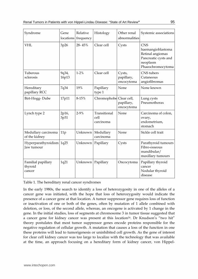
RCC represents 2-3% of all cancers, 7000 people are diagnosed with RCC each year in the UK, and 3600 annually succumb to the disease. It is estimated that 54,390 men and women in the United States will be diagnosed with kidney cancer, while 13,010 will die of their disease in next year. This number can also be expressed as 1 in 72 men and women will be diagnosed with cancer of the kidney during their lifetime and this incidence continues to rise13. There is an apparent increase in the incidence of RCC of 2.5% per year. This may in part be attributed to improved imaging techniques and an increased detection rate of incidental lesions. Approximately 4% of RCCs are of hereditary origin and VHL is the commonest cause. Furthermore, approximately 75% of RCCs are of clear cell histology (cRCC), and VHL is the
hereditary cause of cRCC14. There are at least six other histological subtypes, each associated
with a hereditary syndrome15 (table 1). Hereditary cancer syndromes can be difficult to
identify and early recognition is important, as it facilitates correct management of the
patient, as well as screening of relatives. Principally, mutations can be ‘‘loss-of-function
mutations’’ in tumor suppressor genes and ‘‘gain-of-function-mutations’’ in
protooncogenes, which are then called oncogenes. Research regarding hereditary clear cell
RCC has led to the identification of a relevant gene locus on the short arm of chromosome 3.
This loss-off-function mutation led to the assumption of the existence of a tumor suppressor
gene, and subsequently, further research led to the identification of the VHL gene.
www.intechopen.com
Renal Tumors in Patients with von Hippel-Lindau Disease: “State of Art Review” 95
Syndrome Gene
locations
Relative
frequency
Histology Other renal
abnormalities
Systemic associations
VHL 3p26 28- 45% Clear cell Cysts CNS haemangioblastoma Retinal angiomas Pancreatic cysts and neoplasm Phaeochromocytoma
Tuberous sclerosis
9q34, 16p13
1-2% Clear cell Cysts, papillary, oncocytoma
CNS tubers Cutaneous angiofibromas
Hereditary papillary RCC
7q34 19% Papillary type 1
None None known
Birt-Hogg- Dube 17p11 8-15% Chromophobe Clear cell, papillary, oncocytoma
Lung cysts Pneumothorax
Lynch type 2 2p16, 3p31
2-9% Transitional cell carcinoma
None Carcinoma of colon, ovary, endometrium, stomach
Medullary carcinoma of the kidney
11p Unknown Medullary carcinoma
None Sickle cell trait
Hyperparathyroidism Jaw tumour
1q25 Unknown Papillary Cysts Parathyroid tumours Fibro-osseous mandibular/ maxillary tumours
Familial papillary thyroid cancer
1q21 Unknown Papillary Oncocytoma Papillary thyroid cancer Nodular thyroid disease
Table 1. The hereditary renal cancer syndromes
In the early 1980s, the search to identify a loss of heterozygosity in one of the alleles of a
cancer gene was initiated, with the hope that loss of heterozygosity would indicate the
presence of a cancer gene at that location. A tumor suppressor gene requires loss of function
or inactivation of one or both of the genes, often by mutation of 1 allele combined with
deletion, or loss, of the second allele, whereas, an oncogene is activated by 1 change in the
gene. In the initial studies, loss of segments at chromosome 3 in tumor tissue suggested that
a cancer gene for kidney cancer was present at this location16. Dr Knudson’s “two hit”
theory postulates that most tumor suppressor genes encode proteins responsible for the
negative regulation of cellular growth. A mutation that causes a loss of the function in one
these proteins will lead to tumorigenesis or uninhibited cell growth. As the gene of interest
for clear cell kidney cancer was too large to localize with the technology that was available
at the time, an approach focusing on a hereditary form of kidney cancer, von Hippel-
www.intechopen.com
Renal Cell Carcinoma 96
Lindau, was used as the model of investigation. The hope was that the gene involved in
hereditary kidney cancer may involve the same gene as in the sporadic form of kidney
cancer17. Lubensky and colleague performed a study that supported the two-hit theory of
inheritance in VHL disease. The makeup of 26 renal cysts was studied with microdissection
techniques. They found that each cyst was lined with a single layer of clear epithelial cells.
Twenty-five of the 26 cysts had a mutation of the wild-type allele; in only one case was the
usual heterozygosity of the disease maintained. This finding supports two-hit theory: once
the heterozygosity for VHL is lost in a specific organ, in this case the kidney, the cyst can
progress into a tumor18.
4. Genetics in von Hippel-Lindau
Consistent loss of the short arm of chromosome 3 in VHL associated kidney tumors was
identified19. Genetic linkage analysis was utilized to help identify the VHL gene, which was
identified on the short arm of chromosome 3p20. The loss of the single normal allele of the
VHL gene in the kidney cancer cell samples was critical and suggested that there was an
inherited gene at this location, which was associated with the development of VHL-
associated clear cell kidney cancer. Germline mutations on the VHL gene are identified in
nearly 100% of VHL families21. The disease is caused by germline mutations in the VHL
tumour-suppressor gene; the same gene is inactivated in most cases of sporadic renal-cell
carcinoma and Central Nervous System (CNS) haemangioblastomas. The VHL gene seems
to be pivotal in the processes of angiogenesis, an oxygensensing pathway involving the
hypoxia-inducible factor (HIF), a heterodimeric transcription factor. The VHL gene product
targets the subunit of HIF for polyubiquitylation and proteasomal degradation. This is the
rationale for development of specific inhibitors of hypoxia-inducible factors and their
downstream targets, which lead to potential new treatments. The development of a second
mutation causing deletion of the normal VHL allele leads to tumor formation in affected
individuals. More than 300 different types of mutations have been described that involve the
three exons of the VHL gene.
The different clinical manifestations of VHL can be associated with the location and type of the VHL gene mutation22, 23. Penetrance of the traits is far from complete, and for some such as pheochromocytomas, they tend to be clustered in certain families but do not occur in others24. Maranchie et al.25 identified a significantly higher incidence of renal cell carcinoma in patients with partial germline VHL mutations vs. those with complete VHL gene mutations. It is now known that with mutation analysis (e.g., insertion, deletion, missense, or nonsense) and the location (e.g., codon position) of the mutation, correlations can be made to the phenotype, i.e., the extent of involvement of the various organ systems affected by VHL. Germline mutations in VHL have been identified in almost all patients with von Hippel-
Lindau disease and correlations between the genotype and the phenotype are emerging,
confirming clinical distinction based on the presence or absence of phaeochromocytoma.
Such mutations have been identified in several patients without classic clinical criteria and a
diagnosis of von Hippel-Lindau disease must now be considered in patients with an
apparently sporadic tumour, especially in the case of phaeochromocytomas. Specific
germline mutations in VHL have been associated with a rare recessive haematological
condition (Chuvash polycythaemia) and might also be implicated in some sporadic
apparently congenital polycythaemias26.
www.intechopen.com

Renal Tumors in Patients with von Hippel-Lindau Disease: “State of Art Review” 97
The function of the VHL gene has been evaluated extensively. It is a small gene that encodes 854 nucleotides on 3 exons and is responsible for encoding the VHL protein20. The VHL protein forms a complex with proteins including elongin C, elongin B, and Cul-227-29 and targets the @ subunits of the hypoxia inducible factors, such as HIF-@ and HIF-2@, which are instrumental to ubiquitin mediated degradation30. There are multiple downstream genes, such as Glut 1 (glucose transport), vascular endothelial growth factor (VEGF, blood vessel growth), epidermal growth factor (EGF), transforming growth factor (TGF-@), for which HIF regulates. The expression of these genes increases in clear cell carcinoma when the VHL gene is inactivated (figure 1). Many of the receptors for HIF regulated genes are the targets for the new targeted therapeutic approaches for clear cell carcinoma31. Studies have shown that if the VHL complex is unable to bind to the HIF proteins, tumorigenesis results. Mutations in the oxygen-dependent domain or the VHL binding site of HIF-2-@, but not of HIF-1-@, result in tumorigenesis in xenograft models. Kondo and colleagues33 showed that inhibition of only HIF-2-@ suppresses pVHL deficient tumor growth. This finding established that HIF-2-@ is a critical component to tumorigenesis in clear cell RCC. As previously discussed, HIF proteins are key regulators of oxygen homeostasis, and tight regulation of HIF allows cell survival and growth at the time of hypoxic stress. HIF acts via transcriptional regulation of VEGF, PDGF, EGFR, GLUT1, erythropoietin, and TGF-@. Loss of VHL is thought to result in a pseudohypoxic state, so that cellular response pathways mediated by HIF are activated despite normal oxygen conditions. Understanding the VHL pathway and these pseudohypoxic pathways has provided the opportunity to develop therapies that target the downstream HIF pathway genes VEGF and PDGF with agents such as sunitinib that have a high affinity for the VEGF and PDGF receptors. Other strategies, such as targeting HIF transcription and HIF stability, also are being evaluated in clinical trials.
5. Clinical diagnosis
VHL disease is among the group of disorders known as ‘‘phakomatoses’’ in which lesions
arise from a single germ layer. The lesions of VHL disease are primarily mesodermal in
origin. The clinical manifestations of VHL disease span a spectrum of phenotypic variability.
It has virtually complete penetrance but variable expressivity. The syndrome is
characterized by multiple tumor types, including kidney tumors, pheochromocytomas,
epididymal cystadenomas, retinal angiomas, hemangiomas of the CNS, and pancreatic islet
cell tumors.
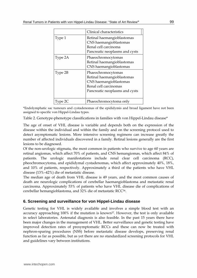
Diagnosis of von Hippel-Lindau disease is often based on clinical criteria. Patients with a family history, and a CNS haemangioblastoma (including retinal haemangioblastomas), phaeochromocytoma, or clear cell renal carcinoma are diagnosed with the disease. Those with no relevant family history must have two or more CNS haemangioblastomas, or one CNS haemangioblastoma and a visceral tumour (with the exception of epididymal and renal cysts, which are frequent in the general population) to meet the diagnostic criteria4, 34. Specific correlations of genotype and phenotype have emerged in affected families. Several familial phenotypes of von Hippel-Lindau disease are now recognised, providing useful information to screen and counsel affected individuals. Type 1 families have a greatly reduced risk of phaeochromocytomas, but can develop all the other tumour types generally associated with the disease. Type 2 families have phaeochromocytomas, but have either a
www.intechopen.com

Renal Cell Carcinoma 98
low-risk (type 2A) or high-risk (type 2B) for renal cell carcinomas. Type 2C families have phaeochromocytomas only, with no other neoplastic findings of VHL (table 2)35.
Fig. 1. 70 The VHL gene complex targets hypoxia-inducible factors (HIF) for ubiquitin-mediated degradation. When there is a mutation in the VHL gene in clear cell kidney cancer, in either the elongin binding or the HIF binding domain (A), HIF is not degraded and this protein over-accumulates. Increased HIF levels lead to increased transcription of a number of downstream pathway genes that are thought to be important in kidney cancer, such as vascular endothelial growth factor (VEGF), the glucose transporter, GLUT1, and transforming growth factor _ (TGF-@) (B). Targeted approaches to therapy currently include tyrosine kinase inhibitors that target the downstream gene receptors (C). Other approaches being developed to target the VHL-HIF pathway in clear cell kidney cancer include the development of small molecules or other agents which block HIF transcription. Reprinted from Linehan et al32.
www.intechopen.com
Renal Tumors in Patients with von Hippel-Lindau Disease: “State of Art Review” 99
Clinical characteristics
Type 1 Retinal haemangioblastomas CNS haemangioblastomas Renal cell carcinoma Pancreatic neoplasms and cysts
Type 2A Phaeochromocytomas Retinal haemangioblastomas CNS haemangioblastomas
Type 2B Phaeochromocytomas Retinal haemangioblastomas CNS haemangioblastomas Renal cell carcinomas Pancreatic neoplasms and cysts
Type 2C Phaeochromocytoma only
*Endolymphatic sac tumours and cystadenomas of the epididymis and broad ligament have not been assigned to specific von Hippel-Lindau types.
Table 2. Genotype-phenotype classifications in families with von Hippel-Lindau disease*
The age of onset of VHL disease is variable and depends both on the expression of the
disease within the individual and within the family and on the screening protocol used to
detect asymptomatic lesions. More intensive screening regimens can increase greatly the
number of affected individuals discovered in a family. Retinal lesions generally are the first
lesions to be diagnosed.
Of the non-urologic stigmata, the most common in patients who survive to age 60 years are
retinal angiomas, which affect 70% of patients, and CNS hemangiomas, which affect 84% of
patients. The urologic manifestations include renal clear cell carcinoma (RCC),
pheochromocytoma, and epididymal cystadenomas, which affect approximately 40%, 18%,
and 10% of patients, respectively. Approximately a third of the patients who have VHL
disease (13%–42%) die of metastatic disease.
The median age of death from VHL disease is 49 years, and the most common causes of death are neurologic complications of cerebellar haemangioblastoma and metastatic renal carcinoma. Approximately 53% of patients who have VHL disease die of complications of cerebellar hemangioblastoma, and 32% die of metastatic RCC36.
6. Screening and surveillance for von Hippel-Lindau disease
Genetic testing for VHL is widely available and involves a simple blood test with an accuracy approaching 100% if the mutation is known37. However, the test is only available in select laboratories. Antenatal diagnosis is also feasible. In the past 15 years there have been major changes in the management of VHL. Better surveillance and genetic testing have improved detection rates of presymptomatic RCCs and these can now be treated with nephron-sparing procedures (NSS) before metastatic disease develops, preserving renal function as far as possible, but as yet there are no standardized screening protocols for VHL and guidelines vary between institutions.
www.intechopen.com
Renal Cell Carcinoma 100
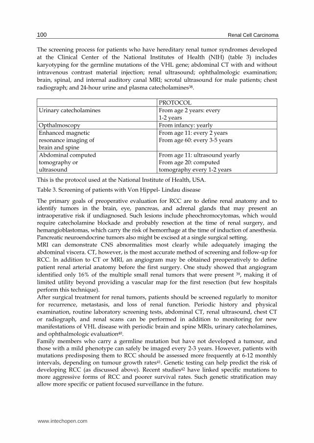
The screening process for patients who have hereditary renal tumor syndromes developed
at the Clinical Center of the National Institutes of Health (NIH) (table 3) includes
karyotyping for the germline mutations of the VHL gene; abdominal CT with and without
intravenous contrast material injection; renal ultrasound; ophthalmologic examination;
brain, spinal, and internal auditory canal MRI; scrotal ultrasound for male patients; chest
radiograph; and 24-hour urine and plasma catecholamines38.
PROTOCOL
Urinary catecholamines From age 2 years: every 1-2 years
Opthalmoscopy From infancy: yearly
Enhanced magnetic resonance imaging of brain and spine
From age 11: every 2 years From age 60: every 3-5 years
Abdominal computed tomography or ultrasound
From age 11: ultrasound yearly From age 20: computed tomography every 1-2 years
This is the protocol used at the National Institute of Health, USA.
Table 3. Screening of patients with Von Hippel- Lindau disease
The primary goals of preoperative evaluation for RCC are to define renal anatomy and to identify tumors in the brain, eye, pancreas, and adrenal glands that may present an intraoperative risk if undiagnosed. Such lesions include pheochromocytomas, which would require catecholamine blockade and probably resection at the time of renal surgery, and hemangioblastomas, which carry the risk of hemorrhage at the time of induction of anesthesia. Pancreatic neuroendocrine tumors also might be excised at a single surgical setting. MRI can demonstrate CNS abnormalities most clearly while adequately imaging the
abdominal viscera. CT, however, is the most accurate method of screening and follow-up for
RCC. In addition to CT or MRI, an angiogram may be obtained preoperatively to define
patient renal arterial anatomy before the first surgery. One study showed that angiogram
identified only 16% of the multiple small renal tumors that were present 39, making it of
limited utility beyond providing a vascular map for the first resection (but few hospitals
perform this technique).
After surgical treatment for renal tumors, patients should be screened regularly to monitor for recurrence, metastasis, and loss of renal function. Periodic history and physical examination, routine laboratory screening tests, abdominal CT, renal ultrasound, chest CT or radiograph, and renal scans can be performed in addition to monitoring for new manifestations of VHL disease with periodic brain and spine MRIs, urinary catecholamines, and ophthalmologic evaluation40. Family members who carry a germline mutation but have not developed a tumour, and those with a mild phenotype can safely be imaged every 2-3 years. However, patients with mutations predisposing them to RCC should be assessed more frequently at 6-12 monthly intervals, depending on tumour growth rates41. Genetic testing can help predict the risk of developing RCC (as discussed above). Recent studies42 have linked specific mutations to more aggressive forms of RCC and poorer survival rates. Such genetic stratification may allow more specific or patient focused surveillance in the future.
www.intechopen.com

Renal Tumors in Patients with von Hippel-Lindau Disease: “State of Art Review” 101
7. Renal cell carcinomas and renal cysts in von Hippel Lindau general features
Renal cell carcinomas are the major malignant neoplasm in von Hippel-Lindau disease and
the primary cause of inherited renal cancer. These tumours are seen in 24–45% of patients,
and adding renal cysts increases the finding of renal lesions to 60%. Renal disease is
multicentric and bilateral in at least 75% of patients who have VHL disease. The mean age at
presentation is 39 +/- 10 years, which is 10 to 20 years earlier than that reported for sporadic
renal disease.
In addition to the young age of presentation, RCC associated with VHL disease also recurs frequently years. Although small renal tumours in this disease tend to be low grade and minimally invasive, their rate of growth varies widely43. Renal lesions are often multiple and bilateral. Walther and colleagues44 estimated that 600 microscopic tumours and 1100 microscopic clear-cell-lined cysts might be present in the kidneys of some 37-year-old patients. Results of an investigation of 228 renal lesions in 28 patients, followed for at least 1 year, showed that transition from a cyst to a solid lesion was rare. Solid tumors in VHL disease have been observed to grow at rates of 0.2 to 2.2 cm/year (mean, 1.6 cm/year), which is somewhat faster than rates observed in sporadic RCC. The change that occurs in renal lesions over time in patients who have VHL disease is generally slow and constant, but it has been followed in only a small number of cases 45. However, complex cystic and solid lesions can contain neoplastic tissue that frequently enlarges. Renal cell carcinomas often remain asymptomatic for long intervals. Thus, serial imaging of the kidneys is useful for early diagnosis. Occasionally, the more advanced cases with these neoplasms can present with haematuria, flank pain, or a flank mass. Renal cysts in von Hippel-Lindau disease are typically asymptomatic and seldom need treatment. Renal cysts are present in 59% to 63% of patients who have VHL disease, and some studies believe the number may be as high as 85% Cysts generally preceded the detection of solid tumors by about 3 to 7 years. Studies have shown that at least 21% of cystic renal lesions contain foci of RCC and RCC is reported to develop in 24% to 45% of patients who have VHL disease. Therefore, complex cysts need monitoring, as they often harbour solid components of renal cell carcinoma. Because of the frequent absence of early clinical symptoms and the importance of early detection, diagnosis during presymptomatic screening has the potential to enhance overall outcome. Contrast-enhanced abdominal CT is the standard for detection of renal involvement in the disease. CT allows detection and quantification by size and number of renal cell carcinomas and cysts, allowing serial monitoring of individual lesions. Imaging is usually recommended in 3–5 mm sections, and before and after intravenous injection of contrast media. Precontrast and postcontrast MRI is an alternative method of detection for patients who have reduced renal function. These carcinomas are yellow or orange and are encapsulated. They can be solid or a mixture of solid and cystic in appearance. Histologically, they are always of the clear-cell subtype, and small carcinomas tend to be low grade. In addition, microscopic examination of small VHL renal lesions consistently demonstrated a pseudo-capsule surrounding these low-grade renal lesions. This pseudo-capsule has since proven to facilitate tumor resection by forming a natural tissue plane for dissection46 . The recurrence rate of renal tumors is high in patients who have VHL disease. Examination of normal renal tissue of patients who have RCC and VHL disease has shown multiple microscopic renal tumors 43. Treatment recommendations depend on tumour size.
www.intechopen.com
Renal Cell Carcinoma 102
8. Radiology of renal masses in VHL
RCC in VHL usually manifests as a complex cystic mass, which is also the most difficult type of renal lesion to accurately assess, especially when detected incidentally. The Bosniak classification was first described in 1986 and determines the management approach to the sporadic cystic renal mass47. The system was devised for CT but can also be applied to MRI. As some patients with VHL require long-term follow-up of renal manifestations with cross-sectional imaging, there are clear advantages of MRI, such as the absence of radiation. CNS manifestations are almost exclusively imaged with MRI in most institutions. MRI has also proved more accurate at assessing cystic pancreatic lesions and distinguishing phaeochromocytoma from adenoma on chemical shift imaging. It is also more sensitive than CT in detecting neuroendocrine tumours of the pancreas. For these reasons, several authors advocate annual surveillance of abdominal manifestations with MRI. These advantages must be weighed against potential risks of nephrogenic systemic fibrosis (NSF) with gadolinium compounds when used in renal impairment, as well as logistical factors, such as cost and limited access of MRI at some institutions. However, patients undergoing a lifetime of screening may accumulate relatively large doses of radiation to the kidneys as a consequence of annual screening with CT. Thus, there is no clear-cut answer which is the best imaging modality48. Before CT was widely available 13-42% of patients with VHL died of metastatic RCC43. The discovery of the VHL gene in 1993 unlocked a better understanding of the pathways involved in tumourigenesis. Surgery is still the only curative treatment of RCC. Bilateral nephrectomy with renal-replacement therapy would eradicate all tissue at risk for developing RCC and is a theoretical, but radical, option in a patient with VHL. However, patients with VHL do worse on renal-replacement therapy. A large study49 showed a 5-year survival rate of 65% in VHL patients on dialysis compared with 71-86% for the non-VHL group. But renal impairment in the absence of renal surgery is rare among VHL patients. As patients with VHL do worse with renal- replacement therapy, the aim of successful management is to preserve renal function balanced against the risk of developing renal metastatic disease. The secondary aim is to minimize the number of procedures performed to accomplish this. To accomplish these aims accurate imaging-guided diagnosis and nephron-sparing procedures are important.
9. Nephron-sparing surgery and other treatments in von Hippel-Lindau disease
RCC in von Hippel-Lindau disease differs from its sporadic counterpart in that the diagnosis is made at a young age, and there are usually multiple bilateral renal tumors. Although these are generally low-stage tumors, they are capable of progression with metastasis and represent a frequent cause of death in patients with von Hippel-Lindau disease. RCC in these patients is characterized histopathologically by both solid tumors and renal cysts that contain either frank carcinoma or a lining of hyperplastic clear cells representing incipient carcinoma. Therefore, adequate surgical treatment of localized RCC in vonHippel-Lindau disease requires excision of all solid and cystic renal lesions. Choyke et al.50 have shown that intraoperative ultrasonography may be a valuable adjunct for this population of patients. In their series, this study identified additional tumors in 25% of patients with hereditary renal cancer undergoing renal exploration.
www.intechopen.com
Renal Tumors in Patients with von Hippel-Lindau Disease: “State of Art Review” 103
The surgical options in patients with bilateral RCC and von Hippel-Lindau disease are bilateral nephrectomy and renal replacement therapy or partial nephrectomy to avoid end-stage renal failure. The general philosophy has been to pursue nephron-sparing surgery if possible, given the multifocal nature of the disease, even for centrally located tumors. The treatment options for patients who have renal involvement in VHL disease have evolved during the last 3 decades. Staged bilateral nephrectomy and hemodialysis have progressed to partial nephrectomy and NSS. The best management approach for VHL disease balances the goals of minimizing the risk of RCC metastasis and preserving renal function and attempts to minimize the total number of surgeries a patient will require over a lifetime51 . There are no specific guidelines regarding the type or timing of surgery in patients who have VHL disease. The treatment strategy of total nephrectomy and Renal Replacement Theraphy (RRT)
removes all the tissue at risk for RCC and, with it, the risk of metastases. Goldfarb and
colleagues52 noted a 5-year survival rate of 65% in patients who had VHL disease and who
were treated with bilateral nephrectomy and RRT. Some transplant centers consider the
presence or potential for VHL-related tumors, including renal and extrarenal tumors, a
contraindication for renal transplantation.
The indications for NSS are localized malignancy in a solitary kidney, bilateral synchronous
renal lesions, and renal tumors in patients whose contralateral kidney is threatened by local,
systemic, or genetic risk factors53.
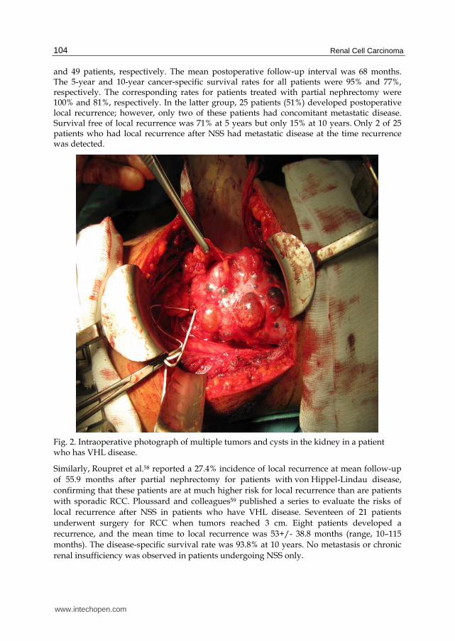
Although early results of partial nephrectomy were promising, subsequent studies suggested a high incidence of postoperative tumour recurrence in the remaining portion of the kidney. It is likely that most of these local recurrences were a manifestation of residual microscopic RCC that was not removed at the time of the original partial nephrectomy. Walther and colleagues54 have reported extensively on their approach of enucleation of small renal tumors associated with VHL disease (figure 2). Preservation of the perirenal fat and fascia are believed to be important in improving the patient’s ability to undergo subsequent renal surgeries. The renal capsule is incised, circumscribing the lesions to be resected. All cystic lesions are excised, if possible, because of the potential for development of RCC46. Smaller cystic lesions are unroofed, and the base is fulgurated (however it has not been shown that this is completely safe). A study from the NIH55 showed that renal tumors less than 3 cm in size are not considered an indication for radical surgery. Patients who have VHL disease generally are followed expectantly until solid renal lesions are larger than 3 cm in diameter. In the previously mentioned NIH study, the 3-cm threshold for surgery was evaluated in 52 patients who had VHL disease. None of these patients had metastatic disease, and none required RRT. Forty-six of these patients underwent NSS rather than radical nephrectomy. Forty-four patients who had VHL disease developed renal tumors larger than 3 cm. From this study, the authors concluded that using a 3-cm tumor diameter as a threshold for NSS may help prevent metastatic disease. These authors also noted the importance of intraoperative Doppler ultrasound for locating tumors and verifying that all tumors have been removed56. A follow-up of this study38 revealed that metastasis did not develop in any patient who had a solid renal tumor smaller than 3 cm. Of 50 patients who had RCC, metastasis developed in 1 patient who had a primary renal tumor that was 4.5 cm in greatest diameter. One multicenter study57 delineated the long-term outcomes after surgical treatment of localized RCC in 65 patients with von Hippel-Lindau disease managed at eight medical centers in the United States. RCC was present bilaterally and unilaterally in 54 and 11 patients, respectively. Radical nephrectomy and partial nephrectomy were performed in 16
www.intechopen.com
Renal Cell Carcinoma 104
and 49 patients, respectively. The mean postoperative follow-up interval was 68 months. The 5-year and 10-year cancer-specific survival rates for all patients were 95% and 77%, respectively. The corresponding rates for patients treated with partial nephrectomy were 100% and 81%, respectively. In the latter group, 25 patients (51%) developed postoperative local recurrence; however, only two of these patients had concomitant metastatic disease. Survival free of local recurrence was 71% at 5 years but only 15% at 10 years. Only 2 of 25 patients who had local recurrence after NSS had metastatic disease at the time recurrence was detected.
Fig. 2. Intraoperative photograph of multiple tumors and cysts in the kidney in a patient who has VHL disease.
Similarly, Roupret et al.58 reported a 27.4% incidence of local recurrence at mean follow-up
of 55.9 months after partial nephrectomy for patients with von Hippel-Lindau disease,
confirming that these patients are at much higher risk for local recurrence than are patients
with sporadic RCC. Ploussard and colleagues59 published a series to evaluate the risks of
local recurrence after NSS in patients who have VHL disease. Seventeen of 21 patients
underwent surgery for RCC when tumors reached 3 cm. Eight patients developed a
recurrence, and the mean time to local recurrence was 53+/- 38.8 months (range, 10–115
months). The disease-specific survival rate was 93.8% at 10 years. No metastasis or chronic
renal insufficiency was observed in patients undergoing NSS only.
www.intechopen.com

Renal Tumors in Patients with von Hippel-Lindau Disease: “State of Art Review” 105
Taken together, these studies suggest that partial nephrectomy can provide effective initial treatment of patients with RCC and von Hippel-Lindau disease but should be withheld until tumor size reaches or eclipses 3.0 cm. Studies show that up to 85% of patients who have VHL disease experience de novo renal tumor recurrence at 10 years57. After nephron-sparing surgery, patients with von Hippel-Lindau disease must be observed closely because most will eventually develop locally recurrent RCC with the concomitant need for repeated renal surgery. In this setting, repeated partial nephrectomy can be challenging because of postoperative fibrosis, and some centers are moving toward thermal ablative modalities to reclaim local control and many surgeons are reluctant to perform additional NSS because of complications related to postoperative fibrosis60. These salvage surgeries were greatly complicated by severe fibrosis, scarring, and the obliteration of normal anatomic planes. The authors concluded that aggressive surgical intervention in these patients is indicated, because none of the patients developed metastatic disease as long as 7 years after surgery, and there was 100% survival in this cohort61. NSS has been performed using the laparoscopic approach in select centers, but the inability to achieve reliable hypothermia along with prolonged ischemia times and increased complication rates have limited in its widespread application. At present, open NSS should be considered the standard of care for treating RCC in the setting of VHL disease. When removal of all renal tissue is necessary to achieve control of malignant disease, renal transplantation can provide satisfactory replacement therapy for end-stage renal disease and appears to be safe despite the tumor diathesis. Newer, minimally invasive techniques62, such as radiofrequency ablation and cryoablation, have been applied recently as an alternative to excisional NSS in patients who have RCC. Ablative techniques may63, 65 be performed by laparoscopic and percutaneous image-guided techniques. Jolly et al.64 concluded in a recent published article that renal survival in patients with VHL who are treated for RCC has improved with time. Their renal survival curves actually reflect the decreasing need to remove all renal tissue to achieve local control of malignancy, in parallel with improved NSS and the emergence of renal salvage techniques such as Radio Frecuency Ablation (RFA) (figure 3a and 3b). As well as surgery, nephron sparing procedures now include RFA and cryoablation. In their experience since 2004 RFA has become the exclusive salvage technique. Overall a third of RFA sessions were performed in single functioning kidneys, including 8 in a total of 8 patients who had already been operated on twice and were not candidates for additional NSS. These all have a role in the surgical management of VHL-associated RCC and experience of
these newer modalities in VHL is increasing, and good preliminary success rates are being
published. A small study by Shingleton and Sewell66 reported the successful treatment of
five small RCCs in four patients with VHL using MRI-guided cryoablation, but greater
evidence is available for imaging- guided RFA treatment of RCCs in the context of VHL,
demonstrating good success rates for exophytic tumours of up to 5 cm in size67, 68.
In conclusion69, as patients with VHL with RCC are increasingly treated with nephron sparing techniques, especially RFA, it must be remembered that only long-term follow-up can hopefully confirm that no unexpected metastasis develops beyond increased renal survival. In patients who have a single kidney or multiple bilateral lesions, collateral damage to normal renal parenchyma must be considered. For patients who have VHL disease, and all patients who have hereditary cancer syndromes, the goal of treatment is cancer control, not cancer cure, and preservation of functional parenchyma to avoid the morbidity associated with renal or adrenal loss.
www.intechopen.com

Renal Cell Carcinoma 106
Fig. 3a. Renal tumor in a patient with solitary right kidney and VHL disease
Fig. 3b. Contrast enhanced CT scan after radiofrequency ablation show the absence of contrast enhancement in the ablated area.
www.intechopen.com

Renal Tumors in Patients with von Hippel-Lindau Disease: “State of Art Review” 107
10. References
[1] Lonser RR, Glenn G, Walther McC, et al. Von-Hippel-Lindau disease. Lancet 2003, 361:
2059-67.
[2] Richard S, Graff J, Lindau J, Resche F. Von Hippel-Lindau disease. The Lancet, Volume
363, Issue 9416, 10 April 2004, Pages 1231-1234.
[3] Kaelin WG Jr. Molecular basis of the VHL hereditary cancer syndrome. Nat Rev Cancer
2003; 2: 673–82.
[4] Melmon KL, Rosen SW. Lindau’s disease. Review of the literature and study of large
kindred. Am J Med 1964, 36: 595–617.
[5] Lamiell JM, Salazar FG, Hsia YE. Von Hippel-Lindau disease affecting 43 members of
single kindred. Medicine 1989; 68: 1–29.
[6] Resche F, Moisan JP, Mantoura J, et al. Hemangioblastomas, hemangioblastomatosis and
von Hippel-Lindau disease. Adv Techn Stand Neurosurg 1993; 20: 197–303.
[7] Galezowski X, Traité iconographique d’opthalmoscopie. In: Baillière, ed. Diagnostic et
traitement desaffections occulaires par les docteurs. Paris, Baillières, 1986.
[8] von Hippel E. Vorstellung eines Patienten mit einer sehr ungewöhnlichen Netzhaut.
XXIV Verstellung der ophthalmologischen Gesellschaft (Heidelberg, 1895), JF
Bergmann Verlag, Wiesbaden, 1896: 269.
[9] von Hippel E. Ueber eine sehr seltene Erkrankung der Netzhaut. Klinische
Beobachtungen. A von Graefe’s Arch Ophthalmol 1904; 59: 83–106.
[10] von Hippel E. Die anatomische Grundlage der von mir beschriebenen “sehr seltene
Erkrankung der Netzhaut”. A von Graefe’s Arch Ophthalmol 1911; 79: 350–77.
[11] Lindau A. Studien über Kleinhirnzysten. Bau, Pathogenese und Beziehungen zur
Angiomatosis retinae. Acta Pathol Microbiol Scand 1926; S1: 1–128.
[12] Davison C, Brock S, Dyke CG. Retinal and central nervous hemangioblastomatosis with
visceral changes (von Hippel-Lindau’s disease). Bull Neurol Instit NY 1936; 5: 72–
93.
[13] SEER Cancer Statistics Review, 1975–2005. NCI 2007; (November 2007 SEER data
submission. Available from URL:
[14] http://seer. cancer.gov/csr/1975_2005/results_merged/sect_01_overview.pdf and
http://seer.cancer.gov/csr/1975_2005/results_merged/sect_11_
kidney_pelvis.pdf).
[15] Pavlovich CP, Schmidt LS. Searching for the hereditary causes of renal-cell carcinoma.
Nat Rev Cancer 2004; 4: 381- 93.
[16] Choyke PL, Glenn GM, Walther MM, et al. Hereditary renal cancers. Radiology 2003;
226:33-46.
[17] Zbar B, Brauch H, Talmadge C, et al. Loss of alleles of loci on the short arm of
chromosome 3 in renal cell carcinoma. Nature 1987; 327: 721– 4.
[18] Knudson AG. Genetics of human cancer. Annu Rev Genet 1986; 20: 231–51.
[19] Lubensky IA, Gnarra JR, Bertheau P, et al. Allelic deletions of the VHL gene detected in
multiple microscopic clear cell renal lesions in von Hippel-Lindau disease patients.
Am J Pathol 1996; 149(6):2089–94.
[20] Tory K, Brauch H, Linehan WM, et al. Specific genetic change in tumors associated with
von Hippel-Lindau disease. J Natl Cancer Inst 1989; 81:1097–10.
www.intechopen.com

Renal Cell Carcinoma 108
[21] Latif F, Tory K, Gnarra JR, et al. Identification of the von Hippel-Lindau disease tumor
suppressor gene. Science 1993; 260:1317–20.
[22] Stolle C, Glenn GM, Zbar B, et al. Improved detection of germline mutations in the von
Hippel-Lindau disease tumor suppressor gene. Hum Mutat 1998; 12:417–23.
[23] Chen F, Kishida T, Yao M, et al. Germline mutations in the von Hippel-Lindau disease
tumor suppressor gene: correlations with phenotype. Hum Mutat 1995; 5:66 –75
[24] Ong KR, Woodward ER, Killick P, et al. Genotype-phenotype correlations in von
Hippel-Lindau disease. Hum Mutat 2007; 28:143–9.
[25] Neumann HPH, Zbar B. Renal cysts, renal cancer, and von Hippel-Lindau disease.
Kidney Int 1997; 51:16 –26.
[26] Maranchie JK, Afonso A, Albert PS, et al. Solid renal tumor severity in von Hippel
Lindau disease is related to germline deletion length and location. Hum Mutat
2004; 23:40–6.
[27] Ang SO, Chen H, Hirota K, et al. Disruption of oxygen homeostasis underlies
congenital Chuvash polycythemia. Nat Genet 2002; 32: 614–21.
[28] Duan DR, Pause A, Burgess WH, et al. Inhibition of transcription elongation by the
VHL tumor suppressor protein. Science 1995; 269: 1402–6.
[29] Kibel A, Iliopoulos O, DeCaprio JA, et al. Binding of the von Hippel-Lindau tumor
suppressor protein to Elongin B and C. Science 1995;269:1444–6.
[30] Pause A, Lee S, Worrell RA, et al. The von Hippel-Lindau tumor suppressor gene
product forms a stable complex with human CUL-2, a member of the Cdc53 family
of proteins. Proc Natl Acad Sci USA 91997; 4:2156–61.
[31] Iliopoulos O, Jiang C, Levy AP, et al. Negative regulation of hypoxiainducible genes by
the von Hippel-Lindau protein. Proc Natl Acad Sci USA 1996; 93:10595–9.
[32] Linehan WM. Editorial: Kidney Cancer, a Unique Opportunity for the Development of
Disease Specific Therapy. J Urol 2002; 168: 2411–2.
[33] Linehan WM, Zbar B. Focus on kidney cancer. Cancer Cell 2004; 6: 223–8.
[34] Kondo K, Klco J, Nakamura E, Lechpammer M, Kaelin WG Jr. Inhibition of HIF is
necessary for tumour suppression by the von Hippel-Lindau protein. Cancer Cell
2002; 1: 237–46.
[35] Escourolle R, Poirer J. Manual of Neuropathology. 2nd edn. Philadelphia: WB
Saunders, 1978: 49–51.
[36] Hes F, Zewald R, Peeters T, et al. Genotype-phenotype correlations in families with
deletions in the von Hippel-Lindau (VHL) gene. Hum Genet 2000; 106: 425–31.
[37] Martz CH. Von Hippel-Lindau disease: a genetic condition predisposing tumor
formation. Oncol Nurs Forum 1991; 18(3):545–51.
[38] Linehan WM, Walther MM, Zbar B. The genetic basis of cancer of the kidney. J Urol
2003; 170:2163- 72.
[39] Herring JC, Enquist EG, Chernoff A, et al. Parenchymal sparing surgery in patients with
hereditary renal cell carcinoma: 10-year experience. J Urol 2001; 165(3):777–81.
[40] Miller DL, Choyke PL, Walther MM, et al. von Hippel-Lindau disease: inadequacy of
angiography for identification of renal cancers. Radiology 1991; 179(3):833–6.
[41] Maher ER, Yates JR, Harries R, et al. Clinical features and natural history of von Hippel-
Lindau disease. Q J Med 1990; 77(283):1151–63.
www.intechopen.com

Renal Tumors in Patients with von Hippel-Lindau Disease: “State of Art Review” 109
[42] Choyke PL, Glenn GM, Walther MM, et al. von Hippel- Lindau disease: genetic, clinical,
and imaging features. Radiology 1995; 194: 629- 42.
[43] Israel GM, Hindman N, Bosniak MA. Evaluation of cystic renal masses: comparison of
CT and MR imaging by using the Bosniak classification system. Radiology 2004;
231:365- 71.
[44] Walther MM, Choyke PL, Glenn G, et al. Renal cancer in families with hereditary renal
cancer: prospective analysis of a tumor size threshold for renal parenchymal
sparing surgery. J Urol 1999; 161(5):1475–9.
[45] Walther MM, Lubensky IA, Venzon D, Zbar B, Linehan WM. Prevalence of microscopic
lesions in grossly normal renal parenchyma from patients with von Hippel-Lindau
disease, sporadic renal cell carcinoma and no renal disease: clinical implications. J
Urol 1995; 154: 2010–14.
[46] Choyke PL, Glenn GM, Walther MM, et al. The natural history of renal lesions in von
Hippel-Lindau disease: a serial CT study in 28 patients. AJR Am J Roentgenol 1992;
159(6):1229–34.
[47] Poston CD, Jaffe GS, Lubensky IA, et al. Characterization of the renal pathology of a
familial form of renal cell carcinoma associated with von Hippel-Lindau disease:
clinical and molecular genetic implications. J Urol 1995; 153(1):22–6.
[48] Bosniak MA. The current radiological approach to renal cysts. Radiology 1986; 158:1-
10.
[49] Meister M., Choyke P., Anderson C., Patel U. Radiological evaluation, management,
and surveillance of renal masses in Von Hippel–Lindau disease. Clinical
Radiology, Volume 64, Issue 6, June 2009, Pages 589-600.
[50] Grubb 3rd RL, Choyke PL, Pinto PA, et al. Management of von Hippel- Lindau
associated kidney cancer. Nat Clin Pract Urol 2005; 2: 248- 55.
[51] Choyke PL, Pavlovich CP, Daryanani KD, et al: Intraoperative ultrasound during renal
parenchymal sparing surgery for hereditary renal cancers: A 10 year experience. J
Urol 2001; 165:397-400.
[52] Fetner CD, Barilla DE, Scott T, et al. Bilateral renal cell carcinoma in von Hippel- Lindau
syndrome: treatment with staged bilateral nephrectomy and hemodialysis. J Urol
1977; 117(4): 534–6.
[53] Goldfarb DA, Neumann HP, Penn I, et al. Results of renal transplantation in patients
with renal cell carcinoma and von Hippel-Lindau disease. Transplantation 1997;
64(12):1726–9.
[54] Duffey BG, Choyke PL, Glenn G, et al. The relationship between renal tumor size and
metastases in patients with von Hippel-Lindau disease. J Urol 2004; 172(1): 63–5.
[55] Walther MM, Thompson N, Linehan W. Enucleation procedures in patients with
multiple hereditary renal tumors. World J Urol 1995; 13(4):248–50.
[56] Walther MM, Choyke PL, Glenn G, et al. Renal cancer in families with hereditary renal
cancer: prospective analysis of a tumor size threshold for renal parenchymal
sparing surgery. J Urol 1999; 161(5):1475–9.
[57] Walther MM, Choyke PL, Hayes W, et al. Evaluation of color Doppler intraoperative
ultrasound in parenchymal sparing renal surgery. J Urol 1994; 152(6 Pt 1): 1984–7.
www.intechopen.com
Renal Cell Carcinoma 110
[58] Steinbach F, Novick AC, Zincke H, et al. Treatment of renal cell carcinoma in von
Hippel- Lindau disease: a multicenter study. J Urol 1995; 153(6):1812–6.
[59] Roupret M, Hopirtean V, Mejean A, et al. Nephron sparing surgery for renal cell
carcinoma and von Hippel-Lindau’s disease: a single center experience. J Urol 2003;
170(5):1752–5.
[60] Ploussard G, Droupy S, Ferlicot S, et al. Local recurrence after nephron-sparing surgery
in von-Hippel-Lindau disease. Urology 2007; 70(3):435–9.
[61] Bratslavsky G, Liu JJ, Ferlicot S, et al. Salvage partial nephrectomy for hereditary renal
cancer: feasibility and outcomes. J Urol 2008; 179(1):67–70.
[62] Thompson RH, Leibovich BC, Lohse CM, et al. Complications of contemporary open
nephron sparing surgery: a single institution experience. J Urol 2005; 174(3):855–8.
[63] Kunkle DA, Egleston BL, Uzzo RG. Excise, ablate or observe: the small renal mass
dilemma-a meta-analysis and review. J Urol 2008; 179(4):1227–33.
[64] Aron M, Gill IS. Minimally invasive nephron-sparing surgery (MINSS) for renal
tumours. Part II: probe ablative therapy. Eur Urol 2007; 51(2):348–57.
[65] Joly D., Méjean A. , Corréas J-M et al. Progress in Nephron Sparing Therapy for Renal
Cell Carcinoma and von Hippel-Lindau Disease. The Journal of Urology, Volume
185, Issue 6, June 2011, Pages 2056-2060.
[66] Hwang JJ, Walther MM, Pautler SE et al: Radio frequency ablation of small renal
tumors: intermediate results. J Urol 2004; 171: 1814.
[67] Shingleton WB, Sewell Jr PE. Percutaneous renal cryoablation of renal tumors in
patients with von Hippel-Lindau disease. J Urol 2002; 167:1268-70.
[68] Gervais DA, McGovern FJ, Arellano RS, et al. Renal cell carcinoma: clinical experience
and technical success with radio- frequency ablation of 42 tumors. Radiology 2003;
226: 417-24.
[69] Su LM, Jarrett TW, Chan DY, et al. Percutaneous computed tomography-guided
radiofrequency ablation of renal masses in high surgical risk patients: preliminary
results. Urology 2003; 61(Suppl. 1):26-33.
[70] Beth Reed A., Parekh D. Surgical Management of von Hippel-Lindau Disease: Urologic
Considerations. Surgical Oncology Clinics of North America - Volume 18, Issue
1 (January 2009) (157-174).
[71] Rosner I, Bratslavsky G., Pinto P., Linehan W. The clinical implications of the genetics of
renal cell carcinoma. Urologic Oncology: Seminars and Original
Investigations, Volume 27, Issue 2, March-April 2009, Pages 131-136.
www.intechopen.com
Renal Cell CarcinomaEdited by Dr. Hendrik Van Poppel
ISBN 978-953-307-844-1Hard cover, 144 pagesPublisher InTechPublished online 16, December, 2011Published in print edition December, 2011
InTech EuropeUniversity Campus STeP Ri Slavka Krautzeka 83/A 51000 Rijeka, Croatia Phone: +385 (51) 770 447 Fax: +385 (51) 686 166www.intechopen.com
InTech ChinaUnit 405, Office Block, Hotel Equatorial Shanghai No.65, Yan An Road (West), Shanghai, 200040, China
Phone: +86-21-62489820 Fax: +86-21-62489821
Surgical and medical oncologists have been unable to decrease renal cell carcinoma mortality for uncertainreasons, although a lot of progress has been made in diagnosis and imaging, recognition of different geneticand pathological entities, management of localized disease and in the research on new drug treatments foradvanced stages of the disease, potentially combined with surgery. The purpose of this book, which tackles anumber of separate interesting topics, is to provide further insight into the disease and the management ofearly and advanced renal cell carcinoma. The volume is divided into different parts; the first part covers thecharacterization of renal masses and the second part covers rare distinct pathological entity. In themanagement section, active surveillance, partial nephrectomy and radiofrequency ablation are presented. Aseparate chapter reviews the management of Von Hippel Lindau disease, and finally, conventional andaberrant signaling pathways are explored.
How to referenceIn order to correctly reference this scholarly work, feel free to copy and paste the following:
Mario Alvarez Maestro, Luis Martinez-Pin ̃eiro and Emilio Rios Gonzalez (2011). Renal Tumors in Patients withvon Hippel-Lindau Disease: "State of Art Review", Renal Cell Carcinoma, Dr. Hendrik Van Poppel (Ed.), ISBN:978-953-307-844-1, InTech, Available from: http://www.intechopen.com/books/renal-cell-carcinoma/renal-tumors-in-patients-with-von-hippel-lindau-disease-state-of-art-review-

© 2011 The Author(s). Licensee IntechOpen. This is an open access articledistributed under the terms of the Creative Commons Attribution 3.0License, which permits unrestricted use, distribution, and reproduction inany medium, provided the original work is properly cited.