volume 7 no 1 1936 1937
DESCRIPTION
UWOMJ University of Western Ontario Medical Journal Schulich School of Medicine & DentistryTRANSCRIPT

UNIVERSITY OF WESTERN ONTARIO
MEDICAL JOURNAL
VOL. VII.,
Published by the
Undergraduate Body of the
University of Western Ontario Medical School
LONDON CANADA

University of Western Ontario
MEDICAL JOURNAL Published Quarterly by
Undergraduate Body of the University of Western Ontario Medical School
Entered as a Second Class Matter at London, Canada.
BOARD OF ADVISORS J. A. MACGREGOR, M.D., F.R.C.P. (C.), LL.D. (West.), Chairman
E. M. WATSON, M.D., F. R. C. P. (Edin.),
Faculty Representative
H. A. SKINNER, M. B. E. P. JOHNS, M.D. H. M. SIMPSON, M.D.
A. C. H. TROTTIER, M.D. E. G. DAVIS, M.D. J. R. ARMSTRONG, M.D.
S. G. CHALK, M.D., W. C. SHARPE. M. B. W. J. BROWN, B.S.A., B.A., M.Sc. L.L.M., F.C.I.S.
W. J. McLEAN, M. B.
STAFF JACK P. WELLS, B.A., M.D., Honorary Editor WILLIAM J. TIGHE, B.A., '38, Editor-in-Chief
W. ELGIN CRYSLER, '38, Abstract Editor
Consulting Editors C. A. BRIGHT, '37
W. E. GIBSON, '37
J. M. GRAHAM, '37
F. 0. TOPPING, '37
Associate Editors JOHN D. MUNN, '38 FRANK BABB, B.A., '38
Assistant Editors NELLES ENGLAND, '39 W. D. SHARPE, B.A., '39
Business Manager HUGH KNOX, B.A., '39
Circulation Manager ED. L. BROWN, '40
Subscription Manager HAROLD KESTER, '40
Advertising Manager HUGH McALPINE, B.A., '39
A sst' Advertising Manager JoHN McLACHLIN
Secretary-Treasurer KEN SYMINGTON, '40

UNIVERSITY OF WESTERN ONTARIO MEDICAL JOURNAL
The University of Ontario Medical
is Published by
Western Journal
The Staff of t he University of Western Ontario Medical J ournal
GENERAL INFORMATION
Articles and communications for publication in the Journal should be addressed to the Editor, University of Western Ontario Medical School, London, Canada.
All correspondence regarding subscriptions, advertisements, etc., should be addressed to the Business Manager, University of Western Ontario Medical Journal, Medical School, London, Canada.
Papers for publication must be typewritten with double spacing and the original copy only be submitted. The author of a paper should always keep a copy.
Charts, diagrams, etc., will be reproduced free of charge. All lines in drawings are to be in black ink.
All references to publications should be given in the following order-name of author; name of periodical; volume; pages; year. Example:
Miller, J. A.: The Pathogenesis of Bronchiectasis. J. Thor. Surg.; 3; 246-269; 1934.
Reprints, which can be supplied from each article, should be ordered from the editor. Prices will be submitted on request.
The annual subscription is $1.00, payable in advance. Remittances should be made payable to the Subscription Manager, University of Western Ontario Medical School, London, Canada.
Non-receipt of copies: Subscribers who have not received each copy are requested to notify the business manager promptly so as to ensure delivery.
When writina advertisers please mention University of Western Ontario Medical Journal.
•
'

•
II
UNIVERSITY OF WESTERN ONTARIO MEDICAL J OU RNAL
Microscope Slides AT NEW LOW PRICES
WRITE FOR QUOTATIONS
AND SAMPLES
CANADIAN LABORATORY SUPPLIES Limited
TORONTO MONTREAL WINNIPEG
PRINTING When placine your neit order for OFFICE STATIONERY, give us a call.
Metcalf 1724 or write us, statine your requirementa.
Your Letterheads, Statementa, EnYelopes, etc.
ahould be in accord with your profession.
HUNTER PRINTING COMPANY ZZ6 Kine Street
LONDON ONTAR IO
W e print the Medical Jouraal
When writine advertiaera please mention Univenity of Western Ontario Medical Journal.

The Unit,ersity of Western Ontario
MEDICAL JOURNAL VOL. VII. No. 1
Infections of the Hand By W. E. CRYSLER, '38
T HE importance of infections of the hand is manifold and real. First, from a pathological viewpoint, superficial infections may extend
to produce an acute suppurative tenosynovitis with ultimate tendon destruction, or osteomyeliti of the terminal phalanx, or both. If untreated or treated improperly, there will be either lo s of the digit or loss of function of that digit. Secondly, from an economic viewpoint, a well-functioning hand i of primary importance to that class of people in whom these conditions commonly occur, viz. the wage earner. Lastly, infections of the hand po, se s a mortality. The majority of them, if diagno ed early and treated properly, can be restored to normal function -if not, part or all of the hand may suffer.
This subject can only be properly presented from two aspects, both of which have a common end and the second of which is absolutely dependent on the first:
(1) An anatomical study of those parts of the hand which are especially involved in infections. This is absolutely es entia], for without such a working anatomical knowledge one cannot hope to efficiently recognize the clinical features and hence treat infections of the hand in their various forms.
(2) With this anatomy in mind, we can describe and understand the clinical aspects of the more common infections and also be equipped to instigate proper surgery.
ANATOMY
The anatomy will include: (1) The fascial compartments of the palm. (2) The tendons and their sheaths. (3) The lymphatics.
These structures are important because: (1) They are the areas which are involved in infections. (2) It is through these routes that infection spreads in the
case of the severe infections of the hand. I.-THE FASCIAL COMPARTMENTS OF THE PALM
The palmar aponeurosis belongs to the same sheet of fascia as the
1
7

2 UNIVERSITY OF WESTERN ONTARIO MEDICAL JOURNAL
transverse carpal ligament. It is characteristically a thick dense sheet of fibrous connective tissue lying immediately subjacent to the subcutaneous connective tissue. It is divisible into three parts:
(1) A thin medial part covering the muscles of the hypothenar eminence.
(2) A thin lateral part covering the muscles of the thenar eminence.
(3) A strong, thick intermediate part which conceals and protects the main vessels, nerves and tendons.
This intermediate part is triangular in shape. Into its upper apical end is inserted the palmaris longus muscle. Its broad distal margin lies at the level of the heads of the metacarpal bones and is divided into four processes, one for each finger.
A septum is given off from the medial and lateral borders which divides the palm into compartments. The radial septum is attached to the metacarpal bone of the middle finger and the ulnar septum is attached to the radial side of the metacarpal bone of the little finger (Fig. 1). These fascial processes divide the palm into three compartments.
(1) The hypothenar compartment (Kana vel's hypothenar space). (2) The thenar compartment (Kanavel's thenar space) . (3) The mid-palmar compartment (Kanavel's mid-palmar space). THE HYPOTHENAR SPACE: This space extends from the radial side
of the metacarpal bone of the little finger to blend with the subcutaneous tissue at the ulnar side of the hand (Fig. 1). It is completely filled with the hypothenar muscles and hence infection absolutely localizes here and tends to point to the surface. Therefore it is of little surgical interest.
THE THENAR SPACE: This compartment lies on the palmar aspect of the adductor pollicis brevis, beginning at the metacarpal bone of the middle finger and extending laterally to the radial border of that muscle. From this point it turns dorsally to reach the level of the flexor surface of the metacarpal bone of the first finger. In other words, it is "J" shaped in cross-section. It is important to remember that this space lies deep in the palm (Fig. 1). Its ulnar boundary is represented superficially by the adductor crease of the thumb.
THE MIDDLE PALMER SPACE: This space extends from the middle metacarpal bone to the radial side of the fifth metacarpal bone. It is bounded posteriorly by the anterior interosseous membrane overlying the metacarpal bones and the interosseous muscles. On its anterior aspect is a second thin sheet of fascia separating it from the tendons and the lumbricals of the little, middle and ring fingers. Distally, it possesses three virtual diverticuli along the second, third and fourth lumbrical muscles (Fig. 1). A small isthmus can be found leading from the proximal end of the space under the tendons and ulnar bursa into the forearm.
Mention may be made here of : (1) The dorsal subcutaneous space, which is an extensive area of

INFECTIONS OF THE HAND 3
loose tissue without definite boundaries. It allows pus to spread over the entire dorsum of the hand.
(2) The dorsal subaponeurotic space. It is limited on its subcutaneous side by the dense aponeuro is of the extensor tendons and upon its deep aspect by the metacarpal bones and dorsal interossei muscles. It has the shape of a truncated cone with the smaller end at the wrist and the broader end at the knuckles. !I.-THE TENDONS AND THEIR SHEATHS (in the palm)
The tendons are those of : ( 1) The flexor digitorum sublimis. (2) The flexor digitorum profundus. ( 3) The flexor pollicis longus.
Fig. !.- The relation of the synovial sheaths of the tendons and of the thenar space and major forearm spnce to various stn1ctures of the hand and forearm. Note that the spaces lie dorsal to the tendons and muscles (from Kanavel).
The four tendons of the flexor digitorum sublimis are arranged in pairs deep to the transverse carpal ligament. The superficial pair is destined for the fore and little fingers and the deep pair for the middle two fingers. They radiate in this fashion as they pass through the palm, lying superficial to the palmar spaces and in a sociation with the digital vessels and nerves, the tendons of the flexor digitorum profundus (which lie deeply) and the lumbricals, which are on the same level as the flexor digitorum profundus tendons.
The flexor digitorum profundus is not divided into its respective

4 UNIVERSITY OF WESTERN ONTARIO MEDICAL JOURNAL
tendons at the level of the transverse carpal ligament. The tendon for the index finger, however, is distinct and separate and only more distally do its tendons separate.
Towards their insertion, the flexor digitorum sublimis tendons become flattened and separate to pass dorsally, thus allowing the flexor digitorum profundus tendons to pass distally. The separated tendon of the flexor digitorum sublimis then unites for a short distance and finally splits again to be inserted at the base of the middle phalanx. The flexor digitorum profundus tendon is inserted into the volar aspect of the base of the terminal phalanx.
The flexor pollicis tendon proceeds distally between the two short muscles of the thumb and between the two sesamoid bones of the head of the metacarpal bone of the thumb. It continues to its insertion at the terminal phalanx. It is ensheathed in a fibro-osseous compartment in its course along the phalanges of the thumb in a manner similar to that of the other fingers.
THE LUMBRICAL MUSCLES: They are four in number. They arise from the tendons of the flexor digitorum profundus. The lateral two arise from the lateral borders of the first and second tendons. The medial two arise from the contiguous borders of the second and third, and the third and fourth. Each muscle passes distally, to end in a delicate tendon which is inserted on the radial side of the respective finger, blending with the extensor tendon on the posterior aspect. It is important to remember the close relationship of these small muscles to the fascial compartments.
THE MAJOR FOREARM SPACE: This space is an extensive area lying deep to the flexor digitorum profundus muscle on the one hand and anterior to the radius, ulna and interosseous membrane on the other. It extends distally to and at the level of the lower border of the pronator quadratus, deep to the ulnar and radial bursae. Proximally, it extends to the origin of the flexor muscles of the forearm.
Each digit has its own tendon sheath, enclosing the few flexor tendons, in the case of the fingers, and the tendon of the flexor pollicis longus in the case of the thumb. These sheaths are attached to the margins of the volar aspect of the phalanges. They are fibro-mucous in structure and possess, in addition, two accessory structures:
(1) The digital vaginal ligaments. These are stout attachments to the lateral borders of the bodies of the first and second phalanges.
(2) The annular and cruciate ligaments at the joints. Embryologically, the tendon is invaginated into this fibrous
mucous-lined sheath and hence the two surfaces in apposition are of mucous tissue. The vinculi longi and brevi represent the morphological remnants of this "mesentery" and it is through these structures that the tendons derive their blood supply. This fact accounts for the tendon destruction which occurs in tenosynovitis since the increased tension deprives the tendon of its blood supply.

INFECTIONS OF THE HAND 5
THE SHEATHS OF THE INDEX, MIDDLE AND RING FINGERS (Fig. 1):
These begin just distal to the distal interphalangeal joint and end approximately a thumb's breadth proximal to the web, or on a line between the proximal palmar crease at the base of the fore-finger and the end of the distal palmar crease at the base of the little finger. At this point they are in close relationship with the major fascial spaces, through the medium of the lumbrical canals. The sheath for the index finger is in r elation with the thenar space; that for the middle finger is not constant-more often it is related to the mid-palmar space and that of the ring finger to the mid-palmar space also.
THE SHEATH OF THE FLEXOR POLLICIS LoNGUS AND THE RADIAL BURSA (Fi g. 1): This sheath begins in a manner similar to those of the index, middle and ring fingers and is related to the tendon as high up as a thumb's breadth proximal to the transversal carpal ligament. It passes through the carpal tunnel and is here known as the radial bursa. At this point it is in close relationship with the ulnar bursa.
In its course it lies superficial to the proximal part of the thenar space and above the wrist it lies on the pronator quadratus and is therefore in close relationship with the major space of the forearm.
THE SHEATH OF THE TENDON OF THE LITTLE FINGER AND THE ULNAR BURSA (Fig. 1): This sheath may terminate at the same level a s the sheaths for the index, middle and ring fingers, or it may continue into the ulnar bursa directly, or the communication may be in all degrees. It differs from the other tendon sheaths in that there is no lumbrical canal relationship on its ulnar side.
The ulnar bursa begins at the proximal end of the finger sheath, rapidly spreads out over the fifth metacarpal bone distally, and more proximally extends over the fourth metacarpal bone. It roofs the mid-palmar space in the hand.
At the wrist it comes into association with the tendons of the flexor digitorum sublimis and the flexor digitorum profundus. It does not completely encircle them. There is an invagination of the tendons into the ulnar side of the bursa.
The ulnar and radial bursae are in close approximation at this area. The percentage of cases in which they communicate is variably quoted .
. Lee McGregor's text states they communicate in 50 % of cases, but others maintain that the figure is much higher. Poirier states that this communication is by means of accessory sheaths. Kanavel's work shows these are most frequently two in number and states that they should not be called accessory, since one of them is almost always present. He prefers to name them the intermediate anterior and posterior synovial sheaths. Ill.-THE LYMPHATICS
For descriptive purposes, the lymphatics may be divided into superficial and deep.
The superficial lymphatics take, in general, from any point in the palm, the shortest course to the dorsum. From here, the ring and little fingers drain to the epitrochlear gland and from there to the humeral
6 UNIVERSITY OF WESTERN ONTARIO MEDICAL JOURNAL
chain of axillary glands. Those from the thumb and index finger pass directly to the axillary glands. The lymphatics from the middle finger pass either:-
(a) to the epitrochlear gland and thence to the axillary glands, or, (b) to the infraclavicular glands directly. The deep lymphatics follow the large blood vessels and are of less
surgical importance. They are, however, related to the palmar fascial spaces.
It is most important to palpate these glands when dealing with an infected hand.
INFECTIONS OF THE HAND Infections of the hand may be distinguished as :
(1) The milder, more localized type. (2) The grave infections.
Of the first, felon and paronychia, and of the second, tenosynovitis, fa cial space abscesses and lymphangiitis will be considered.
1. FELONS A felon may be defined as a subcutaneous infection of the palmar surface of a distal phalanx. It is the commonest acute inflammatory condition of the terminal phalanx.
The connective tissue structure in this location is uch as to produce a closed framework in the distal part of the finger, in which is contained the terminal digital vessels. As a result of inflammation here, there is produced a tense oedema which in turn is sufficient to occlude the vessels. This anatomical fact explains the liability to bone necrosis of the terminal phalanx.
Clinical Features : (1) sticking pain in the distal phalanx which later becomes throbbing; (2) The distal portion of the finger becomes red, swollen and bulbous; (3) The area is tender to touch, mostly over the site of the pus in the early stages. Later, due to tissue destruction, the pain abates.
The phalanx is at first tense with oedema, due to its anatomical structure. The tenseness is followed progressively by induration and then fluctuation.
Treatment : Immediate incision under general anaesthesia is indicated, if possible, before fluctuation can be elicited. Fluctuation here as a rule means bone necrosis.
The incision is usually best made along the lateral medial sides of the terminal phalanx and the tip, in the form of a horseshoe. It ends just distal to the distal flexion crease. It is a better procedure than the median incision because:
(1} It preserves tactile sensibility. (2) It preserves function. ( 3) It allows for better drainage.
A common error is not to incise deeply enough. The infection lies deeply and good drainage must be established so that the necrotic contents can be expelled. The fibrous strands in the subcutaneous space should be divided completely across.
INFECTIONS OF THE HAND 7
Following the incision, a piece of rubber dam may be inserted. Warm boracic dressings are applied, and the patient is advised to keep the arm in an upright position. Later, vaseline dressings are used.
If bone has been destroyed and is floating freely, remove it. The diaphysis is practically always destroyed, but the epiphysis is unharmed. Consequently, replacement is rapid.
2. PARONYCHIA: This is an infection of the nail base, caused either by:
( 1) "Hangnail." (2) Manicuring with septic instruments. Two types of paronychia are recognized : (1) That in which there is an acute inflammation of the sub
epithelial tissue at the site of infection with small abscess formation. (2) A chronic type-the neglected case. Here the infection spreads
around the nail base to a varying degree, thus separating the nail base from the matrix.
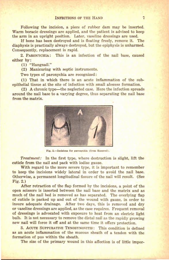
Fig. 2.-Incisions for paronychia (from Kana vel ) .
Treatment: In the first type, where destruction is slight, lift the cuticle from the nail and pack with iodize gauze.
With regard to the more severe type, it is important to remember to keep the incisions widely lateral in order to avoid the nail base. Otherwise, a permanent longitudinal fissure of the nail will result. (See Fig. 2.)
After retraction of the flap formed by the incisions, a point of the open scissors is inserted between the nail base and the matrix and as much of the nail bed is removed as has separated. The overlying flap of cuticle is packed up and out of the wound with gauze, in order to insure adequate drainage. After two days, this is removed and dry or vaseline dressings are applied, as the case requires. Frequent removal of dressings is advocated with exposure to heat from an electric light bulb. It is not necessary to remove the distal nail as the rapidly growing new nail will force it off and at the same time it offers protection.
3. ACUTE SUPPURATIVE TENOSYNOVITIS: This condition is defined as an acute inflammation of the mucous sheath of a tendon with the formation of pus within the sheath.
The size of the primary wound in this affection is of little impor-

8 UNIVERSITY OF WESTERN ONTARIO MEDICAL JOURNAL
tance. It may be anything from a simple pin-prick to an extensive wound.
S ymptoms : (1) Symmetrical swelling of the entire finger. (2) Flexion of the finger . (3 ) Exquisite pain on extension of the finger. ( 4) Exquisite pain over the course of the sheath, limited to the sheath.
In addition, the adjacent fingers may be swollen at their bases and there may be oedema of the dorsum. The entire hand is painful and all fingers are held in flexion, but they do not exhibit the marked rigidity present in the infected finger. The tenderness may subside later, especially after rupture of the sheath. Paraesthesia is common.
Extensions: ( 1) The little finger : to ulnar bursa; to radial bursa; to major forearm space; to the middle palmar space; osseous involvement; rupture to surface. (2) The index, middle and ring fingers: to the fascial compartments to which each sheath is related. (3) The thumb: to the radial bursa; to the ulnar bursa; to the major forearm space; to the thenar space ; osseous involvement.
Ulnar Bursa Infection: This is often difficult to diagnose. The two most important signs are: (1) Extension of exquisite pain and tenderness over the anatomical distribution of the bursa. (2) A point of maximum tenderness j ust proximal to the point where the distal palmar flexion crease crosses the ulnar bursa.
AI o present are: (1) Increased oedema of the dorsum. (2) Fullne s of the palm, but the palmar concavity is still present. (3) Noticeable swelling proximal to t he transverse carpal ligament.
Extension to the radial bursa is marked by increased rigidity of the thumb and pain over t he area of anatomical distribution.
Treatment: Fig. 3. For this- condition it is essential that the patient be hospitalized. Operation is performed early, with the patient under a general anaesthetic and the arm should be so prepared that the operator has a bloodless field to deal with. Extreme care should be taken to prevent sepsis both at operation and in dressings.
The first incision is made at the site of the known infection, on the lateral side of the digit, rather than in the median plane. In order to avoid the digital blood vessels and nerves, a good plan is to take a point anterior or posterior (as the case requires), to the posterior termination of the middle phalangeal flexion crease, when the finger is partially flexed. The distal digit is not incised. On exposing the sheath, it is not to be incised at the joints and the vinculi are not disturbed. This prevents prolapse of the tendon.
Above all, make the incision too free rather than too small . A free incision will drain more rapidly and heal with less destruction than a small incision which drains slowly, with much destruction and resultant loss of function.
If the infection is believed to have extended to the lumbrical canal, continue the primary inci ion a in Fig. 3.
The thenar and mid-palmar spaces are dealt with below. The Ulnar Bursa: If extension has occurred here, the incision is

INFECTIONS OF THE HAND 9
best carried across the palm as in Fig. 3. The incision is begun at the proximal end of the original incision and is carried upwards on the radial side of the hypothenar muscles to the distal margin of the carpal ligament.
The Tendon Sheath of the Thumb and Radial Bursa: The incision is commenced distally (Fig. 3), dissecting down to the sheath and continued proximally along its course to a point one inch below the transverse carpal ligament. Between these two points, the motor branch of the median nerve to the thenar muscles is located.
The Major Forearm Space: The incision is started on the ulnar side of the forearm, one and one-half inches proximal to the tip of the ulnar styloid process on its flexor surface. Again, an extravagant incision is of more value than a meagre one. It is best to treat this complication from the ulnar side, because of the absence of nerves and blood vessels. After making the incision, one dissects between the deep flexors and the ulna to the pus. Occasionally, through and through drainage is required (Fig. 3). Drainage tubes are permissable here.
Fig. 3.- Lines show possible incisions for infection of the tendon sheaths. The dotted lines show incisions made in exceptional cases (!rom Kanavel).
Lastly, with all of these, the insertion of drainage tubes is absolutely contra-indicated, with the exception of the major forearm space. They result in the formation of adhesions, the very thing that the surgeon must attempt to guard against.
Daily soaking of the affected area in hot boracic or N-saline solution with active movement facilitates drainage. Vaseline gauze is applied at first, and is followed by dry dressing when required.
It is best to splint the hand in the position of function, in case adhesions do occur.
4. MAJOR FASCIAL SPACE ABSCESSES: These include the thenar and mid-palmar spaces.

10 UNIVERSITY OF WESTERN ONTARIO MEDICAL JoukNAL
Etiology: 1. Primary: Direct implantation wounds via the lumbrical canals. 2, Secondary: (a) Tenosynovitis with rupture of sheath; (b) Chronic tenosynovitis. 3, Lymphangiitis.
Symptoms: 1, General: The general symptoms are those of an acute inflammatory fever. 2, Local: (a) Obliteration of the palmar concavity with slight bulging is almost pathognomonic. (b) Position of the fingers-the thumb and index fingers will be held in flexion in thenar space infection, and the middle, ring and little fingers, in midpalmar space infection. The rigidity seen in frank tenosynovitis is not present. (c) Greater swelling is present in thenar space infection, with possibly "ballooning' of that space. (d) In thenar space infection there may be detected a brawny induration following the oedema and preceding the tardy fluctuation. Never wait for the latter. With midpalmar space infection, t he palmar fascia is too dense to display this feature. (e) There is doughy, pitting oedema of the dorsum. This is not an indication for incision directed to this area.
F ig. 4.- Typical incisions for opening the lumbr ical space and for opening the lumbrical space in conjunction with the middle palmar spa ce (from Kanavel ) .
Treatment: In treating a tendon sheath, the fascial space is not drained and likewise, when treating a fascial space, the tendon is not drained, if both are involved. Each must be treated separately.
The treatment is divided into prophylactic and active. (1) Prophylactic: (a) Give all wounds aseptic care. (b) Incise any localized infection early, i.e., as soon as an accumu
lation of pus has been diagnosed. Remember that incisions which bring forth only serum harm the patient . Pus rarely accumulates on the dorsum. When one can't be certain if pus is present, apply hot fomentations liberally until the infection has become walled off.

INFECTIONS OF THE HAND 11
(2) Active The Middle Palmar Space: (Fig. 4). The incision is best made
either between the middle and ring or the ring and little fingers, on the palmar aspect, depending on the site of the pus. If there is no choice, the former incision is best. This opens the lumbrical canal. The incision is carried a thumb's breadth and a half into the palm. Artery forceps are inserted in the incision and opened. Rubber dam may be inserted for a day. The usual routine dressings and hot packs are employed.
The Thenar Space: (Fig. 5) : This space is best approached from the dorsum on the radial side of the metacarpal bone for the index finger, at its middle, and on a level with its flexor surface. Closed artery forceps are inserted into the incision as above. They are directed to the ulnar side, keeping close to the flexor surface of th ebone, taking care not to pass beyond the middle metacarpal bone and then opened. The after treatment is the same as for the mid-palmar space.
Fig. 6.-Drainage of the thenar space. Site of incision (a) and drain (h) inserted into this space (from Kanavel) .
5. LYMPHANGIITIS: This condition may involve either the superficial or deep lymphatics. The latter is rare and the former is common.
Etiology : The predisposing cause is generally attributed to a lowered resistance of the individual. The condition is more frequent in the winter months.
The exciting cause may be impossible to elicit. Very frequently there is no evidence of a primary lesion.
The organism most frequently responsible is the streptococcus hemolyticus.
Signs and Symptoms: ( 1) There may or may not be a local reaction with slight swelling. If the infection is severe, the local symptoms are
------------------------------------------------------ ----~
12 UNIVERSITY OF WESTERN ONTARIO MEDICAL JOURNAL
almost negligible and the general or systemic symptoms are severe. It is these latter cases which possess the mortality and there are manifest all the signs and symptoms of septicaemia. If on the other hand, the infection is not severe, there will be more marked local signs and a very misleading one is a doughy, pitting oedema of the dorsum which does not call for incision.
(2) Tenderness over the inflamed area. (3) Red streaks corresponding to the anatomical distribution of
the lymphatics. (4) Usually after twenty-four hours or less there will be tenderness
over the glands which drain the particular areas. (o) Absence of the signs which accompany tenosynovitis and
major fascial space infections. Treatment : Local: (1) Antiseptic hot fomentations have not been proven of
any definite value. Massive applications of fomentations lightly wrung from hot normal saline are the best. To be effective, they must be applied from the finger tip to the shoulder and changed frequently .
(2) Incisions. Again, it cannot be too strongly emphasized that the puffy oedema which occurs on the dorsum of the hand is not to be incised. The presence of the dense lymphatic plexuses in this location and the looseness of the tissues explains the oedema. If incision is prformed, the infection is merely spread and may result in a cellulitis of the skin, not unlike erysipelas. The only sign of localization of infection in the hand worth waiting for is a brawny induration of the area affected. Incisions are to be made then, rather than after waiting for the tardy appearance of fluctuation. The reason is evident--with the oedema, the infection has not localized-with the fluctuation, the stage is too late, for tissue destruction with accompanying loss of function is almost certain.
To incise the bean-like indurated areas which occur along the lymphatics (most commonly on the dorsum) is an error. When performed, only a slight amount of serum is brought to the surface.
Likewise, transverse incisions along the course of lymphatics as an aid to block the spread merely translates this spread into an area of localized tissue damage and is therefore likely to open up new channels of spread.
Consequently, the use of the knife in lymphangiitis is very limiteil and should he employed only:
(1) In localization in tendon sheaths. (2) In the subcutaneous tissues as on the back of the forearm. (3) About abscessed glands. General or Systemic Treatment : (1) Antagonistic drugs: Their
use lies in the hope that since their structure is less stable than that of the cell protoplasm, the toxins will combine with them rather than with the cell protoplasm. Quinine is the oldest of these, and even yet, its beneficial effects are questioned. (2) Food and fluids: The diet will

INFECTIONS OF THE HAND 13
consist of easily digested foods and fluids in all forms and in abundance. (3) Glucose and blood transfusions are required in malignant cases. (4) Supportive measures include fresh air, sunlight, attention to the bowels; in short, all the forces are brought forth that will aid the individual to build up his low resistance.
CONCLUSIONS (1) Infections of the hand are most important to the individual
and they must be looked upon with caution. (2) Adequate anatomical knowledge is essential. (3) Proper surgery, in the light of what has been done by others,
notably Kanavel, is of the utmost importance in treatment. As regards the scalpel, some cases call for radicalism, others for
conservatism, and still others for no scalpel at all.
In the preparation of this article, the text on Infections of the Hand by Dr. A. B. Kanavel of Northwestern University was extensively referred to. The accompanying diagrams were taken from this treatise, and permission to use them is gratefully acknowledged.
"OPERATION" You are carried in a basket, Like a carcass from the shambles, To the theatre, a cockpit Where they stretch you on a table.
Then they bid you close your eyelids, And they mask you with a napkin, And the anesthetic reaches Hot and subtle through your being.
And you gasp and reel and shudder In a rushing, swaying rapture, While the voices at your elbow Fade - receding - fainter - farther.
Lights about you shower and tumble, And your blood seems crystallizingEdged and vibrant, yet within you Racked and hurried back and forward.
Then the lights grow fast and furious. And you hear a noise of waters, And you wrestle, blind and dizzy, In an agony of effort.
Till a sudden lull accepts you, And you sound an utter darkness And awaken . . . with a struggle . . . On a hushed, attentive audience.
-By WILLIAM ERNEST HENLEY.

The International Congress of Anatomists ·=·
By CHARLES C. MACKLIN, M.B., M.D., M.A., Ph.D., F.R.S.C.
THE Fourth Federative International Congress in Anatomy was held on September 2nd to 8th in the School of Medicine of the Royal
University of Milan, Italy, and gathered together about 350 scientists from at least twenty-five different countries widely scattered throughout the world.
This Convention was opened with impressive ceremonies in the Council Hall of the great medieval Castello Sforzesco, where the various delegations were received by Dr. Ferdinando Livini, Professor of Anatomy in the Royal University of Milan and Chairman of the Council of Directors, and by Professor Migliavacca. The great importance with which it was regarded is shown by the eminence of the various representatives. The King of Italy was represented by H. R. H. the Duke of Bergamo, the Fascist Government by His Excellency Signor Bianchini, Under-secretary of State for Finance, and the Vatican by Mgr. Buttava. At the head table beside Professor Livini were seated the Mayor, the Prefect, the Rector of the Royal University of Milan, and the Presidents of the great Anatomical Societies. Among the latter were J obnston for Great Britain and Ireland, Jackson for the United States of America, Policard for France, Stieve for Germany, Boecke for Ht>lland, Hirako for Japan, Loth for Poland, etc. There were many other important dignatories present, including representatives of the Army, Academic and Engineering Societies, the Medical Asociation, the Bar, the Red Cross, and Departments of the Government.
Profesor Livini, who was President of the Congress and representative of the Fascist Educational Association, the National Council of Research, the Italian Association for the Progress of Science and the Royal Institute of Lombardy for Sciences and Letters, made the first speech, in which, after expressing his gratitude to the King, the Duke of Bergamo and Mr. Mussolini for their part in the plans for the success of the Congress, he thanked all the authorities present, extended a warm welcome to the Congressionists and stressed the world-wide importance of the Convention. He then unfolded a verbal picture of modern anatomy, emphasizing that it is now a dynamic science, not a static one. "Anatomy is no longer the science of death," he said, "it is the science of life." It now occupies a position of first rank in the field of the biological sciences, and, as well, continues to be the essential foundation of medicine. That it is highly dynamic is shown by the new paths that it is exploring every day, leading into direct contacts with the
*Dr. Macklin, Professor of Histology and Embryology at University of Western Ontario, was a delegate to this distinguished gathering and delivered a paper, an abstract of which appears elsewhere in this issue.
14

THE INTERNATIONAL CONGRESS OF ANATOMISTS 15
domains of sister sciences and of every department of clinical medicine, and by the increasing use that is being made of experimental animals, and of such newer technics as motion pictures and X-rays in its researches. The wide range of its interests is shown in the list of subjects and abstracts of reports to be presented at this Congress. It is not enough that anatomy be merely "descriptive"; it must interpret living structures in terms of function. The barriers that formerly separated the different sciences from one another are now broken down, never to be replaced. These barriers--artificial and unnatural as they were--have retarded progress greatly. In thus making correlations with related sciences, anatomists are not departing from their own field, but are rendering ever higher service not only to their own but to other sciences. Professor Livini . then traced clearly the development of anatomical science in Italy, to which, more than four centuries ago, Leonardo da Vinci made a great contribution, and inspired a vast amount of later work. His oration was greeted with enthusiastic applause.
The Rector of the Royal University and the Mayor of Milan then delivered enthusiastic and warm addresses of welcome.
His Excellency, Bianchini, expressed gratitude for the duty entrusted to him by Il Duce, "who is following this Congress with great interest," and brought greetings from the Fascist Government, "which is pleased to gather together these illustrious scientists for a noble purpose of benefit to mankind." The speaker expatiated on the advantages accruing to humanity from the study of modern anatomy, particularly emphasizing the value of a knowledge of the human organism in its minuter parts--the cells and tissues as revealed by the microscope--in arriving at a clear understanding of the bodily functions and of pathological processes in the vital substance of the human body. This knowledge was essential for the proper understanding of diseases and their causes, and therefore for their cure and prevention. He then formally opened the Congress in the names of the King and Mr. Mussolini.
Professor Policard replied for the foreign representatives, recalling that this Congress succeeds that of Amsterdam in 1930. He dwelt upon the enormous growth of the anatomical sciences in recent years, particularly in the microscopic field, in which he is a leading worker, and emphasized the great breadth and scope of modern anatomy as shown by the program of this Congress. In the newer investigations now taking place a great number of recent technics were being used, and modern anatomy now was integrated with all of the preclinical and clinical sciences.
While at their own homes, quite early in the year, the members had received printed circulars giving advance information about the Congress, together with invitations to come and participate in it. They had filled out the blank forms provided, giving details about themselves and the subjects which they wished to speak upon or demonstrate, and

16 UNIVERSITY OF WESTERN ONTARIO MEDICAL JOURNAL
they had also sent off to Italy short summaries of their subjects. They indicated their hotel preferences, so that, upon their arrival in Milan, they found reservations waiting for them. They replied to the various gracious invitations to the social functions. Thus, upon presenting themselves at the Secretary's office at 31, via Mangiagalli, there remained nothing for the members to do but to make themselves at home, and to receive the following official literature and other articles:
(1) A bound handbook of 79 pages containing a list of the Congressionists and a complete Program of the Congress. The register was arranged both alphabetically and numerically. A special list of "Relatori," or those who were to give papers or demonstrations, was provided. The program was arranged in morning and afternoon sessions, and for each session there was a list of the contributors with their subjects and usually a short summary of the paper, in one of the four official languages, English, French, Italian and German. The times and places of the various social events were clearly indicated. In the back of this volume was a map of the main part of the City of Milan with street car and omnibus routes marked by a system of red lines with suitable numbers.
(2) A handsome badge of bronze, oval in form, bearing a basrelief of Aesculapius, from which was suspended the member's official number, on a bronze plate, the whole being carried on a silk ribbon of red and blue. This was worn at all times and served to identify the wearer under any circumstance.
(3) A Pass entitling the holder to free transportation upon the street cars and omnibuses of the city for the entire duration of the Congress.
(4) A complimentary copy of the official illustrated guide-book of Milan.
(5) A large folded map of the city with full directions for reaching the numerous places of interest.
(6) A book of complimentary tickets to the various receptions and excursions, etc.
(7) A complimentary copy of the first issue of "Acta Medica Italica," containing 168 pages of descriptive matter and illustrations on the subject of the Italian Universities-a very informative volume on the institutions of higher learning in modern Italy.
(8) A handsome medallion of bronze, about 31;2 inches in diameter, bearing a bas-relief of Malpighi, the discoverer of the capillaries, at the age of 63. On the reverse is the legend: "Mediolani, IV Foederativus Internationalis Anatomicorum Conventus, MCMXXXVI, with the Fascist emblem and the Fascist year, XIII!. It was carried in a suitable case lined with red velvet.
Those who think of anatomy as nothing more than the dissection and description of dead human bodies will have their conceptions revolutionized by an analysis of the one hundred and forty or more reports made at this Congress. For instance, no less than 80 % of them

THE INTERNATIONAL CONGRESS OF ANATOMISTS 17
were concerned with microscopic studies of tissues and cells under many different conditions. This includes, of course, the nervous tissues. Of this, approximately 63 % of the papers were in histology and 17 % were in embryology, though a combination of both points of view was found in many of them. The remaining 20 % were in gross human anatomy, anthropology, Roentgenological studies of the living, etc.
In both microscopic and gross researches, the emphasis was on the appearance and changes of the parts, and their functional interpretation in the living body, and thus Professor Livini's view of dynamic modern anatomy was amply vindicated by the content of this Program. Indeed it may be said that any factor of whatever kind that brings about structural change, is now included in the modern anatomical domain. Thus there were many papers dealing with histological variations in tissues and organs depending on the operation of such factors as age, position and relations of the part, state of functional activity, racial type, effects of hormones, vitamines, etc. As instances of this dominant trend may be cited the following: the report of Dabelow (Munich) on the evolutional, involutional and functional alterations in the human mammary gland from birth to old age; that of Maleci (Padua) on the changing ratio of cells to fibers in the motor nuclei of the seventh cranial nerve from birth to old age; that of Dentici (Palermo) on the variation of histological structure in veins depending on their position and relation to fascial layers as (a) those which are peripheral to the fascial layers as contrasted with (b) those which lie beneath the fascia, and with (c) those of the viscera; that of Stieve (Berlin) on evidence of increased activity of the liver in pregnancy; that of Rossi and Scevola (Pavia) on the variations of the innervation of the uterus in its pregnant and non-pregnant state; that of Champy (Paris) on the action of male hormone and of folliculin on the plumage of birds; that of Zawisch (Vienna) on the influence of adrenalin on bone formation; and that of Moricard (Paris) on the effect of mitosin on cell division, and that of folliculin on the production of ovulation in the rabbit. These titles, taken haphazardly from the program, show the catholicity of interest of modern anatomists, and the enormous emphasis placed upon findings in the living, actively functioning cells and tissues.
Many of the newer technics, referred to by Profes or Policard, were employed by the congressionists in their researches. Thus a number of the reports dealt with results from use of the tissue culture method: as those of Grodzinski (Cracow) on the dynamic changes in the cells of the yolk sac during ingestion of fat; Delorenzi (Turin) on the behavior of the Kupffer cells and their reaction to vital dyes; Levi (Turin) on growth in explanted dorsal root ganglion cells; Rondinini (Bologna) on contraction in striated muscle cells; and Staudacher (Padua) on direct observation of developing contractility in the ureter and bile ducts of the embryo. The method of microincineration was used by Barigozzi (Milan) in investigating the mineral components of

18 UNIVERSITY OF WESTERN ONTARIO MEDICAL JOURNAL
the cells of the salivary glands. In this technic, developed by Policard in France and Scott and others in America, thin sections are stuck to glass slides and reduced to ash by heat; much can thus be learned about the distribution, within the cells, of their non-combustile elements, by microscopic studies of the patterns of the residue as it lies in situ on the slide. Ultracentrifugalization was used by Sestini (Siena) in an analysis of the components of nerve cells. On this continent, Beams and his colleagues have used the ultracentrifuge, which yields approximately the effect of 150,000 times the force of gravity, with good results in the determination of the relative densities of such things as nucleoli, chromatin particles, mitochondria and other cellular constituents. Motion pictures, particularly of the "fast motion" type, have shed much light on movements of cells and tissues unappreciable by ordinary methods of observation, and at this Congress Elias (Padua) studied developing blood areas by means of this technic. Ultraviolet light was used by Ravotti (Milan) in observations on reticulocytes, and vital staining was combined with direct observation of living cells by Vonwilier (Moscow) in the study of living cells of the brain. Federow reported his direct observations of the cells and synapses of sympathetic ganglia under conditions of repose and experimental stimulation. The value of X-rays, not only in teaching but in research into the structure of the living body was enhanced by some of the contributions. Hasselwander (Erlangen) gave his results on the development of the ossification centers, and on variations in these and in skeletal structure in general; on the inheritance of these variations, on the action of the living joints; on the form, position and malposition of various parts of the intestinal tract, and on the use of stereograms in exact methods of study of growth. Not only has roentgenography proved itself to be of great use in the study of gross structure, but it is being applied to cytological study as well, as by Lamarque and Turchini (Montpellier). Histological control of phy iological endocrine studies is now essential in endocrinological research. Granel (Montpellier) showed this in his results on the influence of thymus extract on testis cells, and also of developing testis on the thymus.
Heredity was stressed in many papers: for instance in the communication of Hasselwander (Erlangen) on ossification, that of Benninghof (Kiel) on "constitutional anatomy," and that of Sagols (Montpellier) on the mutations caused by radium, and the value of this knowledge in the elucidation of evolutionary phenomena.
The thyroids of different breeds of dogs, according to Vicari, show actual histological differences characteristic of the breed, and these variations involve the parafollicular as well as the follicular cells. The parafollicular cells are numerous during activation of the follicular epithelium and their number is further increased in pregnancy. These observations suggest that the number of parafollicular cells is related to physiological conditions, and hence that these hitherto somewhat neglected cells are of importance.

THE INTERNATIONAL CONGRESS OF ANATOMISTS 19
Lewis of Harvard (this year's President of the American Association of Anatomists) broke new ground in his study of the forms which cells take when compressed against one another, and has compared them with the forms of bubbles in masses. He told the Congress that the average cell in a compressed group is characteristically 14-hedral, and made the interesting deduction that the "edge canals" or intercellular spaces arise by tension at the edges of apposed cell membranes.
Windle gave the results of his studies on the genesis of somatic movements and the origin and integration of reflexes in embryo mammals. He found that embryronic skeletal muscle may be made to contract by direct faradic stimulation, before the central nervous system is functioning reflexly. When the first reflex-like movements do come, they are from supposedly spinal reflexes, and these are at first local and separate, and are later integrated. In the cat and rat he found that their development is coincidental with the structural development of the first spinal reflex arcs.
Addison (Canadian-born, of Philadelphia) reported that the hypophysis of old albino rats of both sexes sometimes undergoes a curious and notable enlargement, and may even become 20 times the normal weight without disturbing the body size. Increase in the undifferentiated chromophobe cells of the pars anterior was found.
Jackson said that, after prolonged suppression of growth in young albino rats by protein deficiency, as well as by simple underfeeding, the females recovered their body weight upon refeeding, but the males, subjected to the same treatment, grew less rapidly and remained permanently stunted. The mortality of the starved males was particularly high.
Cummins correlated fingerprints with handedness in a study of some 450 persons, and found that whorls were notably increased in lefthanded females, with an accentuation of bimanual inequality in their occurrence. Since dermatoglyphics are definitively formed early in the fetal period, it is evident that functional asymmetry is foreshadowed then.
Three full afternoons were devoted to demonstrations of microscopic slides, photographs, drawings, models and other research material, and at these there was ample opportunity to enter into free discussions on the scientific points brought up.
Some features of the buildings of the Royal University of Milan, in which the Congress was held, may be of interest to local medical men. These were two, the Anatomical Institute and the Institute of Forensic Medicine.
The Anatomical Institute is a handsome edifice, much like our medical school in plan, with an elongated facade looking out upon a fine, wide, tree-lined street, the via Mangiagalli, and long wings projecting back from the ends. In one of these is the airy, capacious and handsome auditorium, with comfortable seats folding back automatically and noiselessly when vacated, thus making of every tier a cross-aisle.

20 UNIVERSITY OF WESTERN ONTARIO MEDICAL JOURNAL
There was an up-to-date equipment of suspended blackboards and beaded movie screens, and a costly and elaborate projection outfit handling not only lantern slides, but microscopic slides, and pictures even of large size. Indeed it seemed that most of the speakers projected their drawings and photographs directly, instead of by lantern slide photographs of them, and in this way the colors and finest details were faithfully reproduced. There was a small reflecting projector which flashed messages silently upon the screen even as they were being written down. The audience was always kept informed, by a system of changing illuminated numbers on an electrical system, of the papers then going on in the other auditorium, in the Institute of Forensic Medicine, about a block away, which was, of course, similarly equipped. The mechanical ventilation was most efficient, all smoke being quickly evacuated. There were excellent motion picture projectors. Such details as the darkening of the windows were well cared for, and the attendants were very competent.
In the fine foyer, each afternoon, free refreshments were served. Close by were the general offices of the Congress, the Post Office where we received our mail from home, and the offi!!e of the General Secretary of the CIT (Italian Touring Company), who was most courteous and efficient in caring for our needs in anything to do with travel. For the demonstration of the microscopic preparations there were two large rooms on the ground floor and two on the second. On the second floor there was also a huge collection of exhibits of the optical companies, such as Zeiss, Leitz, Reichert and Koristka. In this di play were found examples of the most modern microscopes, including an ingenious one, with built-in camera, and a reversed system of objectives and condensers.
The demonstrations of specially dissected gross anatomical parts were shown in the right wing of the building. The commodious dissecting room formed the expanded circular end of this, and extended through two floors. It was flooded with light from the sides and from an octagonal skylight projecting like a cupola from the dome-like roof. The conveniently low pedestal tables, of smooth stone, clean and sanitary, with pleasing rounded ends, were gently hollowed to allow any fluids to drain through a central vent into a receptacle in the single central supporting pillar. The absence of legs was a great advantage to the dissectors, as well as to the janitors. The stools, of white enamelled metal, of about ordinary chair height, could be moved under the tables out of the way. Thus the greatest simplicity was combined with optimum convenience. The side of the room adjoining the rest of the wing was flattened, and here were found blackboards and projection screens. Here, also, was a large opening in the wall through which preparations from the ample store in the next room could be handed. The tables were set radially, and so formed a circle about a central large round table used for the display of dissected parts. A most desirable feature was the ample floor space, making it easy for teachers and students to move about. Lectures could be given right in the room,

..
THE INTERNATIONAL CONGRESS OF ANATOMISTS 21
with lantern slides and specially dissected parts. Sinks were placed at suitable points around the walls. In the neighboring rooms were special equipments, such as X-ray apparatus, balances for weighing the parts, etc. An ample supply of fresh air of proper temperature and humidity was guaranteed. Altogether the plant seemed ideal for the study of gross human anatomy.
The other building was a handsome two-story marble structure, whose pediment, high above the chaste front, bore the legend "Rebus Medicis sub Specie Juris." It contained an equally efficient auditorium and accessory rooms.
Space will not permit more than mention of the splendid new municipal hospital, through which the author was privileged to be conducted in a small party by the chief of surgery, Dr. Aldo Defrise, who had worked at Johns Hopkins Medical School, and spoke excellent English. He is interested especially in research on the respiratory apparatus. The entire Congress was taken through the Sera-therapeutic Institute, a fine large group of buildings of modern design, which performs the diagnostic work of this great community, manufactures sera and vaccines, and looks after the multitudinous duties of a public health institute. About two hours were required merely to pass through the main parts of the buildings and see a little of what was going on. There is an excellent scientific library. Much research work has been done in this Institute. Indeed the publications of the last two years, 1932 and 1933, totalled 86, and the output is probably even greater now. When it is recalled that such men as Golgi and Negri worked here, the importance of the personnel is realized. Here we found stables full of horses used in the making of anti-diphtheric serum, large rooms teeming with guinea pigs, great cages of poisonous snakes for antivenom vaccines. In fact, there was here all that is required for public health work related not only to the civilian population, but to the army also, and even to domestic animals. One can hardly doubt the enormous importance which modern Italy places upon the maintenance of health in man and beast. The genial Director is Professor Serafino Belfanti. It is interesting that the Institute is situated on Darwin Street. Indeed one notices that many of the streets in Milan, and in other Italian cities, bear the names of famous men, scientists like Marconi, as well as artists and literati like Raphael, Sanzi and Dante Alighieri.
The entertainment of the "Congressisti" by the local committee was lavish. There were a number of receptions (including one at the Mayor's palace), a special luncheon, a formal dinner, a symphony orchestra concert, a visit to the Milan Triennial Fair, a tour through Pavia, including the University where Scarpa, Corti, Golgi and many other great investigators had worked, and an ali-day motor tour to Lake Como and San Pellegrino. There the party looked over into Switzerland from high Brunate (reached after a perilous ride up a steep "funicolare," or electric railway), and viewed the splendors of Monte Rosa and her snow-capped companions, which Professor Corti of Turin said

22 UNIVERSITY OF WESTERN ONTARIO MEDICAL JOURNAL
were one hundred miles away. These functions were all quite free, of course, and gave ample opportunity for getting acquainted with the members of the Congress.
While being conducted in a party through the ancient and beautiful Certosa of Pavia, the writer was introduced to Professor Christian Champy of Paris, France. Professor Champy, on learning that the writer came from London, Canada, at once recalled that Dr. Edwin Seaborn, of this city, had carried on research work with him in Paris on the subject of the ovary of the mare and its role in the oestrus cycle, and in the production of hormones, and asked to be remembered cordially to Dr. Seaborn. He spoke of the warm pleasure it gave him to recall Dr. Seaborn, and the excellent 'investigation which this alumnus of our medical school had carried out in Paris, the results of part of which have been published in the Anatomical Record, where they have earned the interest and approval of many scientists working in this field.
This World Congress marks a notable milestone in the history of anatomy. The members are already looking forward to the next one, which, it is expected, will take place in 1940 in London, England.
" The Old Order Changeth " We appear in this issue in a brand new garb. It is with regret and
some misgiving that we bid farewell to the familiar emblem that for RO long has graced our cover. Times will change, though, and plateR will wear out! When such a distinguished organ as our venerable and esteemed contemporary, the British Medical Journal, discards a format centuries old, we deem it no disgrace to imitate her.
The design which now appears on the cover is, for the benefit of graduates, the recently adopted official crest of the Hippocratic Society, undergraduate governing body of the faculty of Medicine. The school is indebted for this masterly bit of design to Dr. Ralph Christianson, '36, now at Montreal General Hospital.
The new cover will, we trust, be an omen of future progress on the Journal. All sections are undergoing reorganization at present and the results will be shown, we hope, in a better and more punctual publication. A new office has been created, that of exchange editor, and John D. Munn, '38, will be its first occupant. It is hoped that by extending our exchange service to many more Medical schools we will be in a position to keep our readers abreast of the many recent developments in mdical education at other centres.

Hypnotism By RONALD BOURNE, B.A., '38
H YPNOTISM, very popular at the end of the last century, has so far departed from medicine that the average general practitioner
knows little of it beyond what he may have seen in some vaudeville theatre, where it was cloaked with the fineries of showmanship. Nevertheless, he is probably unknowingly using it every week in his own practice to a very mild degree, even if only by stroking the brow of a restless patient or telling a squirming boy that removing a sliver is not going to hurt.
Hypnoti:sm is defined by Bernheim as "a particular psychical condition which one can produce and in which suggestibility is increased." The name hypnotism was given to this phenomenon by Braid in 1843 because in his series he used it to induce sleep in his patients. It is, however, an unfortunate term, because sleep is by no means necessary. In fact, it is often absent as shall be shown, and hypersuggestability is much more accurate.
The beginnings of hypnotism are lost in antiquity, first appearing in early Egypt. Medicine, practiced there by the priests, consisted largely of psychotherapy and probably induced celestial hallucinations which would indicate to the patient that be was cured. In India, hypnotism has been practiced as long as written records have been kept, reaching its highest peak by being used during major surgical operations to induce anesthesia.
The modern science was reborn as an art by Mesmer in 1776, and developed into a science by Braid of Manchester in 1843. It then progressed slowly until the foundation of the Institute of Salpetriere by Charcot and Janet in 1880. Under their guidance and that of the opposing school at Nancy under Bernheim and Fore!, it reached a peak at the turn of the century, but abuse in the bands of charlatans and quacks removed it from popular favor.
Recently, however, the pendulum of public opinion has started the reverse swing and hypnotism is once more coming to the fore as a respectable branch of therapeutics. While by no means the panacea that some of its devotees believe it to be, it has its definite place in the armamentarium, not only of the psychiatrist but also of the general practitioner.
There are three theories as to the nature of this phenomenon: 1. The magnetic fluid theory, advanced by Mesmer, and now held
only by spiritualistic cults, who believe that a vapour is transmitted from one individual to another by magnetism.
2. The somatic theory, advanced by Charcot and supported by his school, who believe it to be a peculiar state of the neuromuscular junctions induced by contact with the skin.
3. The psychic theory, most widely accepted now, which conceives 23

24 UNIVERSITY OF WESTERN ONTARIO MEDICAL JOURNAL
it to be a mental state induced by suggestion; the latter is the only reputable means of induction now in use.
Freudian psychologists believe there is in the mind of the subject a transfer of the identity of the hypnotist to the person of the parent of the opposite sex, with a resulting Oedipus attachment. The psychophysiologists, however, believe hypnotism to be the result of stimulation of the hypothalamic sleep centre, produced while the hypnotist rhythmically stimulates the peripheral nerves, as described below.
The field of the mind can be graphically divided into a central area, representing the conscious field, a more peripheral area, the subconscious, and outermost the unconscious, all of which are formative factors in our personality. ,
Hypnotism successively reaches each of these fields, and the stage of hypnosis will depend on the area of the mind involved. When only the conscious level is influenced, it is termed the stage of somnolence. Consciousness is not lost, and suggestion is applicable only by directing the focus of consciousness on whatever the hypnotist mentions. The acceptance or refusal of the suggestion lies with the subject. In mild degrees this cannot be differed from simple waking suggestion; in deeper states it gradually changes to the second level or the stage of hypotaxis, which is concerned more with the subconscious mind. In this stage the patient is very amenable to suggestion, but will not follow it if it be against his moral principles; amnesia is only partial or temporary, unless suggested. Most therapeutic treatments can be given at this level and in many, even below this. In the outer ring or unconscious mind are borne many factors which influence our behaviour and mental health, but are never apparent to us. These factors may be eradicated by bringing them to the conscious level, or new factors can be added during the third or somnambulistic stage of hypnosis. This difficult stage is difficult to induce and is often gained only after long or repeated hypnoses. On waking, the patient has no recollection of what has occurred unless the latter has been suggested.
The act of hypnotising is no more mysterious or complicated than going to sleep. The hypnotist does not, by his dominant will suppress that of the subject, but rather leads the latter, who is willing and co-operative. Thus the stronger the subject's will the more successful is the hypnosis.
Who can hypnotise and who can be hypnotised? As just mentioned, the important factor is not a case of will power, but of confidence and co-operation. Theoretically, any normal, intelligent person can hypnotise, but in practise he must be a person who by his appearance and manner, can gain the confidence of his subject. Hence a person with deep piercing eyes, etc., is much more successful than a ludicrous roilypolly or a timid, fearful person. Similarly, in theory, any normal person can be hypnotised, with the exception of psychotics and the feebleminded, only some of whom can be done. (Charcot states the reverse of this, but huge series of cases disprove his theory.) Actually, figures

HYPNOTISM 25
at large clinics show that from 90 to 98 % of unselected cases, including some psychotics, have been hypnotised. However, many of their unsuccessful cases have been stopped after a single attempt, while some cases require up to sixty or more periods before they can develop sufficient confidence and co-operation to be successfully hypnotised. Further, there are many devices which might be used to detract attention from the central thought while the patient is still being induced. A moderate dose of a mild sedative, judicially used, will often remove any slight fear and restlessnes the patient might present.
The hypnotist should always have the consent of the subject, or of a responsible person, to hypnotise him and to perform whatever therapeutic measures he believes necessary ; he should have a witness present during the hypnosis to safeguard his reputation.
The actual method of induction can be learned faster by one demonstration than by reading a library of books, and once learned, only confidence and experience can permit the application and improvement of the knowledge. Having the patient watch someone else being hypnotised is of great asistance, as it removes much of the mystery and fear of the operation.
Actually, the suggestion is given to the patient that he fall asleep and this is aided by causing such sensations as are usually associated with sleep; for example, eye or ear fatigue and quiet comfort. Fatigue is induced by having him stare at a bright object rotating slowly in a circle ten inches in diameter about ten centimeters before his eyes, or gaze at a bright object just above his normal line of vision. At the same time, the suggestion of drowsiness, fatigue and sleep are given in a lulling monotonous tone until the eyes close; at this time, deep sleep is suggested. A further method of inducing eye fatigue is by havin.g the patient stare into the hypnotist's eyes at a very short distance anp suggesting that he will first undergo a fusion of visual fields, i.e., only one eye, then eye fatigue, and finally, sleep. This, however, puts a very personal note into the technique and has no advantage over the other methods. Aural fatigue may be induced by listening to a faint ticking of a watch or a chime, but concentration on these distracts the patient from the vocal suggestion.
Simple rythmical stroking, done while suggesting sleep, is very efficacious alone, and is better still combined with one of the above methods. While the magnetic fluid theory is not accepted, there is a definite value in the technique of stroking, especially when employed on a baby or an elderly patient.
Hypnoti ts are well advised to seek some unusual means of inducing hypnosis, as this not only augments the confidence of the patient but also avoids the exigency of a patient suddenly being hypnotised at some later time, when confronted by the same stimulus under natural circumstances.
The patient having been thus induced is carried (at the second stage) or attempts are made to deepen the state by further suggestion.

26 UNIVERSITY OF WESTERN ONTARIO MEDICAL JOURNAL
Then the proper therapeutic measures indicated are suggested repeatedly. It is often advisable to give the suggestion of increased susceptability to future hypnoses to ease future treatments, and to suggest a feeling of euphoria, or contentedness before one tells the patient to awaken.
As mentioned above, mental defectives cannot readily be hypnotised, as they cannot concentrate long enough. However, occasionally they can be suddenly terrified into a state of catalepsy and carried along in a hypnotic state from that point.
Hypnotism can be used as an anesthetic or analgesic, to correct some dysfunction, or as a means of psychotherapy.
In accomplishing anesthesia, the subject is put to sleep as described above, and the suggestion is given that all sensation is leaving the area that is painful or which is to be operated upon. At the same time it is advisable to suggest absence of fear or other emotions of withdrawal. If necessary, it can be suggested that such anesthesia will continue to act after awakening until told otherwise, and then awaken the patient. Do not neglect to end such anesthesia. As an analgesic it is used in the same way, and can be used for any of the rheumatic inflammations, functional pains, or obstetrics.
Among functional disorders, the scope of hypnotism is as wide as the nervous system whether somatic or vegetative. It can be used to augment or inhibit the normal tonic impulses to muscles and glands. Thus it can be employed to advantage in menstrual disorders, even to stopping the flow, asthma, gastric upsets, functional diarrhea and constipation. Hemorrhage can even be controlled in many cases.
Finally as a psychotherapeutic agent it can be used to break habits or form new correct ones, but these must be absolute ones. There can be no half-way measures, such as permitting the patient, by suggestion, to have just one cigarette after meals or one drink a day. Again, it may be used with greater ease and more rapidity than psychoanalysis in exploring the subconscious and unconscious minds to detect falsehoods, false impressions, or even suppressed ideas which may be perverting the patient's mental life.
Hypnosis is not contra-indicated in any medical condition and can thus be used where drug therapy might be harmful. It is contraindicated, however, in paranoic and paranoid patients, and should not be repeated too frequently in hysterical patients.
BIBLIOGRAPHY Diethelm, 0.: Treatment in Psychiatry, MacMillan Co., 1936. Forel, A.: Hypnotism and Psychotherapy, 5th ed., Rebman & Co., 1907. Munro, H. S.: Suggestion Therapeutics, Applied Hypnotism, Psychic Science, Masby
Co., 1907. Pike, H. V.: Hypnotism, Mental Health Bulletin, Danville State Hospital; 14:2:16;
1936. Schilders & Kauders: Hypnosis, Nerv. and Ment. Dis. Pub. Co., 1927

The History of Anesthesia·=· By JOHN D. MUNN, '38
T HE history of anesthesia may be broadly divided into two periods, the pre-anesthetic period and the anesthetic period. The pre
anesthetic period ends and the anesthetic period begins with the discovery of ether in 1842 and its general introduction in 1846.
THE PRE-ANESTHETIC PERIOD
The beginning of the use of drugs to produce anesthesia dates from . earliest antiquity and is symbolized in the 21st verse of the second chapter of Genesis : "And the Lord God caused a deep sleep to fall upon Adam and he slept : and He took one of his ribs and closed up the flesh instead thereof."
Homer in "The Odyssey" says: "Helen dropped into the wine of which the soldiers drank, a drug, an antidote of grief and pain inducing oblivion to all ills. He who drinks of this mingled cup sheds not a tear the live long day. Were death to seize his venerated sire or her who gave him birth, or were the sword buried in the bosom of his brother or greatly loved sister, no tear would even then bedew his cheeks."
Poppy and Indian hemp were probably known to the Egyptians and Greeks, mandragora to the Egyptians, Babylonians and the Hebrews. Theophrastus and Discorides were the first to mention the aphrodisiac and soporific properties of mandragora. It is not clear whether the mandrakes which Rachel sought of Leah (Genesis xxx, 14-16) were for the former purposes or to cease the pangs of childbirth.
Hua, a Chinese physician, is said to have used "hashish" in surger y about 200 B.C. The ancient Peruvian Incas probably utilized the anesthetic property of the active principle of coca in trephining.
Surgical sleeping draughts are mentioned by the church Fathers, Hilary and Oregen. The earliest Salernitan reference to the "soporific sponge" occurs in the Jensen imprint of the Anti-dotarium of Nicholas of Salerno, probably written in the eleventh century. The sponge was steeped in a mixture of opium, hyoscyamus, mulberry juice, lettu~e,
hemlock, mandragora and ivy, dried, and when moistened was inhaled by the patient, who was subsequently awakened by applying finall juice to the nostrils. This prescription, Husemann thinks, was derived from older formulae for anodyne applications of similar ingredients to the temples for insomnia or cataplasma for local anesthesia.
It is said that among the ancient Assyrians, the pain of circumcision was prevented by compression of the veins of the neck during the time of operation. This was doubtless an ancient discovery of the possibility of producing temporary unconsciousness by pressure upon the carotid arteries and pneumogastric nerves to which attention has been again directed in modern times.
In 484 B.C. Herodotus refers to the inhalation of vapors of hemp *Delivered before the Osler Society of London, October, 1936.
27

28 UNIVERSITY OF WESTERN ONTARIO MEDICAL J OURNAL
(Cannabis Indica) to produce intoxication. In 23 A.D. Pliny, the Roman historian, speaks of the juice of certain leaves as producing sleep and then in 134 A.D., Galen, the physician and contemporary of Diascorides, describes the properties of mandragora to paralyze sensation and motion.
THE ANESTHETIC PERIOD
It was at the close of the 18th century that modern surgical anesthesia was foreshadowed, with the discovery of hydrogen in 1776, nitrogen and oxygen in 1772 and nitrous oxide in 177 4. "Pneumatic chemistry," as it were, opened up a field of experimentation which made possible surgical operations under conditions which Sir Humphrey Davy described as "uneasiness being swallowed for a few minutes of pleasure."
Soon after the discovery of these gases, attempts were made to put them to practical use. In 1785, Pierson of Birmingham, England, used ether inhalation for asthma and in 1789 the Medical Pneumatic Institute was organized under Dr. Beddoes, where large reservoirs of gases were installed for the treatment of phthisis and other diseases by inhalation. This Institute, which was superintended by Sir Humphrey Davy, while not successful in itself, was important in that it led to Davy's experiments with nitrous oxide. By 1800, Davy had become sufficiently well acquainted with this gas to use it for the alleviation of headache and also for the extraction of one of his own wisdom teeth. This latter event led him to make the historic prediction that, "since nitrous oxide seems capable of destroying physical pain, it may be used in surgical operations where there is no great effusion of blood." The value of this suggestion was not recognized for nearly half a century.
Warren, of Boston, used sulphuric ether in 1805 on a patient suffering from phthisis and in 1806 it was used in attacks of asthma. Faraday seems to have been the first to recognize the value of sulphuric ether as an anesthetic. In 1818 there appeared a paragraph attributed to Faraday in the Quarterly Jour nal of Science and Arts, in which it was pointed out that "when the vapor of ether is mixed with common air and inhaled it produces effects very similar to those occasioned by nitrous oxide."
Henry Hill Hickman in the period from 1820 to 1828, did successful experiments on anesthetising lower animals. Impressed by the agonizing sufferings of those on whom he was called to operate, he resolved to seek some method of alleviating their pain by rendering them unconscious before the operation. After rendering his animals unconscious, he excised ears, amputated legs, made incisions, then dressed the wounds and noted the time they took to heal and the period required for complete recovery. His experiments met with much success, but he died in 1829, his ambition to perform human experiments unrealized.
In 1839 some young people held a quilting party near Athens, Georgia, after which they finished the evening by inhaling ether. At

THE HISTORY OF ANESTHESIA 29
the height of the frolic, a negro boy appeared at the door and was invited to partake of the ether, but he refused. Some of the boys dragged him in and forced a handkerchief covered with ether to his mouth and nose. After a long struggle he became quiet and the boys desisted their efforts to intoxicate him. Instead of getting up, the negro lay as if asleep and, much to the terror of the culprits, did not awaken until medical attention was given an hour later.
It remained for Crawford W. Long of Georgia to intelligently make use of the facts which were common knowledge to all. Long, in 1842, indulged frequently with his pupils in ether frolics, during which he was badly bruised; yet he noticed upon recovery that he had not been conscious of pain. Frequent observations of this fact, in connection with himself and his students, led Long to conceive the idea of using ether to prevent the pain of surgical operations. He was successful in all his operations where he anesthetised the patient, but since his was the narrow sphere of a local country doctor, surgical cases were few and his fame did not spread beyond the restricted world in which he lived. He made no secret of his discovery, but did not advertise it until others had laid claim to the honor.
In 1844, two years and eight months after Long anesthetised the first patient with ether, Horace Wells, a dentist of Hartford, Conn .. attended a lecture on "laughing gas" by C. Q. Cotton, a chemist. The lecture impressed upon him the belief that the administration of nitrous oxide would bring about the result for which he had been looking. He tested his conclusions upon himself and had one of his teeth extracted. The extraction was successful in that it caused no pain. However, when Wells attempted a public demonstration of his theory he failed and, being such a sensitive man, his reason gave way and early in 1848 he died by his own hand.
It was left to William T. C. Morton, a former pupil of Wells, to place the use of ether as an anesthetic upon a sound basis. His discovery was entirely independent of that of Long, who preceded him by four years.
The first public demonstration of surgery without pain was given in the Massachusetts General Hospital's crowded amphitheatre in the presence of the surgical and medical staff on October 16th, 1846, just ninety years ago.
Morton came into the amphitheatre late-delayed by waiting for the completion of a new inhaler. Just a few minutes before, Dr. Warren had remarked: "As Dr. Morton has not arrived, I presume he is otherwise engaged," apparently conveying the idea that Morton was not likely to appear. As he was about to proceed with his operation, Morton entered. Amidst the sea of faces he saw not one that was sympathetic. Blank incredulity, or at the best, curiosity alone was to be seen. Warren turning to him remarked: "Well, sir, your patient is ready." Adjusting his apparatus, Morton calmly administered the anesthetic and turning to Dr. Warren said: "Dr. Warren your patient is ready."
30 UNIVERSITY OF WESTERN ONTARIO MEDICAL JOURNAL
The silence of the tomb reigned in the large amphitheatre while Dr. Warren made his first incision through the skin and dissected out a large tumor; yet the patient made no sign nor did he move a muscle of his body.
This scene was dramatically different to that witnessed in the usual surgical operation of the time. The crudeness of the preanesthetic methods is vividly depicted by Hayden, who describes an operation performed on a woman.
"With meek, imploring look and the air of a startled fawn as her modest gaze meets the bold eyes fixed upon her, she is brought into the amphitheatre, crowded with men anxious to see the shedding of her blood and laid upon a table. With a knowledge and merciful regard as to the intensity of the agony which she is to suffer, opiates and stimulants have been freely given her which, perhaps at this last stage, fl,re again r epeated. She is cheered by kind words and the information that it will soon be over and she freed forever from that which now afflicts her; she is enjoined to be calm and to keep quiet and still. With assistance at hand to hold her struggling form, the operation is commenced.
"But of what avail are all her attempts at fortitude. At the first clear, crisp cut of the scalpel, agonizing screams burst from her, and with convulsive efforts she endeavors to leap from the table, but force is nigh. Strong men throw themselves upon her and pinion her limbs. Shrieks upon shrieks make horrible way into the stillness of the room, until the heart of the boldest sinks into his bosom like a lump of lead.
"At length it is finished and, prostrate with pain, weak from her exertions and bruised by the violence used, she is borne from the amphitheatre to her bed in the wards to recover from the shock by slow degrees."
Later on Morton patented his application of sulphuric ether. While he probably had the right to patent his discovery, the fact that he did so was most regrettable, for the patent right caused much dissatisfaction and adverse comment. Many refused to accept it on the grounds of quackery. One year after its discovery, in 1847, one of the largest hospitals in North America had not tried it at all. When Europe confirmed the efficacy of ether, this opposition subsided.
Bigelow, who attended Morton's demonstration, took some ether with him to London and the first operation (extraction of a tooth) was performed on December 19, 1846, at the home of Bott of Gower Street. Two days later ether was administered to patients at University College Hospital by Squires, Liston operating. In 1847, J. Y. Simpson, a Scottish physician, first used ether in midwifery and finding that the pains of labor might be wholly abolished without interfering with uterine contractions, adopted it in his obstetric practice.
On November 10, 1847, the slow but steady progress of the use of ether received an effectual setback by the publication of Simpson's

THE HISTORY OF ANESTHESIA 31
famous pamphlet qn a "New Anesthetic Agent as Substitute for Sulphuric Ether in Surgery and Midwifery."
Because of Simpson's writing and his efforts on behalf of chloroform, the use of this new anesthetic spread with remarkable activity and soon had almost entirely supplanted ether in general surgery. Its progress was hastened also by its ease of management and speedy action, and because its vapor was much pleasanter to take than ether.
One of the most singular struggles of medical science during modern times occurred in our own days in a Protestant country. Just as there resulted a theological and sectarian condemnation of and opposition to vaccinations, and the use of coca and quinine, so did the advocation of the use of anesthetics in obstetrical cases by James Young Simpson meet with a vigorous storm of protest. The hostility of the Scotch ecclesiastical authorities to the alleviation of pain in childbirth had its source in an old belief in Scotland. In 1591, for example, a lady of rank, one Eufame Macalyane, was charged with seeking the assistance of Agnes Sampson for the relief of pain at the time of the birth of her two sons. She was accordingly burned alive for her "sin" on the Castle Hill of Edinburgh, and this view, which stood for nothing kind, merciful or humane, persisted even to the middle of the 19th century.
Simpson's use of chloroform was denounced from the pulpit as impious and contrary to Holy Writ. Many Biblical texts were cited, whose general declaration was, that to use chloroform was "to avoid one part of the primeval curse on women." As in the time of witchcraft, so strong was the power of the church, so universal the belief in the guilt of all women that, notwithstanding the fact that Simpson wrote pamphlet after pamphlet to defend the blessing he had introduced, he seemed about to be overcome, when he seized a new weapon. According to White (in his History of Warfare of Science With Theology), this was probably the most absurd by which a great cause was ever won. "My opponents forget," he said, "the 21st verse of the second chapter of Genesis. It is the record of the first surgical operation ever performed and the text proves that the Maker of the Universe, before He took the rib from Adam's side for the creation of Eve, caused a deep sleep to fall upon Adam." We are told that this was a stunning blow, but that it did not entirely kill the sectarian opposition, for the leaders of the resistance had strength left to maintain that the "deep sleep of Adam took place before the introduction of pain into the world-in an age of innocence."
However, a new champion, Thomas Chalmers, now intervened and with a few pungent remarks, dispersed the enemy forever, and the greatest victory of science against suffering was assured.
During the year 1870 Simpson wrote to Bigelow in Boston : "Chloroform is the greatest triumph of all for it has, if not entirely, yet nearly entirely, superseded the use of sulphuric ether." In spite of this statement, the use of ether had in the main, held its position

32 UNIVERSITY OF WESTERN ONTARIO MEDICAL JOURNAL
against chloroform in the United States. In 1890, after twenty years filled with records of accidents from the use of chloroform causing cardiac failure, surgeons all over the world began to discard it for ether.
In 1881, Alexander Crombel, surgeon at the Calcutta Medical College Hospital, strongly advocated a combination of the use of morphine and chloroform. H~ said that he had never seen a death from chloroform and ascribed his success to the use of a hypodermic of morphine before the administration of the chloroform. This idea came from Claude Bernard, who reported experiments on dogs along similar lines in 1869.
Among recent advances, it may be noted that anesthetics are used more in combination and sequence than before. In pulmonary anesthesia the establishment of rebreathing upon a scientific basis by Gotch, and the combination of spinal, local, and general anesthetics are noteworthy advances. Added to this list recently are regional, electrical, rectal and intra tracheal insufflation.
The tendency of the present day is toward absolute accuracy in the choice of anesthetics and the amount administered. This, together with a competent anesthetist who has been thoroughly trained for that particular work, insures to the patient of the twentieth century complete oblivion from pain during surgical operation with a minimum of discomfort and risk.
BIBLIOGRAPHY Anestbetics Ancient and Modern-An Historical Sketch of Anesthesia, London,
Burrough's Wellcome & o., 1907. Cumstons: The History of Medicine, Keg an Paul, 1926. Flagg: The Art of Anesthesia, Lippinicott, 1928. Garrison: History of Medicine, Saunders & Co., 1922. Gawthmey: Anesthesia, MacMillan, 1924. Gordon, H. L.: Sir James Simpson, T. Fisher Unwin, 1896. Hayden, Wm. R.: History of Anesthesia, Inter. Jour. Surg .. 1896. Simpson, Sir James: Anesthesia, D. Appleton & Co., New York, 1872. Watson, B. P. : Amer. Jour. Obst. Gyn., 32:4, President's Address.
"ADMITTING ROOM" A square, squat room (a cellar on promotion), Drab to the soul, drab to the very daylight; Plasters astray in unnatural-looking tinware; Scis ors and lint and apothecary's jars.
Here, on a bench a skeleton would writhe from, Angry and sore, I wait to be admitted : Wait till my heart is lead upon my stomach, While at their ease two internes do their chores.
One has a probe-it seems to me a crowbar. A small boy sniffs and shudders after bluestone. A poor old tramp explains his poor old ulcers. Life (I think) a blunder and a shame.
By WILLIAM ERNEST HENLEY.
To Study the Phenomena of Disease without Books
Is to Sail an Uncharted Sea.
-Osler.
RECENT ACCESSIONS TO THE MEDICAL SCHOOL LIBRARY Abbott-Atlas of Congenital Cardiac Disease. 1936. Alexander-Collapse Therapy of Pulmonary Tuberculosis. 1937. American Medical Directory. 1936. American eurological Association (Committee for the Investigation of Eugenical
Sterilization)-Eugenical Sterilization. 1936. (Donation.) Bailey-Text-book of Histology; 9th ed. 1936. Bell-The Treasury of Human Inheritance. Vol. 4. Best and Taylor-Physiological Basis of Medical Practice. 1937. Biggart-Pathology of the Nervous System. 1936. Buckley, Ed.-Second Annual Report of the British Committee on Chronic Rheu-
matic Diseases. 1936. Butler-Syphilis Sive Morbus Humanus. 1936. (Donation.) Chamberlain-Symptoms and Signs in Clinical Medicine. 1936. Christopher-Minor Surgery; 3rd ed. 1936. Christopher-A Text-book of Surgery. 1936. Clark-Applied Pharmacology; 5th ed. 1935. Currie-Manual of Public Health Laboratory Practice. 1936. Cushing-From a Surgeon's Journal, 1915-1918. 1936. Cushny-A Text-book of Pharmacology and Therapeutics; 11th ed. 1936. Davenport and Ekas-Statistical Methods in Biology, Medicine and Psychology;
4th ed. 1936. Dawson-The Causation of Sex; 3rd ed. 1921. Diethelm-Treatment in Psychiatry. 1936. DuBois-Basal Metabolism in Health and Disease; 3rd ed. 1936. Dunbar-Emotions and Bodily Changes. 1935. Dutton-Parenteral Therapy. 1936. Ellis-Psychology of Sex. 1933. Evans-Recent Advances in Physiology; 5th ed. 1936. Fearon- utrition Factors in Disease. 1936. Fieser-The Chemistry of Natural Products Related to Phenanthrene. 1936. Fischer-The Lyophilic Colloids. 1934. Golden-Diagnostic Roentgenology. 1936. Goodall-Puerperal Infection. 1932. Gregory-A B C of the Endocrines. 1935. Groves and Blanchard-Readings in Mental Hygiene. 1936. Hale-White-Materia Medica, Pharmacy, Pharmacology and Therapeutics; 22nd ed.
1935. Handbuch der Normalen und Pathologischen Physiologie. Bd. 7, 1 and 2. Heiser-An American Doctor's Odyssey. 1936. Hitchcock-Physical Chemistry for Students of Biology and Medicine; 2nd ed. 1933. Hutchison and Mottram-Food and Principles of Dietetics; 8th ed. 1936. Irving-A Text-book of Obstetrics. 1936. Jamieson-Regional Anatomy; parts vi and vii. 1936. Kahn-Tissue Immunity. 1936. Kalthoff-The Colorimetric and Potentiometric Determination of pH. 1931. Lake-The Foot. 1935. Levine-Clinical Heart Disease. 1936. Lewis-Vascular Disorders of the Limbs. 1936. Macallum-A Text-book of Pathology; 6th ed. 1936. Macalpine-Cystoscopy and Urography; 2nd ed. 1936. McClendon-Physiological Chemistry; 6th ed. 1936.
33

~~ ll l ll l ll l lll 11 1 11 111 1 11 1 11 1 11 111 1 11 1 11 1 11 111111 1 11 1 ll l ll l ll l ll l ll l 11 1 11 111 1 11 111 1 11 111 1 11 1 JI I II I J I l l! I I ~
~b stracts IJB -~ ~ II I II I II I JJ I JJ I II I IJ I II I II I JJ I II I II I II I II I II I IJ I IJ I II I JI I II I II I I/ I l ll ll l ll l ll l !l l ll l ll l ll l ll l ll l ll l lt l ll ~
ON SURGICAL DIAGNOSIS By W. H. 0GILIVIE
Brit. Med. Jour., Jan. 2, 1937 To us our profession is more than a
trade; it is our life, an expression of our humanity. That humanity has three aspects-science, art, religion; we know, we feel, we believe. But the third, which is perhaps the most important of all, which underlies the others, is least often acknowledged. Many writers speak of the science and art of medicine, but none since Sir Thomas Browne has written on the religion of medicine. The three are necessary to a sane and balanced whole; for just as science and religion without art produce the .fanatic, art and religion without science the mystic, so science and art unleavened by faith produce the virturso, the technical expect. The faith of medicine is expressed in The Hippocratic Oath. "The course I adopt shall be for the benefit of my patients, according to my ability and judgment." The welfare of the patient is the end we seek in surgical decision, which thus becomes the surgeon's religion, a faith that must sometimes repudiate science and transcend diagnosis.
W. E. CRYSLER, '38.
RUPTURE OF THE KIDNEY FOLLOWING PYELOGRAPHY
By H. BARETZ
J. Amer. Med. Ass.; 106; 12; p . 980; March 21, 1936
The normal capacity of the renal pelvis is from 4 to 6 cc. An increase over this amount denotes pyelectasis and injection of the pyelographic medium must proceed with caution. At the first sign of an ache in the renal area the flow must be interrupted. To determine the capacity of the pelvis, aspiration should be a routine procedure. The operator can then, with a fair degree of accuracy, determine the maximum quantity of fluid to be injected.
The signs and symptoms of rupture vary with the extent of the rupture. However, pain, localized or general, is a constant symptom. Nausea, vomiting, fever, chills and elevation of the pulse
34
are commonly seen. Tenderness over the kidney is always present. The diagnosis is easily made on examination of the pyelogram. The extravasation may be extensive or localized to a small area perinephrically. Surgical intervention is indicated if the urography shows extreme extravasation or should there be no improvement in the clinical course.
The author reports 3 cases of . ruptured kidney following the indiscreet use of the pyelogram. These cases illustrate the extreme danger of pyelography in the presence of acute or subacute infectim. Boretz urges visualizatim of the pelvis by the excretory route in the presence of kidney infection to the retrograde method is essential e>..-treme caution must be exercised.
ABE WILENSKY, '38.
STAPHYLOCOCCUS TOXIN, TOXOID AND ANTITOXIN
By C. E. DOLMAN AND J. S. KITCHING Can. Pu b. Health J . ; 529; 27; 11;
November, 1936 The disaster in Bundaberg, Australia,
1928, of 12 deaths following injection of diphtheria toxin-antitoxin mixture infected with staphylococci revived the interest in the toxicity of staphylococcal filtrates.
Staphylococcal filtrates are leucocidic, dermo-necrotic, hemolytic, and lethal, and there seem also to be factors producing plasma coagulation and severe gastroenteritis in man and monkeys. They stimulate in the host the production of specific antibodies. The haemolysin content of filtrates is taken as an index of their exotoxin content. Strains of staphylococcus from apparently healthy nasopharyngeal mucosa may be extremely toxigenic in vitro and are potentially dangerous in vivo.
Staphylococcal Toxoid, formalinized toxin, similar to diphtheria toxoid, is innocuous but stimulates production of specific antibodies. Its use results in an increase of from 5 to 10 times the amount of circulating staphylococcal antitoxin. General results are favourable. Indications: Localized staphylococcal infections, e.g., recurrent boils,
i
~

ABSTRACTS 35
carbuncles, pustular acne, staphylococcal osteomyelitis, etc. Dosage: A course of 8 graduated subcutaneous doses going from 0.05 cc. to 0.50 cc., with 5-7 days between doses. The course may be repeated after another month or two. Mild reactions may occur. Duration of immunity depends upon the person and conditions. Chronic foci should be removed.
Staphylococcal Antitoxin is prepared from serum of horses immunized against pooled strains of staphylococci. It has marked toxin-neutralizing power. Clinical results: acute and generalized infections are favourable. Indications: Early, in treatment of acute and generalized infections where staphylococcus is even the presumptive causal agent, e.g., cellulitis, osteomyelitis, brain abscess, meningitis. Dosage: Varies with the case. 15-30 cc. intramuscularly do not cause undue discomfort. Intravenous and intrathecal routes may be used.
The authors suggest an appreciation of the limitations of staphylococcal antitoxin therapy in acute and generalized infections, and advocate toxoid immunization after the subsidence of acute symptoms.
FRANK BRENT, '38.
ALVEOLAR PORES AND THEIR SIGNIFICANCE IN THE
HUMAN LUNG By C. C. MACKLIN
Arch. Path., Vol. 21, pp. 202-216; February, 1936
The alveolar pores have recently become of interest because of the suggestion by a group of workers at Yale University-Van Allen, Kindskog and Richter-that they may possibly be, at least in part, responsible for the remarkable phenomenon of peripheral spread in the lungs. Their experiments were performed mainly on the lungs of the dog. From theu results they have been leo to postulate a mechanism for a roundabout permeation of air from regions still open to the exterior into a region of abveoli, the supply air tube of which is blocked. Therefore, these alveoli may, as they have said, "breathe" under these circumstances. They have called the process "collateral ventilation."
While working on the morphological and functional relationship of the peribronchial a ir spaces to the bronchus which they envelop, the author's attention was drawn to the possibility of a peripheral thoroughfare for air and
fluid in the normal lung. By injecting a syringeful of hot coloured gelatin into a secondary bronchus of a previously warmed dog's lung, it was noted, that with continuous gentle pressure the gelatin was not restricted to what might be assumed to be the limit of that bronchial subtree. Rather, it continued with the application of gentle pressure, to spread beyond this. Many experiments, on both animal and human lungs, have been done by the author, using various fluids containing visible suspensiom;. It is logical to say from the results obtained that the alveoli possess minute pores capable of permitting "interlobular spread."
It is evident that if particles of larger size than the pores are mixed with the fluid to be injected, they will remain in the part into which they are directly injected. Thus, they will tend to accumulate on its periphery, as the fluid containing them filters out. This factor will prove a valuable anatomical assistant in the accurate limitation of the territbry supplied by a given bronchial stem.
The origin of these pores is debated. Whether natural, or arising from pathology, they are of considerable clinical importance. Therefore, they cannot be overlooked. The possibility of their function in the spread of disease and in the diffusion of dust or bacteria has been considered. Whatever the origin of these pores may be, their presence bears on the problem of the mechanics of external respiration.
J. D. MciNNES, '40.
PREFRONTAL LOBOTOMY IN THE TREATMENT OF MENTAL
DISORDERS By W. FREEMAN AND J. W. WATTS
South. Med. J.; 30; 23-31; 1937. The authors describe an operation in
detail, including diagrams of their incisions and operative field, case histories, photographs and X-rays of their patients.
The operation is said to draw the "sting" of the psychosis, and, while it is still too early to predict the final outcome, the results thus far have been completely successful. While not designed to cure the psychosis, it has relieved the cardinal symptoms of worry, apprehension and anxiety. Nervous tension in patients have improved. There have been no ataxias, paralyses or mental symptoms develop.

36 UNIVERSITY OF WESTERN ONTARIO MEDICAL JOURNAL
The technique is described in detail whereby two small cores of tissue are detached in the medulla of each frontal lobe by means of an instrument designed by the authors. No effort is made to control hemorrhage, and this has not complicated any of the cases.
RON. BOURNE, '38.
GASTRO-INTESTINAL .MANIFESTATIONS OF HYPERTHYROIDISM
By MAXWELL SCARF, M.D. J. Lab. Clin. Med.; 21; 12; Sept., 1936
GASTRO-INTESTINAL SYMPTOMS IN GRAVES' DISEASE
Appeti te : The fire created by the overactive thyroid is sufficiently intense to burn up ingested food more rapidly than it can be assimilated, so that loss of weight can rarely be avoided. Increased appetite is an overrated symptom. The author has shown in his series of 80 cases that 10% have unusually good appetites, 20 % have decreased appetites and the remaining 70 % show no change.
Diarrhoea and Constipation: Loose bowels is considered among the classical signs of Graves' Disease. Goldzieher quotes 30% of his cases with diarrhoea. Tinker quotes in his series of 4,000 patients that only 10% had diarrhoea. In the author's series of 80 cases, 20% had diarrhoea. Hypermobility of the intestine is marked as the barium meal reached the rectum in 3 hours. This hypermobility of the G.-I. contents is held responsible for some measure of the weight lost. Constipation occurs more often in hyperthyroidism than diarrhoea. Most textbooks overlook this. In Shirer's series he shows that twice as many cases of constipation occurred as diarrhoea. In the author's series 30 of the 80 cases had constipation, whereas only 16 had diarrhoea. Thus, as a symptom of hyperthyroidism constipation doubles diarrhoea. But it must be remembered that normal bowel movements occur more frequently than either diarrhoea or constipation. Dunlop, Davis and Packard have reported cases of severe obstipation. Timm believes that parathyroid involvement accounts for this in that the dis.turbed calcium balance cause spasticity of the bowel. Smithies believes that hyperthyroidism stimulates the adrenals which in turn leads to the spasticity producing obstinate constipation and occasionally functional obstruction. The author reports in detail a case of hyperthyroidism ushered in by symptoms of intestinal obstruction.
Pain: Pain occurs in 20% of cases as an ache, soreness, cramp, burning or indigestion. In Verbrycke's series of cases 47% had general abdominal cramps and pains. Hillemand, Mezard and Crile report epigastric pains simulating peptic ulcer. Autopsy of thyrotoxic patients by Robertson and Hargis shows high incidence of actual gastric and duodenal ulcers.
ausea and Vomiting: Both these symptoms may occur not infrequently, and when they do they express severe hyperthroidism. Scarf, the author, in his series of 80 cases, reports that 12 had nausea and 9 had vomiting.
Summary: The author believes that case history and statistical analysis prove the importance of being alert for an underlying or unsuspected hyperthyroid disease when the above symptoms remain unexplained. The quoted symptoms make one so goitre-conscious that perhaps hyperthyroidism will often be mistakenly suspected; but also one will be frequently rewarded by uncovering a hitherto concealed thyroid state.
W. J . F. BuGG, '38.
TREA Tl\IENT OF P E RIPH ERAL OBLITERATIVE ARTERIAL
DISEASES By CALLENS & WILENSKY
J. Amer. Med. Ass.; 107; 24; December 12, 1936
Herein is a report of 29 cases treated for obliterative vascular disease by the use of intermittent venous pressure.
Briefly, this procedure consists of the application to the proximal portion of an extremity of a pneumatic cuff which is inflated to a pressure approximating the diastolic level of arterial pressure. This creates a venous obstruction and when the pressurP. is released, a state of reactive hyperemia develops. An increase of arterial flow as much as 600 % results, depending on the degree and duration of application of venous congestion.
Five patients suffering from ThronboAngiitis obliterans were treated with resulting rapid and complete relief of pain and the ability of ulcers to heal as well as an increase in walking capacity.
Twenty-three patients with Arteriosclerosis (obliterans) were subjected to this intermittent venous occlusion. Of these, 13 were diabetic and 10 nondiabetic. The results in these cases, including all types of arteriosclerosis obliterans with or without gangrene, were as follows:

ABSTRACTS 37
( 1) Complete relief of pain in 17 cases, partial relief in 6 and no relief in 2. When the pain was relieved, it was apparent in from 2 to 48 hours.
(2) With regard to gangrene, four cases were healed, five were healing and two cases were made worse by the treatment.
( 3). The most striking results were obtained in the relief of intermittent claudication. Fifteen cases had increased their walkmg capacity three-fold within two to four weeks.
HERBERT J. LIPSON, '38.
LABORATORY TESTS FOR THE EARLY DIAGNOSIS OF PREGNANCY
By F. A. E. CREW Brit. M ed. J.; 3960; 1092; 1936
All these tests are for the significant increase in the amount of gonadotropic hormones excreted in the urine during pregnancy.
This increase is very rapid--occuring in the first few weeks. Aschkeim-Zondek Test
Batches of immature female mice are injected with 0.4 cc. of prepared urine over regular intervals and killed after 100 hours. In a positive test, white and haemorrhagic spots on the ovary are visible to the naked eye. This test is 99 % accurate, but takes five days to perform. A modification, using rats and 8 to 12 cc. of urine in a series of doses over 6 to 8 hours is claimed to give a result in 30 hours. Friedman T est
This is a better test where ungency is a factor. Two adult virgin rabbits are used. The positive reaction consists of actual ovulation. A trustworthy positive result can be accepted within 30 hours of an injection of 10 cc. of native filtered urine into the ear vein. One rabbit is examined. If there is no evidence of ovulation, the second rabbit receives a second injection of 10 cc. It is examined 30 hours later. Bt·auha T est
This depends upon a marked enlargement of the seminal vescles of the male mouse. However, it takes 7 to 10 days to complete and possesses no special advantages.
In other countries, and under other test conditions, other test animals are to be preferred.
Chemical tests, which are less expensive, have been devised, but as yet are not satisfactorily accurate.
J . B. TINDALL, '38.
SER Ul\1 THER APY
By C. E . DOLMAN
J. Canad. Med. Ass.; 35; 628; 1936 Serum therapy is the transference of
a specific humoral autibody from an animal known to have a high concentration of this autibody in its blood to another animal to which it is desired to endow temporarily with an increased supply of the same autibody. Th.ere an. six principles to be observed:
1. Specificity: This presupposes a correct diagnosis.
2. Potency: Standardization by Government officials has made possible the high concentration of autibody in a minimum amount of alien protein.
3. Early Administration: As the efficacy of the serum diminishes with delay in administration, there will come a time when no amount of potent and specific serum will do any good.
4. Optimal Route: As the purpose of serum is to break up cell-toxin union, neutralize toxin foci, protect the tissues that have resisted the toxin, intravenous alministration is the best route. This acts immediately, whereas intramuscularly, there is a delay of 48 to 72 hours before the maximum titre exists. Exception: Meningitis. Give the serum intrathecally combined with intravenous injection for concomitant septicaencia.
5. Adequate Dosage: Prophylactically, this is standardized. Therapeutically each case must be taken individually. In general, keep a high titre of circulating autibodies until the infection is well localized and the toxaemia is well under controL
6. Accessory Treatment: If pus ia present, drain all pockets.
The difficulties in serum therapy are: 1. Secondary Infections: Be on guard
for this, and also for the inaccessability of infected foci.
2. Allergic Reaction: Have 0.5 to 1 cc. of epinephrine 1:1000 solution handy. Desensitization in individuals who give a positive eye and skin should be attempted.
3. Thermal or Chill Reaction: To minimize this administer warm (body temp.) serum slowly. Be sure the serum contains no precipitate.
4. Serum Sickness: 0.5 to 1 cc. epinephrine 1:1000 is useful.
5. Incompleteness of available specific antibodies: This is slightly overcome by continuing antibacterial antibodies and antitoxins in the same serum.
The paper also discusses controversial

38 UNIVERSITY OF WESTERN ONTARIO MEDICAL JOURNAL
points on well-established serums, and mentions recently prepared anti-bacterial serums that show promising results.
SYDNEY LURIA, '38.
A NEW TREATMENT FOR HRONIC LEUCORRHEA
By BOURNE, BOND & McGARRITY
Brit. Med. J.; pg. 116; 1, 16, 1937 As long as the cause of leucorrhea is
uncertain its treatment must be empirical. Whether the excessive mucous secretion of the cervix be due to an increase from structurally normal glands under the stimulus of chronic inflammation, or to an endocrine disturbance the only ~thod of arresting the discharge which at present has a good chance of success is by the destruction of the whole mucous membrane of the cervical canal.
The earlier methods of treatment were chemical, i.e., application of strong antiseptics. A later method of treatment was an attempt to kill the organism by diathermy or ionization. This was followed with temporary success.
The method described here attempts to destroy the mucous membrane by the application of a chemical agent--zinc chloride. Porous clay pencils of various diameters are used. If these pencils are placed in a saturated solution of zinc chloride for ten minutes, they will become fully charged with the salt and ready for use. The pencil should fit accurately into the cervical canal and its proximal end should be flush with the external os. The most convenient size is 1 IAI" by 14 ". It is left for three hours. A strip of gauze moistened with sodium bicarbonate is loosely packed around the cervix and firmly around the os to maintain the pencil in position.
The absorption of zinc chloride by the tissues is fairly constant according to the size of the pencil. It is sufficient to produce necrosis of the mucosa about 2-4 millimeters deep. A slough of the mucosa is thus produced and discharged in about a week. A number of sections of the cervix have been made at varying intervals after application of the zinc chloride which have demonstrated the complete re-epithelialization of the canal. The area of the necrosis usually includes the majority but not all of the cervical glands--an occasional gland is deep enough for its fundus to escape the necrosing action. It is chiefly from these remaining islets of epithelium that the new lining of the canal is developed after the slough has separated.
There are three reasons for failure: 1. The dose of zinc chloride may be
insufficient to reach the deepest parts of the glands.
2. The cervical canal is irregular so that some recesses may be unoccupied by the round pencil.
3. The deeply lacerated cervix of the multipara cannot hold the pencil in close contact with its walls.
The results to date show that of <il cases treated and examined afterwards, 27 were completely cured, 11 were definitely improved and three were no better.
CLEMENT DELITSKY, 40.
A SIMPLE TREATMENT FOR PSORIASIS
By JOSEPH KRAPHA, JR., M.D., Augusta, Ga., J. Lab. Clin. Med., Vol. 21, No.
11; August, 1936 Psoriasis of unknown etiology is not
hereditary, respects neither age, sex, colour, race nor diet; nor is it transmitted by closest family contacts. Exposure to sun clears up the disease in the South somewhat in prolonged cases. This led the author to try Viosterol as a cure. A chronic case of ten years' standing was treated by 2 gelatin capsules of 3 mins. of Haliver oil with Viosterol daily. Within 60 days the skin was entirely cleared up. Two selected cases were then tried; a nurse at the "U" hospital, and a coloured female patient at the Dermatological Clinic of the "U". The latter case was of 30 years' duration.
The nurse has lesions on her arms, leg, back and scaly patches on her forehead. Viosterol 2 capsules per day for two and one-half months showed no improvement. Viosterol was increased to 10 capsules per day for ten days at 2,000 units of Vit. D per capsule; thus the total dose of 20,000 units was given. Then a rest for ten days followed. The massive dose was then repeated. Improvement was slow, but to date only a few patches were left. The patient was put on a "maintenance" dose of 2 capsules per day.
The coloured female case of 30 years' duration had extensive overlapping fungoid lesions. She had tried every type of salves, and patented ointments with no relief. Placed on a dosage of 2 capsules of Haliver Oil and Viosterol per day for 4 months, she showed questionable improvement. Massive dose of 10 capsules per day was tried with a rest
i I I I
\ I

.ABSTRACTS 39
period of ten days, followed by a repetition of the massive dose. During the month following the skin completely cleared up. She broke out again later and she was replaced on the same massive dosa~e. This treatment entirely cleared up the skin again. She was continued on a "maintenance" dose.
W. J . F. BUGG, '38.
THE EARLY DIOGNOSIS OF INFANTILE PARALYSIS AND ITS
TREATMENT By JAMES N. SMELLIE
The Practitioner; February, 1936 Too many regard poliomyelites as a
disease characterized by the sudden onset of paralysis. This conception must be improved if treatment is to be of value.
In most cases three clinical stages can be recognized.
First Stage- The symptom-complex stimulates that of any acute infection. In sporadic cases diagnosis is impossible.
Second Stage- Post-paralytic State: Lasts 48 to 72 hours. The general signs are a continuation of those in stage one. They are pyrexia and tachycardia greater than expected with the elevation of temperature ; prostration; nausea and even vomiting. Neurological signs are headache; pains in neck, back and limbs; the mental condition is one of apathy and indifference; unsteady voluntary movements and muscular twitchings; finally rigidity of the spinal muscles. Lumbar puncture reveals cerebrospinal fluid under pressure; clear; cell count 50-600.
Third Stage-Paralysis. DIFFERENTIAL DIAGNOSIS
1. From tuberculous meningitis. 2. From other forms of meningitis
such as meningococcal, pneumococcal and influenza!.
3. From conditions showing true or pseudo-paralysis:
1. Peripheral neuritis. 2. Nerve injury. 3. Bone, joint and muscle diseases. 4. Tonic polyneuritis (bilateral and
symmetrical). 5. Post-diphtheritic paralysis. 6. Pseudo-paralysis of scurvy and
pink disease. TREATMENT
Three types of serum are used: Convalescent serum, human immune serum and human antiviral serum. Administration is only of value in the preparalytic stage. A total of 100 cc. distributed
intrathecally, intravenously and intramuscularly is adequate.
Lumber puncture is performed every 12-24 hours as long as pressure remains.
BILL KLINCK, '38.
THE ROLE OF THE HYPOPHYSIS IN EXPERIMENTAL CHRONIC
ADRENAL INSUFFICIENCY By A. GEOLLMAN AND W . M. FIROR
Amer. J. Physiol.; 112; 2; June, 1925 The interrelationship of the hypo
physis and the adrenal gland is quite evident in that:
1. Disease in adrenal cortex causes changes in the pituitary gland, and
2. There are changes in the adrenal due to (1) acromegaly and (2) hypophyseal cachexia and pathology of the hypophysis.
However, the authors have gone a step farther and have shown that in chronic adrenal insufficiency, there is a clinical picture of hypophyseal cachexia which is remedied by extracts derived from the hypophysis and not by adrenal cortical hormone. EFFECTS OF ADRENAL AND HYPOPHYSEAL THERAPY ARE:
1. G1·owth: Adrenal cortical hormone caused no effect. Anterior pituitary growth hormone caused a remarkable growth--either approximating or exceeding the normal growth for this period.
2. The R eproductive System: Adrenal cortical hormone did not repair the reproductive system. Extracts of the pituitary gland produced a ready response.
3. Body T emperature: Adrenal cortical hormone could not elevate the temperature to normal. Dessicated thyroid orally, injection of thyroxine, or anterior pituitary extract containing thyrotropic principle caused the temperature to be raised to normal.
The results in the latter case are interpreted as due to successive changes in the adrenal- pituitary- thyroid complex. ThP- dysfunction of the pituitary here causes thyroid insufficiency, and thus avoids the paradox otherwise present of thyroid hyperactivity seen in acute adrenal insufficiency since in this case the pituitary gland is not affected.
Thus, the authors conclude that interference of normal adrenal activity induces a secondary :hypophyseal insufficiency which produces the syndrome ob erved in animals suffering from chronic adrenal insufficiency.
SYDNEY LURIA, '38.

~II I II I II I I I I I I I II I II I II I I I I II I II I II I II I II I II I II I II I II I II I II I IJ I II I II I II I II I II I II I I I I II I II I II I II I II I II I I ~
8-ii ~ b i to r i a I i}v ~ ~ ~ ~ ~ll l ll l ll l ll l ll l ll l ll l ll l ll l ll l ll l ll l ll l ll l ll l ll l ll l ll l ll l ll l ll l ll l ll l ll l ll l ll l l l l l l ll ll l ll l ll l ll l ll l l l ~
W E begin with this issue, volume VII. of the Medical Journal. It would be fitting at this time to glance in retrospection at the
past of the Journal and to determine, if pos ible, if its usefulness has justified its existence.
This project was born in the mind of one J.P. Wells, B.A., of Meds '30. With an industry and ambition that seems well nigh unbelievable, Jack built the Medical Journal from an idea, into an active and valuable contribution to medical literature, with a total circulation of over 1,800. While the facts of the story are not recorded, it is said that Jack, in one of his summer vacations, called on every practitioner in Western Ontario and sold them all on the idea of an undergraduate medical publication. He laid the foundations and traditions of an institution, which we, his successors, believe worthwhile and sound, and will attempt to perpetuate. It is not without good cause that Jack Wells is, and will always be, Honorary Editor of the Journal.
In the past seven years we have published over 300 papers, the educational value and interest of which give us reason for some pride. For seven years our graduates and alumni, in China, India, the British Isles, Europe and all the far-flung quarters of the earth, have enjoyed an additional bond of union to their alma-mater. For as long it has carried the name of our school, with added prestige, to other centres of learning and hospitals at which our men may some day desire to obtain appointments. These duties it has done well, but they are, we believe, secondary to the primary purpose of the Journal.
This ultimate aim, as we see it, is to provide Western men, graduate and undergraduate, with a medium for publication. A man has not completed his duty to humanity, if he discover and practice, all the available knowledge about his one field of medical endeavor. If he uses such special knowledge and experience only in the limited field with which he personally comes in contact, he has failed. Not the least important of Osler's four maxims, "observe, record, tabulate and communicate," is the last. Don't let your knowledge die with you! Make it available to the rest of the world. Everyone should know how, though, to write intelligently and lucidly, and the easiest way to learn is as an undergraduate, by publishing in Your Journal.
Such success as the Journal has had is due in the main, however, to the loyal support of our alumni. The majority of them have been most faithful, and have responded to our every appeal. Such an undertaking as this requires a bit more than the painstaking industry of its staff. It is a source of great satisfaction to us that we have that co-operation, without which labour is in vain, and effort fruitless.
40