vol.6,n.1,pp.17-24 (jul – sep 2015) journal of surgical ... · of the tooth movement in...
TRANSCRIPT
Vol.6,n.1,pp.17-24 (Jul – Sep 2015) Journal of Surgical and Clinical Dentistry – JSCD
JSCD (Online ISSN: 2358-0356) Openly accessible at http://www.mastereditora.com.br/jscd
ROOT RESORPTION IN ORTHODONTIC TREATMENTWITH EMPHASIS ON DENTAL INTRUSION
FABRÍCIO PINELLI VALARELLI1, DÉBORA CANTISANO BASSO DE SÁ2, TIAGO PELUSO VELHO3,ROGÉRIO DE ALMEIDA PENHAVEL4, EDUARDO ALVARES DAINESI5, KARINA MARIA SALVATOREDE FREITAS6*, RODRIGO HERMONT CANÇADO7
1. Professor of the Master Program in Orthodontics, Faculty Inga, Maringá-PR; 2. Specialist in Orthodontics by FACSETE-ICOS, Joinville-SC; 3.Master Program in Orthodontics, Faculty Inga, Maringá-PR; 4. Professor of the Specialization Course in Orthodontics- FacSete-ICOS, Joinville-SC; 5.Professor of the Master Program in Orthodontics, FAP – UNIES, Bauru-SP; 6. Coordinator of the Master Program in Orthodontics, Faculty Inga,Maringá-PR; 7. Professor of the Master Program in Orthodontics, Faculty Inga, Maringá-PR
* Rua Jamil Gebara, 1-25, apto 111, Bauru, São Paulo, Brasil. CEP: 17017-150. [email protected]
Received: 08/05/2014. Accepted: 08/19/2014
ABSTRACTThe present study on orthodontic root resorption addresses factorsassociated to root resorption occurrences and reports some proce-dures that must be followed before and during orthodontic treat-ment. Factors related to patients and technical procedures areclosely linked to the occurrence of this pathology. Some otherprocedures as: periapical radiographs of anterior tooth; follow-ing-up periapical radiographs of the anterior teeth after six monthsof treatment; if the resorption was diagnosed, treatment disconti-nuance from 60-90 days and reevaluation required to continue thetreatment, are very important and must be considered with eachand every patient undergoing orthodontic treatment.
KEYWORDS: Orthodontics, dental intrusion, root resorption.
1. INTRODUCTIONThe occurrence of root resorption in Orthodontics is
quite evident, so much that several authors state that theorthodontic movements increase the risk of root resorp-tion, which is the main and more frequent cause in thewestern population1,2.
Orthodontists sought the best approach of orthodontictreatments to achieve the best results in shape, functionand dentofacial aesthetic, but not worrying about theoccurrence of root resorption. Currently, professionalsreach the same goals in the completion of orthodontictreatments, but concerning about the root resorption,focusing on prevention. Severe and structurally impor-tant root resorption occurs in 10% of people undergoingorthodontic treatment1. In most orthodontic treatments,no impairment of functional capacity and longevity ofthe affected tooth occur. The forces applied on the teethto achieve effective movements must promote some de-gree of stress on the periodontal tissues, either by hy-poxia, or compression3.
This study addresses the relationship of resorption withmechanical intrusion, because root resorption exhibitshigher incidence in this type of orthodontic mechanics.
When the intrusion is associated with higher corrections,it induces more root resorption. It should be emphasizedthe differentiation of pure or isolated intrusion move-ments, from those of an intrusive mechanics in whichthere is a combination of movement types and greatermovements3.
In orthodontic treatment, many malocclusions have adeep curve of Spee, which contributes to a deep overbite;therefore, it is necessary to level the curve of Spee bothfor functional reasons and those proposed by the ortho-dontic treatment. Accordingly, it is very common to usearchwires with reverse and marked curves to correct theoverbite. This implies in individual tooth movements,with the intrusion and protrusion of the anterior teeth asthe most common effects4,5.
Studies have not exclusively evaluated the intrusivemechanics characterized by the use of archwires withreverse and marked curves and their effects on the de-gree of root resorption4.
This paper aimed, then, to review the literature andsearch for general considerations that help to preventcertain occurrences, so that mainly root resorption canbe minimized6.
Capelozza Filho et al. (1998)1 suggested that the eti-ology of root resorption seems to depend on genetic,physiological and anatomical variables. Thus, didacti-cally they classify the factors in general, local and me-chanical. According to the authors, the general factorsinclude heredity, gender, age, and health status. As forlocal factors, these are represented by the type of maloc-clusion, habits, history of previous trauma, root devel-opment stage, root shape, and oral health. There are alsomechanical factors that are part of the orthodontic forcemagnitude, the force application interval, and the forcetype and duration.
According to Sameshima & Sinclair (2001)7, the re-sorption occurs mainly in maxillary anterior teeth withmarked positive overjet and deep overbite, due to the
Valarelli et al. / J. Surg. Clin. Dent. V.6,n.1,pp.17-24 (Jul - Sep 2015)
JSCD (Online ISSN: 2358-0356) Openly accessible at http://www.mastereditora.com.br/jscd
demand for greater torque, amount of root displacementand intrusion, required to correct this type of malocclu-sion.
Of the tooth movement in Orthodontics, the intrusionand retraction are associated with root resorption. Theintrusion is an aggressive and harmful movement to pe-riodontal structures, so it is often related to externalapical root resorption during orthodontic treatment3.
Apical root resorption is a serious iatrogenic eventassociated with orthodontic treatment. It is believed thatthey result from a complex combination of individualbiology and effects of mechanical forces8. Several fac-tors have been implicated in the initiation and progres-sion of external root resorption during orthodontictreatment, divided into host factors, local factors andfactors related to orthodontic mechanotherapy. Themagnitude of orthodontic forces was shown to be anetiological factor in the external apical root resorption(EARR). The external root resorption is a common se-quel of orthodontic treatment and can occur in the ab-sence of this. Genetic factors account for at least 50% ofthe variation in EARR9. The apical root resorption isdefined as a pathological or physiological process re-sulting in the loss of cementum and dentin10.
The intrusion is often cited as a cause of great riskfor apical root resorption and resorption on inter-root orbifurcation region2,8. Apical root resorption depends onthe intensity of orthodontic movements. In orthodonticmovement, the driving inclination forces promote com-pression of the tooth’s periodontal ligament on the al-veolar bone surface2,3.
Several authors investigated the intrusion as a possi-ble cause of resorption. As a result, they found that theintrusion can be performed with light force to reduce theoverbite while causes negligible apical rootresorption11,12. Compared with the continuous force, or-thodontic intermittent activation may be a reliable me-thod to prevent significant root resorption11,13,14. Oneshould be aware that the extrusion can also cause resorp-tion in susceptible patients12.
Studies show that patients treated with mechanicalintrusion to accent and reverse the curve of Spee hadstatistically greater root resorption than patients withnormal overbite not receiving this mechanics11. In gen-eral, there was no difference in the amount of root re-sorption among the appliance systems and between age,sex and extraction treatment, but in the treatment dura-tion a difference was observed15. There was no differ-ence in root resorption between the conventional and theself-ligated systems16,17.
From the geometric point of view, the shape of theroots can be classified into triangular, rhomboid and qu-adrilateral. By applying the same type of force and toothmovement, the triangular roots tend to concentrate high-er forces on a smaller apical area than the rhomboid and
quadrilateral shapes. Therefore, these types of shortroots tend to undergo more resorption during orthodonticmovements7,12-14,18,19.
In the context of orthodontic technique, some tech-nical and operational aspects are mentioned as enhancersof the highest frequency of root resorptions, for exam-ple:
- The use of intermaxillary elastics;- Extraction in the context of the treatment;- Intrusive mechanical;- Extensive tooth displacements.The literature affirms that only 10% of root resorp-
tion in orthodontics are severe, so it is indicated thatperiapical radiographs of the upper and lower incisorsare routinely performed in adolescents and a series ofradiographs in adults as usual preventive procedure, pre-viously at the beginning treatment10,18,20. During ortho-dontic treatment, it is recommended that periapical ra-diographs of the upper and lower incisors should betaken at every six months for controlling the biologicalcost of mechanotherapy. The higher predisposition toresorption of maxillary incisors is related to the exten-sion of movement of these teeth as a result of malocclu-sion, function and aesthetics correction10. If at the radio-graphic examination, there is evidence of a minimum orno resorption, it can be stated that the patient is at lowrisk of severe resorption at the end of treatment, so thesame treatment regimen is maintained. If detecting amoderate absorption, the patient is at regular risk of se-vere resorption and small risk of marked resorption atthe end of treatment. In these cases, a rest period (pas-sive archwire mechanically stabilized) from 60 to 90days is recommended and the susceptibility must becommunicated to the patient10,21.
Following the literature, the routine requires practi-cality in the management and planning. For this purpose,there are 10 topics to be remembered during orthodontictreatment to prevent the root resorption and its conse-quences:
- Conduct a thorough medical history to find pre-vious treatments, dental trauma history, replantation, andjaw surgeries;
- Make a periapical radiographic evaluation of allteeth during the planning of the case. In 7-10% of casesof patients without orthodontic treatment root resorptionhas been diagnosed, which may be exacerbated duringorthodontic treatment; if not diagnosed during treatmentplanning, they will be later assigned to the treatmentitself.
- harmonize the use of less aggressive forces andmoves to root morphology, maxillary bone crest whenthese aspects are unfavorable;
- When planning external movements, reveal themost probability of causing resorption in such cases;
- Indicate extractions when strictly necessary;
Valarelli et al. / J. Surg. Clin. Dent. V.6,n.1,pp.17-24 (Jul - Sep 2015)
JSCD (Online ISSN: 2358-0356) Openly accessible at http://www.mastereditora.com.br/jscd
- Consider that the use of intrusive mechanical is fa-vorable to the occurrence of root resorption;
- Worry about the distribution of forces preferablyregarding to the occurrence and intensity;
- Six months later, re-evaluate radiographicallywhether or not significant resorption occurred. If diag-nosed, discontinue treatment for 5-8 weeks and thenreturn normally. This maneuver decreases significantlytooth shortening at the ending of orthodontic treatment.
- In cases of retreatment or transference of patients,previously promote a thorough assessment of periapicalradiographs to have knowledge on the diagnosis of thecurrent case situation.
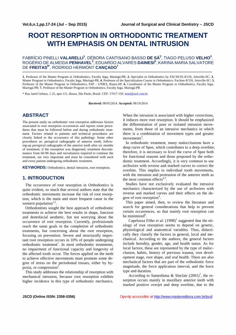
2. CASE REPORTK. F. A, female, aged 10 years and six months old,
attended the clinics complaining about the diastemas. Atextraoral analysis, face balance with convex profile andpresence of passive lip seal was observed (Figure 1).
Figure 1. Initial extraoral photographs.
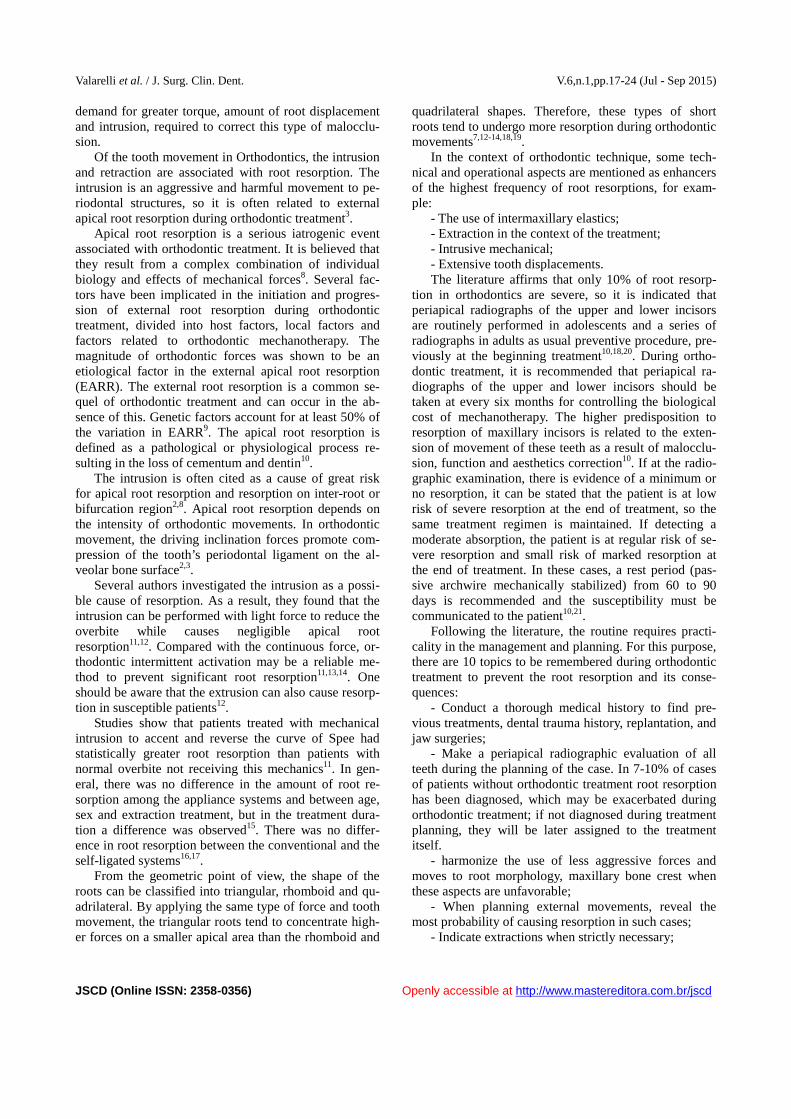
At intraoral and radiograph examination, Class I bi-lateral malocclusion, diastema between the upper frontteeth, overbite of approximately 3-4 mm, slightly flaringof the maxillary central incisors, and presence of somedeciduous teeth were present (Figures 2 to 5).
Figure 2. Initial intraoral photographs.
Valarelli et al. / J. Surg. Clin. Dent. V.6,n.1,pp.17-24 (Jul - Sep 2015)
JSCD (Online ISSN: 2358-0356) Openly accessible at http://www.mastereditora.com.br/jscd
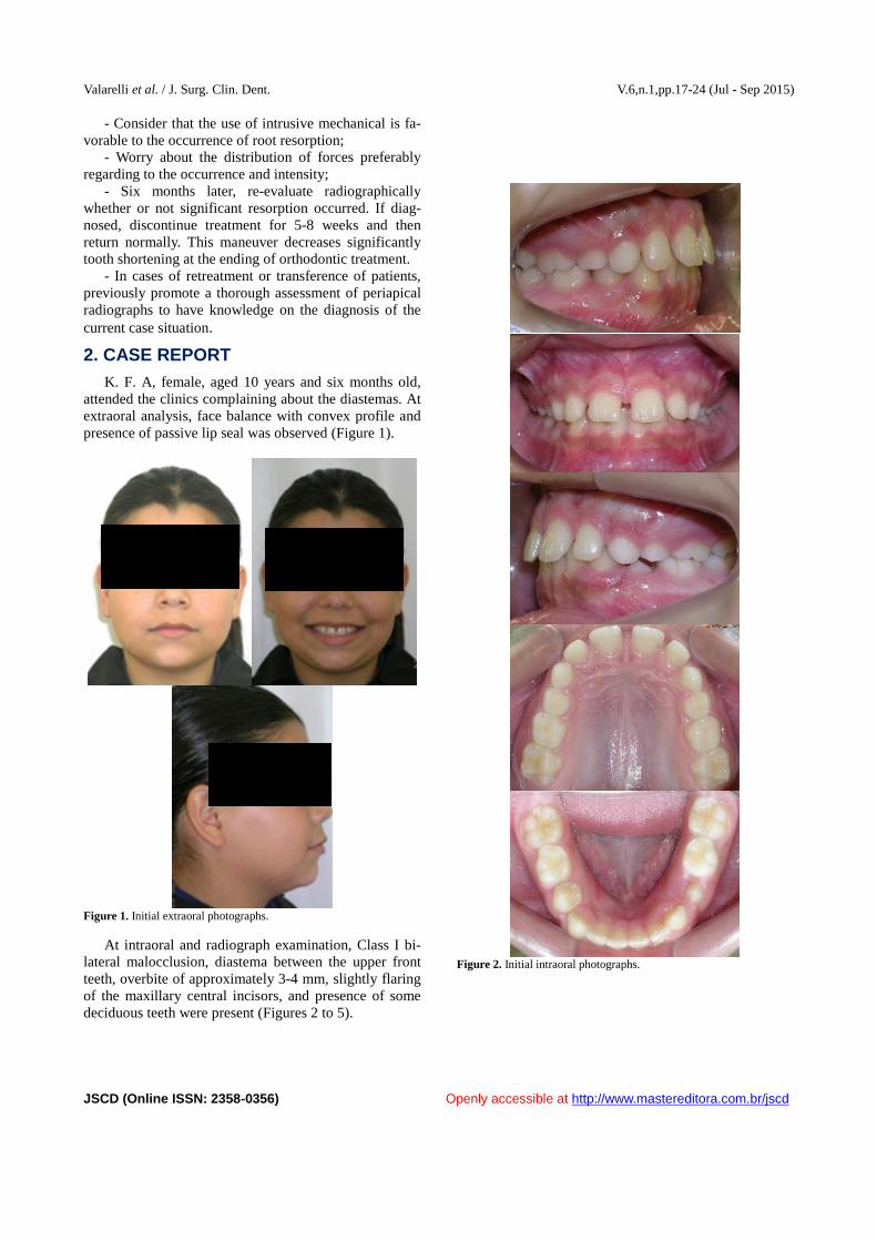
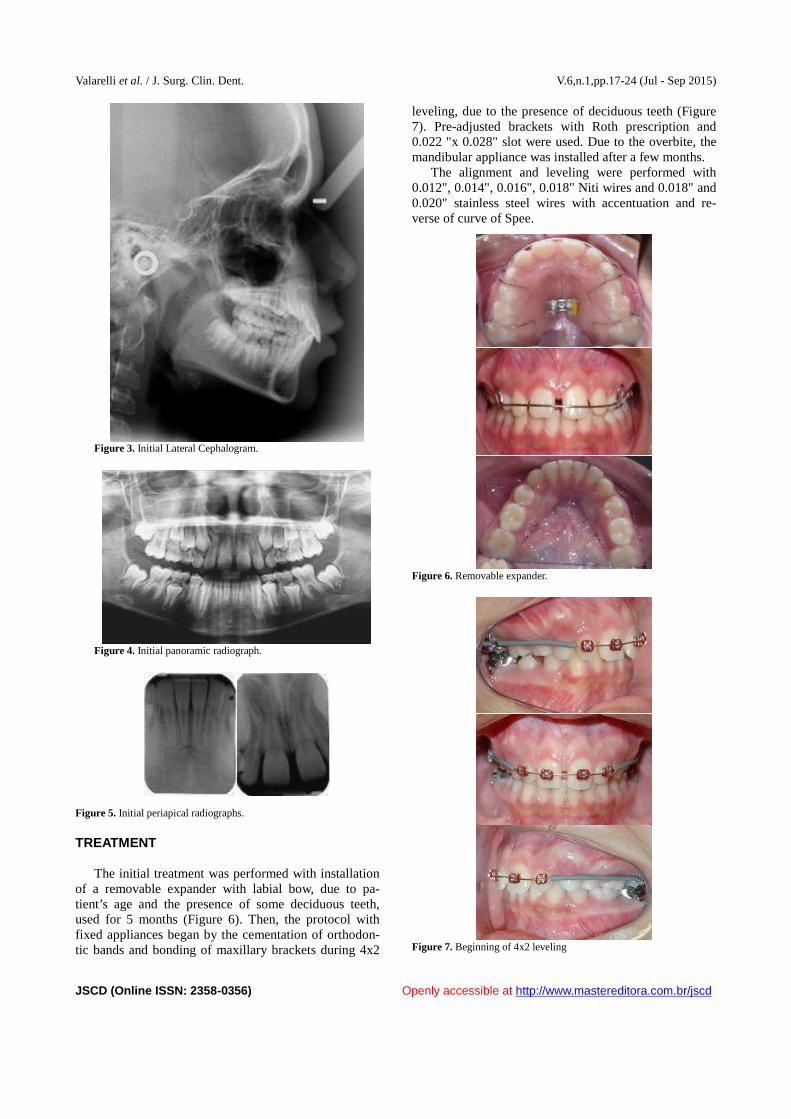
Figure 3. Initial Lateral Cephalogram.
Figure 4. Initial panoramic radiograph.
Figure 5. Initial periapical radiographs.
TREATMENT
The initial treatment was performed with installationof a removable expander with labial bow, due to pa-tient’s age and the presence of some deciduous teeth,used for 5 months (Figure 6). Then, the protocol withfixed appliances began by the cementation of orthodon-tic bands and bonding of maxillary brackets during 4x2
leveling, due to the presence of deciduous teeth (Figure7). Pre-adjusted brackets with Roth prescription and0.022 "x 0.028" slot were used. Due to the overbite, themandibular appliance was installed after a few months.
The alignment and leveling were performed with0.012", 0.014", 0.016", 0.018" Niti wires and 0.018" and0.020" stainless steel wires with accentuation and re-verse of curve of Spee.
Figure 6. Removable expander.
Figure 7. Beginning of 4x2 leveling
Valarelli et al. / J. Surg. Clin. Dent. V.6,n.1,pp.17-24 (Jul - Sep 2015)
JSCD (Online ISSN: 2358-0356) Openly accessible at http://www.mastereditora.com.br/jscd
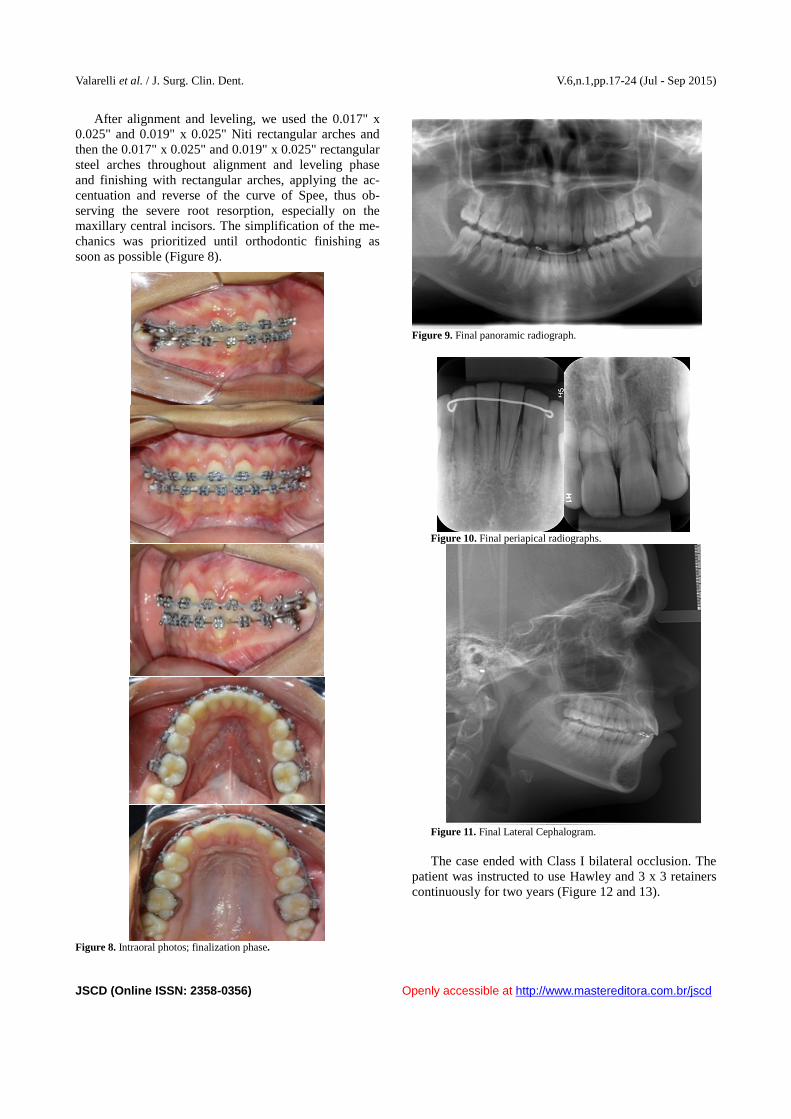
After alignment and leveling, we used the 0.017" x0.025" and 0.019" x 0.025" Niti rectangular arches andthen the 0.017" x 0.025" and 0.019" x 0.025" rectangularsteel arches throughout alignment and leveling phaseand finishing with rectangular arches, applying the ac-centuation and reverse of the curve of Spee, thus ob-serving the severe root resorption, especially on themaxillary central incisors. The simplification of the me-chanics was prioritized until orthodontic finishing assoon as possible (Figure 8).
Figure 8. Intraoral photos; finalization phase.
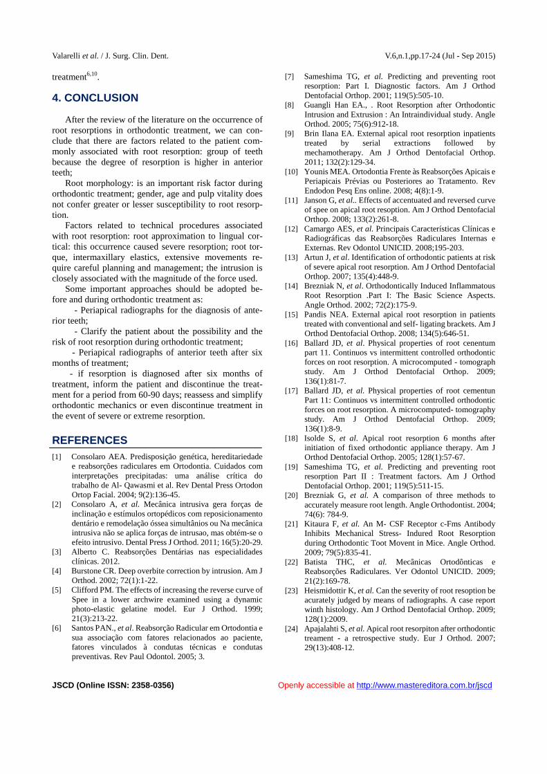
Figure 9. Final panoramic radiograph.
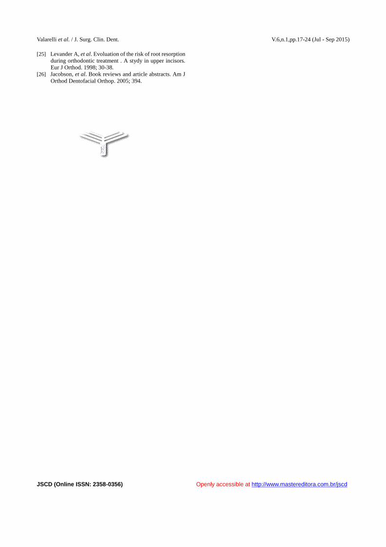
Figure 10. Final periapical radiographs.
Figure 11. Final Lateral Cephalogram.
The case ended with Class I bilateral occlusion. Thepatient was instructed to use Hawley and 3 x 3 retainerscontinuously for two years (Figure 12 and 13).

Valarelli et al. / J. Surg. Clin. Dent. V.6,n.1,pp.17-24 (Jul - Sep 2015)
JSCD (Online ISSN: 2358-0356) Openly accessible at http://www.mastereditora.com.br/jscd
Figure 12. Final photographs.
Figure 13. 3x3 Mandibular retainer and maxillary Hawley retainer.
3. DISCUSSIONThe intrusive mechanics is widely used in the treat-
ment of overbite, because it promotes the intrusion ofanterior teeth. It is known that the intrusive force causesstress mainly on the apex and therefore implies in da-maging this area of the tooth, resulting in apical resorp-
tion3,22.Authors report that the force intensity applied during
the initial period of intrusion can determine the finaldegree of root resorption, as the reaction of the intrudedteeth varies according to the magnitude of the force ex-erted2.
Of the orthodontic movements, the intrusion and roottorque are the most likely causal factors of root resorp-tion, which if combined, further increase the occurrenceof this resorption6,8,23. The studies have recommendedthe application of light forces on the intrusion move-ments that depend on the magnitude of the applied forcefor prevention1,8.
The overbite correction basically involves four typesof tooth movement: anterior intrusion, posterior extru-sion, incisor inclination, and differential growth of themaxillary and mandibular structures. Some types oftreatment are more likely to cause extrusion of posteriorteeth; others to cause intrusion of anterior teeth,4.
The extrusion of the posterior teeth can be obtainedby use of the bite plates, because they prevent the con-tacts of these teeth, allowing the fast development of theposterior dentoalveolar area4,5.
The leveling archwire with reverse and accentuatedcurve is another common approach for correcting deepbite. These archwires provide an intrusion force on ante-rior teeth and extrusion force on posterior teeth, com-bining both teeth movements2,5,11.
The literature reports that root resorption is closelyassociated with certain risk factors that may be related tothe patient and techniques themselves. As regards toOrthodontics, some clinical procedures should beadopted to prevent such resorption3.
As can be seen, a large number of authors stated thatthe highest incidence of root resorption occurs on themaxillary anterior teeth, followed by the mandibularanterior teeth, first molars, canines and premolars7,24.
Concerning to the prevention during orthodontictreatment, the authors suggest a periapical radiographshot of the incisors after 6-9 months of treatment, ascontrol, when the risk of resorption at the end of treat-ment is defined3,10.
A protocol of actions has been recommend to betaken preventively after the detection of some degree ofresorption according to the classification recommendedby Levander and Malmgren at 6-9 months of treatment:instructing the patient's about the susceptibility to thisroot damage, panoramic radiograph to verify the re-maining teeth, and perform the periodic following-up atevery 90 days16,26. The authors do not advocate the restperiod only for teeth with minimal resorption. For othertypes, they recommend mandatory rest from 60 to 90days. For severe resorption, they suggest treatment op-timization to reduce its duration. In extreme resorptioncases, they advocate mandatory discontinuation of
Valarelli et al. / J. Surg. Clin. Dent. V.6,n.1,pp.17-24 (Jul - Sep 2015)
JSCD (Online ISSN: 2358-0356) Openly accessible at http://www.mastereditora.com.br/jscd
treatment6,10.
4. CONCLUSION
After the review of the literature on the occurrence ofroot resorptions in orthodontic treatment, we can con-clude that there are factors related to the patient com-monly associated with root resorption: group of teethbecause the degree of resorption is higher in anteriorteeth;
Root morphology: is an important risk factor duringorthodontic treatment; gender, age and pulp vitality doesnot confer greater or lesser susceptibility to root resorp-tion.
Factors related to technical procedures associatedwith root resorption: root approximation to lingual cor-tical: this occurrence caused severe resorption; root tor-que, intermaxillary elastics, extensive movements re-quire careful planning and management; the intrusion isclosely associated with the magnitude of the force used.
Some important approaches should be adopted be-fore and during orthodontic treatment as:
- Periapical radiographs for the diagnosis of ante-rior teeth;
- Clarify the patient about the possibility and therisk of root resorption during orthodontic treatment;
- Periapical radiographs of anterior teeth after sixmonths of treatment;
- if resorption is diagnosed after six months oftreatment, inform the patient and discontinue the treat-ment for a period from 60-90 days; reassess and simplifyorthodontic mechanics or even discontinue treatment inthe event of severe or extreme resorption.
REFERENCES[1] Consolaro AEA. Predisposição genética, hereditariedade
e reabsorções radiculares em Ortodontia. Cuidados cominterpretações precipitadas: uma análise crítica dotrabalho de Al- Qawasmi et al. Rev Dental Press OrtodonOrtop Facial. 2004; 9(2):136-45.
[2] Consolaro A, et al. Mecânica intrusiva gera forças deinclinação e estímulos ortopédicos com reposicionamentodentário e remodelação óssea simultânios ou Na mecânicaintrusiva não se aplica forças de intrusao, mas obtém-se oefeito intrusivo. Dental Press J Orthod. 2011; 16(5):20-29.
[3] Alberto C. Reabsorções Dentárias nas especialidadesclínicas. 2012.
[4] Burstone CR. Deep overbite correction by intrusion. Am JOrthod. 2002; 72(1):1-22.
[5] Clifford PM. The effects of increasing the reverse curve ofSpee in a lower archwire examined using a dynamicphoto-elastic gelatine model. Eur J Orthod. 1999;21(3):213-22.
[6] Santos PAN., et al. Reabsorção Radicular em Ortodontia esua associação com fatores relacionados ao paciente,fatores vinculados à condutas técnicas e condutaspreventivas. Rev Paul Odontol. 2005; 3.
[7] Sameshima TG, et al. Predicting and preventing rootresorption: Part I. Diagnostic factors. Am J OrthodDentofacial Orthop. 2001; 119(5):505-10.
[8] Guangli Han EA., . Root Resorption after OrthodonticIntrusion and Extrusion : An Intraindividual study. AngleOrthod. 2005; 75(6):912-18.
[9] Brin Ilana EA. External apical root resorption inpatientstreated by serial extractions followed bymechamotherapy. Am J Orthod Dentofacial Orthop.2011; 132(2):129-34.
[10] Younis MEA. Ortodontia Frente às Reabsorções Apicais ePeriapicais Prévias ou Posteriores ao Tratamento. RevEndodon Pesq Ens online. 2008; 4(8):1-9.
[11] Janson G, et al.. Effects of accentuated and reversed curveof spee on apical root resoption. Am J Orthod DentofacialOrthop. 2008; 133(2):261-8.
[12] Camargo AES, et al. Principais Características Clínicas eRadiográficas das Reabsorções Radiculares Internas eExternas. Rev Odontol UNICID. 2008;195-203.
[13] Artun J, et al. Identification of orthodontic patients at riskof severe apical root resorption. Am J Orthod DentofacialOrthop. 2007; 135(4):448-9.
[14] Brezniak N, et al. Orthodontically Induced InflammatousRoot Resorption .Part I: The Basic Science Aspects.Angle Orthod. 2002; 72(2):175-9.
[15] Pandis NEA. External apical root resorption in patientstreated with conventional and self- ligating brackets. Am JOrthod Dentofacial Orthop. 2008; 134(5):646-51.
[16] Ballard JD, et al. Physical properties of root cenentumpart 11. Continuos vs intermittent controlled orthodonticforces on root resorption. A microcomputed - tomographstudy. Am J Orthod Dentofacial Orthop. 2009;136(1):81-7.
[17] Ballard JD, et al. Physical properties of root cementunPart 11: Continuos vs intermittent controlled orthodonticforces on root resorption. A microcomputed- tomographystudy. Am J Orthod Dentofacial Orthop. 2009;136(1):8-9.
[18] Isolde S, et al. Apical root resorption 6 months afterinitiation of fixed orthodontic appliance therapy. Am JOrthod Dentofacial Orthop. 2005; 128(1):57-67.
[19] Sameshima TG, et al. Predicting and preventing rootresorption Part II : Treatment factors. Am J OrthodDentofacial Orthop. 2001; 119(5):511-15.
[20] Brezniak G, et al. A comparison of three methods toaccurately measure root length. Angle Orthodontist. 2004;74(6): 784-9.
[21] Kitaura F, et al. An M- CSF Receptor c-Fms AntibodyInhibits Mechanical Stress- Indured Root Resorptionduring Orthodontic Toot Movent in Mice. Angle Orthod.2009; 79(5):835-41.
[22] Batista THC, et al. Mecânicas Ortodônticas eReabsorções Radiculares. Ver Odontol UNICID. 2009;21(2):169-78.
[23] Heismidottir K, et al. Can the severity of root resoption beacurately judged by means of radiographs. A case reportwinth histology. Am J Orthod Dentofacial Orthop. 2009;128(1):2009.
[24] Apajalahti S, et al. Apical root resorpiton after orthodontictreament - a retrospective study. Eur J Orthod. 2007;29(13):408-12.
Valarelli et al. / J. Surg. Clin. Dent. V.6,n.1,pp.17-24 (Jul - Sep 2015)
JSCD (Online ISSN: 2358-0356) Openly accessible at http://www.mastereditora.com.br/jscd
[25] Levander A, et al. Evoluation of the risk of root resorptionduring orthodontic treatment . A stydy in upper incisors.Eur J Orthod. 1998; 30-38.
[26] Jacobson, et al. Book reviews and article abstracts. Am JOrthod Dentofacial Orthop. 2005; 394.