2010-4 root resorption and orthodontics - aspedia resorption and orthodontics care ... you may wish...
TRANSCRIPT
Creating Brighter Futures
University of Sydney
Root Resorption and Orthodontics

COLGATE IS THE PREFERRED BRAND OF THE ASO NSW
Root Resorption and Orthodontics
CARE COLUMNScienti� c Affairs Manager, Colgate, South Paci� c. Dr Sue Cartwright
Colgate’s support and involvement in professional dental education is crucial. Without it newsletters such as this, many society meetings and activities, university programmes and, indeed, whole hospital and university departments would not exist or function effectively. The Scienti� c Affairs Manager is therefore one of the most important members of the Colgate team for it is they who map out, organise and supervise Colgate’s involvement.
Recently, Dr Barbara Shearer has moved on to take on an even more challenging position internationally with Colgate. Her replacement as the new Scienti� c Affairs Manager, based in Sydney, is Dr Sue Cartwright.
Prior to taking on this position Sue was the Head of the Oral Health Department at AUT University in Auckland, New Zealand. This department runs a Bachelor of Health Science in Oral Health which is equivalent to BOH programmes in Australia. She is a general dentist with 25 years clinical experience and has spent nearly eight years in a teaching role as well. She has completed a Diploma in Clinical Dentistry in Periodontology, and this year completes her Masters thesis in Education.
On behalf of the dental profession the editors of this newsletter welcome Sue to Australia and her new role, and thank Colgate for their continued and generous support of dental education throughout Australia, and beyond.
To contact Dr Sue Cartwright you can email: [email protected] or phone: (02) 9229 5737
Root resorption has long been recognised in dentistry, and especially in orthodontics. It is a process by which cementum, and often dentine, is removed from the root of a tooth by clastic cell activity. Root resorption may be associated with:
1. Physiologic tooth movement,
2. Exfoliation of primary teeth,
3. Tooth impaction,
4. Periapical or periodontal infl ammation,
5. Tooth implantation or replantation,
6. Continuous occlusal trauma,
7. Tumours or cysts,
8. Metabolic or systemic disturbances,
9. Local functional or behavioural problems,
10. Orthodontic treatment,
11. Idiopathic factors.
Orthodontically induced root resorption is commonly seen on dental radiographs as the permanent shortening or blunting of the root. Although it occurs in individuals who have never experienced orthodontic tooth movement,1, 2 the incidence among orthodontically treated individuals is relatively high. The reported rates vary according to whether the assessment is radiographic or histologic.
Incidence
Post-orthodontic panoramic or periapical radiographic studies report that apical root resorption is usually less than 2.5mm3-5 and varying from 6% to 13% for different teeth.6 However histological studies of animal and human root surfaces of orthodontically moved teeth report a greater than 90% incidence of infl ammatory root resorption.7, 8 The results of the histological surveys suggest that a small amount of apical root resorption may be a normal physiological process, perhaps akin to continuous bone remodelling.
Following orthodontic treatment the loss of root structure, in most cases, is minimal and not clinically signifi cant and can be classifi ed as minor or moderate. Severe resorption, defi ned as exceeding 4mm or a third of the original root length, is seen in approximately 1% to 5% of teeth. 9-11
Figure 1a Severe maxillary incisor resorption identifi ed during orthodontic treatment on a reconstructed OPG from a cone beam CT
Figure 1b Incisor roots being resorbed by the erupting ectopic canine.
Orthodontically induced root resorption can be localised to a particular tooth or a generalised shortening of all or most of the teeth. The most commonly involved teeth are the maxillary incisors.12 A higher incidence is also noted for cases where there has been an increased amount of root movement and where roots were very long and narrow prior to treatment.
Endodontically treated teeth
The susceptibility of non-vital and endodontically treated teeth to root resorption during orthodontic treatment is still controversial. Although some studies show an increased incidence of root resorption,13 the results may be skewed, as many of the teeth involved also have a history of trauma. A number of other studies 14,15 have indicated no difference.
How can orthodontic tooth movement cause root resorption?
The biological process of tooth movement appears to be implicated in the process of root resorption. During orthodontic treatment, the periodontal ligament (PDL) is subjected to mechanical forces from the orthodontic appliances. On one side of the tooth the alveolar bone and the PDL are compressed, while on the opposite side the PDL is stretched. The stress of the stretched PDL induces apposition of bone, while compression produces bone resorption and remodelling of the periodontal ligament. This alters the PDL’s vascularity
Dr Sue Cartwright
Creating Brighter FuturesYOU MAY WISH TO SHARE THIS ISSUE OF BRIGHTER FUTURES WITH YOUR HYGIENISTS AND OTHER STAFF MEMBERS
BR
IG
HTE
R FUTURES
2010-4and blood flow resulting in local synthesis and release of various key molecules such as neurotransmitters, cytokines, growth factors, colony stimulating factors and arachidonic acid metabolites necessary for bone remodelling and tooth movement.
Hyalinisation (also termed “sterile necrosis”) is common within the periodontal ligament after a force is placed on a tooth. It usually occurs after a few days, and may remain within the PDL for up to four to eight weeks after initiation of the compressive load.16 During this time, resorption of the alveolar socket is prevented, and undermining resorption of the alveolar bone may occur. In addition, root resorption near the areas of hyalinization will occur. The hyaline is removed from the periodontal ligament by macrophages, with concomitant resorption of the alveolar socket wall facilitating tooth movement.
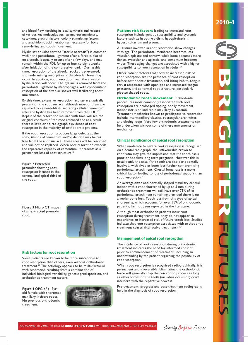
By this time, extensive resorption lacunae are typically present on the root surface, although most of them are repaired by cementoblasts secreting cellular cementum after the hyaline has been removed from the PDL.17 Repair of the resorption lacunae with time will see the original contours of the root restored and as a result there is little or no radiographic evidence of root resorption in the majority of orthodontic patients.
If the root resorption produces large defects at the apex, islands of cementum and/or dentine may be cut free from the root surface. These areas will be resorbed and will not be replaced. When root resorption exceeds the reparative capacity of cementum, it presents as a permanent loss of root structure.18
Figure 2 Extracted premolar showing root resorption lacunae in the coronal and apical third of the root.
Figure 3 Micro CT image of an extracted premolar root.
Risk factors for root resorption
Some patients are known to be more susceptible to root resorption than others, even without orthodontic treatment.19 The aetiology appears to be multi-factorial with resorption resulting from a combination of individual biological variability, genetic predisposition, and orthodontic treatment factors.
Figure 4 OPG of a 13yr old female with shortened maxillary incisors roots. No previous orthodontic treatment.
Patient risk factors leading to increased root resorption include genetic susceptibility and systemic factors such as hypothyroidism, hypopituitarism, hyperpituitarism and trauma.
All tissues involved in root resorption show changes with age. The periodontal membrane becomes less vascular, aplastic and narrow, while bone becomes more dense, avascular and aplastic, and cementum becomes wider. These aging changes are associated with a higher susceptibility to root resorption in adults.20
Other patient factors that show an increased risk of root resorption are the presence of root resorption before orthodontic treatment, nail-biting habits, tongue thrust associated with open bite and increased tongue pressure, and abnormal root structure, particularly pipette shaped roots.
Orthodontic tooth movement. Orthodontic procedures most commonly associated with root resorption are prolonged tipping, bodily movement, incisor intrusion and rapid maxillary expansion. Treatment mechanics known to predispose to resorption include intermaxillary elastics, rectangular arch wires and closing loops. Very few orthodontic treatments can be undertaken without some of these movements or mechanics.
Clinical significance of apical root resorption
When moderate to severe root resorption is recognised on a dental radiograph, the unfavourable crown to root ratio may give the impression that the tooth has a poor or hopeless long term prognosis. However this is usually only the case if the teeth are also periodontally involved, with alveolar bone loss further compromising periodontal attachment. Crestal bone loss is a more critical factor leading to loss of periodontal support than root resorption.21
An average-sized and normally shaped maxillary central incisor with a root shortened by up to 5 mm during orthodontic treatment will still have over 75% of its periodontal attachment remaining provided there is no alveolar bone loss. Tooth loss from this type of apical shortening, which accounts for over 95% of orthodontic patients, has not been reported in the literature.
Although most orthodontic patients incur root resorption during treatment, they do not appear to experience an increased risk of future tooth loss. Studies indicate that root resorption associated with orthodontic treatment ceases after active treatment.22-24
Management of apical root resorption
The incidence of root resorption during orthodontic treatment indicates the need for informed consent prior to commencement of treatment, including an understanding by the patient regarding the possibility of root resorption.
When root resorption is recognised radiographically, it is permanent and irreversible. Eliminating the orthodontic force will generally stop the resorption process as long as other forces on the teeth (including occlusion) don’t interfere with the reparative process.
Pre-treatment, progress and post-treatment radiographs help in the diagnosis of root resorption.
Brighter Futures is published by the Australian Society of Orthodontists (NSW Branch) Inc. in conjunction with the Orthodontic Discipline at the University of Sydney.
The newsletter is intended to help keep the dental profession updated about contemporary orthodontics, and also to help foster co-operation within the dental team.
Without the generous support of Henry Schein Halas and Colgate, who are an integral part of the dental team, this publication would not be possible.
The statements made and opinions expressed in this publication are those of the authors and are not official policy of, and do not imply endorsement by, the ASO (NSW Branch) Inc or the Sponsors.
Correspondence is welcome and should be sent to:
Department of OrthodonticsUniversity of SydneySydney Dental Hospital2 Chalmers Street, Surry Hills NSW 2010
AUTHOR & EDITORS
Dr Daniel TanPRINCIPAL AUTHOR
Prof M Ali DarendelilerDr Dan VickersDr Michael DineenDr Ross AdamsDr Sarah Raphael
www.aso.org.au Your Dental One Stop Shop!
BRIGHTER FUTURES
Where an increased susceptibility to root resorption is suspected, progress radiographs approximately 6 to 12 months after the start of treatment may be indicated. If root resorption is identified, active treatment can be paused or stopped to allow for repair of the root resorption lacunae. There is some evidence that a 2 to 3 month treatment pause decreases further root resorption.25
If treatment is to be continued, then radiographic monitoring on a regular basis is advisable. It may be necessary to terminate treatment prior to achieving the original treatment objectives and then reassess the treatment plan with the patient. Alternative treatment options might include prosthetic solutions to close spaces, releasing teeth from active arch wires, interproximal reduction of enamel instead of extracting teeth to gain space, and early fixation of teeth showing signs of resorption.
In cases of moderate to severe root resorption, no studies have recommended the splinting of the teeth. However, the presence of parafunctional habits or tooth mobility could indicate a permanent lingual splint.26
The use of pharmaceutical or ultrasound agents to stop the process is still controversial. Most of the pharmaceutical agents target odontoclast and osteoclast function. This can also affect the rate of tooth movement and therefore the duration of orthodontic treatment.
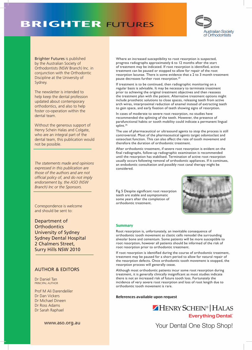
After orthodontic treatment, if severe root resorption is evident on the final radiographs, follow-up radiographic examination is recommended until the resorption has stabilised. Termination of active root resorption usually occurs following removal of orthodontic appliances. If it continues, an endodontic consultation and possibly root canal therapy might be considered.
Fig 5 Despite significant root resorption teeth are stable and asymptomatic some years after the completion of orthodontic treatment.
Summary
Root resorption is, unfortunately, an inevitable consequence of orthodontic tooth movement as clastic cells remodel the surrounding alveolar bone and cementum. Some patients will be more susceptible to root resorption, however all patients should be informed of the risk of root resorption prior to orthodontic treatment.
If root resorption is identified during the course of orthodontic treatment, treatment may be paused for a short period to allow for natural repair of the resorption defects. Once orthodontic tooth movement is stopped, the resorption process will generally cease.
Although most orthodontic patients incur some root resorption during treatment, it is generally clinically insignificant as most studies indicate there is not an increased risk of future tooth loss. Fortunately the incidence of very severe root resorption and loss of root length due to orthodontic tooth movement is rare.
References available upon request