vol. published december 2 dis. aquat. org
TRANSCRIPT
Vol. 5: 163-169, 1988 DISEASES OF AQUATIC ORGANISMS Dis. aquat. Org.
Published December 2
Paramoeba pemaquidensis (Sarcomastigophora: Paramoebidae) infestation of the gills of coho
salmon Oncorhynchus kisutch reared in sea water
Michael L. ~ent'l*, T. K. Sawyer2, R. P. ~ e d r i c k ~
'Battelle Marine Research Laboratory, 439 West Sequim Bay Rd, Sequim, Washington 98382, USA '~escon Associates, Inc., Box 206, Turtle Cove, Royal Oak, Maryland 21662, USA
3~epar tment of Medicine, School of Veterinary Medicine, University of California, Davis, California 95616, USA
ABSTRACT: Gill disease associated with Paramoeba pemaquidensis Page 1970 (Sarcomastigophora: Paramoebidae) infestations was observed in coho salmon Oncorhynchus lasutch reared in sea water Fish reared in net pens in Washington and in land-based tanks in California were affected. Approxi- mately 25 O/O mortality was observed in the net pens in 1985, and the disease recurred in 1986 and 1987. Amoeba infesting the gill surfaces elicited prominent epithelia1 hyperplasia. Typical of Paramoeba spp., the parasite had a Feulgen positive parasome (Nebenkorper) adjacent to the nucleus and floatlng and transitional forms had digitiform pseudopodia. We have established cultures of the organism from coho gills; it grows rapidly on Malt-yeast extract sea water medium supplemented with Klebsiella bacteria. Ultrastructural characteristics and nuclear, parasome and overall size of the organism in study indicated it is most closely related to the free-living paramoeba P. pemaquidensis. The plasmalemma of the amoeba from coho gills has surface filaments. Measurements (in pm) of the amoeba under various conditions are as follows: transitional forms directly from gills 28 (24 to 30), locomotive forms from liquid culture 21 X 17 (15 to 35 X 11 to 25), and locomotive forms from agar culture 25 X 20 (15 to 38 X 15 to 25). Nuclei and parasomes from liquid cultures were 5 to 7 and 3 to 5 X 2 to 3, respectively. The disease was not induced by laboratory exposure of coho salmon to amoeba cultures, but the amoeba was consistently isolated from exposed fish and aquarium detritus for 4 wk after exposure. This suggests that the organism is an opportunistic pathogen which only proliferates on fish gills and causes disease under certain situations yet to be clearly defined. In vitro studies indicated that the optimal salinity for growth of the amoeba was between 15 and 20 ppt, while growth was greatly dminished below 10 ppt. This concurs with field observations where the amoeba was eradicated from fish held in sea water tanks following a reduction in salinity.
INTRODUCTION
Commercial rear ing of salmonid fishes in sea wate r is a n important a n d growing industry in several locations throughout the world. Research on diseases of sal- monids h a s emphasized those occurring during their freshwater development , bu t with t h e increase i n s e a water aquacul ture several n e w protozoan pa thogens of salmonids reared in sea water have recently b e e n iden- tified (Hoffman 1984, Harrell & Scott 1985, Harrell e t al. 1985, Elston e t al. 1987, M u n d a y e t al. 1988).
Present address: Department of Fishenes and Oceans, Pacific Biological Station, Nanaimo. B.C., Canada V9R 5K6
O Inter-Research/Printed in F. R. Germany
Amoebic gill disease h a s affected several fishes rea red i n freshwater (Kubota & Kamata 1971, Sawyer e t al. 1974, 1975, Salas 1981, Daoust & Ferguson 1985, Nash e t al. 1988), b u t few cases h a v e b e e n reported i n marine fishes. Chat ton (1909, 1910) reported gill dis- e a s e associated with a vahlkampfid a m o e b a i n Sym- p h o d u s melops a n d S. tinca (Labridae) a n d proposed that this w a s a temporary infection b y a n otherwise free-living organism. Recently, M u n d a y e t al. (1988) described severe gill disease associated with a Para - moeba-like organism in seawater-reared Atlantic sal- m o n Sa lmo sa la r a n d rainbow trout Salmo g a i r d n e n i n Tasmania. W e have s e e n similar gill infestations in coho salmon Oncorhynchus kisutch reared in s e a wate r a n d described here a r e t h e n~orphology , culture

164 Dis. aquat. Org. 5: 163-169, 1988
characteristics, infectivity, and associated pathology of Paramoeba pemaquidensis from epizootics in Washington and California. USA.
MATERIALS AND METHODS
Infested coho salmon Oncorhynchus kisutch from a net pen facility in Washington state were examined several times in 1985 to 1987 and coho reared in land- based seawater tanks in California were examined in April through June 1987. Wet mount preparations were examined by Nomarsh phase interference microscopy. Gills and visceral organs were fixed in Davidson's solution (Humason 1979), processed for histological examination and stained with either hematoxylin and eosin or Feulgen stain for DNA. Gill tissue for electron microscopy was fixed and processed as described by Harrell et al. (1986). Amoebae from agar cultures were prepared for electron microscopy by fixing amoebae in 1 : 1 5 O/O glutaraldehyde with 0.1 M cacodylate buffer and filtered sea water at 30 ppt.
In vitro culture. Amoebae from the Washington epizootic in 1987 were initially established in culture by placing gill tissue from infested coho salmon in 75 cm2 plastic tissue culture flasks with ca 25 m1 culture medium. The medium consisted of Medium 199 (Sigma, St. Louis, MO., USA), filtered sea water at 25 ppt, 10 % fetal calf serum, 5 % chicken serum, and 100 ~t.g ml-' of the following antimicrobial compounds: neomycin, kanamycin, novobiocin, penicillin, strep- tomycin and nystatin.
Agar cultures were established from liquid cultures w l c h had been passed ca 8 times. Malt-yeast-sea water (MYS) agar was prepared as described by Page (1983) and the surface of the agar was covered with a thin smear of Klebsiella bacteria prior to incubation. Cultures were maintamed at 15 "C unless otherwise indicated. This medium was routinely used for main- taining amoebae, and MYS agar was also prepared with sea water diluted to 5, 10, 15 and 20 ppt to investigate growth at reduced salinities.
Amoebae did not grow on MYS medium made at a salinity of 5 ppt and growth of the amoebae at 6 to 30 ppt was quantitated with the following method. Amoebae were transferred to Lab-Tek@ Flaskettese7 (Miles Scientific, Naperville, Ill., USA) in 6 m1 sea water adjusted to the appropriate salinity. Initial concen- tration of the amoeba was 1.3 X 10³ ml-l. Amoebae were counted after 96 h with the aid of an inverted compound microscope. Klebsiella bacteria at 1 X 10⁶ ml-' were provided as food for the amoebae in each flaskette. Growth at temperatures from 5 to 20 "C was investigated using the same system, and salinities in these cultures were maintained at 25 ppt.
Transmission. To determine the infectivity of the Paramoeba, coho salmon (mean wt 15 g) were exposed to amoebae from agar cultures which had been passed 12 to 15 times. To ensure that they had not been previously exposed to the amoeba, the fish were obtained from a freshwater hatchery and acclimated to sea water in 200 1 aquaria for 2 d prior to exposure at the Battelle Marine Research Laboratory, Sequim, Washington. The fish were maintained in open sys- tems, and the affluent water was sand-filtered. Water temperature was 9 to l l "C and salinity 30 ppt through- out the experiment.
Amoebae were harvested from the agar cultures by washing with sterile sea water. Twenty fish were exposed to 300 amoebae ml-' in a 20 l static bath for 45 min. The affluent water was then turned on to fill the tank, and the flow maintained at 2 l min-l. Fish were fed daily with a commercial pellet diet and detritus was allowed to accumulate at the aquarium bottom. Control fish were treated similarly, except they were not exposed to amoeba cultures.
Five fish samples were examined at 7, 14,23 and 29 d post exposure (PI). Gills from all fish were examined in wet mounts and histological sections. Gill tissue and detritus from the aquaria were cultured for amoebae on MYS agar as described above at 14 , 23 and 29 days PI.
RESULTS
Pathology and treatment
Significant mortalities at the Washington site were associated with the amoeba in the fall of 1985. Epizoo- tics occurred again in fall 1986 and 1987, but mor- talities were fewer and usually only small, emaciated fish were heavily infested. Moribund fish were lethar- gic and often accumulated at the surface or corners of the pens. The water temperature at this site typically ranges from 9 to 12 'C during the fall. In contrast, the disease was first noted in the spring of 1987 at the California site, where the water temperature in the tanks was ca 13 "C.
Histological changes in the gills were consistent at both sites and severity of lesions were in concurrence with density of amoebae. The gills exhibited prominent epithelia1 hyperplasia which often resulted in complete fusion of the secondary lamellae (Fig. 1). Fusion of the distal portion of secondary lamellae resulted in the formation of large interlammelar vesicles. Amoebae were usually confined to the g111 surface and rarely penetrated through the epithelium. No histopathologi- cal changes In visceral organs were associated with amoeba infestations. Some fish at both locations were also infected with a microsporidium, presumably Loma
Kent et al.: Paramoeba from salmon gills 165
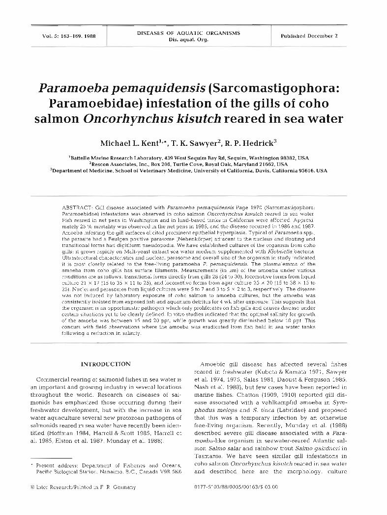
Figs 1 to 4 . Paramoeba pemaqludensis from the gills of coho salmon Oncorhynchus kisutch reared in sea water. m Epithehal hyperplasia, fusion of the secondary lamellae and interlamellar vesicles (V) associated with amoebae (arrows) infestation. H & E. Bar = 50 pm. Transitional form with digitiform pseudopodia from coho gill. Nomarski's phase contrast. Bar = 25 pm. G Locomotive form of Paramoeba from liquid culture. N: nucleus; P: parasome. Normarski's phase contrast. Bar = 25 pm. Fig. 4.
Paramoeba from Liquid culture. N: nucleus; P: parasome. Feulgen stain. Bar = 25 pm
salmonae. In contrast to epithelia1 hyperplasia associ- Amoeba morphology ated with the amoeba, the microsporidium formed xenomas and induced interstitial inflammation and Fresh wet mounts of the gills from fish at both loca- vasculitis when the xenomas were ruptured and spores tion revealed numerous transitional forms with digitate were dispersed throughout primary lamellae. Gill pseudopodia (Fig. 2 ) . A prominent parasome and ves- monogeneans Lamlniscus strelkowi were observed on icular nucleus was observed in amoebae which were some affected fish in Washington in 1986 and 1987. allowed to adhere to glass (locomotive form) (Fig. 3).

Kent et al.: Paramoeba from sallnon gills 167
Gill sections and imprints also revealed a Feulgen positive parasome adjacent to the nucleus (Fig. 4). Most amoebae had only 1 parasome but occasionally 2 were observed. Measurements (in pm) of 20 amoebae from wet mounts of infested gills, agar and liquid cultures are as follows: transitional forms from gills 28 (24 to 301, locomotive forms from liquid culture 21 X 17 (15 to 35 X 11 to 25), and locomotive forms from agar culture 25 X 20 (15 to 38 X 15 to 25). Nuclei and parasomes were measured from liquid cultures; nucleus 5 to 7, parasome 3 to 5 x 2 to 3.
Electron microscopy of amoebae on gill tissue and from agar culture (Fig. 5) revealed a parasome mor- phologically similar to those described previously (Per- kins & Castagna 1971, Cann & Page 1982, Jones 1985). Adherent to the plasmalemma in amoeba from gills were numerous filaments which extended ca 350 nm from the surface (Fig. 6). These filaments were not detected in amoebae from agar cultures.
In vitro growth characteristics
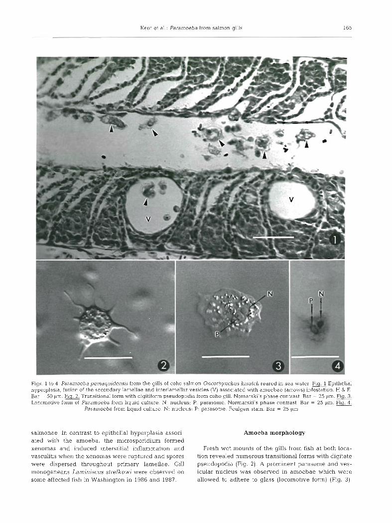
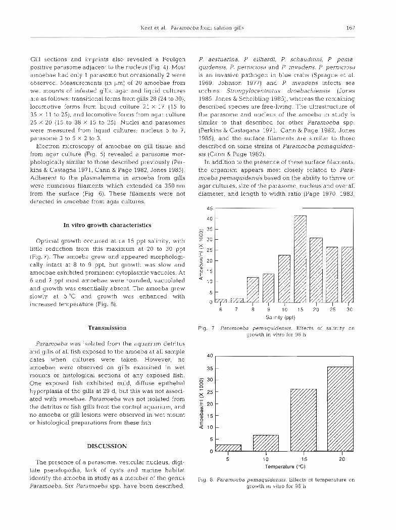
Optimal growth occurred at ca 15 ppt salinity, with little reduction from this maximum at 20 to 30 ppt (Fig.?). The amoeba grew and appeared morphologi- cally intact at 8 to 9 ppt, but growth was slow and amoebae exhibited prominent cytoplasmic vacuoles. At 6 and 7 ppt most amoebae were rounded, vacuolated and growth was essentially absent. The amoeba grew slowly at 5 ° C and growth was enhanced with increased temperature (Fig. 8).
Transmission
Paramoeba was isolated from the aquarium detritus and gills of all fish exposed to the amoeba at all sample dates when cultures were taken. However, no amoebae were observed on gills examined in wet mounts or histological sections of any exposed fish. One exposed fish exhibited mild, diffuse epithelia1 hyperplasia of the gills at 29 d, but this was not associ- ated with amoebae. Paramoeba was not isolated from the detritus or fish gills from the control aquarium, and no amoeba or gill lesions were observed in wet mount or histological preparations from these fish.
P. aestuarina, P. e~lhardi, P. schaudinni, P. pema- quidensis, P, perniciosa and P. invadens. P. perniciosa is an invasive pathogen in blue crabs (Sprague et al. 1969, Johnson 1977) and P. invadens infects sea urchins Strongylocentrotus droebachiensis (Jones 1985, Jones & Scheibling 1985), whereas the remaining described species are free-living. The ultrastructure of the parasome and nucleus of the amoeba in study is similar to that described for other Paramoeba spp. (Perkins & Castagana 197 1, Cann & Page 1982, Jones 1985), and the surface filaments are similar to those described on some strains of Paramoeba pernaquiden- sis (Cann & Page 1982).
In addition to the presence of these surface filaments, the organism appears most closely related to Para- moeba pemaquidensis based on the ability to thrive on agar cultures, size of the parasome, nucleus and overall diameter, and length to width ratio (Page 1970, 1983,
Salinity (ppt)
Fig. 7 Paramoeba pernaquidens~s Effects of salin~ty on growth in vitro for 96 h
DISCUSSION
The presence of a parasome, vesicular nucleus, digi- 5 10 15 20
Temperature ("C) tate pseudopodia, lack of cysts and marine habitat identify the amoeba in study as a member of the genus Fig. 8. Paramoeba pemaquidensis. Effects of temperature on Paramoeba. Six Paramoeba spp. have been described; growth in vitro for 96 h
168 Dis. aquat. Org. 5: 163-169, 1988
Bovee & Sawyer 1979, Cann SL Page 1982). Nucleus, parasome and overall diameters of the strain in study are smaller than those of P. eilhardi and larger than those of P. aestuarina and P. perniciosa. Furthermore, the latter is an obligate parasite of the blue crab Callinectes sapidus and has not been maintained in vitro.
Paramoeba invadens is similar in size to our isolate, but lacks plasmalemmal hairs (Jones 1985). P. invadens also is quite similar in size and culture characteristics to other strains of P. pemaquidensis and the taxonomic distinction of these 2 species is unclear. Furthermore, the presence or absence of plasmalemmal surface fila- ments is not a sufficient criterion alone to differentiate these species because some strains of P. pemaquiden- sis lack these hairs and their presence may depend on culture characteristics. They were observed on some amoebae directly associated with gills (Fig. 6), whereas they were not detected on amoebae from agar cultures. Paramoeba spp. vary greatly in size dependent on culture conditions and density of infection (Sawyer 1969, Cann & Page 1982) and further studies beyond morphological comparisons, such as isoenzyme analy- sis, are needed to clarify the taxonomic relationships of Paramoeba spp.
Along with the preliminary report by Munday et al. (1988), this is the first report of Paramoeba pema- quidensis from the Pacific Ocean and the first report of a Paramoeba infestation in a vertebrate. Amoebae have been associated with fish reared in freshwater, and the pathological response to infestations is typically hyper- plasia of the branchial epithelium (Sawyer et al. 1975, Daoust & Ferguson 1985). The only reports of amoebic gill infestations in marine fishes that we are aware of are those by Chatton (1909, 1910) in Symphodus spp. (Labndae) and a Paramoeba-like amoeba from net pen-reared salmonids in Australia (Munday et al. 1988). We have examined histological material from infested salmon from Australia and the amoeba and associated disease appears identical to those of salmon in Washington and California.
The amoeba of our study exhibited poor growth in vitro at salinities less than 10 ppt (Fig. 7). This concurs with in vitro observations; the amoeba and associated disease was eradicated from fish held in seawater tanks in California following a brief reduction in salinity, and Munday et al. (1988) reported that the Australian amoeba is eradicated by moving infested fish in pens to a n area with a heavy freshwater influx. However, the precise salinity reduction needed to eradicate the infes- tation is yet to be determined.
Neither severe epithelia1 hyperplasia of the gills nor mortality was induced in the 1.aboratory exposure study, which indicate that the amoeba was not pathogenic under the experimental conditions
employed. The amoeba is likely an opportunistic pathogen which proliferates on fish gills and causes disease only under ceretain conditions which are yet to be precisely defined. This is similar to bacterial gill disease of freshwater salmonids. In this disease, certain bacterial epiphytes (Flavobacterium or Cytophaga spp.) are associated with gill hyperplasia, but the dis- ease is difficult to reproduce with bacteria isolated from affected fish (Wakabayashi et al. 1980, Snieszko 1981). Based on our field observations, the water temperature during the exposure experiment appears to be at the low range for the occurrence of the disease, and in vitro growth of the amoeba is enhanced at higher tem- peratures (Fig. 8). Furthermore, Munday et al. (1988) reported most epizootics in Australia at temperatures greater than 17 "C. Also, it is possible that the amoeba attain 'bloom' populations when the water contains abundant food organisms that favor their growth. Repeated passage of the amoeba on artificial media prior to exposure to fish may have also diminished its virulence.
Another factor which may explain the lack of disease in experimentally exposed fish is that massive prolifer- ation of the amoeba may only occur on gills with pre- existing lesions, induced by such agents as other para- sites, chemical or mechanical injury. Goldes et al. (1988) reported moderate to severe gill infestations by the ectoparasitic flagellate Ichtyobodo necator in rain- bow trout exposed to high levels of inert suspended solids in the form of clay kaolin, whereas fish kept in otherwise identical conditions were not infested. Many coho salmon with prominent Paramoeba infestations in our study were also infected with a microsporidium (Loma salmonae) or a monogenean (Laminiscus strel- kow~]. However, the amoeba was the only pathogen detected in other salmon w t h severe epithelial hyper- plasia from the same epizootics.
In agreement with B. L. Munday, Tasmania Institute of Technology, Tasmania (pers. comm.), we feel that the amoeba can be a primary pathogen and cause severe disease in seawater-reared salmon under cer- tain conditions. In addition to clarification of taxonomic relationships of the amoeba from Australia, our isolate, and other strains of Paramoeba pemaquidensjs and P. invadens, further studies are needed to elucidate host-parasite relationships of the disease (e.g determi- nation of more precise conditions under which the amoeba proliferates on gills and causes disease)
Acknowledgements. This work was supported by Battelle Memorial Institute, Cor~ora te Technical Develo~rnent Proiect Number B-0322-4051, colurnbus, Ohio, and ~o rnsea ~ a r k s , Inc. We thank Drs R. A. Elston and J. Groff for helpful sugges- tions, M. T Wilkinson for histological preparations and R. Munn for electron microscopy preparations.

Kent et al.. Paramoeba from salmon gills 169
LlTERATURE CITED
Bovee, E. C.. Sawyer, T K. (1979). Marine flora and fauna of the Northeastern United States. Protozoa: Sarcodina: Amoebidae. NOAA Tech. Rep. Natl. Mar. Fish. Serv. Cir- cular 419, p. 1-56
Cann, J P., Page, F. C. (1982). Fine structure of small free- hving Paran~oeba (Amoebida) and taxonomy of the genus. J mar biol. Ass. U. K. 62: 25-43
Chatton, E . (1909). Une amibe, Amoeba mucicola. n. sp . , parasite des branchies des labres, associee a une tnchodine. C. r. Seanc. Soc. Biol 67: 690-692
Chatton, E. (1910). Protozoaires parasites des branchies des labres, Ameoba mucicola, Chatton, Trichodina labrorum n. sp. Archs. Zool. exp. gen. 5: 239-266
Daoust, P.-Y., Ferguson, H. W. (1985). Nodular gill disease: a unique form of proliferative gill disease in rainbow trout. Salmo gairdnen hchardson. J. Fish Dis. 8: 511-522
Elston, R. A., Kent, M. L., Harrell, L. H. (1987). An intranuclear micorsporidi.um associated with acute anemia in the chinook salmon, Oncorhynchus tshawytscha. J . Protozool. 34: 274-277
Goldes, S. A. , Ferguson, H. W. , Moccia, R. D., Daoust, P.-Y. (1988). Histological effects of the inert suspended clay kaolin on the gills of juvenile rainbow trout, Saln~o gajrd- nerj hchardson. J. Fish Dis. 11: 23-33
Harrell, L. W., Scott, T M. (1985), Kudoa thyrsitis (Gilchirst) (Myxosporea; Multivalvulida) in Atlantic salmon, Salmo salar L. J. Fish Dis. 8: 329-332
Harrell, L. W., Elston, R. A., Scott, T M-, Wilkinson, M. T (1986). A significant new systemic disease of net-pen reared chmook salmon (Oncorhynchus tshawytscha) brood stock. Aquaculture 55: 249-262
Hoffman, G. L. (1984). Two fish pathogens, Parvicapsula sp. and M~traspora cyprjni Myxosporea, new to North America. Symp. Biol. Hung. 23 127-135
Humason, G . L. (1979). Animal tissue techniques. W. H. Free- man, San Francisco
Johnson, P. T (1977). Paramoebiasis in the blue crab, Callinectes sapidus. J . Invert. Pathol. 29. 308-320
Jones. G. M. (1985). Paramoeba invadens n. sp. (Amoebida, Paramoebidae), a pathogenic amoeba from the sea urchin, Strongylocentrotus droebachiensis, in Eastern Canada. J. Protozool. 32: 564-569
Jones, G. M., Scheibling, R. E. (1985). Paramoeba sp. (Amoebida, Paramoebidae) as the possible causative agent of sea urchin mass mortality in Nova Scotia. J . Parasitol. 71: 559-565
Kubota, S., Kamata, T (1971). An amoeba observed on the gills of amago. Fish Pathol. (Japan) 5: 155
Munday, B L , Lange, K., Foster, C. , Lester, R., Handlinger, J . (1988). Amoebic gill disease in sea-caged salmonids in Tasmanian waters. Tasm. Fish. Res. (in press)
Nash, G , Nash, M,, Schlotfeldt, H.-J. (1988). Systemic amoebiasis in cultured European catfish, Silurus glanis L. J . Fish Dis. 11: 57-71
Page, F. C. (1970). Two new species of Paramoeba from Maine. J. Protozool. 17: 421-427
Page. F. C. (1983). Marine Gymnamoebae. Inst. Terrestrial Ecol., Cambridge, UK
Perkins, F. O., Castagana, M. (1971). Ultrastructure of the Nebenkorper or 'secondary nucleus' of the parasitic amoeba Paramoeba perniciosa (Amoebida, Para- moebidae). J . Invert. Pathol. 17: 186-193
Salas, E. C. M. (1981). Sarcodinos de peces e ~nvertebrados acuaticos. Bol. Lima 15: 3-11
Sawyer, T K (1969). Preliminary study on the epizootiology and host-parasite relationship of Paramoeba sp in the blue crab, Callinectes sapidus. Proc. natl Shellfish. Ass. 59: 60-64
Sawyer, T. K., Hnath, J . G., Conrad, J. F. (1974). Thecamoeba hoffmani sp. n. (Amoebida: Thecamoebidae) from the gills of fingerling salmonid fish. J. Parasitol. 60: 677-682
Sawyer, T K., Hoffman, G. L., Hnath, J . G., Conrad, J. F. (1975). Infection of salmonid fish gills by aquatic amebas (Amoebida: Thecamoebidae). In: Ribelin, W. E.. Migaki, G. (eds.) The pathology of fishes. University of Wisconsin Press, Madison, p. 143-150
Snieszko, S. F. (1981). Bacterial gill disease of freshwater fishes. Fish Disease Leaflet 62, U.S. Fish Wildl. Serv., Kearnysville, West Virginia, USA, p 1-11
Sprague, V . , Beckett, R. L., Sawyer, T K. (1969). A new species of Paramoeba (Amoebida, Paramoebidae) parasitic in the crab Callinectes sapidus. J . Invert. Pathol. 2: 167-174
Wakabayashi, H., Egusa, S., Fryer, J . L. (1980). Characteristics of filamentous bacteria isolated from a gill disease of salmonids. Can. J. Fish. Aquat. Sci. 37: 1499-1504
Responsible Subject Editor: Dr I. Paperna; accepted for printing on September 5, 1988