ecology and pathology of amphibian ranaviruses - int-res. · pdf filediseases of aquatic...
TRANSCRIPT
DISEASES OF AQUATIC ORGANISMSDis Aquat Org
Vol. 87: 243–266, 2009doi: 10.3354/dao02138
Published December 3
INTRODUCTION
Amphibian populations are declining globally(Houlahan et al. 2000, Stuart et al. 2004). While thereare a number of factors that have contributed to thesedeclines, emerging infectious diseases have beenlinked to single- and multiple-population die-offs
(Collins & Storfer 2003, Daszak et al. 2003, Wake &Vredenburg 2008). Batrachochytrium dendrobatidisand several viral types within the genus Ranavirushave been associated with most of the reported amphi-bian mass mortality events (Berger et al. 1998, Greenet al. 2002, Carey et al. 2003a). Ranavirus-associatedmortality has been reported on 5 continents, at all lati-
© Inter-Research 2009 · www.int-res.com*Email: [email protected]
REVIEW
Ecology and pathology of amphibian ranaviruses
Matthew J. Gray1,*, Debra L. Miller1, 2, Jason T. Hoverman1
1274 Ellington Plant Sciences Building, Center for Wildlife Health, Department of Forestry Wildlife and Fisheries,Institute of Agriculture, University of Tennessee, Knoxville, Tennessee 37996-4563, USA
2Veterinary Diagnostic and Investigational Laboratory, College of Veterinary Medicine, University of Georgia, 43 Brighton Road, Tifton, Georgia 31793, USA
ABSTRACT: Mass mortality of amphibians has occurred globally since at least the early 1990s fromviral pathogens that are members of the genus Ranavirus, family Iridoviridae. The pathogen infectsmultiple amphibian hosts, larval and adult cohorts, and may persist in herpetofaunal and oste-ichthyan reservoirs. Environmental persistence of ranavirus virions outside a host may be severalweeks or longer in aquatic systems. Transmission occurs by indirect and direct routes, and includesexposure to contaminated water or soil, casual or direct contact with infected individuals, and inges-tion of infected tissue during predation, cannibalism, or necrophagy. Some gross lesions includeswelling of the limbs or body, erythema, swollen friable livers, and hemorrhage. Susceptible amphi-bians usually die from chronic cell death in multiple organs, which can occur within a few days fol-lowing infection or may take several weeks. Amphibian species differ in their susceptibility to rana-viruses, which may be related to their co-evolutionary history with the pathogen. The occurrence ofrecent widespread amphibian population die-offs from ranaviruses may be an interaction of sup-pressed and naïve host immunity, anthropogenic stressors, and novel strain introduction. This reviewsummarizes the ecological research on amphibian ranaviruses, discusses possible drivers of emer-gence and conservation strategies, and presents ideas for future research directions. We also discusscommon pathological signs of ranaviral disease, methods for diagnostic evaluation, and ranavirussurveillance methods. Inasmuch as ranaviral disease is listed as a notifiable disease by the WorldOrganization for Animal Health and is a threat to amphibian survival, we recommend that biosecu-rity precautions are implemented by nations to reduce the likelihood of transporting ranavirus virionsamong populations. Biosecurity precautions include disinfecting footwear and equipment that comesin contact with surface water inhabited by amphibians and testing commercially shipped amphibiansfor the pathogen. We also encourage natural resource organizations to establish routine surveillanceprograms for ranaviruses in wild amphibian populations.
KEY WORDS: Ambystoma tigrinum virus · Anuran · Bohle iridovirus · Urodela · Emerging infectiousdisease · Frog virus 3 · Iridovirus · Salamander
Resale or republication not permitted without written consent of the publisher
OPENPEN ACCESSCCESS
Dis Aquat Org 87: 243–266, 2009
tudes and elevations that amphibians inhabit, and inmost of the major families of Anura and Urodela(Carey et al. 2003a,b, Daszak et al. 2003). Since deter-mining that ranaviruses were an etiologic agent inamphibian die-offs in the early 1990s (Cunningham etal. 1993, 1996, Drury et al. 1995, Jancovich et al. 1997),scientists have been conducting studies on the genet-ics, ecology, and pathology of this pathogen to deter-mine factors that may lead to its emergence.
The ecology of ranaviruses likely involves a complexinteraction of reservoir species, transmission routes,environmental persistence, stressors, and host immu-nity (Fig. 1). Given the growing interest in the effectsof pathogens on amphibians, our goal was to summa-rize the existing literature on the ecology and pathol-ogy of ranaviruses that infect amphibians and to pro-vide ideas for conservation strategies and futureresearch directions. Included is a summation of thecurrent understanding of Ranavirus genetics; however,we do not provide a detailed description of taxonomyor molecular biology, because extensive reviews andanalyses on these topics have been provided previ-ously (e.g. Chinchar 2002, Wang et al. 2003, Williamset al. 2005, Chinchar & Hyatt 2008, Chinchar et al.2009). We also do not critique molecular techniquesused to differentiate ranaviruses; however, we raisesome potential limitations of techniques and directreaders to molecular reviews over ranaviruses fordetails on caveats.
AMPHIBIAN DIE-OFFS AND RANAVIRUSES
Mass mortality of amphibians from ranaviruses havebeen reported in the Americas, Europe, and Asia (Cun-ningham et al. 1996, Carey et al. 2003a, Converse &Green 2005a, Green & Converse 2005, Fox et al. 2006,Ariel et al. 2009, Balseiro et al. 2009, Une et al. 2009).Ranaviruses also have been isolated from wild-captured amphibians in Australia (Speare & Smith1992, Cullen & Owens 2002), but die-offs in the wildfrom ranaviruses are unknown on this continent. Thegreatest number of reported die-offs is from NorthAmerica, where ranaviruses are responsible foramphibian mortality events in 3 Canadian provincesand over 20 states in the USA (Bollinger et al. 1999,Green et al. 2002, Carey et al. 2003a, Greer et al. 2005,Jancovich et al. 2005). Muths et al. (2006) reported that43% of the reported die-offs in the USA from 2000 to2005 were due to ranaviruses. Similarly, Green et al.(2002) reported that 57% of the mortality events inves-tigated by the United States Geological SurveyNational Wildlife Health Center from 1996 to 2001were wholly or partially caused by ranaviruses. It isestimated that from 1 to 3 new states in the USA report
ranavirus die-offs each year (Converse & Green2005a). Together these data suggest that ranavirusesare widespread pathogens that are frequently associ-ated with amphibian die-offs.
Whether ranaviruses represent a significant threat toamphibian biodiversity is currently debated. The im-portance of ranaviruses in amphibian epizootics hasbeen frequently dismissed, because most ranavirus-associated mortality has occurred with common spe-cies (Cunningham et al. 1996, Green et al. 2002, Careyet al. 2003a, Muths et al. 2006). Although the likeli-hood of detection can be low, die-offs have been re-ported in uncommon species. For example, Rana mus-cosa, R. aurora, Anaxyrus (formerly Bufo) boreas, andAmbystoma tigrinum stebbinsi are species of conser-vation concern in North America that have experi-enced die-offs from ranaviruses (Jancovich et al. 1997,Converse & Green 2005a). Thus, die-offs of uncommonspecies may occur more frequently than realized.Ranaviruses can impact population structure and thelikelihood of species persistence (Collins et al. 1988,Schock & Bollinger 2005) by causing annual mass mor-tality events (Berna 1990, Cunningham et al. 1996,Bollinger et al. 1999, Brunner et al. 2004, Greer et al.2005, Teacher 2008). This threat is especially high forless abundant species, where repeated failed re-cruitment could result in local extirpation (Power &Mitchell 2004, de Castro & Bolker 2005). While addi-tional research addressing the population-level effectsof ranaviruses on amphibians is needed, it is clear thatranaviruses are impacting both common and rareamphibian species across the landscape.
RANAVIRUS CHARACTERISTICS
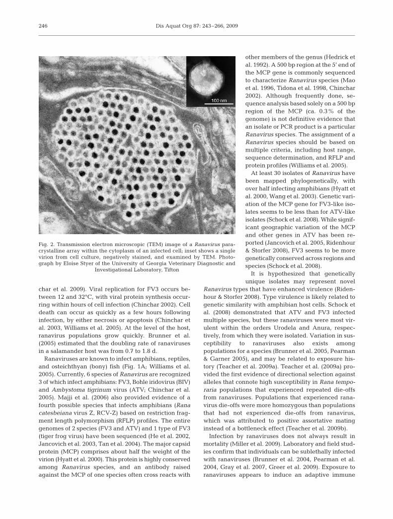
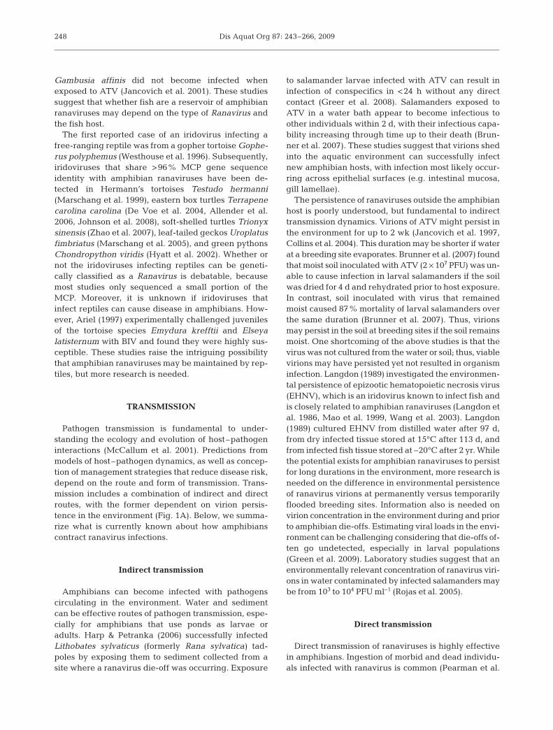
Ranaviruses were first isolated from Lithobates (for-merly Rana) pipiens in the mid-1960s (Granoff et al.1965, Rafferty 1965). Ranavirus is in the family Irido-viridae (Eaton et al. 2007), which contains 5 genera: 2infect invertebrates (Iridovirus and Chloriridovirus)and 3 infect ectothermic vertebrates (Ranavirus, Mega-locytivirus, and Lymphocystivirus; Chinchar et al.2009). Ranaviruses are large, double-stranded DNAviruses (ca. 105 kbp, 150 nm diameter; Williams et al.2005), with a distinctive icosahedral shape that is fre-quently visible in the cytoplasm of infected cells asparacrystalline arrays in electron microscopic images(Fig. 2; Chinchar & Hyatt 2008). The replication cycleof ranaviruses has been studied extensively using frogvirus 3 (FV3), which is the type species for the genus(Chinchar et al. 2005, 2009, Williams et al. 2005). Thegenome encodes around 100 putative gene products(Chinchar 2002), including several that likely playroles in virulence and immune evasion proteins (Chin-
244
Gray et al.: Review: amphibian ranaviruses 245
Possible reservoirs
Bony fish Amphibians
Shed virions
Indirect transmission
Direct transmission
Direct contact, predation, necrophagy
Circulating virions
Amphibian host:
Infected, susceptible, recovered
Reptiles
Environmental persistence
A)
Infected host density Infected
amphibian density
P(Exposure)IT P(Exposure)DT
P(S)i P(S)i P(S)i
Water chemistry, soil type, ambient temperature,
hydroperiod, UV-B
Habitat characteristics
Hatchling Metamorph AdultLarvaePre-metamorphosisPost-metamorphosis
Host susceptibility
Innate and adaptive immune systems
Vertical transmission
Embryo
B)
Natural stressorsDevelopment
Food limitationHost density
PredatorsWater temperature
Co-infections
Anthropogenic stressors
PesticidesFertilizers
Nitrogenous wasteHeavy metalsAcidification
Genetic isolation
Strain noveltyExposure history
Co-evolution
Virulence
P(S)i
P(infection)i
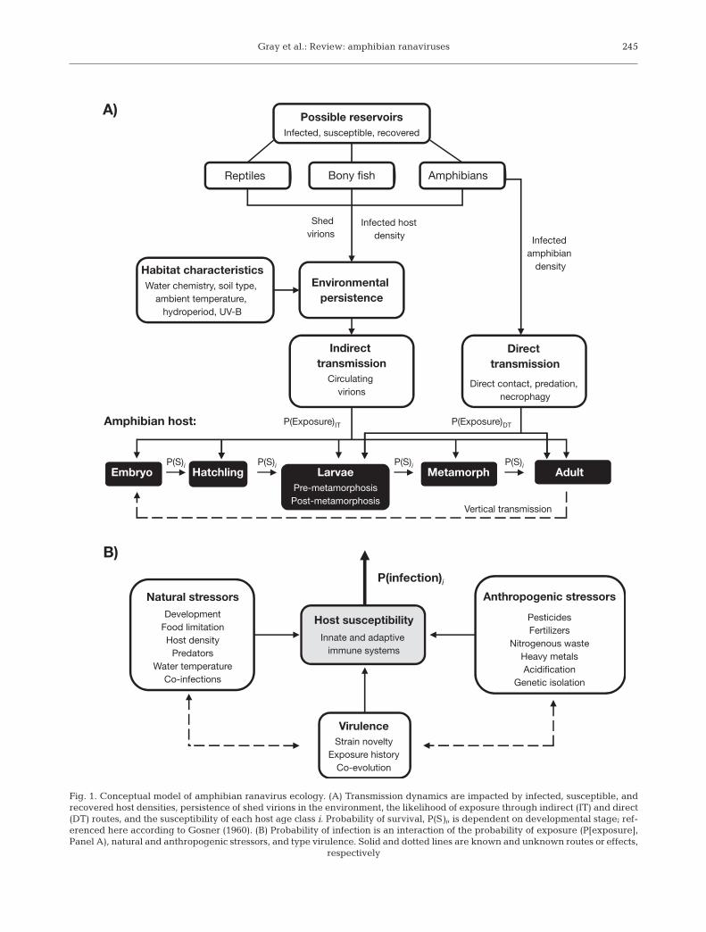
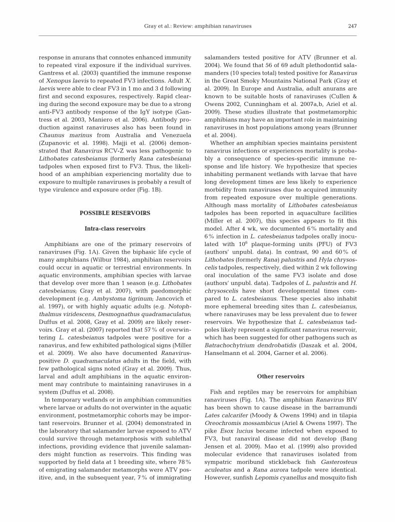
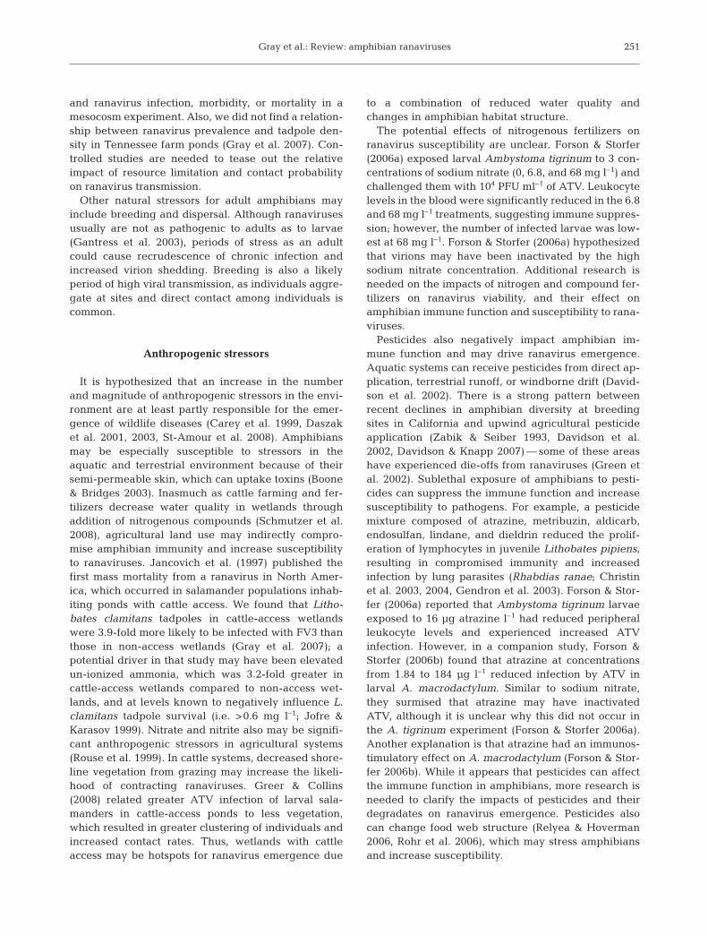
Fig. 1. Conceptual model of amphibian ranavirus ecology. (A) Transmission dynamics are impacted by infected, susceptible, andrecovered host densities, persistence of shed virions in the environment, the likelihood of exposure through indirect (IT) and direct(DT) routes, and the susceptibility of each host age class i. Probability of survival, P(S)i, is dependent on developmental stage; ref-erenced here according to Gosner (1960). (B) Probability of infection is an interaction of the probability of exposure (P[exposure],Panel A), natural and anthropogenic stressors, and type virulence. Solid and dotted lines are known and unknown routes or effects,
respectively
Dis Aquat Org 87: 243–266, 2009
char et al. 2009). Viral replication for FV3 occurs be-tween 12 and 32°C, with viral protein synthesis occur-ring within hours of cell infection (Chinchar 2002). Celldeath can occur as quickly as a few hours followinginfection, by either necrosis or apoptosis (Chinchar etal. 2003, Williams et al. 2005). At the level of the host,ranavirus populations grow quickly. Brunner et al.(2005) estimated that the doubling rate of ranavirusesin a salamander host was from 0.7 to 1.8 d.
Ranaviruses are known to infect amphibians, reptiles,and osteichthyan (bony) fish (Fig. 1A; Williams et al.2005). Currently, 6 species of Ranavirus are recognized3 of which infect amphibians: FV3, Bohle iridovirus (BIV)and Ambystoma tigrinum virus (ATV; Chinchar et al.2005). Majji et al. (2006) also provided evidence of afourth possible species that infects amphibians (Ranacatesbeiana virus Z, RCV-Z) based on restriction frag-ment length polymorphism (RFLP) profiles. The entiregenomes of 2 species (FV3 and ATV) and 1 type of FV3(tiger frog virus) have been sequenced (He et al. 2002,Jancovich et al. 2003, Tan et al. 2004). The major capsidprotein (MCP) comprises about half the weight of thevirion (Hyatt et al. 2000). This protein is highly conservedamong Ranavirus species, and an antibody raisedagainst the MCP of one species often cross reacts with
other members of the genus (Hedrick etal. 1992). A 500 bp region at the 5’ end ofthe MCP gene is commonly sequencedto characterize Ranavirus species (Maoet al. 1996, Tidona et al. 1998, Chinchar2002). Although frequently done, se-quence analysis based solely on a 500 bpregion of the MCP (ca. 0.3% of thegenome) is not definitive evidence thatan isolate or PCR product is a particularRanavirus species. The assignment of aRanavirus species should be based onmultiple criteria, including host range,sequence determination, and RFLP andprotein profiles (Williams et al. 2005).
At least 30 isolates of Ranavirus havebeen mapped phylogenetically, withover half infecting amphibians (Hyatt etal. 2000, Wang et al. 2003). Genetic vari-ation of the MCP gene for FV3-like iso-lates seems to be less than for ATV-likeisolates (Schock et al. 2008). While signif-icant geographic variation of the MCPand other genes in ATV has been re-ported (Jancovich et al. 2005, Ridenhour& Storfer 2008), FV3 seems to be moregenetically conserved across regions andspecies (Schock et al. 2008).
It is hypothesized that geneticallyunique isolates may represent novel
Ranavirus types that have enhanced virulence (Riden-hour & Storfer 2008). Type virulence is likely related togenetic similarity with amphibian host cells. Schock etal. (2008) demonstrated that ATV and FV3 infectedmultiple species, but these ranaviruses were most vir-ulent within the orders Urodela and Anura, respec-tively, from which they were isolated. Variation in sus-ceptibility to ranaviruses also exists amongpopulations for a species (Brunner et al. 2005, Pearman& Garner 2005), and may be related to exposure his-tory (Teacher et al. 2009a). Teacher et al. (2009a) pro-vided the first evidence of directional selection againstalleles that connote high susceptibility in Rana tempo-raria populations that experienced repeated die-offsfrom ranaviruses. Populations that experienced rana-virus die-offs were more homozygous than populationsthat had not experienced die-offs from ranavirus,which was attributed to positive assortative matinginstead of a bottleneck effect (Teacher et al. 2009b).
Infection by ranaviruses does not always result inmortality (Miller et al. 2009). Laboratory and field stud-ies confirm that individuals can be sublethally infectedwith ranaviruses (Brunner et al. 2004, Pearman et al.2004, Gray et al. 2007, Greer et al. 2009). Exposure toranaviruses appears to induce an adaptive immune
246
Fig. 2. Transmission electron microscopic (TEM) image of a Ranavirus para-crystalline array within the cytoplasm of an infected cell; inset shows a singlevirion from cell culture, negatively stained, and examined by TEM. Photo-graph by Eloise Styer of the University of Georgia Veterinary Diagnostic and
Investigational Laboratory, Tifton
Gray et al.: Review: amphibian ranaviruses
response in anurans that connotes enhanced immunityto repeated viral exposure if the individual survives.Gantress et al. (2003) quantified the immune responseof Xenopus laevis to repeated FV3 infections. Adult X.laevis were able to clear FV3 in 1 mo and 3 d followingfirst and second exposures, respectively. Rapid clear-ing during the second exposure may be due to a stronganti-FV3 antibody response of the IgY isotype (Gan-tress et al. 2003, Maniero et al. 2006). Antibody pro-duction against ranaviruses also has been found inChaunus marinus from Australia and Venezuela(Zupanovic et al. 1998). Majji et al. (2006) demon-strated that Ranavirus RCV-Z was less pathogenic toLithobates catesbeianus (formerly Rana catesbeiana)tadpoles when exposed first to FV3. Thus, the likeli-hood of an amphibian experiencing mortality due toexposure to multiple ranaviruses is probably a result oftype virulence and exposure order (Fig. 1B).
POSSIBLE RESERVOIRS
Intra-class reservoirs
Amphibians are one of the primary reservoirs ofranaviruses (Fig. 1A). Given the biphasic life cycle ofmany amphibians (Wilbur 1984), amphibian reservoirscould occur in aquatic or terrestrial environments. Inaquatic environments, amphibian species with larvaethat develop over more than 1 season (e.g. Lithobatescatesbeianus; Gray et al. 2007), with paedomorphicdevelopment (e.g. Ambystoma tigrinum; Jancovich etal. 1997), or with highly aquatic adults (e.g. Notoph-thalmus viridescens, Desmognathus quadramaculatus;Duffus et al. 2008, Gray et al. 2009) are likely reser-voirs. Gray et al. (2007) reported that 57% of overwin-tering L. catesbeianus tadpoles were positive for aranavirus, and few exhibited pathological signs (Milleret al. 2009). We also have documented Ranavirus-positive D. quadramaculatus adults in the field, withfew pathological signs noted (Gray et al. 2009). Thus,larval and adult amphibians in the aquatic environ-ment may contribute to maintaining ranaviruses in asystem (Duffus et al. 2008).
In temporary wetlands or in amphibian communitieswhere larvae or adults do not overwinter in the aquaticenvironment, postmetamorphic cohorts may be impor-tant reservoirs. Brunner et al. (2004) demonstrated inthe laboratory that salamander larvae exposed to ATVcould survive through metamorphosis with sublethalinfections, providing evidence that juvenile salaman-ders might function as reservoirs. This finding wassupported by field data at 1 breeding site, where 78%of emigrating salamander metamorphs were ATV pos-itive, and, in the subsequent year, 7% of immigrating
salamanders tested positive for ATV (Brunner et al.2004). We found that 56 of 69 adult plethodontid sala-manders (10 species total) tested positive for Ranavirusin the Great Smoky Mountains National Park (Gray etal. 2009). In Europe and Australia, adult anurans areknown to be suitable hosts of ranaviruses (Cullen &Owens 2002, Cunningham et al. 2007a,b, Ariel et al.2009). These studies illustrate that postmetamorphicamphibians may have an important role in maintainingranaviruses in host populations among years (Brunneret al. 2004).
Whether an amphibian species maintains persistentranavirus infections or experiences mortality is proba-bly a consequence of species-specific immune re-sponse and life history. We hypothesize that speciesinhabiting permanent wetlands with larvae that havelong development times are less likely to experiencemorbidity from ranaviruses due to acquired immunityfrom repeated exposure over multiple generations.Although mass mortality of Lithobates catesbeianustadpoles has been reported in aquaculture facilities(Miller et al. 2007), this species appears to fit thismodel. After 4 wk, we documented 6% mortality and6% infection in L. catesbeianus tadpoles orally inocu-lated with 106 plaque-forming units (PFU) of FV3(authors’ unpubl. data). In contrast, 90 and 60% ofLithobates (formerly Rana) palustris and Hyla chrysos-celis tadpoles, respectively, died within 2 wk followingoral inoculation of the same FV3 isolate and dose(authors’ unpubl. data). Tadpoles of L. palustris and H.chrysoscelis have short developmental times com-pared to L. catesbeianus. These species also inhabitmore ephemeral breeding sites than L. catesbeianus,where ranaviruses may be less prevalent due to fewerreservoirs. We hypothesize that L. catesbeianus tad-poles likely represent a significant ranavirus reservoir,which has been suggested for other pathogens such asBatrachochytrium dendrobatidis (Daszak et al. 2004,Hanselmann et al. 2004, Garner et al. 2006).
Other reservoirs
Fish and reptiles may be reservoirs for amphibianranaviruses (Fig. 1A). The amphibian Ranavirus BIVhas been shown to cause disease in the barramundiLates calcarifer (Moody & Owens 1994) and in tilapiaOreochromis mossambicus (Ariel & Owens 1997). Thepike Esox lucius became infected when exposed toFV3, but ranaviral disease did not develop (BangJensen et al. 2009). Mao et al. (1999) also providedmolecular evidence that ranaviruses isolated fromsympatric moribund stickleback fish Gasterosteusaculeatus and a Rana aurora tadpole were identical.However, sunfish Lepomis cyanellus and mosquito fish
247
Dis Aquat Org 87: 243–266, 2009
Gambusia affinis did not become infected whenexposed to ATV (Jancovich et al. 2001). These studiessuggest that whether fish are a reservoir of amphibianranaviruses may depend on the type of Ranavirus andthe fish host.
The first reported case of an iridovirus infecting afree-ranging reptile was from a gopher tortoise Gophe-rus polyphemus (Westhouse et al. 1996). Subsequently,iridoviruses that share >96% MCP gene sequenceidentity with amphibian ranaviruses have been de-tected in Hermann’s tortoises Testudo hermanni(Marschang et al. 1999), eastern box turtles Terrapenecarolina carolina (De Voe et al. 2004, Allender et al.2006, Johnson et al. 2008), soft-shelled turtles Trionyxsinensis (Zhao et al. 2007), leaf-tailed geckos Uroplatusfimbriatus (Marschang et al. 2005), and green pythonsChondropython viridis (Hyatt et al. 2002). Whether ornot the iridoviruses infecting reptiles can be geneti-cally classified as a Ranavirus is debatable, becausemost studies only sequenced a small portion of theMCP. Moreover, it is unknown if iridoviruses thatinfect reptiles can cause disease in amphibians. How-ever, Ariel (1997) experimentally challenged juvenilesof the tortoise species Emydura krefftii and Elseyalatisternum with BIV and found they were highly sus-ceptible. These studies raise the intriguing possibilitythat amphibian ranaviruses may be maintained by rep-tiles, but more research is needed.
TRANSMISSION
Pathogen transmission is fundamental to under-standing the ecology and evolution of host–pathogeninteractions (McCallum et al. 2001). Predictions frommodels of host–pathogen dynamics, as well as concep-tion of management strategies that reduce disease risk,depend on the route and form of transmission. Trans-mission includes a combination of indirect and directroutes, with the former dependent on virion persis-tence in the environment (Fig. 1A). Below, we summa-rize what is currently known about how amphibianscontract ranavirus infections.
Indirect transmission
Amphibians can become infected with pathogenscirculating in the environment. Water and sedimentcan be effective routes of pathogen transmission, espe-cially for amphibians that use ponds as larvae oradults. Harp & Petranka (2006) successfully infectedLithobates sylvaticus (formerly Rana sylvatica) tad-poles by exposing them to sediment collected from asite where a ranavirus die-off was occurring. Exposure
to salamander larvae infected with ATV can result ininfection of conspecifics in <24 h without any directcontact (Greer et al. 2008). Salamanders exposed toATV in a water bath appear to become infectious toother individuals within 2 d, with their infectious capa-bility increasing through time up to their death (Brun-ner et al. 2007). These studies suggest that virions shedinto the aquatic environment can successfully infectnew amphibian hosts, with infection most likely occur-ring across epithelial surfaces (e.g. intestinal mucosa,gill lamellae).
The persistence of ranaviruses outside the amphibianhost is poorly understood, but fundamental to indirecttransmission dynamics. Virions of ATV might persist inthe environment for up to 2 wk (Jancovich et al. 1997,Collins et al. 2004). This duration may be shorter if waterat a breeding site evaporates. Brunner et al. (2007) foundthat moist soil inoculated with ATV (2 × 10⁷ PFU) was un-able to cause infection in larval salamanders if the soilwas dried for 4 d and rehydrated prior to host exposure.In contrast, soil inoculated with virus that remainedmoist caused 87% mortality of larval salamanders overthe same duration (Brunner et al. 2007). Thus, virionsmay persist in the soil at breeding sites if the soil remainsmoist. One shortcoming of the above studies is that thevirus was not cultured from the water or soil; thus, viablevirions may have persisted yet not resulted in organisminfection. Langdon (1989) investigated the environmen-tal persistence of epizootic hematopoietic necrosis virus(EHNV), which is an iridovirus known to infect fish andis closely related to amphibian ranaviruses (Langdon etal. 1986, Mao et al. 1999, Wang et al. 2003). Langdon(1989) cultured EHNV from distilled water after 97 d,from dry infected tissue stored at 15°C after 113 d, andfrom infected fish tissue stored at –20°C after 2 yr. Whilethe potential exists for amphibian ranaviruses to persistfor long durations in the environment, more research isneeded on the difference in environmental persistenceof ranavirus virions at permanently versus temporarilyflooded breeding sites. Information also is needed onvirion concentration in the environment during and priorto amphibian die-offs. Estimating viral loads in the envi-ronment can be challenging considering that die-offs of-ten go undetected, especially in larval populations(Green et al. 2009). Laboratory studies suggest that anenvironmentally relevant concentration of ranavirus viri-ons in water contaminated by infected salamanders maybe from 103 to 104 PFU ml–1 (Rojas et al. 2005).
Direct transmission
Direct transmission of ranaviruses is highly effectivein amphibians. Ingestion of morbid and dead individu-als infected with ranavirus is common (Pearman et al.
248
Gray et al.: Review: amphibian ranaviruses
2004, Harp & Petranka 2006). Some larval species (e.g.Ambystoma tigrinum, Spea multiplicata) have canni-balistic phenotypes (Hoffman & Pfennig 1999, Pfennig& Murphy 2000), and many amphibian species con-sume embryos (Alford 1999), which can be infectedwith ranavirus (Tweedell & Granoff 1968). Several stud-ies have documented that ingestion of ranavirus-in-fected animal tissue can cause infection (Jancovich etal. 1997, Pearman et al. 2004, Harp & Petranka 2006,Brunner et al. 2007, Cunningham et al. 2007a). Thus,ranaviruses may limit the occurrence of cannibalisticphenotypes in amphibian populations through differ-ential survival (Pfennig et al. 1991, Parris et al. 2005).Larval salamanders that were infected with ranaviruswere depredated 48% less by dragonfly Anax juniuslarvae than controls (Parris et al. 2004), although themechanism could not be identified. Failed predation at-tempts that damage skin may also facilitate indirecttransmission of free-floating virions in the environmentor direct transmission through contact with infected in-dividuals. Transmission of ranavirus across damagedand intact skin has been shown (Brunner et al. 2007,Cunningham et al. 2007a). Brunner et al. (2007) demon-strated that 1 s of direct skin contact between infectedand uninfected larval salamanders was sufficient tocause infection. Transmission of ranavirus among anu-ran metamorphs through direct contact has also beenreported (Cullen et al. 1995).
Vertical transmission of ranaviruses involving infectionof the egg or sperm in vivo in amphibians has not beenproven. Docherty et al. (2003) isolated an iridovirus fromthe testes of a salamander, providing initial evidence thatvertical transmission may be possible. Infection of eggsor sperm also might occur during transport to or in thecloaca from epithelial shedding of virions. However,postgametogenesis transmission generally is not consid-ered vertical because unrelated individuals could be in-fected as well, especially if virions are shed from thevent. Duffus et al. (2008) detected a ranavirus-positiveembryo from an egg mass that was produced by a posi-tive male and negative female Lithobates sylvaticus. Theroute for transmission was unclear in their study, andcontamination from cloacal or epidermal shedding of thevirus from the male could not be ruled out. Several re-searchers have reported that eggs or captive tadpolesraised from egg masses collected in the wild tested pos-itive for Ranavirus (Greer et al. 2005, Duffus et al. 2008).To date, a study has not been performed to test verticaltransmission of ranaviruses in amphibians where in vitrocontamination was controlled. At the population level, itmay be unimportant whether infection of eggs or spermoccurs in the sex organs or subsequently via cloacalor epidermal shedding, considering that transmissionduring or after gametogenesis likely results in the samesurvival endpoint.
STRESSORS
Stressors are defined as factors that cause endoge-nous production of corticosteroid hormones in organ-isms, which can aid in short-term survival; however,chronic exposure to stressors can result in deleteriouseffects on the immune system (Hill & Wyse 1989). Ingeneral, secretion of corticosterone and aldosteronecauses immune suppression by decreasing T-cell pro-liferation and antibody production, and by inducinglymphocyte apoptosis (Rollins-Smith & Blair 1993,Ottaviani & Franceschi 1996, Ducoroy et al. 1999). Cor-ticosteroids are produced during normal physiologicalprocesses, but their production also can be initiated inresponse to external stimuli (Carey et al. 1999). Exter-nal stimuli can be natural stressors, such as changes inambient temperature, food limitation, or threat of pre-dation, or they can be anthropogenic in origin, such asdecreases in water quality from agricultural opera-tions. Stressors can increase the likelihood of pathogeninfection and morbidity due to reduced immune func-tion (Carey et al. 1999). Below we summarize the cur-rent understanding of the role that stressors play in theemergence of ranaviruses in amphibian populations,which likely includes a combination of natural andanthropogenic factors interacting with host immunityand type virulence (Fig. 1B).
Natural stressors
For amphibians, development represents a signifi-cant natural stressor. Changes in tadpole immunityduring development have been extensively studied forXenopus laevis (Rollins-Smith 1998). For this species,immunity increases from the embryo stages throughtadpole pro-metamorphosis then decreases rapidly asendogenous corticosteroids are produced (Flajnik et al.1987). It is hypothesized that natural immunosuppres-sion associated with metamorphic climax is an adapta-tion to facilitate reconstruction of the organ systems forpostmetamorphic life (Rollins-Smith 1998). The im-mune function at metamorphosis is probably lowerthan in all other developmental stages, and representsa period of high pathogen susceptibility (Carey et al.1999). Mass mortality of larvae from ranaviruses hasbeen reported frequently in anurans during metamor-phosis (Speare & Smith 1992, Green & Converse 2005,Greer et al. 2005). Recently metamorphosed juvenilesalso appear to be highly susceptible (Cullen & Owens2002, Brunner et al. 2004, Schock et al. 2008). Follow-ing tail resorption, juvenile immunity increases untilsexual maturity, with the adult immune functionhigher than in all other stages (Rollins-Smith 1998,Gantress et al. 2003). Gantress et al. (2003) demon-
249
Dis Aquat Org 87: 243–266, 2009
strated that adult X. laevis were able to resist highdoses of FV3, with only transitory signs of disease. Oth-ers have reported greater infection rates and morbidityin tadpoles and juvenile amphibians compared toadults (Gruia-Gray & Desser 1992, Cullen & Owens2002, Green et al. 2002, Collins et al. 2004). The trendfor greater susceptibility of larvae and metamorphscompared to adults appears to be consistent amongamphibian species, except for anurans in the UK. Cun-ningham et al. (1996, 2007a,b) have reported morbiditycaused by ranaviruses in Rana temporaria and Bufobufo adults only. High occurrence of adult mortalitymay be a consequence of easier detection, becausemost die-off reports are submitted by the public in theUK (A. G. F. Teacher, Royal Holloway, University ofLondon, pers. comm.). The relative susceptibility of lar-val versus adult R. temporaria is the focus of ongoingcontrolled studies (A. A. Cunningham, Zoological Soci-ety of London, pers. comm.). Very little information isavailable about the susceptibility of early stages inamphibian development. Tweedell & Granoff (1968)reported >99% mortality for Lithobates pipiensembryos exposed to 102–104 PFU of FV3. However, inongoing research at the University of Tennessee, wedocumented that the egg stage was least susceptible toranavirus compared to hatchling, tadpole, and meta-morph stages for 7 North American anuran species(N. Haislip et al. unpubl. data). Indeed, more researchis needed investigating the relative susceptibility ofamphibians during different developmental stagesand why adult anurans in the UK appear to have highsusceptibility.
Water temperature represents a natural environ-mental stressor that could impact susceptibility toranaviruses, particularly for those species whose lar-vae develop during early spring or winter. Cold-induced immunosuppression has been demonstratedin several anuran species (e.g. Cooper et al. 1992,Miodonski et al. 1996) and in Notophthalmus viri-descens (Raffel et al. 2006). Maniero & Carey (1997)found that T-lymphocyte proliferation and serum com-plement activity were lower in Lithobates pipiensmaintained at 5°C compared to controls held at 22°C.They hypothesized that reduced T-lymphocyte pro-duction would cause decreased signaling of B-lympho-cytes, which would reduce antibody production andcompromise immunity (Maniero & Carey 1997). Raffelet al. (2006) reported a similar trend in field-collectedN. viridescens; fewer lymphocytes and eosinophilswere detected circulating in the blood during winterand spring. Field surveillance for ranavirus supportsthe cold-induced immunosuppression hypothesis. Wefound that L. catesbeianus tadpoles collected in Ten-nessee were 7.7-fold more likely to be infected withFV3 in winter than in summer (Gray et al. 2007). Simi-
larly, Lithobates (formerly Rana) clamitans tadpoleswere 4.7-fold more likely to be infected with FV3 inautumn than in summer (Gray et al. 2007). Interest-ingly, Rojas et al. (2005) found that even though ATVreplication in vitro was more than twice as fast at 26°Ccompared to 10 or 18°C, mortality of larval salaman-ders exposed to ATV and held at the lower tempera-tures was at least 2.5-fold greater than that for sala-manders held at 26°C. Moreover, ATV virion titer insalamanders that died at 26°C was less than that forsalamanders held at 10 or 18°C (Rojas et al. 2005), sug-gesting that ranavirus virulence may be greater atlower water temperatures. An important observation isthat the majority of ranavirus die-offs have been re-ported during summer months (Collins et al. 2004,Converse & Green 2005a,b); however, this could be aconsequence of greater detection by scientists con-ducting fieldwork at this time, the seasonality of manyamphibian populations, or factors other than watertemperature stressing amphibians. Indeed, it is possi-ble that mass mortality could occur frequently duringwinter in larval populations or in hibernating adultswithout detection. Presumably, seasonal fluctuations intemperature are less important for species with larvaethat develop over short duration or at tropical latitudes.The effects of ranavirus on amphibian species withoutlarvae are unknown.
Exposure to predators and resource limitation caninduce stress in amphibian larvae, which may increasetheir susceptibility to ranavirus infection and morbid-ity. Several studies have reported that tadpoles rearedwith predators, competitors, or at low resource levelshave higher levels of corticosterone compared withcontrol tadpoles (Glennemeier & Denver 2002, Rot-Nikcevic et al. 2005). No studies have been publishedon the impacts of predation or competition on the sus-ceptibility of amphibian hosts to ranaviruses. However,Hyla versicolor treated with exogenous corticosteronecontracted twice the number of Alaria sp. trematodeinfections than control tadpoles (Belden & Kiesecker2005). We hypothesize that ranavirus susceptibility willincrease in systems with high insect and fish predatordensities. High predator densities may partly explainranavirus emergence in summer.
Density-dependent infection and mortality associ-ated with ranaviruses have been suggested based onfield observations at die-off sites (Green et al. 2002,Brunner et al. 2004), and appears to be an importantregulating mechanism in ATV-salamander systems(Greer et al. 2008). Density-dependent relationshipswith pathogens can occur either from competition-induced stress or higher transmission from increasedconspecific or congeneric interactions (Allen 2003,2007). Harp & Petranka (2006) did not detect a rela-tionship between Lithobates sylvaticus tadpole density
250
Gray et al.: Review: amphibian ranaviruses
and ranavirus infection, morbidity, or mortality in amesocosm experiment. Also, we did not find a relation-ship between ranavirus prevalence and tadpole den-sity in Tennessee farm ponds (Gray et al. 2007). Con-trolled studies are needed to tease out the relativeimpact of resource limitation and contact probabilityon ranavirus transmission.
Other natural stressors for adult amphibians mayinclude breeding and dispersal. Although ranavirusesusually are not as pathogenic to adults as to larvae(Gantress et al. 2003), periods of stress as an adultcould cause recrudescence of chronic infection andincreased virion shedding. Breeding is also a likelyperiod of high viral transmission, as individuals aggre-gate at sites and direct contact among individuals iscommon.
Anthropogenic stressors
It is hypothesized that an increase in the numberand magnitude of anthropogenic stressors in the envi-ronment are at least partly responsible for the emer-gence of wildlife diseases (Carey et al. 1999, Daszaket al. 2001, 2003, St-Amour et al. 2008). Amphibiansmay be especially susceptible to stressors in theaquatic and terrestrial environment because of theirsemi-permeable skin, which can uptake toxins (Boone& Bridges 2003). Inasmuch as cattle farming and fer-tilizers decrease water quality in wetlands throughaddition of nitrogenous compounds (Schmutzer et al.2008), agricultural land use may indirectly compro-mise amphibian immunity and increase susceptibilityto ranaviruses. Jancovich et al. (1997) published thefirst mass mortality from a ranavirus in North Amer-ica, which occurred in salamander populations inhab-iting ponds with cattle access. We found that Litho-bates clamitans tadpoles in cattle-access wetlandswere 3.9-fold more likely to be infected with FV3 thanthose in non-access wetlands (Gray et al. 2007); apotential driver in that study may have been elevatedun-ionized ammonia, which was 3.2-fold greater incattle-access wetlands compared to non-access wet-lands, and at levels known to negatively influence L.clamitans tadpole survival (i.e. >0.6 mg l–1; Jofre &Karasov 1999). Nitrate and nitrite also may be signifi-cant anthropogenic stressors in agricultural systems(Rouse et al. 1999). In cattle systems, decreased shore-line vegetation from grazing may increase the likeli-hood of contracting ranaviruses. Greer & Collins(2008) related greater ATV infection of larval sala-manders in cattle-access ponds to less vegetation,which resulted in greater clustering of individuals andincreased contact rates. Thus, wetlands with cattleaccess may be hotspots for ranavirus emergence due
to a combination of reduced water quality andchanges in amphibian habitat structure.
The potential effects of nitrogenous fertilizers onranavirus susceptibility are unclear. Forson & Storfer(2006a) exposed larval Ambystoma tigrinum to 3 con-centrations of sodium nitrate (0, 6.8, and 68 mg l–1) andchallenged them with 104 PFU ml–1 of ATV. Leukocytelevels in the blood were significantly reduced in the 6.8and 68 mg l–1 treatments, suggesting immune suppres-sion; however, the number of infected larvae was low-est at 68 mg l–1. Forson & Storfer (2006a) hypothesizedthat virions may have been inactivated by the highsodium nitrate concentration. Additional research isneeded on the impacts of nitrogen and compound fer-tilizers on ranavirus viability, and their effect onamphibian immune function and susceptibility to rana-viruses.
Pesticides also negatively impact amphibian im-mune function and may drive ranavirus emergence.Aquatic systems can receive pesticides from direct ap-plication, terrestrial runoff, or windborne drift (David-son et al. 2002). There is a strong pattern betweenrecent declines in amphibian diversity at breedingsites in California and upwind agricultural pesticideapplication (Zabik & Seiber 1993, Davidson et al.2002, Davidson & Knapp 2007) — some of these areashave experienced die-offs from ranaviruses (Green etal. 2002). Sublethal exposure of amphibians to pesti-cides can suppress the immune function and increasesusceptibility to pathogens. For example, a pesticidemixture composed of atrazine, metribuzin, aldicarb,endosulfan, lindane, and dieldrin reduced the prolif-eration of lymphocytes in juvenile Lithobates pipiens,resulting in compromised immunity and increasedinfection by lung parasites (Rhabdias ranae; Christinet al. 2003, 2004, Gendron et al. 2003). Forson & Stor-fer (2006a) reported that Ambystoma tigrinum larvaeexposed to 16 µg atrazine l–1 had reduced peripheralleukocyte levels and experienced increased ATVinfection. However, in a companion study, Forson &Storfer (2006b) found that atrazine at concentrationsfrom 1.84 to 184 µg l–1 reduced infection by ATV inlarval A. macrodactylum. Similar to sodium nitrate,they surmised that atrazine may have inactivatedATV, although it is unclear why this did not occur inthe A. tigrinum experiment (Forson & Storfer 2006a).Another explanation is that atrazine had an immunos-timulatory effect on A. macrodactylum (Forson & Stor-fer 2006b). While it appears that pesticides can affectthe immune function in amphibians, more research isneeded to clarify the impacts of pesticides and theirdegradates on ranavirus emergence. Pesticides alsocan change food web structure (Relyea & Hoverman2006, Rohr et al. 2006), which may stress amphibiansand increase susceptibility.
251
Dis Aquat Org 87: 243–266, 2009
OTHER HUMAN IMPACTS
Habitat fragmentation can negatively impact amphi-bian populations through demographic and geneticisolation (Marsh & Trenham 2001). It is hypothesizedthat increased occurrence of inbreeding in isolatedpopulations will lead to loss of genetic heterozygosity,which may increase pathogen infection and morbidityrates (Altizer et al. 2003). Pearman & Garner (2005)provided evidence that genetically isolated Ranalatastei populations have a greater chance of experi-encing mass mortality from ranavirus introduction.They hypothesized that increased susceptibility mayhave been a consequence of inbreeding depression orloss of pathogen resistance alleles from genetic drift(Pearman & Garner 2005). Inbred Xenopus laevis tad-poles challenged with FV3 died 3-fold faster than out-bred tadpoles, and inbred adults recovered from FV3infection 2-fold slower than outbred adults (Gantresset al. 2003). These results collectively suggest thatinbreeding as a consequence of genetic isolation mayincrease the likelihood of a ranavirus epizootic occur-ring. However, inbreeding as a consequence of direc-tional selection associated with a ranavirus die-off mayprovide a selective advantage (Teacher et al. 2009a).
Another consequence of habitat fragmentation is in-creased probability of contact among infected individ-uals. Several studies have documented increased nest-edness and elevated relative abundance of someamphibian species inhabiting breeding sites located inagricultural landscapes (Knutson et al. 1999, Kolozs-vary & Swihart 1999, Gray et al. 2004a,b). This mayresult from reflected movement of dispersing individu-als back to breeding sites due to perceived imperme-ability of an anthropogenically disturbed landscape(Rothermel & Semlitsch 2002, Gray et al. 2004b, Ritten-house & Semlitsch 2006). On the other hand, corridorsmay serve as conduits of pathogen transmission amongpopulations (Hess 1996). The impact of ranaviruses ongenetically isolated populations versus its impact onpopulations afforded interdemic movement needs tobe determined.
Amphibian declines associated with disease haveoccurred in some cases at high elevation (Brem & Lips2008, Gahl & Calhoun 2008). It is hypothesized thatchanges in ambient temperature and weather patternsfrom global warming, greater levels of UV-B radiationentering the atmosphere from ozone depletion, andupwind transport and deposition of pesticides may beanthropogenic stressors at high elevation sites (David-son et al. 2002, Pounds et al. 2006, Bancroft et al. 2008,Muths et al. 2008). The relationship of ranavirus out-breaks and elevation remains unclear. Gahl & Calhoun(2008) reported a greater occurrence of ranavirusmortality events at anuran breeding sites positioned at
higher elevation, yet their sites differed by only 150 min elevation. We found an inverse relationshipbetween ranavirus prevalence and elevation among 3sites that differed by 1070 m in elevation; however, themechanisms responsible for this trend are unknown(Gray et al. 2009). More studies are needed that exam-ine the elevational trends of ranavirus outbreaks andidentify possible factors driving any relationships.
It is also possible that novel ranaviruses can be trans-ported and introduced by humans into naïve popula-tions, which Cunningham et al. (2003) coined patho-gen pollution. We suspect that introduction of novelranaviruses that are genetically similar to endemictypes are probably the greatest risk. Several studieshave demonstrated that novel ranaviruses are morepathogenic than endemic types (Majji et al. 2006, Stor-fer et al. 2007); however, virulence appears to berelated to the genetic similarity with coevolved typesand host specificity. As discussed earlier, ATV wasmore pathogenic to salamanders compared to anurans,and FV3 was more pathogenic to anurans than to sala-manders (Schock et al. 2008). Although ranvirusesappear to be widespread, it is possible that some pop-ulations lack evolutionary exposure. In the case ofcompletely naïve populations, the pathogenicity ofranaviruses remains unknown.
Ranaviruses could be transported among watershedsby recreationists, farmers, and researchers. Transportcould occur on fomites such as boots, fishing andresearch gear, farm equipment, and boats. Fishermanand bait industries also transport infected amphibiansacross watersheds. Frequently, trout (subfamily Salmo-ninae) fishermen in the Appalachian Mountains ofeastern North America use adult plethodontid sala-manders for bait (Copeland et al. 2009), and, in thesouthwestern USA, largemouth bass Micropterussalmoides fishermen use larval Ambystoma tigrinumas bait (Picco et al. 2007). The Canadian Ministry ofNatural Resources (CMNR) allows use of Lithobatespipiens for fishing (CMNR 2009), which is a knowncarrier of ranavirus (Greer et al. 2005). Jancovich et al.(2005) provided molecular evidence for the highgenetic variability among ATV isolates in the westernUSA, and hypothesized that a mechanism for this vari-ability may be the interstate transport of salamandersfor fishing bait. Picco et al. (2007) reported that 85% ofbait shops in Arizona contained ATV-positive salaman-ders. Transportation of live amphibians also occursamong countries for pet trade, food, and traditionalmedicines (Schlaepfer et al. 2005). In 3 major ports ofentry in the USA, 4.66 million live frogs on average areimported annually, with 8.5% infected with ranavirus(Schloegel et al. 2009). Importation of ranavirus-infected frogs is a concern if these animals enter thepet trade, aquaculture, or bait fish industries, which
252
Gray et al.: Review: amphibian ranaviruses
many of them do (Schloegel et al. 2009). Thus, novelranaviruses could be introduced by humans into naïvepopulations by transporting contaminated water orfomites, using infected individuals for fish bait, orreleasing non-native amphibians.
PATHOLOGY AND DIAGNOSTICS
Accurate diagnosis of amphibian diseases requiresan understanding of gross and histopathological signs.Molecular techniques are also available to assist indiagnosis. Below we summarize what is known aboutthe pathology and diagnostics of ranaviral disease inamphibians. We also discuss how quickly ranaviral dis-ease can progress in infected amphibians, and providedirection on surveillance of ranaviruses in populations.
Field signs and gross lesions
Field signs (e.g. lordosis, erratic swimming, lethargy)and gross lesions (e.g. swelling, erythema) indicative ofranavirus infection depend on amphibian developmen-tal stage. Tweedell & Granoff (1968) reported loss ofpigmentation, lordosis, epithelial sloughing, and pe-techiation in Lithobates pipiens embryos. For subclini-cal infections (i.e. infection, but no apparent disease),no field signs or gross lesions are generally observed(Miller et al. 2009). However, in sublethal infections (i.e.morbidity, but not mortality), field signs and grosschanges are usually apparent, but the severity dependson the extent of disease. Morbid amphibian larvae of-ten display erratic swimming, lethargy, and lack ofequilibrium (Jancovich et al. 1997, Bollinger et al. 1999,Docherty et al. 2003, Converse & Green 2005a). Grosslesions in larvae include erythema at the base of thegills, ventrum, and legs, and swelling of the legs, body,and gular region (Fig. 3; Jancovich et al. 1997, Bollingeret al. 1999, Docherty et al. 2003, Converse & Green2005a). Cutaneous polyps also have been reported inAmbystoma tigrinum larvae (Jancovich et al. 1997,Bollinger et al. 1999). In adults, erythema of the legsand ventrum, ulceration of the skin, and erythema nearthe vent or plantar surfaces of the feet have been re-ported (Fig. 3; Cunningham et al. 1996, Converse &Green 2005a, Miller et al. 2008). In fatal cases of larvaeand adults, intracoelomic lesions may be present, andinclude petechial or ecchymotic hemorrhages of the in-ternal organs (especially the mesonephros [kidneys],reproductive organs, and liver) and pale swollen livers(Cunningham et al. 1996, Docherty et al. 2003, Miller etal. 2007, 2008). Additionally, the gastrointestinal tractmay be empty and the gall bladder may be enlarged,both of which are consistent with anorexia.
Histological lesions
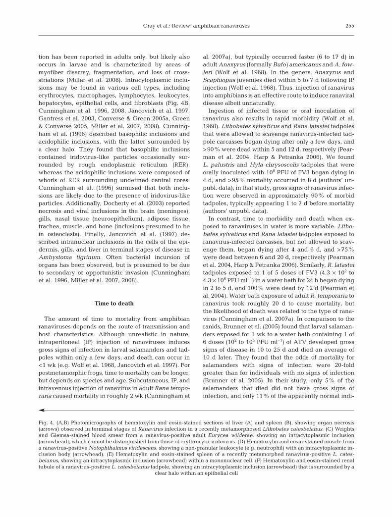
Similar to gross changes, histological changes maybe minimal or absent in subclinically infected individ-uals. We found non-specific histological changes (i.e.minimal to mild lymphocytolysis, lymphoid depletion,and mild vacuolation of hepatocytes and renal tubularepithelium) in ranavirus-positive Lithobates cates-beianus and L. clamitans tadpoles collected from farmponds in Tennessee (Gray et al. 2007, Miller et al.2009). Non-specific changes may be due to variousendogenous or exogenous challenges to the host, suchas steroid release secondary to stress, antigen expo-sure, and systemic illness. Although changes may benon-specific even in fatal cases (Driskell et al. 2009),extensive organ necrosis may be observed in both lar-vae and adults, with the liver, spleen, kidney, andintestines most affected (Fig. 4A; Bollinger et al. 1999,Gantress et al. 2003, Robert et al. 2005, Converse &Green 2005a, Miller et al. 2007, 2008). The renaltubular epithelium can be attenuated, with variousdegrees of vacuolation and necrosis in more severelyaffected cells (Gantress et al. 2003, Converse & Green2005a, Miller et al. 2008). Skeletal muscle degenera-
253
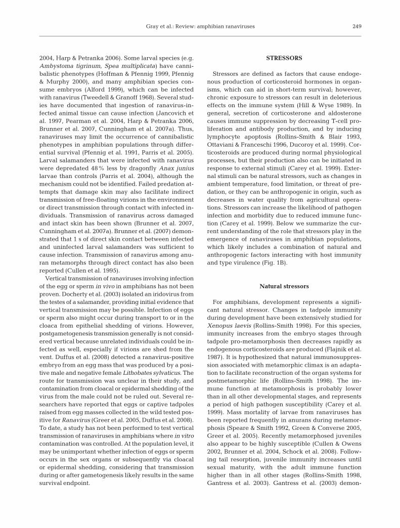
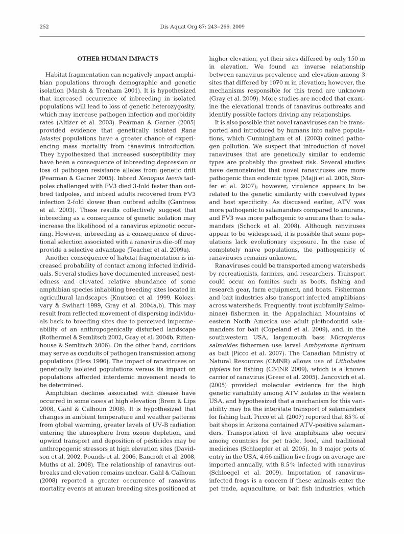
Fig. 3. Gross lesions observed in clinical (sublethal andlethal) infections with Ranavirus. (A) Ulceration (arrows) andhemorrhage of the skin in a Lithobates catesbeianus tadpole.(B) Red (hemorrhagic) legs (arrowhead) in a Hyla chrysoscelistadpole. (C) Swollen red legs (arrowheads) in a L. cates-beianus tadpole. (D) Irregular areas of erythema (arrows) onthe serosa of internal organs in a L. catesbeianus tadpole.(E) Variably sized tan foci (arrows) on the liver of an adultTheloderma corticale that represent areas of necrosis when
viewed in histological sections
Dis Aquat Org 87: 243–266, 2009254
Gray et al.: Review: amphibian ranaviruses
tion has been reported in adults only, but likely alsooccurs in larvae and is characterized by areas ofmyofiber disarray, fragmentation, and loss of cross-striations (Miller et al. 2008). Intracytoplasmic inclu-sions may be found in various cell types, includingerythrocytes, macrophages, lymphocytes, leukocytes,hepatocytes, epithelial cells, and fibroblasts (Fig. 4B;Cunningham et al. 1996, 2008, Jancovich et al. 1997,Gantress et al. 2003, Converse & Green 2005a, Green& Converse 2005, Miller et al. 2007, 2008). Cunning-ham et al. (1996) described basophilic inclusions andacidophilic inclusions, with the latter surrounded bya clear halo. They found that basophilic inclusionscontained iridovirus-like particles occasionally sur-rounded by rough endoplasmic reticulum (RER),whereas the acidophilic inclusions were composed ofwhorls of RER surrounding undefined central cores.Cunningham et al. (1996) surmised that both inclu-sions are likely due to the presence of iridovirus-likeparticles. Additionally, Docherty et al. (2003) reportednecrosis and viral inclusions in the brain (meninges),gills, nasal tissue (neuroepithelium), adipose tissue,trachea, muscle, and bone (inclusions presumed to bein osteoclasts). Finally, Jancovich et al. (1997) de-scribed intranuclear inclusions in the cells of the epi-dermis, gills, and liver in terminal stages of disease inAmbystoma tigrinum. Often bacterial incursion oforgans has been observed, but is presumed to be dueto secondary or opportunistic invasion (Cunninghamet al. 1996, Miller et al. 2007, 2008).
Time to death
The amount of time to mortality from amphibianranaviruses depends on the route of transmission andhost characteristics. Although unrealistic in nature,intraperitoneal (IP) injection of ranaviruses inducesgross signs of infection in larval salamanders and tad-poles within only a few days, and death can occur in<1 wk (e.g. Wolf et al. 1968, Jancovich et al. 1997). Forpostmetamorphic frogs, time to mortality can be longer,but depends on species and age. Subcutaneous, IP, andintravenous injection of ranavirus in adult Rana tempo-raria caused mortality in roughly 2 wk (Cunningham et
al. 2007a), but typically occurred faster (6 to 17 d) inadult Anaxyrus (formally Bufo) americanus and A. fow-leri (Wolf et al. 1968). In the genera Anaxyrus andScaphiopus juveniles died within 5 to 7 d following IPinjection (Wolf et al. 1968). Thus, injection of ranavirusinto amphibians is an effective route to induce ranaviraldisease albeit unnaturally.
Ingestion of infected tissue or oral inoculation ofranavirus also results in rapid morbidity (Wolf et al.1968). Lithobates sylvaticus and Rana latastei tadpolesthat were allowed to scavenge ranavirus-infected tad-pole carcasses began dying after only a few days, and>90% were dead within 5 and 12 d, respectively (Pear-man et al. 2004, Harp & Petranka 2006). We foundL. palustris and Hyla chrysoscelis tadpoles that wereorally inoculated with 106 PFU of FV3 began dying in4 d, and >95% mortality occurred in 8 d (authors’ un-publ. data); in that study, gross signs of ranavirus infec-tion were observed in approximately 90% of morbidtadpoles, typically appearing 1 to 7 d before mortality(authors’ unpubl. data).
In contrast, time to morbidity and death when ex-posed to ranaviruses in water is more variable. Litho-bates sylvaticus and Rana latastei tadpoles exposed toranavirus-infected carcasses, but not allowed to scav-enge them, began dying after 4 and 6 d, and >75%were dead between 6 and 20 d, respectively (Pearmanet al. 2004, Harp & Petranka 2006). Similarly, R. latasteitadpoles exposed to 1 of 5 doses of FV3 (4.3 × 10² to4.3 × 106 PFU ml–1) in a water bath for 24 h began dyingin 2 to 5 d, and 100% were dead by 12 d (Pearman etal. 2004). Water bath exposure of adult R. temporaria toranavirus took roughly 20 d to cause mortality, butthe likelihood of death was related to the type of rana-virus (Cunningham et al. 2007a). In comparison to theranids, Brunner et al. (2005) found that larval salaman-ders exposed for 1 wk to a water bath containing 1 of6 doses (102 to 105 PFU ml–1) of ATV developed grosssigns of disease in 10 to 25 d and died an average of10 d later. They found that the odds of mortality forsalamanders with signs of infection were 20-foldgreater than for individuals with no signs of infection(Brunner et al. 2005). In their study, only 5% of thesalamanders that died did not have gross signs ofinfection, and only 11% of the apparently normal indi-
255
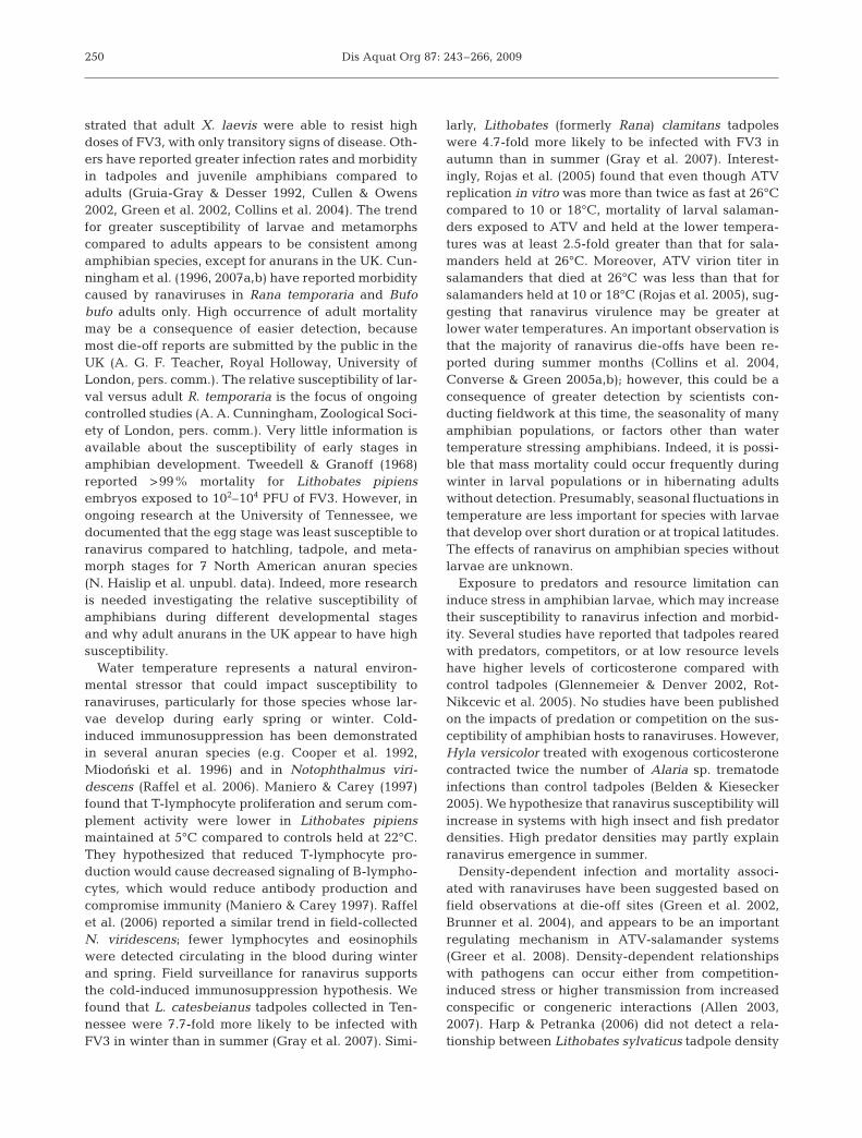
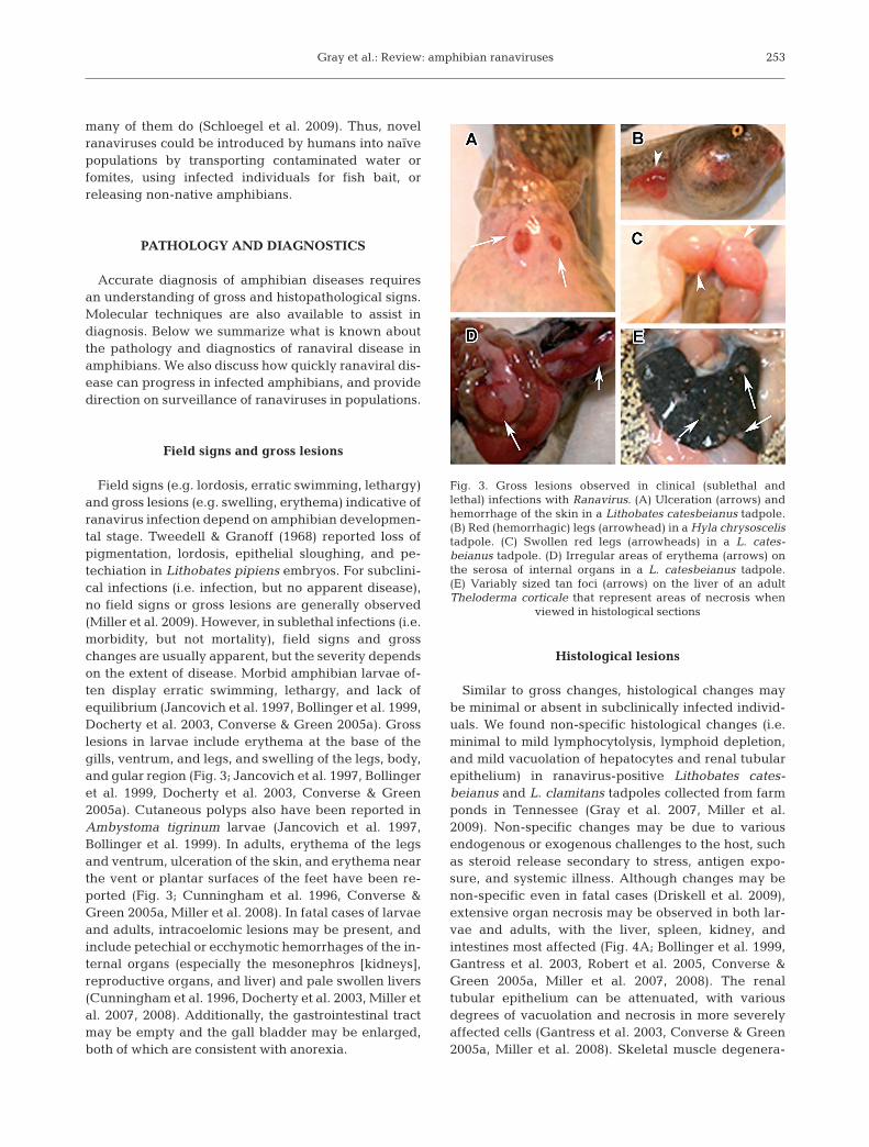
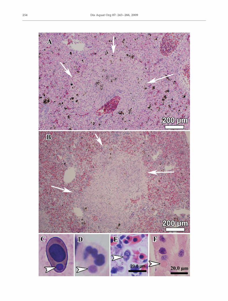
Fig. 4. (A,B) Photomicrographs of hematoxylin and eosin-stained sections of liver (A) and spleen (B), showing organ necrosis(arrows) observed in terminal stages of Ranavirus infection in a recently metamorphosed Lithobates catesbeianus. (C) Wrightsand Giemsa-stained blood smear from a ranavirus-positive adult Eurycea wilderae, showing an intracytoplasmic inclusion(arrowhead), which cannot be distinguished from those of erythrocytic iridovirus. (D) Hematoxylin and eosin-stained muscle froma ranavirus-positive Notophthalmus viridescens, showing a non-granular leukocyte (e.g. neutrophil) with an intracytoplasmic in-clusion body (arrowhead). (E) Hematoxylin and eosin-stained spleen of a recently metamorphed ranavirus-positive L. cates-beianus, showing an intracytoplasmic inclusion (arrowhead) within a mononuclear cell. (F) Hematoxylin and eosin-stained renaltubule of a ranavirus-positive L. catesbeianus tadpole, showing an intracytoplasmic inclusion (arrowhead) that is surrounded by a
clear halo within an epithelial cell
Dis Aquat Org 87: 243–266, 2009
viduals tested positive for the ranavirus (Brunner et al.2005). Collectively, these results suggest that rana-virus-associated morbidity can occur within days ofvirion exposure in water, but time to mortality isdependent on developmental stage, and ranaviral dis-ease in salamanders appears to progress slower than inanurans.
Diagnostics
In general, several tests are necessary to differenti-ate between ranavirus infection and disease. Usefuldiagnostic tools for characterizing ranaviruses includehistology, cytology, virus isolation, electron micro-scopy, and molecular modalities (i.e. PCR, RFLP,sodium dodecyl sulfate polyacrylamide gel electro-phoresis [SDS-PAGE]). Most of these tests can be per-formed on dead or living organisms, but the timebetween death and the postmortem examinationshould be minimized. Although formalin-fixed speci-mens are preferred for histological examination,ethanol-fixed specimens may be used, but prolongedstorage in ethanol can dry tissues and decrease theirusefulness for histology (Green et al. 2009). For PCR,fresh or frozen tissues are preferred, but ethanol-fixedspecimens also can be used. Although formalin fixa-tion can damage DNA, Kattenbelt et al. (2000) demon-strated that Ranavirus DNA can be successfully ex-tracted from formalin-fixed tissues. Thus, formalin-fixed-paraffin-embedded tissues can be used in PCRassays for ranavirus testing and make it possible toconduct retrospective studies (Cullen & Owens 2002,Miller et al. 2008, Driskell et al. 2009). Fresh or frozentissues are necessary for virus isolation and culture,because viable virus is necessary. Finally, collection ofwhole blood is useful for inspection of ranavirus inclu-sion bodies in erythrocytes and leukocytes (Converse& Green 2005a, Green & Converse 2005, Miller et al.2007), but it is important to note that viral inclusionsfound only in erythrocytes may represent erythrocyticiridoviruses — the pathogenicity of which remainsuncertain (Green & Converse 2005). Although manyspecies or age classes of amphibians are too small tocollect substantial amounts of blood, usually enoughblood can be collected to make a blood smear. Bloodmay be collected antemortem from the ventral vein inadults or tail vein in salamanders, or collected post-mortem from the heart of larval or adult amphibians(Wright 2001).
Virus isolation may be used to test for the presence ofviable virus and aids in characterizing the virus type,but it cannot be used to diagnose the presence of dis-ease. Cultured virus is used to perform some moleculartests such as SDS-PAGE and RFLP. Cultured virus can
also yield better products for sequencing. Currently,ranaviruses are cultured on a variety of fish cell lines(e.g. fathead minnow epithelioma papilloma cyprinicells); however, amphibian cell lines are becomingincreasingly available. It is possible that culturing maybe optimized when amphibian cell lines are used, butthis remains to be tested. One caveat is that virus isola-tion is often unsuccessful (Cullen & Owens 2002), withsuccess being dependent upon a variety of factors,including degree of postmortem autolysis, tissue type,and the ability to optimize the incubation conditions(e.g. temperature) for a specific ranavirus. Thus, infec-tion cannot be ruled out based solely on negative isola-tion results (Cullen & Owens 2002).
Electron microscopy is used for confirmation of cul-tured virus, and detection of virions within fixed andparaffin-embedded tissues (Gray et al. 2007, Burton etal. 2008, Miller et al. 2009). However, electron micro-scopy is only reliable for identification to the familylevel (Iridoviridae); it cannot be used to definitivelyverify a ranavirus. Thus, other methods (e.g. PCR andsequencing) are necessary to further classify irido-viruses.
Molecular testing for ranaviruses can be performedon fresh, fixed, or paraffin-embedded tissues, as wellas on blood (Green et al. 2009). Although used infre-quently, amphibian feces or swabs of the oral cavity,cloaca, and skin lesions can be useful non-lethal meth-ods for diagnostic testing (Driskell et al. 2009, Gray etal. 2009), and may provide evidence of viral shedding.For PCR testing, one caveat is that a positive PCRresult only confirms the presence of virions (non-viableor viable). Thus, it is important to perform supportivetests (e.g. histological examination) to differentiate be-tween ranavirus infection (i.e. presence of the patho-gen within the animal) versus disease (i.e. negativelyimpacting health such as cellular degeneration andnecrosis). Also, false-negative and -positive results arepossible, which is discussed in the section ‘Surveil-lance’ that follows. Although conventional PCR hasbeen routinely used for Ranavirus testing and is neces-sary for sequencing PCR products, real-time quantita-tive PCR (qPCR) can be used to quantitate viral load(Yuan et al. 2006, Storfer et al. 2007). Viral load isquantified by standardizing the amount of genomicDNA across samples. Although the correlation be-tween viral load and ranaviral disease remains un-clear, we have found that viral load and mortality ratesare positively correlated (authors’ unpubl. data). It isimportant to note that the brightness of conventionalPCR bands does not equate to viral load even ifgenomic DNA is standardized. Further, several studieshave reported that qPCR may be more sensitive thanconventional PCR at detecting virus infection (Brunneret al. 2005, Pallister et al. 2007, Driskell et al. 2009).
256
Gray et al.: Review: amphibian ranaviruses
Currently, RFLPs, protein profiles, and sequenceanalysis of the MCP gene are used most often to iden-tify unique types of Iridoviridae (Williams et al. 2005,Majji et al. 2006, Schock et al. 2008). Given the conser-vative nature of the MCP, this portion of the Ranavirusgenome may not be ideal to separate Ranavirus types.Ridenhour & Storfer (2008) provide an alternativeapproach to MCP analysis for differentiating rana-viruses that uses multiple DNA sequences from theentire genome and Bayesian- and maximum-likeli-hood-based analysis. Holopainen et al. (2009) also pro-vide a novel approach to differentiate ranavirusesusing PCR and restriction enzyme analysis of DNApolymerase and a neurofilament triplet H1-like proteingene. As stated earlier, sequence analysis based solelyon a 500 bp region of the MCP is insufficient to classifyan iridovirus as a Ranavirus species.
Histological examination is the best method to con-firm the presence of disease versus infection. Specifi-cally, the extent to which organs are affected by rana-virus can be determined by histological examinationonly, and changes due to opportunistic or concurrentpathogens can be assessed (Miller et al. 2008). Tech-niques such as immunohistochemical staining (IHC)and electron microscopy can be applied to histologicalsections and allow identification of ranavirus in spe-cific cells (Cunningham et al. 1996, 2008, Burton et al.2008, Balseiro et al. 2009). Negative aspects of histol-ogy include the requirement of lethal collection andcost. Generally, lethal collections are not possible forinvestigations with imperiled species. Costs for prepar-ing slides for histological examination can be high,especially if serial sectioning of the tissues is needed,which can be important to gain a representative viewof all organs. At this time, commercially available anti-bodies for IHC are not available for ranavirus, reduc-ing the applicability for most researchers. Also, manylaboratories do not have electron microscopes, due tothe initial cost and maintenance of this equipment.Thus, we recommend that teams of researchers worktogether to apply as many diagnostic methods as possi-ble for investigations of surveillance as well as morbid-ity and mortality events.
Surveillance
Pathogen surveillance is fundamental to diseasemonitoring. We are unaware of any active widespreadranavirus surveillance programs, although in the USA,the Tennessee Wildlife Resources Agency is currentlysupporting an initial sampling effort (n = 40 locations)across 2 physiographic regions (Cumberland Plateauand Tennessee River Ridge and Valley). Through theproject RANA, the European Commission is develop-
ing the framework necessary for future surveillance ofranaviruses (European Commission 2009). Also, in theUK, the non-profit Amphibian and Reptile Conserva-tion Trust (www.arc-trust.org/) assists in reporting die-offs from ranaviruses and other causes. We encourageother natural resource organizations and nations toconsider similar programs for monitoring ranavirusesand other amphibian pathogens. Surveillance is impor-tant to detect epizootics and build an understanding ofhow pathogen prevalence varies between disturbedand undisturbed sites and among amphibian species.Surveillance also can be used to direct conservationstrategies in areas with high prevalence to minimizethe likelihood of a die-off.
Four techniques have been used to test for rana-viruses: whole organism, internal organs, tail or toeclips, and skin swabs (Gray et al. 2007, 2009, Greer &Collins 2007, St-Amour & Lesbarrères 2007). We rec-ommend testing for ranaviruses from internal organs(especially liver and kidney), because they are morelikely to provide evidence of systemic infection as op-posed to tail or toe clips and swabs (i.e. external sur-faces), which may simply represent surface exposureto the virus. Whole-organism testing via grindingamphibians using a laboratory-grade tissue blender(e.g. Stomacher 80; Greer & Collins 2007) includesinternal organs, but also virions potentially from theenvironment. In a pilot study with Lithobates cates-beianus larvae, we determined that tail clips had a20% false-negative and 6% false-positive rate for sys-temic infection (i.e. when compared to test results frominternal organs; authors’ unpubl. data); skin swabs hada 22% false-negative and 12% false-positive rate forsystemic infection in that study. Thus, when lethalorganism collection is not feasible or allowed, we rec-ommend testing tail clips over skin swabs for rana-viruses. Greer & Collins (2007) also found that tail clipsfrom postmetamorphic salamanders resulted in falsenegatives, but this effect was dependent on postinfec-tion duration. Fourteen days postinfection, tail clip andwhole-organism tests were identical (Greer & Collins2007). St-Amour & Lesbarrères (2007) reported a 3%false-negative rate from toe clips. Animals that are col-lected for ranavirus testing should be euthanized andeither stored at –80°C or preserved in 95% EtOH.Samples should remain frozen until laboratory testing,as multiple cycles of freezing and thawing can affecttest accuracy.
RANAVIRUS EMERGENCE
An emerging pathogen is defined as one that causesdisease and has recently increased in prevalence orgeographic range, been isolated from a new host, or is
257
Dis Aquat Org 87: 243–266, 2009
genetically distinct from other known pathogen spe-cies (Daszak et al. 2000). Inasmuch as the majority ofreported die-offs associated with ranaviruses haveoccurred since the mid-1990s (Green et al. 2002, Muthset al. 2006, Cunningham et al. 2007b), it appears thatthis pathogen is emerging. However, an increase in thenumber of diagnoses identifying ranaviruses as an eti-ologic agent may be a consequence of greater surveil-lance and advancements in molecular techniques forvirus detection (Williams et al. 2005). Thus, scientistsmust exercise caution in concluding disease emer-gence based on surveillance data alone. We suggestthat at least 2 of the above criteria be used as evidenceof ranavirus emergence.
Emerging pathogens can be novel or endemic(Rachowicz et al. 2005). Novel emerging pathogenstypically result from the introduction of new Ranavirustypes, and endemic emerging pathogens are usually aconsequence of the decreased host immune functionassociated with stressors (Carey et al. 2003a, Rachow-icz et al. 2005). Where evidence exists that ranavirusesare emerging, either scenario is possible (see previoussections ‘Anthropogenic stressors’ and ‘Other humanimpacts’), which can be determined through phylo-genic concordance analysis (Storfer et al. 2007). Thisanalysis compares concordance of host and pathogenphylogenies through simulations and maximum-likeli-hood estimation (Farris et al. 1995, Shimodaira &Hasegawa 1999, Goldman et al. 2000). Storfer et al.(2007) used this analysis and provided evidence thatATV was novel in 3 salamander populations, yet en-demic in 8 populations located across western NorthAmerica. It is reasonable to surmise that ranavirusemergence is not a consequence of virus evolution,because this pathogen can infect multiple host speciesand sections of the genome are conserved (Chinchar etal. 2009). However, considerable genetic variability ofATV isolates and genetic evidence of host switchesprovides support that rapid evolution of ranaviruses ispossible (Jancovich et al. 2005, Storfer et al. 2007,Ridenhour & Storfer 2008, Schock et al. 2008).
CONSERVATION
Threat of ranaviruses to amphibian species survival
Epidemic disease models predict that pathogenscannot directly cause local population extinction iftransmission is density dependent, because as individ-uals die, host density drops below a threshold for effi-cient transmission (Anderson & May 1979). However,empirical evidence of thresholds for wildlife diseases israre (Lloyd-Smith et al. 2005a). Presence of reservoirs,high environmental persistence of pathogens, and fre-
quency-dependent transmission can increase the like-lihood of disease-induced extinction (Woodroffe 1999,de Castro & Bolker 2005). All of these characteristicsare possible in ranavirus–host systems. Duffus et al.(2008) provided evidence of ranavirus reservoirs andfrequency-dependent transmission in an amphibiancommunity with known annual die-offs. Long environ-mental persistence of ranavirus virions is also possible(Langdon 1989). However, this model is not accuratefor all amphibian systems. For example, there are fewranavirus reservoirs, environmental persistence ofvirions is low, and density-dependent transmission islikely in ATV–salamander systems (Greer et al. 2008).Thus, the likelihood of local extirpation of a speciesfrom ranaviruses is dependent on the amphibian com-munity and the characteristics of their habitat that con-tribute to persistence and transmission of virions.
Several studies have reported population declines inamphibian species following ranavirus epizootics (e.g.Collins et al. 1988, Cunningham et al. 1996, Greer et al.2005, Schock & Bollinger 2005, Teacher 2008, Ariel etal. 2009). Local population declines or extirpations canhave rippling effects that increase the possibility ofmetapopulation or species extinction (Hanski 1999).Superspreading events may also occur with rana-viruses from a few highly infectious individuals or inpopulations exposed to anthropogenic stressors(Lloyd-Smith et al. 2005b). The intricacies of ranavirus-disease dynamics remain to be determined, and likelyinclude a complex interaction of reservoir species,transmission routes, virion persistence, stressors, andhost immunity (Fig. 1). However, enough evidenceexists to conclude that ranaviruses are a significantpathogen driving local population dynamics in someareas and resulting in at least localized die-offs ofamphibians.
Possible strategies
Very few studies have been conducted on the use ofconservation strategies to reduce disease emergence;however, the lack of this research should not precludescientists from making logical recommendations. Sim-ple strategies can be implemented if factors that causeemergence are understood. Although ranaviruses mayemerge in a population due to natural stressors orvirion shedding by native species, emergence also canoccur as a result of human activity. Most often, human-induced emergence is related to immune suppressionfrom anthropogenic stressors or the introduction ofnovel virus types. Thus, strategies that reduce anthro-pogenic stressors or the chance of pathogen pollutionshould reduce the likelihood of human-induced rana-virus emergence.
258
Gray et al.: Review: amphibian ranaviruses
Establishing undisturbed vegetation buffers aroundamphibian breeding sites has been recommended tominimize human impacts on the aquatic environment(Semlitsch & Bodie 2003). Semlitsch & Bodie (2003)recommended at least 30 m of vegetation to buffer theaquatic environment. We documented that excludingcattle from wetlands with electric fencing reducedranavirus prevalence in some amphibian species (Grayet al. 2007). For that study (op. cit.), cattle were fencedfrom 20 to 200 m from amphibian breeding sites; thus,a 20 m buffer may be sufficient to reduce cattle effectsand ranavirus emergence. Prudent agricultural prac-tices such as restricting chemical applications to calmdays and ensuring that aircraft-applied pesticides arenot released over amphibian breeding sites wouldreduce the potential effects of chemicals on ranavirusemergence.
Conservation strategies also could be implementedto reduce the likelihood of pathogen pollution. Giventhat larval and adult amphibians can be sublethallyinfected with ranaviruses (Brunner et al. 2004, Gray etal. 2007, Picco et al. 2007, Greer et al. 2009), trans-portation of amphibians among watersheds should beregulated to reduce novel ranavirus introduction.Many states in the USA and many Canadian provincesallow use of amphibians as bait for fishing (Picco &Collins 2008, CMNR 2009). In Tennessee, use ofamphibians for fishing is restricted to the watershedwhere they were captured. We encourage similar ormore stringent regulations in states or nations withoutrestrictions, and recommend that regional transport oflive amphibians be accompanied with certification ofRanavirus-negative test results. We also discouragethe commercial sale of amphibians for fish bait. Storferet al. (2007) demonstrated that an ATV type isolatedfrom larval Ambystoma tigrinum in a bait shop wasmore virulent than wild types. For amphibians tradedinternationally, Gray et al. (2007) suggested that man-datory testing for ranaviruses be considered an expor-tation requirement. Recognizing that ranaviruses areassociated with mass mortality of amphibians and thatthis pathogen can be transported in sublethally in-fected individuals (Schloegel et al. 2009), the WorldOrganization for Animal Health (OIE, www.oie.int/eng/en_index.htm) recently listed ranaviral disease asa notifiable disease. Guidelines have been establishedby the OIE for testing amphibians prior to internationalshipment and for declaration of ranavirus-free animals.The OIE also is in the process of preparing guidelinesfor diagnostic testing of amphibians for ranaviruses.
Given the potentially long duration of environmentalpersistence for ranavirus virions (Langdon 1989), disin-fecting equipment that contacts water or soil whereamphibians live should be performed. Langdon (1989)demonstrated that 70% EtOH was effective in inacti-
vating FV3. Bryan et al. (2009) found that solutionsof 3% bleach or 0.75% Nolvasan (2% chlorhexidinediacetate; Fort Dodge Animal Health) applied for 1 minwere effective in inactivating ranaviruses. Althoughdisinfecting equipment may be impractical for thepublic, scientists working in aquatic environmentsshould follow this practice (Green et al. 2009). Naturalresource agencies should consider developing educa-tional websites and public outreach brochures on thebenefits of disinfecting equipment and recreationalgear that comes in contact with water to control dis-eases in amphibians and other aquatic vertebrates.Examples of outreach education sheets on herpetofau-nal diseases, collecting and shipping protocols for mor-bid or dead amphibians, and disinfecting proceduresare available on the Southeast Partners in Amphibianand Reptile Conservation website (www.separc.org).
RESEARCH DIRECTIONS
Much remains to be learned about the genetics, evo-lution, and ecology of amphibian ranaviruses, and howstressors interact to impact the emergence of thispathogen. Research directions in Ranavirus geneticshave been reviewed by Williams et al. (2005), but in-clude using knock-down and knock-out technology todetermine gene functions (Xie et al. 2005, Sample et al.2007). Advances in the genomics of Ranavirus can beused to develop vaccines and control transmission incaptive facilities (Nakajima et al. 2002, Williams et al.2005, Caipang et al. 2006).
Evidence exists that Ranavirus types can infectmultiple hosts, but susceptibility varies among amphi-bian species (Storfer et al. 2007, Schock et al. 2008).Studies are needed that determine species-specificsusceptibility to different Ranavirus types across geo-graphic regions. This information is fundamental tounderstanding which amphibian species are most atrisk of ranavirus epizootics. It also is important toinvestigate the extent of ranavirus emergence and theprevalence of endemic and novel viral types. Large-scale surveillance programs are needed to identifyareas of ranavirus emergence. The occurrence of noveland endemic types can be determined by comparingviral and host phylogenies. Storfer et al. (2007) pro-vided a good example of using phylogenetic concor-dance analyses to accomplish this task for ATV andAmbystoma tigrinum. Similar studies are needed forother amphibians and Ranavirus species. These ad-vances will help in identifying the mechanism of rana-virus emergence (i.e. pathogen pollution or stressors;Storfer et al. 2007).
Although several studies have been performed onthe ecology of ranaviruses (e.g. Brunner et al. 2004,
259
Dis Aquat Org 87: 243–266, 2009
2007, Pearman et al. 2004, Duffus et al. 2008), ourunderstanding of how this pathogen infects amphi-bians and is maintained in the environment remainslimited. Studies are needed on the environmental per-sistence of ranaviruses in water, soil, and decaying tis-sue, and on fomites. Vertical transmission has beenhypothesized by several investigators (Greer et al.2005, Harp & Petranka 2006, Duffus et al. 2008); how-ever, a study that controls for in vitro contaminationhas not been performed. We know that metamorphsare highly susceptible to ranaviruses followed by lar-vae and adults in some cases (Gantress et al. 2003,Brunner et al. 2004); however, we do not completelyunderstand the pathogencity of ranaviruses duringearly developmental stages. Studies are needed thattest for the effects of ranaviruses on embryos andhatchlings and that compare infection, recovery, andmortality rates among pre- and pro-metamorphosislarvae, metamorphs, and adults (Fig. 1A). Further ex-ploration is needed into the high susceptibility of adultamphibians in the UK. Additional controlled studiesare needed to test the susceptibility of native and intro-duced reptiles and fish to Ranavirus types isolatedfrom amphibians to identify the likelihood of viralmaintenance in other ectothermic reservoirs (Moody &Owens 1994, Ariel 1997, Ariel & Owens 1997). Theseadvances are important to determine how ranavirusesare maintained in the environment and to identify themost susceptible stages in amphibian development.
Only a few studies have been conducted on the roleof stressors in ranavirus emergence (Forson & Storfer2006a,b, Gray et al. 2007). It appears some agriculturalchemicals have immunosuppressive effects on certainamphibians that could lead to increased susceptibilityto ranavirus infection (Forson & Storfer 2006a). How-ever, only 2 pesticides (atrazine and carbaryl) and 1fertilizer (sodium nitrate) have been studied and onlyfor 2 amphibian species (Forson & Storfer 2006a,b,A. Storfer, Washington State University, unpubl. data).Future studies need to include a cadre of pesticides,fertilizers, and mixtures of chemicals with >1 amphi-bian species (e.g. Relyea 2004, Hayes et al. 2006) tofully understand the effects of these possible stressorson ranavirus dynamics in amphibian communities.Also, no studies have been conducted on the effectsof heavy metals and acidification. Studies on anthro-pogenic stressors should be crossed with naturalstressors, such as water temperature, food limitation,and presence of a predator to better understand theimpact of ranaviruses within communities. Factorsassociated with elevation (e.g. increased UV-B radia-tion, temperature) also need to be investigated (Gahl &Calhoun 2008).
Concurrent infections of ranaviruses with otheramphibian pathogens have been documented (e.g.
Cunningham et al. 1996, Bollinger et al. 1999, Fox et al.2006, Schmutzer 2007, Miller et al. 2008). However, todate, only 1 controlled study has been published onhow ranavirus interacts with other pathogens to inducemorbidity (Cunningham et al. 2007a). It is hypothesizedthat primary infection by one pathogen will lead to sec-ondary infection by other pathogens; however, this hasnever been tested via ordered experimental challenges.Possible significant amphibian pathogens includeother ranaviruses, Batrachochytrium dendrobatidis,Aeromonas hydrophila, helminthic and myxozoanparasites, Saprolegnia sp., and Perkinsus-like agents(Green & Converse 2005, Converse & Green 2005a,b,Davis et al. 2007, Romansic et al. 2007, Rohr et al. 2008).Selection of pathogens should correspond to the likeli-hood of co-occurrence in the wild, which is possible formost of these pathogens. Co-infection studies are fun-damental to understanding the significance of patho-gens as primary or secondary invaders and to assigningpathogen risk levels. Identification of primary patho-gens can help direct intervention strategies that mini-mize their effects in amphibian communities.
Most of the controlled ecological experiments per-formed on amphibian ranaviruses have been with ATVand Ambystoma trigrium. Although ATV and A. tigri-num are a good model to explore host–pathogendynamics (Collins et al. 2004), variations in host re-sponses likely exist for other ranaviruses and amphi-bian species. To date, no controlled studies have beenconducted on plethodontid salamanders — the mostdiverse family of urodeles (Gray et al. 2009). Moreover,global anuran species richness is approximately 10-fold that of urodeles, yet only a handful of controlledecological studies exist. We also encourage scientiststo study other ranavirus species, particularly FV3given its global distribution in wild and captive popu-lations (Zupanovic et al. 1998, Zhang et al. 2001, Greeret al. 2005, Fox et al. 2006, Cunningham et al. 2007b,Gray et al. 2007, Miller et al. 2007, Pasmans et al. 2008,Schock et al. 2008).
It remains unclear whether amphibian ranavirusesare capable of causing extinction (Greer et al. 2009).This lack of understanding is a consequence of infre-quent surveillance for this pathogen in common anduncommon species in an amphibian community. Giventhat amphibian ranaviruses can infect multiple speciesin a community, extirpation of uncommon susceptiblespecies is most likely. Annual broad-scale surveillancecoupled with population monitoring is needed to de-termine if amphibian ranaviruses are contributing toglobal amphibian declines. We strongly encouragegovernment agencies and conservation organizationsto develop annual monitoring sites for ranaviruses. Theprevalence of Ranavirus should be estimated, and,when possible, the histopathological changes in tissues
260
Gray et al.: Review: amphibian ranaviruses
examined, especially during a die-off. We also recom-mend simultaneous testing for Batrachochytrium den-drobatidis, due to its role in amphibian declines inmany parts of the world. Through broad-scale surveil-lance and controlled experiments, we will develop agreater understanding of the ecology and pathology ofamphibian ranaviruses and factors that may contributeto emergence.
Acknowledgements. Funds for our research on ranaviruseshave been provided by the University of Tennessee Instituteof Agriculture, East Tennessee Research and Education Cen-ter, University of Georgia Veterinary Diagnostic and Investi-gational Laboratory, Tennessee Wildlife Resources Agency,and the Association of Reptilian and Amphibian Veterinari-ans. M.J.G. and J.T.H. are supported by the University of Ten-nessee Center for Wildlife Health located in the Departmentof Forestry, Wildlife and Fisheries, and D.L.M. is supported bythe University of Georgia College of Veterinary Medicine. Wethank M. Berrill, J. L. Brunner, V. G. Chinchar, and 3 anony-mous referees for reviewing initial drafts of our manuscript.We also acknowledge A. Teacher, A. Duffus, and A. Cunning-ham for providing insight into die-offs in the UK.
LITERATURE CITED
Alford RA (1999) Ecology: resource use, competition and pre-dation. In: McDiarmid RW, Altig R (eds) Tadpoles: thebiology of anuran larvae. University of Chicago Press,Chicago, IL, p 240–278
Allen LJS (2003) An introduction to mathematical biology.Prentice Hall, Upper Saddle River, NJ
Allen LJS (2007) An introduction to stochastic processes withapplications to biology. Prentice Hall, Upper Saddle River,NJ
Allender MC, Fry MM, Irizarry AR, Craig L, Johnson AJ,Jones M (2006) Intracytoplasmic inclusions in circulatingleukocytes from an eastern box turtle (Terrapene carolinacarolina) with iridoviral infection. J Wildl Dis 42:677–684
Altizer S, Harvell D, Friedle E (2003) Rapid evolutionarydynamics and disease threats to biodiversity. Trends EcolEvol 18:589–596
Anderson RM, May RM (1979) Population biology of infec-tious diseases: Part I. Nature 280:361–367
Ariel E (1997) Pathology and serological aspects of Bohle iri-dovirus infections in six selected water-associated reptilesin North Queensland. PhD thesis, James Cook University,Townsville
Ariel E, Owens L (1997) Epizootic mortalities in tilapia Oreo-chromis mossambicus. Dis Aquat Org 29:1–6
Ariel E, Kielgast J, Svart HE, Larsen K, Tapiovaara H, BangJensen B, Holopainen R (2009) Ranavirus in wild ediblefrogs Pelophylax kl. esculentus in Denmark. Dis AquatOrg 85:7–14
Balseiro A, Dalton KP, Del Cerro A, Marquez I, Parra F, PrietoJM, Casias R (2009) Outbreak of common midwife toadvirus in alpine newts (Mesotriton alpestris cyreni) andcommon midwife toads (Alytes obstetricans) in northernSpain: comparative pathological study of an emergingranavirus. Vet J (in press) doi:10.1016/j.tvjl.2009.07.038
Bang Jensen B, Ersbøll AK, Ariel E (2009) Susceptibility ofpike Esox lucius to a panel of Ranavirus isolates. DisAquat Org 83:169–179
Bancroft BA, Baker NJ, Blaustein AR (2008) A meta-analysisof the effects of ultraviolet B radiation and its synergisticinteractions with pH, contaminants, and disease onamphibian survival. Conserv Biol 22:987–996
Belden LK, Kiesecker JM (2005) Glucocorticosteroid hormonetreatment of larval treefrogs increases infection by Alariasp. trematode cercariae. J Parasitol 91:686–688
Berger L, Speare R, Daszak P, Green DE and others (1998)Chytridiomycosis causes amphibian mortality associatedwith population declines in the rain forests of Australia andCentral America. Proc Natl Acad Sci USA 95:9031–9036
Berna HJ (1990) Ecology and life history of the tiger salamanderAmbystoma tigrinum nebulosum Hallowell on the KaibabPlateau. MS thesis, Arizona State University, Tempe, AZ
Bollinger TK, Mao JH, Schock D, Brigham RM, Chinchar VG(1999) Pathology, isolation, and preliminary molecularcharacterization of a novel iridovirus from tiger salaman-ders in Saskatchewan. J Wildl Dis 35:413–429
Boone M, Bridges C (2003) Effects of pesticides on amphibianpopulations. In: Semlitsch RD (ed) Amphibian conserva-tion. Smithsonian Institute, Washington, DC, p 187–198
Brem FMR, Lips KR (2008) Batrachochytrium dendrobatidisinfection patterns among Panamanian amphibian species,habitats and elevations during epizootic and enzooticstages. Dis Aquat Org 81:189–202
Brunner JL, Schock DM, Davidson EW, Collins JP (2004)Intraspecific reservoirs: complex life history and the per-sistence of a lethal ranavirus. Ecology 85:560–566
Brunner JL, Richards K, Collins JP (2005) Dose and host char-acteristics influence virulence of ranavirus infections.Oecologia 144:399–406
Brunner JL, Schock DM, Collins JP (2007) Transmission dyna-mics of the amphibian ranavirus Ambystoma tigrinumvirus. Dis Aquat Org 77:87–95
Bryan LK, Baldwin CA, Gray MJ, Miller DL (2009) Efficacy ofselect disinfectants at inactivating Ranavirus. Dis AquatOrg 84:89–94
Burton EC, Miller DL, Styer EL, Gray MJ (2008) Amphibianocular malformation associated with Frog virus 3. Vet J177:442–444
Caipang CM, Takano T, Hirono I, Aoki T (2006) Genetic vac-cines protect red seabream, Pagrus major, upon challengewith red seabream iridovirus (RSIV). Fish ShellfishImmunol 21:130–138
Canadian Ministry of Natural Resources (CMNR) (2009) Baitfor personal use. Available at: www.mnr.gov.on.ca/en/Business/LetsFish/2ColumnSubPage/STEL02_165361.html(accessed on 29 June 2009)
Carey C, Cohen N, Rollins-Smith L (1999) Amphibian declines:an immunological perspective. Dev Comp Immunol 23:459–472
Carey C, Bradford DE, Brunner JL, Collins JP and others(2003a) Biotic factors in amphibian declines. In: Linder G,Krest SK, Sparling DW (eds) Amphibian declines: anintegrated analysis of multiple stressor effects. Society ofEnvironmental Toxicology and Chemistry, Pensacola, FL,p 153–208
Carey C, Pessier AP, Peace AD (2003b) Pathogens, infec-tious disease, and immune defenses. In: Semlitsch RD(ed) Amphibian conservation. Smithsonian Institute,Washington, DC, p 127–136
Chinchar VG (2002) Ranaviruses (family Iridoviridae): emerg-ing cold-blooded killers. Arch Virol 147:447–470
Chinchar VG, Hyatt AD (2008) Iridoviruses: general features.In: Mahy BWJ, Van Regenmortel MHV (eds) Encyclopediaof virology, Vol 3. Elsevier, Oxford, p 167–174
Chinchar VG, Bryan L, Wang J, Long S, Chinchar GD (2003)
261
Dis Aquat Org 87: 243–266, 2009
Induction of apoptosis in frog virus 3-infected cells.Virology 306:303–312
Chinchar VG, Essbauer S, He JG, Hyatt A, Miyazaki T, SeligyV, Williams T (2005) Iridoviridae. In: Fauguet CM, MayoMA, Maniloff J, Desselberger U, Ball LA (eds) Virus taxon-omy: 8th report of the international committee on the tax-onomy of viruses. Elsevier, London, p 163–175
Chinchar VG, Hyatt AD, Miyazaki T, Williams T (2009)Family Iridoviridae: poor viral relations no longer. In:Van Etten JL (ed) Current topics in microbiology andimmunology, Vol 328. Lesser known large dsDNA viruses.Springer-Verlag, Berlin, p 123–170
Christin MS, Gendron AD, Brousseau P, Menard L and others(2003) Effects of agricultural pesticides on the immunesystem of Rana pipiens and on its resistance to parasiticinfection. Environ Toxicol Chem 22:1127–1133
Christin MS, Menard L, Gendron AD, Ruby S and others(2004) Effects of agricultural pesticides on the immunesystem of Xenopus laevis and Rana pipiens. Aquat Toxicol67:33–43
Collins JP, Storfer A (2003) Global amphibian declines: sort-ing the hypotheses. Divers Distrib 9:89–98
Collins JP, Jones TR, Berna HJ (1988) Conserving genetically-distinctive populations: the case of the Huachuca tigersalamanders (Ambystoma tigrinum stebbinsi Lowe). In:Szaro RC, Severson KC, Patton DR (eds) Management ofamphibians, reptiles, and small mammals in North Amer-ica. GTR-RM-166, US Department of Agriculture ForestService, Rocky Mountain Forest and Range ExperimentStation, Fort Collins, CO, p 45–53
Collins JP, Brunner JL, Jancovich JK, Schock DM (2004) Amodel host–pathogen system for studying infectious dis-ease dynamics in amphibians: tiger salamanders (Ambys-toma tigrinum) and Ambystoma tigrinum virus. Herpetol J14:195–200
Converse KA, Green DE (2005a) Diseases of salamanders. In:Majumdar SK, Huffman JE, Brenner FJ, Panah AI (eds)Wildlife diseases: landscape epidemiology, spatial distrib-ution and utilization of remote sensing technology. Penn-sylvania Academy of Science, Easton, PA, p 118–130
Converse KA, Green DE (2005b) Diseases of tadpoles. In:Majumdar SK, Huffman JE, Brenner FJ, Panah AI (eds)Wildlife diseases: landscape epidemiology, spatial distrib-ution and utilization of remote sensing technology. Penn-sylvania Academy of Science, Easton, PA, p 72–88
Cooper EL, Wright RK, Klempau AE, Smith CT (1992) Hiber-nation alters the frog immune system. Cryobiology 29:616–631
Copeland JE, Mears GL, Caldwell RS (2009) Salamanders asfishing bait in the Blue Ridge Physiographic Province ofEast Tennessee. J Tenn Acad Sci 84:52–54
Cullen BR, Owens L (2002) Experimental challenge and clini-cal cases of Bohle iridovirus (BIV) in native Australiananurans. Dis Aquat Org 49:83–92
Cullen BR, Owens L, Whittington RJ (1995) Experimentalinfection of Australian anurans (Limnodynastes ter-raereginae and Litoria latopalmata) with Bohle iridovirus.Dis Aquat Org 23:83–92
Cunningham AA, Langton TES, Bennett PM, Drury SEN,Gough RE, Kirkwood JK (1993) Unusual mortality associ-ated with poxvirus-like particles in frogs (Rana tempo-raria). Vet Rec 133:141–142
Cunningham AA, Langton TES, Bennett PM, Lewin JF, DrurySEN, Gough RE, MacGregor SK (1996) Pathological andmicrobiological findings from incidents of unusual mortal-ity of the common frog (Rana temporaria). Philos Trans RSoc Lond B 351:1539–1557
Cunningham AA, Daszak P, Rodriguez JP (2003) Pathogenpollution: defining a parasitological threat to biodiversityconservation. J Parasitol 89(Suppl):78–83
Cunningham AA, Hyatt AD, Russell P, Bennett PM (2007a)Emerging epidemic diseases of frogs in Britain are depen-dent on the source of ranavirus agent and the route ofexposure. Epidemiol Infect 135:1200–1212
Cunningham AA, Hyatt AD, Russell P, Bennett PM (2007b)Experimental transmission of a ranavirus disease of com-mon toads (Bufo bufo) to common frogs (Rana temporaria).Epidemiol Infect 135:1213–1216
Cunningham AA, Tems CA, Russell PH (2008) Immunohisto-chemical demonstration of ranavirus antigen in the tissuesof infected frogs (Rana temporaria) with systemic haemor-rhagic or cutaneous ulcerative disease. J Comp Pathol138:3–11
Daszak P, Cunningham AA, Hyatt AD (2000) Wildlife ecology— emerging infectious diseases of wildlife: threats to bio-diversity and human health. Science 287:443–449
Daszak P, Cunningham AA, Hyatt AD (2001) Anthropogenicenvironmental change and the emergence of infectiousdiseases in wildlife. Acta Trop 78:103–116
Daszak P, Cunningham AA, Hyatt AD (2003) Infectious dis-ease and amphibian population declines. Divers Distrib 9:141–150
Daszak P, Strieby A, Cunningham AA, Longcore JE, BrownCC, Porter D (2004) Experimental evidence that the bull-frog (Rana catesbeiana) is a potential carrier of chytrid-iomycosis, an emerging fungal disease of amphibians.Herpetol J 14:201–207
Davidson C, Knapp RA (2007) Multiple stressors and amphi-bian declines: dual impacts of pesticides and fish on yel-low-legged frogs. Ecol Appl 17:587–597
Davidson C, Shaffer HB, Jennings MR (2002) Spatial tests ofthe pesticide drift, habitat destruction, UV-B, and climate-change hypotheses for California amphibian declines.Conserv Biol 16:1588–1601
Davis AK, Yabsley MJ, Keel MK, Maerz JC (2007) Discoveryof a novel alveolate pathogen affecting southern leopardfrogs in Georgia: description of the disease and hosteffects. EcoHealth 4:310–317
de Castro F, Bolker B (2005) Mechanisms of disease-inducedextinction. Ecol Lett 8:117–126
De Voe R, Geissler K, Elmore S, Rotstein D, Lewbart G, Guy J(2004) Ranavirus-associated morbidity and mortality in agroup of captive eastern box turtles (Terrapene carolinacarolina). J Zoo Wildl Med 35:534–543
Docherty DE, Meteyer CU, Wang J, Mao J, Case S, ChincharGD (2003) Diagnostic and molecular evaluation of threeiridovirus-associated salamander mortality events. J WildlDis 39:556–566
Driskell EA, Miller DL, Swist SL, Gyimesi ZS (2009) PCRdetection of Ranavirus in adult anurans from the LouisvilleZoological Garden. J Zoo Wildl Med 40:559–563
Drury SE, Gough RE, Cunningham AA (1995) Isolation of aniridovirus-like agent from common frogs (Rana tempo-raria). Vet Rec 137:72–73
Ducoroy P, Lesourd M, Padros MR, Tournefier A (1999) Nat-ural and induced apoptosis during lymphocyte develop-ment in the axolotl. Dev Comp Immunol 23:241–252
Duffus ALJ, Pauli BD, Wozney K, Brunetti CR, Berrill M(2008) Frog virus 3–like infections in aquatic amphibiancommunities. J Wildl Dis 44:109–120
Eaton HE, Metcalf J, Penny E, Tcherepanov V, Upton C,Brunetti CR (2007) Comparative genomic analysis of thefamily Iridoviridae: re-annotating and defining the coreset of iridovirus genes. Virol J 4:1–17
262
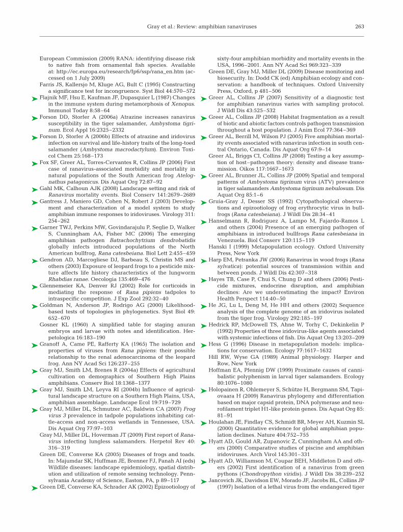
Gray et al.: Review: amphibian ranaviruses
European Commission (2009) RANA: identifying disease riskto native fish from ornamental fish species. Availableat: http://ec.europa.eu/research/fp6/ssp/rana_en.htm (ac-cessed on 1 July 2009)
Farris JS, Kallersjo M, Kluge AG, Bult C (1995) Constructinga significance test for incongruence. Syst Biol 44:570–572
Flajnik MF, Hsu E, Kaufman JF, Dupasquier L (1987) Changesin the immune system during metamorphosis of Xenopus.Immunol Today 8:58–64
Forson DD, Storfer A (2006a) Atrazine increases ranavirussusceptibility in the tiger salamander, Ambystoma tigri-num. Ecol Appl 16:2325–2332
Forson D, Storfer A (2006b) Effects of atrazine and iridovirusinfection on survival and life-history traits of the long-toedsalamander (Ambystoma macrodactylum). Environ Toxi-col Chem 25:168–173
Fox SF, Greer AL, Torres-Cervantes R, Collins JP (2006) Firstcase of ranavirus-associated morbidity and mortality innatural populations of the South American frog Atelog-nathus patagonicus. Dis Aquat Org 72:87–92
Gahl MK, Calhoun AJK (2008) Landscape setting and risk ofRanavirus mortality events. Biol Conserv 141:2679–2689
Gantress J, Maniero GD, Cohen N, Robert J (2003) Develop-ment and characterization of a model system to studyamphibian immune responses to iridoviruses. Virology 311:254–262
Garner TWJ, Perkins MW, Govindarajulu P, Seglie D, WalkerS, Cunningham AA, Fisher MC (2006) The emergingamphibian pathogen Batrachochytrium dendrobatidisglobally infects introduced populations of the NorthAmerican bullfrog, Rana catesbeiana. Biol Lett 2:455–459
Gendron AD, Marcogliese DJ, Barbeau S, Christin MS andothers (2003) Exposure of leopard frogs to a pesticide mix-ture affects life history characteristics of the lungwormRhabdias ranae. Oecologia 135:469–476
Glennemeier KA, Denver RJ (2002) Role for corticoids inmediating the response of Rana pipiens tadpoles tointraspecific competition. J Exp Zool 292:32–40
Goldman N, Anderson JP, Rodrigo AG (2000) Likelihood-based tests of topologies in phylogenetics. Syst Biol 49:652–670
Gosner KL (1960) A simplified table for staging anuranembryos and larvae with notes and identification. Her-petologica 16:183–190
Granoff A, Came PE, Rafferty KA (1965) The isolation andproperties of viruses from Rana pipiens: their possiblerelationship to the renal adenocarcinoma of the leopardfrog. Ann NY Acad Sci 126:237–255
Gray MJ, Smith LM, Brenes R (2004a) Effects of agriculturalcultivation on demographics of Southern High Plainsamphibians. Conserv Biol 18:1368–1377
Gray MJ, Smith LM, Leyva RI (2004b) Influence of agricul-tural landscape structure on a Southern High Plains, USA,amphibian assemblage. Landscape Ecol 19:719–729
Gray MJ, Miller DL, Schmutzer AC, Baldwin CA (2007) Frogvirus 3 prevalence in tadpole populations inhabiting cat-tle-access and non-access wetlands in Tennessee, USA.Dis Aquat Org 77:97–103
Gray MJ, Miller DL, Hoverman JT (2009) First report of Rana-virus infecting lungless salamanders. Herpetol Rev 40:316–319
Green DE, Converse KA (2005) Diseases of frogs and toads.In: Majumdar SK, Huffman JE, Brenner FJ, Panah AI (eds)Wildlife diseases: landscape epidemiology, spatial distrib-ution and utilization of remote sensing technology. Penn-sylvania Academy of Science, Easton, PA, p 89–117
Green DE, Converse KA, Schrader AK (2002) Epizootiology of
sixty-four amphibian morbidity and mortality events in theUSA, 1996–2001. Ann NY Acad Sci 969:323–339
Green DE, Gray MJ, Miller DL (2009) Disease monitoring andbiosecurity. In: Dodd CK (ed) Amphibian ecology and con-servation: a handbook of techniques. Oxford UniversityPress, Oxford, p 481–506
Greer AL, Collins JP (2007) Sensitivity of a diagnostic testfor amphibian ranavinus varies with sampling protocol.J Wildl Dis 43:525–532
Greer AL, Collins JP (2008) Habitat fragmentation as a resultof biotic and abiotic factors controls pathogen transmissionthroughout a host population. J Anim Ecol 77:364–369
Greer AL, Berrill M, Wilson PJ (2005) Five amphibian mortal-ity events associated with ranavirus infection in south cen-tral Ontario, Canada. Dis Aquat Org 67:9–14
Greer AL, Briggs CI, Collins JP (2008) Testing a key assump-tion of host–pathogen theory: density and disease trans-mission. Oikos 117:1667–1673
Greer AL, Brunner JL, Collins JP (2009) Spatial and temporalpatterns of Ambystoma tigrinum virus (ATV) prevalencein tiger salamanders Ambystoma tigrinum nebulosum. DisAquat Org 85:1–6
Gruia-Gray J, Desser SS (1992) Cytopathological observa-tions and epizootiology of frog erythrocytic virus in bull-frogs (Rana catesbeiana). J Wildl Dis 28:34–41
Hanselmann R, Rodriguez A, Lampo M, Fajardo-Ramos Land others (2004) Presence of an emerging pathogen ofamphibians in introduced bullfrogs Rana catesbeiana inVenezuela. Biol Conserv 120:115–119
Hanski I (1999) Metapopulation ecology. Oxford UniversityPress, New York
Harp EM, Petranka JW (2006) Ranavirus in wood frogs (Ranasylvatica): potential sources of transmission within andbetween ponds. J Wildl Dis 42:307–318
Hayes TB, Case P, Chui S, Chung D and others (2006) Pesti-cide mixtures, endocrine disruption, and amphibiandeclines: Are we underestimating the impact? EnvironHealth Perspect 114:40–50
He JG, Lu L, Deng M, He HH and others (2002) Sequenceanalysis of the complete genome of an iridovirus isolatedfrom the tiger frog. Virology 292:185–197
Hedrick RP, McDowell TS, Ahne W, Torhy C, Dekinkelin P(1992) Properties of three iridovirus-like agents associatedwith systemic infections of fish. Dis Aquat Org 13:203–209
Hess G (1996) Disease in metapopulation models: implica-tions for conservation. Ecology 77:1617–1632
Hill RW, Wyse GA (1989) Animal physiology. Harper andRow, New York
Hoffman EA, Pfennig DW (1999) Proximate causes of canni-balistic polyphenism in larval tiger salamanders. Ecology80:1076–1080
Holopainen R, Ohlemeyer S, Schütze H, Bergmann SM, Tapi-ovaara H (2009) Ranavirus phylogeny and differentiationbased on major capsid protein, DNA polymerase and neu-rofilament triplet H1-like protein genes. Dis Aquat Org 85:81–91
Houlahan JE, Findlay CS, Schmidt BR, Meyer AH, Kuzmin SL(2000) Quantitative evidence for global amphibian popu-lation declines. Nature 404:752–755
Hyatt AD, Gould AR, Zupanovic Z, Cunningham AA and oth-ers (2000) Comparative studies of piscine and amphibianiridoviruses. Arch Virol 145:301–331
Hyatt AD, Williamson M, Coupar BEH, Middleton D and oth-ers (2002) First identification of a ranavirus from greenpythons (Chondropython viridis). J Wildl Dis 38:239–252
Jancovich JK, Davidson EW, Morado JF, Jacobs BL, Collins JP(1997) Isolation of a lethal virus from the endangered tiger
263

Dis Aquat Org 87: 243–266, 2009
salamander Ambystoma tigrinum stebbinsi. Dis AquatOrg 31:161–167
Jancovich JK, Davidson EW, Seiler A, Jacobs BL, Collins JP(2001) Transmission of the Ambystoma tigrinum virus toalternative hosts. Dis Aquat Org 46:159–163
Jancovich JK, Mao JH, Chinchar VG, Wyatt C and others(2003) Genomic sequence of a ranavirus (family Iridoviri-dae) associated with salamander mortalities in NorthAmerica. Virology 316:90–103
Jancovich JK, Davidson EW, Parameswaran N, Mao J andothers (2005) Evidence for emergence of an amphibian iri-doviral disease because of human-enhanced spread. MolEcol 14:213–224
Jofre MB, Karasov WH (1999) Direct effect of ammonia onthree species of North American anuran amphibians.Environ Toxicol Chem 18:1806–1812
Johnson AJ, Pessier AP, Wellehan JFX, Childress A andothers (2008) Ranavirus infection of free-ranging and cap-tive box turtles and tortoises in the United States. J WildlDis 44:851–863
Kattenbelt JA, Hyatt AD, Gould AR (2000) Recovery of rana-virus dsDNA from formalin-fixed archival material. DisAquat Org 39:151–154
Knutson MG, Sauer JR, Olsen DA, Mossman MJ, HemesathLM, Lannoo MJ (1999) Effects of landscape compositionand wetland fragmentation on frog and toad abundanceand species richness in Iowa and Wisconsin, USA. Con-serv Biol 13:1437–1446
Kolozsvary MB, Swihart RK (1999) Habitat fragmentation andthe distribution of amphibians: patch and landscape corre-lates in farmland. Can J Zool 77:1288–1299
Langdon JS (1989) Experimental transmission and patho-genicity of epizootic hematopoietic necrosis virus (EHNV)in redfin perch, Perca fluviatilis L. and 11 other teleosts.J Fish Dis 12:295–310
Langdon JS, Humphrey JD, Williams LM, Hyatt AD, West-bury HA (1986) First virus isolation from Australian fish:an iridovirus-like pathogen from redfin perch, Perca fluvi-atilis L. J Fish Dis 9:263–268
Lloyd-Smith JO, Cross PC, Briggs CJ, Daugherty M and oth-ers (2005a) Should we expect population thresholds forwildlife diseases? Trends Ecol Evol 20:511–519
Lloyd-Smith JO, Schreiber SJ, Getz WM (2005b) Super-spreading and the effect of individual variation on diseaseemergence. Nature 438:355–359
Majji S, LaPatra S, Long SM, Sample R, Bryan L, Sinning A,Chinchar VG (2006) Rana catesbeiana virus Z (RCV-Z): anovel pathogenic ranavirus. Dis Aquat Org 73:1–11
Maniero GD, Carey C (1997) Changes in selected aspects ofimmune function in the leopard frog, Rana pipiens, associ-ated with exposure to cold. J Comp Physiol B 167:256–263
Maniero GD, Morales H, Gantress J, Robert J (2006) Genera-tion of a long-lasting, protective, and neutralizing anti-body response to the ranavirus FV3 by the frog Xenopus.Dev Comp Immunol 30:649–657
Mao J, Tham TN, Gentry GA, Aubertin A, Chinchar VG(1996) Cloning, sequence analysis, and expression of themajor capsid protein of the iridovirus frog virus 3. Virology216:431–436
Mao J, Green DE, Fellers G, Chinchar VG (1999) Molecularcharacterization of iridoviruses isolated from sympatricamphibians and fish. Virus Res 63:45–52
Marschang RE, Becher P, Posthaus H, Wild P and others(1999) Isolation and characterization of an iridovirus fromHermann’s tortoises (Testudo hermanni). Arch Virol 144:1909–1922
Marschang RE, Braun S, Becher P (2005) Isolation of a rana-
virus from a gecko (Uroplatus fimbriatus). J Zoo WildlMed 36:295–300
Marsh DM, Trenham PC (2001) Metapopulation dynamicsand amphibian conservation. Conserv Biol 15:40–49
McCallum H, Barlow N, Hone J (2001) How should pathogentransmission be modelled? Trends Ecol Evol 16:295–300
Miller DL, Rajeev S, Gray MJ, Baldwin CA (2007) Frog virus 3infection, cultured American bullfrogs. Emerg Infect Dis13:342–343
Miller DL, Rajeev S, Brookins M, Cook J, Whittington L, Bald-win CA (2008) Concurrent infection with Ranavirus,Batrachochytrium dendrobatidis, and Aeromonas in acaptive anuran colony. J Zoo Wildl Med 39:445–449
Miller DL, Gray MJ, Rajeev S, Schmutzer AC, Burton EC,Merrill A, Baldwin CA (2009) Pathological findings in lar-val and juvenile anurans inhabiting farm ponds in Ten-nessee, USA. J Wildl Dis 45:314–324
Miodonski AJ, Bigaj J, Mika J, P8ytycz B (1996) Season-spe-cific thymic architecture in the frog, Rana temporaria:SEM studies. Dev Comp Immunol 20:129–137
Moody NJG, Owens L (1994) Experimental demonstration ofthe pathogenicity of a frog virus, Bohle iridovirus, for a fishspecies, barramundi Lates calcarifer. Dis Aquat Org 18:95–102
Muths E, Gallant AL, Campbell EHC, Battaglin WA and oth-ers (2006) The Amphibian Research and Monitoring Initia-tive (ARMI): 5-year report. US Geological Survey Scien-tific Investigations Report 2006-5224
Muths E, Pilliod DS, Livo LJ (2008) Distribution and environ-mental limitations of an amphibian pathogen in the RockyMountains, USA. Biol Conserv 141:1484–1492
Nakajima K, Ito T, Kurita J, Kawakami H and others (2002)Effectiveness of a vaccine against red sea bream iridoviraldisease in various cultured marine fish under laboratoryconditions. Fish Pathol 37:90–91
Ottaviani E, Franceschi C (1996) The neuroimmunology ofstress from invertebrates to man. Prog Neurobiol 48:421–440
Pallister J, Gould A, Harrison D, Hyatt A, Jancovich J, HeineH (2007) Development of real-time PCR assays for thedetection and differentiation of Australian and Europeanranaviruses. J Fish Dis 30:427–438
Parris MJ, Davis A, Collins JP (2004) Single-host pathogeneffects on mortality and behavioral responses to predatorsin salamanders (Urodela: Ambystomatidae). Can J Zool82:1477–1483
Parris MJ, Storfer A, Collins JP, Davidson EW (2005) Life-history responses to pathogens in tiger salamander(Ambystoma tigrinum) larvae. J Herpetol 39:366–372
Pasmans F, Blahak S, Martel A, Pantchev N, Zwart P (2008)Ranavirus-associated mass mortality in imported redtailed knobby newts (Tylototriton kweichowensis): a casereport. Vet J 176:257–259
Pearman PB, Garner TWJ (2005) Susceptibility of Italian agilefrog populations to an emerging strain of Ranavirus paral-lels population genetic diversity. Ecol Lett 8:401–408
Pearman PB, Garner TWJ, Straub M, Greber UF (2004)Response of the Italian agile frog (Rana latastei) to a Rana-virus, frog virus 3: a model for viral emergence in naivepopulations. J Wildl Dis 40:660–669
Pfennig DW, Murphy PJ (2000) Character displacement inpolyphenic tadpoles. Evolution 54:1738–1749
Pfennig DW, Loeb MLG, Collins JP (1991) Pathogens as afactor limiting the spread of cannibalism in tiger sala-manders. Oecologia 88:161–166
Picco AM, Collins JP (2008) Amphibian commerce as a likelysource of pathogen pollution. Conserv Biol 22:1582–1589
264
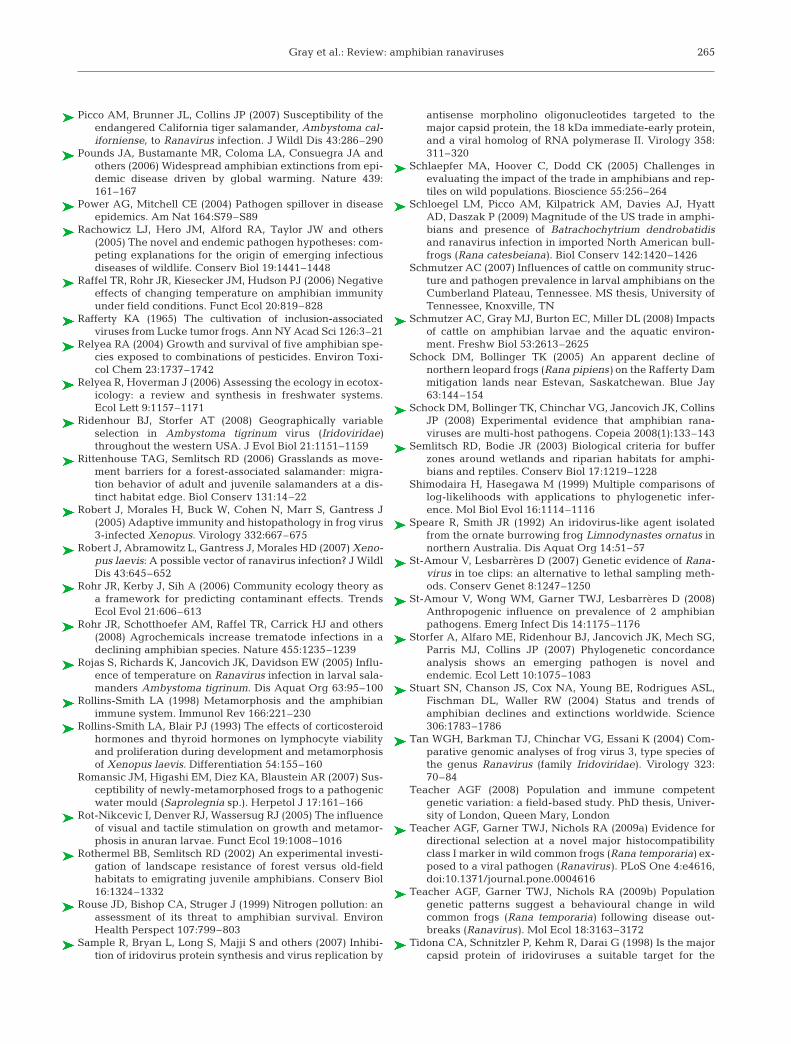
Gray et al.: Review: amphibian ranaviruses
Picco AM, Brunner JL, Collins JP (2007) Susceptibility of theendangered California tiger salamander, Ambystoma cal-iforniense, to Ranavirus infection. J Wildl Dis 43:286–290
Pounds JA, Bustamante MR, Coloma LA, Consuegra JA andothers (2006) Widespread amphibian extinctions from epi-demic disease driven by global warming. Nature 439:161–167
Power AG, Mitchell CE (2004) Pathogen spillover in diseaseepidemics. Am Nat 164:S79–S89
Rachowicz LJ, Hero JM, Alford RA, Taylor JW and others(2005) The novel and endemic pathogen hypotheses: com-peting explanations for the origin of emerging infectiousdiseases of wildlife. Conserv Biol 19:1441–1448
Raffel TR, Rohr JR, Kiesecker JM, Hudson PJ (2006) Negativeeffects of changing temperature on amphibian immunityunder field conditions. Funct Ecol 20:819–828
Rafferty KA (1965) The cultivation of inclusion-associatedviruses from Lucke tumor frogs. Ann NY Acad Sci 126:3–21
Relyea RA (2004) Growth and survival of five amphibian spe-cies exposed to combinations of pesticides. Environ Toxi-col Chem 23:1737–1742
Relyea R, Hoverman J (2006) Assessing the ecology in ecotox-icology: a review and synthesis in freshwater systems.Ecol Lett 9:1157–1171
Ridenhour BJ, Storfer AT (2008) Geographically variableselection in Ambystoma tigrinum virus (Iridoviridae)throughout the western USA. J Evol Biol 21:1151–1159
Rittenhouse TAG, Semlitsch RD (2006) Grasslands as move-ment barriers for a forest-associated salamander: migra-tion behavior of adult and juvenile salamanders at a dis-tinct habitat edge. Biol Conserv 131:14–22
Robert J, Morales H, Buck W, Cohen N, Marr S, Gantress J(2005) Adaptive immunity and histopathology in frog virus3-infected Xenopus. Virology 332:667–675
Robert J, Abramowitz L, Gantress J, Morales HD (2007) Xeno-pus laevis: A possible vector of ranavirus infection? J WildlDis 43:645–652
Rohr JR, Kerby J, Sih A (2006) Community ecology theory asa framework for predicting contaminant effects. TrendsEcol Evol 21:606–613
Rohr JR, Schotthoefer AM, Raffel TR, Carrick HJ and others(2008) Agrochemicals increase trematode infections in adeclining amphibian species. Nature 455:1235–1239
Rojas S, Richards K, Jancovich JK, Davidson EW (2005) Influ-ence of temperature on Ranavirus infection in larval sala-manders Ambystoma tigrinum. Dis Aquat Org 63:95–100
Rollins-Smith LA (1998) Metamorphosis and the amphibianimmune system. Immunol Rev 166:221–230
Rollins-Smith LA, Blair PJ (1993) The effects of corticosteroidhormones and thyroid hormones on lymphocyte viabilityand proliferation during development and metamorphosisof Xenopus laevis. Differentiation 54:155–160
Romansic JM, Higashi EM, Diez KA, Blaustein AR (2007) Sus-ceptibility of newly-metamorphosed frogs to a pathogenicwater mould (Saprolegnia sp.). Herpetol J 17:161–166
Rot-Nikcevic I, Denver RJ, Wassersug RJ (2005) The influenceof visual and tactile stimulation on growth and metamor-phosis in anuran larvae. Funct Ecol 19:1008–1016
Rothermel BB, Semlitsch RD (2002) An experimental investi-gation of landscape resistance of forest versus old-fieldhabitats to emigrating juvenile amphibians. Conserv Biol16:1324–1332
Rouse JD, Bishop CA, Struger J (1999) Nitrogen pollution: anassessment of its threat to amphibian survival. EnvironHealth Perspect 107:799–803
Sample R, Bryan L, Long S, Majji S and others (2007) Inhibi-tion of iridovirus protein synthesis and virus replication by
antisense morpholino oligonucleotides targeted to themajor capsid protein, the 18 kDa immediate-early protein,and a viral homolog of RNA polymerase II. Virology 358:311–320
Schlaepfer MA, Hoover C, Dodd CK (2005) Challenges inevaluating the impact of the trade in amphibians and rep-tiles on wild populations. Bioscience 55:256–264
Schloegel LM, Picco AM, Kilpatrick AM, Davies AJ, HyattAD, Daszak P (2009) Magnitude of the US trade in amphi-bians and presence of Batrachochytrium dendrobatidisand ranavirus infection in imported North American bull-frogs (Rana catesbeiana). Biol Conserv 142:1420–1426
Schmutzer AC (2007) Influences of cattle on community struc-ture and pathogen prevalence in larval amphibians on theCumberland Plateau, Tennessee. MS thesis, University ofTennessee, Knoxville, TN
Schmutzer AC, Gray MJ, Burton EC, Miller DL (2008) Impactsof cattle on amphibian larvae and the aquatic environ-ment. Freshw Biol 53:2613–2625
Schock DM, Bollinger TK (2005) An apparent decline ofnorthern leopard frogs (Rana pipiens) on the Rafferty Dammitigation lands near Estevan, Saskatchewan. Blue Jay63:144–154
Schock DM, Bollinger TK, Chinchar VG, Jancovich JK, CollinsJP (2008) Experimental evidence that amphibian rana-viruses are multi-host pathogens. Copeia 2008(1):133–143
Semlitsch RD, Bodie JR (2003) Biological criteria for bufferzones around wetlands and riparian habitats for amphi-bians and reptiles. Conserv Biol 17:1219–1228
Shimodaira H, Hasegawa M (1999) Multiple comparisons oflog-likelihoods with applications to phylogenetic infer-ence. Mol Biol Evol 16:1114–1116
Speare R, Smith JR (1992) An iridovirus-like agent isolatedfrom the ornate burrowing frog Limnodynastes ornatus innorthern Australia. Dis Aquat Org 14:51–57
St-Amour V, Lesbarrères D (2007) Genetic evidence of Rana-virus in toe clips: an alternative to lethal sampling meth-ods. Conserv Genet 8:1247–1250
St-Amour V, Wong WM, Garner TWJ, Lesbarrères D (2008)Anthropogenic influence on prevalence of 2 amphibianpathogens. Emerg Infect Dis 14:1175–1176
Storfer A, Alfaro ME, Ridenhour BJ, Jancovich JK, Mech SG,Parris MJ, Collins JP (2007) Phylogenetic concordanceanalysis shows an emerging pathogen is novel andendemic. Ecol Lett 10:1075–1083
Stuart SN, Chanson JS, Cox NA, Young BE, Rodrigues ASL,Fischman DL, Waller RW (2004) Status and trends ofamphibian declines and extinctions worldwide. Science306:1783–1786
Tan WGH, Barkman TJ, Chinchar VG, Essani K (2004) Com-parative genomic analyses of frog virus 3, type species ofthe genus Ranavirus (family Iridoviridae). Virology 323:70–84
Teacher AGF (2008) Population and immune competentgenetic variation: a field-based study. PhD thesis, Univer-sity of London, Queen Mary, London
Teacher AGF, Garner TWJ, Nichols RA (2009a) Evidence fordirectional selection at a novel major histocompatibilityclass I marker in wild common frogs (Rana temporaria) ex-posed to a viral pathogen (Ranavirus). PLoS One 4:e4616,doi:10.1371/journal.pone.0004616
Teacher AGF, Garner TWJ, Nichols RA (2009b) Populationgenetic patterns suggest a behavioural change in wildcommon frogs (Rana temporaria) following disease out-breaks (Ranavirus). Mol Ecol 18:3163–3172
Tidona CA, Schnitzler P, Kehm R, Darai G (1998) Is the majorcapsid protein of iridoviruses a suitable target for the
265
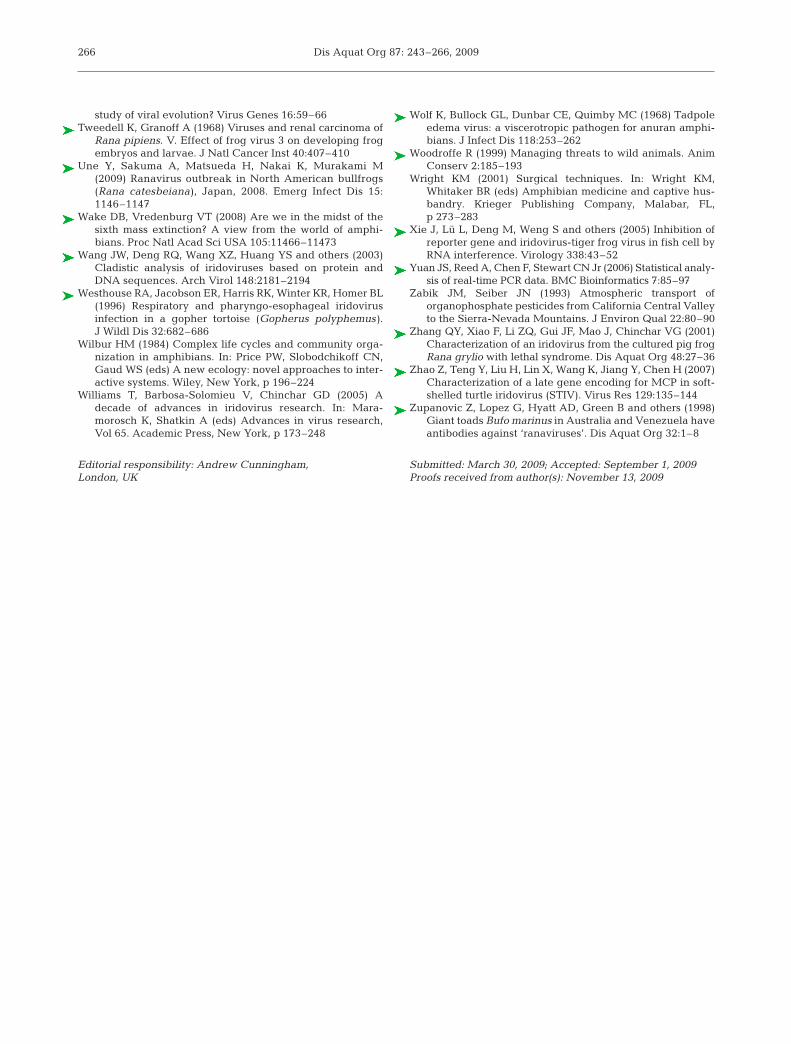
Dis Aquat Org 87: 243–266, 2009
study of viral evolution? Virus Genes 16:59–66 Tweedell K, Granoff A (1968) Viruses and renal carcinoma of
Rana pipiens. V. Effect of frog virus 3 on developing frogembryos and larvae. J Natl Cancer Inst 40:407–410
Une Y, Sakuma A, Matsueda H, Nakai K, Murakami M(2009) Ranavirus outbreak in North American bullfrogs(Rana catesbeiana), Japan, 2008. Emerg Infect Dis 15:1146–1147
Wake DB, Vredenburg VT (2008) Are we in the midst of thesixth mass extinction? A view from the world of amphi-bians. Proc Natl Acad Sci USA 105:11466–11473
Wang JW, Deng RQ, Wang XZ, Huang YS and others (2003)Cladistic analysis of iridoviruses based on protein andDNA sequences. Arch Virol 148:2181–2194
Westhouse RA, Jacobson ER, Harris RK, Winter KR, Homer BL(1996) Respiratory and pharyngo-esophageal iridovirusinfection in a gopher tortoise (Gopherus polyphemus).J Wildl Dis 32:682–686
Wilbur HM (1984) Complex life cycles and community orga-nization in amphibians. In: Price PW, Slobodchikoff CN,Gaud WS (eds) A new ecology: novel approaches to inter-active systems. Wiley, New York, p 196–224
Williams T, Barbosa-Solomieu V, Chinchar GD (2005) Adecade of advances in iridovirus research. In: Mara-morosch K, Shatkin A (eds) Advances in virus research,Vol 65. Academic Press, New York, p 173–248
Wolf K, Bullock GL, Dunbar CE, Quimby MC (1968) Tadpoleedema virus: a viscerotropic pathogen for anuran amphi-bians. J Infect Dis 118:253–262
Woodroffe R (1999) Managing threats to wild animals. AnimConserv 2:185–193
Wright KM (2001) Surgical techniques. In: Wright KM,Whitaker BR (eds) Amphibian medicine and captive hus-bandry. Krieger Publishing Company, Malabar, FL,p 273–283
Xie J, Lü L, Deng M, Weng S and others (2005) Inhibition ofreporter gene and iridovirus-tiger frog virus in fish cell byRNA interference. Virology 338:43–52
Yuan JS, Reed A, Chen F, Stewart CN Jr (2006) Statistical analy-sis of real-time PCR data. BMC Bioinformatics 7:85–97
Zabik JM, Seiber JN (1993) Atmospheric transport oforganophosphate pesticides from California Central Valleyto the Sierra-Nevada Mountains. J Environ Qual 22:80–90
Zhang QY, Xiao F, Li ZQ, Gui JF, Mao J, Chinchar VG (2001)Characterization of an iridovirus from the cultured pig frogRana grylio with lethal syndrome. Dis Aquat Org 48:27–36
Zhao Z, Teng Y, Liu H, Lin X, Wang K, Jiang Y, Chen H (2007)Characterization of a late gene encoding for MCP in soft-shelled turtle iridovirus (STIV). Virus Res 129:135–144
Zupanovic Z, Lopez G, Hyatt AD, Green B and others (1998)Giant toads Bufo marinus in Australia and Venezuela haveantibodies against ‘ranaviruses’. Dis Aquat Org 32:1–8
266
Editorial responsibility: Andrew Cunningham, London, UK
Submitted: March 30, 2009; Accepted: September 1, 2009Proofs received from author(s): November 13, 2009