vitamin d status and hyperparathyroidism in postmeno
TRANSCRIPT

69
ORIGINAL ARTICLE
ABSTRACTAim: to determine the prole of vitamin D and
parathyroid hormone (PTH) and the proportion of vitamin D inadequacy in a population of postmenopausal osteoporotic patients from a rheumatologic outpatient clinic.
Methods: a cross sectional study was conducted between October and December 2006 in the Rheumatology Clinic, Cipto Mangunkusumo Hospital with osteoporosis conrmed by bone mineral densitometry (T score less than –2.5 at the lumbar spine or hip). Patients were excluded if there was a history of oral glucocorticoid treatment within 30 days, vitamin D supplementation, and have renal and/or liver function impairments. Forty-two postmenopausal osteoporotic patients aged 51-77 years old who had been postmenopausal for 5-28 years were included in this study. Vitamin D inadequacy was dened as the plasma levels of 25(OH)D less than 50 nmol/L whereas hyperparathyroidism was dened as the PTH level more than 69 pg/dL.
Results: vitamin D inadequacy was found in 61.9 % of patients and 34.6% of them or 23.8% of total patients were also having high PTH level. There was an inverse correlation between 25(OH)D with PTH levels and positive correlation between duration of menopause and PTH level. Vitamin D inadequacy is common (61.9 %) in postmenopausal osteoporotic patients who visited Rheumatology out patient clinic Cipto Mangunkusumo Hospital Jakarta.
Conclusion: the low concentration of 25(OH)D was correlated with PTH level and duration of menopause. This nding should be conrmed in a larger epidemiological study, either hospital-or community-based to assess vitamin D status among postmenopausal women in Indonesia.
Key words: vitamin D, calcium, parathyroid hormone, postmenopausal osteoporosis.
Vitamin D Status and Hyperparathyroidism in Postmeno-pausal Osteoporotic Patients in Cipto Mangunkusumo Hospital, JakartaSumariyono Sarmidi, Bambang Setiyohadi, Suryo Anggoro KW
Department of Internal Medicine, Faculty of Medicine-dr. Cipto Mangunkusumo Hospital, Jl. Diponegoro no. 71 Jakarta. E-mail: [email protected].
INTRODUCTIONPostmenopausal osteoporosis is a common health
problem today with a wide clinical spectrum and potentially debilitating if the osteoporotic bone becomes fractured.1 The pathogenesis of postmenopausal osteoporosis involves many factors such as estrogen deciency, low calcium intake, vitamin D deciency and secondary hyperparathyroidism. Less severe vitamin D deciency, also called vitamin D insufciency2 or inadequacy, has been a major research topic in the last decade due to its essential role in calcium homeostasis and bone metabolism.
Vitamin D is a prohormone which undergoes two-step metabolism to produce the active metabolite i.e. 1,25-dihydroxyvitamin D [1,25(OH)2D3] with its emerging important actions.3 Vitamin D is synthe-sized in the skin during exposure to sunlight, when 7-dehydrocholesterol (7-DHC) in the skin undergoes photoconversion to previtamin D3.
4 Vitamin D is then transported to the liver and is hydroxylated to 25(OH)D3 in the liver and then converted to 1,25(OH)2D3 in the kidney. 5 Many factors affect the cutaneous production of vitamin D3 such as sun exposure, melanin and age. Melanin is an excellent natural sunscreen and competes with 7-DHC for ultraviolet B photon, therefore increased skin melanin pigmentation decreases the photosynthesis of vitamin D3.
6 Aging significantly diminished the concentration of unesteried 7-DHC in the epidermis.7
Most post menopausal osteoporosis studies, particularly the vitamin D status, were carried out in four-season countries like European countries, United States and Australia. Countries that lie far from the equator, such as northern Europe, northern part of United States and Canada have been associated with high vitamin D inadequacy, especially in winter season. During winter, the sunlight must pass a much longer distance through the atmosphere where most of the ultraviolet B light is absorbed, thus, vitamin D synthesis is virtually absent.8 However, the expected association

70
Sumariyono Sarmidi, et al Acta Med Indones-Indones J Intern Med
between latitude and vitamin D inadequacy was not found in the international MORE study.9 Vitamin D fortification policies, dietary habits, time spent outdoors, clothing habits, skin type and pig-mentation were argued to influence the vitamin D levels in that study.
Vitamin D status is assessed by measuring the level of serum 25(OH)D, the major circulating metabolite of vitamin D. A low serum 25(OH)D concentration is the hallmark of vitamin D deciency. The low level of 25(OH)D in elderly has been associated with a high serum PTH.10 The low serum 25(OH)D concentration leads to a decrease of 1,25(OH) 2D3 and calcium absorption. The lower calcium concentration then causes an increase of PTH secretion, which stimulates the production of 1,25(OH) 2D3. This mechanism has been proposed to explain whereby vitamin D deciency could contribute to the pathogenesis of hip fracture.11
Data on vitamin D status from equatorial countries like Indonesia, where the sunlight exposure is high, are limited. It was assumed that the vitamin D levels among Indonesian postmenopausal women would be higher than the Caucasians. This study was aimed at evaluating vitamin D status and parathyroid hormone levels among postmenopausal osteoporotic patients in Cipto Mangunkusumo Hospital.
METHODS
Study Designs and SubjectsThis was a cross-sectional study in postmenopausal
osteoporotic women living in Jakarta and its surroundings. The study population was postmenopausal osteoporotic patients who visited the Rheumatology Clinic, Cipto Mangunkusumo Central Hospital. Samples were patients who came between October and December 2006.
Patients would have been eligible if they had been postmenopausal (i.e. the absence of menses) for at least 5 years and osteoporotic as dened by a T-score below 2,5 on hip or spine bone mineral densitometry (dual-energy X ray absorptiometry; Lunar GE Prodigy). Eligible patients were asked to sign a written informed consent prior to enrollment into the study. Patients were excluded from the study if there had been a history of glucocorticoid treatment in the last 30 days, abnormal liver and/or renal functions, or if the patient had refused to participate in the study. Patients were recruited consecutively (purposive sampling) until the required minimum sample was achieved.
Blood Chemistry AssaysPatients who met the inclusion criteria underwent
blood chemistry test including serum creatinin, liver
function test, and serum albumin. Next, serum 25(OH)D, PTH, and calcium levels were measured. Blood sample was taken between 8 and 10 a.m. from the patients after fasting overnight. Serum 25(OH)D was measured by enzyme-link immunosorbent assay (ELISA) method (ELLISA Reader 530). The normal range is 47.7-144 nmol/L. Vitamin D inadequacy was defined when the level of 25(OH)D was < 50 nmol/L. Intact PTH assay was done by using immunochemi-luminescent technique (IMMULITE 2000). The normal values of PTH are 10-69 pg/mL. Hyperparathyroidism is dened if the level of PTH is > 69 pg/mL. Total serum calcium level was measured to assess the calcium status. The normal values are 8.4-9.7 mg/dL.
Data AnalysisCharacteristics of the study subjects were pre-
sented descriptively. Correlative analyses were done among variables using Pearson correlation coefcient. Statistical analysis was done using the SPSS for Windows PC version 12.0 (SPSS, Inc, Chicago, IL) computer software.
RESULTSA total of 42 patients aged between 53 and 77 years
had been recruited. There were 4 (0.5%) patients aged 60 years or less, 8 (19.0%) aged 61-65 years, 18 (42.9%) aged 66-70 years and 12 (28.6%) aged more than 70 years. The 25(OH)D, PTH, and total serum calcium levels are given in Table 1. Almost all patients had pain as they were suffering from several rheumatologic diseases, e.g. osteoarthritis, rheumatoid arthritis, and bromyalgia; one patient presented with scleroderma. Low serum 25(OH)D levels were found in 26 (61.9%) patients while increased PTH levels were found in 10 (23.8%) patients. Nine out of 26 patients (34.6%) with low 25(OH)D levels had high PTH levels.
71
Vol 40 • Number 2 • April 2008 Vitamin D Status and Hyperparathyroidism in Postmenopausal Osteoporotic
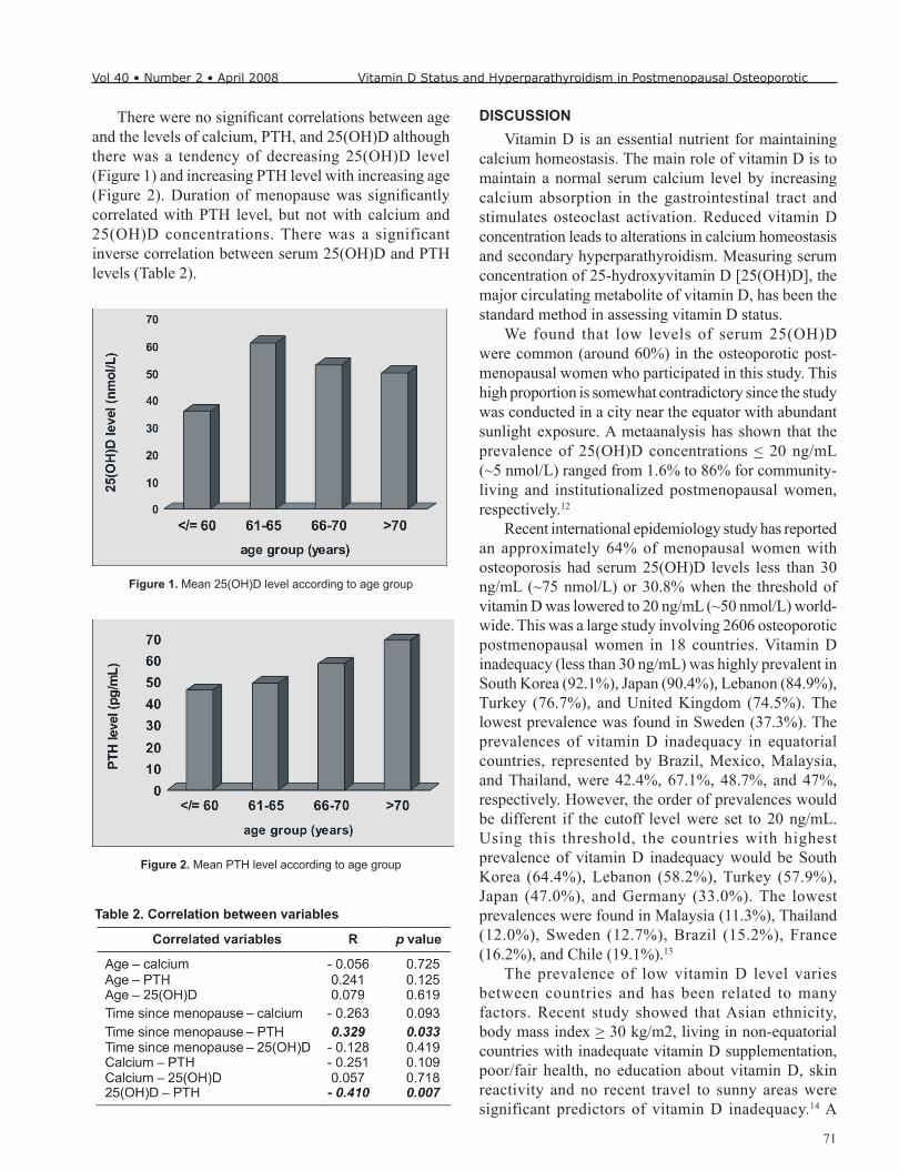
There were no signicant correlations between age and the levels of calcium, PTH, and 25(OH)D although there was a tendency of decreasing 25(OH)D level (Figure 1) and increasing PTH level with increasing age (Figure 2). Duration of menopause was signicantly correlated with PTH level, but not with calcium and 25(OH)D concentrations. There was a significant inverse correlation between serum 25(OH)D and PTH levels (Table 2).
DISCUSSIONVitamin D is an essential nutrient for maintaining
calcium homeostasis. The main role of vitamin D is to maintain a normal serum calcium level by increasing calcium absorption in the gastrointestinal tract and stimulates osteoclast activation. Reduced vitamin D concentration leads to alterations in calcium homeostasis and secondary hyperparathyroidism. Measuring serum concentration of 25-hydroxyvitamin D [25(OH)D], the major circulating metabolite of vitamin D, has been the standard method in assessing vitamin D status.
We found that low levels of serum 25(OH)D were common (around 60%) in the osteoporotic post-menopausal women who participated in this study. This high proportion is somewhat contradictory since the study was conducted in a city near the equator with abundant sunlight exposure. A metaanalysis has shown that the prevalence of 25(OH)D concentrations < 20 ng/mL (~5 nmol/L) ranged from 1.6% to 86% for community-living and institutionalized postmenopausal women, respectively.12
Recent international epidemiology study has reported an approximately 64% of menopausal women with osteoporosis had serum 25(OH)D levels less than 30 ng/mL (~75 nmol/L) or 30.8% when the threshold of vitamin D was lowered to 20 ng/mL (~50 nmol/L) world-wide. This was a large study involving 2606 osteoporotic postmenopausal women in 18 countries. Vitamin D inadequacy (less than 30 ng/mL) was highly prevalent in South Korea (92.1%), Japan (90.4%), Lebanon (84.9%), Turkey (76.7%), and United Kingdom (74.5%). The lowest prevalence was found in Sweden (37.3%). The prevalences of vitamin D inadequacy in equatorial countries, represented by Brazil, Mexico, Malaysia, and Thailand, were 42.4%, 67.1%, 48.7%, and 47%, respectively. However, the order of prevalences would be different if the cutoff level were set to 20 ng/mL. Using this threshold, the countries with highest prevalence of vitamin D inadequacy would be South Korea (64.4%), Lebanon (58.2%), Turkey (57.9%), Japan (47.0%), and Germany (33.0%). The lowest prevalences were found in Malaysia (11.3%), Thailand (12.0%), Sweden (12.7%), Brazil (15.2%), France (16.2%), and Chile (19.1%).13
The prevalence of low vitamin D level varies between countries and has been related to many factors. Recent study showed that Asian ethnicity, body mass index > 30 kg/m2, living in non-equatorial countries with inadequate vitamin D supplementation, poor/fair health, no education about vitamin D, skin reactivity and no recent travel to sunny areas were significant predictors of vitamin D inadequacy.14 A
Figure 1. Mean 25(OH)D level according to age group
Figure 2. Mean PTH level according to age group

72
Sumariyono Sarmidi, et al Acta Med Indones-Indones J Intern Med
study in Jakarta and its neighboring city, Bekasi, found that among residents of an institutionalized care (panti werdha), the prevalence of vitamin D deciency was 35.1% using the same cutoff level of 50 nmol/L.15 In North America, vitamin D level less than 20 ng/mL (~50 nmol/L) was found in only 18.2% of women receiving osteoporosis therapies. However, this study has included Caucasian healthy women who were well-educated and more aware of the importance of vitamin D to bone health.16 The high proportion of vitamin D inadequacy in our patients might be partly caused by a different study population from those epidemiology studies stated above. Many studies have included active people who were still exposed to the sunlight. Our study subjects were patients in the Rheumatology Clinic suffering from various rheumatic diseases with considerable pain. These kinds of patients probably limit their activities and thus limiting their exposure to the sunlight. Although previtamin D below the skin is decreased with increasing age, sun exposure will still activate the synthesis of vitamin D beneath the skin; particularly if the skin is exposed to the ultraviolet B light long enough during the peak hours.
There has been a study which has the same patients’ characteristics with ours. i.e. in an outpatient rheumatology clinic in Spain. They reported a higher prevalence of low serum 25(OH)D concentration below 50 nmol/L, i.e. 84%.17 Hypovitaminosis D was common in hospitalized patients including those without apparent risk factors for vitamin D deciency.18
Our results also showed a tendency of reduced vitamin D levels with the increasing age. However, the lowest vitamin D levels were found in patients aged less than 60 years. This contradictory nding could be explained by the small number of patients (4 patients) who came with specific conditions. The first was a severely osteoporotic patient with hip and vertebral fractures who was unable to walk and sitting in a wheel chair; her vitamin D level was 38.6 nmol/L. The second patient had scleroderma with the vitamin D level of 47.6 nmol/L; it is possible that the previtamin D level is also low in this skin disorder. The last two patients were women who wore full-covered clothing. Study has shown that clothing could prevent the photosynthesis of vitamin D3.
19 None of these patients had a high PTH level. This would probably occur for several reasons: The effect of vitamin D on PTH level occurs through two pathways, direct and indirect ways. The direct effect is through the vitamin D receptor (VDR) in parathyroid gland which has antiproliferative effect and suppression of PTH gene transcription. The indirect effect is through its effect on calcium absorption in gastrointestinal
tract: low vitamin D level causes to reduce calcium absorption in the gastrointestinal tract and to produce hypocalcemia which, in turn, stimulates the secretion of PTH. Although the level of vitamin D is decreasing, if the calcium intake is high and the age is relatively younger, the total calcium absorbed will be better in than the higher age group and the stimulation of PTH secretion due to hypocalcemia which is also lower.
Hyperparathyroidism depends on several things, i.e. hypocalcemia, vitamin D deciency and age. In this current study, there was an inverse correlation between vitamin D and PTH levels (Table 2) but only one-third of the patients with low vitamin D level had high PTH. A similar rate was found also in other study that found secondary hyperparathyroidism in only one-third of postmenopausal women with established osteoporosis.20 This result supports the theory and study that vitamin D level inuences the PTH secretion, the lower the vitamin D level, the higher PTH secretion or the higher the vitamin D level, the lower the PTH secretion through its antiproliferative effect in parathyroid gland. However, the secretion of PTH does not solely depend on vitamin D; there are several other factors that increase PTH level.21 Increasing serum PTH level could be the early biochemical changes of vitamin D insufciency. PTH levels begin to rise as serum 25(OH)D levels fall below 80 nmol/L after being adjusted for serum ionized calcium, body weight, and age.22 The risk of developing secondary hyperparathyroidism due to vitamin D deciency was higher in hospitalized elderly.23
Hypocalcemia could also stimulate PTH secretion. However, it should be remembered that in secondary hyperparathyroidism due to hypocalcemia, the serum calcium level usually is normal low or slightly below normal.24 Our study results were consistent with this theory. The negative correlation between calcium and PTH levels was weak and not statistically signicant. This could happen because, after PTH is secreted, calcium mobilization from the bones and renal calcium reabsorption were increased, and thus the serum calcium level was maintained around normal level.
CONCLUSION Vitamin D inadequacy is common (61.9 %) in
postmenopausal osteoporotic patients who visited to Rheumatology out patient clinic Cipto Mangunku-sumo Hospital Jakarta. The low concentration of 25(OH)D was correlated with PTH level and duration of menopause. This finding should be confirmed in a larger epidemiological study, either hospital- or community-based, to know the vitamin D status among postmenopausal women in Indonesia.

73
Vol 40 • Number 2 • April 2008 Vitamin D Status and Hyperparathyroidism in Postmenopausal Osteoporotic
REFERENCES1. Rosen CJ. Postmenopausal osteoporosis. N Engl J Med.
2005;353:595-603.2. Chapuy MC, Preziosi P, Maamer M, Arnaud S, Galan P,
Hercberg S, Meunier PJ. Prevalence of vitamin D insufciency in an adult normal population. Osteoporos Int. 1997;439:43.
3. Dusso AS, Brown AJ, Slatopolsky E. Vitamin D. Am J Physiol Renal Physiol. 2005;289:F8-28.
4. Holick MF. McCollum Award Lecture, 1994: Vitamin D – new horizons for the 21st century. Am J Clin Nutr. 1994;60:619-30.
5. Holick MF. Sunlight and vitamin D for bone health and prevention of autoimmune diseases, cancers, and cardiovascular disease. Am J Clin Nutr. 2004;80(suppl):1678S-88S.
6. Clemens TL, Adams JS, Henderson SL, Holick MF. Increased skin pigment reduces the capacity of the skin to synthesize vitamin D. Lancet. 1982;1:74-6.
7. Holick MF. Vitamin D: photobiology, metabolism, mechanism of action, and clinical applications. Primer on the metabolic bone diseases and disorders of mineral metabolism. 5th ed. Philadelphia: Lippincott; 1999.
8. Webb AR, Kline L, Holick MF. Influence of season and latitude on the cutaneous synthesis of vitamin D3: exposure to winter sunlight in Boston and Edmonton will not promote vitamin D synthesis in human skin. J Clin Endocrinol Metab. 1988;67:373-8.
9. Lips P, Duong T, Oleksik A, et al. A global study of vitamin D status and parathyroid function in postmenopausal women with osteoporosis: baseline data from the multiple outcomes of Raloxifene evaluation clinical trial. J Clin Endocrinol Metab. 2001;86:1212-21.
10. Vieth R, Ladak Y, Walsh PG. Age-related changes in the 25-hydroxyvitamin D versus parathyroid hormone relationship suggest a different reason why older adults require more vitamin D. J Clin Endocrinol Metab. 2003;88:185-91.
11. Lips P. Vitamin D deciency and secondary hyperparathyroid-ism in the elderly: consequencesfor bone loss and fractures and therapeutic implications. Endocr Rev. 2001;2:477-501.
12. Gaugris S, Heaney RP, Boonen S, Kurth H, Bentkover JD, Sen SS. Vitamin D inadequacy among post-menopausal women: a systematic review. Q J Med. 2005;98:667-76.
13. Lips P, Hosking D, Lippuner K, Norquist JM, Wehren L, Maalouf G, et al. The prevalence of vitamin D inadequacy amongst women with osteoporosis: an international epidemiological investigation. J Int Med. 2006;260:245-4.
14. Rizzoli R, Eisman JA, Norquist J, Ljunggren O, Krishnarajah G, Lim S-K, Chandler J. Risk factors for vitamin D inadequacy among women with osteoporosis: an international epidemiologi-cal study. Int J Clin Pract. 2006;60:1013-9.
15. Setiati S. The effect of ultraviolet B from sun exposure on 25(OH)D and parathyroid hormone levels in Indonesian elderly women. Dissertation. Jakarta: Faculty of Public Health, University of Indonesia; 2006.
16. Holick MF, Siris ES, Binkley N, Beard MK, Khan A, et al. Prevalence of vitamin D inadequacy among postmenopausal North American women receiving osteoporosis therapy. J Clin Endocrinol Metab. 2005;90:3215-24.
17. Aguado P, del Campo MT, Garces MV, Gonzales-Casaus ML, Bernad M, et al. Low vitamin D levels in outpatient postmenopausal women from a rheumatology clinic in Madrid, Spain: their relationship with bone mineral density. Osteoporosis Int. 2000;11:739-44.
18. Thomas MK, Lloyd-Jones DM, Thadhani RI, Shaw AC, Deraska DJ, Kitch BT. Hypovitaminosis D in medical inpatients. N Engl J Med. 1998;338:777-83.
19. Matsuoka LY, Wortsman J, Dannenberg MJ, Hollis BW, Lu Z, Holick MF. Clothing prevents ultraviolet-B radiation-dependent photosynthesis of vitamin D. J Clin Endocrinol Metab. 1992;75:1099-103.
20. Sahota O, Mundey MK, San P, Godber IM, Lawson N, Hosking DJ. The relationship between vitamin D and parathyroid hormone: calcium homeostasis, bone turnover, and bone mineral density in postmenopausal women with established osteoporosis. Bone. 2004;35:312-9.
21. Jupper H, Brown EM, Kronenberg HM. Parathyroid hormone. Primer on the metabolic bone diseases and disorders of mineral metabolism. 5th ed. Philadelphia: Lippincott; 1999.
22. Need AG, O’Louoghlin PD, Morris HA, Horowitz M, Nordin BEC. The effects of ageand other variables on serum parathyroid hormone in postmenopausal women attending an osteoporosis center. J Clin Endocrinol Metab. 2004;89:1646-9.
23. Giusti A, Barone A, Razzano M, Pizzonia M, Oliver M, Palummeri E, Pioli G. High prevalence of secondary hyperparathyroidism due to hypovitamonosis D in hospitlized elderly with and without hip fracture. J Endocrinol Invest. 2006;29:809-13.
24. Leman J, Favus MJ. The intestinal calcium absorbtion of calcium, magnesium and phosphate. Primer on the metabolic bone diseases and disorders of mineral metabolism. 5th ed. Philadelphia: Lippincott; 1999.