visual prototypes - philosophy of science association · 3 rudolf arnheim's views on visual...
TRANSCRIPT

Visual Prototypes
Pauline Sargent
NSF Postdoctoral FellowUniversity of California, San Diego
Philosophy Department, #01199500 Gilman Drive
La Jolla, CA 92093-0119
[email protected](619) 459-2706
1

This work was partially supported by a fellowship from the Science & TechnologyStudies Program of the National Science Foundation.

Visual Prototypes
Abstract
In this paper I introduce the concept of "visual prototype" to capture one
particular kind of work done by visual representation in the practice of science. I
use an example from neuroscience; more particularly, the example of the visual
representation of the pattern of convolutions (the gyri and sulci) of the human
cortex as this pattern is constructed and used in the practice of mapping the
functional human brain. I argue that this work--which is visual representation as
distinct from linguistic representation--is an essential piece of the human brain
mapping project.
Word Count: Abstract: 94Article: 4814
Note: Because of the visual nature of this article (i.e. the large number of figuresnecessary to the argument), it is perhaps not suitable for publication in Philosophyof Science. The present version of this paper is designed for presentation (withtransparencies). A different version could be prepared for publication.


Visual Prototypes
Introduction
In current philosophical discussions there appears to be general agreement
that it is misleading to characterize scientific knowing as a fundamentally linguistic
practice. Implications from two distinct paths of investigation--work in the
philosophy of science on the nature of scientific theories and/or models, and work
in philosophy of mind on the nature of representation--are converging on the
importance of non-verbal practices in the discovery and construction of scientific
knowledge. This general agreement is bringing needed philosophical attention to
the pictures, diagrams, and visual images of all sorts that are typically found in
scientific publications. However, as yet, there is little agreement as to why these
visual images are important, or h o w they are involved scientific knowing. What
might be helpful now are descriptions and analysis of very specific work done by
visual representation in very specific scientific practices. This paper is an attempt to
present an example of such work--visual prototype construction in neuroscience--in
a way that might deepen the general view of the importance of pictures to the

practice of science.1
One way to approach the study of visual representation in science is to try to
expand our view of scientific languages to include the non-verbal representational
practices of graphs, drawings, photographs, etc.. This approach emphasizes the
language-like qualities of scientific visual representation.2 Another possible
approach is to point to certain elements of scientific practice which are non-verbal,
but which are nonetheless cognitive, communicative, and representational. Rather
than stretching our view of language to include non-verbal elements, this second
approach tries to expand our view of scientific thinking, scientific communication,
and even scientific claims and theories, to include non-verbal, non-linguistic
elements.3 One problem with this approach is the fact that the verbal and the
visual--language and visual representation--are highly integrated in actual scientific
practice. It is very difficult, and perhaps not ultimately useful, to try to completely
isolate the verbal from the visual. However, given the fact that the verbal and the
visual are interwoven in the practice of science, and the fact that philosophy has a
history of analyzing the practice as thoroughly linguistic; it is necessary to show
1 See Shelley 1996 for the example of visual abduction in archaeology.
2 Martin Rudwick takes this approach (see Rudwick 1976 as an early example which initiated thisdiscussion in the history of science).
3 Rudolf Arnheim's views on visual thinking are central to this approach.
what visual representation adds to the practice. In this context, I am trying to show
the non-language-like aspects of pictorial thinking and pictorial communication.
In this paper I take this second approach to the study of one kind of visual
representation found in the practice of neuroscience--pictures of the human brain. I
introduce the concept of visual prototype to capture the role of these particular
pictures. I argue that these pictures provide evidence for high level cognitive
activity (e.g. abstraction, generalization, and category construction) which is
non-linguistic (i.e. not in any natural language) and yet is representational--it is
about the brain. This activity is not only individualistic (inside individual heads),
but it is distributed or social. Communication is a major function of the visual
representations I will be looking at.
Constructing Visual Prototypes
In psychology, Eleanor Rosch and her colleagues have convincingly shown
that humans, in their everyday cognitive practices, sort objects in the world into
categories constructed around prototypes. They argue that such category
construction works from the inside out, rather than from the outside in. Rather
than being circumscribed by well defined boundaries, categories have an "internal
structure" that is "composed of a 'core meaning' which consists of the 'clearest
cases' (best examples) of the category, 'surrounded' by other category members of

decreasing similarity to that core meaning" (1973, p. 112). The internal structure of
common object categories is based on such things as: common attributes, motor
movements in common, objective similarity in shape, and identifiability of
averaged shapes (1978). In practice, "categories tend to become defined in terms of
prototypes or prototypical instances that contain the attributes most representative
of items inside and least representative of items outside the category" (1978, p. 30).4
Rosch et al are concerned with categorization as it happens within a culture
in daily life, and to the extent to which scientists also live within such cultures, her
model can be seen as applicable to scientists. However, it is an additional question
to ask whether Rosch's model applies to category formation within the practice of
science. It is possible that the classical model of categorization (logical, bounded
entities) is true of scientific practice, whereas the non-classical (Roschian) model is
true of everyday cognition. Several philosophers of science (e.g. Giere 1994,
Nersessian 1992, Bechtel & Abrahamsen 1990) have recently used Rosch's work to
argue that this non-classical model fits scientific practice better than the classical
model.
4 Arnheim made a similar point in 1969. He drew the distinction between container concepts and types: “A container concept is the set of attributes by which a kind of entity can be identified. A type is thestructural essence of such a kind of entity." He then argues that "The abstraction characteristic ofproductive thinking are types rather than containers--in science as well as in art” (1969, pp. 174-175). An example of type vs. container concept: “the container concept of intelligence as the set of personscapable of tackling certain test questions [versus] the type concept of intelligence as a structural patternof mental behavior” (1969, p. 178).

My intent here is to extend Rosch's insights to the study of the role of visual
representation in science and to argue that a primary function of visual
representation in science is in the construction of visual (visuospatial)5 prototypes
and visual (visuospatial) prototypicality. If we understand the extent to which
scientific categories are held together by internal structure rather than surrounded
by well defined boundaries, then we might see more clearly the prominent--even,
essential--role of visual representation in the discovery and construction of
scientific categories.
Using a specific example in neuroscience, I will argue that certain practices of
visual representation involve the construction of visual prototypes which form the
internal structure of 'Roschian' categories. Where Rosch needed to show that
people in her laboratory experiments actually used prototypes rather than external
boundaries, I will try to show that the practice of mapping the brain uses visual
representations for the explicit and purpose of constructing the prototypical brain as
a necessary prerequisite for mapping the brain. One reason that scientists construct
and use visual prototypes may be that their everyday cognition uses Roschian
prototypes. Another reason may be that visual imagery is, as argued by Kosslyn
(1994) and others, a major part of everyday cognition. But an additional reason--the
5 Although I will continue general practice by referring to the graphic representations in science as"visual representation" it is more accurate to see these representations as visuospatial such that thethird dimension of 3-D renderings is also included in the class.

one I want to explore here--is that visual representation is the most appropriate
kind of representation for this particular scientific purpose.
The visual representations that I am investigating are external, materialized
objects such as drawings or photographs. As distinct from the internal mental
processes and images studied in cognitive science, the external, materialized visual
representations constructed and used in the practice of science are visible, tangible,
public objects. These objects function within the social context of scientific practice
both as elements of distributed cognition and as significant vehicles of
communication amongst scientists (and to the public beyond). Rosch's questions
can be asked of these visual representations and representational practices. For
example, do neuroscientific textbooks use visual representations of prototypes in the
teaching of categories, or do they use definitions and descriptions of boundaries? Is
there a public (i.e. not privately mental) practice of prototype construction that can
be observed and analyzed as a distinct process? Are visual representations involved
in this practice? Can particular visual representations which are used repeatedly be
seen as prototypes or focal points in category construction?
Human Brain Mapping
A very active area of current neuroscientific research is the functional
mapping of the human brain. This mapping endeavor is primarily focused on the

cerebral and cerebellar cortex and is motivated by the current theory of cortical
localization which claims that areas of human brain cortex are specialized for
different mental functions: distinct mental functions are performed by distinct sets
of neurons. Within this general claim, there is a wide range of views about how a
particular function such as language might be arranged in the brain (e.g. as a
number of relatively small groups of neurons distributed over a relatively large area
of the brain, or as one or two larger groups of neurons localized to two sites such as
Broca’s and Wernicke’s areas). The overwhelming challenges of this research are, so
to speak, to carve the mind and the brain at their natural joints such that particular
parts of the mind are the right sorts of thing to localize in the particular places
identified in the brain.
The history of this scientific research program is best characterized as the
history of a practice rather than the history of a theory. It would be false to say that
the claim of cortical localization that is now standard doctrine in neuroscience has a
history that goes back to ancient Greece and Egypt. However, the practice of locating
mental functions in specific locations within the head does have that history. The
linguistic claims made within the practice have radically changed throughout this
history, but the practice of brain mapping has a definite continuity. In early
European writings of the Middle Ages, for example, the different mental functions
were thought to be "common sense," "imagination," "reasoning," and "memory."
And the different locations were the ventricles or spaces (thought to be empty)
within the brain. Thus, the claim was that a mental function was "performed in" a
particular part of the brain. These mental activities, which were not physical
processes at all, took place within the empty cells within the brain. Today, on the
other hand, the differentiation of mental functions to be located in the cortex alone
is in a state of great proliferation. There is no single list of mental functions that
everyone in the field would agree on, and any list that named all the functions
being looked at would be very long and would have many areas of overlap and
contradiction. Also, the number of locations within the brain is limited only by the
scale at which one is working. The meaning of "performed in (or by)" or
"specialized for" is also currently understood in several different ways, and all of
them very different from the medieval view of non-physical functions happening
in empty spaces within the brain. However, throughout the history of the practice
of brain mapping the focus has been on the same question, “What area of the brain
can be identified with--or simply associated with--what mental function?” The goal
of this practice is some sort of map or atlas that locates individual mental functions
in particular anatomically defined parts of the human brain. This practice has been
going on since, at least, 300 BC in Greece.
Toga & Mazziotta, in their book Brain Mapping: The Methods , describe the
cartography of the brain as fundamentally similar to cartography of the earth.

"Whether the cartographic topic is a description of a shoreline or the boundaries of
the basal ganglia, the fundamental issues are the same" (1996, 3). The objective is to
construct good maps which "represent reality as we know it" (1996, 3). This analogy
has been adopted by the field and the name "brain mapping" is now used
throughout the practice. There is a journal called Human Brain Mapping, another
called BrainMap, an annual conference called "The International Conference on
Functional Mapping of the Human Brain," and a recently formed professional
organization: Organization of Human Brain Mapping.
Although the metaphor of "mapping" is very apt, there is one very
significant way in which mapping the brain is not like mapping the earth.
Cartography has as its goal the localization of significant geographical features on
our one and only home planet--Earth. Functional brain mapping, on the other
hand, has as its goal the localization of significant neurological functions on the
physical anatomy of all human brains, any human brain, or the human brain. As
Toga & Mazziotta put it, "neuroanatomy must represent a variable physical reality
that differs from individual to individual" (1996, p. 389). Knowing the kind of
variation and the amount of such variation that exists among individual human
brains is an essential piece of the brain mapping project. This problem is certainly
not new, nor is it unique to the human brain. The point that I want to make here is
that visual representation has a major role in the discovery and construction of this
kind of knowledge. The visual representation of both the commonality and the
variation among brains is a necessary part of the process of constructing the human
brain.
Visual Prototypes of the Brain
The history of brain mapping, or functional localization, has been written by
several individuals; however, one of these histories is particularly useful to the
present study: An Illustrated History of Brain Function: Imaging the Brain f rom
Antiquity to the Present by Edwin Clarke and Kenneth Dewhurst originally
published in 1972. This book is rare in the history of science in that it gives full
recognition to the significant role of visual representation in the discovery and
construction of scientific knowledge.6
Here I will focus on the evolution of imaging the cerebral convolutions: the
pattern of sulci and gyri of the brain. Clark & Dewhurst characterize the evolution
of these visual images as a progression towards increasingly naturalistic depiction:
the depictions become more accurate, more realistic, conforming more closely to
nature. In repeating this story here, my intention is to critique some of the basic
assumptions held by Clarke & Dewhurst. Although, I appreciate their focus on
visual representation, I think they have missed some of the key aspects of how
6 Current works, such as Rudwick 1992, are changing this situation.

visual prototype construction works in practice.
The progressive story is one that is told--and can only be told--in retrospect
from within the particular perspective of current knowledge: the history is
understood as a progression towards the present state of knowledge. Current
knowledge is that the cerebral cortex is a highly complex functional structure. All of
the higher cognitive faculties (motor control, visual processing, sensory processing,
consciousness, memory, language, etc.) are currently attributed to the cerebral cortex
and the cerebellum. The cerebral cortex is a convoluted or folded sheet of neurons
whose axons extend out of the inner side of this layer towards the center of the
brain. The cerebral cortex is thus the convoluted outer surface (after removal of the
skull, blood vessels, cerebral-spinal fluid, and several layers of tissue.
It is thought that the highly convoluted nature of the cortical sheet "arose
during evolution of the primate brain as the volume of the cerebral cortex increased
more rapidly than the volume of the cranium" (Kandel et al, 1991). However, the
causal processes involved in the formation of the exact folds or pattern of sulci
(crevices) and gyri (crests) are still an open question. The pattern of cerebral
convolutions is thought to be standard enough across different human brains to
warrant naming certain gyri and sulci, and these features (e.g. the central or
Rolandic sulcus, the precentral gyrus) are considered landmarks of the standard
brain. It is also generally thought that these landmarks are places or structures in
which functions can be. That is, it is currently thought that there is a relationship
between the folding pattern of the cortex and the location of certain functions
within the cortex. For example, it is said that motor functions are located in the
precentral gyrus: the precentral gyrus is often called the motor strip. Thus the
representation of the pattern of gyri and sulci in "the human brain" is fundamental
to the functional mapping of the human brain.
Taking current knowledge about the significance of the convolutions as a
given, we can now look back to see when these scientific objects were first
visualized. Clarke & Dewhurst found some 13th century representations of brain
function which show wavy lines in the area of the brain (see Figure 1), but it would
be difficult to argue that they are intentional representations of the pattern of
cerebral convolutions. Leonardo da Vinci drew a sketch of the surface of an ox brain
(see Figure 2) in which a convolution pattern can be identified: current knowledge
tells us that this sketch is a depiction of an ox's brain even though da Vinci does not
identify it as such. The sketch is dated between 1504 and 1507, but da Vinci is an
historical anomaly in this story (as he is in other histories of art and science).
Generally speaking, illustrations prior to the early 19th century depict the cortex
without any specific pattern. In naturalistic pre-19th century illustrations the
cortical surface is depicted to look like "coils of small intestine" (as described by
Erasistratus in the third century BC.) or "a plate of macaroni" as a later observer

noted. A few of the illustrations used by Clarke & Dewhurst are included here in
Figure 3. As Clarke & Dewhurst argue, prior to the end of the 18th century, even
though there were many advances in the techniques of illustrating, "the idea of a
definite pattern with named gyri and sulci had yet to be established" (1996, p. 85). It
is important to note that this yet to be established "idea" must be seen as both a
linguistic idea and as a visual idea: the convolutions were not named, but also their
pattern had not been visualized. Towards the end of the 18th century, several
drawings stand out as increasingly "realistic," or conforming to nature, as Clarke &
Dewhurst put it (see Figure 4). Clarke & Dewhurst attribute this advance to artistic
trends which were interacting with anatomical illustration practices generally.7
It was not until Franz Joseph Gall (1758-1828) introduced the "science" of
cranioscopy or phrenology that the pattern of gyri and sulci was fully recognized.
Gall postulated that the brain was the organ of the mind with mental and moral
faculties located in specific areas of its surface. The individual locations were also
called organs and Gall identified 27 of them (see Figure 5, top). There is a distinct
lack of empirical evidence in the historical material that would justify Gall's
delineation and localization of the faculties, or his assumption that there was a
direct correlation between the size of a particular organ and the wealth of that
7 This same story is told in other case studies within the history of science. See, for example, Edgerton1991 and Rudwick 1992.
particular faculty in a person's character. He argued that the outer shape of the skull
reflected the sizes of the various mental organs. Thus, an excess or a deficit of a
faculty could be detected by examining the skull. A bump on the skull, depending
on where it was located, might mean an excellent capacity for memory, or it might
mean an excess of covetousness. An indentation would indicate a deficit in one of
the faculties. It is interesting to note that these faculties are sometimes moral goods
(love of offspring), sometimes neutral (memory, sense of place), and sometimes
moral evils (tendency to murder). Thus a bump on the skull can be a good thing or
a bad thing depending on where it is located. Following Gall, others identified up to
160 different such organs including things like republicanism, faithful love, and
submissiveness.
Gall's so-called "cult of cranial bumps" (Clarke & Dewhurst 1996, p. 104) was
later completely discredited by the neuroscientific community, however his work is
also seen as the first instantiation of the current theory of cortical localization and as
the beginning of the modern practice of functional mapping of the human brain.
Now that functional localization is accepted doctrine, Gall's contributions are being
revisited. In reference to one of Gall's illustrations (see Figure 5, bottom) Clarke &
Dewhurst argue that
by placing the numbers of his organs directly on the convolutions of the brainas well as on areas mapped on the surface of the cranium [Gall] broughttogether our two main themes: localization of brain function and themorphological arrangement of the cerebral gyri. We have finally arrived at a

theory of localized function of the cerebral convolutions. (Clarke & Dewhurst1996, p. 98)
Clarke & Dewhurst argue that "the late 18th century anatomists were
beginning to depict the cerebral gyri with scrupulous care and artistic talent, even
though there seemed little reason, from a functional point of view for such minute
attention" (1996, p. 104). Gall's system transformed this activity into "an urgent and
meaningful desire to know more about the convolutions and sulci" (1996, p. 104).
By the middle of the 19th century, due to developments in macroscopical,
embryological, and comparative anatomy, "the chaos of cerebral convolutions [was
transmuted] into an orderly gyral pattern" (1996, p. 104) in both human and animal
brains (see Figure 6). What Clarke & Dewhurst call "the final phase of unraveling,
defining, tracing and naming the cerebral convolutions" (1996, p. 110) shows the
beginning of a return to more schematic drawings (see Figure 7). Clarke &
Dewhurst do not point this out, but the shading that in the previous "realistic"
drawings indicated the convex shape of the gyri, has been dropped. The lines that
indicate the sulci give a certain amount of depth to the drawing, but the drawings
seem to be aiming for a more abstract representation of the gyri and sulci than the
previous drawings. The location of the lines has become more important than the
texture of the surface. My own interpretation of this progression is that once the
significant structural elements of a natural object have been identified, then the
emphasis on realism, naturalism, and artistic merit is relaxed. Once the community

has been convinced that these particular structural elements are "real" (they are
significant, they matter, they exist over multiple examples in nature, they constitute
a pattern in nature) then it is no longer necessary to portray them with realistic clues
or naturalistic embellishments. When the community is convinced, the "facts" no
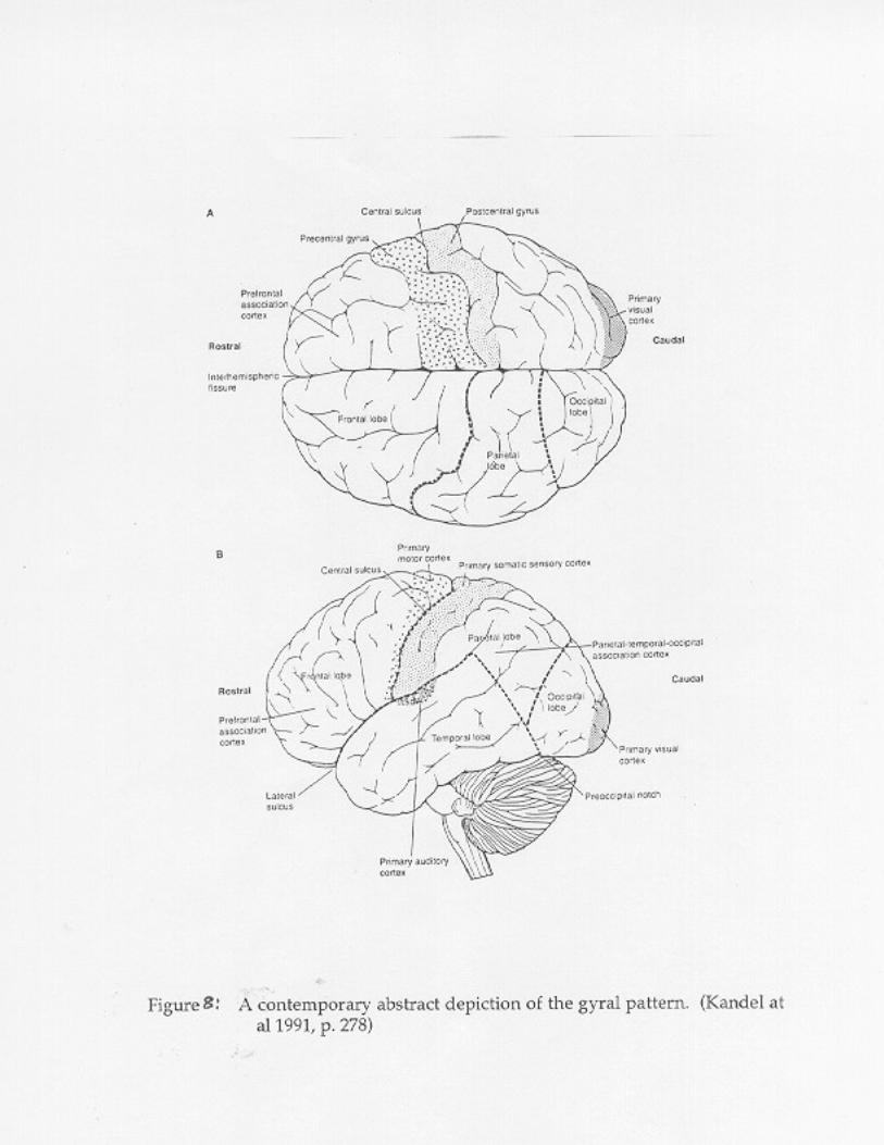
longer need to be depicted convincingly. See Figure 8 for an example of the very
abstract--yet accurate--representations of the cerebral convolutions used in
contemporary texts.
The Clarke & Dewhurst story of the progressive depiction of the convolutions
ends with a reference to photography as the ultimately realistic and naturalistic
method of representation of the cortical surface. However, it is very interesting to
read, in their caption under a photograph of the cerebral surface as seen in life
during brain surgery, the following statement. "Many will agree that there is a faint
resemblance to the abdominal contents of an animal with its tight packed coils of
ileum, which brings us back full circle to Erasistratus in the third century BC." (1996,
p. 113, emphasis in original). This comment, and looking at the photograph (see
Figure 9), seems to indicate that contrary to what Clarke & Dewhurst say,
photography is not the ultimate in scientific illustration. Instead, realistic,
naturalistic scientific illustration has its place, but schematic illustration is always
necessary. Each individual viewer has to learn to see the abstracted pattern in the
actual object. This requires the education or training of one's visual system. Clarke

& Dewhurst have cut the story short by focusing on one piece of the history and
taking for granted the many schematic drawings and models of the cerebral
convolutions that follow that piece of the history. The story continues with these
schematic drawings in which the cerebral convolutions are clearly represented with
little or no concern for realism. However, at the same time, the story also continues
with new kinds of "realistic" representation (x-ray, neuroimaging) that go beyond
photography of the surface of the brain.
An alternative approach is to frame the story as the development of a visual
representation of "the human brain:" the prototypical human brain. This visual
representation--or complex set of visual representations--includes both schematic
and naturalistic representations. Criteria for which kind of visual image might be
useful in a given context depends on the purpose of the immediate practice. Of
course, some of these images are "realistic" (photographs, neuroimages), but these
images must be accompanied by visual abstractions which instruct the viewer on
how to see the realism in the images. Even the most naturalistic scientific
illustrations are not transparent windows on reality. All of these visual images are
representations of the brain.
Recent developments in neuroimaging technologies--positron emission
tomography (PET), and functional magnetic resonance imaging (fMRI), for
example--offer new ways of visualizing individual functioning human brains.

These technologies also give rise to new challenges for integrating data from
individual brains into knowledge about the functional human brain. There are
currently two distinct approaches to constructing atlases of brain function: the
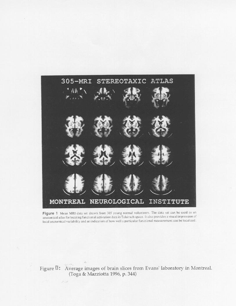
average brain atlas versus the probabilistic atlas. Evans et al (1996) describe the
difference between these two approaches as the difference between removing
anatomical variation and quantifying it. They argue for the probabilistic approach
and are developing an atlas which attempts to visually quantify MRI and PET data
over more than 300 individual brains (see Figure 10 & 11). Woods (1996), on the
other hand, argues for the use of the average brain atlas. "A brain that minimizes
the average amount of distortion required to register it to any individual in the
population is an ideal choice to serve as a target atlas for that population" (1996 p.
336). Woods offers several different ways of visualizing the extent of the variation.
Figure 12 gives a clear impression of the problem, but does little to quantify it.
Figure 13, on the other hand, attempts to give a more quantified sense of variation
of location of specific sulci.
The current state of development of brain atlases offers a preview of the
continuation of the story. Having established the commonality of features such as
the sulci and gyri in the human brain, the science has now moved into a phase of
reestablishing the variability of these features. It is very difficult to say, at the
moment, where this story will go from here. Various on-line atlases or databases are

in a state of rapid development and testing. For example, Evans's group in
Montreal has recently created an "on-line interface to a 3D MRI simulated brain
database" called BrainWeb. Their hope is that the database will serve as a "gold
standard" for the brain mapping community. The future of the visual prototype of
the brain appears to be in virtual cyberspace.
Conclusion
The purpose of this survey of only a very small percentage of the pictures of
the brain used in the practice of functional human brain mapping has been to show
some of the work that goes into the construction of visual prototypes in at least one
scientific practice. Whole technologies are developed (from perspective drawing
through computer visualization) for the purpose of creating stable, exemplary,
comprehensible images of the prototypical brain. Practices such as the localization
of specific functions in specific parts of the brain require the construction of these
visual representations, not only for the individual cognitive tasks of figuring out
what goes where, but also for the distributed (social) tasks of coordinating research
among communities of scientists.
20

References
Arnheim, Rudolf. 1969. Visual Thinking. Berkeley, CA: University of CaliforniaPress.
Bechtel, William & Adele A. Abrahamsen. 1990. "Beyond the exclusivelypropositional era." Synthese Vol. 82: 223-253.
Clarke, Edwin & Dewhurst, Kenneth. 1996. An Illustrated History of BrainFunction: Imaging the Brain f rom Antiquity to the Present, Second edition,revised and enlarged, originally published in 1972. San Francisco: NormanPublishing.
Edgerton, Samuel Y. 1991. The Heritage of Giotto's Geometry: Art and Science onthe Eve of the Scientific Revolution. Ithaca, NY: Cornell University Press.
Evans, A. C., Collins, D. L., & Holmes, C. J. 1996. "Computational approaches toquantifying human neuroanatomical variability." In Brain Mapping: TheMethods. Eds. Arthur W. Toga & John C. Mazziotta. San Diego, CA: Academic Press, pp. 343-362.
Giere, R. N. 1994. "The cognitive structure of scientific theories." Philosophy ofScience, Vol. 61, pp. 276-296.
Kandel, Eric R., Schwartz, J. H. & Jessell, T. M., Eds. 1991. Principles of NeuralScience, Third Edition. Norwalk, CT: Appleton & Lange.
Kosslyn, Stephen M. 1994. Image and Brain: The Resolution of the Imagery Debate.Cambridge, MA: The MIT Press.
Nersessian, Nancy. 1992. "How do scientists think? Capturing the dynamics ofconceptual change in science." In Cognitive Models of Science, MinnesotaStudies in the Philosophy of Science, Vol. 15. Ed. R. N. Giere. Minneapolis: University of Minnesota Press, pp. 3-44.
Rosch, Eleanor. 1978. "Principles of categorization." In Cognition andCategorization, pp. 27-48. Eds. Eleanor Rosch & Barbara B. Lloyd. Hillsdale,NJ: Lawrence Erlbaum Associates, Publishers.
_______. 1973. "On the internal structure of perceptual and semantic categories." In Cognitive Development and the Acquisition of Language. Ed. Timothy E.Moore. New York: Academic Press, pp. 111-144.
Rudwick, Martin J. S. 1992. Scenes From Deep Time: Early Pictorial Representationsof the Prehistoric World. Chicago: The University of Chicago Press.
_______. 1976. "The Emergence of a Visual Language for Geological Science1760-1840." History of Science, xiv (1976), 149-195.
Shelley, C. 1996. "Visual Abductive Reasoning in Archaeology." Philosophy ofScience, Vol 63:#2:278-301.
Toga, Arthur W. & Mazziotta, John C.. 1996. Brain Mapping: The Methods. SanDiego, CA: Academic Press, Inc.
Woods, Roger P. 1996. Correlation of Brain Structure and Function. In Brain
20

Mapping: The Methods. Eds. Arthur W. Toga & John C. Mazziotta. SanDiego, CA: Academic Press.
20