visual inspection with lugol's iodine (vili): evidence to...
TRANSCRIPT

Visual inspection with Visual inspection with LugolLugol’’s iodine (s iodine (VILI): VILI):
Evidence to dateEvidence to date
Original source:
Alliance for Cervical Cancer Prevention (ACCP)www.alliance-cxca.org
Overview:
❚ Description of VILI and how it works❚ Infrastructure requirements❚ What test results mean❚ Test performance❚ Strengths and limitations❚ Program implications in low-resource settings
Types of visual inspection tests:
❚ Visual inspection with Lugol’s iodine (VILI),also known as Schiller’s test, uses Lugol’s iodine instead of acetic acid.
❚ Visual inspection with acetic acid (VIA) can be done with the naked eye (also called cervicoscopy or direct visual inspection, [DVI]), or with low magnification (also called gynoscopy, aided VI, or VIAM).

What does VILI involve?
❚ Performing a vaginal speculum exam during which a health care provider applies Lugol’s iodine solution to the cervix.
❚ Viewing the cervix with the naked eye to identify color changes on the cervix.
❚ Determining whether the test result is positive or negative for possible precancerous lesions or cancer.

How VILI works:
❚ Squamous epithelium contains glycogen, whereas precancerous lesions and invasive cancer contain little or no glycogen.
❚ Iodine is glycophilic and is taken up by the squamous epithelium, staining it mahogany brown or black.
❚ Columnar epithelium does not change color, as it has no glycogen.❚ Immature metaplasia and inflammatory lesions are at most only
partially glycogenated and, when stained, appear as scattered, ill-defined uptake areas.
❚ Precancerous lesions and invasive cancer do not take up iodine (as they lack glycogen) and appear as well-defined, thick, mustard or saffron yellow areas.

What infrastructure does VILI require?
❚ Private exam room❚ Examination table ❚ Trained health professionals❚ Adequate light source❚ Sterile vaginal speculum ❚ New examination gloves, or HLD surgical gloves❚ Large cotton swabs❚ Lugol’s iodine solution and a small bowl❚ Containers with 0.5% chlorine solution❚ A plastic bucket with a plastic bag❚ Quality assurance system to maximize accuracy

Categories for VILI test results:
Clinically visible ulcerative, cauliflower-like growth or ulcer; oozing and/or bleeding on touch.
Suspicious for cancer
Well-defined, bright yellow iodine non-uptake areas touching the squamo-columnar junction (SCJ) or close to the os if SCJ is not seen.
Test-positive
Squamous epithelium turns brown and columnar epithelium does not change color; or irregular, partial or non-iodine uptake areas appear.
Test-negative
Clinical FindingsVILI Category
VILI: test-negative
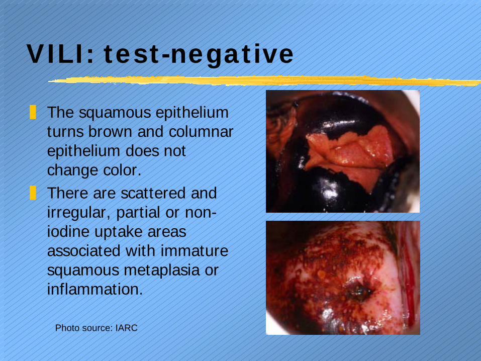
❚ The squamous epithelium turns brown and columnar epithelium does not change color.
❚ There are scattered and irregular, partial or non-iodine uptake areas associated with immature squamous metaplasia or inflammation.
Photo source: IARC

VILI: test-positive
❚ Well-defined, bright yellow iodine non-uptake areas touching the squamocolumnarjunction (SCJ).
❚ Well-defined, bright yellow iodine non-uptake areas close to the os if SCJ is not seen, or covering the entire cervix.
Photo source: IARC

VILI: Suspicious for cancer
❚ Clinically visible ulcerative, cauliflower-like growth or ulcer; oozing and/or bleeding on touch.
Photo source: IARC

Management options if the VILI result is positive:
❚ Offer to treat immediately, (without colposcopy or biopsy, known as the “test-and-treat” or “single-visit” approach).
❚ Refer for colposcopy and biopsy and then offer treatment if a precancerous lesion is confirmed.
Management options if the VILI result is suspicious for cancer:
❚ Refer for colposcopy and biopsy and further management. Further management options include:❙ Surgery❙ Radiotherapy❙ Chemotherapy❙ Palliative care

Test performance:Sensitivity and Specificity
❚ Sensitivity: The proportion of all those with disease that the test correctly identifies as positive.
❚ Specificity: The proportion of all those without disease (normal) that the test correctly identifies as negative.
VILI test performance:
❚ Sensitivity = 87.2%❚ Specificity = 84.7%❚ These results are from a cross-sectional
study involving 4,444 women. (Sankaranarayanan et al., 2003).

Strengths of VILI:
❚ Simple, easy-to-learn approach that is minimally reliant upon infrastructure.
❚ Low start-up and sustaining costs.❚ Many types of health care providers can perform
the procedure.❚ High sensitivity results in a low proportion of
false negatives.❚ Test results are available immediately.❚ Decreased loss to follow-up.

Limitations of VILI:
❚ Moderate specificity may result in over-referral and over-treatment in a single-visit approach.
❚ Less accurate when used in post-menopausal women.
❚ There is a need for developing standard training methods and quality assurance measures.
❚ Rater dependent.

Conclusions:
❚ VILI is a promising new approach.❚ Adequate training and ongoing supervision are essential
to enable health care providers to evaluate the features of a lesion and make accurate assessments.
❚ More research is needed to establish the most appropriate and feasible approach to reducing false-positives and over-treatment (when offered as part of a single-visit, “test-and-treat” approach).
❚ Properly designed studies on VILI are essential to evaluating the effectiveness in reducing cervical cancer incidence and mortality.
References:
❚ ACCP. Visual screening approaches: Promising alterative screening strategies. Cervical Cancer Prevention Fact Sheet. (October 2002).
❚ Sankaranarayanan R, Wesley R, Thara S, Dhakad N, ChandralekhaB, Sebastian P, Chithrathara K, Parkin DM, Nair MK. Test characteristics of visual inspection with 4% acetic acid (VIA) and Lugol's iodine (VILI) in cervical cancer screening in Kerala, India.International Journal of Cancer 106(3):404-408. (September 1, 2003).
❚ Sankaranarayanan R,Rajkumar R, Arrossi S, Theresa R, Esmy PO, Mahé C, Muwonge R, Parkin DM, Cherian J. Determinants of participation of women in a cervical cancer visual screening trial in rural south India. Cancer Detection and Prevention 27(6):415-523 (November-December 2003).
For more information on cervical cancer prevention:
❚ The Alliance for Cervical Cancer Prevention (ACCP) www.alliance-cxca.org
❚ ACCP partner organizations:❙ EngenderHealth www.engenderhealth.org❙ International Agency for Research on Cancer
(IARC) www.iarc.fr❙ JHPIEGO www.jhpiego.org❙ Pan American Health Organization (PAHO)
www.paho.org❙ Program for Appropriate Technology in Health
(PATH) www.path.org
1
Visual inspection withVisual inspection withLugolLugol’’ss iodine (iodine (VILI): VILI):
Evidence to dateEvidence to date
Original source:
Alliance for Cervical Cancer Prevention (ACCP)www.alliance-cxca.org
Slide overview: This presentation provides a summary of the latest evidence, as of 2003, on visual inspection with Lugol’s iodine (VILI) as a test for cervical cancer.

2
Overview:
❚ Description of VILI and how it works❚ Infrastructure requirements❚ What test results mean❚ Test performance❚ Strengths and limitations❚ Program implications in low-resource settings
Slide overview: In this presentation, we will discuss the following topics.

3
Types of visual inspection tests:
❚ Visual inspection with Lugol’s iodine (VILI),also known as Schiller’s test, uses Lugol’s iodine instead of acetic acid.
❚ Visual inspection with acetic acid (VIA) can be done with the naked eye (also called cervicoscopy or direct visual inspection, [DVI]), or with low magnification (also called gynoscopy, aided VI, or VIAM).
Slide overview: This is a partial list of the types of vision-based tests available for testing for cervical cancer or precancer. The key differences in these tests are whether or not magnification is used, and whether acetic acid or some other technique of highlighting abnormalities is used.
•Note for bullet 1: The screening test using iodine (VILI) is similar in approach tothe Schiller’s iodine test advocated in the 1930s and widely used early in the 20th
century before the development of cytology. Schiller’s test was well known for its low specificity, however, it is noteworthy that experience gained through the use of Lugol’s iodine application in colposcopy has helped refine VILI and avoided many false-positive findings.•Note for after last bullet: This talk focuses on VILI.

4
What does VILI involve?
❚ Performing a vaginal speculum exam during which a health care provider applies Lugol’siodine solution to the cervix.
❚ Viewing the cervix with the naked eye to identify color changes on the cervix.
❚ Determining whether the test result is positive or negative for possible precancerous lesions or cancer.
Slide overview: VILI is simple to administer, and a range of types of health care providers can perform the procedure with appropriate training.Note fur bullet 3: Results of the test are available immediately and do not require
laboratory support.
5
How VILI works:
❚ Squamous epithelium contains glycogen, whereas precancerous lesions and invasive cancer contain little or no glycogen.
❚ Iodine is glycophilic and is taken up by the squamous epithelium, staining it mahogany brown or black.
❚ Columnar epithelium does not change color, as it has no glycogen.❚ Immature metaplasia and inflammatory lesions are at most only
partially glycogenated and, when stained, appear as scattered, ill-defined uptake areas.
❚ Precancerous lesions and invasive cancer do not take up iodine (as they lack glycogen) and appear as well-defined, thick, mustard or saffron yellow areas.
Slide overview: Application of iodine results in brown or black color staining in areas containing glycogen. In areas lacking glycogen, iodine is not absorbed and such areas remain colorless or turn yellow.
Note for bullet 1: Glycogen is a sugar stored by normal cells.Note after the last bullet: Gross cancerous lesions are usually apparent before the
application of iodine.

6
What infrastructure does VILI require?
❚ Private exam room❚ Examination table ❚ Trained health professionals❚ Adequate light source❚ Sterile vaginal speculum ❚ New examination gloves, or HLD surgical gloves❚ Large cotton swabs❚ Lugol’s iodine solution and a small bowl❚ Containers with 0.5% chlorine solution❚ A plastic bucket with a plastic bag❚ Quality assurance system to maximize accuracy
Slide overview: The supplies and equipment required to provide VILI testing arelisted here. Most of these supplies are available at even the most basic levels of the health care system in low-resource countries, although not always.
Note for bullet 4: Preferably, a bright halogen lamp that can be easily directed atthe cervix. The light source needs to be something other than daylight. It can be a flashlight or torch, or a gooseneck lamp. The stronger and more consistent the light source, the easier it will be for health care providers to identify abnormalities.•Note for bullet 7: Cotton swabs can be handmade using cotton batting and broomsticks or ring forceps.Note for bullet 9: For decontamination, an aluminium/steel/plastic container is
used for immersing the gloves, and a plastic bucket or container for decontamination of instruments.Note for bullet 10 (second to last): A bucket is used to dispose of contaminated
swabs and other waste items.Note for last bullet: Elements of a quality assurance system include (but are not
limited to) supervision, periodic refresher trainings, evaluation of on-going program activities and long-term impact, a mechanism for constructive feedback from women and health care providers, and an effective information system.Note at the end: Other necessary supplies that should be available at any clinic
setting include cotton balls, gauze, and rubber or plastic sheets for the table.

7
Categories for VILI test results:
Clinically visible ulcerative, cauliflower-like growth or ulcer; oozing and/or bleeding on touch.
Suspicious for cancer
Well-defined, bright yellow iodine non-uptake areas touching the squamo-columnar junction (SCJ) or close to the os if SCJ is not seen.
Test-positive
Squamous epithelium turns brown and columnar epithelium does not change color; or irregular, partial or non-iodine uptake areas appear.
Test-negative
Clinical FindingsVILI Category
Slide overview: There are three categories of test results. Each is described in more detail on the following slides.
8
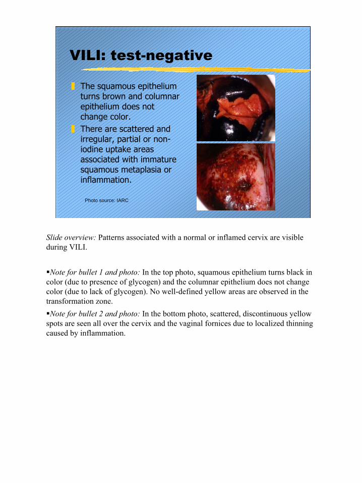
VILI: test-negative
❚ The squamous epithelium turns brown and columnar epithelium does not change color.
❚ There are scattered and irregular, partial or non-iodine uptake areas associated with immaturesquamous metaplasia or inflammation.
Photo source: IARC
Slide overview: Patterns associated with a normal or inflamed cervix are visible during VILI.
Note for bullet 1 and photo: In the top photo, squamous epithelium turns black in color (due to presence of glycogen) and the columnar epithelium does not change color (due to lack of glycogen). No well-defined yellow areas are observed in the transformation zone.Note for bullet 2 and photo: In the bottom photo, scattered, discontinuous yellow
spots are seen all over the cervix and the vaginal fornices due to localized thinning caused by inflammation.

9
VILI: test-positive
❚ Well-defined, bright yellow iodine non-uptake areas touching the squamocolumnarjunction (SCJ).
❚ Well-defined, bright yellow iodine non-uptake areas close to the os if SCJ is not seen, or covering the entire cervix.
Photo source: IARC
Slide overview: The well-defined, mustard-yellow lesions in the transformation zone indicate cervical intraepithelial neoplasia.
Note for bullet 1: The squamocolumnar junction (SCJ) is the point at which columnar cells meet ectocervical squamous cells on the cervix. This junction marks the furthest extent of the transformation zone towards or, in the case of post-menopausal women, into the cervical canal.

10
VILI: Suspicious for cancer
❚ Clinically visible ulcerative, cauliflower-like growth or ulcer; oozing and/or bleeding on touch.
Photo source: IARC
Slide overview: Because invasive cervical cancer lacks glycogen, the growth does not stain with iodine and turns yellow. Gross cancerous lesions can be apparent before the application of iodine.
11
Management options if the VILI result is positive:
❚ Offer to treat immediately, (without colposcopyor biopsy, known as the “test-and-treat” or “single-visit” approach).
❚ Refer for colposcopy and biopsy and then offer treatment if a precancerous lesion is confirmed.
Slide overview: Women testing positive may be offered further testing (colposcopy or biopsy) or treatment, or both, immediately after testing. Note after bullet 2: The different approaches to screening, diagnosis, and
treatment, based on VILI, are being evaluated in terms of safety, acceptability to women, and effectiveness in preventing invasive cancer.

12
Management options if the VILI result is suspicious for cancer:
❚ Refer for colposcopy and biopsy and further management. Further management options include:❙ Surgery❙ Radiotherapy❙ Chemotherapy❙ Palliative care
Slide overview: If a VILI test result is suspicious for cancer, refer for further testing (colposcopy or biopsy) and management.
13
Test performance:Sensitivity and Specificity
❚ Sensitivity: The proportion of all those with disease that the test correctly identifies as positive.
❚ Specificity: The proportion of all those without disease (normal) that the test correctly identifies as negative.
Slide overview: The test performance of each screening method is rated by its sensitivity and specificity. Before discussing VILI’s test performance, it is important to understand what sensitivity and specificity mean.

14
VILI test performance:
❚ Sensitivity = 87.2%❚ Specificity = 84.7%❚ These results are from a cross-sectional
study involving 4,444 women. (Sankaranarayanan et al., 2003).
Slide overview: VILI’s 87.2% sensitivity ensures that a large proportion of high-grade disease is identified. Its specificity implies that about 15 percent of women tested may be treated unnecessarily in a single-visit “test-and-treat” approach.
Note for bullet 1: High sensitivity in detecting high-grade lesions or cancer means there are few false-negatives and most precancerous lesions are detected. The diagnostic properties of VILI are being evaluated in 12 ACCP cross-sectional studies established in India, Congo, Mali, Niger, Guinea, and Burkina Faso. The screening tests are provided by a range of health care providers including trained nurses, health workers, or graduate students. VILI’s performance in these ACCP studies is consistent with the findings presented here.Note for bullet 2: Further investigation with diagnostic procedures, such as
colposcopy and biopsy, may reduce over-treatment caused by VILI’s lower specificity.
15
Strengths of VILI:
❚ Simple, easy-to-learn approach that is minimally reliant upon infrastructure.
❚ Low start-up and sustaining costs.❚ Many types of health care providers can perform
the procedure.❚ High sensitivity results in a low proportion of
false negatives.❚ Test results are available immediately.❚ Decreased loss to follow-up.
Slide overview: VILI has the following strengths as an alternative test for precancer or cancer in low-resource settings.•Note for bullet 1: Assuming sufficiently trained providers are available, VILI is asimple approach. Health care providers can be trained in a short period of time (1 to 2 weeks).•Note for bullet 2: In most settings, costs associated with launching and sustaining VILI-based programs are lower than other methods (except VIA). VILI can be performed in extremely low-resource settings.Notes on bullet 3: In situations in which health care providers can receive adequate
and ongoing training, VILI has the potential for adequate population coverage.Note for bullet 4: Therefore, a high proportion of precancerous lesions are
detected. Note for bullet 5: Because results are available immediately, further investigations
(such as colposcopy and biopsy), and treatment (such as cryotherapy or LEEP) can occur during the same visit, if appropriate.Note for bullet 6: This means that additional visits for investigations and
treatments are reduced.
16
Limitations of VILI:
❚ Moderate specificity may result in over-referral and over-treatment in a single-visit approach.
❚ Less accurate when used in post-menopausal women.
❚ There is a need for developing standard training methods and quality assurance measures.
❚ Rater dependent.
Slide overview: VILI also has limitations.
Note for bullet 1: The single-visit “test-and-treat” approach results in over-referral and over-treatment of women. Over-referral has important cost implications in settings with scarce resources.Note for bullet 2: It may be difficult to interpret the color patterns associated with
Lugol’s iodine application in post-menopausal women because of the degeneration and atrophy of the epithelium. Menopause reduces the production of glycogen, and the color pattern resulting from iodine application becomes confusing.•Note for bullet 3: The Alliance for Cervical Cancer Prevention (ACCP) is currentlyinvestigating these elements.•Note for bullet 4: “Rater dependent” means the test's performance depends on the abilities of the person doing the test (versus a machine, as for HPV testing). This means that even when service providers have training, test performance may vary depending on service delivery conditions and other factors.

17
Conclusions:
❚ VILI is a promising new approach.❚ Adequate training and ongoing supervision are essential
to enable health care providers to evaluate the features of a lesion and make accurate assessments.
❚ More research is needed to establish the most appropriate and feasible approach to reducing false-positives and over-treatment (when offered as part of a single-visit, “test-and-treat” approach).
❚ Properly designed studies on VILI are essential to evaluating the effectiveness in reducing cervical cancer incidence and mortality.
Slide overview: VILI is a promising, new approach to cervical cancer prevention,however, it remains to be determined if VILI-based screening programs will result in a reduced disease burden. Note for bullet 2: The feasibility of utilizing VILI for wide-scale screening will be
determined, to a larger extent, by the capacity to maintain effective training and monitoring efforts. Note for bullets 3 and 4: Current ACCP research may provide valuable information
on VILI’s sensitivity and specificity, program effectiveness in detecting high-grade lesions, and prevention of cervical cancer.

18
References:
❚ ACCP. Visual screening approaches: Promising alterative screening strategies. Cervical Cancer Prevention Fact Sheet. (October 2002).
❚ Sankaranarayanan R, Wesley R, Thara S, Dhakad N, ChandralekhaB, Sebastian P, Chithrathara K, Parkin DM, Nair MK. Test characteristics of visual inspection with 4% acetic acid (VIA) and Lugol's iodine (VILI) in cervical cancer screening in Kerala, India.International Journal of Cancer 106(3):404-408. (September 1, 2003).
❚ Sankaranarayanan R,Rajkumar R, Arrossi S, Theresa R, Esmy PO, Mahé C, Muwonge R, Parkin DM, Cherian J. Determinants of participation of women in a cervical cancer visual screening trial in rural south India. Cancer Detection and Prevention 27(6):415-523 (November-December 2003).
19
For more information on cervical cancer prevention:
❚ The Alliance for Cervical Cancer Prevention (ACCP) www.alliance-cxca.org
❚ ACCP partner organizations:❙ EngenderHealth www.engenderhealth.org❙ International Agency for Research on Cancer
(IARC) www.iarc.fr❙ JHPIEGO www.jhpiego.org❙ Pan American Health Organization (PAHO)
www.paho.org❙ Program for Appropriate Technology in Health
(PATH) www.path.org