visual field screening and analysis part 2 - … · 10/17/2011 · visual field screening and...
TRANSCRIPT
1
Visual Field Screening and Analysis
PART 2A SYSTEMATIC APPROACH TO
INTERPRETATIONRevised 1/16/2011
VF ScreeningSystematic Approach to Interpretation
Patient infoField testing strategy and test point patternReliability indicators
Is the VF reliable? Decision - normal or abnormal VF?
Is true, organic VF loss present?“True, organic VF loss” = VF loss due to a disease n ot
due to an artifact like a lensholder, lens rim, dro opy lid, small pupil, wrong age entered, tired patient etc.
If abnormal - diagnostic decision - cause of VF defec t? Site of lesion?

2
TAN 30 test
Pt info: 45 y/oTest: N 30-5
Reliabiltyindicators:Fix errorsFalse positive errors
Normal?
Testing Strategy and Test Point Pattern
� Single intensity strategy - artifactuous misses on edge due to subthreshold testing - common
� Appropriate testing level?� Central reference level on HFA
Central reference level is an index of the patient’s sensitivity – the higher the central reference level the higher the instrument thinks the patient’s sensitivity is
� Central reference too high →→→→ stimuli too dim →→→→artifactuous misses
� Appropriate age/birthdate entered?� Age too low →→→→ stimuli too dim →→→→
artifactuous misses (reduced specificity)

3
Testing Strategy and Test Point Pattern (cont’d.)
• Test point patterno Few points are tested in general VF screening, i.e. ,
40 points on HFA Central 40 screening test, 17 areasareas(10ºx10º squares) on the FDT/Matrix C20-5 test, 19 areasareas on the FDT/Matrix N30-5 test.VERY IMPORTANT: Since there are very few points or areas tested in the general screening tests that we do one (1) miss may be the only sign of a VF defect, particularly on FDT/Matrix
o If more points tested, i.e., 80 points →→→→ more often get 2, 3 or more misses at site of a true VF defect
Inferior VF loss on FDT N30-5 & on HFA C30-2Only one miss on FDT due to few test points on N30- 5 and because
most of VF loss is outside of the central 20º where the N30 tests
4
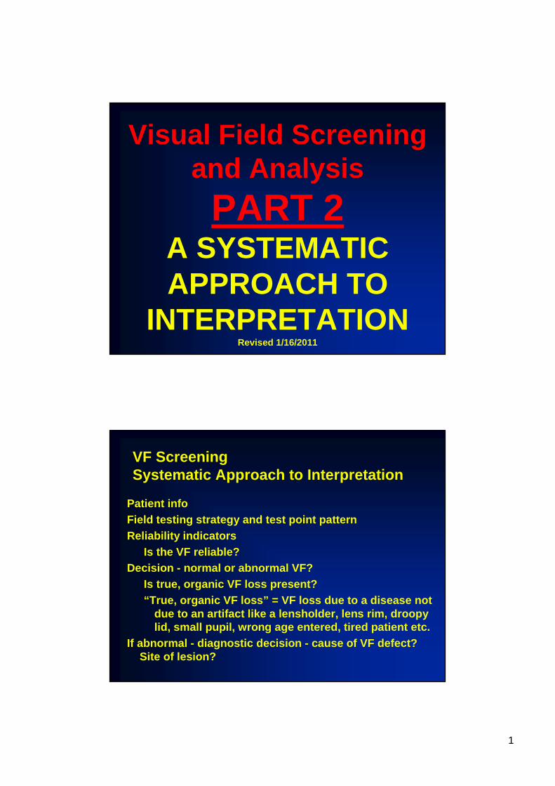
More points tested – more points missed in the VF de fectDicon Central 40 ( 40 points) Dicon Central 80 ( 80 points)
2 misses in nasal step area OD 4 misses in na sal step area OD
Interpretation Strategies
• The greater the number of adjacent misses the great er the likelihood of true VF loss rather than artifact
• Patient has 2 chances to hit each point (exception Octopus ). Misses are retested.
• Artifactuous misses more common in peripheral edge of the field, i.e., very common outside of central 30° Do not test beyond 30°in general VF screening
• Artifactuous misses more common beyond 20°in superior VF due to the normally steeper decline in sensitivity, the even greater decline and increased fluctuation with age in this area, the much greater chance of obstruction from lids, lashes, brow (deep -set eyes) etc.
5
EGFDTN30-5Screening
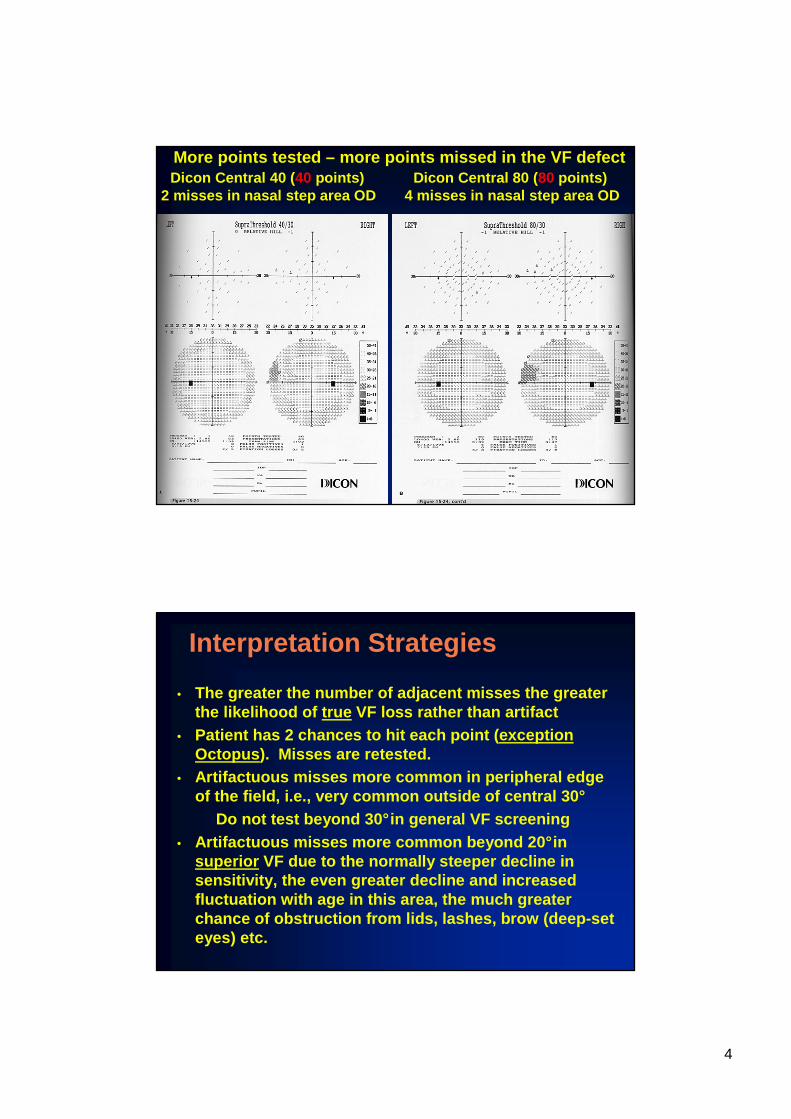
FDT C20-5
One miss sup in OS
Real VF loss vs. artifact?What should be done to differentiate true from artifact?
6
Assessment of Test ReliabilityFixation Losses – Ways to Evaluate Fixation
• Fixation loss indexo Automatic fixation monitoro Heijl-Krakau (blind spot) monitor
• Gaze tracker on new HFA (at the bottom of the printout)o Shows continuous graph of fixation quality
and blinkso Upward deflection is a fixation losso Downward deflection is a blink
• Perimetrist monitors fixation and writes commentso Cannot monitor fixation on the FDT since no
way to see the eye
“Fixation Losses”on Blind Spot Monitor-Possible Causes
• True fixation losses/shifts• Head misalignment after BS
localization• False positive responses during the
test• False negative responses during BS
localization• Poor BS localization• High refractive error• Small/ hypoplastic ONH
7
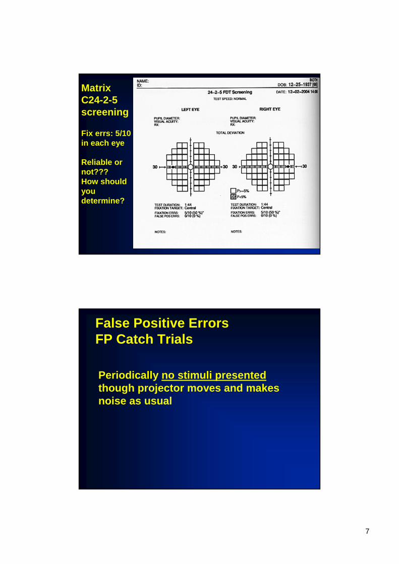
Matrix C24-2-5screening
Fix errs: 5/10 in each eye
Reliable or not??? How should you determine?
False Positive ErrorsFP Catch Trials
Periodically no stimuli presentedthough projector moves and makes noise as usual
8
False Positive Responses
• Patient responds to something other than seeing the stimulus
• May respond to stimulus projector movement or the noise caused by the projector movement
• May respond to “timing” the interval between stimuli
False Positive Responses Possible Effects
• If false positives occur on threshold sampling may cause perimeter to think the patient has higher than his/her actual sensitivity. Stimuli are made too dim →→→→ many misses
on the screening• If during the screening test VF defects may not
be detected → → → → patient hits the button when the stimulus is presented in a VF defect where the patient could not actually see it
• Many “fixation losses” →→→→ patient hits the button when the stimulus is actually in the blindspot
9
False Positive ResponsesWhat to do about them• If one FP occurs remind the patient to
“Hit the button only when they see or think they see the light (or flickering/wavy area)”
• If 2 FPs advise the patient to hit the button only when they know they see the light. Repeat these instructions.
• If even more FPs stop test, advise the patient that they were hitting the button when there was no light. Repeat instructions above. Rerun test.
False Negative ErrorsFN Catch Trial
• Periodically perimeter presents a much brighter than previously seen stimulus to patient at a location that has been previously found to be normal →→→→ if missed → false negative response

10
HFA C40
2/2 False Negatives
Reliable VF??Normal or abnormal VF?
Is there a pattern to the misses?
HFA C80
Reliable VF?
Normal or abnormal VF?
Is there a pattern to the misses?
11
False Negative ErrorsPossible Causes
• Fatigue• Inattention• VF loss
Artifactual VF LossCommon Causes
• Small pupil• Lensholder, lens rim artifact• Fatigue• Cataract, media opacity
12
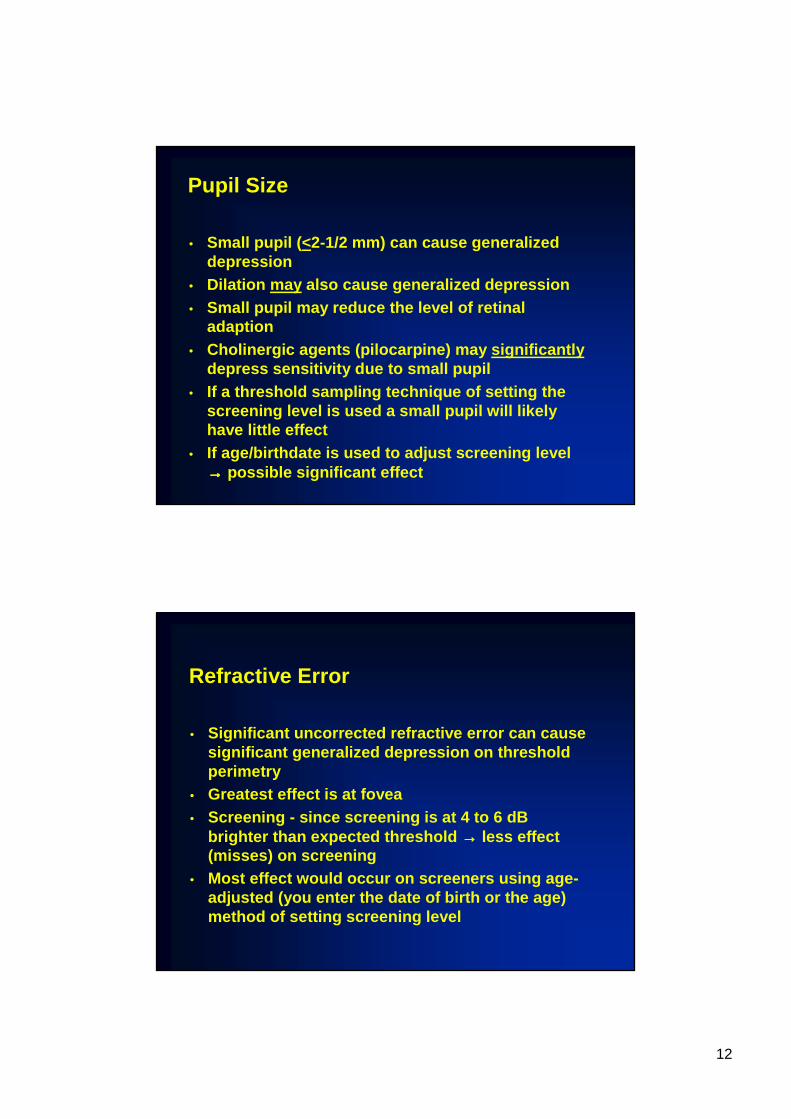
Pupil Size
• Small pupil (< 2-1/2 mm) can cause generalized depression
• Dilation may also cause generalized depression• Small pupil may reduce the level of retinal
adaption• Cholinergic agents (pilocarpine) may significantly
depress sensitivity due to small pupil• If a threshold sampling technique of setting the
screening level is used a small pupil will likely have little effect
• If age/birthdate is used to adjust screening level →→→→ possible significant effect
Refractive Error
• Significant uncorrected refractive error can cause significant generalized depression on threshold perimetry
• Greatest effect is at fovea• Screening - since screening is at 4 to 6 dB
brighter than expected threshold →→→→ less effect (misses) on screening
• Most effect would occur on screeners using age-adjusted (you enter the date of birth or the age) method of setting screening level
13
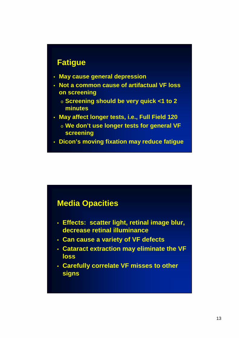
Fatigue
• May cause general depression• Not a common cause of artifactual VF loss
on screeningo Screening should be very quick <1 to 2
minutes• May affect longer tests, i.e., Full Field 120
o We don’t use longer tests for general VF screening
• Dicon’s moving fixation may reduce fatigue
Media Opacities
• Effects: scatter light, retinal image blur, decrease retinal illuminance
• Can cause a variety of VF defects• Cataract extraction may eliminate the VF
loss• Carefully correlate VF misses to other
signs
14
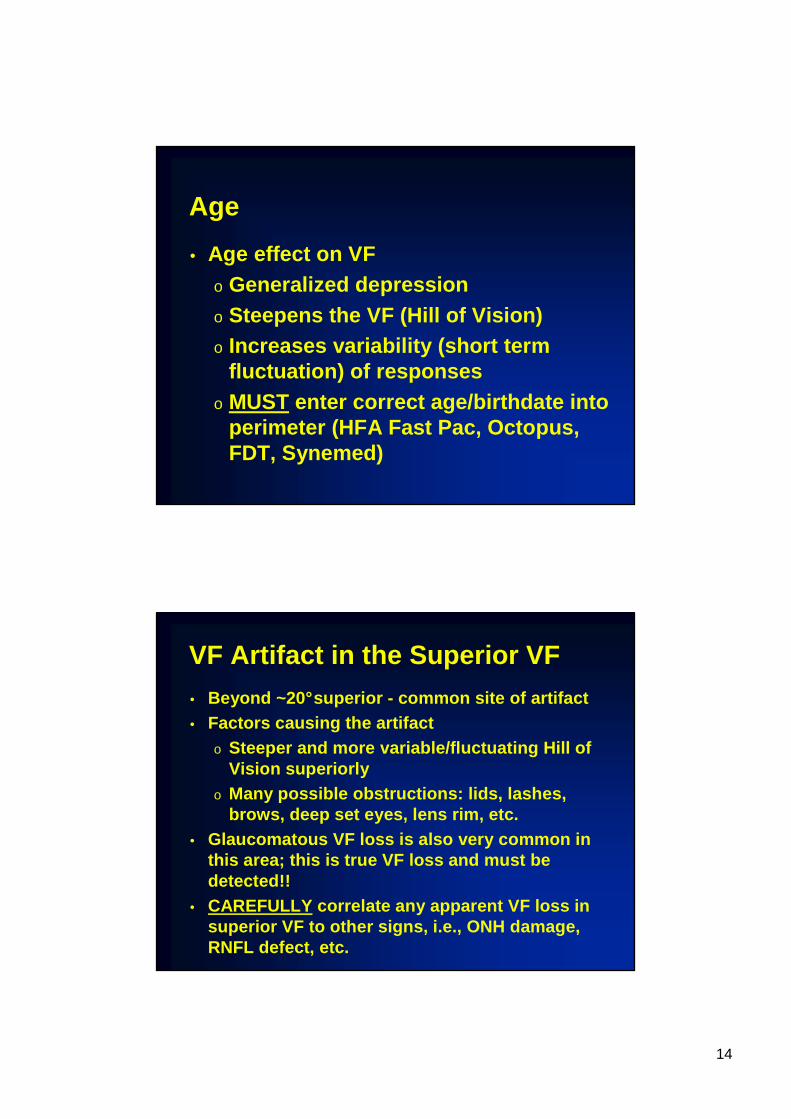
Age
• Age effect on VFo Generalized depressiono Steepens the VF (Hill of Vision)o Increases variability (short term
fluctuation) of responses o MUST enter correct age/birthdate into
perimeter (HFA Fast Pac, Octopus, FDT, Synemed)
VF Artifact in the Superior VF
• Beyond ~20°superior - common site of artifact• Factors causing the artifact
o Steeper and more variable/fluctuating Hill of Vision superiorly
o Many possible obstructions: lids, lashes, brows, deep set eyes, lens rim, etc.
• Glaucomatous VF loss is also very common in this area; this is true VF loss and must be detected!!
• CAREFULLY correlate any apparent VF loss in superior VF to other signs, i.e., ONH damage, RNFL defect, etc.
15
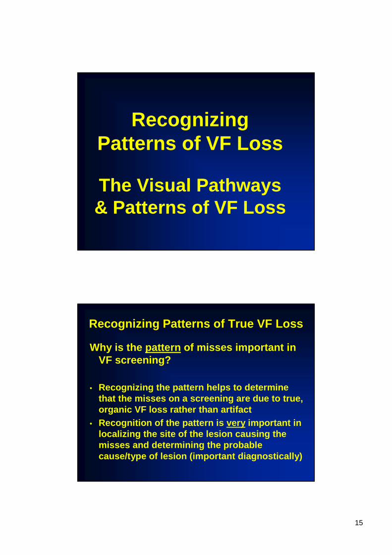
Recognizing Patterns of VF Loss
The Visual Pathways & Patterns of VF Loss
Recognizing Patterns of True VF Loss
Why is the pattern of misses important in VF screening?
• Recognizing the pattern helps to determine that the misses on a screening are due to true, organic VF loss rather than artifact
• Recognition of the pattern is very important in localizing the site of the lesion causing the misses and determining the probable cause/type of lesion (important diagnostically)

16
HFA C40 2/2 False Negatives
Reliable VF??Normal or abnormal VF?
Is there a pattern to the misses?Yes –inferior arcuate and superior
nasal step
The Visual PathwaysFour Territories
• Territory 1 - outer retina, choroid• Territory 2 - inner retina and optic nerve
(ON) o Ganglion cells and their axons, ONH,
ON up to the chiasm• Territory 3 - optic chiasm• Territory 4 - post chiasmal visual pathway
o Optic tract, LGNo Visual radiationso Visual cortex
17
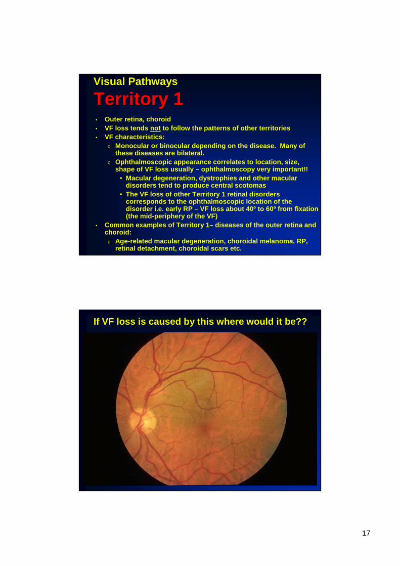
Visual Pathways
Territory 1Territory 1• Outer retina, choroid• VF loss tends not to follow the patterns of other territories• VF characteristics:
o Monocular or binocular depending on the disease. M any of these diseases are bilateral.
o Ophthalmoscopic appearance correlates to location, s ize, shape of VF loss usually – ophthalmoscopy very impor tant!!
• Macular degeneration, dystrophies and other macular disorders tend to produce central scotomas
• The VF loss of other Territory 1 retinal disorders corresponds to the ophthalmoscopic location of the disorder i.e. early RP – VF loss about 40º to 60º from fixation (the mid-periphery of the VF)
• Common examples of Territory 1– diseases of the oute r retina and choroid:
o Age-related macular degeneration, choroidal melanom a, RP, retinal detachment, choroidal scars etc.
If VF loss is caused by this where would it be??
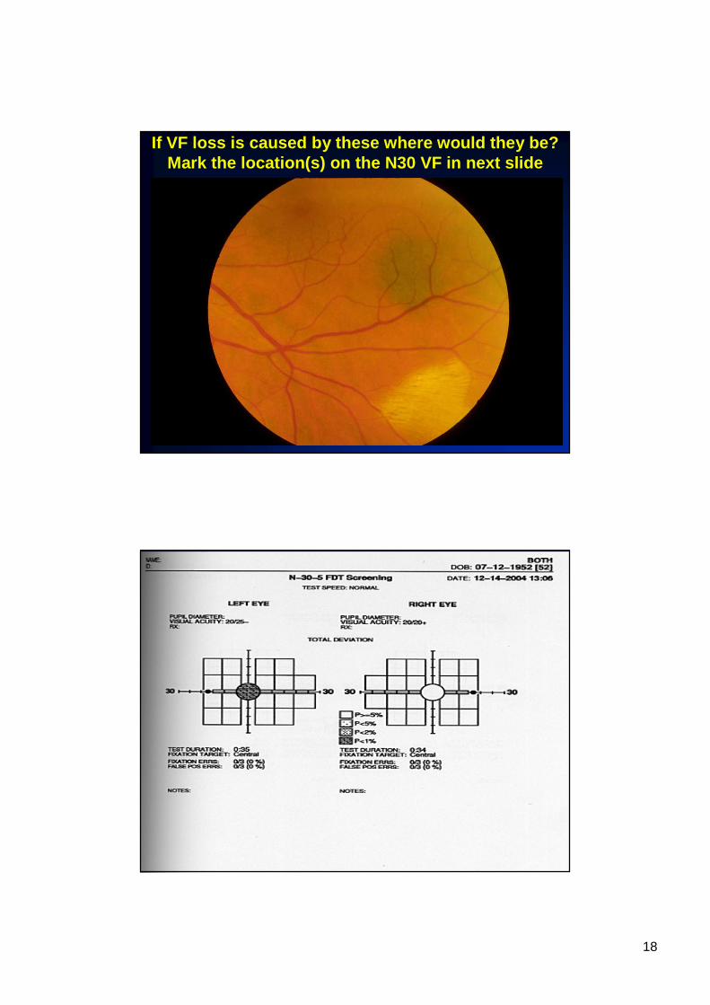
18
If VF loss is caused by these where would they be?Mark the location(s) on the N30 VF in next slide
19
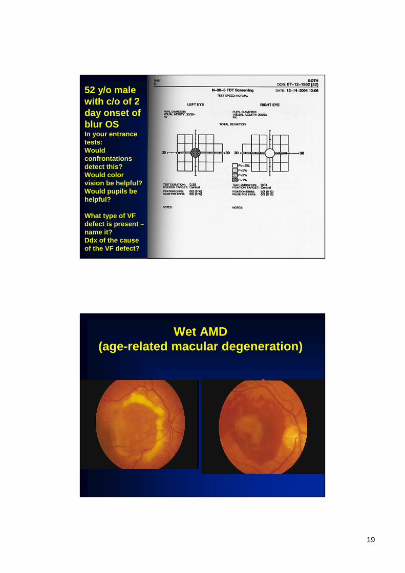
52 y/o male with c/o of 2 day onset of blur OSIn your entrance tests: Would confrontations detect this?Would color vision be helpful? Would pupils be helpful?
What type of VF defect is present –name it? Ddx of the cause of the VF defect?
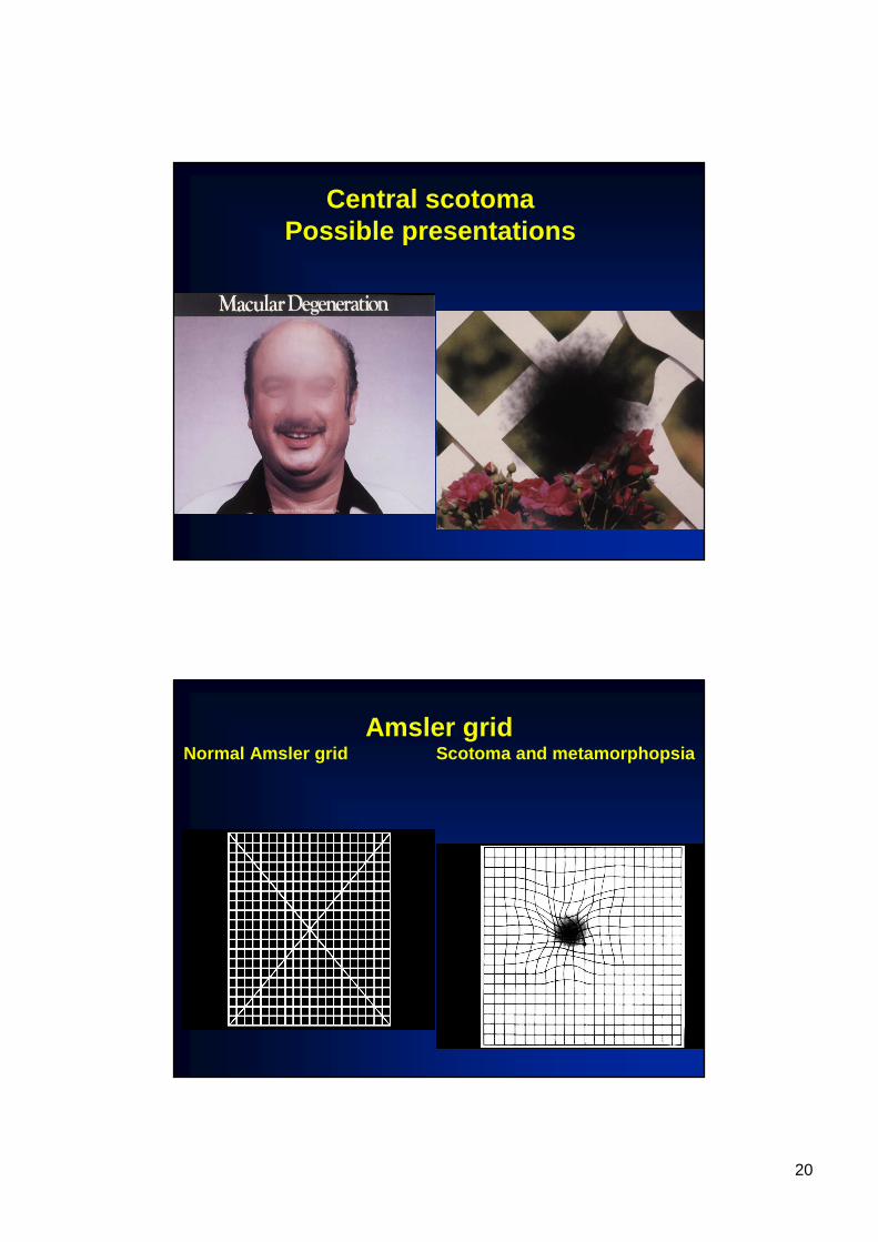
Wet AMD(age-related macular degeneration)
20
Central scotoma Possible presentations
Amsler gridNormal Amsler grid Scotoma and meta morphopsia

21
If this produces a VF defect where would it be? At fixation.Are there other tests in the comp exam that could s how evidence of
this? VAs, color vision (blue-yellow defect)
The pointer is used to mark the fovea; patient is in structed to look at the tip of the pointer. If this causes a VF defect where would it be in the VF?Immediately adjacent to fixation in the superior te mporal quadrant

22
You are performing BIO on a patient’s right eye, standing to the patient’s right side with the patie nt
looking up to his left. Area of fundus in this BIO view? If this causes VF defect where would it be?
OPTOMAP of OS Will the lesion show on CF confrontations? On FDT?
Where would this lesion be in the VF?Answer: About 60º nasal and just above the nasal hor izontal midline
23
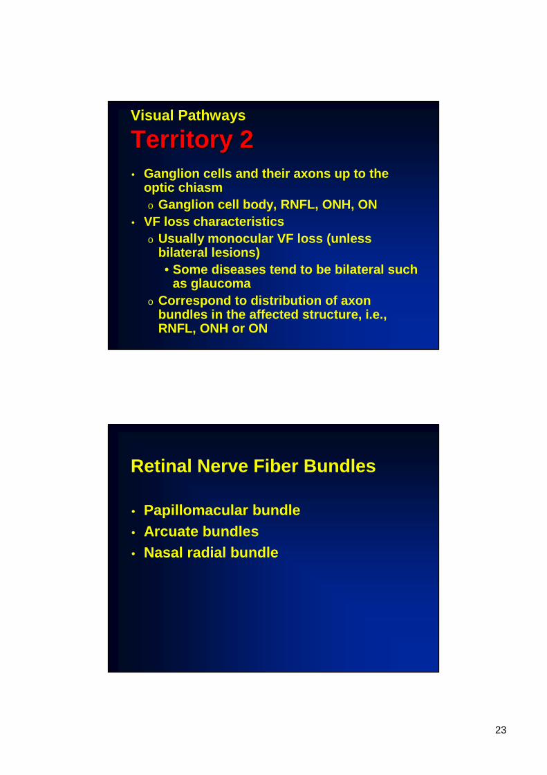
Visual Pathways
Territory 2Territory 2• Ganglion cells and their axons up to the
optic chiasmo Ganglion cell body, RNFL, ONH, ON
• VF loss characteristicso Usually monocular VF loss (unless
bilateral lesions)• Some diseases tend to be bilateral such
as glaucomao Correspond to distribution of axon
bundles in the affected structure, i.e., RNFL, ONH or ON
Retinal Nerve Fiber Bundles
• Papillomacular bundle• Arcuate bundles• Nasal radial bundle

24
Papillomacular (PM) Bundle
• About 70-90% of all ganglion cell axons• From macula to temporal side of ONH
o Enters ONH from about 8:00 to 10:00 on right ONH and from 2:00 to 4:00 on left ONH
• Very fine caliber bundles →→→→ very thin RNFL with very fine bundles →→→→ PM bundles are very difficult to visualize clinically
• Damage to PM bundle →→→→ central scotoma or centrocecal scotoma
Damage to PM BundlePossible Clinical Signs
• Central or centrocecal scotoma• Reduced visual acuity• Reduced contrast sensitivity• Color vision loss/change
o By Kollner’s rule - R-G defect for ganglion cells (RNFL, ONH, ON)
• Decreased direct pupillary light reflex causing: L- N dissociation and APD if unilateral or if asymmetric damage
• PM RNFL loss (but very hard to see clinically) &/or temporal pallor of ONH (hard to detect due to the wide variation of color of temporal rim in normals –myopes tend to have a pale temporal rim)

25
Arcuate Bundles
• From all retina temporal, superior and inferior to fovea
• All temporal retinal fibers are arcuate fibers• A few nasal retina fibers are arcuates• IMPORTANT: temporal retina means temporal
to an imaginary vertical line through the fovea . This corresponds to the vertical midline of the VF
• Arcuates from temporal retina arc over/under the PM bundle - they do not cross the temporal horizontal raphe' which corresponds to the nasal horizontal midline of the VF
Arcuate Bundles
• Thicker bundles – much easier to visualize ophthalmoscopically than PM bundle or NR bundle
• Enter superior and inferior ONHo For right ONH from about 10:00 to 1:00
and 8:00 to 5:00o For left ONH from about 11:00 to 2:00
and 7:00 to 4:00

26
OPTOMAP of OSNote defect in inf arcuate bundle loss
Key VF LandmarksAnatomic CorrelatesVF LandmarkVertical midline
Fixation pointNasal horizontal
midlineTemporal horizontal
midline
Blindspot
Anatomic CorrelateNone- imaginary vertical line
through the foveaFoveaTemporal horizontal raphe’
None – the temporal horizontal midline is not an important landmark because there is no anatomic correlate
Optic nervehead

27
RNFL BundlesCorresponding VF Regions• PM bundle -
centrocecal region• Arcuate bundles -
Bjerrum or arcuate region
• Nasal radial bundle -temporal wedge region
• Note: damage may produce loss of whole region or, more likely, VF loss in small part of the region.
Damage to Arcuate BundlePossible VF Defects
• Arcuate scotoma - arc-shaped scotoma in arcuate region
• Vertical enlargement of BS (Seidel’s sign)• Paracentral scotoma - small scotomas within 20°or
30°of fixation• Nasal step - scotoma immediately adjacent to nasal
horizontal midline, has edge at the nasal horizonta l midline
• Complete arcuate scotoma - entire arcuate region is abnormal

28
Damage to Arcuate BundlesKey Characteristics of VF Loss
• Unilateral (unless bilateral lesions)• In arcuate region of VF• Does not cross (“respects”) the nasal
horizontal midline
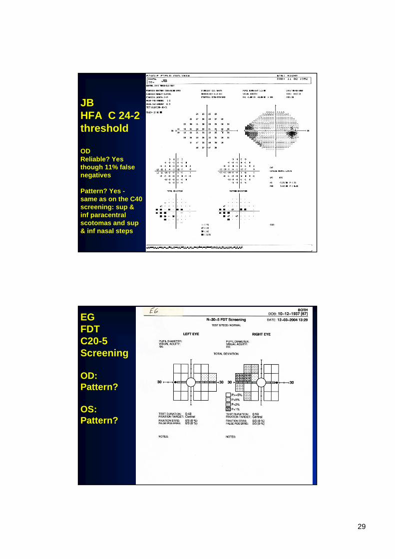
JBHFA C40
ODReliable? YesNormal? NoPattern? Yes -sup & infparacentralscotoma and sup & inf nasal step
OS: Very reliableNo misses
29
JBHFA C 24-2threshold
ODReliable? Yes though 11% false negatives
Pattern? Yes -same as on the C40 screening: sup & inf paracentralscotomas and sup & inf nasal steps
EGFDTC20-5Screening
OD: Pattern?
OS: Pattern?
30
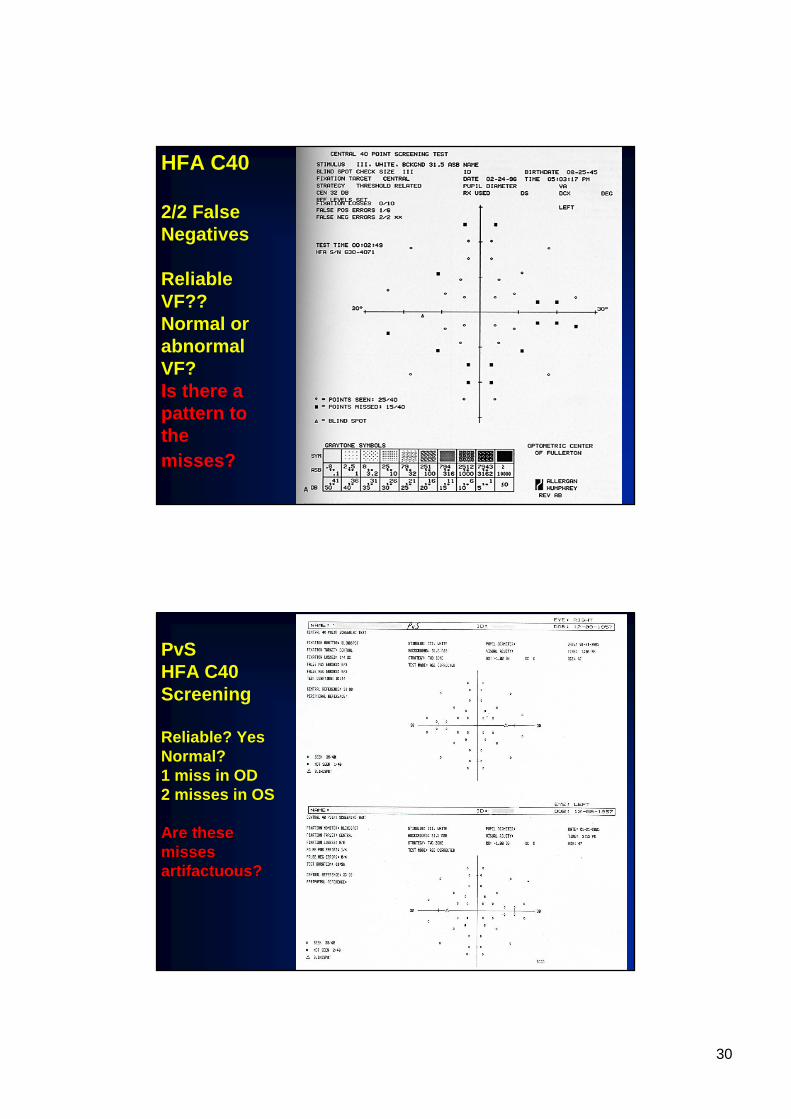
HFA C40
2/2 False Negatives
Reliable VF??Normal or abnormal VF?Is there a pattern to the misses?
PvSHFA C40Screening
Reliable? YesNormal? 1 miss in OD2 misses in OS
Are these Are these misses misses artifactuous?artifactuous?
31
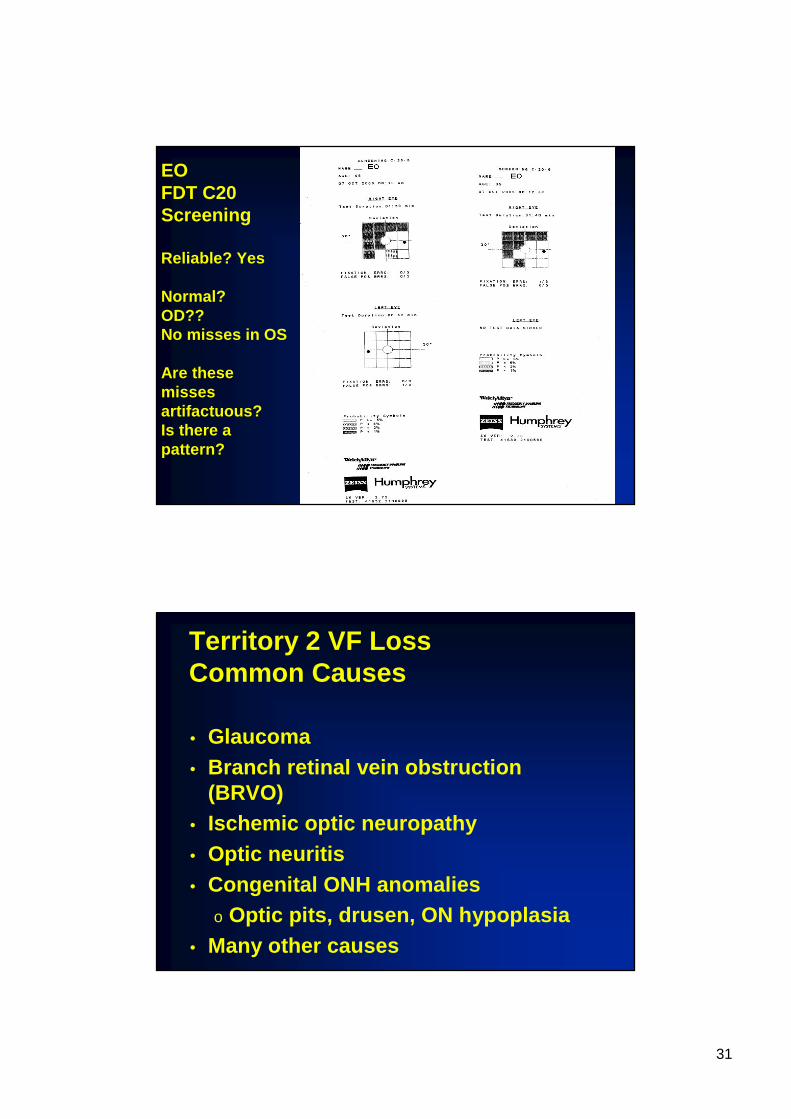
EOFDT C20Screening
Reliable? Yes
Normal? OD??No misses in OS
Are these Are these misses misses artifactuous? artifactuous? Is there a Is there a pattern? pattern?
Territory 2 VF LossCommon Causes
• Glaucoma• Branch retinal vein obstruction
(BRVO)• Ischemic optic neuropathy• Optic neuritis• Congenital ONH anomalies
o Optic pits, drusen, ON hypoplasia• Many other causes

32
Nasal Radial Bundles
• From retina nasal to ONH and somewhat superior and inferior nasal to ONH
• Enter nasal ONH from about 1:00 to 5:00 for right ONH
• NR bundles do not respect the nasal horizontal midline of the eye →→→→ no respect for temporal horizontal midline of VFo The temporal horizontal midline is NOT
diagnostically important • Damage if at ONH produces wedge- or pie-
shaped VF defect pointing to the temporal side of the BS
Optic Nervehead
• Cup contains no axons• Rim tissue contains axons
33
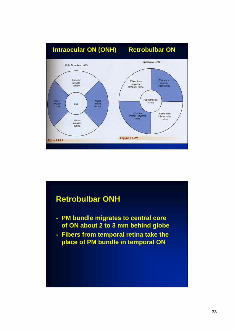
Intraocular ON (ONH) Retrobulbar ON
Retrobulbar ONH
• PM bundle migrates to central core of ON about 2 to 3 mm behind globe
• Fibers from temporal retina take the place of PM bundle in temporal ON
34
Territory 2 DamageDifferentiation of the Cause
• Ophthalmoscopy very helpful• Retrobulbar ON damage will progress down
ONH (retrograde atrophy → shows as pallor of the ONH rim tissue) and will appear in ONH in 1 to 3 months after the damage
• Before 1 to 3 months neuroimaging may be needed differentiate the cause of retrobulbar ON damage
• Pupil signs - decreased direct light reflex, L-N dissociation and +APD (APD ifasymmetric or unilateral)
FDT C20-5
Reliable?
Normal or abnormal VF?
Pattern?
If this is true, organic VF loss where could the lesion be that caused it?

35
Territory 3 Territory 3 -- Optic ChiasmOptic ChiasmKey Anatomy
• Nasal retina fibers from each eye cross in median bar of chiasm (53-55%)o Fibers from inferior nasal retina are inferior in
chiasm and loop into the contralateral optic nerve• Anterior knee of von Willebrand
o Fibers from superior nasal retina are superior in chiasm, may loop into the contralateral tract• Posterior knee of von Willebrand
• Temporal retina fibers from each eye continue posteriorly in lateral sides/angles of chiasmo Fibers from inferior temporal are inferiorlyo Fibers from superior temporal are superiorly

36
Nasal Retinal Fibers vs Temporal Retinal Fibers
• All temporal retinal fibers are in the arcuate bundleso Temporal fibers are those that arise from ganglion
cells temporal to an imaginary vertical line that goes through the fovea
o Damage to temporal fibers causes pallor at the superior & inferior poles of the ONH
• Nasal retinal fibers are:o Nasal radial bundles → enter the nasal ONHo PM bundles → enter the temporal ONHo Very few arcuate bundles → enter the sup & inf ONH
Damage to all nasal fibers causes a band of pallor across the ONH (nasal rim & temporal rim pallor)
Patterns of Chiasmal VF Loss
• Anterior junctional scotoma• Bitemporal heteronymous VF loss• Inferior bitemporal VF loss• Binasal heteronymous VF loss

37
Anterior Knee of Von WillebrandAnterior Junctional Scotoma
• Fibers from inferior nasal retina OU loop anteriorly into the inferior posterior contralatera l ONH where it meets the chiasm
• Damage (usually from below anterior chiasm) causes superior temporal quadranopsia to contralateral side and optic nerve VF loss (central scotoma, arcuate, etc.) ipsilateral to lesion -anterior junctional scotoma
• Common cause is post-fixed chiasm (chiasm posteriorly displaced so that the anterior chiasm sits over pituitary) with pituitary adenoma
Left anterior junctional scotomaOS: ON nerve fiber bundle (PM bundle) VF defectOD: Sup temp VF defect, respects vertical midline
38
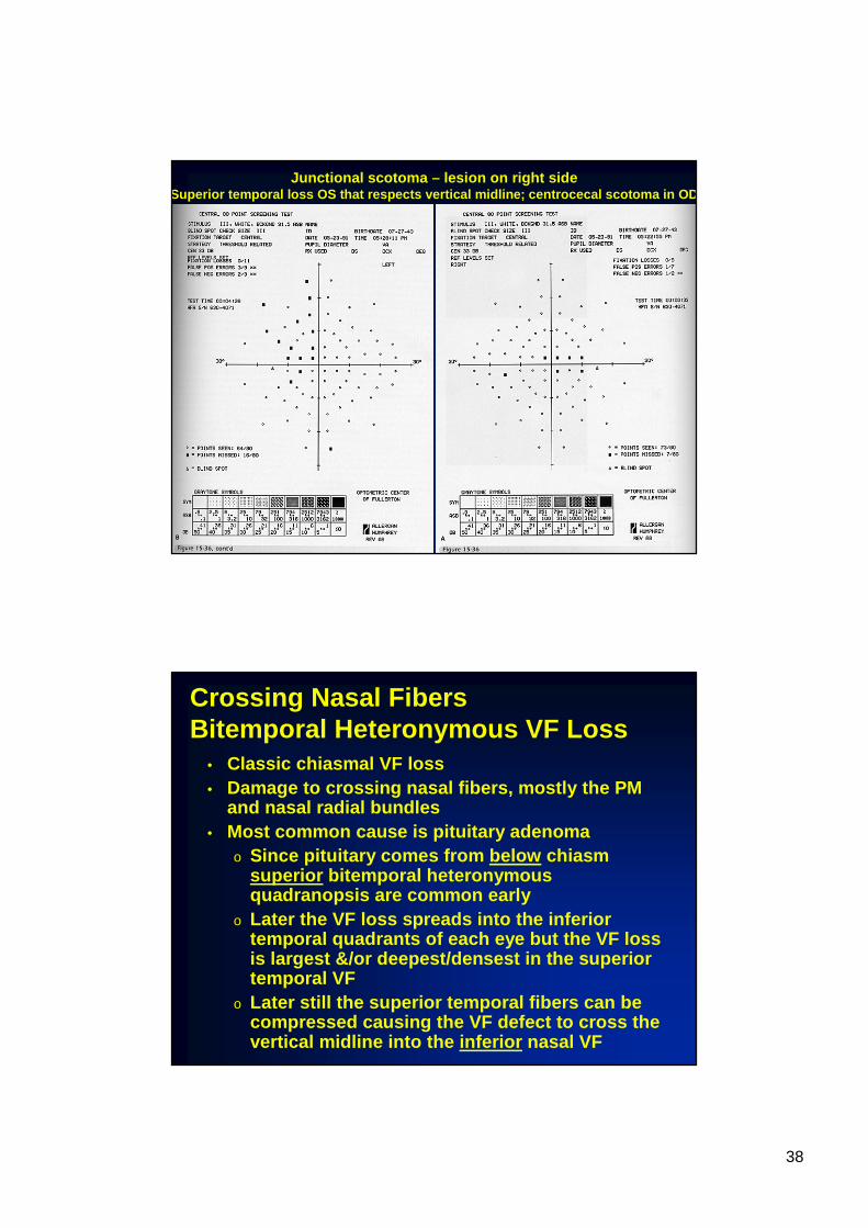
Junctional scotoma – lesion on right side Superior temporal loss OS that respects vertical midl ine; centrocecal scotoma in OD
Crossing Nasal FibersBitemporal Heteronymous VF Loss
• Classic chiasmal VF loss• Damage to crossing nasal fibers, mostly the PM
and nasal radial bundles• Most common cause is pituitary adenoma
o Since pituitary comes from below chiasm superior bitemporal heteronymous quadranopsis are common early
o Later the VF loss spreads into the inferior temporal quadrants of each eye but the VF loss is largest &/or deepest/densest in the superior temporal VF
o Later still the superior temporal fibers can be compressed causing the VF defect to cross the vertical midline into the inferior nasal VF
39
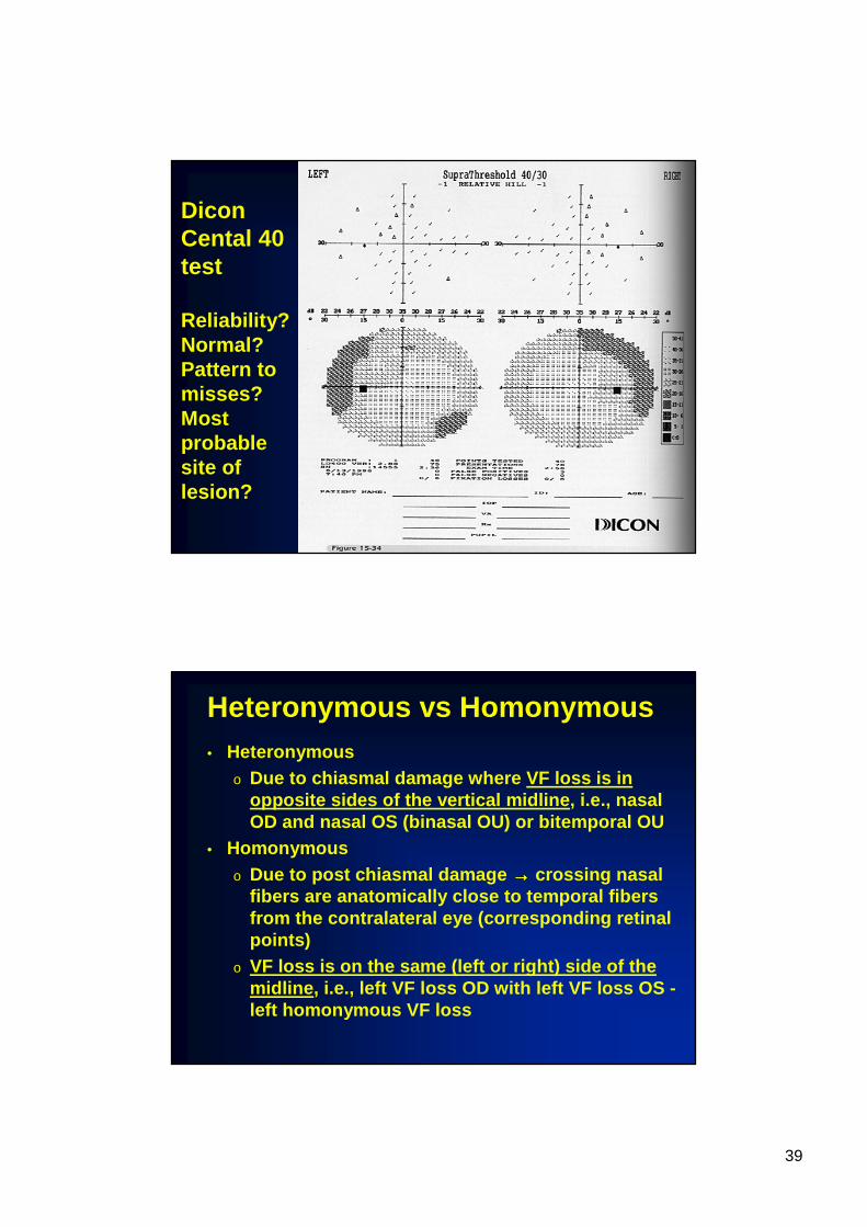
Dicon Cental 40 test
Reliability? Normal? Pattern to misses? Most probable site of lesion?
Heteronymous vs Homonymous• Heteronymous
o Due to chiasmal damage where VF loss is in opposite sides of the vertical midline , i.e., nasal OD and nasal OS (binasal OU) or bitemporal OU
• Homonymouso Due to post chiasmal damage →→→→ crossing nasal
fibers are anatomically close to temporal fibers from the contralateral eye (corresponding retinal points)
o VF loss is on the same (left or right) side of the midline , i.e., left VF loss OD with left VF loss OS -left homonymous VF loss
40
Inferior BitemporalHeteronymous Quadranopsia
• Lesions coming from superior and posterior to posterior notch of the chiasm compress crossing superior nasal fibers
• VF loss appears initially in the inferior temporal VF of each eye and/or is deepest in the inferior temporal VF
• Most common cause is craniopharyngioma
Binasal VF Loss
• Temporal fibers occupy the lateral sides of the chi asm• Lesions on both lateral angles of the chiasm may cause
binasal VF loss• Very, very uncommon to get true (does not cross the
vertical midline) binasal since there must be lesion s in 2 places
• VF loss that is due to arcuate damage to each eye i n Territory 2 is very common but it is not true binas al VF that respects the vertical midline. It is MUCH more common than true binasal VF loss.
• Can cause binasal heteronymous hemianopsia
41
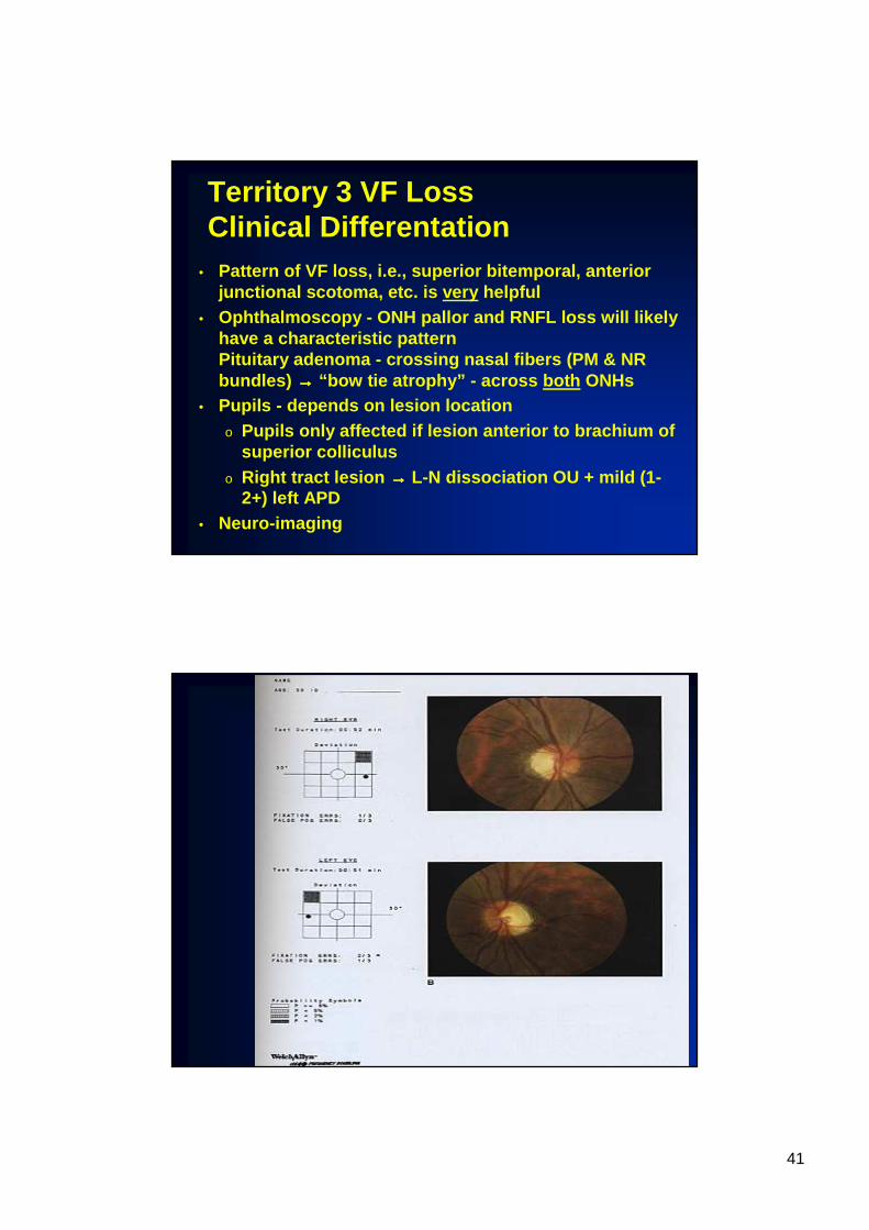
Territory 3 VF LossClinical Differentation
• Pattern of VF loss, i.e., superior bitemporal, ante rior junctional scotoma, etc. is very helpful
• Ophthalmoscopy - ONH pallor and RNFL loss will likel y have a characteristic patternPituitary adenoma - crossing nasal fibers (PM & NR bundles) →→→→ “bow tie atrophy” - across both ONHs
• Pupils - depends on lesion locationo Pupils only affected if lesion anterior to brachium of
superior colliculuso Right tract lesion →→→→ L-N dissociation OU + mild (1-
2+) left APD• Neuro-imaging

42
QUICK REVIEW
• Territory 1- outer retina/choroid“What you see is what you get”What you get in the VF should be what you see in the eyeVF loss does not follow the patterns of other territories (unless by coincidence)Monocular lesions most common but binocular lesions are not uncommonMacular lesions can occur and cause central scotoma; ARMD is common cause
QUICK REVIEW
• Territory 2 – RGCs and their axons up to but not including the chiasm
One or more of 3 nerve fiber bundles PM – macular dysfunction → ↓VA, ↓color vision, central or centrocecalscotoma, pallor of temporal rim, loss of RNFL in PM bundleArcuate bundles ( inf or sup or both) →various VF defects confined to the arcuate (Bjerrum) area of the VF & does not cross the nasal horizontal midline

43
QUICK REVIEW
• Territory 2 – continuedNasal radial bundle – corresponds to temporal wedge of the VF because the fibers go directly into the ONH from retina nasal to the ONH → wedge shaped VF defects temporal to the BS
IMPORTANT DATES IN OHP 1
• Tuesday 1/25/11 – last lecture!!!!• Monday 1/31/11 2:00 to ~3:30 – VF
review by Danielle Leong – optional but recommended
• Monday 2/7/11 – final lab proficiency • Monday 2/14/11 12:30 to 3:00 – final
exam

44
Territory 4Territory 4Post-Chiasmal Visual Pathways
• Optic tracts• LGN• Visual radiations
o Temporal lobeo Parietal lobeo Occipital lobe
• Visual cortex
45
Territory 4 VF LossCommon Characteristics
• Homonymouso Respects vertical midlineo VF loss is to the samesame side in both
eyes, i.e., left side or right side• VF defect is contralateral to lesion• VF defect may look similar or even
identical in the two eyes (congruity)
Homonymous VF Loss
• Characteristic of post chiasmal lesion• VF is damaged to the same side of the
vertical midline in each eye, i.e., right side OD & right side OS - right homonymous hemianopsia
• A characteristic of post chiasmal VF loss ONLY

46
Congruity• A characteristic of post chiasmal VF loss only• As the visual radiations approach the visual cortex
the fibers from corresponding retina points are anatomically close together →→→→ damage to fibers from one eye is more likely to be associated with damage to the other fibers from the other eye
• VF defects are much more similar, usually identical, in lesions of the visual cortex
•• Congruity increases the more posterior the lesion Congruity increases the more posterior the lesion is in territory 4is in territory 4
•• CannotCannot judge congruity if the VF defect is judge congruity if the VF defect is total/complete (all points in the whole total/complete (all points in the whole hemifieldhemifield ) ) and absolute (and absolute ( nono sensitivity at all)sensitivity at all)
Dicon Central 40
Reliability?Normal? Pattern? Site of the lesion(s)?
47
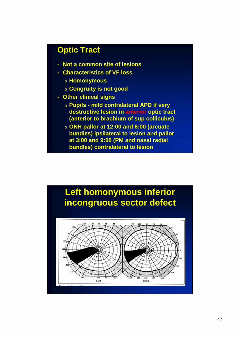
Optic Tract
• Not a common site of lesions• Characteristics of VF loss
o Homonymouso Congruity is not good
• Other clinical signso Pupils - mild contralateral APD if very
destructive lesion in anterior optic tract (anterior to brachium of sup colliculus)
o ONH pallor at 12:00 and 6:00 (arcuate bundles) ipsilateral to lesion and pallor at 3:00 and 9:00 (PM and nasal radial bundles) contralateral to lesion
Left homonymous inferior incongruous sector defect

48
Temporal Lobe
• Fibers from superior retina are located more medially toward interhemispheric fissure
• Fibers from inferior retinas loop far out around the inferior horn at lateral ventricle - Meyer’s loopo Long course makes the inferior
fibers much more likely to be damaged than superior fibers
49
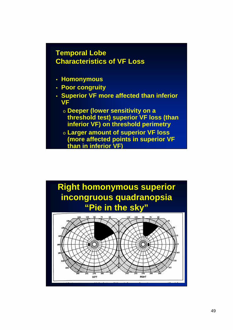
Temporal LobeCharacteristics of VF Loss
• Homonymous• Poor congruity• Superior VF more affected than inferior
VFo Deeper (lower sensitivity on a
threshold test) superior VF loss (than inferior VF) on threshold perimetry
o Larger amount of superior VF loss (more affected points in superior VF than in inferior VF)
Right homonymous superior incongruous quadranopsia
“Pie in the sky”
50
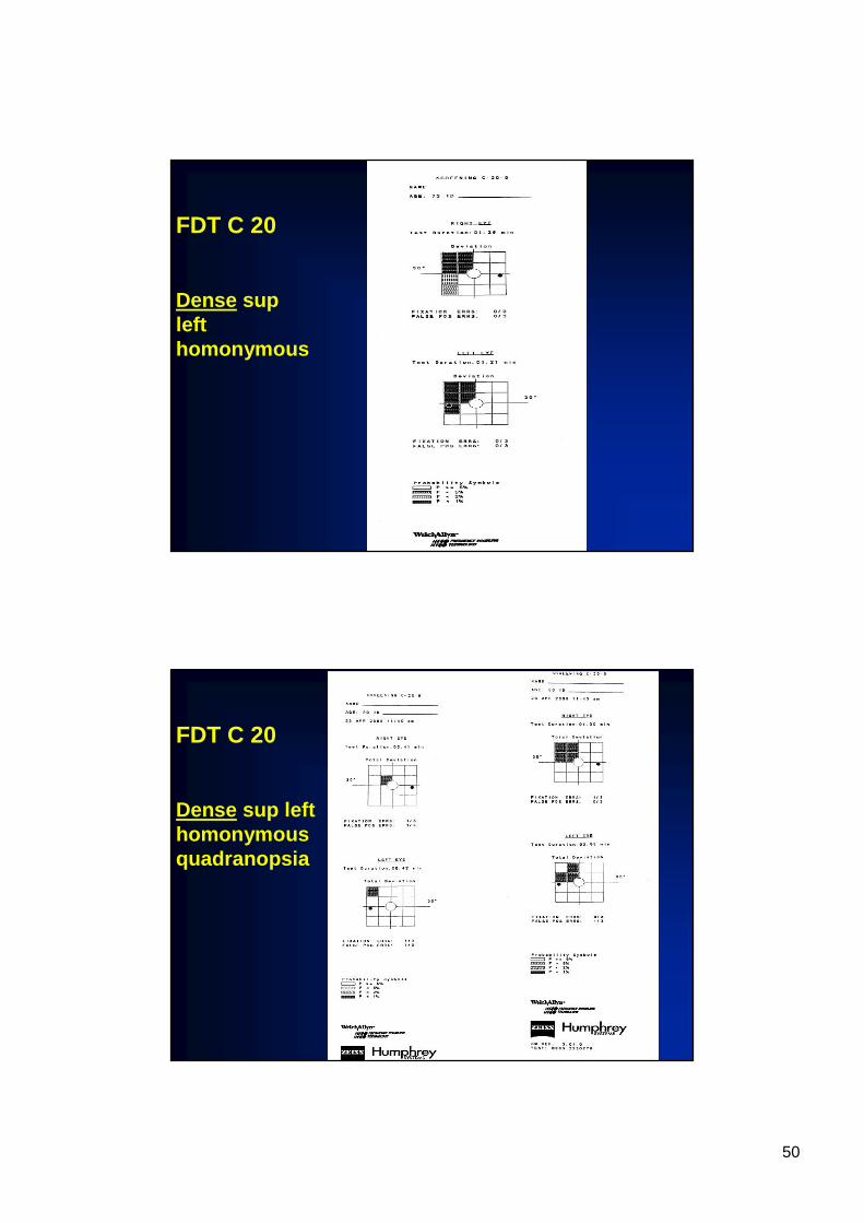
FDT C 20
Dense sup left homonymous
FDT C 20
Dense sup left homonymousquadranopsia

51
FDT C 20
Dense sup left homonymousquadranopsia
Parietal Lobe
• Fibers from corresponding points are physically closer together →→→→greater congruity
• Fibers from superior retina are more likely affected →→→→ VF loss in inferior field or densest VF loss inferiorly
52
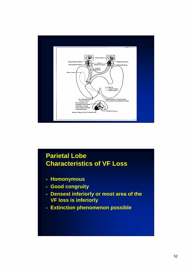
Parietal LobeCharacteristics of VF Loss
• Homonymous• Good congruity• Densest inferiorly or most area of the
VF loss is inferiorly• Extinction phenomenon possible

53
Inferior left homonymous quadranopsia“Pie on floor” defect
FEFDT C 20
Dense inf left homonymousquadranopsia
54
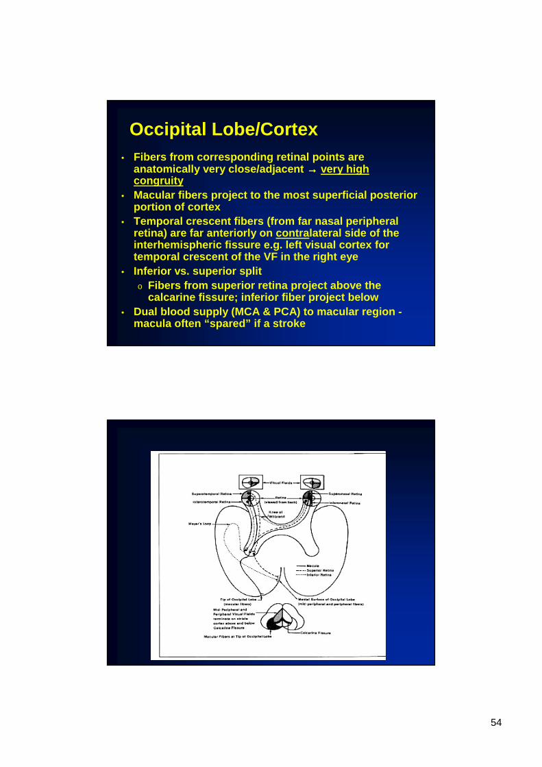
Occipital Lobe/Cortex• Fibers from corresponding retinal points are
anatomically very close/adjacent →→→→ very high congruity
• Macular fibers project to the most superficial post erior portion of cortex
• Temporal crescent fibers (from far nasal peripheral retina) are far anteriorly on contra lateral side of the interhemispheric fissure e.g. left visual cortex for temporal crescent of the VF in the right eye
• Inferior vs. superior splito Fibers from superior retina project above the
calcarine fissure; inferior fiber project below• Dual blood supply (MCA & PCA) to macular region -
macula often “spared” if a stroke

55
Dual (PCA & MCA) blood supply to visual cortex - at occipital pole (macular region)
Occipital Lobe/CortexCharacteristics of VF Loss
• Homonymous• High congruity• Macular sparing common with
stroke• Small, highly congruent
homonymous scotomas are common
56
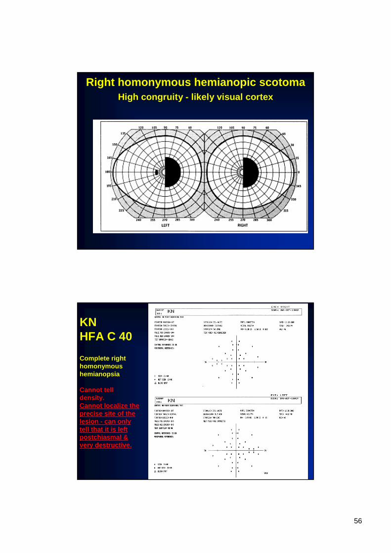
Right homonymous hemianopic scotomaHigh congruity - likely visual cortex
KNHFA C 40
Complete righthomonymoushemianopsia
Cannot tell density. Cannot localize the precise site of the lesion - can only tell that it is left postchiasmal & very destructive.
57
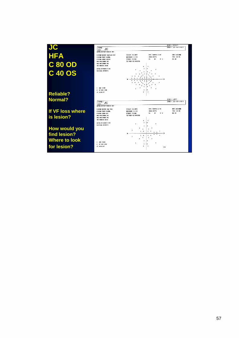
JCHFA C 80 OD C 40 OS
Reliable?Normal?
If VF loss where is lesion?
How would you find lesion?Where to look for lesion?