virulence and survival mechanisms in borrelia and
TRANSCRIPT
Mia Åstrand
Virulence and survival mechanisms in Borrelia and Klebsiella infections – a structural bioinformatics perspective
Mia Å
strand /
/ Virulence and
survival mechanism
s in Borrelia
and Kleb
siella infectio
ns // 2
02
1
ISBN 978-952-12-4075-1
9 789521 240751

Mia Åstrand
Was born on June 2, 1985, in Pietarsaari, Finland. She graduated from University of Helsinki in 2010 with a Master of Science in Biology and obtained a bachelor’s degree in Pharmacy in 2016. This PhD thesis project in Biochemistry has taken place during 2016-2021 under the supervision of Adjunct Professor Tiina A. Salminen at the Faculty of Science and Engineering.

VirulenceandsurvivalmechanismsinBorreliaandKlebsiellainfections–astructural
bioinformaticsperspective
MiaÅstrand
Biochemistry Faculty of Science and Engineering
Åbo Akademi University Turku, Finland 2021

FromtheFacultyofScienceandEngineering,A7 boAkademiUniversity,A7 boAkademiGraduateSchool&NationalDoctoralProgrammeinInformationalandStructuralBiology
Supervisedby
AdjunctProfessorTiinaA.Salminen
StructuralBioinformaticsLaboratory,BiochemistryFacultyofScienceandEngineering,InFLAMESFlagshipResearchCenterA7 boAkademiUniversityTurku,Finland
Reviewedby
AdjunctProfessorSallaRuskamo
FacultyofBiochemistryandMolecularMedicineBiocenterOuluUniversityofOulu,Finland
and
AdjunctProfessor RiikkaIhalin
DepartmentofLifeTechnologiesFacultyofTechnologyUniversityofTurkuTurku,Finland
Opponent
AssistantProfessorRonnieBerntsson
DepartmentofMedicalBiochemistryandBiophysicsUmeåUniversityUmeå,Sweden
ISBN 978-952-12-4075-1 (printed)ISBN 978-952-12-4076-8 (Digital)Painosalama, Turku, Finland 2021

AbstractBacteriauseawidevarietyofmechanismstoestablishaninfectionintheirhostand to compete with other microorganisms for space and nutrients. Thesevirulence and survival mechanismsmainly rely on the functions of proteins,which perform a multitude of highly specialized tasks to ensure bacterialcolonization and survival in the host, e.g., through adhesion to host tissues,nutrientimport,immuneevasionmechanismsandtoxindelivery.Asthefunctionofaproteinisgovernedbyitsstructure,determiningitsthree-dimensional structure makes it possible to gain knowledge also about itsfunction. This thesis is focused on virulence factors inBorrelia andKlebsiellabacteriaandIhaveusedstructuralbioinformaticsmethodstoprovideadetailedunderstandingofhowstructuralfeaturesgovernthefunctionofaprotein.The BmpA, BmpB, BmpC and BmpD proteins of the Lyme disease-causingbacteriaBorreliaburgdorferi are anexampleofhowbacteria can surviveandreproducesuccessfullyinahostdespiteaverylimitedbiosyntheticcapacity.Byanalysing3Dstructuralmodelsof theBmpproteins,weshowedthattheyareinvolvedinthepurinesalvagepathway,whichisusedbytheBorreliabacteriatoobtain vital purinenucleotides even though they lack the enzymesneeded tosynthesize them. BmpD was shown experimentally to bind to the purinesadenosine and inosine. Our structural comparison showed a high similaritybetweenBmpA,BmpBandBmpD,whileBmpCdiffered significantly from theothers, indicating a preference for a different ligand. The evolutionaryrelationshipsbetweentheBorreliaBmpproteinswerestudiedinaphylogeneticanalysis,which provided an improved classification of the Bmp proteins andrevealed significant differences between the Lyme disease-causing and therelapsingfever-causingBorrelia.To successfully colonize a host,Borreliabacteria also need to adhere to hosttissues,andthisisdonethroughspecializedsurface-locatedproteinsthatbindtoreceptorsonhostcellsorintheextracellularmatrix.Basedonourmutationalstudies,theBorreliagariniiDbpAandDbpBproteinsareessentialforadhesionto cells in the nervous system and mediate adherence by binding to theproteoglycans decorin and biglycan on the surface of HBMECs (human brainmicrovascularendothelialcells).Negativelychargedglycosaminoglycan(GAG)chainsontheproteoglycansinteractwithpositivelychargedlysineresiduesonthe surface of the Dbp proteins and these interactions were inhibited bymutating the lysines. Our structural analysis showed significant changes insurface potential for the mutated proteins, which explained the loss ofelectrostatic interactions, and highlighted the importance of individual lysineresiduesforproteoglycanbinding.

ThehighlyantibioticresistantbacteriumKlebsiellapneumoniaeusessecretionsystems to inject toxic effectors into host cells or rival microorganisms. TheeffectorsofthetypeVIsecretionsystem(T6SS)cansuppressthehostimmunesystem andVgrG proteins play a key role in the function of this system.OurexperimentalstudiesshowedthatK.pneumoniaeVgrG4causesaROS-mediatedtoxic effect in the host and that its C-terminal DUF2345-containing part issufficientforROSinduction.Structuralmodellingpredictedthatthisregionhasabeta-helicalfold,whichischaracteristicforVgrGspikes.TheimmunityproteinSel1EpreventsthetoxiceffectsofVgrG4fromharmingthebacteriaitself,andbycombining a structural analysiswith a study of its evolutionary conservationpatternswehighlightedresiduespossiblyinvolvedininteractionswithVgrG4.Insummary,thisworkdeepensourunderstandingofthestructureandfunctionof bacterial proteins essential for virulence and survival. Insights into thedetailed molecular mechanisms of a protein’s function can greatly aid thedevelopmentofhighlytargetednoveltreatmentmethods.

SammanfattningBakterier använder sig av en mängd olika mekanismer för att infektera sinvärdorganismochförattkonkurreraomutrymmeochnäringsämnenmedandramikroorganismer. Dessa virulens- och överlevnadsmekanismer baserar sigfrämst på funktionen hos olika proteiner som utför en mängd mycketspecialiserade uppgifter för att säkerställa kolonisering och överlevnad ivärdorganismen,t.ex.genomadhesiontillvärdensvävnader,genomupptagavnäringsämnen, undvikande av immunförsvaret och leverans av toxiskasubstanser.Eftersom ett proteins funktion avgörs av dess struktur kan man få framinformationomproteinetsfunktiongenomattbestämmadesstredimensionellastruktur.DenhäravhandlingenärfokuseradpåvirulensfaktoreriBorrelia-ochKlebsiella-bakterierochstrukturbioinformatikmetoderharanväntsförattgeendetaljeradförståelseförhurstrukturellaegenskaperstyrettproteinsfunktion.BmpA,BmpB,BmpCochBmpDproteinernaiBorreliaburgdorferi,somorsakarborrelios,ärexempelpåhurbakterierkanöverlevaochförökasigframgångsriktienvärdorganismtrotsenmycketbegränsadbiosyntetiskkapacitet.Genomattanalysera 3D-strukturmodeller för Bmp proteinerna visade vi att de ärinvolveradeidensåkalladepurinesalvage-rutten,sombakteriernaanvänderförattfåtillgångtilllivsnödvändigapurin-nukleotider,trotsattdesaknardeenzymsom behövs för att syntetisera dem. Experimentella studier visade att BmpDbindertillpurinernaadenosinochinosin.Vårstrukturellajämförelsevisadeattdet finns en stor likhetmellan BmpA, BmpB och BmpDmedan BmpC tydligtskiljersigfråndeandra,vilketindikerarattdenbindertillenannorlundaligand.De evolutionära förhållandena mellan Borrelias Bmp proteiner studeradesgenomenfylogenetiskanalys,somresulteradeienförbättradklassificeringavBmpproteinernaochvisadepåtydligaskillnadermellandeBorrelia-bakteriersomorsakarborreliosochdesomorsakaråterfallsfeber.För att framgångsrikt kolonisera en värdorganismbehöverBorrelia-bakterierockså fästa sig vid värdens vävnader, och detta görs genom specialiseradeytproteinersombindertillreceptorerpåvärdcellensytaelleridetextracelluläramatrixet. Baserat på våra mutationstudier är DbpA och DbpB proteinerna iBorreliagariniiväsentligaföradhesiontillcellerinervsystemetochförmedlardetta genom att binda till proteoglykanerna decorin och biglykan på ytan avmikrovaskuläraendotelcellerimänniskanshjärna(humanbrainmicrovascularendothelial cells,HBMECs).Negativt laddade glykosaminoglykankedjor (GAG)påproteoglykanerna interagerarmedpositivt laddade lysinerpåytanavDbpproteinerna, och dessa interaktioner inhiberas när lysinerna muteras. Vårstrukturella analys visade tydliga skillnader i ytpotential för de muteradeproteinerna,vilketförklararförlustenavdeelektrostatiskainteraktionerna,ochbetonarviktenavindividuellalysinerförproteoglykanbindning.

Den antibiotikaresistenta bakterien Klebsiella pneumoniae använder sig avspeciella utsöndringssystem för att överföra toxiska effektormolekyler tillvärdceller eller konkurrerande mikroorganismer. Effektormolekylerna iutsöndringssystem VI (type six secretion system, T6SS) kan hämmavärdorganismens immunförsvar och VgrG proteinerna utgör en viktig del isystemet. Våra experimentella studier visade att VgrG4 från K. pneumoniaeorsakar en toxisk effekt i värdcellen och att detta styrs genom inducering avoxidativ stress. Proteinets C-terminala DUF2345-innehållande del visade sigvaratillräckligfördettaochgenomstrukturellmodelleringvisadeviattdennaregionhar enbeta-helikal struktur, vilket är typiskt för spike-delenhosVgrGproteiner.ImmunitetsproteinetSel1EförhindrardentoxiskaeffektenavVgrG4attskadabakteriensjälv,ochgenomattkombinerastrukturellaanalysermedstudier av proteinets evolutionära konservationsmönster kunde vi peka utaminosyrorsompotentielltinteragerarmedVgrG4.Dettaarbetebidrartillenfördjupadkunskapomstrukturenochfunktionenhosbakteriellaproteinersomärnödvändigaförvirulensochöverlevnad.Insikteride detaljerademolekyläramekanismerna för ett proteins funktion kan bidramed viktig information vid utvecklingen av nya och specifikt målinriktadebehandlingsmetoder.

Tableofcontents
Listoforiginalpublications..........................................................................................I
Contributionsoftheauthor........................................................................................II
Acknowledgements.......................................................................................................III
Abbreviations...................................................................................................................V
1. Introduction................................................................................................................1
2. Reviewoftheliterature..........................................................................................3 Bacterialpathogens.......................................................................................................3 Hostdefensemechanisms...........................................................................................5 2.2.1 Innateimmunity..................................................................................................................5 2.2.2 Adaptiveimmunity............................................................................................................6 2.2.3 Antibiotics..............................................................................................................................7
Bacterialvirulencemechanisms...............................................................................8 2.3.1 Adhesion.................................................................................................................................8 2.3.2 Invasionstrategiesandevasionofhostdefenses..............................................10 2.3.3 Nutrientacquisition........................................................................................................11
Interbacterialcompetitioninsidethehost.........................................................14 2.4.1 Contact-independentmethods..................................................................................14 2.4.2 Contact-dependentmethods......................................................................................15 2.4.3 Effectorsandimmunityproteins..............................................................................18
MainvirulencemechanismsusedbyBorreliaandKlebsiella.......................19 2.5.1 Borrelia.................................................................................................................................19 2.5.2 Klebsiella..............................................................................................................................21
3. Aims.............................................................................................................................23
4. Methods......................................................................................................................24 Sequenceanalysis........................................................................................................24 Sequencealignment....................................................................................................25 Modellingprotein3Dstructures............................................................................25 Modelanalysis..............................................................................................................27 Phylogeneticanalysis.................................................................................................27 Moleculardynamicssimulations...........................................................................28

Visualizationandstructuralanalysis...................................................................29 Experimentalwork......................................................................................................29 4.8.1 BmpDcrystallization......................................................................................................29
5. ResultsandDiscussion..........................................................................................31 Borreliaburgdorferi–BmpA,BmpB,BmpCandBmpD..................................31 5.1.1 Introduction.......................................................................................................................31 5.1.2 TheB.burgdorferiBmpproteinsaresubstrate-bindingproteins..............31 5.1.3 StructuralcharacterizationoftheBmpligand-bindingsite.........................33 5.1.4 TheBmpproteinsareconservedinBorreliaspecies......................................38 5.1.5 TheBmpproteinsplayaroleinthepurinesalvagepathway......................42
Borreliagarinii–DbpAandDbpB..........................................................................44 5.2.1 Introduction.......................................................................................................................44 5.2.2 StructuraloverviewoftheDbpproteins...............................................................45 5.2.3 SequenceanalysisoftheB.gariniiDbpproteins...............................................46 5.2.4 GAG-bindingsitesinDbpAandDbpB.....................................................................46 5.2.5 Proteinstabilityanalysis..............................................................................................50
Klebsiellapneumoniae–VgrG4andSel1E...........................................................50 5.3.1 Introduction.......................................................................................................................50 5.3.2 K.pneumoniaeVgrGs......................................................................................................51 5.3.3 StructuralcharacterizationofVgrG4......................................................................52 5.3.4 SequenceanalysisandmodellingoftheimmunityproteinSel1E.............57 5.3.5 PredictionofpotentialbindinginterfacebetweenSel1EandVgrG4.......59
6. Conclusion.................................................................................................................62
7. References.................................................................................................................65

I
ListoforiginalpublicationsIÅstrandM, Cuellar J, Hytönen J, Salminen TA (2019). Predicting the ligand-binding properties of Borrelia burgdorferi s.s. Bmp proteins in light of theconservedfeaturesofrelatedBorreliaproteins. JournalofTheoreticalBiology462:97–108.IICuellarJ,ÅstrandM,ElovaaraH,PietikäinenA,GuédezG,SalminenTA,HytönenJ.StructuralandbiomolecularanalysesoftheBmpDlipoprotein(BB0385)fromBorrelia burgdorferi reveal a substrate-binding protein of an ABC-typenucleosidetransporter.InfectImmun.2020;88(4):e00962-19.IIIPietikäinenA,ÅstrandM,CuellarJ,GladerO,ElovaaraH,RouhiainenM,SaloJ,Furihata T, Chiba K, Salminen TA, Hytönen J. Conserved lysine residues indecorin bindingproteins of Borrelia garinii are critical in adhesion to humanbrain microvascular endothelial cells. Mol Microbiol. 2021 Jan 29. doi:10.1111/mmi.14687.IVStoreyD,McNallyA,ÅstrandM,Sá-PessoaGracaJSantosJ,Rodriguez-EscuderoI,ElmoreB,PalaciosL,MarshallH,HobleyL,MolinaMartinM,CidVJ,SalminenTA, Bengoechea JA.Klebsiella pneumoniae type VI secretion system-mediatedmicrobial competition is PhoPQ controlled and reactive oxygen speciesdependent.PLoSPathog.2020;16(3):e1007969.
II
Contributionsoftheauthor
I. The computational work, i.e., sequence alignment, modeling,structural analysisandphylogenetic tree,wasdoneby theauthor.Theauthoralsoanalyzedalltheresultsandwrotethemanuscript.
II. TheauthordidtheBmpDcrystallizationsetupandanalyzedthefinal3DstructureofBmpDanditsligandinteractions.Thecorrespondingparts of the manuscript were also written by the author.
III. The author performed the computational work, i.e., sequencealignment,modelingandmoleculardynamicssimulations,analyzedtheresultsandwrotethecorrespondingpartsandallthestructuralanalysispartsofthemanuscript.
IV. TheauthordidtheVgrG4andSel1models, thestructuralanalysesand the functional site predictions, and wrote these parts in themanuscript.

III
Acknowledgements
The work for this thesis was carried out at the Structural BioinformaticsLaboratory(SBL),FacultyofScienceandEngineering,A7 boAkademiUniversityduring2016–2021.Ithasbeenaveryinteresting,rewardingandenjoyabletimeandIwouldliketothankeveryonewhomadethispossibleandeveryonewhohasbeenapartoftheseyears.
IamverygratefulthatIgottodomyPhDattheSBL,andIwouldliketothankitsleader,ProfessorMarkJohnson.Mostofall,IwouldliketothankmysupervisorAdjunctProfessorTiinaA.Salminen,forallyoursupportandpatience,andforpassing on your extensive knowledge about structural bioinformatics. I haveenjoyedthisworkimmenselyandIamverygratefulthatyougavemethechancetoworkonsuchinterestingprojects.IalsowanttothankDr.GabrielaGuédezforbeing my second supervisor and for patiently teaching me about X-raycrystallography.
Iwould like to thank theNationalDoctoral Programme in Informational andStructuralBiology(ISB)foracceptingmeandforgrantingmefunding.ThanksespeciallytothedirectorProfessorMarkJohnson,andtothecoordinatorFredrikKarlssonforalwaysbeingsoveryniceandhelpful.
IwouldliketothankAdjunctProfessorRiikkaIhalinandAdjunctProfessorSallaRuskamo forreviewingmythesisand for taking the timetogivemevaluablecomments which helped to improve the thesis. I am also grateful to ourcollaborators Associate Professor Jukka Hytönen and Professor José A.Bengoecheaandtheirresearchgroups.Iespeciallywanttothankmyco-authorDr. Julia Cuellar, who I collaborated closely with on two papers and whosepositiveattitudealwaysmade thework feelboth funandeasy. Ialsowant tothankDr.AnnukkaPietikäinen,foraveryniceandefficientcollaborationonmylatest publication. I am furthermore grateful to the members of my thesiscommittee; Associate Professor Jukka Hytönen, Adjunct Professor Outi Salo-Ahen,AdjunctProfessorJarmoKäpyläandDr.GabrielaGuédez,fortheirvaluableideasandcommentsregardingmythesisprojects.
IamhappyandgratefultohavebeenabletoworkwithallthewonderfulpeopleofSBL.Thepositive,funandhelpfulatmospherehasmadeitaveryniceplacetowork.LeonorLopesdeCarvalhoandKätheDahlströmwere thebestpossiblecolleaguesandfriendswhenIstartedmyPhD-studiesandIwanttothankyoubothforallyourhelp,butmostlyforallthefunwehavehad.Leonor,IespeciallyrememberalltheniceearlymorningteabreakswehadinthebeginningofmyPhD.ThanksforsharingallyourexperienceonbeingaPhD-student.Käthe,youalwaysfeltlikemybigsisterinthelab,anditwasapleasureworkingacrossfromyouinourlittlecorneroftheoffice.MahletTamirat,whostartedoutatthesametimeasme,hasbeenagreatsupportduringtheseyearsandIamveryhappythat

IV
I got to domy PhDwith you. I was also very glad for your company on thedisastrousBerlinconferenceJ.MarionAlixandIdaAlankoweremyconstantcompanions for lunches and tea breaks during the last few years and I amgrateful for all the laughs we have had together. I am also very thankful toMarion,IdaandMahletforthevirtualcoffeebreaksduringthepandemic.Theygavemesomemuch-neededbreaksandalwaysputasmileonmyface.PolytimiDimitriou,yoursunnysmileandlaughbrighteneduptheofficeanditwasalwaysnicetohaveatalkwithyou.Parthiban,thanksforyourcompanyinthecoffeeroom,andforgenerouslysharingyourknowledgeaboutMD-simulations.IamalsoverygratefultoOutiSalo-Ahen,forbeingpartofmythesiscommittee,forcollaborationsandforalwaysbeingsokindandhelpful.IalsowanttoextendmythankstoJukkaLehtonen, forall thehelpwithcomputerproblemsandforallyour interesting lunch time stories. I alsowant to thank the people in PeterSlotte’s lab,especiallyOskarEngberg, foralwaysinvitingmeforcoffeebreaksandforteachingmehowtoworkwithlipids.
Iwouldalsoliketothankmyfamilyandfriends.TackMamma,EmmaochMalinförattnifunnitsdärochgettmigenvälbehövligmotvikttilljobbet,ochförattnialltidstöttatmigtrotsattniintealltidförståtterpåvadjagjobbarmed.Leif,tackfördualltidfinnsdärmedlugn,tålamodochhumornärjagbehöverdetsommestoch för att duhjälptmigmed allt från tekniskaproblem till diskussioner omgrammatikochgrafiskutformning.Tackocksåtill Jonna,minbästavänsedanlågstadiet.Tackförattdualltidfinnsdärtrotsattviborsålångtifrånvarandra.Jag är också tacksam förmina ”Åbo-vänner” Jessi, Johanna, Sandra,AnneochFebe. Det är alltid lika roligt att träffa er även om vi nuförtiden är litemerautspridda.JagvillocksåtackaMiaochHanna,vårabokcirkelträffar,museibesökochspelkvällarhargettmigmångaroligaochgivandeandningspauser.I am also grateful for the generous funding I have received from MagnusEhrnrooth foundation, Svenska kulturfonden and Tor, Joe och Pentti BorgsMinnesfond,ÅboAkademi.

V
Abbreviations
3D Three-dimensionalABC ATP-bindingcassetteABPS AdaptivePoisson-BoltzmannSolverATP AdenosinetriphosphateBbss BorreliaburgdorferisensustrictoBg BorreliagariniiBLAST BasicLocalAlignmentSearchToolBmp BasicmembraneproteinCASP CriticalAssessmentofproteinStructurePredictionCDI Contact-dependentinhibitionCryo-EM CryogenicElectronMicroscopyDAMP Damage-associatedmolecularpatternsDbp Decorin-bindingproteinExP ExportedproteinGAG GlycosaminoglycanI-TASSER IterativeThreadingASSEmblyRefinementKp KlebsiellapneumoniaeLB LymeborreliosisLG LeandGascuelMAPK Mitogen-activatedproteinkinaseMD MolecularDynamicsML MaximumLikelihoodNBD Nucleotide-bindingdomainNCBI NationalCenterforBiotechnologyInformationNF-kB Nuclearfactor-κBNMR NuclearmagneticresonanceOME OutermembraneexchangesystemsPa PseudomonasaeruginosaPAAR Proline-alanine-alanine-argininePAMP Pathogen-associatedmolecularpatternsPDB ProteinDataBankPnrA PurinenucleosidereceptorAPRR PatternrecognitionreceptorRF RelapsingfeverRMSD RootmeansquaredeviationRMSF RootmeansquarefluctuationROS ReactiveoxygenspeciesSBP Substate-bindingproteinSLR Sel1-likeRepeatsTLR Toll-likereceptorTMD TransmembranedomainTp TreponemapallidumTPR TetratricopeptiderepeatTssX TypesixsecretionXTxSS TypeXsecretionsystemUniProtKB UniversalProteinKnowledgebaseVgrG Valine-glycinerepeatproteinG


1
1. Introduction Bacteriaaremicroscopicorganismsthatthriveinallkindsofenvironmentsandhaveanabilitytoadapttothemostextremecircumstances.Somebacterialiveinourbodiesandarevital for thebody’snormal functions,whileotherbacteriacontain virulence factors that cause disease in their host. The discovery ofantibiotics in the20th century enabledus to treat infections causedby thesepathogenic bacteria, but the rapid life cycles of bacteria mean that they arequickly developing resistance tomany antibiotics (Hutchings et al., 2019). Incombinationwithouroveruseofantibiotics,thishasledtoanalarmingsituationwheremoreandmorebacteriaarerapidlydevelopingantibioticresistance.Atthesametime,therateofantibioticdevelopmenthassteadilydecreasedinthelastfewdecades,creatingasituationwhereevencommoninfectionscanbecomelife-threatening. Bacteriausecomplexmechanismstoinfecttheirhostsandtocompetewithotherbacteria for resources. The constant arms race between bacteria and theircompetitorshaveledtothedevelopmentofamultitudeofdifferentmechanismsandmostbacteriahavealargearsenalofweaponsforattackingbothhostcellsandrivalbacteria(BlivenandMaurelli,2016).Severalmechanismscanoftenbeusedsimultaneouslyandarecoordinatedtoproduceaspecificeffectattherightplace,attherighttime.Ithasalsobeensuggestedthatantibioticresistancecouldbereducedbyusingantibioticsthattargetbacterialvirulencemechanismssincethis would give a lower selection pressure (Ruer et al., 2015). Most of thevirulencemechanismsrelyonproteinstoperformdifferentfunctionsthatharmthe target cell in some way, for example by rupturing the target cell or bybreakingdownitsDNA(WebbandKahler,2008). Proteinsarelargemacromoleculesthatcanbeconsideredtheworkersofacell.Theyareinvolvedinallmajorprocessesneededtosustainlife.Ontheorganismleveltheyplaykeyrolesin,forexample,theimmunesystem,themetabolismandthe nervous system, and on the cellular level they are involved in e.g., DNAreplication, enzymatic reactions and transporter functions. Proteins are builtfrom20standardaminoacids,combinedincountlessnumbersofways,andwithco-andposttranslationalmodifications,theyproduceavastarrayofproteinsofdifferent sizes and shapes. The sequence of amino acids specifies the three-dimensionalstructureoftheprotein,which isessential forthefunctionoftheprotein. Bioinformaticsisamultidisciplinaryfieldthatcombinesbiology,mathematics,computer science and statistics to study protein function, evolutionaryrelationshipsorpredictproteinstructures(AamerMehmood,2014).ThefieldwasoriginallyinitiatedbyMargaretDayhoffinthe1950s,whenshestartedusingcomputationaltoolstostudyproteinsequences(Gauthieretal.,2019).Tofullyunderstand the function of a protein however, it is vital to know its three-

2
dimensional structure. This can be determined either through experimentalmethods,likeX-raycrystallography, Cryogenicelectronmicroscopy(cryo-EM), Nuclearmagneticresonance(NMR)orbyuseofcomputationaltools.Structuralbioinformatics is a branch of bioinformatics that uses various computationaltoolstopredictandanalyzethe3Dstructuresofproteins(Chandraetal.,2010).Fromthestructureofaprotein,itsfunctioncanbeinferredanddetailedanalysesofhowproteinsinteractwitheachotherandwithothermoleculescanbemade.However,whenanalyzingproteinstructures,itisimportanttorememberthatproteins are highly dynamic molecules that constantly fluctuate betweenconformations,andinsomecasesdonotformstablestructuresunlessboundtoanothermolecule(DysonandWright,2005;Henzler-WildmanandKern,2007).Since the number of sequenced proteins is much larger than the number ofexperimentally determined protein structures, the function ofmany proteinsremainsunknown.Asaresultofthisdiscrepancy,novelwaysoftreatingdiseasesare left undiscovered due to the lack of structural information. Here,computationalstructurepredictionmethodscanplayanimportantrole,astheycanpredictthestructureofaproteinmuchfasterandmoreinexpensivelythantheexperimentalmethods. Inthisthesis,informationobtainedfromproteinsequencesandstructuresarecombinedwith the results of functional studies andwith analyses of proteininteractions to study specific proteins that are involved in host infection andintermicrobial competition. This integrated approach provides detailedinformationon theproteinsand their functionalpropertiesandenablesus todefinetheirrolesinthecomplexnetworksofinterbacterialandhost-pathogeninteractions.

3
2. Reviewoftheliterature
BacterialpathogensBacteriaareprokaryoticmicroorganismsthatcome inmanyshapesandsizesand that exist in almost every conceivable environment on earth. Unlikeeukaryotes,prokaryoteshaveacellwall,whichissometimessurroundedbyasticky capsule, they lack complex organelles and besides their singlechromosome they also contain small rings of DNA, called plasmids. Hair-likeappendagescalledfimbriaeandpiliareusedforcommunicationandattachment,and longer appendages called flagella are used for movement. Bacteria aretraditionallydividedintoeitherGram-negativeorGram-positivebacteriaafterHansChristianGram,whodiscoveredastainingmethodwhichcandifferentiatebacteriabasedontheircellwallstructure.InGram-positivebacteria,thecellwallismadeupofaplasmamembrane,aperiplasmicspaceandaverythicklayerofpeptidoglycan,containingteichoicandlipoteichoicacidsinsideit(CabeenandJacobs-Wagner,2005).TheGram-negativecellwallontheotherhandisamoremultilayered structure,with a lipopolysaccharide (LPS)-rich outermembraneand a periplasmic space between the inner and outer membranes. A thinpeptidoglycan layer in theperiplasm is connected to theoutermembranevialipoproteins(figure1).Many bacteria live in our bodies and form commensal relationships that aremutuallybeneficial.Ourbodiesofferanutrient-richenvironmentforthebacteriaand they in turnprovideuswith essential vitamins, digestnutrients thatourbodiescannotbreakdown,andhelppreventcolonizationbypathogenicbacteria(Brestoff and Artis, 2013). Pathogenic bacteria contain virulence factors thatenable them to evade the immune systemand causedisease in thehost. Thevirulence of bacteria can be defined as the result of interactions between amicrobe and a susceptible host (Casadevall and Pirofski, 2019). Hospital-acquiredinfectionsareanexampleofhownormallyharmlessbacteriacancausediseasewhen the immune systemof the host isweakened. In relation to theestimatednumberofbacterialspecies,veryfewspeciesactuallycausediseaseand from an evolutionary perspective it is usually more beneficial for thebacteria to live in a commensal relationshipwith its host. Despite this, somebacteria do cause disease and it has been speculated that for some bacteriadiseaseisarequirementforsuccessfultransmissiontoanewhost.

4
Figure1.DifferencesinmembranestructurebetweenGram-positiveandGram-negativebacteria.Thefigureshowsasimplifiedbacterialcellwherethemaincomponentsaremarked.Theclose-upsshow the specific cell wall features of Gram-positive and Gram-negative bacteria. The Gram-positivecellwallconsistofaplasmamembrane,athinperiplasmicspaceandathickpeptidoglycanlayerwithlipoteichoicandteichoicacids.TheGram-negativecellwallconsistoftwomembranes,aninnerplasmamembraneandanoutermembrane.Betweenthemistheperiplasmicspacewhichalsocontainsathinlayerofpeptidoglycan.
Togainaccesstoahost,bacteriamustpenetrateitsexternalbarriersandthenevadeitsdefensestoensuresurvivalandreplication(Senetal.,2016).Foreverydefensemechanism employed by the host, bacteria have developed effectivemechanismstocounteractthem,thuscreatinganarmsracebetweenhostandpathogen(WebbandKahler,2008).Manyofthesemechanismsarealsousedincompetitionwithotherbacterial species,enabling thebacteria tooutcompetetheirrivalsforaccesstohostnutrients.The aim of this literature review is to give a general overview of bacterialmechanisms used for host infection and interbacterial competition and toemphasize themechanisms that are relevant for theprojects included in thisthesis.
5
HostdefensemechanismsPathogenicbacteriaenteringthebodyofahostwillencounterawiderangeofobstaclesproducedbythedefensesystemofthehost.First,physicalbarrierstrytoblockpathogensfromgainingaccessintothehost.Whenthepathogenshavepenetrated these initial barriers, they aremet by the innate immune system,which produces the first immune responses against the intruder. After a fewdays,thehighlyspecificadaptiveimmuneresponseisactivated,killinginvadingpathogensinahighlyspecificmanner.Incaseswherethehost’sowndefensesarenotenough,pharmaceuticalantimicrobial compounds (antibiotics) canbeusedtoeradicatetheinfection.
2.2.1 InnateimmunityPhysical barriers, like the skin, the respiratory tract and the gastrointestinaltract,preventpathogensfromenteringourbodies,andsecretionssuchasmucus,salivaandtearswashawayforeignparticlesfromtheepithelialsurfacesofthebody(Wilsonetal.,2002).Thesebarriersalsoproducechemicalsubstancesthatareharmfultothepathogens,e.g.,antibacterialpeptides,enzymesandstomachacid. Antibacterial peptides are produced by many cell types in the body,especiallybyepithelialcellsthatcomeindirectcontactwithpathogens(PatelandAkhtar,2017).Theyareahighlydiversegroupofmoleculesthatoftentargetthecellmembraneorothernon-specific targets, therebymaking itharder forbacteriatodevelopresistance(Nguyenetal.,2011).Commensal,non-pathogenicbacteria normally inhabit the epithelial surfaces, and the invading pathogensmustcompetewiththemforspaceandnutrients.Oncethepathogenpassestheseinitial barriers, they encounter the cells of the innate immune system,whichproducesnon-specificreactionsagainstinvadingmicrobes.The initial innate cellular response to pathogens is mainly mediated byphagocyticcellslikeneutrophilsandmacrophages,whichdestroythepathogensbyengulfingthemintheprocessofphagocytosis(Nicholson,2016).Theinnateimmunesystemcellsareactivatedwhenspecificreceptorsrecognizeconservedmolecular patterns on the bacteria. These so-called pathogen-associatedmolecularpatterns(PAMPs)areanintegralpartofbacteriaandareessentialfortheir survival and ability to cause infection (Akira et al., 2006). PAMPs arerecognizedbypattern-recognitionreceptors(PRRs),whicharehighlyconservedreceptorsthatarecontinuallyexpressed,bothontheinsideandtheoutsideofcells(Nicholson,2016).TheToll-likereceptor(TLR)familyareamongthemostimportant pattern recognition receptors and can detect a large variety ofmicrobialpatterns(AkiraandTakeda,2004).Theimmunesystemcanalsobeactivated by damage-associated molecular patterns (DAMPs), which aremoleculesreleasedbythebodyasaresultofthetissuedamageandcelllysisthatoccurs during the infection (Kaur and Secord, 2019). In addition to these

6
molecularpatterns,thebodycanreacttothelossofspecificmoleculesthatareexpressedbyhealthyandnormalcells.Inductionofoxidativestress isanotherwayforthehosttocombatpathogens(Pohanka, 2013). Reactive oxygen species (ROS) are normally produced as aresultofaerobicrespirationandinthepresenceoffreemetalsbutcanalsobeinduced as a defense mechanism against pathogens. ROS damagesmacromoleculesanddisrupts signalingpathwaysby causing strandbreaks inDNAorbyoxidatingproteins, lipidsorDNA (AutenandDavis,2009).Duringinfections, ROS is triggered by the phagocytosis of bacteria and a so-calledrespiratoryburst isactivated insidethephagosome.ROScanalsobereleasedinto the extracellular space, targeting bacteria that escapes phagocytosis(Nguyenetal.,2017).The complement system plays a major role in the initial immune responseagainstbacterial infectionsandconsistofanetworkofproteins that togetherenhancestheabilityoftheimmunesystemtodestroypathogens,bymarkingthepathogens for phagocytosis, by producing proinflammatory molecules or byinducing cell lysis (Dunkelberger and Song, 2010). Together, these initialresponsesoftheinnateimmunesystemactivatethehighlyspecificreactionsoftheadaptiveimmunesystem(IwasakiandMedzhitov,2015).
2.2.1.1 NutritionalimmunityAnotherdefensivestrategyagainst invadingpathogens is thesequestrationofvitalnutrients.Manyenzymesandproteinsrequiretracemineralslikeiron,zincandmanganesetofunctionandtheseelementsareconsequentlyvitalforbothhostandpathogen.Themethodsemployedbythehostforpreventingpathogenaccess tominerals is callednutritional immunity. Sequestrationof iron is themost common form of nutritional immunity and most of the iron insidevertebrate bodies is stored intracellularly, either in the iron storage proteinferritinorinthehemeofhemoglobinormyoglobin(Skaar,2010).Inresponsetoinflammatorysignalsthebodycanpreventirontransferintothecirculationbyinhibitingferroportin,anironexporter,andbysecretinglactoferrin,whichbindstoFe3+withhighaffinity,therebyfurtherrestrictingthepathogen’saccesstoiron(Núñezetal.,2018).Anyremainingextracellularfreeironisquicklyboundtotransferrinensuringthatvirtuallynofreeironisaccessibletopathogens(CassatandSkaar,2013).However,asthehostshavedevelopedsystemstosequesterminerals from the pathogens, the pathogens have consequently developedmechanisms for circumventing them (see more details in section 2.3.3.2)(HennigarandMcClung,2014).
2.2.2 AdaptiveimmunityComparedtotheinnateimmunesystem,itnormallytakesseveraldaysbeforetheadaptiveimmunesystemisactivated,butthenitprovidesahighlyspecific

7
responsetopathogensandalsocreatesanimmunologicalmemory.Theadaptiveimmuneresponsereliesontwomaintypesoflymphocytes,BcellsandTcells.Both cell types have surface receptors that bind with high specificity tomolecules recognized as non-self, antigens. B cells recognize antigens on thesurface of pathogens whereas T cells only recognize fragments of antigensexposedonthesurfaceofantigen-presentingcells(Actor,2019).Fragmentsofantigens can be presented on the surface ofmany cell types, for example onphagocyticcellsoftheinnateimmunesystem,oronthesurfaceofBcells.WhenBcellsrecognizeaspecificantigen,theyaretriggeredtostartdividingandbecomeantibody-producingplasmacellsormemorycells(BonillaandOettgen,2010).Bindingofanantibodytoanantigenactivates,forexample,theclassicalpathwayinthecomplementsystem,resultingincelllysis,orthephagocyticcells,increasingphagocytosis(Forthal,2014).Itcanalsocausecellstoaggregateorbeimmobilized,ortheantibodiescanphysicallypreventpathogensfromattachingtohostcells.WhenaTcellhasbeenactivated,itproliferatesintosubtypesthatperformdifferentfunctions:TkillercellsdestroyinfectedcellswhereasThelpercells secrete cytokines, which activate macrophages or stimulate B cells toincreasetheproductionofantibodies.
2.2.3 AntibioticsAlthough not part of the natural defense systems of the host, antimicrobialcompounds in the formofpharmaceuticals, i.e.,antibiotics,canalsoprovideasignificant defense against infectious bacteria. Antibiotics are antimicrobialcompoundsdesignedtokillorimpedethegrowthofpathogenicbacteriainthebody (Hutchings et al., 2019). Many antibiotics originate from compoundsproduced naturally by plants, bacteria or fungi, who use them as chemicalweapons or as defense mechanisms against competitors. Antibiotics mainlytarget essential functions or structures of the bacteria and are generallyclassified according to theirmechanismof action (Kapoor et al., 2017). Beta-lactamsandglycopeptideantibioticspreventcellwall synthesisbydisruptingthe formation of the peptidoglycan layer (Bush, 2012). Macrolides andtetracyclines are examples of antibiotics that target the protein synthesismachinery. They bind to ribosomal subunits and inhibit protein synthesis bypreventingtRNAbindingorbycausingunfinishedproteinchainstodetachfromthe ribosome (Mccoy et al., 2011). DNA synthesis can be disrupted byfluoroquinolones, which inhibit the DNA topoisomerases needed for DNAreplication(Aldredetal.,2014).Vitalmetabolicpathwayscanalsobetargeted,e.g., the synthesis of folic acid, needed for DNA synthesis, is inhibited bysulfonamidesandtrimethoprim(Fernández-Villaetal.,2019).

8
BacterialvirulencemechanismsBacteria have developed a wide range of mechanisms to subvert the hostimmune system and to sustain an infection (Wilson et al., 2002). They useadhesion mechanisms to attach themselves to a host cell, produce toxins oreffectors that destroy or damage host cells and evade the host defensemechanismsbymaskingthemselvesindifferentwaystopreventrecognitionbythehost.
2.3.1 AdhesionInordertocolonizeahost,thebacteriafirstneedtoattachthemselvesfirmlytothehostsurfaces.Mechanicalforces(coughing,saliva,bloodflowetc.)inthehostconstantly try to remove bacteria from its surfaces, and forming a stableadherenceisthereforeacrucialfirststepintheinfectionprocess(Wilsonetal.,2002).Whenbacteriacomeintocontactwiththehostsurfaces,theycansensechanges in their environment, which in turn trigger changes in metabolism,respiration and expression of virulence-specific genes (Stones and Krachler,2016).Therearealargenumberofadhesionmolecules(adhesins)onthesurfaceof bacteria, and these can form a wide variety of interactions with the hostsurfaces. Some interactions are transient and nonspecific whereas otherinteractionsarestableandhighlyspecific.Thedifferentkindsofadhesinsarenormallyexpressedinacoordinatedfashionatdifferentstagesoftheinfection(KlemmandSchembri,2000).Adhesinsaredividedintotwomaingroups:fimbrial(pili)andafimbrialadhesins(Pizarro-CerdáandCossart,2006;RibetandCossart,2015;Wilsonetal.,2002).Fimbriaeareshorthair-likepolymericstructuresonthesurfaceofbacteriawithaproteinatthetip,whichdeterminesthebindingspecificitytomoleculesonthehostcell.Afimbrialadhesinsarebacterialsurfaceproteinsthatbindtoavarietyof molecules on the host surface, like integrins and cadherins, as well ascomponentsoftheextracellularmatrix(collagens,laminins,proteoglycansetc.)and usually mediate more intimate, short-range contacts than the fimbria(Wilsonetal.,2002).TheadhesinOspC(OuterSurfaceProteinC)isanexampleofanafimbrialadhesinandplaysacrucialroleinBorreliainfectionsbybindingto the extracellular matrix components dermatan sulfate and/or fibronectin.BacteriawithOspCthatcanbindtodermatansulfatearecapableofcolonizingmice joints,whereasbacteriawithOspC that canonlybind to fibronectinareunabletocolonizejoints(Linetal.,2020).Adhesionisalsoanimportantfirststepintheformationofbiofilms.Bacteriaarecapable of aggregating into multicellular communities with othermicroorganismsandtogethertheysecreteapolysaccharide-richmatrixthatcanprotect them against many host defense mechanisms, including antibiotics(RibetandCossart,2015).

9
2.3.1.1 BacterialadhesiontoproteoglycansManypathogensadheretohosttissuesbybindingtoproteoglycans,whicharewidelyexpressedinmanytissues.Proteoglycansareglycosylatedproteinsfoundonhostcellsurfaces,intracellularcompartmentsandintheextracellularmatrix,where they mediate cellular processes like adhesion, signaling and motility(RostandandEsko,1997).Theyconsistofacoreproteinthatiscovalentlyboundtooneormoreglycosaminoglycan(GAG)chains(figure2)(Varkietal.,1999),whicharemadeupofrepeatingdisaccharideunitsconsistingofanaminosugarandauronicacid.EachGAGismadeupofaspecificcombinationofdisaccharideunits and negatively charged sulfate groups are attached to some of them,creatingauniquesulfationpattern.ThesesulfategroupsmediateelectrostaticinteractionstoGAG-bindingproteins(Hilemanetal.,1998).Proteoglycansarethushighlyvariablemoleculesandtheirdistributionandcompositioncanvarydepending on tissue type (García et al., 2016).Which tissues a pathogen cancolonizemaybedeterminedbyitspreferenceforaspecificGAG.B.burgdorferihas, forexample,beenshowntobindtoproteoglycans invarioushosttissues(e.g. endothelial and neural tissues) and to bind to different types of GAGsdependingonthetissue(Leongetal.,1998).
Figure 2. General structure of proteoglycans and glycosaminoglycans (GAGs) shown ascomponentsoftheextracellularmatrix(EM).ProteoglycansconsistofacoreproteinandattachedGAGchains.AGAGchainismadeupofrepeatingdisaccharideunitsconsistingofanaminosugarandauronicacid,whichcanvarybetweendifferentGAGs.Negativelychargedsulfategroupsareattached to the GAG chains in specific sulfation patterns. The GAG chains interact with othermoleculesbyformingelectrostaticinteractionsthroughthesulfategroups.

10
2.3.2 InvasionstrategiesandevasionofhostdefensesAfteradheringtoahostcell,manybacterianeedtogainfurtheraccessintothehost. Most bacteria have either an extracellular or an intracellular lifestyle(Wilsonet al., 2002), but somebacteria canuseboth strategies. Extracellularbacteriaproduceenzymesthatbreakdownthetissuebarriersandthenestablishanichewithinthetissue,butwithoutenteringhostcells.Intracellularpathogenson the other hand penetrate the host cells and modify the cells internalenvironment in order to survivewithin the host cellwithout being degraded(Thakuretal.,2019).Avoiding the effects of antimicrobial peptides is one vital strategy used bybacteriatoevadetheimmunesystem(ReddickandAlto,2014;Sperandioetal.,2015). This can be done e.g. by changing the charge of membrane lipids tofunctionasanelectrostaticbuffer(Ernstetal.,2009),tosecreteproteasesthatbreak down antimicrobial peptides (McGillivray et al., 2009) or to usetransporterstoexporttheantimicrobialpeptidesorimportthemfordestruction(Eswarappaetal.,2008;Shaferetal.,1998).Anotherevasionmechanismistomask the bacterial surface in a capsule consisting of polysaccharides, whichhidesthespecificbacterialmoleculesrecognizedbytheimmunesystem,whilestill allowing adhesins to penetrate (Finlay and McFadden, 2006; Paton andTrappetti,2019).Lipopolysaccharide,flagellaandpeptidoglycanaresomeofthemost easily recognizable bacterial surface molecules and bacteria use manydifferentmethods tohide these fromthe immunesystem(Hajametal.,2017;Kawasakietal.,2004;SorbaraandPhilpott,2011).Bymakingsmallalterationsto these kinds of molecules, the bacteria can avoid being recognized by theimmunesystem.Antigenicvariationisanexampleofhowbacteriacanmodifythesurfaceantigens recognizedby theadaptive immunesystem,and therebyavoid immune activation (Palmer et al., 2016). As many immune cells arephagocytic,bacteriahavedevelopedmechanismsforavoidingphagocytosisorintracellularkillinginphagolysosomes,e.g.bycausinglysisofthephagocyticcellor by preventing opsonization (Uribe-Querol and Rosales, 2017). Molecularmimicryisamethodusedbybacteriatohidethemselvesfromthehostimmunesystembymimickinghostcellsurfacemolecules(Brownetal.,2008).HumancellsarecoatedinsialicacidresiduesandthebacteriaNeisseriameningitidiscanescapedetectionbydecorating its surfacewith theseresidues (Parsonsetal.,1996).BacteriacanalsomodifytheirPAMPsinsuchawaythatthePRRscannolongerrecognizethem(Matsuura,2013).Thepathogenshavealsoevolvedmechanismstoavoidthedamagescausedbyoxidative stress, for example by repairing damaged DNA and proteins, or byproducingantioxidantsanddetoxificationenzymesthatneutralizeROS(Reniere,2018).Furthermore,ithasalsobeenshownthatsomebacteriacaninduceROSintargetcellsanduseittotheirownadvantage(Dongetal.,2015;Herschetal.,2020). Bacteria can also use othermethods for causing host damage, e.g., by

11
attackingcrucialsignalingpathways(e.g.MAPKandNF-kB)orproteinsecretorypathways(Duesberyetal.,1998;Kimetal.,2007;Sanadaetal.,2012).Manyofthe specific effectors used to attack host cells are also used in interbacterialcompetitionsandwillbediscussedinmoredetailinsection2.4.
2.3.2.1 AntibioticresistanceBacteria are constantly exposed to antimicrobial compounds in their naturalenvironmentsanddevelopresistancetotheantibioticsthroughtheprocessofnatural selection. Resistance can be acquired through mutations or throughhorizontalgenetransfer(Holmesetal.,2016).Inhorizontalgenetransfers,thebacteriaobtaingeneticmaterialfromtheenvironmentorbydirecttransferfromotherbacteria.Thismechanismenablesrapidacquisitionofantibioticresistancegenesandisthemaincauseoftheincreasingantibioticresistancefoundamongpathogenicbacteriatoday(MunitaandArias,2016).Themechanismsusedtoconferantibioticresistancecanbecategorizedbasedonthebiochemicalpathwaysused(MunitaandArias,2016).Acommonlyusedstrategyistochemicallyalterordestroytheantibioticcompound.Theantibioticcanbemodifiede.g.,byacetylationorphosphorylation,andoftenresults inasterichindrancethatpreventstheantibioticfrombindingtoitstarget.Bacteriacanalsopreventtheantibioticfromreachingitstargetbydecreasingitsuptakeintothecellorbyactivelypumpingtheantibioticoutofthecellthrougheffluxpumps.Thetargetsitecanalsobeprotectedindifferentwaystopreventbindingoftheantibiotic,e.g.,thetetracyclineresistancedeterminantTet(M)canremovetetracyclinefromtheribosomeandthenaltertheconformationoftheribosometopreventtetracyclinebinding(Dönhöferetal.,2012).Modificationofthetargetsiteisanotherwaytopreventantibioticbinding.Thiscanbedonee.g.,throughpointmutationsorbychemicalmodificationssuchasmethylation.
2.3.3 NutrientacquisitionNutrients, like carbohydrates,nucleic acids, aminoacids, lipidsand transitionmetalslikeiron,zincandmanganese,areneededasbuildingblocksforcellsandto produce energy for cellular growth and replication. The metabolicrequirementsofbacteriacanvarygreatly,andmostbacteriahavehighlyflexiblemetabolic systems that can be quickly adapted to differing environments.Pathogenic strains generally have a higher degree of flexibility than non-pathogenic strains (Passalacqua et al., 2016). The host environment containsplenty of nutrients and potential niches for bacteria. However, the hostmetabolismisalsotightlyregulatedandinresponsetopathogensitcanbeevenfurtherrestrictedasameanstopreventbacterialcolonization.Inordertogainafoothold, the bacteria use several different strategies to overcome thesedefenses.

12
Insidethehost,manyofthenutrientsneededbybacteriaarelockedinsidelargemacromolecules and are not easily accessible to the bacteria. However, theessentialcarbonandnitrogencompoundsfoundinthemacromoleculescanbeobtained through the action of degradative enzymes like proteases andphospholipases (Lehman et al., 2019). For the intracellular bacteriaMycobacterium tuberculosis, host cell lipids are the main carbon and energysource(Rameshwarametal.,2018)andtheyuselipolyticenzymestohydrolyzehost lipids into free fattyacids thatcanbeused for thebacteria’sownneeds.Somebacteriacanhijackhostpathwaysandusethemfortheirownpurposes.The intracellular bacteria Legionella pneumophila ubiquitinates host cellproteins,whichtargetthemfordegradation,andtherebyproducesfreeaminoacidsforitsownuse(Priceetal.,2011).Genesinvolvedinmetabolicpathwayscanalsobeacquiredthroughhorizontalgenetransfer,justlikeothervirulencegenes(AbuKwaikandBumann,2015),andenablethebacteriatotakeadvantageofhostnutrientsbyprovidingnovelwaysofaccessinghostnutrients.2.3.3.1 NucleicaciduptakeNucleotidesplayessential roles asbuildingblocks forRNAandDNA,provideenergy for cellular processes (adenosine triphosphate (ATP)), function ascofactorsandplaykeyrolesinsignalingandregulation(Kilstrupetal.,2005).Mostbacteriaarecapableofproducingnucleotidesdenovo(figure3),whereasothers must obtain nucleobases and nucleosides from the surroundingenvironmentandusesalvagepathwaystoconvertthemintonucleotides.Amongothers,thelactobacilli(Kilstrupetal.,2005)andthespirochetes(Petterssonetal., 2007) are unable to de novo synthesize nucleotides and rely on salvagepathways. The Lyme disease-causingBorrelia spirochetes have a parasitizinglifestyle andneed to obtainmost of its nutrients from thehost (Radolf et al.,2012). Someof thekeyenzymesof the salvagepathwayaremissing in thesebacteria,andtheyaretherefore,forexample,completelydependentonaccesstohostdeoxynucleotides(Lawrenceetal.,2009).
Figure 3. Chemical structures of nucleobases, nucleosides and nucleotides (nucleosidemonophosphates). The conventional numbering of nucleobase atoms is shown in the adeninestructure.FigureadaptedfromPublicationII.

13
2.3.3.2 MetaluptakeIron, zinc, copper and manganese are crucial for a wide range of cellularprocesses,suchasaminoacidsynthesis,DNAsynthesis,asenzymecofactors,andin electron transport, and are thus essential compounds for most livingorganisms. However, at higher levels thesemetals are toxic to cells, and aretherefore tightly regulated (Passalacquaetal.,2016).Thehost iron levelsarealwayskeptataleveltoolowforoptimalbacterialgrowthandbacteriathereforerequire specialized uptake systems to obtain the needed iron (Rohmer et al.,2011).Manybacteriaalsousethelowironlevelsasasignalthattheyareinsideavertebratehostandneedtoactivatevirulencefactors.Bacteriaproducehigh-affinity siderophores that can bind to free iron, and the siderophore-ironcomplexesarethentakenupbythebacteria(HennigarandMcClung,2014).Thehostcounteractsthisbyproducingsiderocalins,whichbindtothesiderophoresandpreventtheiruptakebythebacteria.Somebacteria,likeNeisseria,havenosiderophores but can hijack the siderophores of other bacteria (Cornelissen,2018).Ironcanalsobeaccessedfromhemoglobin,usingheme-bindingproteins.TheIsd(iron-responsivesurfacedeterminant)systeminStaphylococcusaureusconsists of an efficient chain of heme-binding proteins that captures hemedirectlyfromhemoglobinandtransportsitintothecell(Griggetal.,2010).Somebacteriahavetakenadifferentapproachtotheproblemandeliminatedtheneedfor iron altogether. The genomes of Lyme disease Borrelia encode very fewmetal-requiring proteins and has substituted iron for manganese in these,enabling the bacteria to survive in iron-limited environments withoutspecializediron-acquisitionsystems(PoseyandGherardini,2000).
2.3.3.3 ABC-transportersinnutrientimportABC(ATP-bindingcassette)transportersareusedasimportersforawiderangeofmolecules (aminoacids,metal ions, vitamins, peptides etc.) andhavebeenshowntobeessentialforbacterialsurvivalinsidethehost(Tanakaetal.,2018).ABCtransportersarefoundinbothprokaryotesandeukaryotesandisoneofthelargest familiesof transporter systems (Davidsonet al., 2008a).Theyuse thehydrolysisofATPtotransportsubstratesovercellmembranes(Reesetal.,2009)andconsistoftwotransmembranedomains(TMD)andtwonucleoside-bindingdomains(NBD)(figure4).TheTMDsformatranslocationchannelthroughthemembraneandtheNBDsbindtoandhydrolyzeATP,poweringthetransportofsubstrates through the ABC-transporter. In bacteria, the ABC-transportersrequire a substrate-binding protein (SBP) to deliver the substrate to themembrane-boundtransportercomplex(LichtandSchneider,2011).

14
Figure4.BacterialABC-transportersystemsconsistoftwotransmembranedomains(TMDs),twonucleotide-bindingdomains(NBDs)andasubstrate-bindingprotein(SBP).ThegeneralfunctionofABC-transportersystemsisshown.First,theSBPbindstoasubstratemolecule(purple)andtransportsittothemembraneboundTMDs.TheTMDsopeninanATP-dependentmannerandallowthesubstratemoleculetopassthroughthemembrane.FigureadaptedfromPublicationII.
InterbacterialcompetitioninsidethehostInsideahost,bacteriaalsoneedtocompetewithotherbacteria,pathogenicorcommensal,fornutrientsandspace(StubbendieckandStraight,2016).Bacteriacanout-competeotherspeciesbydepletingallavailablenutrients(exploitativecompetition)orinamoredirectwaybyproducingtoxinsthatinhibitsthegrowthor division of other cells or by physically damaging the cell (interferencecompetition)(García-Bayona and Comstock, 2018). Bacteria employ a largevarietyofantibacterial toxins,rangingfromsmallmoleculesto largeproteins,andsometoxins targetonlycloselyrelatedstrainswhileothers targetawidevariety of strains (Hibbing et al., 2010). Small molecules can normally passthrough the target cell membrane by simple diffusion, whereas largerproteinaceous toxins require more advanced transport mechanisms. Themethodsusedforinterbacterialcompetitioncanbebroadlydividedintocontact-independentandcontact-dependentmethods.Studieshaveshownthatbacteriacanrespondtothreatsinadistance-dependentway,wherecontact-independentmethodsareusedtorespondtothreatsthatarefurtheraway,whereascontact-dependentmethodsareusedformoreimmediatethreats(Westhoffetal.,2017).
2.4.1 Contact-independentmethodsContact-independentmethodsdonotrequirephysicalcontactbetweencellsandaremainlymediatedthroughsecretionoftoxicsubstances.Bacteriahavealargearsenal of toxicmolecules that are released from the cell throughmembranepores,throughactivetransportorbymeansofmembranevesicles(Granatoetal., 2019) and some largerprotein toxins are even released through cell lysis(García-BayonaandComstock,2018).Thesemoleculesarehighlydiverseandtheir functions are still largely unknown, however, many of them affect thegrowth and development of the target bacteria (Stubbendieck and Straight,2016).

15
Manyofthemechanismsusedbybacteriarelyonchemicaltoxins,moleculesthatchemically interfere with the function of another cell (Granato et al., 2019).Bacteriocinsarealargeanddiversegroupofantibacterialpeptidesorproteins,whichnormallytargetcloselyrelatedbacterialstrains,andthatrequirespecificreceptorsonthetargetcell(Hassanetal.,2012).Somebacteriausebiologicalweaponsintheformofphages,virusesincorporatedintothebacterialgenome,and these can be released to kill other bacterial cells by injecting their owngeneticmaterialintothecell(Patzetal.,2019).Mechanicalweaponscanbeusedto physically damage the target cell, e.g., tailocins, which are phage-likemolecules thatpuncture themembraneof a target cell (Granatoet al., 2019).Bacteriacanalso releaseextracellularvesicles,both todefendagainstattacksfrom other bacteria and to deliver toxic molecules to competing bacteria(StubbendieckandStraight,2016).Thevesiclesbudofffromthesurfaceoftheproducingbacteriaandfusewiththemembraneofthetargetbacteria.
2.4.2 Contact-dependentmethodsThecontact-dependentmethodsrequireclosecontactbetweenthebacteriaanditstargetandrelyonatransferofproteinsdirectlyintoatargetcellorintotheextracellularenvironment(GreenandMecsas,2015).Thesecretedproteinscanbe used to induce toxic effects in the host, to facilitate adhesion, to obtainnutrientsfromtheenvironmentortocompetewithotherbacteriainestablishinga niche. There are two main types of mechanisms used: contact-dependentgrowthinhibition(CDI)systemsandsecretionsystems.IntheCDIsystem,anoutermembranebeta-barreltransporter(CdiB)transportsCdiA,alargeproteincontainingatoxindomain,tothecellsurfacewhereitbindstoaspecificreceptoronthetargetcell(Aokietal.,2005;Jonesetal.,2017;Willettetal.,2015).Animmunityprotein(CdiI)alsobelongstothissystemanditsroleistopreventautointoxication.ThetoxindomainofCdiAcanbehighlyvariableandenablethebacteriatodelivertoxinswithawiderangeofactivities.However,mostofthetoxinsrecognizedsofarhaveenzymaticactivitiesthattargetnucleicacids(AllenandHauser,2019).Specializedsecretionssystemsareusedbybacteriatosecreteproteins(effectormolecules)acrossmembranes(GreenandMecsas,2016;Kleinetal.,2020)andthesesystemsarealsousedbypathogenicbacteriatotransportvirulencefactorsintohost cells (figure5). Some secretedproteins are toxic to thehost cell ordisruptvitalfunctions,whereasotherproteinshelpthebacteriatoattachtocells,establishthemselvesinaspecificnicheortocompetewithotherbacteriaintheenvironment. Bacteria can transfer a large number of different effectormoleculesbyusingthesecretionsystemsandsincesomeeffectormoleculescanhave synergistic or conflicting effects the translocation have to be strictlycoordinated and timed in order to produce the right effects at the right time(StonesandKrachler,2016).

16
InbothGram-negativeandGram-positivebacteria,theSec(generalsecretion)andTat(twinargininetranslocation)pathwaysareusedforgeneraltransportofproteins between intracellular compartments and over the cytoplasmicmembrane(Frainetal.,2019;Tsirigotakietal.,2017).Thesesecretionsystemsarehighlyconservedwithinalldomainsoflifeandproteinstransportedbythesesystemsnormallyremaininsidethecellorintheperiplasmicspace.However,theyworkinconcertwithsecretionsystemsthatspanonlytheoutermembraneanddelivereffectorstothesesystemsforfurthertransportoutsidethecell.Since Gram-negative bacteria consist of an inner and an outer membraneseparatedbytheperiplasmicspace,secretionofproteinstotheoutsideofthebacteriarequiresspecializedsystems thatcanpenetrate these layers.Todatetherearenineknownsecretionsystems,allofthemsecretingdifferentkindsofsubstratesusingdifferentmechanisms(Bhoiteetal.,2020;GreenandMecsas,2016;Lasicaetal.,2017).T2SS(Type2secretionsystem),T5SSandT8SScanonlysecreteproteinsacrosstheoutermembraneandreliesontheSecandTatpathwaystofirstdelivertheproteinsintotheperiplasmicspace(Fanetal.,2016;Korotkov and Sandkvist, 2019). The T8SS is dedicated exclusively to thetransportofcurliaspartofthecurlibiogenesispathway(Bhoiteetal.,2020).T1SS,T3SS,T4SSandT6SSarecapableoftransportingproteinsacrossboththeinner and outer membranes and, except for T1SS, these systems can alsopenetratethehostmembraneandtransportproteinsdirectlyintothehostcell(Cherrak et al., 2019; Dey et al., 2019; Sgro et al., 2019; Spitz et al., 2019).RecentlyitwasdemonstratedthataYersiniapseudotuberculosisT6SSsystemcanalso release effectors directly into the extracellular space in a contact-independentmanner(Songetal.,2021).Theeffectorsarethentakenupbythehostcellthroughspecificproteinsontheoutermembrane.TheT9SShasbeendiscoveredonlyinBacteroidetes,wherethesystemseemstobeinvolvedeitherinmotilityorfunctionasaweapon(Lasicaetal.,2017).Itsmajorcomponentshavebeenidentifiedbutitsmechanismofactionisstillnotfullyknown.
Figure5.Simplifiedoverviewofcurrentlyknownbacterialsecretionsystems.T2SS,T5SSandT8SSrequire the help of the Sec and Tat secretion systems to first transport substrates into theperiplasmicspace.T3SS,T4SSandT6SScantransporttheirsubstratesdirectlyintoatargetcell.TM:targetcellmembrane,OM:Outermembrane,IM:Innermembrane,MM:mycomembrane.

17
Gram-positivebacterialackanoutermembranebuthaveaverythickcellwallconsistingofpeptidoglycan.InadditiontotheSecandTatpathways,manyGram-positivebacteriauseanadditionalfactor(SecA2)forSecsecretion(Tsirigotakietal.,2017).Proteinscanalsobereleasedintotheextracellularspacethroughpassive diffusion through the peptidoglycan layer. Certain Gram-positivebacteria, like theMycobacteria, contain a so calledmycomembrane, a dense,hydrophobic lipid layer, outside the cell wall and a T7SS system have beenidentifiedinthesebacteriaandisthoughttotransportproteinsacrossboththeinnermembraneandthemycomembrane(Houbenetal.,2014).
2.4.2.1 T6SSThe T6SS was first described in Vibrio cholerae as a novel mechanism forextracellularproteinsecretion(Pukatzkietal.,2006)andwasshowntobeactiveduring chronic Pseudomonas aeruginosa infections (Mougous et al., 2006).BacteriausetheT6SSbothtoinfectitshostandtocompetewithotherbacteria(Hood et al., 2010; Murdoch et al., 2011; Schwarz et al., 2010). It is a largecomplex of proteins that together form a sophisticated machinery that cantransfer effector molecules into target cells (figure 6). The complex isstructurallysimilartothebacteriophageinjectionsystems(Leimanetal.,2009)and is encoded ina gene clusterwith13 conserved coregenes,whichareallessentialforthefunctionofthesystem(Cianfanellietal.,2016).Inadditiontothe coregenes, the clustersmayencodeaccessoryproteins andeffectors andother proteins that may be needed for the construction or regulation of thesystem(Cianfanellietal.,2016).
Figure6.TheT6SSsystem.Theproteincomplexconsistsofabaseplatestructure(TssAEFGK),amembrane complex (TssLMJ), an inner tubeconsistingofstackedringsofHcp,anoutertubemadeupofTssBC,andVgrGandPAARproteinsat the tip of the structure. The membranecomplex spans both the inner (IM) and outermembranes (OM) and the tip of the structurepenetratesthetargetcellmembrane(TM).

18
The secretion machinery is anchored to the bacterial membrane through amembrane complex,which consists of the proteins TssJ (Type six secretion),TssLandTssM.Abaseplatestructureisconnectedtothemembranecomplexandprovides a platform for assembly of the tube and sheath structures. Thebaseplateisformedbyfivedifferentproteins:TssA,TssE,TssF,TssGandTssK.An inner tube is made up of stacked rings of Hcp, which show a structuralsimilaritytogp19ofthebacteriophageT4tail-tube(Leimanetal.,2009).Outsideoftheinnertube,theproteinsTssBandTssCformacontractilesheath,whichcanbecontractedandtherebycausetheinnertubetobepushedthroughthecellwallandintothetargetcell.TheinnertubeiscappedbyaVgrGproteinandaPAARrepeatprotein,whichformasharptipthatpenetratesthecellmembranesandarereleasedintothetargetcellwhenthecomplexpiercesthecellwall.VgrGisstructurallyverysimilartothegp27/gp5complexthatformsthespikeofthebacteriophageT4(Leimanetal.,2009).TherecanbemorethanonetypeofVgrGprotein in a species and these can have slightly different functions and beassociatedwithdifferenteffectorsandPAARproteins(Cianfanellietal.2016b).In Serratia marcescens, cargo effectors show a preference for a certain VgrGproteinsandspecificPAAR-VgrGcombinationsarerequiredforproperassemblyoftheT6SS(Cianfanellietal.2016b).
2.4.3 EffectorsandimmunityproteinsBacteriauseahugearrayofdifferenttoxinsandeffectorstoattackneighboringbacterial cells, andmanyof themarealsoused toattackhostcells.Thewordeffector refers to molecules that require specialized transporter systems fortheir delivery and exert their function in concert with other effectors in acoordinated fashion.Toxinsandeffectorsoftenperformsimilar functions,buttoxinsdonotrequireanyspecializedtransportercomplexesandtheyfunctionindependentlyofothertoxins(Galán,2009).Effectorsarehighlydiverseinsize,shapeandsequencebuttheystillhavesomecharacteristicsincommon(Ruheetal., 2020). TheN-terminal part is normally a conserved region thatmediatesexportanddeliveryintothetargetcell,whereastheC-terminalpartcontainsthetoxicactivity.Theeffectorsarealsomodularandcarrydifferentkindsoftoxindomains, which can be broadly divided into groups based on the kind ofmechanismstheyuse(Ruheetal.,2020;Russelletal.,2014).Phospholipasesandpore-formingtoxinsbothtargetthecellmembrane(Russellet al., 2014). Phospholipases are primarily delivered by T6SSs and are oftenfused to Hcp, VgrG or PAAR proteins. They hydrolyze the cell membranephospholipidsanddisruptthecellmembrane.Pore-formingtoxinschangethepermeabilityofthebacterialmembranes,causingawiderangeofeffectsonthetargetcell,fromactivationofinflammasomesanddisruptedproteinsynthesistocell death (Peraro and Van Der Goot, 2016). The cell wall peptidoglycan isanothertargetforeffectors.Effectorscandisruptitintwoways:bypreventingthesynthesisofthepeptidoglycanorbyenzymaticallydegradingit(Ruheetal.,

19
2020).Bothmethodsultimatelycauselysisofthetargetcell.Severalenzymaticeffectors,e.g.DNases,deaminasesandNADases,aretargetedtothecytoplasm(Jurėnas and Journet, 2020), where they cause damage to DNA or to otheressentialmacromoleculesinthecell.Extracellularmetallophoreeffectors,whichscavengeforzinc,copperormanganeseandimportthembackintothebacteria,havealsobeendiscovered(Hanetal.,2019;Sietal.,2017;Wangetal.,2015).Eacheffector isnormallyencodedwithan immunityproteinthatprotects theproducingbacteriafromthetoxiceffectsofitsowneffectorsandfromthoseofsibling bacteria. The immunity proteins often bind to the active site of theeffectortophysicallyblockthesiteorsomecanevenreversetheeffectscausedbytheeffector(Coulthurst,2019;Tingetal.,2018).Immunitygeneshavealsobeen foundwithout a cognate effector, so-called “orphaned” immunity genes,which provide the bacteriumwith protection even if it lacks its own effector(Hersch et al., 2020). They are often genes acquired through horizontal genetransferorimmunitygeneswhoseeffectorhavebeenlost,andtheycanprotectthebacteriumagainsteffectorsfromotherstrains(Coulthurst,2019).Immunityproteinscaneitherbeexpressedcontinuouslyorbeinducedbythepresenceofotherbacteria,bynutrientavailabilityorby thegrowthphaseof thebacteria(Herschetal.,2020).
MainvirulencemechanismsusedbyBorreliaandKlebsiella
Allbacteriahavetheirownuniquewaysofsurvivingandreproducinginsideahost, shaped by the evolutionary arms race that takes place between eachpathogenanditshost(BlivenandMaurelli,2016).Thepathogenhasdevelopedstrategiesthatenableittosurviveinitsspecifichostenvironment,andthesearecontinuouslyrefinedinresponsetonewdefensemechanismsbythehost.Asthehostenvironmentchanges,bacterialgenesthatarenolongernecessarymaybediscardedandnewgenesthatprovideanadvantagemightbeacquiredthroughhorizontalgenetransfers.ThemainvirulencemechanismsusedbyBorreliaandKlebsiellaarediscussedbelowandillustratedinfigure7.
2.5.1 BorreliaBacteria of the genus Borrelia belong to the Spirochaetaceae family and areGram-negativebacteria characterizedby a helical structure anddidermic cellenvelopes(Radolfetal.,2012).Theycancausethevector-transmitteddiseasesLyme borreliosis (LB) and relapsing fever (RF). LB, which is common in thenorthernhemisphere,istransmittedbyIxodesticksandinitiallycausesflu-likesymptomsthatcanlaterdevelopintochronicsymptomsaffectingthejoints,thenervoussystemandtheheart(Schnarretal.,2006).RFiscommonlyfoundin

20
temperateandtropicalregionsandcausesrecurrentfeverepisodes(Talagrand-Rebouletal.,2018).About20BorreliaspeciescauseLBandthesearegenerallyreferredtoastheB.burgdorferisensulatocomplex(hereafterLBBorrelia).B.burgdorferisensustricto(hereafterB.burgdorferi)referstoaspecieswithinthesensulato-complex.Unlikemanybacteria,theBorreliagenomeisnotknowntoencodeanytoxinsorsecretionsystems(Fraseretal.,1997).ThismeansthatBorreliareliesonothervirulencemechanisms tosustain infections, suchasadhesion tohostsurfacesand immune evasion. Bymainly trying to evade the host immune responses,ratherthanactivelydamagingthehostcells,thesymptomsofinfectionarethuscausedonlybytheinflammatoryresponsesproducedbythehost(Kerstholtetal., 2020).Borrelia enters themammalianhost through the tickbite, and ticksalivaalsocontainsproteinsthathelpthebacteriaevadetheinitialdetectionbythe immune system, e.g. by inhibiting ROS production and by preventingcomplement,antibody-mediatedkillingandthereleaseofantimicrobialpeptides(Radolfetal.,2012).Borreliaspreadsinthehostthroughthebloodcirculationandinteractswithandadheres to host surfaces through adhesins located on the bacterial outermembrane.Borrelia encodemany different adhesins and some of them havebeenshowntobeessentialforhostinfectionwhilethefunctionofothersarestillunknown(BrissetteandGaultney,2014).Thespecificroleofanadhesinduringhostinfectionsisdifficulttostudysinceadhesinscanhaveredundantfunctionsor be expressed only in certain tissues, or at different stages of the infection(PetzkeandSchwartz,2015).Likeotherextracellularpathogens,Borreliaoftenattachtohostcellsurfacesortotheextracellularmatrix,anetworkofproteinsandcarbohydrates that surrounds the cells.Theadhesinson thebacteria canbind to different components e.g., collagen, fibronectin, glycosaminoglycans(GAGs),integrinsanddecorin(BrissetteandGaultney,2014).The outer surface of the Borrrelia contains an unusually large number oflipoproteins, but no lipopolysaccharides, like other Gram-negative bacteria(PetzkeandSchwartz,2015).Thelipoproteinsplayvitalrolesinadhesion,andin immune evasion mechanisms like antigenic variation and complementevasion.ThevlsantigenicvariationsystemisfoundinallLB-causingBorreliaandisanessentialimmuneevasionmechanismneededforthelong-termsurvivalofLBBorrelia inmammalianhosts. It consistsof thevlsEgeneandanumberofsilentcassettesthatcontainvariantsofthecentralcassettefoundinvlsE(Norris,2015).Duringmammalianinfections,recombinationeventstakeplacebetweenthese regions, ensuring that the antigenic surface of theprotein is constantlychanging and thereby prevents recognition by the host immune system. Thecomplementsystemprovidesthefirstrapiddefenseagainstpathogensandcankill Gram-negative bacteria within minutes by forming a membrane attackcomplex that causes cell lysis (Heesterbeek et al., 2018).Borrelia use several

21
differentmechanismstoevadedetectionbythecomplement,e.g.,outersurfaceproteinscalledCRASPs(complementregulator-acquiringsurfaceproteins),thatbindtocomplementregulatorproteinsofthefactorHfamily,whichinhibitsthecomplementpathway(DeTaeyeetal.,2013).Borreliaarehighlymotilebacteriaandcanmoveveryfastinthetissues,whichenablesthemtoavoidengulfmentbyphagocytes(Radolfetal.,2012).TheLBBorreliaalsohavearathersmallgenomethatismissingseveralvitalmetabolicpathways(Fraseretal.,1997).Theyaree.g.unabletosynthesizeaminoacids,nucleotidesandfattyacids,andrelysolelyonglycolysisforenergyproduction(Kerstholtetal.,2020).TheparasiticlifestyleofBorreliaenablesthemtoobtainallnecessarynutrientsfromtheirhost.AlthoughtheBorreliagenomeencodesfewer transporter proteins than other bacteria, many of them have a broadsubstrate-specificityandcan importawiderangeof substrates (Saier Jr. andPaulsen,2000).
Figure 7. Main virulencemechanisms used byBorrelia andKlebsiella bacteria. Obtaining vitalnutrientsfromthehostcanbeachievedthroughspecializedimportersorbysecretingproteinsthatcapturespecificnutrient,e.g.,iron.ForKlebsiella,athickcapsuleplaysavitalpartinprotectingthe bacteria against recognition by the immune system. Antigenic variation is anotherway toescapedetectionbyhostimmunity.Adhesinsenabletissuecolonizationbyadheringthebacteriatohost cellsor to theextracellularmatrix. InBorrelia, flagellaenablebacteria tomovequicklythrough the host tissues andhelp themevade phagocytosis. Lipopolysaccharides and secretedtoxins and effectors cause harmful effects in the host. See text for more details about whichmechanismsarefoundinBorreliaandKlebsiellarespectively.
2.5.2 KlebsiellaK. pneumoniae is considered an urgent threat to human health due to itsincreasingresistancetoantibiotics(PaczosaandMecsas,2016).Itisresponsiblefor over 70% of humanKlebsiella-infections (Pitout et al. 2015) and causesinfectionsoftherespiratoryandurinarytracts,aswellassepsisandabscessesoftheliver(Brobergetal.2014).Virulencegenesandantimicrobialresistancegenes are easily shared between Klebsiella bacteria, causing hypervirulent,

22
multi-resistant strains (Bengoechea and Sa Pessoa, 2019). TheWorld HealthOrganizationlistsK.pneumoniaeasoneofthespeciesthatshouldbeprioritizedwhendevelopingnewantibiotics(WHO,2017).K. pneumoniae is known as a stealth pathogen due to its skills in avoidingdetectionbytheinnateimmunesystem(BengoecheaandSaPessoa,2019).Itcanshield itsPAMPs frombeing recognizedby the immune system(Llobet et al.,2015), avoid phagocytosis (March et al., 2013) andprevent the effects of thecomplementsystem(Álvarezetal.,2000).Anothercharacteristic featureofK.pneumoniae infections seems to be a reduction of the early inflammatoryresponses.Ifthebodycannotproperlyinitiatethisearlyresponse,itresultsinamore severe infection. It has been shown that K. pneumoniae inhibits thisresponsebyblockingtheactivationoftheNF-kBandMAPKpathways(Regueiroetal.,2011).The best characterized virulence factors in K. pneumoniae are capsule,lipopolysaccharide,fimbriaeandsiderophores(PaczosaandMecsas,2016).Thecapsule is essential for K. pneumoniae, as strains lacking a capsule show asignificantreductioninvirulence(Cortésetal.,2002;Lawloretal.,2006).Thecapsuleprotectsthebacteriafromtheactionsoftheinnateimmunesystem,suchascomplement,phagocytosisandantimicrobialpeptides(PaczosaandMecsas,2016). The thickness of the capsule seems to be more important than itscompositionwhenitcomestoprovidingprotectionandthisisevidencedbythethicker hypercapsule seen in hypervirulentK. pneumoniae strains (Patro andRathinavelan, 2019). LPS are found in the outermembrane ofGram-negativebacteriaandconsistofanOantigen,acoreoligosaccharideandlipidA.Itisthemajor factor that protects K. pneumoniae against complement (Paczosa andMecsas, 2016). Strains lacking a full-length O antigen are sensitive tocomplement killing, even if the bacteria is protected by a capsule, whereasstrains containing an intact O antigen are resistant (Merino et al., 1992).Fimbriaemediateattachmenttohostcellsandplayanimportantpartinbiofilmformation, while siderophores are used to scavenge iron from the hostenvironment(PaczosaandMecsas,2016).

23
3. AimsThe aim of this thesis was to make a detailed structure-function analysis ofspecific proteins involved in interbacterial competition and host-pathogeninteractionsinBorreliaandKlebsiellabacteria.Theindividualaimswere:AimI–Toelucidatethefunction,ligand-bindingpropertiesandstructureoftheB.burgdorferiBmpproteinsThe aim of this project was to determine the exact function of the BorreliaburgdorferibasicmembraneproteinsBmpA,BmpB,BmpCandBmpDanddefinetheirroleinBorreliainfections.AlthoughtheBmpproteinsintheLymediseasecausing B. burgdorferi are known to play an important part during humaninfections,theirfunctionhasremainedunknown.TheobjectiveofpublicationIwastocreate3DstructuralmodelsoftheBmpproteinsandtostudystructuraldifferencesbetweentheproteinsaswellastheirligand-bindingpropertiesandevolutionaryrelationships.InpublicationII,theobjectivewastoexperimentallyanalyzetheligand-bindingofBmpDandtodetermineitsX-raystructuretogiveastructuralexplanationforitsligand-bindingcapacity.Aim II – To reveal key residues involved in host cell adhesion in theB.gariniiDbpproteinsInthisproject,theaimwastodeterminetheroleoftheBorreliagariniiDbpAandDbpB proteins in mediating attachment to host cells in the nervous system.Borrelia garinii is the main cause of Lyme disease in Europe and is oftenassociatedwithneuroborreliosis.TheadhesinsDbpAandDbpBarevitalforB.garinii virulence inmammalian hosts. The objective of publication IIIwas tocreate3DstructuralmodelsoftheDbpproteinsandtomakeadetailedanalysisof howmutations in potential binding-site residues affect the function of theproteins.AimIII–TopredictthestructuresforK.pneumoniaeVgrG4andSel1EandanalyzetheirpossibleinteractionsitesThe aim of this project was to determine the structure and function of theKlebsiellapneumoniaeVgrG4proteinandtostudyitsroleinhostinfectionandintermicrobialcompetition.ThehighlyantibioticresistantK.pneumoniaecausesawiderangeofinfectionsintherespiratorysystem,includinglife-threateninghospital-acquired infections. VgrG proteins are an integral part of the T6SS,which is used for secreting toxic effectors into host cells or competitors. InpublicationIV,theobjectivewastocreate3DstructuralmodelsforVgrG4anditsimmunityprotein,Sel1E,andtopredictpossibleinteractionsitesbetweenthem.

24
4. MethodsEachsectionbeginswithabriefintroductiontothemethodsusedinthisthesisand is followedby short descriptions of how themethodswereused in eachproject.Themainmethodsusedarealsosummarizedinfigure8.
SequenceanalysisTheanalysisofproteinsequences isafundamentalpartofbioinformaticsandthere are several widely used databases and tools for exploring proteinsequences(Chenetal.,2017).Fromtheaminoacidsequenceofaprotein,itispossible to find informationabout itsmolecularmass,pI,stabilityetc., to findfunctional domains and motifs as well as to predict cell location, secondarystructureandevenpossibleinteractionpartners.Amino acid sequences can be obtained from the UniProtKB(https://www.uniprot.org/) and from the NCBI protein database(https://www.ncbi.nlm.nih.gov/). The UniProtKB (Bateman et al., 2017) is alargerepositoryofproteinsequencescontainingdetailedannotationsaboutthesequencesandlinkstoothersequenceanalysisresources,e.g.InterPro(Finnetal., 2017),which classifiesproteins into familiesandprovides informationonfunctionallyimportantdomains(Mitchelletal.,2019).TheBasicLocalAlignmentSearchTool(BLAST)isaninvaluabletoolinbioinformatics(Altschuletal.,1990).TheBLASTalgorithmusessequencedatabasestofindlocalsimilaritiesbetweensequencesandthencalculatesthestatisticalsignificanceofthematches.ABLASTsearch with an unknown sequence can give information about the proteinsfunction, evolutionary relationships, protein family and can also provideinformationabouthomologousproteinswithaknown3Dstructure.TheproteinstructuresidentifiedbyBLASTsearchescanberetrievedfromtheProteinDataBank (rcsb.org, Berman et al., 2000), which contains the 3D structures ofbiologicalmacromoleculessolvedbyX-raycrystallography,NMRorCryo-EM. AllsequencesusedinthisworkhavebeenretrievedfromtheUniProtKBorfromtheNCBIdatabases.TheInterPro(Finnetal.,2017)sequenceanalysistoolwasusedtoidentifyproteinfamiliesanddomains.Secondarystructurepredictionsweremadewith PSIPred (Jones, 1999), and LipoP (Juncker andWillenbrock,2003),andSignalP(AlmagroArmenterosetal.,2019)wasusedtopredictsignalpeptides. Localization predictions were made for the K. pneumoniae Sel1proteins using CELLO (Yu et al., 2004), Psortdb (Peabody et al., 2016) andPhobius(Källetal.,2007).TheevolutionaryconservationpatternsoftheSel1proteinswereanalyzedwithConSurf(Ashkenazyetal.,2016).

25
SequencealignmentSequence alignments are used to identify regions of similarity (conservation)between sequences and can give information about structural, functional orevolutionary relationships.Multiple sequence alignments align three ormoresequencesandisthestartingpointforawiderangeofbioinformaticsanalyses(Chowdhury and Garai, 2017). Scoring methods are used to determine howsimilar the residues in a specific position in the alignment are. Two types ofscoringareusedforaminoacidsequencealignments:anidentityscore,whichindicatesifidenticalresiduesarefoundinbothpositionsandasimilarityscore,whichalsotakesthephysicochemicalpropertiesofaminoacidsintoaccount.Asubstitutionforaresiduewithsimilarpropertieswillthereforescorehigher.ThesequencealignmentshavebeenmadeintheBODILmodellingenvironment(Lehtonen et al., 2004)usingMALIGN, formultiple sequence alignments, andVERTAA for structure-based alignments. In publication I and III themultiplesequence alignments were aligned to the pre-aligned structure-basedalignmentsusingMALIGN.InpublicationIVthesamemethodwasusedtomakeanalignmentoftheN-terminalpartofVgrG4.
Modellingprotein3Dstructures Protein function is closely linked toprotein structureandchanges ina singleaminoacidcancompletelyalterorabolishthefunctionofaproteinifitislocatedinastructurallyimportantregion(SadowskiandJones,2009).Knowingthe3Dstructureofaproteinisthereforeoftenanecessarysteptowardselucidatingitsfunction.Protein3DstructurescanbedeterminedexperimentallyusingX-raycrystallography, NMR and Cryo-EM, or predicted computationally usinghomologymodeling,threadingorabinitiomodeling(Chandraetal.,2010).Theexperimental methods are considered highly reliable and produce highresolutionstructures,however,theyarealsotime-consumingandexpensive(Kc,2017). The computational methods, on the other hand, are faster andinexpensive in comparison and function as an important complement to theexperimentalmethods.Related proteins, i.e., proteins that are homologous, often share a similarstructure.Inhomologymodelingthispremiseisusedtomodelproteinsbasedon an experimentally solved structure of a related protein (França, 2015). Asequence alignment between the structural template and the sequence ofinterestisthefirst,andalsothemostessential,stepinthehomologymodelingprocess. A correct sequence alignment is a prerequisite for creating a goodqualitymodel(Haddadetal.,2020).Thehigherthesequenceidentitybetweentheproteinsthemorelikelythatthemodelwillbecorrect.However,asproteinstructuresaremoreconservedthansequences(sequencesmaychangewithout

26
resulting in structural changes) it is possible to create reliable models eventhoughthesequenceidentityislow(>30%)(Illergårdetal.,2009). Modellerisoneofthemostcommonlyusedhomologymodelingprogramsandittries to find themost probable structure for a sequence by satisfying spatialrestraintsintheformofprobabilitydensityfunctions(ŠaliandBlundell,1993).C-alpha-C-alphadistances,main-chainN-Odistances,main-chainandside-chaindihedralanglesarerestrained,andtheprogramproducesamodelwhichfulfilstheserestraintsaswellaspossible.Proteinthreadingisanotherfrequentlyusedtemplate-basedmodelingmethodanditisusede.g.byI-TASSER(Yangetal.,2015),anonlinemodelingserverthathasconsistentlyrankedhighintheCASPexperimentswhereproteinstructuremodeling methods are evaluated (Kryshtafovych et al., 2019). ThreadingmethodsalignaproteinsequencetoproteinsinthePDBandtrytofindmatchesthatitcanuseastemplatestoproduceamodel.Thefinalmodelisconstructedbyreassemblingstructuralfragmentsbasedonthethreadingtemplates(Royetal.,2011).Threadingisnormallyusedwhentherearenohomologousproteinsavailable in the PDB but the protein still shares part of the structure withproteins in the PDB (Xu et al., 2007). Threading does not require as high asequenceidentityashomologymodelingandcanproducegoodalignmentsevenwithasequenceidentitybelow25%.Ifnostructuraltemplateisavailableforaprotein,abinitiomodelingcanalsobeused to predict its structure. This method predicts the native structure of aproteinusingonly its aminoacid sequence to find the conformationwith theglobalenergyminimum(Dornetal.,2014).Untilveryrecentlythismethodhasnotbeenabletoproduceveryaccuratemodels.InthelatestCASPexperiments(https://predictioncenter.org/casp14/index.cgi, 22.1.2021) however, proteinstructureswereforthefirsttimepredictedwithanexceptionallyhighaccuracybytheartificialintelligencemethodAlphaFold,developedbyDeepMind(Jumperetal.,2020).If structural templates are available, the first step in the homologymodelingprocessistocreateastructure-basedalignmentwherethestructuraltemplatesare superimposed and provide a structural framework on which to build acorrectalignmentformodeling.Thesequenceofinterestisthenalignedtothestructure-basedalignmentandthestructuralfeaturescanbeusedtoprovideamorereliablealignmentformodeling.HomologymodelsoftheBmpproteins,theDbpproteinsandtheVgrG4proteinweremadewithModeller(ŠaliandBlundell,1993).10modelswerecreatedforeach protein and the model with the lowest energy according to Modellerobjectivefunctionwaschosenforfurtheranalyses.ThemodelingofVgrG4wasdone in four separateparts.TheN-terminalpart (residues1-563), the repeat

27
part(637-677)andtheC-termpart(678-739)wheremodeledwithModeller,whilethehelix-part(residues564-636)wasmodeledusingthemodellingserverI-TASSER(Yangetal.,2015).Astructure-basedalignmentwasmadeusingthecrystal structures ofPseudomonas aeruginosa VgrG1 (PDB ID: 4uhv (Spínola-Amilibiaetal.,2016),4mtk(Sychevaetal.tobepublished))andtheEscherichiacoliv3393protein(PDBID:2p5z(Leimanetal.,2009))andVgrGsequencesofsimilar length to PaVgrG1 and to K. pneumoniae VgrG4 were aligned to theprealignedstructure-basedalignment,creatingthealignmentusedformodeling.DuetotherepetitivepatternsfoundintheVgrGspike,wecouldusearepeatedpartofPaVgrG1(residues551-589)tomodeltherepeatpartofVgrG4.TheC-terminalpartwasmodeledbasedonP.aeruginosaVgrG1residues550-611.
Sel1D and Sel1Eweremodeled using the I-TASSER server and based on thehomologsfoundintheBLASTsearchwespecifiedthattheprogramshouldusetheP.aeruginosaPa5087protein(PDBID:5jkp)asthetemplate.
ModelanalysisThequalityofproteinmodelsneedstobecarefullyevaluatedbeforeanyfurtheranalyzes are done. A visual inspection of the model superimposed on thetemplate is usually the first step. The RMSD (root mean square deviation)calculatesanaveragedistancebetweenatomsinthetwostructuresandgivesanindicationoftheoverallsimilarity(KufarevaandAbagyan,2012).Therearealsoseveral online protein quality evaluation servers that assess protein modelsbasedone.g.,stereochemicalorelectrostaticpropertiesorsimilaritiestoknownstructures.AllmodelswerevisuallyinspectedandevaluatedbysuperimpositionwiththetemplatestructuresusingtheprogramVERTAAintheBODILmodelingenvironment (Lehtonen et al., 2004) and in PyMOL (The PyMOL MolecularGraphicsSystem,Version2.0Schrödinger,LLC).Themodelswerealsoevaluatedby the onlinemodel quality evaluation serversQmean (Benkert et al., 2009),PROSAweb(WiedersteinandSippl,2007),ProQ(WallnerandElofsson,2003)andMODFOLD(MaghrabiandMcguffin,2017).TheABPSplugininPyMOLwasusedtocalculatetheelectrostaticpotentialoftheDbp(publicationIII)andSel1proteinsurfaces(publicationIV).
PhylogeneticanalysisPhylogeneticanalysesusingDNAorproteinsequencescanbeusedtodeterminethe evolutionary relationships between species or strains (Ajawatanawong,2017).Theevolutionaryrelationshipsaredepictedintheformofatree,whichiscalculated based on amultiple sequence alignment consisting of homologoussequences.Theaccuracyofthetreeisentirelydependentonthequalityofthesequencealignment.Themethodsused tocalculatephylogenetic treescanbedivided into twomaingroups:distance-basedandcharacter-based (Yangand

28
Rannala,2012).Indistance-basedmethods,adistancevalueiscalculatedbasedonthesequencesimilaritiesinthemultiplesequencealignmentandadistancematrixisthenusedtoinferthephylogenetictree.Neighborjoiningisoneofthemost commonly used distance-based methods and is generally a very fastmethodbutisnotconsideredtobereliableforhighlyvariablesequences.Amongthe character-basedmethods,maximumparsimony,maximum likelihood andBayesian inference are the most frequently used methods (Ajawatanawong,2017).Thesemethodsareconsiderablymoretimeconsumingthanthedistance-basedmethodsbecausetheycalculatethephylogenetic treedirectlybasedonsequence information.Allpossible trees thatcanbeconstructedbasedonthemultiplesequencealignmentarescored,andthebesttreeisdeterminedbasedonthehighestscore.However,asitisnotcomputationallypracticaltocalculateallpossibletreesforlargersequencealignments,mostmethodsusesomeformofheuristicapproachtospeeduptheprocess(YangandRannala,2012).The phylogenetic tree (publication I) was generated using the MaximumLikelihood (ML)method inMEGA7 (Kumar et al., 2016). The Le and Gascuel(2008) substitution matrix (LG)(Le and Gascuel, 2008) with gammadistributions and invariant sites (G + I) was determined to be the mostappropriate evolutionary model for the data set using the “Find BestDNA/Protein Models” function in MEGA and was used for the ML analysis.Completedeletionofgapsandmissingdatawascarriedout toexcludehighlyvariableregionsfromanalysisandbootstrapping(500replications)wasusedtoevaluate branch support. Bootstrapping is commonly used for evaluating theconfidenceofphylogenetictrees(Felsenstein,1985;HillisandBull,2010)and500replicatesisthedefaultsettinginMEGA7(Kumaretal.,2016).
MoleculardynamicssimulationsMoleculardynamics(MD)simulationsaimtoimitatethemotionsofatomsinamolecularstructuresetinanenvironmentthatmimicsthenormalsettingofthatmolecule(Patodia,2014).Forproteins,thismeansthatthemotionsofaproteinstructurearesimulated inawater-filledenvironment that imitates itsnormalcellularenvironment.Thesystemisheldatadefinedtemperatureandpressureandionscanbeaddedtoneutralizethechargeontheprotein.Aforcefieldisusedtocalculatethepotentialenergyofthesystem.MD-simulationscanbeusedforawiderangeofpurposes,e.g.tostudyproteinstabilityorfolding,toanalyzetheeffects of mutations or to observe conformational changes or ligand-binding(HollingsworthandDror,2018).The MD simulations of the Dbp models (publication III) were done withDesmondMolecularDynamicsSystem(2019-4)inMaestro(Bowersetal2006).Theproteinsweresolvatedinanoctahedralbox,with20Ådistancebetweenthesolute surface atoms and the edge of the box, using a TIP3P water model(Jorgensen1983).Cl-atomswereaddedtoneutralize thesystems.Aconstant

29
temperatureof300K(maintainedbytheNosé-Hooverchainthermostat)andaconstant pressure of 1.01325 bar (maintained by the Martyna-Tobias-Kleinbarostat)wasupheldusingtheOPLS3eforcefield(Harder2016).Thedefaultequilibration protocol in Desmond was used to relax the systems and thesimulationswere run for 200 ns. All simulationswere done in triplicates byrandomizing the initialvelocitiesand theresulting trajectorieswereanalyzedwiththeSimulationInteractionsDiagramprograminDesmond.
VisualizationandstructuralanalysisManual visualization and analysis of proteins structures and their potentialinteractions are one of the most important ways of evaluating models,simulationsordockings.Bysuperimposingmodelsontheirstructuraltemplates,it ispossible toeasilyobserve similarities and identifydifferences. IndividualframesofanMD-simulationandthedifferentposesofdockedmoleculescanbeanalyzed in great detail. Visualization software allows the user to showindividualaminoacidresiduesindifferentformats,tovisualizepotentialbondsandtomeasureatomicdistances.Pymol(ThePyMOLMolecularGraphicsSystem,Version2.0Schrödinger,LLC.)wasused for visual inspectionof themodels and for creatinghigh resolutionpictures. Labelswere added inMicrosoft® PowerPoint or in the GNU ImageManipulation Program. ESPript 3.0 (Robert and Gouet, 2014) was used topreparethealignmentfigures.GraphicalfigureswerecreatedinPowerPoint.
ExperimentalworkExceptfortheBmpDcrystallizationdescribedbelow,allexperimentalworkinthis thesis was performed by our collaborators in their laboratories. TheexperimentsareexplainedindetailintheMaterialandMethodssectionsofthepublications.
4.8.1 BmpDcrystallizationThesitting-dropvapordiffusionmethodwasusedtoobtaincrystalsofrBmpD.9.6mg/ml protein in 50mMTris/HCl pH 8.0 bufferwasmixed 2:1with thereservoir solution of 0.2 M sodium chloride, 0.1 M Tris, 20% (wt/vol)polyethylene glycol 6000 (pH 8.0), supplemented with 15% 2-methyl-2,4-pentanediol(MPD)ascryoprotectant.Thecrystalswereobservedafter5days.

30
Figure8.Summaryofthestructuralbioinformaticsmethodsusedinthisthesis.

31
5. ResultsandDiscussion
Borreliaburgdorferi–BmpA,BmpB,BmpCandBmpD
5.1.1 IntroductionTheB.burgdorferiBmpproteinswerefirstcharacterizedinthebeginningofthe1990swhenantibodiesagainstBmpA(thencalledP39)weredetectedinserumsamplesfromhumanLBpatients(Simpsonetal.,1990).ThegenewaslocatedinthesinglechromosomefoundinB.burgdorferiandsoonthreeadditionalgenes(bmpB,bmpCandbmpD)werediscoveredinthesamelocation,allhomologoustobmpA (Aron et al., 1994;Ramamoorthy et al., 1996; Simpson et al., 1994).AntibodiesagainstBmpAarespecificandcanbeusedfordiagnosingLBpatients(Vermaetal.,2009).AntibodiesagainstBmpBandBmpDhavealsobeenfoundin LB patients,whereasBmpC is shown to be continuously expressed duringinfectioninmice(TingLiangetal.,2002).Thus,theBmpproteinsareexpressedduringinfectionsinhumans,buttheirexactfunctionisunknown.TheaimofthisworkwastodeterminethestructureandfunctionoftheBmpproteinsandtodefinetheirroleinLBinfections.
5.1.2 TheB.burgdorferiBmpproteinsaresubstrate-bindingproteinsThefourBmpproteinsofB.burgdorferiB31havealengthof339(BmpA),341(BmpB,BmpD)and353(BmpC)aminoacidsandsharesequenceidentitiesintherangeof37-50%.TheirexpressionduringBorreliainfectionsindicatethattheBmpproteinsplayanessentialpartintheinfectionprocess.However,astherehas been conflicting reports about their location and function, their exactfunctionhas remainedunknown.Vermaet al. (2009)predicted that theBmpproteinsareadhesionproteinsbindingtolaminin.However,sinceBmpB,BmpCandBmpDarepredictedtobelocatedontheinnermembraneoftheperiplasmicspace,thiswouldprecludearoleinadhesion(Dowdelletal.,2017).OthershavepredictedthattheBmpproteinsareABC-transporterproteins(Gherardinietal.,2010;KanehisaandGoto,2000).OurownsequenceanalyseswithInterProalsopredictedthattheBmpproteinscontainan”ABCtransportersubstrate-bindingproteinPnrA-like”(IPR003760)domainanda“Periplasmicbindingprotein-likeI”(IPR028082)domain.TheBmpproteinswerealsopredictedbyLipoP(JunckerandWillenbrock,2003)tohavealipoproteinsignalsequenceattheN-terminalpart.Lipoproteinsignalsequencesareusedbybacteriatoensurethatproteinsare positioned in their correct location in a membrane (Zückert, 2014). Thesignalsequenceconsistsofaregionofapproximately20aminoacids,fromthefirstaminoacid(Met)tothefirstcysteineresidue(Zückert,2014).Theproteinsareattachedtothemembraneviaadiacylglycerylgroup,whichisboundtothesulfhydrylgroupofthecysteine,andanacylgroupattachedtotheaminogroup

32
(Buddelmeijer, 2015). The signal sequence is then cleaved from the proteinbeforeitisattachedtothemembrane.
5.1.2.1 AnalysesoftheBmpstructureandmodelsABLASTsearch (Altschuletal.,1990)wasdone to findhomologousproteinswith a known3D structure. The relatedproteinPnrA inTreponemapallidum(hereafterTp)hasasequenceidentityof27.5-31.3%totheBmpproteinsandhasbeenshowntobeanABC-transportersubstrate-bindingproteinthatbindstopurinenucleosides(Dekaetal.,2006).ThisproteinwassubsequentlyusedasastructuraltemplateformodelingtheBmpproteins.Homology modeling, the methods used for modeling the Bmp proteins, is afrequently used protein structure prediction method, which is based on theassumption that proteinswith similar sequences also have similar structures(França,2015;RostandSander,1996).Thefunctionofaproteinisdependentonitsstructureandproteinstructuresarethusmoreconservedthansequencesinordertopreserveproteinfunctions.Itisthereforepossibletomodelaproteinbased on a related 3D structure even though the sequence identity is low(Illergårdetal.,2009).Qualityassessments,performedbyvisualinspectionandbyassessmentservers,showedthatthemodelsoftheBmpproteinswereofhighquality.Therootmeansquaredeviations(RMSD)betweentheBmpproteinsandPnrAwereintherangeof0.42-0.5Å, indicatinghighlysimilarstructures.Thequality assessment servers also gave high scores to the models, furthersupportingtheirquality.In parallel with the modeling, the crystal structure of BmpD was solved(Publication II, Table 1). The crystal structure superimposes on the Tp PnrAstructurewithanRMSDof1Å,indicatingaveryhighstructuralsimilaritydespitethe lowsequence identityof27.8%. In thecrystal structure,BmpDwasalso,surprisingly,boundtoapurinenucleoside,whichhadnotbeenaddedduringthecrystallizationsetup.AsTpPnrAisalsoknowntobindtopurinenucleoside,thisfurther supports the notion that PnrA and the Bmp proteins have a similarfunctionaswellasasimilarstructure.TheBmpproteins share the typical foldof substrate-bindingproteins (SBPs),which consist of two alpha/beta domains linked by three connecting loops(figure9)(Scheepersetal.,2016).Asubstrate-bindingsiteisformedinthecleftbetweenthetwodomainsandresiduesfrombothdomainsbindtothesubstrate.SBPscanhavemanydifferentfunctionsandbindtoseveraldifferentsubstrates,but they all have a highly conserved fold, despite having low sequencesimilarities.Accordingtotheclassificationsystem,whichdividestheSBPsintosevenstructuralclusters(Scheepersetal.,2016),theBmpproteinsbelongtotheB-Icluster.Thisgroupconsistsmainlyofproteinsbindingtosugars,alcoholsandautoinducer2.WhenasubstratebindstotheSBPit inducesaconformational

33
change to a closed formation, which is recognized by the TMDs and triggershydrolysisofATPandopeningofthetranslocationchannel(seefigure4).TheBmpDmodelwasshowntobehighlysimilartotheBmpDcrystalstructureanddifferenceswerefoundmainlyonthesurfaceoftheproteins.ThestructuressuperimposewithanRMSDof1Å.HelicesIandJareshorterinthemodelthaninthecrystalstructureandtherearesomesmalldifferencesinthepositionofsurfaceloops.Especiallytheregionsurroundingtheligandbindingsiteisverysimilar in both of them, except for some differences in water moleculessurroundingtheligand,reflectingtheslightlydifferentbindingmodesforinosineandadenosine(seemoredetailsbelow).
Figure9.CrystalstructureofB.burgdorferiBmpDboundtoadenosine.Thecentralbetastrandsinthecenterofeachdomainareshowninpurpleandnumbered1-12.ThesurroundingalphahelicesareinlightpinkandmarkedA-I.Theligand,adenosine,isshownaswhitesticksinthebindingsiteformedinthecleftbetweenthedomains.Thethreeloops(I-III)shownabovetheligandconnectthetwodomains.FigureadaptedfromPublicationII.
5.1.3 StructuralcharacterizationoftheBmpligand-bindingsiteNitrogenous compounds like nucleobases and nucleosides are needed by allorganismstoformthenucleotidesneededasbuildingblocksforDNA,RNAandATP.Nucleosides consist of a ribose part connected to a base part. In purinenucleosides,thebasepartismadeupofafusedimidazoleandpyrimidinering(see figure 3), compared to the single pyrimidine ring found in pyrimidinenucleosides.UsingLC-MSanalysis,thepurinenucleosideinBmpDwasshowntobe adenosine. However, ligand-binding assays demonstrated that BmpD also

34
bindstoinosine,andtherefore,itseemslikelythattheBmpproteinsarecapableofbindingtoseveralsimilarnucleosides.Infact,TpPnrAwasalsocrystallizedwith several different purine nucleosides (Deka et al., 2006) andmany otherSBPsareknowntobindtoseveralstructurallysimilarligands(Davidsonetal.,2008b). Our structural analyses indicate that conformational changes andadditional watermolecules can compensate for differences in the nucleosidestructures and ensure that corresponding interactions are formed betweenBmpDandtheligand(seemoredetailsinsection5.1.3.2).The inosine molecule bound to Tp PnrA was included in modeling the Bmpproteins,andthisenabledustomakeadetailedstudyofthesubstrate-bindingpropertiesoftheligand-bindingsitesoftheBmpmodels.ThecrystalstructureoftheA.pernixK1solute-bindingprotein(PDBID:4pev,tobepublished),boundtoadenosine,wasalsoincludedintheanalysis.5.1.3.1 Inosinebinding-sitecomparisonTheribosepartofinosineinTpPnrAformshydrogenbondswiththesidechainsofD108,D238,K260andwiththemainchainnitrogenofG212(figure10A),andtheseinteractionsareconservedalsointheA.pernixK1solute-bindingproteinand in theBmpproteins,witha fewexceptions.D238 is replacedbyN241 inBmpC(figure10E)butcanstillformthehydrogenbondstoO3’andO5’liketheaspartateinPnrA.D108isE98inBmpD(figure10F),whichcanformthesameinteractionsasaspartatedespitethelongersidechain. TheresiduesbindingtothebasepartofthepurinenucleosideareconservedinBmpA,BmpBandBmpD,butnotinBmpC.F36andF186inPnrAformaromaticstackinginteractionswiththeimidazoleandpyrimidineringsonthepurinebase.D27formshydrogenbondswithN1andO6andisconsideredacrucialresidueforpurinebindinginTpPnrA(Dekaetal.,2006).Thesubstrate-bindingsiteintheBmpCmodeldifferssignificantlyfromtheotherBmpswithregardstotheresidues interacting with the purine base part. F36 and F186 in PnrA arereplacedbyY29andL191inBmpC(figure10E).Whilethestackinginteractionwith Y29 is maintained, L191 is not capable of forming aromatic stackinginteractions.BmpCalsohasapositivelychargedhistidine(H21)insteadofthenegatively charged aspartate atD27 inPnrA, and this residue is furthermoreturned away from the binding site, making hydrogen bonding to N1 and O6unlikely.

35
Figure10.Theligand-bindingsiteinthecrystalstructuresoftheT.pallidumPnrA-inosinecomplex(PDBID:2fqw,(Dekaetal.,2006))(A),theA.pernixK1solute-bindingprotein(SBP)incomplexwith adenosine (PDB ID: 4pev, to be published) (B) and theBmpmodels in complexwith thepotentialligandinosine,BmpA(C),BmpB(D),BmpC(E)andBmpD(F).TheatomnamesfortheoxygensintheribosepartaremarkedingreyinA(O2’-O5’).Thenameofthecompletelyconservedlysineisunderlinedandresiduesthatbindtotheribosepartareinitalics.FigureadaptedfromPublicationI.
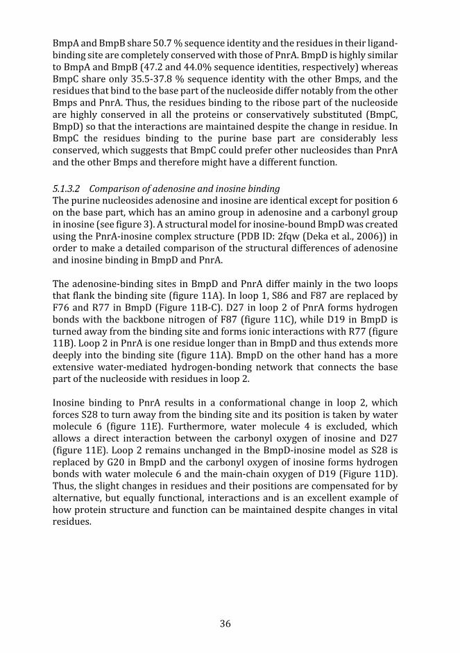
36
BmpAandBmpBshare50.7%sequenceidentityandtheresiduesintheirligand-bindingsitearecompletelyconservedwiththoseofPnrA.BmpDishighlysimilartoBmpAandBmpB(47.2and44.0%sequenceidentities,respectively)whereasBmpCshareonly35.5-37.8%sequenceidentitywiththeotherBmps,andtheresiduesthatbindtothebasepartofthenucleosidediffernotablyfromtheotherBmpsandPnrA.Thus,theresiduesbindingtotheribosepartofthenucleosideare highly conserved in all the proteins or conservatively substituted (BmpC,BmpD)sothattheinteractionsaremaintaineddespitethechangeinresidue.InBmpC the residues binding to the purine base part are considerably lessconserved,whichsuggeststhatBmpCcouldpreferothernucleosidesthanPnrAandtheotherBmpsandthereforemighthaveadifferentfunction.
5.1.3.2 ComparisonofadenosineandinosinebindingThepurinenucleosidesadenosineandinosineareidenticalexceptforposition6onthebasepart,whichhasanaminogroupinadenosineandacarbonylgroupininosine(seefigure3).Astructuralmodelforinosine-boundBmpDwascreatedusingthePnrA-inosinecomplexstructure(PDBID:2fqw(Dekaetal.,2006))inordertomakeadetailedcomparisonofthestructuraldifferencesofadenosineandinosinebindinginBmpDandPnrA.Theadenosine-bindingsites inBmpDandPnrAdiffermainlyinthetwoloopsthatflankthebindingsite(figure11A).Inloop1,S86andF87arereplacedbyF76andR77 inBmpD(Figure11B-C).D27 in loop2ofPnrA formshydrogenbondswith thebackbonenitrogenofF87(figure11C),whileD19 inBmpD isturnedawayfromthebindingsiteandformsionicinteractionswithR77(figure11B).Loop2inPnrAisoneresiduelongerthaninBmpDandthusextendsmoredeeplyintothebindingsite(figure11A).BmpDontheotherhandhasamoreextensivewater-mediated hydrogen-bonding network that connects the basepartofthenucleosidewithresiduesinloop2.Inosine binding to PnrA results in a conformational change in loop 2, whichforcesS28toturnawayfromthebindingsiteanditspositionistakenbywatermolecule 6 (figure 11E). Furthermore, water molecule 4 is excluded, whichallows a direct interaction between the carbonyl oxygen of inosine and D27(figure11E).Loop2remainsunchangedintheBmpD-inosinemodelasS28isreplacedbyG20inBmpDandthecarbonyloxygenof inosineformshydrogenbondswithwatermolecule6andthemain-chainoxygenofD19(Figure11D).Thus,theslightchangesinresiduesandtheirpositionsarecompensatedforbyalternative,butequally functional, interactionsand isanexcellentexampleofhowproteinstructureandfunctioncanbemaintaineddespitechangesinvitalresidues.

37
Figure 11. A. Crystal structure of BmpDbound to adenosine. The close-up shows the electrondensitymapfortheligandandthepositionofthetwoloopswhoseresiduesbindtothebasepartofthepurinenucleoside.B-E.Comparisonoftheligand-bindingsiteofBmpD(B),PnrAboundtoadenosine(PDBID:2fqy)(C), themodelof theBmpD-inosinecomplex(D),andPnrAboundtoinosine (PDB ID: 2fqw) (E). All the potential hydrogen bonds are shown as dashed lines. Theinteractionsof thebasepartarehighlightedtodisplaythedifferences,whereastheribosepartformsidenticalinteractionsinallthestructures.Adenosineisshownaswhitesticksandinosineas wheat sticks, water molecules are shown as spheres (1-7); lighter colored spheres arecompletelyconserved,anddarkerspheresdifferbetweenthestructures.F176(BmpD)andF186havebeenomittedforclarity.FigureadaptedfromPublicationII.

38
5.1.4 TheBmpproteinsareconservedinBorreliaspeciesThemajority of protein sequences are given a putative functional annotationbasedpurelyonsequencesimilaritysearches,wherethefunctionofthetophitisautomaticallytransferredtothenewsequence(BrownandSjölander,2006).This frequently leads toannotationerrorsand togeneralizedandunspecifiedannotations.ThiswasthecasefortheBmpproteins,asasearchoftheUniProtKBdatabase (Bateman et al., 2017) revealed that many Bmp sequences wereannotated as “basic membrane protein”, “ABC transporter substrate-bindingprotein” or “nucleoside-binding protein”. Another group of proteins wereannotatedas“exportedprotein”.ByinferringaphylogenetictreeoftheBorreliaBmpsequencesweaimedtoimprovetheclassificationoftheBmpproteinsandto identify functional differences between them. Furthermore, we wanted todetermineiftherewouldbeacleardistinctionbetweentheBmpproteinsofLBandRFBorrelia.TheMaximum Likelihood (ML)method is frequently used to study sequenceevolution(YangandRannala,2012)andwasthereforechosenforouranalysis.MLgivesaphylogenetictreewiththehighestlikelihoodofproducingtheoriginalmultiple sequence alignment. The multiple sequence alignment in our caseconsistedofBmpandexportedprotein(ExP)sequencesfromknownLBandRFBorreliastrains,selectedfromtheUniProtKB(PublicationI,figureS1).TheExPsequenceswereincludedintheanalysisinordertoclarifytheirrelationshiptotheBmpproteinsand,ifpossible,determinetheirfunction.The ML-tree forms four main branches which further diverge into separatebranchesforthedifferentBmpproteinsand,withsomeexceptions,theLBandRFBorreliaproteinsthenformdistinctsubbranches(figure12).ThefirstbranchcontainstheExP/BmpCproteins,thesecondbranchtheBmpD/BmpA2proteins,the third branch the BmpB proteins and the last branch contains the BmpAproteins.Themainbranchesaresupportedbyhighbootstrapvalues,exceptforthe branch that divides into the ExP/BmpC and the BmpD/BmpA2 branches(bootstrapvalue44).Lowbootstrapvaluesusuallysuggestthatsequenceshavediverged during a relatively short evolutionary time frame (Soltis and Soltis,2014,2003).ThefirstmainbranchissplitintoseparatebranchesfortheExPandtheBmpCproteinsand theseare further split into subbranches forLBandRFBorreliasequences.TheExPproteinsarefoundwithinthesamebranchastheT.pallidumRfuAprotein (Dekaetal.,2013),which isalsoanABC typesubstrate-bindingproteinandsharestheirtypicalstructure,butwhichbindstoriboflavininsteadof purine nucleosides. It is thus involved in a completely differentmetabolicpathwaythanthenucleoside-bindingproteins.ThebindingsiteinRfuAislocatedinthecleftbetweenthetwodomains,asinothersubstrate-bindingproteins,butrequiresaverydifferentbindingsitetoaccommodatetheriboflavinmolecule,

39
whichconsistofa three-ringed isoalloxazineattached toaribitylchain.Thus,sincetheExPproteinsaremorecloselyrelatedtoRfuAthantotheBmpproteinstheycouldlikewisefunctionasriboflavin-bindingproteins.
Figure12.MaximumlikelihoodtreeoftheBorreliaBmpandExPproteins.EachmainbranchisindividuallycoloredandLBBorreliaspeciesaremarkedwithcirclesandRFBorreliawithtriangles.Bootstrapvaluesshowbranchsupport.Strainnamesareincludedifthereismorethanonestrainperspecies.ProteinshavebeennamedBmpX-likeiftheirnamesarenotspecifiedintheUniProtKBdatabase,andiftheUniProtKBnamesdifferfromhowthetreeclassifiestheprotein,theUniProtnameisfoundinparenthesis.FigurefromPublicationI.
TheBmpCproteinsarelocatedinthesamemainbranchastheExPproteinsandshareamorerecentcommonancestorwiththemthanwiththerestoftheBmpproteins.However, thebinding site ofBmpC, thoughdifferent from theotherBmpproteins,isevenmoredifferentthantheRfuAbindingsite,whichindicatesthattheBmpCproteinsarenotriboflavin-bindingproteins.Sequenceanalysisof

40
theBmpCproteins, aswell as the structural analysis, show that the residuesbindingtotheribosepartofthenucleosideareconservedwhereastheresiduessurroundingthepurinebasepartshowconsiderablevariation.InthepositioncorrespondingtoH21(figure10E),BmpCproteinsfromotherLBBorreliahaveeither histidine or asparagine, whereas in the RF Borrelia this position hasprolineorserine.InallotherBmpsthispositionhasahighlyconservedaspartatethatformsinteractionswiththebasepartoftheligand.Histidine,asparagineorserinecouldpossiblyformsimilarinteractionswiththeligand,butprolinecouldnot.L191isaleucineoranarginineinLBBorreliaandglutamateoraspartateinRFBorrelia.Theseresiduescannotformthestackinginteractionwiththebasepart of the ligand, as phenylalanine does in the other Bmpproteins. And thechargedglutamateoraspartateinthispositionofRFBorreliasuggeststhattheycouldformionicinteractionsorhydrogenbondstotheligandmolecule.N30isconserved in all LBBorrelia but in RFBorrelia the corresponding residue isphenylalanineorleucine.Takentogether,itisnotclearhowthesevariationsinthe ligand-binding site affect the function of theBmpCproteins, but it seemslikelythatthesubstratetheybindtocouldcontainaribosering,oraribose-likestructure, as the residues surrounding that part are highly conserved. Thevariationsbetween theLBandRFBorrelia also indicate apossible functionaldifference between these two groups, which could reflect the metabolicdistinctionsbetweentheLBandRFBorrelia.RFBorreliahasacompletepurinesalvagepathwaywhilethepathwayinLBBorreliaismissingsomekeyenzymes(Petterssonetal.,2007).TheRFBorreliacontainsanadditionalBmpprotein,whichisnotfoundintheLBBorrelia.ThisproteinhasbeennamedBmpA2byLescotetal.(2008)butinourphylogenetic tree the BmpA2 proteins are located in the same branch as theBmpDproteins.However, the ligand-binding residues of theBmpA2proteinsdifferfromtheBmpDproteinsbyhavingaglycineinsteadofN28(PublicationI,figureS2).TheasparagineresidueinthispositionisconservedwithinallotherBmpproteinsincludedinthisstudy(withtheexceptionoftheBmpCproteinsofRFBorrelia,whichhaveaphenylalanineora leucine instead).Theasparagineside chain interacts with the purine nucleoside in the hydrogen-bondingnetwork formedbetweenthebasepartof the ligandand loop2of theprotein(figure11),andasglycinelacksasidechainitcannotformthesameinteractions.Thus,theseresultsindicatethatBmpA2,likeBmpC,couldinteractwithaslightlydifferentligandthantheotherBmpproteins.NoRFBorreliasequencesarefoundintheBmpBbranch,whichisinagreementwith the analyses done by Lescot et al. 2008,where noBmpBproteinswereidentifiedintheRFBorrelia(Lescotetal.,2008).FortheBmpAproteinsthesubbranches are completely separate and do not form a common branch beforedivergingintoindividualbranchesforLBandRFBorrelia.

41
5.1.4.1 GenomiccomparisonandgeneexpressionTheB. burgdorferi Bmpproteins are expressed simultaneously in vitro but tovaryingdegrees(Dobrikovaetal.,2001).BmpAisexpressedataconsiderablyhigherlevelthantheotherBmpproteins,andespeciallyBmpCisexpressedinverylowamounts.BmpBistranscribedtogetherwithBmpAwhereastheotherproteinshavetheirowntranscriptionalstartsites(Ramamoorthyetal.,2005).ThissuggeststhattheBmpAproteinmighthaveamore importantrole inthepurinesalvagepathwayorthatitisinvolvedintransportingasubstratethatisinhighdemandbythebacterium.The differences between the Bmp proteins of LB and RF Borrelia are alsonoticeableatthegenomelevel.Inbothgroups,therearefourdifferentbmpgenesbut their order in the genome differs (figure 13). In B. afzelii and B. gariniiadditional bmp genes are found, which are most likely the result of geneduplicationevents.InbothB.afzeliiandB.gariniithereisanadditionalBmpAgene,whichclusterstogetherwiththeotherBmpAproteinsinthephylogenetictreeandhasahighsequenceidentity(>94%)tothecanonicalBmpAproteinineachspecies.TodistinguishthemfromtheBmpA2proteins,whichformsagroupofitsown,welabeledthemBmpAaandBmpAb.TheBmpAbproteinofB.afzeliicontains a serine instead of G202 and theB. garinii BmpA b protein has anasparagine instead of D19, however, neither change will likely impact thebindingsiteproperties.SinceG202 formsahydrogenbond to theribosepartthroughitsbackbonenitrogen,achangetoserinewouldnotaffectthis.However,thelargersidechainofserinecouldimpactthebindingpocketifthesidechainprotrudesintoit.Thesidechainnitrogenandoxygenoftheasparaginewouldstillbeabletoformthesamehydrogenbondsastheaspartate.

42
Figure13.GeneorderoftheBorreliabmpgenes,basedonthephylogeneticanalysis.Xindicatesfragments ormissing sequences in the NCBI database (Agarwala et al., 2017). The red arrowindicates that theorderof the genes is reversed compared to theothers. Figure adapted fromPublicationI.
5.1.5 TheBmpproteinsplayaroleinthepurinesalvagepathwayHaving determined the structures and ligand-binding properties of the Bmpproteins,wealsostudiedtheirroleinawidercellularcontext.B.burgdorferiisanauxotrophicbacteriumandcannotsynthesizeaminoacids,fattyacid,vitamincofactorsornucleotidesdenovo (Fraser et al., 1997;Gherardini et al., 2010).Bacteriathatlackthecapacitytosynthesizeimportantnutrientsdenovohavetorely on salvage pathways to obtain the necessary substances from the hostenvironment.
Purine salvage pathways are used to re-form purine nucleotides fromnucleobasesandnucleosidesobtainedfromthehost(Warneretal.,2014).Thepurine salvage pathway in B. burgdorferi, however, is missing some of theessential enzymes needed (Pettersson et al., 2007). In particular, the vitalenzyme ribonucleotide reductase (RNR) is missing in B. burgdorferi. Thisenzyme, which catalyzes the reduction of ribonucleotides into theircorrespondingdeoxyribonucleotides,isconsideredessentialforDNAsynthesisinallorganisms(Gherardinietal.,2010).B.burgdorfericompensatesforthelackofthisenzymebyrelyingonaccesstohostdeoxyribonucleotidesbutcanalso

43
make use of 2′-deoxyribosyltransferase, an enzyme that can transferdeoxyribosemoietiesfromhost-derivednucleosidestoanucleobase(Lawrenceetal.,2009).
Borrelia obtain the required nucleobases and nucleosides from the hostenvironmentandconvert these intonucleotides.OurresultsshowthatBmpDplayapartintheuptakeofnucleosidesinB.burgdorferi,bytransportingpurinenucleosidesintheperiplasmicspacetoABC-transportersattheinnermembrane(figure14).Purinenucleosidemonophosphatesfromthehostarefirstconvertedto nucleosides by a nucleotidase enzyme on the outside of the bacterial cell(Lawrenceetal.,2009)beforetheypassthroughintotheperiplasmicspaceviaouter membrane porins, such as p66, which allows the diffusion of smallhydrophilicmolecules(Barcena-Uribarrietal.,2010).BmpDisanchoredtotheinnermembranebya fattyacidchain(Dowdelletal.,2017)andbindsto freepurine nucleosides and transports them to ABC transporters (BB0677 toBB0679).Thenucleoside is imported into thebacterialcytoplasmby theABCtransporter,andtheredeoxynucleotidekinase(BB0239)converts itback toanucleosidemonophosphate(Gherardinietal.,2010).Adenylatekinase(BB0417)andnucleosidediphosphatekinase (BB0463) thenaddadditionalphosphatesandformsnucleosidediphosphates,andfinallynucleosidetriphosphates,whichcansubsequentlybeusedasbuildingblocksforRNA(Jewettetal.,2009).
Figure14.SchematicviewofthepurinesalvagepathwayinB.burgdorferi.Step1,nucleotides((i.e.,GMP, IMP, and AMP) are converted into nucleosides (guanosine, inosine, and adenosine) by anucleotidase enzyme by removing the phosphate group. Steps 2-3, The nucleosides aretransportedintotheperiplasmicspacethroughtheoutermembraneandbindtoSBPs,likeBmpD.Step4,theSBPproteintransportsthenucleosidetoamembrane-boundABCtransportersystem(BB0677toBB0679),wheretheyaretransportedthroughtheinnermembraneintothecytoplasm.Step5,deoxynucleotidekinase(BB0239)addsaphosphategrouptothenucleoside,reforminganucleoside monophosphate. Adenylate kinase (BB0417) and nucleoside diphosphate kinase(BB0463) then add additional phosphates and first form nucleoside diphosphates and finallynucleoside triphosphates (ATPandGTP),which canbe incorporated intoRNA.FigureadaptedfromPublicationII.

44
Borreliagarinii–DbpAandDbpB
5.2.1 IntroductionB.garinii(Bg),belongingtotheB.burgdorferisensulatocomplex,isoneofthemain species that cause LB in Europe and is often associated withneuroborreliosis (Stanek and Strle, 2018). The B. garinii decorin-bindingproteins A and B (DbpA, DbpB) are adhesins expressed during mammalianinfections andmediate attachment to theproteoglycansdecorin andbiglycan(BrissetteandGaultney,2014;Linetal.,2017).DifferentBorreliagenospeciesbindtotheproteoglycanGAGchainswithdifferentaffinitiesandthisaffectsthetissuetropismofthespirochetes(Benoitetal.,2011;Linetal.,2014;Saloetal.,2011).DbpproteinsformelectrostaticinteractionswiththenegativelychargedGAG chains on the proteoglycans (Gandhi and Mancera, 2008). Brown et al.(1999)haveshownthatthreelysineresiduesarecriticalforGAG-bindinginB.burgdorferi and form the so called canonical GAG-binding site (Brown et al.,1999).The Dbp proteins were first discovered inB. burgdorferi as surface proteinsinvolved in bacterial adherence to decorin (Guo et al., 1995). They werecharacterizedassurface-exposedproteinsoftheMSCRAMM(microbialsurfacecomponent-recognizingadhesivematrixmolecule)family(Guoetal.,1998)andshowntobeexpressedinvivoduringinfections(Hansonetal.,1998).AntibodiesagainstDbpAcaninhibitthegrowthofB.burgdorferibothinvivoandinvitroandimmunization with DbpA gives complete protection. DbpA and DbpB wereshowntomediateattachmenttotheextracellularmatrix(Guoetal.,1998)andtobeabletoattachtomammaliancells(Fischeretal.,2003).TheDbpproteinsseemtobeimportantfortheearly stagesofbacterialdissemination,whichispartlymediatedthroughthelymphaticsystem(Imaietal.,2013),andhavealsobeenshowntoberequiredforarthritisdevelopment(Saloetal.,2015).SinceBgisknowntocauseneurologicalsymptomsinLBpatients(StanekandStrle,2018)anddecorinisexpressedinthenervoussystem(Hanemannetal.,1993; Kallmann et al., 2002), the aim was to study if the Dbp proteins areinvolved in colonization of the central nervous system. Our collaboratorsmutatedspecificlysinesonthesurfaceoftheBgSBK40DbpA(K78/80/82A)andDbpB(K79A)proteinsandperformedbindingexperimentstostudytheeffectsonBgdecorin-andbiglycanbindingtohumanbrainmicrovascularendothelialcells(HBMECs).Toanalyzethestructuraleffectsofthemutations,Icreated3Dstructural models of the Bg SBK40 DbpA and DbpB proteins and used MD-simulationstostudythestabilityoftheproteins.

45
5.2.2 StructuraloverviewoftheDbpproteinsOurexperimentsshowedthattheDbpmutantproteins(DbpAK78/80/82AandDbpBK79A)wereunabletobindtobiglycanordecorin,andthatBorreliastrainsexpressing the mutant proteins showed significantly reduced binding toHBMECs.Toprovideastructuralexplanationforthis,Icreatedhomologymodelsforthewt(wildtype)andmutantDbpAandDbpBproteins.ForDbpA,theBgPBrDbpAstructure(PDBID:2mtd(MorganandWang,2015)),whichhasasequenceidentityof89.6%toDbpA,wasusedasastructuraltemplateformodeling(figure15).ForDbpBtheB.burgdorferisensustricto(BbssB31)DbpBstructure(PDBID:2mvg, (FengandWang,2015)),whichhasa sequence identityof61%toDbpB,wasused.Visualinspectionofthesuperimposedstructuresshowedthatthemodelswere highly similar to the template structures, and thiswas alsoconfirmedbytheRMSDvaluesbetweenthetemplatestructuresandthemodelswhich were 0.49 Å for DbpA and 0.51 Å for DbpB, indicating very similarstructures.Furthermore,bothmodelsreceivedveryhighscoresintheModFOLD(MaghrabiandMcguffin,2017)andProQ(WallnerandElofsson,2003)qualityevaluation. TheModFOLD globalmodel quality scorewas above 0.5 for bothDbpAandDbpB(score>0.4indicatescompleteandconfidentmodels)andtheProQLGscoreswere4.988and4.724forDbpAandDbpBrespectively(values>4indicatesextremelygoodmodels).
Figure15.MultiplesequencealignmentusedforcreatinghomologymodelsfortheBgSBK40DbpAandDbpBproteins.ThestructuraltemplatesBgPBrDbpA(PDBID:2mtd,Morgan&Wang2015)andBbssB31DbpB(PDBID:2mvg,(Feng&Wang2015)arealignedwiththeBgSBK40DbpAandDbpB and with the extensively studied DbpA protein of Bbss 297. The secondary structureinformationforthetemplatesisshownabove(2mtd)andbelow(2mvg)thealignment.Blackboxesandblueasterisksindicatethepositionofthemutatedresidues.Completelyconservedresiduesareshownwitharedbackgroundwhereassimilarresiduesareshowninredandboxed.FigurefromPublicationIII.

46
TheoverallfoldoftheDbpAandDbpBmodelsaresimilartootherknownDbpstructuresandconsistoffivealphahelicesandalinkerregionconnectinghelices1and2(FengandWang,2015;Fortuneetal.,2014;MorganandWang,2015;Wang,2012).ThelinkerformsanunstructuredloopinDbpBandinmostotherDbpstructures,whereasinDbpAithasawell-structuredhelicalshape.Helix5issignificantly shorter in DbpB than in DbpA and the C-terminal end isconsequentlylongerandmoreunstructured.
5.2.3 SequenceanalysisoftheB.gariniiDbpproteinsK80inDbpAandK79inDbpBcorrespondtoK82inB.burgdorferi,whichisoneofthethreelysineresidues(K82,K163andK170)showntobecrucialforGAG-binding (Brown et al., 1999). From a multiple sequence alignment with allcurrentlyavailableBgDbpsequences(publicationIII,figureS1)itcouldbeseenthatthisresidueiscompletelyconservedamongalltheincludedBgsequences,highlightingitsimportanceasaGAG-bindingresidue.Thetwoadditionallysines(K78,K82),foundincloseproximitytoDbpAK80,correspondtoK77andK81inBg Pbr DbpA, where they have been shown to form a second binding site,independentfromthecanonicalbindingsite(MorganandWang,2015).UnlikeK80, K78 and K82 are not conserved in all Bg sequences. K78 is mainlysubstitutedforaspartateorglutamatewhereasK82issubstitutedforarginineorglutamine.InDbpB,thecorrespondingpositionscontainanaspartate(D77)andaglutamine(Q81),andthus theseresiduesareunlikely to formasecondbinding site in the DbpB proteins. The substituted residues in the DbpAsequencesarealllarge,polarorchargedresiduesandthepotentialinteractionsformedwiththeGAGchainscanpossiblybemaintainedtosomedegreedespitethe differences. Arginine is positively charged just like lysine, and has beenreported to form even stronger ionic interactions than lysine (Gandhi andMancera, 2008). Glutamate, aspartate and glutamine are negatively chargedresidues;however,theycouldpossiblyformhydrogenbondstotheGAGchains.Analternativepossibilityisthatthesecondbindingsiteformedbytheseresiduesare only found in some of the Dbps, and in others are compensated for byresidues in other positions. It has, in fact, been shown that some Dbps haveseveral binding sites and that these can be uniquely adapted to each protein(FengandWang,2015;Morganetal.,2015;MorganandWang,2015).
5.2.4 GAG-bindingsitesinDbpAandDbpBThecanonicalGAG-bindingsiteinDbpAismadeupofresiduesK80,K161andK168(figure16A).K78andK82arelocatedontheoppositesideoftheproteinwhere they form a secondbinding site, similar toBg PbrDbpA (Morgan andWang,2015).ThesecondbindingsiteinBgPbrDbpAconsistoffourresidues(K77,K81, K173 andK176), is located on the opposite side of the protein inrelationtothecanonicalbindingsite,andhasbeenshowntoactindependently

47
(MorganandWang,2015). InBgSBK40DbpA,K174andE177correspondtoK173andK176inBgPbrDbpA,andK174couldparticipateinasecondbindingsitewhilethenegativelychargedE177isunabletoformtheionicinteractionsnormallyfoundwithGAGchains.ThesecondbindingsitehavethesameaffinityforGAGsasthecanonicalbindingsite(MorganandWang,2015)andthus,bymutatingresiduesfrombothbindingsitesweensuredthatweobtainedanon-functionalprotein thatcouldbeused tostudy theroleof thedecorin-bindingproteinsinadherencetoHBMECs.InBgPbrDbpA,K173andK176wereshowntohaveagreater impactonGAG-binding thanK77andK81.However, inourexperimentsthemutatedDbpA(K87/80/82A),whichstillcontainsK174,didnotbindtoproteoglycansandthestrainexpressingthetriplemutantonlyshowedweakbindingtoHBMECs.ThissuggeststhatK174doesnotplayasignificantroleinGAG-bindinginBgSBK40DbpA.ThecanonicalbindingsiteinBgSBK40DbpBismadeupofresiduesK79,K160andR167 (figure16B).BbssB31DbpBhasbeen shown to contain twoGAG-binding sites, and the second binding site is formed by the unstructured C-terminaltailwhich,interestingly,seemstobetheprimaryGAG-bindingsiteinBbss B31 DbpB (Feng andWang, 2015). In DbpB of Bg SBK40 however, thecanonical binding site still plays an important role, as shown by the lack ofproteoglycanbindingseenintheDbpBK79Amutant.TheC-terminaltailinBbssB31DbpBconsistsoffourlysineswhereasinBgSBK40DbpBitcontainsthreelysinesandanasparagine.Asparaginecannot formionic interactionswiththeGAGsandthisdifferencemightexplainthegreaterimportanceofthecanonicalbinding site inBgSBK40DbpB.Furthermore, as the canonicalbinding site inBbssB31DbpBismadeupoftwolysinesandaglutaminethissuggeststhatthelysine-richC-terminal tailcompensates for themissing lysine in thecanonicalbindingsite.Incontrast,thecanonicalGAG-bindingresiduesinBgSBK40aretwolysinesandanarginine(K79,K160,R167)andasargininehavebeenshowntobindevenmorestronglythanlysines,thisfurtherhighlightstheimportanceofthecanonicalGAG-bindingsiteinBgSBK40DbpB.FurtherstudiesareneededtoelucidatetheroleofthepotentialC-terminalbindingsiteinBgSBK40.

48
Figure16.TheGAG-bindingsitesinthe3DstructuresofBgSBK40DbpAandDbpB.A.BgSBK40DbpA(pink)superimposedonthestructuraltemplate(lightblue)BgPBrDbpA(PDBID:2mtd,(Morgan&Wang,2015)).TheGAG-bindingsitesareshownintheclose-ups.ThecanonicalbindingsiteconsistofresiduesK80,K161andK168.ThesecondbindingsiteislocatedontheoppositesideoftheproteinandconsistofresiduesK78andK82andresiduesK174andE177.K174andK177corresponds toK173andK176 inBgPBrDbpA(see text formoredetails).B.BgSBK40DbpB(green)superimposedonthestructuraltemplate(lightblue)BbssB31DbpB(PDBID:2mvg,(Feng&Wang,2015)).ThecanonicalbindingsiteofDbpBconsistofresiduesK79,K160andR167.Thepotential secondbinding site is located in theC-terminal tail (residuesK181,K182,N183 andK184).ThelinkerinDbpAformsahelicalstructurewhichismissinginDbpB.Helix5isshorterinDbpBthaninDbpAanditsC-terminalendthereforeformsalongerflexibleloop.Mutatedresiduesaremarkedinitalicsandresiduesontheoppositesideoftheproteinareingrey.FigureadaptedfromPublicationIII.

49
SinceGAGsareknowntointeractwithDbpsthroughelectrostaticinteractions,theeffectsofthemutationswerefurtheranalyzedbystudyingtheelectrostaticsurface potentials of the proteins. The mutations in the Dbp proteins causesignificantchangesinsurfacepotentialandsincetheDbpproteinsareknowntobindtoGAGsthroughionicinteractions,thesechangescanresultineliminatedorreducedbinding(figure17).Electrostaticinteractionsarealsoimportantforlong-rangerecognitionofbindingpartners(Vasconetal.,2020),whichmeansthatchangesinsurfacepotentialcouldmakeitlesslikelyforthebindingpartnerstoformtheinitialcontactneededfortheinteraction.
Figure 17. Electrostatic surface potential maps for the Bg SBK40 DbpA and DbpB models,calculatedwiththeAPBStool(AdaptivePoisson–BoltzmannSolver)inPyMOL.Thecolorsareinthe range of -2 (red) to +2 (blue). Positively charged areas on the surface, created by lysineresidues,areinvolvedinionicinteractionswithGAGs.TheDbpAK78A/K80A/K82AmutationsandtheDbpBK79Amutationcausesignificantchangesinthesurfacepotentialoftheproteins,whichpreventsGAG-binding.FigurefromPublicationIII.

50
5.2.5 ProteinstabilityanalysisMD simulationswere done to studywhether themutationswould affect thestability of the protein. Experiments had shown that themutantDbpA had alowermeltingtemperaturethanthewtprotein,whichcouldbetheresultofanunstable protein fold. However, the 3D fold of both the wt and the mutantproteins remained intact throughout the simulations and an increase inflexibilitywasonlyseenintheterminalregionsandinthelinker.Interestingly,thelinkerinwtDbpBwasmoreflexiblethaninthemutantDbpB(PublicationIII,figureS3C)whereasnosuchdifferencecouldbeobservedforDbpA.ThiscanbeexplainedbythesmallersizeandhydrophobicnatureofA79comparedtoK79,whichallowsthelinkertoreachfurtherdowntowardsthecanonicalbindingsite.IthasbeenshownthattheshapeofthelinkerhasanimpactontheGAG-bindingaffinity ofDbpAs (Morganet al., 2015),where amore compact shape gives amoreexposedbindingsiteandthusincreasesthebindingaffinity.Asthelinkerin the mutant DbpB can extend further down towards the binding site it ispossible that itwould partly cover the binding site, preventing access to thecanonical binding residues. The backbone residues near K79/A79 are alsoslightlymoreflexibleinthemutantDbpBwhichcanbeexplainedbythedifferentpropertiesof lysineandalanine.K79 formselectrostatic interactionswith thesurroundingresidues,whichmakesitspositionsmorefixed,whereasA70cannotformsimilarinteractionsand,thus,confersflexibility.
Klebsiellapneumoniae–VgrG4andSel1E
5.3.1 IntroductionThetypeVIsecretionsystem(T6SS)isoneofthemanyvirulencefactorsusedbybacteriatoinvadehostcells.InK.pneumoniae,ithasbeenshowntoplayaroleininterbacterialcompetition,hostinvasionandcolonization,aswellasintype-1fimbriaeexpression(Hsiehetal.,2019).TheVgrGproteinsareanessentialpartoftheT6SSandbindtoeffectorproteins,whichtheytransportintothetargetcell, or carry internal effectordomains (evolvedVgrGs) (Hachani et al., 2014;Pukatzki et al., 2007). The VgrG proteins were first characterized in Rhs(recombinationhotspot)elements inE.coliasproteinscontainingarecurringvaline-glycinedipeptide repeat (Wang et al., 1998) and similar proteins havebeenfoundinawiderangeofGram-negativebacteria(Pukatzkietal.,2007).OurcollaboratorsinProfessorJoséA.Bengoechea’sresearchgrouphasshownthattheVgrG4proteininK.pneumoniaestrain52.145(Kp52.145)istoxictootherbacteriaandto fungi,andthat thiseffect ismediatedthroughROSgeneration(Storeyetal.,2020). Incontrast,VgrG1andVgrG2inthesameK.pneumoniaestraindidnothaveacytotoxiceffect.

51
VgrG proteins are often encoded in close proximity to an immunity proteinwithinthesamelocus.TheVgrG4locuscontainsfivegenes,whichcodeforsel-1repeatproteins(Sel1A-E),amotifofteninvolvedinprotein-proteininteractions(D’Andrea and Regan, 2003). Two proteins with sel-1 repeats have beendiscovered also in P. aeruginosa (Yang et al., 2016) and these function asimmunity proteins for the effector protein PldB, suggesting that the sel-1proteins in the VgrG4 locus could have a similar function. ExperimentsperformedbyourcollaboratorssubsequentlydemonstratedthatSel1EfunctionsastheimmunityproteinforVgrG4(Storeyetal.,2020).
In this project, the functional differences between the three Kp 52.145 VgrGproteinswere studied and the region of VgrG4 responsible for the observedcytotoxiceffectwasdetermined.A3DstructuralmodelofVgrG4wascreatedtostudy the structural basis for its cytotoxic effect. The structure of the VgrG4immunityproteinSel1Ewasalsomodeled,andcomparedtoSel1D,whichdoesnot inhibit the function of VgrG4, to identify residues potentially involved inbindingtoVgrG4.
5.3.2 K.pneumoniaeVgrGsThe genome of Kp 52.145 comprises three T6SS loci, each coding for a VgrGprotein:VgrG1(W9BN84),VgrG2(W9BP38/W9BGC5)andVgrG4(W9BAA4).VgrGproteinsaretrimericproteinsconsistingofanN-terminalheadpart,wherethethreemonomersresideside-by-side,andanelongatedspikepart,wherethemonomers are intertwined into a triangular-shaped beta helix (figure 18)(Cianfanellietal.,2016).ManyVgrGsalsocontainanextendedC-terminalpart(Records,2011),whichisoftenhighlyvariableandplaysaroleindeterminingeffectorspecificity(Bondageetal.,2016;Wettstadtetal.,2019).ItalsobindstoPAARproteins,whichareshowntobevitalforsecretionofVgrGproteins(Woodetal.,2019b).AVgrGproteincanrequirebindingbyaspecificPAARtofunction(Wood et al., 2019b) and PAARproteins have also been shown to be able todeliver effectors to the VgrG protein by interacting with the effectors, andsometimeswith chaperones, beforemediating binding to the tip of the VgrGspike(Burkinshawetal.,2018).

52
Figure18.GenericstructureofaVgrGprotein, illustratedbytheP.aeruginosaVgrG1structure(PDBID:4uhv,(Spínola-Amilibiaetal.,2016)).A.TrimericstructureofVgrGwitheachmonomercoloredinaseparatecolorandthehead,neckandspikeregionsdepicted.Themonomersinthespikepartare intertwinedto formthecharacteristicbeta-helical fold.B.Top-downviewof thehead-part, showing theside-by-sidearrangementof the threemonomers in theheadpart.C.AsinglemonomerofVgrG.
The Kp 52.145 VgrG proteins are evolved VgrGs and contain a conservedDUF2345 domain and an extended C-terminal part of varying length. VgrG1consistsof851aminoacids,VgrG2of823aminoacidsandVgrG4of899aminoacids, and theyshareabout40%sequence identity.ThedifferencesbetweenthemarelocatedintheC-terminalendoftheproteins.AstheexperimentsdonebyourcollaboratorsshowedthatonlyVgrG4causesacytotoxiceffectintargetcells, this indicates that there is a clear functional difference between theKp52.145VgrGproteins.
5.3.3 StructuralcharacterizationofVgrG4The3DmodelofVgrG4wascreatedinModeller(ŠaliandBlundell,1993)usingthePseudomonasaeruginosaVgrG1(PaVgrG1)structure(PDBID:4uhv(Spínola-Amilibia et al., 2016) as the structural template. However,modelingwas notstraightforwardasVgrG4isabout250aminoacidslongerthanPaVgrG1andhas

53
a longerC-terminalpart.PaVgrG1has the typical trimeric structureofVgrGs,with an N-terminal head part and a C-terminal spike part, connected by adisorganizedneckregion(Spínola-Amilibiaetal.,2016).Residues1-563(head,neck,N-terminalpartofthespike)ofVgrG4couldbeconfidentlymodeledbasedonthePaVgrG1crystalstructure(figure19).Theheadpartconsistsof24betastrandsand4alphahelicesfromeachmonomer.ThespikepartofPaVgrG1ismadeupof16betastrandsthatformthebetahelicalfold.InVgrG4ontheotherhand,thisrepetitivebetastrandfoldisdisruptedbyanalphahelicalpart,whichlacks a corresponding part in PaVgrG1. This part (residues 564-636) wasmodeledseparatelybyI-TASSERastwolong,almostparallelalphahelicesthatprotrudefromthespikefold.Afterthehelices,thebetastrandfoldcontinuesfor12strandspermonomer,asVgrG4has5betastrandsmorethanPaVgrG1.TheVgrGspikestructuresareexamplesofbetahelical foldswhicharebasedonaregularrepeatpatternformedbyeightresidues:5-6residuesmakeupthebetastrandand2-3residuesformaloopbetweentwobetastrands(Spínola-Amilibiaetal.,2016).Ineachbetastrand,everyotherresiduepointtowardstheinteriorofthespike,andthesearepredominantlyhydrophobic,whiletheresiduesontheoutsideofthespikearemainlypolarorchargedresidues.ThemodeledpartofVgrG4endswiththeputativePAARinteractionsite,asthefoldoftheremainingpartoftheproteincouldnotbemodeledbasedonPaVgrG1asthestructuraltemplate.TheVgrG4sequencecontainsaregionrichinprolinesaftertheputativePAARinteractionsiteandasprolinesareunfavoredresiduesin beta strands, this indicates that the beta helical structure is most likelydisruptedatthispointandtheremainingpartoftheproteinformsadifferentkindoffold.InPaVgrGtheC-terminalendturnsupwardsalongthelengthofthespikewhere it formsalphahelices.As this leaves theendof the spike free tointeract with PAAR proteins it is possible that the longer C-terminal part ofVgrG4could formeven largeradditionaldomainson theoutsideof thespike.ThishasindeedbeenseeninotherVgrGs.TheC-terminalendsofVgrG2binE.coli(EAEC)Sci-1andVgrG4bandVgrG5inP.aeruginosaPAO1,containTTR-likedomainsthatcanfunctionasanadaptorforeffectorbinding(Flaugnattietal.,2016;Wettstadtetal.,2019).InadditiontotheTTR-likedomain,VgrG2binP.aeruginosaPAO1alsocontainsametallopeptidasedomainwhichcauses toxiceffectsintargetcells(Woodetal.,2019a).Likewise,theV.choleraeVgrG3proteincontainsapeptidoglycandegradingdomainatitsC-terminalend(Brooksetal.,2013).ThisindicatesthatthelargedifferencesinlengthbetweenVgrGs,andthevariabilityof theC-terminalparts can correspond toverydifferent functionaldomains. It also suggests that outside of the conserved head-spike structurecommontoallVgrGs,domainsofvaryingsizeandfunctioncanexist.

54
Figure 19. 3D model for K. pneumoniae VgrG4. A. The bar depicts the VgrG4 sequence andillustratesthedifferentpartsthatweremodeled:N-term:residues1-563,Helix:residues564-636,Repeat:residues637-677,C-term:residues678-739.TheVgrG4region(570-899)containingtheDUF2345domain(residues612-759)caninducetoxiceffectsinbacteriaandyeast,whilethe838-899 region cannot. B. Trimeric homologymodel of VgrG4. The teal-colored region depicts thefunctional part of VgrG4 (residues 570-899) and starts with the unique helical domains andcontinueswiththebetahelicalspike,whichismadeupof12betastrandsineachmonomer.Theindependently modeled Helix part is inserted in the estimated location, however, its exactorientationinrelationtotherestoftheproteinisnotknown.The3DstructureofVibriocholeraePAAR(PDBID:4jiv)isshowntoindicatethelocationofthelikelyinteractionsitewiththePAARproteinfoundinthesamelocusasVgrG4.FigureadaptedfromPublicationIV.

55
Our collaborators performed experiments with truncated variants of VgrG4,which showed that residues 612-759 are sufficient for triggering the ROS-mediatedtoxiceffectuniquetoVgrG4.Theseresiduescompriseasmallpartofthealphahelicalpart,aswellastheremainingC-terminalbetastrands(figure19)andcorrespondtothepredictedlocationoftheDUF2345domain.However,as the DUF2345 domain is also found in VgrG1 and VgrG2, the functionaldifferencesbetweentheproteinsaremost likelyexplainedbyspecificresiduedifferenceswithinthisregion,ratherthananydifferenceintheir3Dfold.Afterthepublicationofthiswork,anewstructureofthespikepartofanE.coliVgrG protein was published (Flaugnatti et al., 2020). This structure issignificantly longer than the PaVgrG1 structure (figure 20) and contains twoalpha helices located on the outside of the spike, just as predicted for K.pneumoniaeVgrG4.Thehelicesareofdifferentlengthsandarelocatedparallelto the spike. Furthermore, the E. coli structure also contains a TTR(transthyretin-like) domain,which is located on the outside of the spike andconnectedtoitbyashortstretchofaminoacids.However,thestructureofthisconnectingregionwasnotsolved.ThePaVgrG1andtheE.colistructuressharethe typical intertwinedbeta-helicalrepeat foldbutdifferslightly in theupperpartofthespike,wheretheE.colistructurehasafive-strandedantiparallelbetasheet,comparedtothethree-strandedantiparallelpartinPaVgrG1.Attheendofthebeta-helicalspike,thePaVgrG1structureformsa loopthatturnsupwardsalong the lengthof thespikeandends inashortalphahelix,while theE.colistructurefoldintotheTTRdomain.TheE.coliVgrGproteinismoresimilarinboth sequence and length toVgrG4, compared to PaVgrG1, and the structurecould therefore be used to refine the modeling of the VgrG4 spike part(unpublished). The residues involved in the helices are the same as waspredictedbysecondarystructurepredictionsandoverall, the foldofVgrG4 ishighlysimilartotheE.colistructure.

56
Figure20.ComparisonofthespikepartsofthePaVgrG1,E.coliVgrG(EcVgrG),andthestructuralmodels forK. pneumoniae VgrG4.A-B: PaVgrG1 trimer andmonomer. C:K. pneumoniae VgrG4model based on PaVgrG1 as structural template (VgrGPa). D-E:E. coli VgrG1 trimer (with TTRdomain)andmonomer.F:K.pneumoniaeVgrG4modelbasedonE.coliVgrGasstructuraltemplate(VgrG4Ec).Theantiparallelbetasheetsareshownindashedboxes.TheapproximatelocationoftheseparatelymodeledhelicesisshowningreybackgroundinC,andthepartsofthemodelthatwereincorrect,inlightoftherefinedmodelbasedontheE.colistructure,areshowningrey.

57
5.3.4 SequenceanalysisandmodellingoftheimmunityproteinSel1EThe Sel1 proteins are located in the same locus as VgrG4 and, according toexperimentsdonebyourcollaborators,Sel1EisabletoprotectagainstVgrG4toxicity,whereasSel1Disnot.Toelucidatethestructuralbasisforthisfunctionaldifference,thestructuresofSel1EandSel1Dweremodeledandcompared,andtheevolutionaryconservationofthesequenceswasstudied.The Sel1 proteins contain Sel1-like repeats (IPR006597) and belong to theTetratricopeptide-like helical domain superfamily (IPR011990). Thetetratricopeptide repeat (TPR) is an alpha/alpha repeatmotif consisting of aconsensussequenceof34aminoacidsthatformtwoantiparallelalphaheliceswhich are connected by a turn (Mittl and Schneider-Brachert, 2007).Alpha/alpharepeatproteinscancontainavaryingnumberofrepeatsandthusform proteins of different sizes. TPR motifs are mostly involved in protein-protein interactions and can be found in functionally very different proteins(D’AndreaandRegan,2003).TheSel1repeat(SLR)sequenceishighlysimilartothe TPR motif but is longer (36-44 residues) (Mittl and Schneider-Brachert,2007).Sel1DandSel1Eshare69%sequenceidentityanddifferencesbetweenthemaremainly found in theterminalregions(figure21).Sel1Ehasabout70residuesmorethanSel1DandsecondarystructurepredictionsindicatethatitsC-terminalend contains a long loop regionwith very fewpredicted secondary structureelements. Sequence analyses did not find any information about potentialdomains or functions for this C-terminal region. Signal sequence predictionsperformedbySignalP(AlmagroArmenterosetal.,2019)andLipoP(JunckerandWillenbrock, 2003) predicted an N-terminal lipoprotein signal sequence inSel1D,butnotinSel1E.TheN-terminusofSel1DisslightlylongerthaninSel1Eand the first 18 residues are predominantly hydrophobic and ends with acysteine, which is characteristic for all lipoprotein signal peptides in Gram-negative bacteria (Zückert, 2014). In Sel1E, the N-terminal is shorter andcontainsmorepolaror charged residues. Lipoprotein signal sequences targetproteinsfortransportoutsidethecytoplasmicmembrane,andthelackofsignalsequence in Sel1E suggests that Sel1D and Sel1E can be located in differentcellular compartments. However, ScanProsite (de Castro et al., 2006) andlocationpredictionsdonebyPsortdb(Peabodyetal.,2016),CELLO(Yuetal.,2004)andPhobius (Källetal.,2007)didnotgiveanyconclusive informationaboutthecellularlocationoftheproteins.

58
Figure 21. Sequence alignment of Sel1D and Sel1E. The parts of the proteins that could beconfidentlymodeled(Sel1D:residues42-303,Sel1E:28-289)areshownwithpinkbackground.ThepredictedsignalsequenceofSel1Disshownwithgreenbackground.PositionsinSel1DandSel1EpredictedbyConSurftobebothvariableandexposedaremarkedwithblackdots.ResiduesconservedinbothSel1DandSel1Eareinbold.
Formodelingof theSel1proteins, the threading-basedmethod I-TASSERwasused (Yang et al., 2015).We specified as a restraint for I-TASSER to use thecrystalstructureofP.aeruginosaPa5087(PDBID:5jkp,(Yangetal.,2016))asstructuraltemplateformodeling.Pa5087shares27and28%sequenceidentitywith Sel1D and Sel1E, respectively. However, since there was no suitabletemplateavailablefortheC-terminalpartsoftheproteins,theseweremodeledindependentlybyI-TASSER.Residues42-303(Sel1D)and28-289(Sel1E)weremodeled with high confidence based on Pa5087. The remaining C-terminalresiduesweremodeledquitedifferently inSel1DandSel1Eduetothe lackofsuitabletemplatestructures.TheconfidentlymodeledpartsofSel1DandSel1EarehighlysimilartoeachotherandtoPa5087,asshownbyanRMSDvalueof0.3ÅbetweenbothSel1-proteinsandPa5087.TheproteinsconsistofsixSLRrepeats, each made up of two antiparallel alpha helices (figure 22). In theMODFOLD (Maghrabi and Mcguffin, 2017) residue accuracy predictions theregion between residues 42-303 (Sel1D) and 28-289 (Sel1E) shows a highaccuracy,whereasthec-terminalpartsshowloweraccuracy.Takentogether,theI-TASSERmodelssuggestthattheN-terminalendoftheSel1DandSel1Eproteinsshare the typical fold of SLR-proteins while the structure of the C-terminalregions could not be accurately determined due to a lack of homologousstructures.

59
Figure22.3DstructuralmodelofK.pneumoniaeSel1E(residues28-289).ThesixSLR-motifsareshownindifferentcolors.
5.3.5 PredictionofpotentialbindinginterfacebetweenSel1EandVgrG4The structures of TPR- and SLR proteins are characterized by similar repeatmotifs,buttheiroverallstructurescandiffergreatlyduetoavaryingnumberofrepeats,differentanglesbetweenrepeats,aswellastheoverallcurvatureoftheprotein(MittlandSchneider-Brachert,2007).Together,thisgivestheproteinsahighly versatile structure, which can bind a wide range of molecules usingseveraldifferentbindingmodes(Perez-RibaandItzhaki,2019).TheconcaveandconvexsurfacescreatedbythecurvedshapeofmostTPR-andSLRproteinsgivetheproteinsmanypotentialbindingsites.The versatile SLR-structure, with its many possible binding sites, makespredicting the interaction site between VgrG4 and Sel1E a complex task.However, as there is a clear functionaldifferencebetweenSel1DandSel1Eadetailed comparison of the structures and their surface properties made itpossible topinpointresiduesthatmightplayarole in interactionswithotherproteins. The ConSurf server (Ashkenazy et al., 2016) was used to depictevolutionaryconservationbasedonmultiplesequencealignmentsfortheSel1proteins. ConSurf scores each residue based on a conservation scale, rangingfromhighly conserved to highly variable. The server also predictswhether aresidueisburiedorexposed,i.e.,iftheresidueislocatedonthesurfaceoftheprotein,andthuscapableofinteractingwithothermolecules,orif it isburiedwithintheproteinandmorelikelytohaveastructuralrole.WhencomparingtheconservationpatternsforSel1DandSel1E,virtuallythesamepositionsinbothproteinsarepredictedasconservedandvariable.AsidefromthevaryinglengthsoftheC-terminalparts,thereareonlyafewresiduedifferencesbetweenSel1DandSel1EandmostoftheseresiduesarealsopredictedbyConSurftobeexposedandhighlyvariable(figure23).

60
Figure 23. Comparison of electrostatic surface potentials for Sel1D and Sel1E. Due to the lowconfidenceoftheC-terminalpartsofthemodels,onlythehighconfidenceregionsofthemodelswereusedforthecomparison.Theclose-upsshowresidueswhichcontributetotheelectrostaticpotentialdifferences,seetextformoredetails.TheelectrostaticsurfaceswerecalculatedwiththeAPBS tool (AdaptivePoisson–BoltzmannSolver) inPyMOLand thecolour ranges from-2 to2.FigureadaptedfromPublicationIV.
InmanyTPR-andSLRstructures,chargedsurfaceareashavebeenshowntoplayarole in ligandbinding (Hanetal.,2007;Zeytunietal.,2011).The immunityproteins Pa5086, Pa5087 and Pa5088 in P. aeruginosa have significantdifferencesintheelectrostaticsurfacepotentials(Wenetal.,2020;Yangetal.,2016). A similar pattern was found also in the Sel1 proteins when theelectrostatic surface potential of the Sel1D and Sel1E structureswas studiedusing the APBS plugin in PyMol. There were two regions with significantdifferencesbetweenSelDandSel1E(figure23),bothlocatedintheC-terminalhalfof thestructures. In the first region,Sel1DhasamorepositivelychargedpatchcomparedtoSel1E,whichisexplainedmainlybythetwoarginines(figure23A) found in thisarea (glutamateand leucine inSel1E). In theother region,however,Sel1DhasaglutamateandanaspartatewhichgivesitamorenegativechargethanSel1E,whichhasaglycineandatyrosine(figure23B).TheresiduesinthesetworegionsareamongtheresiduesthatarepredictedbyConSurftobevariableandsurfaceexposed.Takentogetherthisindicatesthattheseresidues

61
could be important for the function of the proteins and that their differingpropertiesmightenable themtobind todifferentpartners.As theC-terminalpart of VgrG4, which is responsible for its cytotoxic effect, consist of manycharged surface residues, it is possible that Sel1E could form electrostaticinteractionswithspecificresiduesonthesurfaceofVgrG4andtherebypreventsitsproperfunction.

62
6. ConclusionInthisthesis,virulencefactorsfromBorreliaandKlebsiellahavebeenstudiedand structure-function analyses have provided insights into their role in thevirulenceandsurvivalmechanismsusedbythesepathogenicbacteria.InpublicationI,3DstructuralmodelswerecreatedfortheB.burgdorferiBmpproteinsandtheirpotentialligand-bindinginteractionswereanalyzedindetail.Furthermore, the evolutionary relationships between the Borrelia Bmpsequenceswerestudiedthroughaphylogeneticanalysis.Thestructuralanalysesdemonstrated that the Bmp proteins are substrate-binding proteins thattransportpurinenucleosides toABCtransporters for import into thebacteriaandthusplayavitalrole inthepurinesalvagepathway.However, the ligand-binding site of BmpC differed from the other Bmp proteins to a significantdegree,suggestingthatitmostlikelyhasadifferentligandpreferencethantheother Bmp proteins. The phylogenetic analysis provided an improvedclassificationoftheBmpproteins,showedthatthepreviouslyidentifiedBmpA2proteinsaremorecloselyrelatedtoBmpDandthattheExPproteinsmostlikelyfunctionasriboflavin-bindingproteins.InpublicationII,thecrystalstructureofBmpDconfirmedthestructuralfoldseeninthemodelsandshowedthatBmpDwasboundtoadenosine.ExperimentalresultsfurtherdemonstratedthatBmpDcanalsobindtoinosine,indicatingthattheBmpproteinscanpossiblytransportseveraldifferentpurinenucleosides.Furtherstudieswouldbeneededtofullydetermine the substrate scope of the Bmp proteins, and in particular that ofBmpC.InpublicationIII,homologymodelswerecreatedfortheB.gariniiDbpproteinsandadetailedanalysisoftheirpotentialGAG-bindingsiteswereperformedtoexplain the structural basis for the reduced GAG-binding seen in the lysinemutants.Theanalysisshowedthatthemutationscauseasignificantchangeinthe electrostatic surface potential, thus reducing GAG-binding, which mainlyoccurs through ionic interactions. DbpA contains two GAG-binding sites, onopposite sides of the protein, whereas DbpB relies mainly on the canonicalbindingsite,butpotentiallyhasasecondbindingsitelocatedattheC-terminaltail.MD-simulationsweredonetostudythestabilityoftheproteinfoldinthemutants,butthiswasnotaffectedbythemutationsandtheproteinsremainedstable throughout the simulations. The Dbp proteins were shown to beimportant forB. garinii binding toHBMECs in the brain ofmice, and furtherstudiescouldbemadetodeterminetheexactbindingmodeoftheDbpproteins.In publication IV, the 3D structures for the K. pneumoniae VgrG4 and Sel1Eproteins were predicted, and their role in T6SS-mediated intermicrobialcompetitionsandhostinteractionswerestudied.VgrG4causesaROS-mediatedtoxiceffect inhostcellsandtheeffectorfunctionwaslocatedtotheDUF2345domain-containingregionintheC-terminaloftheprotein.Sel1Ewasshownto

63
be the immunity protein of VgrG4 and was able to abolish the toxic effectsinducedbyVgrG4.StructuralanalysisofSel1EhighlightedresiduespotentiallyinvolvedininteractionswithVgrG4.Futurestudiesshouldfocusondeterminingthe full-length structure of VgrG4 and the exact bindingmode of VgrG4 andSel1E.The proteins studied in this work illustrate two very different virulencestrategies.TheBorreliaBmpproteinsareusedtoobtainvitalnutrientsthatthebacteriacannotsynthesizethemselvesandtheDbpproteinsensurethetissuecolonizationrequiredforasuccessfulinfection.Whilethesemethodscanbeseenasnecessaryforthesurvivalofthebacteria,theVgrG4proteinofK.pneumoniae,ontheotherhand,isanexampleofadirectassaultonatargetcell,withtheaimof causing damage. These examples demonstrate that interactions between apathogenanditshost,andbetweencompetingmicrobes,occuronmanylevels,from the direct attacks with harmful effectors to the more subtle evasionmechanismthatkeepthepathogenundetectedbytheimmunesystem.Bacteriauseseveralmechanismssimultaneously,usuallyinahighlycoordinatedmanner,andincombinationwiththefunctionalredundancyoftenseenamongvirulencefactors,thismakesstudyingthesemechanismsfarfromtrivial.Gainingmoreknowledgeof the complexnetworkof interactions takingplacebetween bacteria and its surroundings will aid our understanding of howbacteriaavoid the immunesystem,howtheyareable toobtainnutrientsandhow they compete with other microorganisms to gain advantages. This isessentialinformationneededwhendevelopingnewtreatmentmethodsthatarespecifically targeted to cause maximum damage to vital bacterial pathways.Structural bioinformatics methods are a valuable tool for studying thesebacterial virulence and survivalmechanisms. The combineduse of sequence-basedstudieswithdetailedstructureanalysesprovidesanin-depthknowledgeofthemolecularinteractionsbehindthesemechanisms,whichisarequirementwhendevelopingdrugmoleculesthatcantargetspecificproteins.Theresultofthisthesiscontributestoourunderstandingofbacterialvirulenceandsurvivalmechanismsinseveralways.(i)BypredictingthefunctionoftheBmpproteins and improving the classificationof theproteins (publication I),which greatly aids the study of functional differences between highly similarproteins. (ii) By providing a detailed structural explanation of ligand-bindingproperties (publication II and III), giving information on potential targets forinhibitorytreatments. (iii)Bypredictingthepossible interactionsitebetweentwo proteins (publication IV), which provides valuable information formutationalstudiesintofunctionallyimportantresidues.Despitethemanykindsofantibioticsavailabletoday,bacterialinfectionsarestillamajorcauseofillnessanddeathintheworld.Onlybyunderstandingthedetailsoftheintricatemechanismsusedbybacteriacanwehopetodevelopefficient

64
treatments targeting the most vulnerable parts of the systems. Targetingbacterialvirulencemechanismscouldbeaneffectivestrategy,whichwouldalsopotentially reduce the development of antibiotic resistance due to loweredselectionpressures.

65
7. ReferencesAamerMehmood,M.,2014.Useof
BioinformaticsToolsinDifferentSpheresofLifeSciences.J.DataMiningGenomicsProteomics05.https://doi.org/10.4172/2153-0602.1000158
AbuKwaik,Y.,Bumann,D.,2015.HostDeliveryofFavoriteMealsforIntracellularPathogens.PLoSPathog.11,1–8.https://doi.org/10.1371/journal.ppat.1004866
Actor,J.K.,2019.TLymphocytes:RingleadersofAdaptiveImmuneFunction.Introd.Immunol.45–62.https://doi.org/10.1016/b978-0-12-816572-0.00004-8
Agarwala,R.,Barrett,T.,Beck,J.,Benson,D.A.,Bollin,C.,Bolton,E.,Bourexis,D.,Brister,J.R.,Bryant,S.H.,Canese,K.,Charowhas,C.,Clark,K.,DiCuccio,M.,Dondoshansky,I.,Feolo,M.,Funk,K.,Geer,L.Y.,Gorelenkov,V.,Hlavina,W.,Hoeppner,M.,Holmes,B.,Johnson,M.,Khotomlianski,V.,Kimchi,A.,Kimelman,M.,Kitts,P.,Klimke,W.,Krasnov,S.,Kuznetsov,A.,Landrum,M.J.,Landsman,D.,Lee,J.M.,Lipman,D.J.,Lu,Z.,Madden,T.L.,Madej,T.,Marchler-Bauer,A.,Karsch-Mizrachi,I.,Murphy,T.,Orris,R.,Ostell,J.,O’Sullivan,C.,Palanigobu,V.,Panchenko,A.R.,Phan,L.,Pruitt,K.D.,Rodarmer,K.,Rubinstein,W.,Sayers,E.W.,Schneider,V.,Schoch,C.L.,Schuler,G.D.,Sherry,S.T.,Sirotkin,K.,Siyan,K.,Slotta,D.,Soboleva,A.,Soussov,V.,Starchenko,G.,Tatusova,T.A.,Todorov,K.,Trawick,B.W.,Vakatov,D.,Wang,Y.,Ward,M.,Wilbur,W.J.,Yaschenko,E.,Zbicz,K.,2017.DatabaseResourcesoftheNationalCenterforBiotechnologyInformation.NucleicAcidsRes.45,D12–D17.https://doi.org/10.1093/nar/gkw1071
Ajawatanawong,P.,2017.MolecularPhylogenetics:ConceptsforaNewcomer.Adv.Biochem.Eng.Biotechnol.160,185–196.https://doi.org/10.1007/10_2016_49
Akira,S.,Takeda,K.,2004.Toll-likereceptorsignalling.Nat.Rev.Immunol.4,499–511.https://doi.org/10.1038/nri1391
Akira,S.,Uematsu,S.,Takeuchi,O.,2006.Pathogenrecognitionandinnateimmunity.Cell124,783–801.https://doi.org/10.1016/j.cell.2006.02.015
Aldred,K.J.,Kerns,R.J.,Osheroff,N.,2014.MechanismofQuinoloneActionandResistance.Biochemistry53,1565–1574.
Allen,J.P.,Hauser,A.R.,2019.DiversityofContact-DependentGrowthInhibitionSystemsofPseudomonasaeruginosa.J.Bacteriol.201,1–12.https://doi.org/10.1128/JB.00776-18
AlmagroArmenteros,J.J.,Tsirigos,K.D.,Sønderby,C.K.,Petersen,T.N.,Winther,O.,Brunak,S.,vonHeijne,G.,Nielsen,H.,2019.SignalP5.0improvessignalpeptidepredictionsusingdeepneuralnetworks.Nat.Biotechnol.37.https://doi.org/10.1038/s41587-019-0036-z
Altschul,S.F.,Gish,W.,Miller,W.,Myers,E.W.,Lipman,D.J.,1990.Basiclocalalignmentsearchtool.J.Mol.Biol.215,403–410.https://doi.org/10.1016/S0022-2836(05)80360-2
Álvarez,D.,Merino,S.,Tomás,J.M.,Benedí,V.J.,Albertí,S.,2000.CapsularpolysaccharideisamajorcomplementresistancefactorinlipopolysaccharideOsidechain-deficientKlebsiellapneumoniaeclinicalisolates.Infect.Immun.68,953–955.https://doi.org/10.1128/IAI.68.2.953-955.2000
Aoki,S.K.,Pamma,R.,Hernday,A.D.,Bickham,J.E.,Braaten,B.A.,Low,D.A.,2005.Microbiology:Contact-dependentinhibitionofgrowthinEscherichiacoli.Science(80-.).309,1245–1248.https://doi.org/10.1126/science.1115109
64
Aron,L.,Alekshun,M.,Perlee,L.,Schwartz,I.,Godfrey,H.P.,Cabello,F.C.,1994.CloningandDNAsequenceanalysisofbmpC,ageneencodingapotentialmembranelipoproteinofBorreliaburgdorferi.FEMSMicrobiol.Lett.123,75–82.https://doi.org/10.1111/j.1574-6968.1994.tb07204.x
Ashkenazy,H.,Abadi,S.,Martz,E.,Chay,O.,Mayrose,I.,Pupko,T.,Ben-Tal,N.,2016.ConSurf2016:animprovedmethodologytoestimateandvisualizeevolutionaryconservationinmacromolecules.NucleicAcidsRes.44,W344–W350.https://doi.org/10.1093/nar/gkw408
Auten,R.L.,Davis,J.M.,2009.Oxygentoxicityandreactiveoxygenspecies:Thedevilisinthedetails.Pediatr.Res.66,121–127.https://doi.org/10.1203/PDR.0b013e3181a9eafb
Barcena-Uribarri,I.,Thein,M.,Sacher,A.,Bunikis,I.,Bonde,M.,Bergström,S.,Benz,R.,2010.P66porinsarepresentinbothLymediseaseandrelapsingfeverspirochetes:acomparisonofthebiophysicalpropertiesofP66porinsfromsixBorreliaspecies.BiochimBiophysActa.1798,1197–1203.
Bateman,A.,Martin,M.J.,O’Donovan,C.,Magrane,M.,Alpi,E.,Antunes,R.,Bely,B.,Bingley,M.,Bonilla,C.,Britto,R.,Bursteinas,B.,Bye-AJee,H.,Cowley,A.,DaSilva,A.,DeGiorgi,M.,Dogan,T.,Fazzini,F.,Castro,L.G.,Figueira,L.,Garmiri,P.,Georghiou,G.,Gonzalez,D.,Hatton-Ellis,E.,Li,W.,Liu,W.,Lopez,R.,Luo,J.,Lussi,Y.,MacDougall,A.,Nightingale,A.,Palka,B.,Pichler,K.,Poggioli,D.,Pundir,S.,Pureza,L.,Qi,G.,Rosanoff,S.,Saidi,R.,Sawford,T.,Shypitsyna,A.,Speretta,E.,Turner,E.,Tyagi,N.,Volynkin,V.,Wardell,T.,Warner,K.,Watkins,X.,Zaru,R.,Zellner,H.,Xenarios,I.,Bougueleret,L.,Bridge,A.,Poux,S.,Redaschi,N.,Aimo,L.,ArgoudPuy,G.,Auchincloss,A.,Axelsen,K.,Bansal,P.,Baratin,D.,Blatter,M.C.,Boeckmann,B.,Bolleman,J.,Boutet,E.,Breuza,L.,Casal-Casas,C.,DeCastro,E.,Coudert,E.,Cuche,B.,Doche,M.,Dornevil,D.,
Duvaud,S.,Estreicher,A.,Famiglietti,L.,Feuermann,M.,Gasteiger,E.,Gehant,S.,Gerritsen,V.,Gos,A.,Gruaz-Gumowski,N.,Hinz,U.,Hulo,C.,Jungo,F.,Keller,G.,Lara,V.,Lemercier,P.,Lieberherr,D.,Lombardot,T.,Martin,X.,Masson,P.,Morgat,A.,Neto,T.,Nouspikel,N.,Paesano,S.,Pedruzzi,I.,Pilbout,S.,Pozzato,M.,Pruess,M.,Rivoire,C.,Roechert,B.,Schneider,M.,Sigrist,C.,Sonesson,K.,Staehli,S.,Stutz,A.,Sundaram,S.,Tognolli,M.,Verbregue,L.,Veuthey,A.L.,Wu,C.H.,Arighi,C.N.,Arminski,L.,Chen,C.,Chen,Y.,Garavelli,J.S.,Huang,H.,Laiho,K.,McGarvey,P.,Natale,D.A.,Ross,K.,Vinayaka,C.R.,Wang,Q.,Wang,Y.,Yeh,L.S.,Zhang,J.,2017.UniProt:Theuniversalproteinknowledgebase.NucleicAcidsRes.45,D158–D169.https://doi.org/10.1093/nar/gkw1099
Bengoechea,J.A.,SaPessoa,J.,2019.Klebsiellapneumoniaeinfectionbiology:Livingtocounteracthostdefences.FEMSMicrobiol.Rev.43,123–144.https://doi.org/10.1093/femsre/fuy043
Benkert,P.,Künzli,M.,Schwede,T.,2009.QMEANserverforproteinmodelqualityestimation.NucleicAcidsRes.37,510–514.https://doi.org/10.1093/nar/gkp322
Benoit,V.M.,Fischer,J.R.,Lin,Y.P.,Parveen,N.,Leong,J.M.,2011.AllelicvariationoftheLymediseasespirocheteadhesinDbpAinfluencesspirochetalbindingtodecorin,dermatansulfate,andmammaliancells.Infect.Immun.79,3501–3509.https://doi.org/10.1128/IAI.00163-11
Berman,H.M.,Westbrook,J.,Feng,Z.,Gilliland,G.,Bhat,T.N.,Weissig,H.,Shindyalov,I.N.,Bourne,P.E.,2000.TheProteinDataBank.NucleicAcidsRes.28,235–42.
Bhoite,S.,Gerven,N.Van,Chapman,M.R.,Remaut,H.,Brussels,B.,Brussel,V.U.,Biology,S.,2020.CurliBiogenesis:BacterialamyloidassemblybytheTypeVIIIsecretionpathway8,1–12.https://doi.org/10.1128/ecosalplus.

65
ESP-0037-2018.CurliBliven,K.A.,Maurelli,A.T.,2016.Evolution
ofBacterialPathogensWithintheHumanHost.Microbiol.Spectr.4,1–13.https://doi.org/10.1128/microbiolspec.vmbf-0017-2015
Bondage,D.D.,Lin,J.-S.,Ma,L.-S.,Kuo,C.-H.,Lai,E.-M.,2016.VgrGCterminusconfersthetypeVIeffectortransportspecificityandisrequiredforbindingwithPAARandadaptor-effectorcomplex.Proc.Natl.Acad.Sci.U.S.A.113,E3931-40.https://doi.org/10.1073/pnas.1600428113
Bonilla,F.A.,Oettgen,H.C.,2010.Adaptiveimmunity.J.AllergyClin.Immunol.125,S33–S40.https://doi.org/10.1016/j.jaci.2009.09.017
Brestoff,J.,Artis,D.,2013.CommensalbacteriaattheinterfaceofhostmetabolismandtheimmunesystemPopulationgenetictoolstodissectinnateimmunityinhumans.NatImmunol14,676–684.https://doi.org/10.1038/ni.2640.Commensal
Brissette,C.A.,Gaultney,R.A.,2014.That’smystory,andI’mstickingtoit-anupdateonB.burgdorferiadhesins.Front.Cell.Infect.Microbiol.4,1–13.https://doi.org/10.3389/fcimb.2014.00041
Brooks,T.M.,Unterweger,D.,Bachmann,V.,Kostiuk,B.,Pukatzki,S.,2013.LyticactivityoftheVibriocholeraetypeVIsecretiontoxinVgrG-3isinhibitedbytheantitoxinTsaB.J.Biol.Chem.https://doi.org/10.1074/jbc.M112.436725
Brown,D.,Sjölander,K.,2006.Functionalclassificationusingphylogenomicinference.PLoSComput.Biol.2,0479–0483.https://doi.org/10.1371/journal.pcbi.0020077
Brown,E.L.,Guo,B.P.,O’Neal,P.,Höök,M.,1999.AdherenceofBorreliaburgdorferi.J.Biol.Chem.274,26272–26278.https://doi.org/10.1074/jbc.274.37.26272
Brown,S.A.,Palmer,K.L.,Whiteley,M.,2008.Revisitingthehostasagrowth
medium.Nat.Rev.Microbiol.6,657–666.https://doi.org/10.1038/nrmicro1955
Buddelmeijer,N.,2015.Themolecularmechanismofbacteriallipoproteinmodification-How,whenandwhy?FEMSMicrobiol.Rev.39,246–261.https://doi.org/10.1093/femsre/fuu006
Burkinshaw,B.J.,Liang,X.,Wong,M.,Le,A.N.H.,Lam,L.,Dong,T.G.,2018.AtypeVIsecretionsystemeffectordeliverymechanismdependentonPAARandachaperone-co-chaperonecomplex.Nat.Microbiol.3,632–640.https://doi.org/10.1038/s41564-018-0144-4
Bush,K.,2012.Antimicrobialagentstargetingbacterialcellwallsandcellmembranes.OIERev.Sci.Tech.31,43–56.https://doi.org/10.20506/rst.31.1.2096
Cabeen,M.T.,Jacobs-Wagner,C.,2005.Bacterialcellshape.Nat.Rev.Microbiol.3,601–610.https://doi.org/10.1038/nrmicro1205
Casadevall,A.,Pirofski,L.,2019.BenefitsandCostsofAnimalVirulenceforMicrobes1–7.
Cassat,J.E.,Skaar,E.P.,2013.Ironininfectionandimmunity.CellHostMicrobe13,509–519.https://doi.org/10.1016/j.chom.2013.04.010
Chandra,N.,Anand,P.,Yeturu,K.,2010.Structuralbioinformatics:Derivingbiologicalinsightsfromproteinstructures.Interdiscip.Sci.Comput.LifeSci.2,347–366.https://doi.org/10.1007/s12539-010-0045-6
Chen,C.,Huang,H.,Wu,C.,2017.ProteinBioinformaticsDatabasesandResources.MethodsMol.Biol.1558,3–39.
Cherrak,Y.,Flaugnatti,N.,Durand,E.,Journet,L.,Cascales,E.,2019.StructureandActivityoftheTypeVISecretionSystem.ProteinSecret.Bact.329–342.https://doi.org/10.1128/microbiolspec.psib-0031-2019
Chowdhury,B.,Garai,G.,2017.Areviewon

66
multiplesequencealignmentfromtheperspectiveofgeneticalgorithm.Genomics109,419–431.https://doi.org/10.1016/j.ygeno.2017.06.007
Cianfanelli,F.R.,Monlezun,L.,Coulthurst,S.J.,2016.Aim,Load,Fire:TheTypeVISecretionSystem,aBacterialNanoweapon.TrendsMicrobiol.24,51–62.https://doi.org/10.1016/j.tim.2015.10.005
Cornelissen,C.N.,2018.SubversionofnutritionalimmunitybythepathogenicNeisseriae.Pathog.Dis.76,1–14.https://doi.org/10.1093/femspd/ftx112
Cortés,G.,Borrell,N.,DeAstorza,B.,Gómez,C.,Sauleda,J.,Albertí,S.,2002.MolecularanalysisofthecontributionofthecapsularpolysaccharideandthelipopolysaccharideOsidechaintothevirulenceofKlebsiellapneumoniaeinamurinemodelofpneumonia.Infect.Immun.70,2583–2590.https://doi.org/10.1128/IAI.70.5.2583-2590.2002
Coulthurst,S.,2019.TheTypeVIsecretionsystem:Aversatilebacterialweapon.Microbiol.(UnitedKingdom)165,503–515.https://doi.org/10.1099/mic.0.000789
D’Andrea,L.D.,Regan,L.,2003.TPRproteins:Theversatilehelix.TrendsBiochem.Sci.28,655–662.https://doi.org/10.1016/j.tibs.2003.10.007
Davidson,A.L.,Dassa,E.,Orelle,C.,Chen,J.,2008a.Structure,function,andevolutionofbacterialATP-bindingcassettesystems.Microbiol.Mol.Bio.Rev.72,317–364,tableofcontents.https://doi.org/10.1128/MMBR.00031-07
Davidson,A.L.,Dassa,E.,Orelle,C.,Chen,J.,2008b.Structure,Function,andEvolutionofBacterialATP-BindingCassetteSystems.Microbiol.Mol.Biol.Rev.72,317–364.https://doi.org/10.1128/MMBR.00031-07
deCastro,E.,Sigrist,C.J.A.,Gattiker,A.,Bulliard,V.,Langendijk-Genevaux,
P.S.,Gasteiger,E.,Bairoch,A.,Hulo,N.,2006.ScanProsite:DetectionofPROSITEsignaturematchesandProRule-associatedfunctionalandstructuralresiduesinproteins.NucleicAcidsRes.34.https://doi.org/10.1093/nar/gkl124
Deka,R.K.,Brautigam,C.A.,Biddy,B.A.,Liu,W.Z.,Norgard,M.V.,2013.EvidenceforanABC-typeriboflavintransportersysteminpathogenicspirochetes.MBio.https://doi.org/10.1128/mBio.00615-12
Deka,R.K.,Brautigam,C.A.,Yang,X.F.,Blevins,J.S.,Machius,M.,Tomchick,D.R.,Norgard,M.V.,2006.ThePnrA(Tp0319;TmpC)lipoproteinrepresentsanewfamilyofbacterialpurinenucleosidereceptorencodedwithinanATP-bindingcassette(ABC)-likeoperoninTreponemapallidum.J.Biol.Chem.https://doi.org/10.1074/jbc.M511405200
Dey,S.,Chakravarty,A.,GuhaBiswas,P.,DeGuzman,R.N.,2019.ThetypeIIIsecretionsystemneedle,tip,andtranslocon.ProteinSci.28,1582–1593.https://doi.org/10.1002/pro.3682
Dobrikova,E.Y.,Bugrysheva,J.,Cabello,F.C.,2001.TwoindependenttranscriptionalunitscontrolthecomplexandsimultaneousexpressionofthebmpparalogouschromosomalgenefamilyinBorreliaburgdorferi.Mol.Microbiol.39,370–378.https://doi.org/10.1046/j.1365-2958.2001.02220.x
Dong,T.G.,Dong,S.,Catalano,C.,Moore,R.,Liang,X.,Mekalanos,J.J.,2015.Generationofreactiveoxygenspeciesbylethalattacksfromcompetingmicrobes.Proc.Natl.Acad.Sci.U.S.A.112,2181–2186.https://doi.org/10.1073/pnas.1425007112
Dönhöfer,A.,Franckenberg,S.,Wickles,S.,Berninghausen,O.,Beckmann,R.,Wilson,D.N.,2012.StructuralbasisforTetM-mediatedtetracyclineresistance.Proc.Natl.Acad.Sci.U.S.A.109,16900–16905.https://doi.org/10.1073/pnas.1208037109

67
Dorn,M.,ESilva,M.B.,Buriol,L.S.,Lamb,L.C.,2014.Three-dimensionalproteinstructureprediction:Methodsandcomputationalstrategies.Comput.Biol.Chem.53,251–276.https://doi.org/10.1016/j.compbiolchem.2014.10.001
Dowdell,A.S.,Murphy,M.D.,Azodi,C.,Swanson,S.K.,Florens,L.,Chen,S.,Zückert,W.R.,2017.ComprehensivespatialanalysisoftheBorreliaburgdorferilipoproteomerevealsacompartmentalizationbiastowardthebacterialsurface.J.Bacteriol.199,1–20.https://doi.org/10.1128/JB.00658-16
Duesbery,N.S.,Webb,C.P.,Leppla,S.H.,Gordon,V.M.,Klimpel,K.R.,Copeland,T.D.,Ahn,N.G.,Oskarsson,M.K.,Fukasawa,K.,Paull,K.D.,VandeWoude,G.F.,1998.ProteolyticinactivationofMAP-kinase-kinasebyanthraxlethalfactor.Science(80-.).280,734–737.https://doi.org/10.1126/science.280.5364.734
Dunkelberger,J.R.,Song,W.C.,2010.Complementanditsroleininnateandadaptiveimmuneresponses.CellRes.20,34–50.https://doi.org/10.1038/cr.2009.139
Dyson,H.J.,Wright,P.E.,2005.Intrinsicallyunstructuredproteinsandtheirfunctions.Nat.Rev.Mol.CellBiol.6,197–208.https://doi.org/10.1038/nrm1589
Ernst,C.M.,Staubitz,P.,Mishra,N.N.,Yang,S.J.,Hornig,G.,Kalbacher,H.,Bayer,A.S.,Kraus,D.,Peschel,A.,2009.ThebacterialdefensinresistanceproteinMprFconsistsofseparabledomainsforlipidlysinylationandantimicrobialpeptiderepulsion.PLoSPathog.5,1–9.https://doi.org/10.1371/journal.ppat.1000660
Eswarappa,S.M.,Panguluri,K.K.,Hensel,M.,Chakravortty,D.,2008.TheyejABEFoperonofSalmonellaconfersresistancetoantimicrobialpeptidesandcontributestoitsvirulence.Microbiology154,666–678.https://doi.org/10.1099/mic.0.2007/011114-0
Fan,E.,Chauhan,N.,Udatha,D.B.R.K.G.,Leo,
J.C.,Linke,D.,2016.TypeVSecretionSystemsinBacteria.Microbiol.Spectr.4,1–24.https://doi.org/10.1128/microbiolspec.vmbf-0009-2015
Felsenstein,J.,1985.ConfidenceLimitsonPhylogenies :AnApproachUsingtheBootstrapAuthor(s):JosephFelsensteinPublishedby :SocietyfortheStudyofEvolutionStableURL :http://www.jstor.org/stable/2408678.Evolution(N.Y).39,783–791.
Feng,W.,Wang,X.,2015.StructureofdecorinbindingproteinBfromBorreliaburgdorferianditsinteractionswithglycosaminoglycans.Biochim.Biophys.Acta-ProteinsProteomics1854,1823–1832.https://doi.org/10.1016/j.bbapap.2015.08.003
Fernández-Villa,D.,Aguilar,M.R.,Rojo,L.,2019.Folicacidantagonists:Antimicrobialandimmunomodulatingmechanismsandapplications.Int.J.Mol.Sci.20,1–30.https://doi.org/10.3390/ijms20204996
Finlay,B.B.,McFadden,G.,2006.Anti-immunology:Evasionofthehostimmunesystembybacterialandviralpathogens.Cell124,767–782.https://doi.org/10.1016/j.cell.2006.01.034
Finn,R.D.,Attwood,T.K.,Babbitt,P.C.,Bateman,A.,Bork,P.,Bridge,A.J.,Chang,H.Y.,Dosztanyi,Z.,El-Gebali,S.,Fraser,M.,Gough,J.,Haft,D.,Holliday,G.L.,Huang,H.,Huang,X.,Letunic,I.,Lopez,R.,Lu,S.,Marchler-Bauer,A.,Mi,H.,Mistry,J.,Natale,D.A.,Necci,M.,Nuka,G.,Orengo,C.A.,Park,Y.,Pesseat,S.,Piovesan,D.,Potter,S.C.,Rawlings,N.D.,Redaschi,N.,Richardson,L.,Rivoire,C.,Sangrador-Vegas,A.,Sigrist,C.,Sillitoe,I.,Smithers,B.,Squizzato,S.,Sutton,G.,Thanki,N.,Thomas,P.D.,Tosatto,S.C.E.,Wu,C.H.,Xenarios,I.,Yeh,L.S.,Young,S.Y.,Mitchell,A.L.,2017.InterProin2017-beyondproteinfamilyanddomainannotations.NucleicAcidsRes.45,D190–D199.https://doi.org/10.1093/nar/gkw1107

68
Fischer,J.R.,Parveen,N.,Magoun,L.,Leong,J.M.,2003.Decorin-bindingproteinsAandBconferdistinctmammaliancelltype-specificattachmentbyBorreliaburgdorferi,theLymediseasespirochete.Proc.Natl.Acad.Sci.U.S.A.100,7307–7312.https://doi.org/10.1073/pnas.1231043100
Flaugnatti,N.,Le,T.T.H.,Canaan,S.,Aschtgen,M.S.,Nguyen,V.S.,Blangy,S.,Kellenberger,C.,Roussel,A.,Cambillau,C.,Cascales,E.,Journet,L.,2016.AphospholipaseA1antibacterialTypeVIsecretioneffectorinteractsdirectlywiththeC-terminaldomainoftheVgrGspikeproteinfordelivery.Mol.Microbiol.99,1099–1118.https://doi.org/10.1111/mmi.13292
Flaugnatti,N.,Rapisarda,C.,Rey,M.,Beauvois,S.G.,Nguyen,V.A.,Canaan,S.,Durand,E.,Chamot-Rooke,J.,Cascales,E.,Fronzes,R.,Journet,L.,2020.StructuralbasisforloadingandinhibitionofabacterialT6SSphospholipaseeffectorbytheVgrGspike.EMBOJ.39,1–14.https://doi.org/10.15252/embj.2019104129
Forthal,D.N.,2014.Functionsofantibodies.Microbiolspectr2,1–17.
Fortune,D.E.,Lin,Y.P.,Deka,R.K.,Groshong,A.M.,Moore,B.P.,Hagman,K.E.,Leong,J.M.,Tomchick,D.R.,Blevins,J.S.,2014.IdentificationoflysineresiduesintheBorreliaburgdorferiDbpAadhesinrequiredformurineinfection.Infect.Immun.82,3186–3198.https://doi.org/10.1128/IAI.02036-14
Frain,K.M.,Robinson,C.,vanDijl,J.M.,2019.TransportofFoldedProteinsbytheTatSystem.ProteinJ.38,377–388.https://doi.org/10.1007/s10930-019-09859-y
França,T.C.C.,2015.Homologymodeling:animportanttoolforthedrugdiscovery.J.Biomol.Struct.Dyn.33,1780–1793.https://doi.org/10.1080/07391102.2014.971429
Fraser,C.M.,Casjens,S.,Huang,W.M.,Sutton,G.G.,Clayton,R.,Lathigra,R.,White,O.,Ketchum,K.A.,Dodson,R.,
Hickey,E.K.,Gwinn,M.,Dougherty,B.,Tomb,J.F.,Fleischmann,R.D.,Richardson,D.,Peterson,J.,Kerlavage,A.R.,Quackenbush,J.,Salzberg,S.,Hanson,M.,VanVugt,R.,Palmer,N.,Adams,M.D.,Gocayne,J.,Weidman,J.,Utterback,T.,Watthey,L.,McDonald,L.,Artiach,P.,Bowman,C.,Garland,S.,Fujii,C.,Cotton,M.D.,Horst,K.,Roberts,K.,Hatch,B.,Smith,H.O.,Venter,J.C.,1997.GenomicsequenceofaLymediseasespirochaete,Borreliaburgdorferi.Nature390,580–586.https://doi.org/10.1038/37551
Galán,J.E.,2009.Commonthemesinthedesignandfunctionofbacterialeffectors.CellHostMicrobe5,571–579.
Gandhi,N.S.,Mancera,R.L.,2008.Thestructureofglycosaminoglycansandtheirinteractionswithproteins.Chem.Biol.DrugDes.72,455–482.https://doi.org/10.1111/j.1747-0285.2008.00741.x
García-Bayona,L.,Comstock,L.E.,2018.Bacterialantagonisminhost-associatedmicrobialcommunities.Science(80-.).361.https://doi.org/10.1126/science.aat2456
García,B.,Merayo-Lloves,J.,Martin,C.,Alcalde,I.,Quirós,L.M.,Vazquez,F.,2016.Surfaceproteoglycansasmediatorsinbacterialpathogensinfections.Front.Microbiol.7,1–11.https://doi.org/10.3389/fmicb.2016.00220
Gauthier,J.,Vincent,A.T.,Charette,S.J.,Derome,N.,2019.Abriefhistoryofbioinformatics.Brief.Bioinform.20.
Gherardini,F.,Boylan,J.,Lawrence,K.,Skare,J.,2010.MetabolismandphysiologyofBorrelia,in:Samuels,S.,Radolf,J.D.(Eds.),Borrelia:MolecularBiology,HostInteractionandPathogenesis.CaisterAcademicPress,Norfolk,UnitedKingdom.,pp.103–138.
Granato,E.T.,Meiller-Legrand,T.A.,Foster,K.R.,2019.TheEvolutionandEcologyofBacterialWarfare.Curr.Biol.29,R521–R537.https://doi.org/10.1016/j.cub.2019.04.024
Green,E.R.,Mecsas,J.,2016.Bacterial

69
SecretionSystems:AnOverview.Microbiol.Spectr.4,1–19.https://doi.org/10.1128/microbiolspec.vmbf-0012-2015
Green,E.R.,Mecsas,J.,2015.BacterialSecretionSystems–Anoverview.Am.Soc.Microbiol.4,1–32.https://doi.org/10.1128/microbiolspec.VMBF-0012-2015.Bacterial
Grigg,J.C.,Ukpabi,G.,Gaudin,C.F.M.,Murphy,M.E.P.,2010.StructuralbiologyofhemebindingintheStaphylococcusaureusIsdsystem.J.Inorg.Biochem.104,341–348.https://doi.org/10.1016/j.jinorgbio.2009.09.012
Guo,B.P.,Brown,E.L.,Dorward,D.W.,Rosenberg,L.C.,Höök,M.,1998.Decorin-bindingadhesinsfromBorreliaburgdorferi.Mol.Microbiol.30,711–723.https://doi.org/10.1046/j.1365-2958.1998.01103.x
Guo,B.P.,Norris,S.J.,Rosenberg,L.C.,Hook,M.,1995.AdherenceofBorreliaburgdorferitotheproteoglycandecorin.Infect.Immun.63,3467–3472.https://doi.org/10.1128/iai.63.9.3467-3472.1995
Hachani,A.,Allsopp,L.P.,Oduko,Y.,Filloux,A.,2014.TheVgrGproteinsare“àlacarte”deliverysystemsforbacterialtypeVIeffectors.J.Biol.Chem.289,17872–17884.https://doi.org/10.1074/jbc.M114.563429
Haddad,Y.,Adam,V.,Heger,Z.,2020.Tenquicktipsforhomologymodelingofhigh-resolutionprotein3Dstructures.PLoSComput.Biol.16,1–19.https://doi.org/10.1371/journal.pcbi.1007449
Hajam,I.A.,Dar,P.A.,Shahnawaz,I.,Jaume,J.C.,Lee,J.H.,2017.Bacterialflagellin—apotentimmunomodulatoryagent.Exp.Mol.Med.49,e373–e373.https://doi.org/10.1038/emm.2017.172
Han,D.,Oh,J.,Kim,K.,Lim,H.,Kim,Y.,2007.CrystalstructureofYrrB:ATPRproteinwithanunusualpeptide-bindingsite.Biochem.Biophys.Res.Commun.360,784–790.
https://doi.org/10.1016/j.bbrc.2007.06.129
Han,Y.,Wang,T.,Chen,G.,Pu,Q.,Liu,Q.,Zhang,Y.,Xu,L.,Wu,M.,Liang,H.,2019.APseudomonasaeruginosatypeVIsecretionsystemregulatedbyCueRfacilitatescopperacquisition.PLoSPathog.15,1–25.https://doi.org/10.1371/journal.ppat.1008198
Hanemann,C.O.,Kuhn,G.,Lie,A.,Gillen,C.,Bosse,F.,Spreyer,P.,Muller,H.W.,1993.ExpressionofdecorinmRNAinthenervoussystemofrat.J.Histochem.Cytochem.41,1383–1391.https://doi.org/10.1177/41.9.8354878
Hanson,M.S.,Cassatt,D.R.,Guo,B.P.,Patel,N.K.,Mccarthy,M.P.,Dorward,D.W.,Höök,M.,1998.ActiveandpassiveimmunityagainstBorreliaburgdorferidecorinbindingproteinA(DbpA)protectsagainstinfection.Infect.Immun.66,2143–2153.https://doi.org/10.1128/iai.66.5.2143-2153.1998
Hassan,M.,Kjos,M.,Nes,I.F.,Diep,D.B.,Lotfipour,F.,2012.Naturalantimicrobialpeptidesfrombacteria:Characteristicsandpotentialapplicationstofightagainstantibioticresistance.J.Appl.Microbiol.113,723–736.https://doi.org/10.1111/j.1365-2672.2012.05338.x
Heesterbeek,D.A.C.,Angelier,M.L.,Harrison,R.A.,Rooijakkers,S.H.M.,2018.ComplementandBacterialInfections:FromMolecularMechanismstoTherapeuticApplications.J.InnateImmun.10,455–464.https://doi.org/10.1159/000491439
Hennigar,S.R.,McClung,J.P.,2014.NutritionalImmunity:StarvingPathogensofTraceMinerals.Am.J.LifestyleMed.10,170–173.https://doi.org/10.1177/1559827616629117
Henzler-Wildman,K.,Kern,D.,2007.Dynamicpersonalitiesofproteins.Nature450,964–972.https://doi.org/10.1038/nature06522
Hersch,S.J.,Manera,K.,Dong,T.G.,2020.

70
DefendingagainsttheTypeSixSecretionSystem:beyondImmunityGenes.CellRep.33,108259.https://doi.org/10.1016/j.celrep.2020.108259
Hibbing,M.E.,Fuqua,C.,Parsek,M.R.,Peterson,S.B.,2010.Bacterialcompetition:Survivingandthrivinginthemicrobialjungle.Nat.Rev.Microbiol.8,15–25.https://doi.org/10.1038/nrmicro2259
Hileman,R.E.,Fromm,J.R.,Weiler,J.M.,Linhardt,R.J.,1998.Glycosaminoglycan-proteininteractions:Definitionofconsensussitesinglycosaminoglycanbindingproteins.BioEssays20,156–167.https://doi.org/10.1002/(SICI)1521-1878(199802)20:2<156::AID-BIES8>3.0.CO;2-R
Hillis,D.M.,Bull,J.J.,2010.SocietyofSystematicBiologistsAnEmpiricalTestofBootstrappingasaMethodforAssessingConfidenceinPhylogeneticAnalysisPublishedby :OxfordUniversityPressfortheSocietyofSystematicBiologistsStableURL :http://www.jstor.org/stable/299242,182–192.
Hollingsworth,S.A.,Dror,R.O.,2018.MolecularDynamicsSimulationforAll.Neuron99,1129–1143.https://doi.org/10.1016/j.neuron.2018.08.011
Holmes,A.H.,Moore,L.S.P.,Sundsfjord,A.,Steinbakk,M.,Regmi,S.,Karkey,A.,Guerin,P.J.,Piddock,L.J.V.,2016.Understandingthemechanismsanddriversofantimicrobialresistance.Lancet387,176–187.https://doi.org/10.1016/S0140-6736(15)00473-0
Hood,R.D.,Singh,P.,Hsu,F.,Güvener,T.,Carl,M.A.,Trinidad,R.S.,Silverman,J.M.,Ohlson,B.B.,Hicks,K.G.,Rachael,L.,Li,M.,Schwarz,S.,Wang,W.Y.,Merz,A.J.,David,R.,Mougous,J.D.,2010.ATypeVISecretionSystemofPseudomonasaeruginosaTargetsaToxintoBacteria.Cell7,25–37.https://doi.org/10.1016/j.chom.2009.12.007.A
Houben,E.N.G.,Korotkov,K.V.,Bitter,W.,2014.Takefive-TypeVIIsecretionsystemsofMycobacteria.Biochim.
Biophys.Acta-Mol.CellRes.1843,1707–1716.https://doi.org/10.1016/j.bbamcr.2013.11.003
Hsieh,P.F.,Lu,Y.R.,Lin,T.L.,Lai,L.Y.,Wang,J.T.,2019.KlebsiellapneumoniaeTypeVISecretionSystemContributestoBacterialCompetition,CellInvasion,Type-1FimbriaeExpression,andinVivoColonization.J.Infect.Dis.219,637–647.https://doi.org/10.1093/infdis/jiy534
Hutchings,M.,Truman,A.,Wilkinson,B.,2019.Antibiotics:past,presentandfuture.Curr.Opin.Microbiol.51,72–80.https://doi.org/10.1016/j.mib.2019.10.008
Illergård,K.,Ardell,D.H.,Elofsson,A.,2009.Structureisthreetotentimesmoreconservedthansequence-Astudyofstructuralresponseinproteincores.ProteinsStruct.Funct.Bioinforma.77,499–508.https://doi.org/10.1002/prot.22458
Imai,D.M.,ScottSamuels,D.,Feng,S.,Hodzic,E.,Olsen,K.,Barthold,S.W.,2013.Theearlydisseminationdefectattributedtodisruptionofdecorin-bindingproteinsisabolishedinchronicmurinelymeborreliosis.Infect.Immun.81,1663–1673.https://doi.org/10.1128/IAI.01359-12
Iwasaki,A.,Medzhitov,R.,2015.Controlofadaptiveimmunitybytheinnateimmunesystem.Nat.Immunol.16,343–353.https://doi.org/10.1038/ni.3123
Jewett,M.W.,Lawrence,K.A.,Bestor,A.,Byram,R.,Gherardini,F.,Rosa,P.A.,2009.GuaAandGuaBareessentialforBorreliaburgdorferisurvivalinthetick-mouseinfectioncycle.J.Bacteriol.https://doi.org/10.1128/JB.00450-09
Jones,A.M.,Low,D.A.,Hayes,C.S.,2017.Can’tyouhearmeknocking:contact-dependentcompetitionandcooperationinbacteria.Emerg.Top.LifeSci.1,75–83.https://doi.org/10.1042/etls20160019
Jones,D.T.,1999.Proteinsecondary

71
structurepredictionbasedonposition-specificscoringmatrices.J.Mol.Biol.292,195–202.https://doi.org/10.1006/jmbi.1999.3091
Jumper,J.,Evans,R.,Pritzel,A.,Green,T.,2020.HighAccuracyProteinStructurePredictionUsingDeepLearning[WWWDocument].https://deepmind.com/blog/article/alphafold-a-solution-to-a-50-year-old-grand-challenge-in-biology.
Juncker,A.,Willenbrock,H.,2003.PredictionoflipoproteinsignalpeptidesinGramnegativebacteria.ProteinSci.12,1652–1662.https://doi.org/10.1110/ps.0303703.Biosynthesis
Jurėnas,D.,Journet,L.,2020.Activity,delivery,anddiversityofTypeVIsecretioneffectors.Mol.Microbiol.1–12.https://doi.org/10.1111/mmi.14648
Käll,L.,Krogh,A.,Sonnhammer,E.L.L.,2007.Advantagesofcombinedtransmembranetopologyandsignalpeptideprediction-thePhobiuswebserver.NucleicAcidsRes.35,429–432.https://doi.org/10.1093/nar/gkm256
Kallmann,B.A.,Wagner,S.,Hummel,V.,Buttmann,M.,Bayas,A.,Tonn,J.C.,Rieckmann,P.,2002.Characteristicgeneexpressionprofileofprimaryhumancerebralendothelialcells.FASEBJ.16,589–591.https://doi.org/10.1096/fj.01-0594fje
Kanehisa,M.,Goto,S.,2000.KEGG:KyotoEncyclopediaofGenesandGenomes.NucleicAcidsRes.28,27–30.
Kapoor,G.,Saigal,S.,Elongavan,A.,2017.Actionandresistancemechanismsofantibiotics:Aguideforclinicians.J.Anaesthesiol.Clin.Pharmacol.33,300–305.
Kaur,B.P.,Secord,E.,2019.InnateImmunity.Pediatr.Clin.NorthAm.66,905–911.https://doi.org/10.1016/j.pcl.2019.06.011
Kawasaki,K.,Ernst,R.K.,Miller,S.I.,2004.3-O-DeacylationofLipidAbyPagL,aPhoP/PhoQ-regulatedDeacylaseofSalmonellatyphimurium,Modulates
SignalingthroughToll-likeReceptor4.J.Biol.Chem.279,20044–20048.https://doi.org/10.1074/jbc.M401275200
Kc,D.B.,2017.Recentadvancesinsequence-basedproteinstructureprediction.Brief.Bioinform.18,1021–1032.https://doi.org/10.1093/bib/bbw070
Kerstholt,M.,Netea,M.G.,Joosten,L.A.B.,2020.Borreliaburgdorferihijackscellularmetabolismofimmunecells:Consequencesforhostdefense.TicksTick.Borne.Dis.101386.https://doi.org/10.1016/j.ttbdis.2020.101386
Kilstrup,M.,Hammer,K.,Jensen,P.R.,Martinussen,J.,2005.Nucleotidemetabolismanditscontrolinlacticacidbacteria.FEMSMicrobiol.Rev.29,555–590.https://doi.org/10.1016/j.femsre.2005.04.006
Kim,J.,Thanabalasuriar,A.,Chaworth-Musters,T.,Fromme,J.C.,Frey,E.A.,Lario,P.I.I.,Metalnikov,P.,Rizg,K.,Thomas,N.A.,Lee,S.F.,Hartland,E.L.,Hardwidge,P.R.R.,Pawson,T.,Strynadka,N.C.,Finlay,B.B.,Schekman,R.,Gruenheid,S.,2007.TheBacterialVirulenceFactorNleAInhibitsCellularProteinSecretionbyDisruptingMammalianCOPIIFunction.CellHostMicrobe2,160–171.https://doi.org/10.1016/j.chom.2007.07.010
Klein,T.A.,Ahmad,S.,Whitney,J.C.,2020.Contact-DependentInterbacterialAntagonismMediatedbyProteinSecretionMachines.TrendsMicrobiol.28,387–400.https://doi.org/10.1016/j.tim.2020.01.003
Klemm,P.,Schembri,M.A.,2000.Bacterialadhesins:Functionandstructure.Int.J.Med.Microbiol.290,27–35.https://doi.org/10.1016/S1438-4221(00)80102-2
Korotkov,K.V.,Sandkvist,M.,2019.Architecture,Function,andSubstratesoftheTypeIISecretionSystem.ProteinSecret.Bact.227–244.https://doi.org/10.1128/ecosalplus.e

72
sp-0034-2018Kryshtafovych,A.,Schwede,T.,Topf,M.,
Fidelis,K.,Moult,J.,2019.Criticalassessmentofmethodsofproteinstructureprediction(CASP)—RoundXIII.ProteinsStruct.Funct.Bioinforma.87,1011–1020.https://doi.org/10.1002/prot.25823
Kufareva,I.,Abagyan,R.,2012.Methodsofproteinstructurecomparison.MethodsMol.Biol.857,231–257.https://doi.org/10.1007/978-1-61779-588-6_10
Kumar,S.,Stecher,G.,Tamura,K.,2016.MEGA7:MolecularEvolutionaryGeneticsAnalysisVersion7.0forBiggerDatasets.Mol.Biol.Evol.33,1870–1874.https://doi.org/10.1093/molbev/msw054
Lasica,A.M.,Ksiazek,M.,Madej,M.,Potempa,J.,2017.TheTypeIXSecretionSystem(T9SS):HighlightsandRecentInsightsintoItsStructureandFunction.Front.Cell.Infect.Microbiol.7.https://doi.org/10.3389/fcimb.2017.00215
Lawlor,M.S.,Handley,S.A.,Miller,V.L.,2006.Comparisonofthehostresponsestowild-typeandcpsBmutantKlebsiellapneumoniaeinfections.Infect.Immun.74,5402–5407.https://doi.org/10.1128/IAI.00244-06
Lawrence,K.A.,Jewett,M.W.,Rosa,P.A.,Gherardini,F.C.,2009.Borreliaburgdorferibb0426encodesa2′-deoxyribosyltransferasethatplaysacentralroleinpurinesalvage.Mol.Microbiol.https://doi.org/10.1111/j.1365-2958.2009.06740.x
Le,S.Q.,Gascuel,O.,2008.AnImprovedGeneralAminoAcidReplacementMatrix.Mol.Biol.Evol.25,1307–1320.https://doi.org/10.1093/molbev/msn067
Lehman,M.K.,Nuxoll,A.S.,Yamada,K.J.,Kielian,T.,Carson,S.D.,Fey,P.D.,2019.Protease-MediatedGrowthofStaphylococcusaureusonHostProteinsIsopp3Dependent10,1–17.
Lehtonen,J.V,Still,D.-J.,Rantanen,V.-V.,
Ekholm,J.,Björklund,D.,Iftikhar,Z.,Huhtala,M.,Repo,S.,Jussila,A.,Jaakkola,J.,Pentikäinen,O.,Nyrönen,T.,Salminen,T.,Gyllenberg,M.,Johnson,M.S.,2004.BODIL:amolecularmodelingenvironmentforstructure-functionanalysisanddrugdesign.J.Comput.Aided.Mol.Des.18,401–19.
Leiman,P.G.,Basler,M.,Ramagopal,U.a,Bonanno,J.B.,Sauder,J.M.,Pukatzki,S.,Burley,S.K.,Almo,S.C.,Mekalanos,J.J.,2009.TypeVIsecretionapparatusandphagetail-associatedproteincomplexesshareacommonevolutionaryorigin.Proc.Natl.Acad.Sci.U.S.A.106,4154–9.https://doi.org/10.1073/pnas.0813360106
Leong,J.M.,Wang,H.,Magoun,L.,Field,J.A.,Morrissey,P.E.,Robbins,D.,Tatro,J.B.,Coburn,J.,Parveen,N.,1998.DifferentclassesofproteoglycanscontributetotheattachmentofBorreliaburgdorferitoculturedendothelialandbraincells.Infect.Immun.66,994–999.https://doi.org/10.1128/iai.66.3.994-999.1998
Lescot,M.,Audic,S.,Robert,C.,Nguyen,T.T.,Blanc,G.,Cutler,S.J.,Wincker,P.,Couloux,A.,Claverie,J.M.,Raoult,D.,Drancourt,M.,2008.ThegenomeofBorreliarecurrentis,theagentofdeadlylouse-bornerelapsingfever,isadegradedsubsetoftick-borneBorreliaduttonii.PLoSGenet.4.https://doi.org/10.1371/journal.pgen.1000185
Licht,A.,Schneider,E.,2011.ATPbindingcassettesystems:structures,mechanisms,andfunctions.Cent.Eur.J.Biol.6,785–801.https://doi.org/10.2478/s11535-011-0054-4
Lin,Y.P.,Benoit,V.,Yang,X.,Martínez-Herranz,R.,Pal,U.,Leong,J.M.,2014.Strain-SpecificVariationoftheDecorin-BindingAdhesinDbpAInfluencestheTissueTropismoftheLymeDiseaseSpirochete.PLoSPathog.10.https://doi.org/10.1371/journal.ppat.1004238
Lin,Y.P.,Li,L.,Zhang,F.,Linhardt,R.J.,2017.Borreliaburgdorferi

73
glycosaminoglycan-bindingproteins:ApotentialtargetfornewtherapeuticsagainstLymedisease.Microbiol.(UnitedKingdom)163,1759–1766.https://doi.org/10.1099/mic.0.000571
Lin,Y.P.,Tan,X.,Caine,J.A.,Castellanos,M.,Chaconas,G.,Coburn,J.,Leong,J.M.,2020.Strain-specificjointinvasionandcolonizationbyLymediseasespirochetesispromotedbyoutersurfaceproteinC.PLoSPathog.16,1–29.https://doi.org/10.1371/journal.ppat.1008516
Llobet,E.,Martínez-Moliner,V.,Moranta,D.,Dahlström,K.M.,Regueiro,V.,Tomása,A.,Cano,V.,Pérez-Gutiérrez,C.,Frank,C.G.,Fernández-Carrasco,H.,Insua,J.L.,Salminen,T.A.,Garmendia,J.,Bengoechea,J.A.,2015.Decipheringtissue-inducedKlebsiellapneumoniaelipidastructure.Proc.Natl.Acad.Sci.U.S.A.112,E6369–E6378.https://doi.org/10.1073/pnas.1508820112
Maghrabi,A.H.A.,Mcguffin,L.J.,2017.ModFOLD6:anaccuratewebserverfortheglobalandlocalqualityestimationof3Dproteinmodels.NucleicAcidsRes.45.https://doi.org/10.1093/nar/gkx332
March,C.,Cano,V.,Moranta,D.,Llobet,E.,Pérez-Gutiérrez,C.,Tomás,J.M.,Suárez,T.,Garmendia,J.,Bengoechea,J.A.,2013.RoleofBacterialSurfaceStructuresontheInteractionofKlebsiellapneumoniaewithPhagocytes.PLoSOne8,1–16.https://doi.org/10.1371/journal.pone.0056847
Matsuura,M.,2013.Structuralmodificationsofbacteriallipopolysaccharidethatfacilitategram-negativebacteriaevasionofhostinnateimmunity.Front.Immunol.4,1–9.https://doi.org/10.3389/fimmu.2013.00109
Mccoy,L.S.,Xie,Y.,Tor,Y.,2011.Antibioticsthattargetproteinsynthesis.WileyInterdiscip.Rev.RNA2,209–232.https://doi.org/10.1002/wrna.60
McGillivray,S.M.,Ebrahimi,C.M.,Fisher,N.,
Sabet,M.,Zhang,D.X.,Chen,Y.,Haste,N.M.,Aroian,R.V.,Gallo,R.L.,Guiney,D.G.,Friedlander,A.M.,Koehler,T.M.,Nizet,V.,2009.ClpXcontributestoinnatedefensepeptideresistanceandvirulencephenotypesofBacillusanthracis.J.InnateImmun.1,494–506.https://doi.org/10.1159/000225955
Merino,S.,Camprubi,S.,Alberti,S.,Benedi,V.J.,Tomas,J.M.,1992.MechanismsofKlebsiellapneumoniaeresistancetocomplement-mediatedkilling.Infect.Immun.60,2529–2535.https://doi.org/10.1128/iai.60.6.2529-2535.1992
Mitchell,A.L.,Attwood,T.K.,Babbitt,P.C.,Blum,M.,Bork,P.,Bridge,A.,Brown,S.D.,Chang,H.Y.,El-Gebali,S.,Fraser,M.I.,Gough,J.,Haft,D.R.,Huang,H.,Letunic,I.,Lopez,R.,Luciani,A.,Madeira,F.,Marchler-Bauer,A.,Mi,H.,Natale,D.A.,Necci,M.,Nuka,G.,Orengo,C.,Pandurangan,A.P.,Paysan-Lafosse,T.,Pesseat,S.,Potter,S.C.,Qureshi,M.A.,Rawlings,N.D.,Redaschi,N.,Richardson,L.J.,Rivoire,C.,Salazar,G.A.,Sangrador-Vegas,A.,Sigrist,C.J.A.,Sillitoe,I.,Sutton,G.G.,Thanki,N.,Thomas,P.D.,Tosatto,S.C.E.,Yong,S.Y.,Finn,R.D.,2019.InterProin2019:Improvingcoverage,classificationandaccesstoproteinsequenceannotations.NucleicAcidsRes.47,D351–D360.https://doi.org/10.1093/nar/gky1100
Mittl,P.R.E.,Schneider-Brachert,W.,2007.Sel1-likerepeatproteinsinsignaltransduction.Cell.Signal.19,20–31.https://doi.org/10.1016/j.cellsig.2006.05.034
Morgan,A.,Sepuru,K.M.,Feng,W.,Rajarathnam,K.,Wang,X.,2015.FlexibleLinkerModulatesGlycosaminoglycanAffinityofDecorinBindingProteinA.Biochemistry54,5113–5119.https://doi.org/10.1021/acs.biochem.5b00253
Morgan,A.M.,Wang,X.,2015.Structuralmechanismsunderlyingsequence-dependentvariationsinGAGaffinitiesofdecorinbindingproteinA,aBorreliaburgdorferiadhesin.Biochem.J.467,439–451.

74
https://doi.org/10.1042/BJ20141201
Mougous,J.D.,Cuff,M.E.,Raunser,S.,Shen,A.,Zhou,M.,Gifford,C.A.,Goodman,A.L.,Joachimiak,G.,Ordoñez,C.L.,Walz,T.,Joachimiak,A.,Mekalanos,J.J.,2006.AvirulencelocusofPsedomonasaeruginosaencodesaproteinsecretionapparatus312,1526–1530.
Munita,J.M.,Arias,C.A.,2016.Mechanismsofantibioticresistance.Microb.Spectr.4,1–24.https://doi.org/10.3389/fmicb.2015.00034
Murdoch,S.L.,Trunk,K.,English,G.,Fritsch,M.J.,Pourkarimi,E.,Coulthurst,S.J.,2011.TheopportunisticpathogenSerratiamarcescensutilizestypeVIsecretiontotargetbacterialcompetitors.J.Bacteriol.193,6057–6069.https://doi.org/10.1128/JB.05671-11
Nguyen,G.T.,Green,E.R.,Mecsas,J.,2017.NeutrophilstotheROScue:MechanismsofNADPHoxidaseactivationandbacterialresistance.Front.Cell.Infect.Microbiol.7.https://doi.org/10.3389/fcimb.2017.00373
Nguyen,L.T.,Haney,E.F.,Vogel,H.J.,2011.Theexpandingscopeofantimicrobialpeptidestructuresandtheirmodesofaction.TrendsBiotechnol.29,464–472.https://doi.org/10.1016/j.tibtech.2011.05.001
Nicholson,L.B.,2016.Theimmunesystem.EssaysBiochem.60,275–301.https://doi.org/10.1042/EBC20160017
Norris,S.J.,2015.vlsAntigenicVariationSystemsofLymeDiseaseBorrelia :EludingHostImmunitythroughbothRandom,SegmentalGeneConversionandFrameworkHeterogeneity.Mob.DNAIII471–489.https://doi.org/10.1128/9781555819217.ch22
Núñez,G.,Sakamoto,K.,Soares,M.P.,2018.InnateNutritionalImmunity.J.Immunol.201,11–18.https://doi.org/10.4049/jimmunol.1800325
Paczosa,M.K.,Mecsas,J.,2016.Klebsiellapneumoniae:GoingontheOffensewithaStrongDefense.Microbiol.Mol.Biol.Rev.80,629–661.https://doi.org/10.1128/mmbr.00078-15
Palmer,G.H.,Bankhead,T.,Seifert,H.S.,2016.AntigenicVariationinBacterialPathogens.Microbiolspectr4.
Parsons,N.J.,Boons,G.J.,Ashton,P.R.,Redfern,P.D.,Quirk,P.,Gao,Y.,Constantinidou,C.,Patel,J.,Bramley,J.,Cole,J.A.,Smith,H.,1996.LacticacidisthefactorinbloodcellextractswhichenhancestheabilityofCMP-NANAtosialylategonococcallipopolysaccharideandinduceserumresistance.Microb.Pathog.20,87–100.https://doi.org/10.1006/mpat.1996.0008
Passalacqua,K.D.,Charbonneau,M.-E.,O’Riordan,M.X.D.,2016.BacterialMetabolismShapestheHost–PathogenInterface.Microbiol.Spectr.4,1–21.https://doi.org/10.1128/microbiolspec.vmbf-0027-2015
Patel,S.,Akhtar,N.,2017.Antimicrobialpeptides(AMPs):Thequintessential‘offenseanddefense’moleculesaremorethanantimicrobials.Biomed.Pharmacother.95,1276–1283.https://doi.org/10.1016/j.biopha.2017.09.042
Patodia,S.,2014.MolecularDynamicsSimulationofProteins:ABriefOverview.J.Phys.Chem.Biophys.4,4–7.https://doi.org/10.4172/2161-0398.1000166
Paton,J.C.,Trappetti,C.,2019.StreptococcuspneumoniaeCapsularPolysaccharide.Microbiol.Spectr.7,1–15.https://doi.org/10.1128/microbiolspec.gpp3-0019-2018
Patro,L.P.P.,Rathinavelan,T.,2019.TargetingtheSugaryArmorofKlebsiellaSpecies.Front.Cell.Infect.Microbiol.9.https://doi.org/10.3389/fcimb.2019.00367
Patz,S.,Becker,Y.,Richert-Pöggeler,K.R.,Berger,B.,Ruppel,S.,Huson,D.H.,Becker,M.,2019.Phagetail-likeparticlesareversatilebacterial

75
nanomachines–Amini-review.J.Adv.Res.19,75–84.https://doi.org/10.1016/j.jare.2019.04.003
Peabody,M.A.,Laird,M.R.,Vlasschaert,C.,Lo,R.,Brinkman,F.S.L.,2016.PSORTdb:Expandingthebacteriaandarchaeaproteinsubcellularlocalizationdatabasetobetterreflectdiversityincellenvelopestructures.NucleicAcidsRes.44,D663–D668.https://doi.org/10.1093/nar/gkv1271
Peraro,M.D.,VanDerGoot,F.G.,2016.Pore-formingtoxins:Ancient,butneverreallyoutoffashion.Nat.Rev.Microbiol.14,77–92.https://doi.org/10.1038/nrmicro.2015.3
Perez-Riba,A.,Itzhaki,L.S.,2019.Thetetratricopeptide-repeatmotifisaversatileplatformthatenablesdiversemodesofmolecularrecognition.Curr.Opin.Struct.Biol.54,43–49.https://doi.org/10.1016/j.sbi.2018.12.004
Pettersson,J.,Schrumpf,M.E.,Raffel,S.J.,Porcella,S.F.,Guyard,C.,Lawrence,K.,Gherardini,F.C.,Schwan,T.G.,2007.PurinesalvagepathwaysamongBorreliaspecies.Infect.Immun.https://doi.org/10.1128/IAI.00199-07
Petzke,M.,Schwartz,I.,2015.BorreliaburgdorferiPathogenesisandtheImmuneResponse.Clin.Lab.Med.35,745–764.https://doi.org/10.1016/j.cll.2015.07.004
Pizarro-Cerdá,J.,Cossart,P.,2006.Bacterialadhesionandentryintohostcells.Cell124,715–727.https://doi.org/10.1016/j.cell.2006.02.012
Pohanka,M.,2013.Roleofoxidativestressininfectiousdiseases.Areview.FoliaMicrobiol.(Praha).58,503–513.https://doi.org/10.1007/s12223-013-0239-5
Posey,E.J.,Gherardini,F.C.,2000.LackofaRoleforIronintheLymeDiseasePatho288,245–261.
Price,C.T.D.,Al-quadan,T.,Santic,M.,Rosenshine,I.,2011.Host
ProteasomalDegradationgenerateaminoacidsessentialforintracellularbacterialgrowth334.
Pukatzki,S.,Ma,A.T.,Revel,A.T.,Sturtevant,D.,Mekalanos,J.J.,2007.TypeVIsecretionsystemtranslocatesaphagetailspike-likeproteinintotargetcellswhereitcross-linksactin.Proc.Natl.Acad.Sci.U.S.A.104,15508–15513.https://doi.org/10.1073/pnas.0706532104
Pukatzki,S.,Ma,A.T.,Sturtevant,D.,Krastins,B.,Sarracino,D.,Nelson,W.C.,Heidelberg,J.F.,Mekalanos,J.J.,2006.IdentificationofaconservedbacterialproteinsecretionsysteminVibriocholeraeusingtheDictyosteliumhostmodelsystem.Proc.Natl.Acad.Sci.103,1528–1533.https://doi.org/10.1073/pnas.0510322103
Radolf,J.D.,Caimano,M.J.,Stevenson,B.,Hu,L.T.,2012.Ofticks,miceandmen:understandingthedual-hostlifestyleofLymediseasespirochaetes.Nat.Rev.Microbiol.10,87–99.https://doi.org/10.1038/nrmicro2714
Ramamoorthy,R.,McClain,N.A.,Gautam,A.,Scholl-Meeker,D.,2005.ExpressionofthebmpBGeneofBorreliaburgdorferiismodulatedbytwodistincttranscriptionterminationevents.J.Bacteriol.187,2592–2600.https://doi.org/10.1128/JB.187.8.2592-2600.2005
Ramamoorthy,R.,Povinelli,L.,Philipp,M.T.,1996.Molecularcharacterization,genomicarrangement,andexpressionofbmpD,anewmemberofthebmpclassofgenesencodingmembraneproteinsofBorreliaburgdorferi.Infect.Immun.64,1259–1264.
Rameshwaram,N.R.,Singh,P.,Ghosh,S.,Mukhopadhyay,S.,2018.Lipidmetabolismandintracellularbacterialvirulence:Keytonext-generationtherapeutics.FutureMicrobiol.13,1301–1328.https://doi.org/10.2217/fmb-2018-0013
Records,A.R.,2011.TheTypeVISecretionSystem:AMultipurposeDeliverySystemwithaPhage-LikeMachinery.Mol.Plant-MicrobeInteract.24,751–

76
757.https://doi.org/10.1094/MPMI-11-10-0262
Reddick,L.E.,Alto,N.M.,2014.Bacteriafightingback:Howpathogenstargetandsubvertthehostinnateimmunesystem.Mol.Cell54,321–328.https://doi.org/10.1016/j.molcel.2014.03.010
Rees,D.C.,Johnson,E.,Lewinson,O.,2009.ABCtransporters:thepowertochange.Nat.Rev.Mol.CellBiol.10,218–227.https://doi.org/10.1038/nrm2646
Regueiro,V.,Moranta,D.,Frank,C.G.,Larrarte,E.,Margareto,J.,March,C.,Garmendia,J.,Bengoechea,J.A.,2011.KlebsiellapneumoniaesubvertstheactivationofinflammatoryresponsesinaNOD1-dependentmanner.Cell.Microbiol.13,135–153.https://doi.org/10.1111/j.1462-5822.2010.01526.x
Reniere,M.L.,2018.Reduce,induce,thrive:Bacterialredoxsensingduringpathogenesis.J.Bacteriol.200,1–12.https://doi.org/10.1128/JB.00128-18.
Ribet,D.,Cossart,P.,2015.Howbacterialpathogenscolonizetheirhostsandinvadedeepertissues.MicrobesInfect.17,173–183.https://doi.org/10.1016/j.micinf.2015.01.004
Robert,X.,Gouet,P.,2014.DecipheringkeyfeaturesinproteinstructureswiththenewENDscriptserver.NucleicAcidsRes.42.https://doi.org/10.1093/nar/gku316
Rohmer,L.,Hocquet,D.,Miller,S.I.,2011.Arepathogenicbacteriajustlookingforfood?Metabolismandmicrobialpathogenesis.TrendsMicrobiol.19,341–348.https://doi.org/10.1016/j.tim.2011.04.003
Rost,B.,Sander,C.,1996.Sequence-StructureGapByStructurePredictions.Annu.Rev.Biophys.Biomol.Struct.25,113–136.
Rostand,K.S.,Esko,J.D.,1997.Microbialadherencetoandinvasionthroughproteoglycans.Infect.Immun.65,1–8.https://doi.org/10.1128/iai.65.1.1-8.1997
Roy,A.,Kucukural,A.,Zhang,Y.,2011.I-
TASSER:Aunifiedplatformforautomatedproteinstructureandfunctionprediction.NatProtoc5,725–738.https://doi.org/10.1038/nprot.2010.5.I-TASSER
Ruer,S.,Pinotsis,N.,Steadman,D.,Waksman,G.,Remaut,H.,2015.Virulence-targetedAntibacterials:Concept,Promise,andSusceptibilitytoResistanceMechanisms.Chem.Biol.DrugDes.86,379–399.https://doi.org/10.1111/cbdd.12517
Ruhe,Z.C.,Low,D.A.,Hayes,C.S.,2020.PolymorphicToxinsandTheirImmunityProteins:Diversity,Evolution,andMechanismsofDelivery.Annu.Rev.Microbiol.74,497–520.https://doi.org/10.1146/annurev-micro-020518-115638
Russell,A.B.,Peterson,S.B.,Mougous,J.D.,2014.TypeVIsecretionsystemeffectors:poisonswithapurpose.Nat.Rev.Microbiol.12,137–48.https://doi.org/10.1038/nrmicro3185
Sadowski,M.I.,Jones,D.T.,2009.Thesequence-structurerelationshipandproteinfunctionprediction.Curr.Opin.Struct.Biol.19,357–362.https://doi.org/10.1016/j.sbi.2009.03.008
SaierJr.,M.H.,Paulsen,I.T.,2000.Wholegenomeanalysesoftransportersinspirochetes:BorreliaburgdorferiandTreponemapallidum.JMolMicrobiolBiotechnol2,393–9.
Šali,A.,Blundell,T.L.,1993.ComparativeProteinModellingbySatisfactionofSpatialRestraints.J.Mol.Biol.234,779–815.https://doi.org/10.1006/jmbi.1993.1626
Salo,J.,Jaatinen,A.,Söderström,M.,Viljanen,M.K.,Hytönen,J.,2015.DecorinbindingproteinsofBorreliaburgdorferipromotearthritisdevelopmentandjointspecificpost-treatmentDNApersistenceinmice.PLoSOne10,1–17.https://doi.org/10.1371/journal.pone.0121512
Salo,J.,Loimaranta,V.,Lahdenne,P.,Viljanen,M.K.,Hytönen,J.,2011.DecorinbindingbyDbpAandBof

77
Borreliagarinii,Borreliaafzelii,andBorreliaburgdorferisensustricto.J.Infect.Dis.204,65–73.https://doi.org/10.1093/infdis/jir207
Sanada,T.,Kim,M.,Mimuro,H.,Suzuki,M.,Ogawa,M.,Oyama,A.,Ashida,H.,Kobayashi,T.,Koyama,T.,Nagai,S.,Shibata,Y.,Gohda,J.,Inoue,J.I.,Mizushima,T.,Sasakawa,C.,2012.TheShigellaflexnerieffectorOspIdeamidatesUBC13todampentheinflammatoryresponse.Nature483,623–626.https://doi.org/10.1038/nature10894
Scheepers,G.H.,LycklamaaNijeholt,J.A.,Poolman,B.,2016.Anupdatedstructuralclassificationofsubstrate-bindingproteins.FEBSLett.590,4393–4401.https://doi.org/10.1002/1873-3468.12445
Schnarr,S.,Franz,J.K.,Krause,A.,Zeidler,H.,2006.Lymeborreliosis.BestPract.Res.Clin.Rheumatol.20,1099–1118.https://doi.org/10.1016/j.berh.2006.08.006
Schwarz,S.,West,T.E.,Boyer,F.,Chiang,W.C.,Carl,M.A.,Hood,R.D.,Rohmer,L.,Tolker-Nielsen,T.,Skerrett,S.J.,Mougous,J.D.,2010.Burkholderiatypevisecretionsystemshavedistinctrolesineukaryoticandbacterialcellinteractions.PLoSPathog.6,77–78.https://doi.org/10.1371/journal.ppat.1001068
Sen,R.,Nayak,L.,De,R.K.,2016.Areviewonhost–pathogeninteractions:classificationandprediction.Eur.J.Clin.Microbiol.Infect.Dis.35,1581–1599.https://doi.org/10.1007/s10096-016-2716-7
Sgro,G.G.,Oka,G.U.,Souza,D.P.,Cenens,W.,Bayer-Santos,E.,Matsuyama,B.Y.,Bueno,N.F.,DosSantos,T.R.,Alvarez-Martinez,C.E.,Salinas,R.K.,Farah,C.S.,2019.Bacteria-killingtypeIVsecretionsystems.Front.Microbiol.10,1–20.https://doi.org/10.3389/fmicb.2019.01078
Shafer,W.M.,Qu,X.D.,Waring,A.J.,Lehrer,R.I.,1998.ModulationofNeisseria
gonorrhoeaesusceptibilitytovertebrateantibacterialpeptidesduetoamemberoftheresistance/nodulation/divisioneffluxpumpfamily.Proc.Natl.Acad.Sci.U.S.A.95,1829–1833.https://doi.org/10.1073/pnas.95.4.1829
Si,M.,Zhao,C.,Burkinshaw,B.,Zhang,B.,Wei,D.,Wang,Y.,Dong,T.G.,Shen,X.,2017.ManganesescavengingandoxidativestressresponsemediatedbytypeVIsecretionsysteminBurkholderiathailandensis.Proc.Natl.Acad.Sci.2233–2242.https://doi.org/10.1073/pnas.1614902114
Simpson,W.J.,Cieplak,W.,Schrumpf,M.E.,Barbour,A.G.,Schwan,T.G.,1994.NucleotidesequenceandanalysisofinBorreliaburgdorferiencodingtheP39antigenthegeneimmunogenic.FEMSMicrobiol.Lett.119,381–388.https://doi.org/10.1111/j.1574-6968.1994.tb06917.x
Simpson,W.J.,Schrumpf,M.E.,Schwan,T.G.,1990.ReactivityofhumanLymeborreliosisserawitha39-kilodaltonantigenspecifictoBorreliaburgdorferi.J.Clin.Microbiol.28,1329–37.
Skaar,E.P.,2010.Thebattleforironbetweenbacterialpathogensandtheirvertebratehosts.PLoSPathog.6,1–2.https://doi.org/10.1371/journal.ppat.1000949
Soltis,D.E.,Soltis,P.S.,2014.UpdateonMolecularSystematicsTheRoleofPhylogeneticsinComparativeGenetics1132,1790–1800.https://doi.org/10.1104/pp.103.022509.and
Soltis,D.E.,Soltis,P.S.,2003.ApplyingtheBootstrapinPhylogenyReconstruction.Stat.Sci.18,256–267.https://doi.org/10.1214/ss/1063994980
Song,L.,Pan,J.,Yang,Y.,Zhang,Z.,Cui,R.,Jia,S.,Wang,Z.,Yang,C.,Xu,L.,Dong,T.G.,Wang,Y.,Shen,X.,2021.Contact-independentkillingmediatedbyaT6SSeffectorwithintrinsiccell-entryproperties.Nat.Commun.12.https://doi.org/10.1038/s41467-
78
020-20726-8Sorbara,M.T.,Philpott,D.J.,2011.
Peptidoglycan:Acriticalactivatorofthemammalianimmunesystemduringinfectionandhomeostasis.Immunol.Rev.243,40–60.https://doi.org/10.1111/j.1600-065X.2011.01047.x
Sperandio,B.,Fischer,N.,Sansonetti,P.J.,2015.Mucosalphysicalandchemicalinnatebarriers:Lessonsfrommicrobialevasionstrategies.Semin.Immunol.27,111–118.https://doi.org/10.1016/j.smim.2015.03.011
Spínola-Amilibia,M.,Davó-Siguero,I.,Ruiz,F.M.,Santillana,E.,Medrano,F.J.,Romero,A.,2016.ThestructureofVgrG1fromPseudomonasaeruginosa,theneedletipofthebacterialtypeVIsecretionsystem.ActaCrystallogr.Sect.D,Struct.Biol.https://doi.org/10.1107/S2059798315021142
Spitz,O.,Erenburg,I.N.,Beer,T.,Kanonenberg,K.,Holland,I.B.,Schmitt,L.,2019.TypeISecretionSystems—OneMechanismforAll?ProteinSecret.Bact.215–225.https://doi.org/10.1128/microbiolspec.psib-0003-2018
Stanek,G.,Strle,F.,2018.Lymeborreliosis-fromtickbitetodiagnosisandtreatment.FEMSMicrobiol.Rev.42,233–258.https://doi.org/10.1093/femsre/fux047
Stones,D.H.,Krachler,A.M.,2016.Againstthetide:TheroleofbacterialAdhesioninhostcolonization.Biochem.Soc.Trans.44,1571–1580.https://doi.org/10.1042/BST20160186
Storey,D.,McNally,A.,Åstrand,M.,Santos,J.P.G.,Rodriguez-Escudero,I.,Elmore,B.,Palacios,L.,Marshall,H.,Hobley,L.,Molina,M.,Cid,V.J.,Salminen,T.A.,Bengoechea,J.A.,2020.KlebsiellapneumoniaetypeVIsecretionsystem-mediatedmicrobialcompetitionisPhoPQcontrolledandreactiveoxygenspeciesdependent,PLoSPathogens.https://doi.org/10.1371/journal.ppat.1007969
Stubbendieck,R.M.,Straight,P.D.,2016.
Multifacetedinterfacesofbacterialcompetition.J.Bacteriol.198,2145–2155.https://doi.org/10.1128/JB.00275-16
Talagrand-Reboul,E.,Boyer,P.H.,Bergström,S.,Vial,L.,Boulanger,N.,2018.Relapsingfevers:Neglectedtick-bornediseases.Front.Cell.Infect.Microbiol.8.https://doi.org/10.3389/fcimb.2018.00098
Tanaka,K.J.,Song,S.,Mason,K.,Pinkett,H.W.,2018.Selectivesubstrateuptake:TheroleofATP-bindingcassette(ABC)importersinpathogenesis.Biochim.Biophys.Acta-Biomembr.1860,868–877.https://doi.org/10.1016/j.bbamem.2017.08.011
Thakur,A.,Mikkelsen,H.,Jungersen,G.,2019.Intracellularpathogens:Hostimmunityandmicrobialpersistencestrategies.J.Immunol.Res.2019.https://doi.org/10.1155/2019/1356540
TingLiang,F.,KennethNelson,F.,Fikrig,E.,2002.BriefDefinitiveReportMolecularAdaptationofBorreliaburgdorferiintheMurineHost.J.Exp.Med00,275–280.https://doi.org/10.1084/jem.20020770
Ting,S.Y.,Bosch,D.E.,Mangiameli,S.M.,Radey,M.C.,Huang,S.,Park,Y.J.,Kelly,K.A.,Filip,S.K.,Goo,Y.A.,Eng,J.K.,Allaire,M.,Veesler,D.,Wiggins,P.A.,Peterson,S.B.,Mougous,J.D.,2018.BifunctionalImmunityProteinsProtectBacteriaagainstFtsZ-TargetingADP-RibosylatingToxins.Cell175,1380-1392.e14.https://doi.org/10.1016/j.cell.2018.09.037
Tsirigotaki,A.,DeGeyter,J.,Šoštarić,N.,Economou,A.,Karamanou,S.,2017.ProteinexportthroughthebacterialSecpathway.Nat.Rev.Microbiol.15,21–36.https://doi.org/10.1038/nrmicro.2016.161
Uribe-Querol,E.,Rosales,C.,2017.Controlofphagocytosisbymicrobialpathogens.Front.Immunol.8,1–23.https://doi.org/10.3389/fimmu.2017.01368
79
Varki,A.,Cummings,R.,Esko,J.,1999.Chapter11ProteoglycansandGlycosaminoglycans,in:EssentialsofGlycobiology.ColdSpringHarbor(NY):ColdSpringHarborLaboratoryPress,pp.1–23.
Vascon,F.,Gasparotto,M.,Giacomello,M.,Cendron,L.,Bergantino,E.,Filippini,F.,Righetto,I.,2020.Proteinelectrostatics:Fromcomputationalandstructuralanalysistodiscoveryoffunctionalfingerprintsandbiotechnologicaldesign.Comput.Struct.Biotechnol.J.18,1774–1789.https://doi.org/10.1016/j.csbj.2020.06.029
Verma,A.,Brissette,C.A.,Bowman,A.,Stevenson,B.,2009.BorreliaburgdorferiBmpAisalaminin-bindingprotein.Infect.Immun.77,4940–4946.https://doi.org/10.1128/IAI.01420-08
Wallner,B.,Elofsson,A.,2003.Cancorrectproteinmodelsbeidentified?ProteinSci.12,1073–86.https://doi.org/10.1110/ps.0236803
Wang,T.,Si,M.,Song,Y.,Zhu,W.,Gao,F.,Wang,Y.,Zhang,L.,Zhang,W.,Wei,G.,Luo,Z.Q.,Shen,X.,2015.TypeVISecretionSystemTransportsZn2+toCombatMultipleStressesandHostImmunity.PLoSPathog.11,1–25.https://doi.org/10.1371/journal.ppat.1005020
Wang,X.,2012.Solutionstructureofdecorin-bindingproteinAfromBorreliaburgdorferi.Biochemistry51,8353–8362.https://doi.org/10.1021/bi3007093
Wang,Y.D.,Zhao,S.,Hill,C.W.,1998.RhselementscomprisethreesubfamilieswhichdivergedpriortoacquisitionbyEscherichiacoli.J.Bacteriol.180,4102–4110.https://doi.org/10.1128/jb.180.16.4102-4110.1998
Warner,D.F.,Evans,J.C.,Mizrahi,V.,2014.NucleotideMetabolismandDNAReplication.Microbiol.Spectr.2,1–20.https://doi.org/10.1128/microbiolspec.mgm2-0001-2013
Webb,S.A.R.,Kahler,C.M.,2008.Bench-to-bedsidereview:Bacterialvirulenceandsubversionofhostdefences.Crit.
Care12,234.https://doi.org/10.1186/cc7091
Wen,H.,Geng,Z.,Gao,Z.,She,Z.,Dong,Y.,2020.CharacterizationofthePseudomonasaeruginosaT6SSPldBimmunityproteinsPA5086,PA5087andPA5088explainsanovelstockpilingmechanism.ActaCrystallogr.Sect.FStruct.Biol.Commun.76,222–227.https://doi.org/10.1107/S2053230X2000566X
Westhoff,S.,vanWezel,G.P.,Rozen,D.E.,2017.Distance-dependentdangerresponsesinbacteria.Curr.Opin.Microbiol.36,95–101.https://doi.org/10.1016/j.mib.2017.02.002
Wettstadt,S.,Wood,T.E.,Fecht,S.,Filloux,A.,2019.DeliveryofthePseudomonasaeruginosaphospholipaseeffectorsPldAandPldBinaVgrG-AndH2-T6SS-dependentmanner.Front.Microbiol.10,1–6.https://doi.org/10.3389/fmicb.2019.01718
WHO,2017.Prioritizationofpathogenstoguidediscovery,researchanddevelopmentofnewantibioticsfordrug-resistantbacterialinfections,includingtuberculosis.
Wiederstein,M.,Sippl,M.J.,2007.ProSA-web:Interactivewebservicefortherecognitionoferrorsinthree-dimensionalstructuresofproteins.NucleicAcidsRes.35,407–410.https://doi.org/10.1093/nar/gkm290
Willett,J.L.E.,Gucinski,G.C.,Fatherree,J.P.,Low,D.A.,Hayes,C.S.,2015.Contact-dependentgrowthinhibitiontoxinsexploitmultipleindependentcell-entrypathways.Proc.Natl.Acad.Sci.U.S.A.112,11341–11346.https://doi.org/10.1073/pnas.1512124112
Wilson,J.W.,Schurr,M.J.,LeBlanc,C.L.,Ramamurthy,R.,Buchanan,K.L.,Nickerson,C.A.,2002.Mechanismsofbacterialpathogenicity.Postgrad.Med.J.78,216–224.https://doi.org/10.1136/pmj.78.918.216
Wood,T.E.,Howard,S.A.,Förster,A.,Nolan,L.M.,Manoli,E.,Bullen,N.P.,Yau,

80
H.C.L.,Hachani,A.,Hayward,R.D.,Whitney,J.C.,Vollmer,W.,Freemont,P.S.,Filloux,A.,2019a.ThePseudomonasaeruginosaT6SSDeliversaPeriplasmicToxinthatDisruptsBacterialCellMorphology.CellRep.29,187-201.e7.https://doi.org/10.1016/j.celrep.2019.08.094
Wood,T.E.,Howard,S.A.,Wettstadt,S.,Filloux,A.,2019b.PAARproteinsactasthe‘sortinghat’ofthetypeVIvsecretionsystem.Microbiol.(UnitedKingdom)165,1208–1218.https://doi.org/10.1099/mic.0.000842
Xu,J.,Jiao,F.,Yu,L.,2007.Proteinstructurepredictionusingthreading.MethodsMol.Biol.413,91–121.https://doi.org/10.1385/1-59745-574-1:91
Yang,J.,Yan,R.,Roy,A.,Xu,D.,J,P.,Zhang,Y.,2015.TheI-TASSERSuite:Proteinstructureandfunctionprediction.Nat.Methods12,7–8.https://doi.org/10.1038/nmeth.3213
Yang,X.,Li,Z.,She,Z.,Geng,Z.,Xu,J.,Gao,Z.,2016.StructuralanalysisofPseudomonasaeruginosaH3-T6SSimmunityproteins590,2787–2796.https://doi.org/10.1002/1873-
3468.12291Yang,Z.,Rannala,B.,2012.Molecular
phylogenetics:Principlesandpractice.Nat.Rev.Genet.13,303–314.https://doi.org/10.1038/nrg3186
Yu,C.-S.,Lin,C.-J.,Hwang,J.-K.,2004.PredictingsubcellularlocalizationofproteinsforGram-negativebacteriabysupportvectormachinesbasedonn-peptidecompositions.ProteinSci.13,1402–1406.https://doi.org/10.1110/ps.03479604
Zeytuni,N.,Ozyamak,E.,Ben-Harush,K.,Davidov,G.,Levin,M.,Gat,Y.,Moyal,T.,Brik,A.,Komeili,A.,Zarivach,R.,2011.Self-recognitionmechanismofmamA,amagnetosome-associatedTPR-containingprotein,promotescomplexassembly.Proc.Natl.Acad.Sci.U.S.A.108.https://doi.org/10.1073/pnas.1103367108
Zückert,W.R.,2014.SecretionofBacterialLipoproteins:ThroughtheCytoplasmicMembrane,thePeriplasmandBeyond.Biochim.Biophys.Acta-Mol.CellRes.1843,1509–1516.https://doi.org/10.1016/j.bbamcr.2014.04.022


Mia Åstrand
Virulence and survival mechanisms in Borreliaand Klebsiella infections – a structural bioinformatics perspective
Mia Å
strand /
/ Virulence and
survival mechanism
s in Borrelia
and Kleb
siella infectio
ns // 2
02
1
ISBN 978-952-12-4076-8