viktor's notes – perinatal period - neurosurgery residentneurosurgeryresident.net/ped....
TRANSCRIPT

PERINATAL PERIOD Ped9 (1)
Perinatal PeriodLast updated: September 5, 2017
DELIVERY ROOM.....................................................................................................................................1
EVALUATION..........................................................................................................................................1
POSTDELIVERY CARE..............................................................................................................................2
URINATION.............................................................................................................................................2
CIRCUMCISION........................................................................................................................................2
DEFECATION...........................................................................................................................................2
PREMATURITY..........................................................................................................................................2
POSTDATISM, POSTMATURITY................................................................................................................4
SMALL-FOR-GESTATIONAL-AGE (SGA, DYSMATURITY, INTRAUTERINE GROWTH RESTRICTION, IUGR).......................................................................................................................................................4
LARGE-FOR-GESTATIONAL-AGE (LGA)................................................................................................6
PERINATAL ASPHYXIA.............................................................................................................................6
NEWBORN RESUSCITATION.....................................................................................................................7
HYPOXIC-ISCHEMIC ENCEPHALOPATHY (HIE).....................................................................................9
TREATMENT..........................................................................................................................................11
HYPOTHERMIA.......................................................................................................................................11
BIRTH TRAUMA......................................................................................................................................11
HEAD TRAUMA.....................................................................................................................................11
CRANIAL NERVE INJURY......................................................................................................................12
BRACHIAL PLEXUS INJURIES................................................................................................................12
OTHER PERIPHERAL NERVE INJURIES..................................................................................................13
FRACTURES..........................................................................................................................................13
SOFT-TISSUE INJURIES.........................................................................................................................13
PERIVENTRICULAR / INTRAVENTRICULAR HEMORRHAGE (PVH-IVH)............................................13
PERIVENTRICULAR LEUKOMALACIA (PVL)........................................................................................18
JAUNDICE → see p. 826 >>, p. 1959 >>
NEONATAL HYPOGLYCEMIA / HYPERGLYCEMIA → see p. 2750 >>
NEONATAL HEMOLYTIC ANEMIAS (incl. erythroblastosis fetalis, Rh incompatibility, ABO incompatibility, hydrops fetalis) → see p. 1612 (1-6) >>
PHYSIOLOGIC ANEMIA OF INFANCY, ANEMIA OF PREMATURITY → see p. 1559 (4) >>
NEONATAL BLOOD LOSS ANEMIA → see p. 1557 (2) >>
NEONATAL SEIZURES → see p. E9 >>
PEDIATRIC MORTALITY RATES → see p. Ped11 >>
DELIVERY ROOM
EVALUATION
Apgar score at 1 and 5 min is used to evaluate all newborns immediately after birth (assessment of oxygenation): Dr. Virginia Apgar
CRITERIA MNEMONICSCORE
0 1 2
1) color Appearance all blue, palepink body, blue
extremitiesall pink
2) heart rate* Pulse absent < 100 / min > 100 / min
3) reflex response to nasal catheter / tactile stimulation
Grimace none grimace sneeze, cough
4) muscle tone Activity limpsome flexion of
extremitiesactive
5) respiration Respiration absent irregular, slow good, crying
at 1 min: 8-10 - no need for vigorous resuscitation.
5-7 → stimulation and supplemental O2
< 5 → assisted ventilation, possible cardiac support
at 5 min (reflects adequacy of resuscitation and degree of perinatal asphyxia):
7-10 – normal
4-6 – intermediate
0-3 – low

PERINATAL PERIOD Ped9 (2)
*heart rate obtained by palpating umbilical stump (at level of insertion of infant's abdomen) or by direct auscultation of precordium.
many normal newborns have transient cyanosis that clears by 5-min Apgar score!
low Apgar score is not per se indicator* of perinatal asphyxia - components of score depend on physiologic maturity, fetal cardiorespiratory and neurologic conditions, and maternal perinatal therapy. *but is associated with risk of long-term neurologic dysfunction
infants with prolonged (> 10 min) low Apgar score have progressively increasing mortality in 1st year of life; those who survive may have cerebral palsy.
Additional immediate examination (recommended by Robert A. Hoekelman) when infant does not need immediate resuscitation:
1) auscultate anterior thorax
2) palpate abdomen
3) inspect whole body (incl. oral cavity & perineum)
4) pass 14 F tube (through nose, nasopharynx, esophagus) into stomach;
– palpate tube tip in epigastrium or auscultate for bubbling in epigastrium when air is blown through tube;
– aspirate gastric contents (esp. in prematures or delivered by section) to prevent aspiration;
– detectable anomalies – posterior choanal atresia*, esophageal atresia**.
*alternatively may be tested by holding infant’s mouth closed and occluding each nostril alternately (N.B. infants cannot breath through mouth [obligate nasal breathers] – pinching both nostrils will cause significant distress!!!)
**suggested by large amount of saliva in mouth.
In addition to Apgar scoring, neonates should be evaluated within 24 hours:
evaluation should ideally be performed under radiant warmer with family close by.
for details of examination further see p. Exam11 >>
1. Gross deformities (e.g. clubfoot, polydactyly), birth trauma and other important abnormalities (such as heart murmurs*).
*murmur heard in first 24 h is most commonly patent ductus arteriosus (murmur usually disappears within 3 days).
9% infants have abnormalities (mainly orthopedic), but many congenital abnormalities cannot be identified during first examination - inform parents that not all problems are evident at birth (record this in writing).
2. Gestational age (primary determinant of organ maturity!) – when gestational age is uncertain or when infant seems large or small for age; can be determined in days immediately after birth using NEW BALLARD SCORE (typically accurate to ± 2 wk); each neonate is classified: premature, full-term, postmature. see p. Exam11 >>
3. Body measurements – 1length, 2weight, 3head circumference are plotted against gestational age. see p. Exam11 >> (for length, weight), p. D5 >> (for head circumference)
– influenced by genetic factors and intrauterine conditions. also see p. 2735 >>
– through plotting of weight vs gestational age , each infant is classified :
a) SMALL for gestational age (SGA) see below >>
b) APPROPRIATE for gestational age (AGA) (growth parameters are between 10th and 90th percentiles for specific time of gestation)
c) LARGE for gestational age (LGA) see below >>
Body size per se should not be used to infer gestational age or maturation!
N.B. if head circumference is > 90th percentile (regardless of other parameters), specific cerebral pathology should be investigated!
POSTDELIVERY CARE- for term infants with clear amniotic fluid, adequate respiratory effort, and good muscle tone.
1) clearing of airway (if needed) see below (resuscitation) >>
2) drying see below (resuscitation) >>
3) provision of warmth see below (resuscitation) >>
4) assessing Apgar score. see above >>
low-risk delivery team consists of 2 persons: team leader to assess newborn and institute any necessary resuscitation; one assistant to aid in basic newborn resuscitation.

PERINATAL PERIOD Ped9 (3)
neonate is bathed, wrapped, and brought to family.
infants should remain with their mothers during and after routine care.
head should be covered with cap to prevent heat loss.
rooming-in and early breastfeeding should be encouraged.
neonates are bathed once their temperature has stabilized at 37° C for 2 h.
umbilical cord clamp can be removed when cord appears dry (usually at 24 h);
– keep umbilical stump clean and dry to prevent infection - some centers apply ISOPROPYL ALCOHOL several times day or single dose of bacteriostatic TRIPLE DYE.
– cord is observed daily for redness or drainage (cord is entry portal for infection!!!).
neonates DISCHARGED within 48 h should be evaluated within 2-3 days to assess feeding success (breast or bottle), hydration, and jaundice (for those at increased risk).
term neonates lose 5-8%* of birth weight in first 3 days (urinary and insensible fluid losses, passage of meconium, loss of vernix caseosa, drying of umbilical cord, suboptimal caloric intake). see p. Ped11 >> *prematures - up to 15%
URINATION
most neonates void within 24 h after birth (average time of 1st void is 7-9 h after birth).
most void at least 2 times in 2nd 24 h of life.
in 1st 2 days, urine may stain diaper orange / pink (normal urate crystals).
delay in voiding ← tight foreskin, posterior urethral valves.
normal well hydrated newborn wets ≥ 6-8 diapers per day.
CIRCUMCISION
- can be performed (using local anesthesia) within 1st few days of life.
decision regarding circumcision is ultimately matter of personal choice, not medical indication.
if abnormality of glans / penis is present → delay circumcision (prepuce may be used later in plastic surgical repair).
Circumcision must be delayed until at least first void!
circumcision should not be performed if there is risk of bleeding disorders.
benefits : circumcision prevents inflammation of glans and prepuce, decreases incidence of penis cancer, UTIs.
complications : local infection and bleeding.
DEFECATION
if meconium has not been passed within 24 h after birth → evaluate for anatomic abnormalities (imperforate anus, Hirschsprung's disease, cystic fibrosis).
– normal newborn defecates after every feeding ÷ once every 4-5 days.
– breast-fed babies have loose stools with small curds, and bowel movements may be explosive.
PREVENTIVE INTERVENTIONS
- see p. 2700 >>
SCREENINGS
- see p. 4800 >>
PREMATURITY- infant born before 37 wk gestation; etiology → see p. 2648 >>
EPIDEMIOLOGY
≈ 10% pregnancies in USA (17.9% for black infants).
one of chief causes of neonatal morbidity and mortality (incidence of complications and mortality is roughly proportional to degree of prematurity).
PATHOPHYSIOLOGY
Organ maturation - structural & functional development.
maturation is measured by comparison with adult level of organ function.
various organ systems mature at different rates and at different times during gestation.
TERM INFANT has sufficient function of most organs to allow independent function.
– some organs (e.g. liver, kidney) accelerate in function during immediate perinatal period, whereas few organs (e.g. brain, lung) continue to mature for many years after birth.

PERINATAL PERIOD Ped9 (4)
PRETERM INFANT has inadequate function of some vital organs (e.g. lung) at birth, but within short period of time these organs will have accelerated development* → independent function of preterm infant at gestationally young age.
*i.e. PREMATURE BIRTH alters normal sequence of organ maturation.
N.B. neonates < 23-24 weeks' gestation do not have sufficient lung development (absent capillary network adjacent to immature ventilatory units) - cannot survive.
close correlation between somatic growth and maturation of vital organs, but various factors may accelerate or retard these processes.
e.g. biochemical lung maturation:
accelerated by fetal malnutrition and betamethasone;
delayed by maternal diabetes (→ fetal hyperinsulinemia).
CLINICAL FEATURES
- correlate with gestational age:
1. Low birth weight (< 2500 g)
2. Physical signs of immaturity:
1) thin, shiny, dark pink skin through which underlying veins are easily seen.
2) little subcutaneous fat, hair (but abundant lanugo on back).
3) ear pinnae are easily folded into various positions without ready recoil.
4) absent plantar creases.
5) palpable breast tissue < 1 cm in diameter.
6) genitalia - scrotum has few rugae, testes may be undescended / labia majora do not yet cover labia minora (labia minora are prominent), prominent clitoris.
7) “floppy” muscular tone – large range of motion:
– spontaneous activity↓
– no flexion of extremities with posture at rest (i.e. extremities are not held in flexed position typical of term infants).
– scarf sign (elbow easily reaches beyond midline up to anterior axillary line; vs. term infant – cannot reach midline of thorax):
– heel-to-ear maneuver (heel reaches head while pelvis is held flat):
– ventral suspension – infant assumes semicircular posture when suspended prone in air with chest resting on examiner’s hand:
8) reflexes develop at different times: Moro reflex begins by 28-32 wk and is well established by 37 wk; palmar reflex starts at 28 wk and is well established by 32 wk; tonic neck reflex starts at 35 wk and is most prominent at 1 mo post term.
3. Multisystem complications (determine prognosis) - relate to dysfunction of immature organ systems:

PERINATAL PERIOD Ped9 (5)
1) inadequate surfactant production → respiratory distress syndrome (RDS) → bronchopulmonary dysplasia. see p. 2130 (5) >> and p. 2157 >>
2) immaturity of respiratory center → apneic spells. see p. 2116 (2-3) >>
3) periventricular leukomalacia / hemorrhage. see below >>
4) sepsis or meningitis are 4 times more likely! (due to indwelling intravascular catheters, endotracheal tubes, areas of skin breakdown, markedly reduced serum Ig levels).
5) patent ductus arteriosus (incidence increases with increasing prematurity).
6) exceptionally large body surface area to volume ratio → hypothermia.
7) small stomach and immature sucking and swallowing reflexes → aspiration.
8) necrotizing enterocolitis (→ bowel perforation, strictures, short bowel syndrome) - most common surgical emergency in premature infant!
9) retinopathy of prematurity; ↑incidence of myopia and strabismus.
10) hypoglycemia / hyperglycemia. see p. 2750 >>
11) hyperbilirubinemia occurs more commonly (inadequate hepatic excretion); kernicterus may occur at [bilirubin] as low as 10 mg/dL in small, sick, premature infants.
12) immature kidneys' inability to excrete fixed acids (accumulate with high-protein formula feedings and as result of bone growth) → metabolic acidosis.
13) anemia of prematurity. see p. 1559 (4) >>
14) skin immaturity (poorly cornified epidermis, immature stratum corneum until 32-34 weeks' gestation) - little barrier function:
– insensible water loss↑ (transepidermal water loss is most important route for water depletion in extremely immature infant) with accompanying heat loss.
H: increase fluid administration, place in humidified environment (or use plastic blanket or other barrier if placed in radiant warmer).
– ↑risk for infection with organisms that colonize skin surface (e.g. staphylococcal species).
– ↑risk for toxicities from topically applied substances.
– skin integrity is easily disrupted with adhesives.
EVALUATION
- routinely (for all prematures): also see p. Exam11 >>
1) pulse oximetry
2) serum Ca, electrolytes, bilirubin
3) CBC
4) blood culture
5) hearing evaluation
6) cranial ultrasound screening (for periventricular pathology)
7) ophthalmologic screening (for retinopathy of prematurity).
MANAGEMENT
best provided in neonatal ICU or special care nursery.
careful attention to thermal environment (servo-controlled humidified incubators, or at least plastic barriers if placed in open radiant warmers).
airway suctioning must be not vigorous and not frequent (→ hypoxia, periventricular hemorrhage / leukomalacia).
RDS is not associated with mucous production in first 24 hours of life - suctioning must be minimal during this time.
ARTIFICIAL SURFACTANT instillation effectively reduces death secondary to RDS.
– indications: all infants < 30 weeks' gestation, infants > 30 weeks' gestation with clinical signs of RDS.
– prophylactic dose is administered before first breath* or within 15 min following birth.
*more uniform and effective drug distribution when lungs are fluid-filled without air-fluid interfaces
infants are continually monitored for apnea, bradycardia, and hypoxemia until they are 34.5-35 wk adjusted age.
infants < 34 wk gestation (inadequate coordination of sucking and swallowing reflexes) must be fed by NGT (breast milk or formula) ± intravenously (e.g. 10% glucose with maintenance electrolytes) → breastfeeding when > 34 weeks. further see p. Ped11 >>
parents should be encouraged to visit and interact with infant.
scrupulous handwashing before and after all patient contact.
before discharge from hospital - car seat monitoring using pulse oximetry – if can maintain patent airway and good O2 saturations while positioned in car seat?
FOLLOW-UP
Prematures are at risk for developmental and cognitive delays - careful neurodevelopmental follow-up during first year:

PERINATAL PERIOD Ped9 (6)
1) developmental milestones
2) muscle tone
3) language skills
4) growth (weight, length, head circumference weekly).
5) visual skills
6) auditory function
POSTDATISM, POSTMATURITYPostdatism (post term pregnancy) - pregnancy of ≥ 42 weeks. see p. 2649 >>
little fetal growth occurs after 40th week; growth plateaus after 42nd week.
Postmaturity - failing placental function after 42nd wk → placental insufficiency syndrome:
1) soft tissue wasting.
2) fetal hypoxia, oligohydramnios, meconium aspiration (may be unusually severe because post-term amniotic fluid volume is decreased and aspirated meconium is less diluted).
3) hypoglycemia (insufficient glycogen stores at birth are rapidly consumed anaerobically during hypoxia).
infants are unusually alert and appear mature with fully developed primitive reflexes but have decreased amount of soft-tissue mass (particularly subcutaneous fat → thick parchment skin hangs loosely on extremities and is often dry and peeling with cracks); fingernails and toenails are long and may be stained with meconium passed in utero.
prognosis and treatment depend on complications.
SMALL-FOR-GESTATIONAL-AGE (SGA, DYSMATURITY, INTRAUTERINE GROWTH RESTRICTION, IUGR)
SGA / IUGR = fetal weight ≤ 10th percentile for gestational age.
At term, infant is low-birth-weight if < 2500 g (if < 1500 g, very low-birth-weight)
fetal growth rate is 5 gm/d at 14-15 weeks' gestation, 10 gm/d at 20 weeks, and 30 gm/d at 32-34 weeks; growth rate slows after 36 weeks' gestation.
during 1st trimester, growth parameters (i.e. weight, length, head circumference) are fairly uniform in all fetuses!
Somatic growth occurs by two processes: hyperplasia and hypertrophy.
Hyperplasia - increase in size of tissue / organ due to increase in cell number.
growth during 1st half of pregnancy is achieved by hyperplasia - problems that interfere with somatic growth early in pregnancy inhibit cell division, causing decrease in total body cell number → SYMMETRIC GROWTH RETARDATION (i.e. weight↓, abdominal circumference↓, length↓, femur length↓, biparietal diameter & head circumference↓) - 20% IUGR cases.
factors that adversely affect hyperplastic growth of fetus:
1. Chromosomal abnormalities, nonchromosomal congenital syndromes
2. Congenital infection (e.g. CMV, Toxoplasma, rubella)
3. Cell toxins (e.g. alcohol, opioids, cocaine)
Hypertrophy - increase in size of tissue / organ due to increase in cell size.
growth during last trimester of pregnancy and postnatally is achieved primarily by hypertrophy – problems that interfere with somatic growth in last stage of pregnancy inhibit normal cell growth → ASYMMETRIC (HEAD SPARING) GROWTH RETARDATION (i.e. body weight is primarily affected, with preservation of brain, cranium, and long bones growth! = all measurements are normal, except abdominal circumference↓) - 80% IUGR cases.
factors that adversely affect hypertrophic growth of fetus - affect FETAL NUTRITION:
1. Placental insufficiency (preeclampsia, primary hypertension, renal disease, long-standing diabetes)
2. Placental involution (in postmaturity)
3. Maternal malnutrition
4. Multiple gestation
5. Maternal use of cigarettes
best treatment – early delivery.
Placental infarction:

PERINATAL PERIOD Ped9 (7)
Source of picture: “WebPath - The Internet Pathology Laboratory for Medical Education” (by Edward C. Klatt, MD) >>
Placental infarction (necrotic chorionic villi):
Source of picture: “WebPath - The Internet Pathology Laboratory for Medical Education” (by Edward C. Klatt, MD) >>
Placental villi with increased pink collagen (fibrosis as consequence of fetal demise):
Source of picture: “WebPath - The Internet Pathology Laboratory for Medical Education” (by Edward C. Klatt, MD) >>
Marked involution of placental villi (because of prolonged fetal demise) – fibrosis, villi quite small (placenta is of 2nd trimester):
Source of picture: “WebPath - The Internet Pathology Laboratory for Medical Education” (by Edward C. Klatt, MD) >>
CLINICAL FEATURES
despite small size, SGA infants have physical characteristics (skin appearance, ear cartilage, sole creases) and behavior (alertness, spontaneous activity, zest for feeding) similar to those of normal-sized infants of like gestational age.
Full-term SGA infants have increased risk of:
1) perinatal asphyxia* → meconium aspiration**, neurologic deficiencies.

PERINATAL PERIOD Ped9 (8)
2) hypoglycemia (due to lack of adequate glycogen stores), hypocalcemia.
3) polycythemia (due to chronic mild hypoxia caused by placental insufficiency).
4) sequelae of etiologic factors (e.g. malformations).
*if intrauterine growth restriction is caused by placental insufficiency, each uterine contraction slows / stops maternal placental perfusion by compressing spiral arteries; H: fetal heart rate monitoring during labor (fetal compromise → rapid cesarean section to avoid asphyxia).
**hypoxic (esp. postmature) infant passes meconium and begins deep gasping movements.
N.B. full-term SGA infants do not have complications related to organ system immaturity that premature infants of similar size have!
DIAGNOSIS
serial weight and fundal height measurements; lagging (> 4 cm) fundal height → ultrasound.
MANAGEMENT
prenatal – bed rest, adequate nutrition & hydration, smoking cessation.
oligohydramnios / poor fetal status / no growth on serial sonography → deliver.
if asphyxia can be avoided, neurologic prognosis is quite good.
if cause is chronic placental insufficiency, adequate nutrition may allow remarkable “catch-up” growth after delivery.
LARGE-FOR-GESTATIONAL-AGE (LGA)LGA - fetal weight ≥ 90th percentile for gestational age.
Predominant causes:
1) genetically determined size (i.e. maternal history of large infants)
2) maternal diabetes (→ high fetal insulin level → anabolic effect)
3) rare cause of macrosomia - Beckwith-Wiedemann syndrome - duplication of 11p with insulin and IGF-II genes → macrosomia, macroglossia, omphalocele, hypoglycemia, predisposition to cancers (Wilms tumor, hepatoblastoma).
4) hydrops fetalis.
Clinically: large, obese, plethoric.
often listless and limp.
may feed poorly.
Complications:
1. Delivery complications (e.g. shoulder dystocia, asphyxia), birth trauma (e.g. fractures of clavicles or limbs). H: cesarean section!
2. Infants of diabetic mothers:
1) hypoglycemia in first 1-2 h (because of state of hyperinsulinism and sudden termination of maternal glucose); H: close prenatal control of mother's diabetes + prophylactic IV 10% dextrose for infant until early frequent feedings can be established.
2) hyperbilirubinemia – caused by intolerance for oral feedings in earliest days of life, high Hct.
3) delayed pulmonary maturation.
PERINATAL ASPHYXIA ≈ ½ newborn deaths (many of which are extremely premature infants) occur during first 24 hours
following birth; number of these early deaths have component of asphyxia.
for surviving infants, effective management of asphyxia in first few minutes of life may influence long-term outcome!
PATHOPHYSIOLOGY
fetus / newborn subjected to asphyxia begins “diving” reflex (maintains perfusion and oxygen delivery to vital organs):
1) pulmonary vascular resistance↑ → pulmonary blood flow↓, blood flow directly to left atrium↑.

PERINATAL PERIOD Ped9 (9)
2) systemic cardiac output is redistributed - increased flow to heart, brain, and adrenal gland + decreased flow to rest of body.
3) systemic BP↑ (due to increased release of epinephrine); with ongoing hypoxia and acidosis, myocardium fails and BP begins to decrease → tissue ischemia and hypoxia.
Respiratory pattern:
rapid respirations → respiratory efforts eventually cease with continued asphyxia (primary apnea);
H: infant responds to stimulation with reinstitution of breathing.
if asphyxia continues, infant begins irregular gasping efforts, which slowly decrease in frequency and eventually cease (secondary apnea);
H: secondary apnea require positive-pressure ventilation (PPV) to restore breathing (i.e. secondary apnea does not respond to stimulation); longer infant is asphyxiated, longer onset of spontaneous respirations is delayed following initiation of PPV
N.B. primary and secondary apnea cannot be clinically distinguished - if infant does not readily respond to stimulation, PPV should be instituted.
Brain
newborn BP range at which CBF autoregulation is maintained is quite narrow (≈ 10-20 mmHg, vs. ≈ 40 mmHg range in adults); autoregulatory zone is also set at lower level.
as BP falls, CBF falls below critical levels.
local GABA release reduces cerebral oxygen demand, transiently minimizing impact of asphyxia.
6-24 hours after initial asphyxial injury, new phase of neuronal damage may occur due to reperfusion (increases over first 24-48 hours and then starts to resolve).
mechanisms of neuronal damage - release of excitatory amino acids (→ intracellular calcium↑), release of free radicals (→ membrane lipid peroxidation).
N.B. not all brain cells die at once following anoxia; rather, many cells go through reoxygenation and reperfusion period (with neuronal hyperexcitability and intracellular edema)
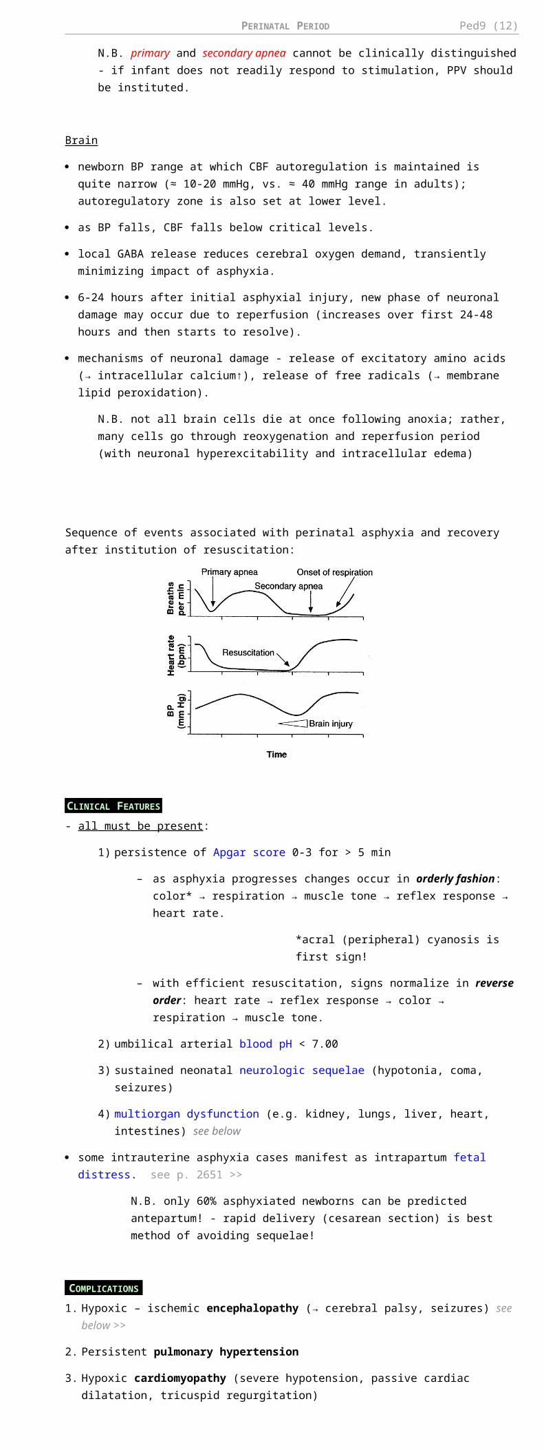
Sequence of events associated with perinatal asphyxia and recovery after institution of resuscitation:
CLINICAL FEATURES
- all must be present:
1) persistence of Apgar score 0-3 for > 5 min
– as asphyxia progresses changes occur in orderly fashion: color* → respiration → muscle tone → reflex response → heart rate.
*acral (peripheral) cyanosis is first sign!
– with efficient resuscitation, signs normalize in reverse order: heart rate → reflex response → color → respiration → muscle tone.
2) umbilical arterial blood pH < 7.00
3) sustained neonatal neurologic sequelae (hypotonia, coma, seizures)
4) multiorgan dysfunction (e.g. kidney, lungs, liver, heart, intestines) see below
some intrauterine asphyxia cases manifest as intrapartum fetal distress. see p. 2651 >>
N.B. only 60% asphyxiated newborns can be predicted antepartum! - rapid delivery (cesarean section) is best method of avoiding sequelae!
COMPLICATIONS
1. Hypoxic – ischemic encephalopathy (→ cerebral palsy, seizures) see below >>
2. Persistent pulmonary hypertension
3. Hypoxic cardiomyopathy (severe hypotension, passive cardiac dilatation, tricuspid regurgitation)
4. Poor gastrointestinal motility; necrotizing enterocolitis occurs rarely.
5. Acute tubular necrosis (!), oliguric renal failure (→ high-output tubular failure → significant water and electrolyte imbalances).
6. Adrenal hemorrhage and necrosis
7. Hypoglycemia

PERINATAL PERIOD Ped9 (10)
8. Polycythemia
9. Hypocalcemia
10. DIC
NEWBORN RESUSCITATION
Who should not be resuscitated:
a) extreme prematurity (< 23 weeks' gestation)
b) extremely low birth weight (< 400 g)
c) chromosomal anomalies inconsistent with life (e.g. trisomy 13).
Equipment for Neonatal Resuscitation
Respiration Suction Fluids Drugs Procedures
- Oxygen supply (preferably warmed and humidified)- Assorted masks- Neonatal bag and tubing to connect to oxygen source- Manometer- Endotracheal tubes (2.5-4)- Tape and scissors- Laryngoscope (0 and 1 sized blades)- Extra bulbs and batteries
- Bulb syringe- Regulated mechanical suction (max 136 cmH2O)- Suction catheters (6F, 8F, 10F)- Suction tubing- Suction canister- Replogle or Salem pump (10F catheter)- Feeding tube (8F catheter)- Syringe, catheter tipped, 20 mL- Meconium aspirator (endotracheal tube adapter)
- IV catheters 22G- Tape and sterile dressing material- D10W- Isotonic saline solution- T-connectors- Syringes 1-20 mL
- EPINEPHRINE (1:10,000)- SODIUM BICARBONATE (0.5 mEq/mL)
- NALOXONE
- Umbilical vein catheters (2.5F, 5F)- Chest tube (10F catheter)- Sterile procedure trays (e.g. scalpels, hemostats, forceps)
Birth may reveal conditions that posed no problem during intrauterine life - for all deliveries, at least one* person should be present who is skilled in neonatal resuscitation and has responsibility for only infant!
*if delivery is high risk - 4 skilled individuals (team leader and 3 assistants)
6-10% newborns require resuscitation at delivery; incidence increases significantly if birth weight is < 1500 g.
Perinatal asphyxia and extreme prematurity are states that most frequently require complex resuscitation;
also 80% low birth weight infants require resuscitation and stabilization at delivery.
1-3 are considered routine care for most term infants:
1. Suctioning of mouth-nose-pharynx should be done before thorax delivery (esp. if delivered through meconium-stained amniotic fluid) and then done intermittently.
– appropriately sized large-bore catheter using mechanical suction device with pressure limit of 100 mmHg (136 cmH2O).
– avoid deep oropharyngeal suctioning (may induce vagal response - central apnea, profound bradycardia, hypotension, laryngospasm).
– instillation of saline into trachea (for thick mucous) also stimulates sensory neurons leading to these sequelae and has no place in immediate resuscitation period.
– vigorous suctioning of nares with catheter can lead to edema → respiratory distress after infant leaves delivery room.
– vigorous frequent airway suctioning in prematures is associated with hypoxia, periventricular hemorrhage / leukomalacia.
2. Positioning on preheated overhead radiant warmer (after quickly drying infant and removing wet linen). see below (HYPOTHERMIA) >>
– supine, neck in neutral position (rolled towel under shoulders).
– insensible water loss is increased in radiant warmer (esp. for premature infant).
3. Tactile stimulation (e.g. flicking soles, rubbing back) – may be necessary* to initiate and encourage regular, spontaneous breathing.
*drying and suctioning often is enough stimulation
– no response to stimulation = secondary apnea → O2 therapy, bag-mask ventilation, intubation & positive pressure ventilation (PPV), chest compressions. see below
– if mother received opioids during last 4 hours and newborn is not breathing → NALOXONE (prematures - ½ dose); contraindication - mother addicted to narcotics.
– SURFACTANT is routinely administered to prematures. see below
1-3 are considered routine care for most term infants.

PERINATAL PERIOD Ped9 (11)
4. Heart rate > 100/min + adequate respiratory effort + cyanosis → O2 supplement at 8-10 L/min through face mask attached to self-inflatable or anesthesia bag.
– if oxygen is to be provided for prolonged period, then heated humidified oxygen should be supplied via oxy-hood (unheated nonhumidified oxygen can quickly cool infant via large surface area of lungs).
– adjust FiO2 to keep target saturations; FiO2 of 1 for short duration is not detrimental to prematures.
Target saturations - 90-96% (88-92% in preterm infant)
Heart rate < 100/min OR respiratory distress (inadequate respiratory activity) OR central cyanosis (despite 100% O2) → bag-mask ventilation to provide continuous positive airway pressure (CPAP) - aids in development of functional residual capacity (i.e. lung recruitment).
– self-inflating and flow-inflating bags remain standard of care.
– infant must be positioned properly and upper airway must be cleared of secretions; mask must be correct size and form tight seal on face.
– look for rise and fall in chest (if no chest rise occurs, either airway is blocked or insufficient pressure is being generated by bag squeezing).
– sufficient, but not excessive, pressure must be used to adequately inflate lungs (pressure release valve must limit positive pressure to 30-40 cmH2O; 20-25 cmH2O for prematures).
– initial inflation of newborn's lungs with 30-40 cmH2O pressure for 5 seconds results in more rapid formation of functional residual capacity; once initial lung recruitment is obtained, avoid overdistension* (→ pneumomediastinum, pneumothorax), but also provide adequate PEEP to prevent atelectasis.
*esp. in prematures when exogenous surfactant is administered → rapid increase of lung compliance.
– ventilate 40-60 breaths per minute initially → fewer assisted breaths if spontaneous respiratory efforts increase.
– if supplemental oxygen is not available, use room air (FiO2= 0.21).
N.B. large controlled multicenter trials indicate that room air is just as effective as 100% oxygen when resuscitating term infants!
– some infants respond to brief mechanical ventilation and begin independent ventilation; others need continued ventilatory support.
– ventilation effectiveness is evaluated by observing increase in heart rate.
– if heart rate does not rise to > 100, use FiO2 of 1.
Heart rate < 60/min following 30 seconds of effective positive pressure ventilation → chest compressions (major difference from adult resuscitation!); see p. 3901 >>
Neonates tend to develop bradycardia with hypoxemia!
– compress 90 times per minute.
– one ventilation is interposed after every 3 chest compressions (i.e. 30 breaths per min).
– evaluate heart rate and color every 30 seconds.
Chest compressions are discontinued as soon as heart rate is > 60 BPM.
if after 30-60 sec HR remains < 60/min → intubation & mechanical ventilation;
– for prematures use Miller size 0 blade, for term infants - size 1 blade.
– appropriate size of endotracheal tube (ETT) is based on weight of infant:
Weight ETT size
ETT measurement at lip
< 1000 g 2.5 7 cm at lip
1000-2000 g
2.5-3 8 cm at lip
2000-3000 g
3-3.5 9 cm at lip
> 3000 g 3.5-4 10 cm at lip
– ventilation is provided via bag or ventilator after infant is intubated.
– immediate increase in heart rate is excellent indicator of appropriate ETT placement.
if after 30-60 sec* mechanical ventilation HR still remains < 60/min → EPINEPHRINE (1:10,000) 0.01-0.03 mg/kg; IV via umbilical venous catheter is preferred route (if epinephrine is given via ETT, follow with 0.5-1 mL of saline flush to ensure that drug is delivered to lung).
*no need to wait if born without pulse
BRADYCARDIA in distressed child is sign of impending cardiac arrest!!!
(newborns, infants, and young children tend to develop bradycardia with hypoxemia, whereas older children tend initially to have tachycardia).
N.B. primary importance is establishment of effective ventilation - without ventilation, other therapies, including drugs, will not be effective in establishing adequate heart rate and perfusion.
PERINATAL PERIOD Ped9 (12)
5. Signs of bleeding, shock → euvolemia restoration (0.9% SALINE*, lactated ringers, 5% albumin, Plasmanate, O-negative blood cross matched with mother); if necessary, add DOPAMINE.
*most frequently used agent for volume expansion in neonates
– dosage for volume expansion - 10 mL/kg IV over 5-10 min (infuse more cautiously in extremely preterm infants – risk of periventricular hemorrhage due to hypervolemia).
– neonatal cardiovascular system is very sensitive to preload, requiring adequate intravascular volume to maintain adequate cardiac output!!!
– hypovolemia may be masked in newborn (significant peripheral vasoconstriction caused by elevated catecholamines following delivery).
– systolic BP may be falsely elevated with pain.
6. For prolonged resuscitation, use SODIUM BICARBONATE (0.5 mEq/mL) 2 mEq/kg* IV to reverse metabolic acidosis. *if base deficit is known, more precise dose can be administered
– SODIUM BICARBONATE should not be used until adequate ventilation is obtained because of concomitant CO2 production following use of this drug (if SODIUM BICARBONATE is used in face of respiratory acidosis and elevated pCO2, acidosis will not be corrected!).
– SODIUM BICARBONATE in delivery room has been associated with ↑risk of intraventricular hemorrhage in very low birthweight infants.
N.B. recent studies show that 0.9% SALINE provides better cardiac & BP support to correct both acidosis and underlying etiology of metabolic acidosis.
Discontinuing resuscitation may be justified in infants who have not responded to continuous and appropriate resuscitation for full 10 minutes and who have no heart rate or respiratory effort (no signs of life).
ALGORITHM for resuscitation of neonates:
POSTRESUSCITATION
- see Hypoxic-Ischemic Encephalopathy >>
SPECIAL PROBLEMS DURING RESUSCITATION
Extreme prematurity see above (PREMATURITY) >>
Choanal atresia see p. 2172 (2) >>
Pierre Robin syndrome see p. 97 >>
Tracheal webbing see p. 2160 >>
Esophageal atresia ± tracheoesophageal fistula see p. 1923 >>
HYPOXIC-ISCHEMIC ENCEPHALOPATHY (HIE)- (sub)acute brain lesions due systemic hypoxemia or reduced cerebral blood flow – i.e. encephalopathy from asphyxia.

PERINATAL PERIOD Ped9 (13)
exact cause and exact time of brain injury often remain uncertain.
e.g. acute perinatal / intrapartum events are found in only 20% children with cerebral palsy
abnormal brain might be underlying risk factor.
pathophysiology – see above (PERINATAL ASPHYXIA) >>
BRAIN PATHOLOGY - depends on brain maturity at time of insult and duration of ischemia:
Total brief (> 20-25 min) asphyxia results in DIFFUSE lesions rarely compatible with life.
profound asphyxia lasting < 10 min in otherwise healthy newborn is not thought to cause any permanent brain damage.
Partial asphyxia for minutes ÷ hours results in predominantly supratentorial lesions:
In preterm infants, damage is at GERMINAL MATRIX area → periventricular hemorrhages, leukomalacia; see below >>
After 36 weeks of gestation, lesions primarily involve:
1. CEREBRAL CORTEX (laminar neuronal necrosis in depths of sulci → ulegyria) – lesions are diffuse or localized watershed* (e.g. in parasagittal location).
2. BASAL GANGLIA (→ status marmoratus** with choreoathetosis and related movement disorders).
3. BRAIN STEM.
4. CEREBELLAR PURKINJE CELLS (→ cerebellar atrophy).
*especially after fetal hypotension
**marble white discoloration due to patchy neuronal loss, gliosis, and hypermyelination (at ≈ 6 months of age); full evolution of neuropathology may take months to years.
Less severe intrauterine anoxic episodes of undetermined duration may involve neurons DIFFUSELY or may preferentially affect HIPPOCAMPAL AREAS.
N.B. any cerebral edema is at its maximum around 72 hours after insult!
total time span of hypoxic– ischemic damage leading to permanent cortical brain tissue loss is 4–6 months.
very severe hypoglycemia may itself damage neonatal brain - damage appears similar to hypoxic– ischemic injury (with initial edema and subsequent atrophy), but is limited to occipital and posterior parietal regions.
CLINICAL FEATURES
Best gauged by SARNAT CLASSIFICATION (in conjunction with EEG, neuroimaging, and brain stem auditory and cortical evoked responses):
FACTOR STAGE I (MILD) STAGE II (MODERATE) STAGE III (SEVERE)
duration* < 24 h 2-14 days hours to weeks
consciousness levelhyperalertness and irritability
lethargy deep stupor or coma
muscle tone normal or slightly↑significant hypotonia or proximal limb weakness
flaccidity
posture mild distal flexion strong distal flexionintermittent decerebration
tendon reflexes ↑ ↑ or ↓ depressed or absent
segmental myoclonus present present absent
seizures none common (70%)frequent** during 24-48 hrs, then usually stop
EEG normallow voltage, periodic or paroxysmal, epileptiform activity
periodic pattern → isoelectric
COMPLEX REFLEXES:
sucking weak weak absent
Moro responseexaggerated (strong, low threshold)
weak, incomplete, high threshold
absent
grasping normal to exaggerated exaggerated absent
oculovestibular normal overactive weak / absent
tonic neck slight strong absent
oculocephalic (doll's eye)
normal overreactivereduced or absent (+ skewed eye deviation, nystagmus, bobbing)
AUTONOMIC FUNCTION: SYMPATHIC↑ PARASYMPATHIC↑ DEPRESSED

PERINATAL PERIOD Ped9 (14)
pupils dilated constricted variable or fixed
respiration regularvariable in rate and depth, periodic
irregular apnea (requires ventilatory support)
heart rate normal or tachycardic low resting, < 120 / min bradycardia
bronchial and salivary secretions
sparse profuse variable
GI motility normal or ↓ ↑, diarrhea variable
risk of death < 1% 5% > 60%
risk of severe handicap < 1% 20-50% > 70%
*then CNS examination becomes normal
**correlate with reperfusion injury
General course: depression → hyperalertness and hyperreflexia → coma.
in USA, stage 3 incidence is 2-4 cases per 1000 births.
symptoms of moderate / severe HIE almost always manifest at birth or within few hours*.
– infants who sustain hypoxic / ischemic insults weeks ÷ months before birth may seem normal at birth (but later show signs of static encephalopathy or seizures); others already exhibit signs of chronic cerebral disease at birth (with overt microcephaly and spasticity).
involvement of multiple organs besides brain is hallmark of HIE.
see above (perinatal asphyxia, complications) >>
sequelae – microcephaly, mental retardation, epilepsy, cerebral palsy.
* even in absence of obvious neurologic deficits in newborn period, long-term functional impairments may be present (e.g. learning difficulties)! - prognosis is always guarded! - all children with moderate / severe HIE should be monitored well into their school ages!
DIAGNOSIS
- primarily clinical!
neuroimaging may or may not reveal abnormal findings!
– cranial ultrasound: cerebral edema may be evident (decreased ventricular size or indistinct sulci and gyral patterns); ischemic lesions appear as diffuse or localized echodensities within brain parenchyma (→ multicystic encephalomalacia, severe atrophy in cortical areas*, atrophic ventricular enlargement).
*white matter may suffer secondary atrophy as cortical neurones disappear
– pulsed Doppler - diastolic velocities↑ with reduced resistive index (< 0.50), sign of luxury perfusion, or decreased peak-systolic velocities, sign of low blood flow.
– CT is best method to confirm cerebral edema, evolving zones of infarction (areas of reduced density)
– MRI is valuable at 6 months ÷ 1 year - status of myelination (e.g. delayed), white-gray tissue injury, preexisting developmental defects of brain; MRI is also useful during follow-up.
many centers are using amplitude-integrated electroencephalography (aEEG); single-channel aEEG performed within few hours of birth can help evaluate severity of brain injury.
standard EEG is obtained as soon as infant is stable.
N.B. in assisted ventilation, drugs for muscle paralysis and morphine (for sedation) can mask seizures! - PHENOBARBITAL may be used prophylactically in heavily sedated or paralyzed infants.
increased incidence of deafness! – perform full-scale hearing test.
impaired visual evoked responses (VER) and brain stem auditory evoked responses (BAER) may imply poor prognosis.
Corticosubcortical ischemic injury in full-term neonate, 3 days old:
Coronal plane ultrasound - slight echodensity of parenchyma and attenuated visibility of cerebral sulci.
Pulsed Doppler - normal peak-systolic velocity (40 cm s−1 ), but increased end-diastolic velocity (23 cm s−1 ) and decreased resistive index (0.43).
Source of picture: Ronald G. Grainger, David J. Allison “Grainger & Allison’s Diagnostic Radiology: A Textbook of Medical Imaging”, 4th ed. (2001); Churchill Livingstone, Inc.; ISBN-13: 978-0443064326 >>

PERINATAL PERIOD Ped9 (15)
A. CT of normal term newborn - clear differentiation between white and grey matter.
B, C. CT of severe partial hypoxia (12-hour-old term newborn) - no differentiation between white and grey matter; preserved normal tissue attenuation in posterior fossa but lateral ventricles (arrows in C) are compressed owing to edema. Since there are already extensive changes at age 12 hours, insult occurred in utero, at least 24 hours before scan.
Multicystic encephalomalacia of 18-month-old girl who suffered severe partial hypoxia at birth (T2-MRI) - most of cerebrum has undergone cystic destruction; mesencephalon and vermis have normal appearance; dilated lateral ventricles; head is small and hydrocephalic but this is not caused by hydrocephalus.
Profound asphyxia, end-stage of 2-year-old boy who suffered profound asphyxia at birth (T2-MRI):
A. Axial MR - symmetrical increased signal in posterior putamen and inferolateral nucleus of thalami (arrows).
B. Axial MR - subtle volume loss in front of and behind central sulcus, with increased signal in subcortical white matter.
C. Sagittal MR - abnormal signal extending between cortical changes and changes in basal ganglia.
TREATMENT
transfer to tertiary neonatal intensive care unit.
most infants need ventilatory support during first week (avoid hyperventilation → severe hypoperfusion of brain).
maintain mean BP > 35 mm Hg (for term infants); DOPAMINE (inotropic of choice) or DOBUTAMINE can maintain adequate cardiac output.
avoiding hyperthermia after hypoxic-ischemic event at birth is essential!!!
current data are insufficient to recommend brain HYPOTHERMIA for all infants with suspected asphyxia; but it is slowly emerging as useful therapy for mild-to-moderate cases!
– cooling must begin within 1 hour of injury.
– keep 3-4°C below baseline temperature for up to 48-72 hours.
– two methods may be used:
a) selective head cooling cap (Cool-Cap) – FDA approved for moderate to severe HIE!
b) whole body hypothermia
– rewarming is carried out gradually (over 6-8 hours).

PERINATAL PERIOD Ped9 (16)
seizures should be treated early with PHENOBARBITAL (drug of choice!!!) or LORAZEPAM (second drug of choice); PHENYTOIN IV (third drug of choice) may be added.
– drugs can be weaned and stopped during first month of life (unless persistent neurological abnormalities and clinical or EEG evidence of seizures → treatment is continued for several months to 1 year).
Even asymptomatic seizures (seen only on EEG) continue to injure brain!
hypotonia and feeding difficulties often persist, requiring tube feeding for weeks ÷ months.
fluids 60-80 mL/kg/d for infant in humidified incubator (rates much higher for infant in dry radiant warmer environment).
– in first 2-4 days, restrict IV fluids to 2/3 of daily requirement (for gestational age and nursing environment) because of high frequency of acute tubular necrosis and IADH.
– for those on high-frequency ventilators (venous return may be impaired), fluid volumes must be increased.
– electrolytes (sodium, potassium, chloride) should not be added initially because fluid shifts from other body compartments allow for adequate electrolyte supply until adequate renal function is documented.
hypoglycemia may occur rapidly in critically ill / premature infants - GLUCOSE 4-6 mg/kg/min IVI should be started for those who do not tolerate enteral feedings.
– avoid dextrose boluses (→ transient hyperosmolarity and rebound hypoglycemia).
Avoid hypoglycemia or hyperglycemia (both damage brain).
monitor weight, hydration status, urine output, serum [sodium].
HYPOTHERMIA- core temperature < 35-35.5° C.
intrauterine thermoregulation is passive - no use of calories or oxygen by fetus (allows for maximal intrauterine growth without fetal energy expenditure for thermal homeostasis).
neonates respond to cooling by sympathetic nerve discharge (norepinephrine) in brown fat – it is used for heat production in newborn period.
Main source of heat production in newborn is nonshivering thermogenesis!
Factors for increased heat losses in newborns:
1) large skin surface area–to–body weight ratio → heat and fluid evaporative loss↑.
2) thin skin with blood vessels near surface provides poor insulation.
3) newborn (especially if premature) has limited capacity to change body position for heat conservation (by decreasing exposed surface area in flexed position).
4) very limited capacity for metabolic heat production - limited energy stores (subcutaneous fat and brown fat), esp. in premature and growth-retarded infants; infants do not shiver effectively.
heat loss increases metabolic rate and uses more oxygen (dangerous if already in respiratory compromise) → rapidly used glucose and glycogen reserves → metabolic acidosis, hypoglycemia.
hypothermia also may be caused by pathologic conditions that impair thermoregulation (e.g. sepsis, intracranial hemorrhage).
PROPHYLAXIS
newborns should be dried with prewarmed blankets or towels.
place on prewarmed heat source; open bed warmers, which use radiant heat, are used in most delivery rooms (convenient access to newborn)
– sick neonates should be maintained in neutral thermal environment to minimize metabolic rate.
– proper incubator temperature depends on birth weight and postnatal age (alternatively, servomechanism can be set to maintain skin temperature at 36.5° C).
N.B. open bed warmers encourage evaporative heat losses - prematures (< 1500 g) should be covered in plastic wrap to prevent excessive heat loss (full resuscitation, including line placement, can and should be performed with plastic wrap in place).
use of warmed humidified* oxygen for bag-valve-mask device; intubated infant should be placed on heated ventilator circuit.
*gas heating and humidification by infant results in massive heat exchange and insensible water loss due to large surface area of lungs
BIRTH TRAUMA forces of labor and delivery occasionally cause physical injury to infant.
INCIDENCE is decreasing due to increasing use of cesarean section (in place of difficult versions, vacuum extractions, or mid- or high-forceps deliveries).
risk factors : small pelvic measurements, large for gestational age infant, breech or other abnormal presentation, primipara.

PERINATAL PERIOD Ped9 (17)
HEAD TRAUMA
Risk factors - primiparas, large infants, preterm delivery, difficult delivery.
Operative delivery (vacuum extraction, forceps, cesarean section) or rapid birth (esp. in breech presentation - rapid head moulding during final moments of birth) - increased risk of skull fractures, intracranial hemorrhage!
Intracranial hemorrhage
gestational age is best indicator of probable site of intracranial hemorrhage:
supratentorial subdural hemorrhage – exclusively full-term or large infants with difficult deliveries.
supratentorial SAH of venous origin – full-term newborns who have focal seizures and benign clinical course; SAH may be found in premature infants, but there is no recognized clinical syndrome.
periventricular hemorrhage – premature infants of ≤ 32 weeks gestation. see below >>
hypoxia-ischemia often precedes bleeding (hypoxia-ischemia damages endothelium, impairs cerebrovascular autoregulation, and can increase cerebral blood flow and venous pressure, all of which make hemorrhage more likely).
diagnosed by CT (ultrasound is not good method - extracerebral fluid collections over hemispheres and posterior fossa masses are very difficult to detect).
– evaluate for skin petechiae or hemorrhage from other sites - suggests systemic disorder (e.g. vit. K deficiency, hemophilia, DIC).
can be life threatening (esp. if born prematurely).
prognosis for SAH is generally good; prognosis for subdural hemorrhage is guarded (some infants do well);
treatment is supportive;
– give vitamin K (if it was not previously given);
– symptomatic subdural hematomas → daily subdural taps. see p. TrH13 >>
– in posterior cranial fossa hematoma, surgical drainage may be lifesaving!
Slow deformational forces → tears in tentorium (less commonly, in falx, in junction between falx and tentorium) → subdural hematoma.
Forceps delivery → ‘ping-pong’ skull fracture - depressed fracture without fracture line (may require elevation for cosmetic reasons). also see p. TrH5 >>
– may be associated with subdural bleeding, SAH, or brain contusion / laceration.
Vaginal delivery may result in:
1) MOLDING (bone overlapping at sutures) - disappears within 2-14 days after birth.
2) CAPUT SUCCEDANEUM – SWELLING in presenting portion of scalp (above periosteum) secondary to compression by cervix (due to vacuum effect after amniotic sac rupture); resolves within 2 weeks.
3) SUBGALEAL HEMATOMA – BLOOD ACCUMULATION in sub-galeal loose areolar tissue.
– soft fluctuant swelling over entire scalp (i.e. not limited by periosteal insertions).
– self-limiting condition.
Needle / incisional drainage may result in infection!!!
4) CEPHALHEMATOMA – subperiosteal BLOOD ACCUMULATION; may develop after instrumental delivery.
– not present at birth, appears within 24 hours
– fluid-blood collection is limited by periosteal insertion at suture lines, i.e. does not extend across suture (vs. caput succedaneum).
– initially soft, but may develop raised bony margin within 2-3 days (rapid Ca deposition at edges of raised periosteum).
– small percentage have associated linear fracture in underlying bone.
– commonly unilateral parietal.
– visible on plain radiograph as subperiosteal elevation.
– usually resorb spontaneously within few weeks (occasionally calcify and form bony protrusion - self-correcting cosmetic deformity - calcified tissue is gradually absorbed by expanding calvarium and appearance becomes normal before age 1-2 years).
Needle / incisional drainage may result in infection!!!

PERINATAL PERIOD Ped9 (18)
CRANIAL NERVE INJURY
Most often - FACIAL NERVE:
a) most injuries - pressure on nerve in utero (head lying against shoulder, sacral promontory, or uterine fibroid).
b) forceps pressure.
injury usually occurs at or distal to exit from stylomastoid foramen.
facial asymmetry is most apparent during crying (differentiate from mandibular asymmetry resulting from intrauterine pressure; maxillary and mandibular occlusal surfaces are not parallel, vs. facial nerve injury).
testing or treatment is not needed for peripheral CN7 injuries or mandibular asymmetry - they usually resolve by age 2-3 months.
BRACHIAL PLEXUS INJURIES
etiology – stretching by shoulder dystocia, breech extraction, neck hyperabduction in cephalic presentations.
pathology – simple stretching, hemorrhage within nerve, tearing of nerve or root, avulsion of roots with accompanying cervical cord injury (ipsilateral pyramidal signs).
associated injuries – fractures of clavicle or humerus or subluxations of shoulder or cervical spine.
clinical features → see p. PN9 >>
treatment – hand support, passive range-of-motion exercises.
– usually improve rapidly.
– if significant deficit persists > 3 mo → MRI to determine extent of injury to plexus, roots, and cervical cord; surgical repair may be helpful.
– if entire brachial plexus is injured → neurosurgical exploration; prognosis for recovery is poor; extremity's growth may be impaired.
OTHER PERIPHERAL NERVE INJURIES
usually not related to labor and delivery (i.e. usually secondary to local traumatic event, e.g. injection in or near sciatic nerve).
treatment - placing muscles antagonistic to those paralyzed at rest until recovery; neurosurgical exploration is seldom indicated.
recovery is complete.
FRACTURES
Most common - MIDCLAVICULAR FRACTURE.
occurs with shoulder dystocia.
neonate is irritable and does not move arm (either spontaneously or when Moro reflex is elicited).
most clavicular fractures are greenstick - heal rapidly and uneventfully (large callus forms within week, and remodeling is completed within month).
treatment - sling by pinning shirt sleeve of involved side to opposite side of infant's shirt.
HUMERUS AND FEMUR FRACTURES in difficult deliveries.

PERINATAL PERIOD Ped9 (19)
most are greenstick, mid-shaft fractures → excellent remodeling, even if moderate angulation occurs initially.
epiphysis fractures also bear excellent prognosis.
SOFT-TISSUE INJURIES
- if they are presenting part or fulcrum for forces of uterine contraction.
periorbital and facial tissues in face presentations; scrotum or labia during breech deliveries.
edema and ecchymosis.
added burden of bilirubin may produce sufficient hyperbilirubinemia to require phototherapy, ± exchange transfusion.
PERIVENTRICULAR / INTRAVENTRICULAR HEMORRHAGE (PVH-IVH)
- hemorrhage into germinal matrix seen exclusively in PRETERM INFANTS after asphyxial insult.
Intraventricular hemorrhage in TERM INFANTS – from choroid plexus
INCIDENCE (inversely proportional to gestational age) – 12-50% among newborns < 1500 g.
ETIOPATHOPHYSIOLOGY
hemorrhage occurs into subependymal friable, richly vascular germinal matrix (lies on lateral wall of lateral ventricles between thalamus and caudate, near foramina of Monro; from lateral ventricle separated only by ependyma).
germinal matrix is site of neuronal proliferation as neuroblasts divide and migrate into cerebral parenchyma;
– by ≈ 20 weeks' gestation, neuronal proliferation is completed; however, glial cell proliferation is still ongoing;
– germinal matrix supports division of glioblasts and glial differentiation until ≈ 32 weeks' gestation, at which time regression is nearly complete.
N.B. germinal matrix is present only in fetus and prematures < 32 weeks!
metabolically active differentiating cells of germinal matrix are rich in mitochondria (quite sensitive to ischemia).
– supplying this area is primitive and fragile retelike capillary network.
– arterial supply - recurrent artery of Heubner and lateral striate arteries.
– venous supply - thalamostriate veins.
– as result of respiratory distress from immature lungs along with episodes of hypoxemia and fluctuations in cerebral perfusion, vessels in germinal matrix have tendency to rupture.
N.B. only sites in adult brain where neurons still being produced – olfactory bulb and hippocampus!
GERMINAL MATRIX - dense layer of small dark blue cells below ependyma of lateral ventricle:
Source of picture: “WebPath - The Internet Pathology Laboratory for Medical Education” (by Edward C. Klatt, MD) >>
t wo major pathophysiologic factors :
(1) loss of cerebral autoregulation (autoregulatory abilities vary proportionally to gestational age - range of perfusion pressures over which premature neonate can control regional CBF is narrower and lower than that of infants born at term) → pressure-passive circulatory pattern.
– conditions that interfere with autoregulation: hypocarbia / hypercarbia, hypoxia, acidosis
(2) abrupt alterations in cerebral blood flow and pressure (esp. hypotension followed by hypertension; increased venous pressure)

PERINATAL PERIOD Ped9 (20)
– conditions that overwhelm autoregulatory abilities: asynchrony between spontaneous and mechanical breaths; birth (esp. vacuum-assisted delivery); frequent noxious procedures of caregiving; instillation of mydriatics; tracheal suctioning; pneumothorax; rapid volume expansion; rapid colloid infusion (e.g. exchange transfusion); infusions of hypertonic solutions (e.g. sodium bicarbonate); seizures; changes in pH, PaCO2, and PaO2.
source of bleeding – capillaries (possess neither tight junctions between endothelial cells nor strong basement membrane - increased flow and pressure may rupture delicate capillaries).
Intraventricular hemorrhage is uncommon in infants who are born at term; for them site of hemorrhage is choroid plexus or venous sinus thrombosis
CLINICAL FEATURES
Grades (worsening prognosis):
Grade I – confined to germinal matrix, i.e. subependymal (usually asymptomatic - most infants do well!)
Grade II – small blood amount (< 40% of ventricular volume) in ventricles without ventricular enlargement (nonspecific irritability or lethargy - most infants do well!)
Grade III – blood in ventricles, ventricular dilation (mortality < 10%); sequelae - static or reversible or progressive posthemorrhagic hydrocephalus → 30-40% incidence of cerebral palsy and mental retardation.
Grade IV (periventricular hemorrhagic venous infarction) – additional hemorrhage into parenchyma which involves periventricular motor tracts (poor prognosis – 27-80% mortality!; 90% incidence of cerebral palsy and mental retardation); it is secondary to lower grade (I-III) hemorrhage which leads to congestion in periventricular white matter → venous infarction → secondary hemorrhage; almost always unilateral and anterior; clinically:
1) severe apnea, bradycardia, hypotension, altered mental status.
2) extensor posturing and opisthotonos; clonic limb movements may occur concurrently (without EEG seizure activity).
3) deviated eyes (converged or diverged), pupils fixed in midposition.
4) fullness of fontanels.
5) many become flaccid and unresponsive and die within minutes or hours; less dramatic deterioration may occur over few days.
6) smaller lesions develop periventricular porencephalic cysts.
N.B. prognosis (of subsequent handicap) is better than in PERIVENTRICULAR LEUKOMALACIA!
Intraventricular hemorrhage (IVH) extending from germinal matrix hemorrhage of 28 week gestational age newborn:
Source of picture: “WebPath - The Internet Pathology Laboratory for Medical Education” (by Edward C. Klatt, MD) >>
Severe IVH - blood filling and distending all of lateral ventricles, extending into brain parenchyma, and extending down third ventricle and out into subarachnoid space:

PERINATAL PERIOD Ped9 (21)
Source of picture: “WebPath - The Internet Pathology Laboratory for Medical Education” (by Edward C. Klatt, MD) >>
Sequelae:
1. Destruction of periventricular cerebral parenchyma (esp. motor tracts*) → cerebral palsy (!!!), mental retardation, seizures.
*tracts innervating lower extremities are nearest to ventricles, followed by those innervating trunk, arm, and, finally, face → greater degree of motor dysfunction of extremities as compared to face (spastic hemiplegia in unilateral lesions and spastic diplegia or quadriplegia in bilateral lesions)
2. Posthemorrhagic hydrocephalus
Because PVH-IVH development is related to alterations in cerebral blood flow, injury to other portions of brain may occur:
1) GLOBAL HYPOXIC-ISCHEMIC INJURY
2) PERIVENTRICULAR LEUKOMALACIA (PVL) - nonhemorrhagic ischemic necrosis.
DIAGNOSIS
1) fall of hematocrit ≥ 10%
2) CSF bloody or normal → xanthochromic.
3) cornerstone of diagnosis - ultrasound - sensitivity 96%, specificity 94% (has replaced CT!) - delineates site of blood in parenchyma and ventricles, ventricular size, and shifts of major structures. see p. D57 >>
American Academy of Neurology recommendation - all infants < 30 weeks gestational age must be screened* by cranial ultrasonography at 7-14 days postnatal life and at 36-40 weeks postmenstrual age.
*PVH can occur without clinical signs
– serial weekly examinations are necessary for follow-up (progression of hemorrhage, development of posthemorrhagic hydrocephalus).
– HYPERECHOGENIC area on inferior wall of lateral ventricle, overlying caudate nucleus head.
– when it ruptures into ventricular lumen, it appears as intraluminal strongly echogenic material (when it is small – must be distinguished from choroid plexus –colour Doppler may help); later, intraventricular clot becomes less echogenic in its centre but is surrounded by echoic line.
– parenchymal hemorrhage is seen as unilateral or asymmetric echodense area, radiating from external angle of ventricle, which evolves to anechoic cavitation (porencephalic cyst).
Sagittal sonogram (intraventricular hemorrhage): hyperechogenic blood fills lumen of lateral ventricle:
Source of picture: Ronald G. Grainger, David J. Allison “Grainger & Allison’s Diagnostic Radiology: A Textbook of Medical Imaging”, 4th ed. (2001); Churchill Livingstone, Inc.; ISBN-13: 978-0443064326 >>

PERINATAL PERIOD Ped9 (22)
Coronal sonogram - right intraventricular hemorrhage with parenchymal hemorrhagic infarction (straight arrows), mass effect on interhemispheric fissure (curved arrow), dilatation of left ventricle (frontal horn F, temporal horn T):
Grade III - hyperechoic areas represent hemorrhage in lateral ventricle; ventricles are markedly dilated:
Grade IV (periventricular hemorrhagic infarction):
A. Axial T2-MRI - anterior periventricular cavity contains residual blood clot.
B. Axial T1-MRI in 2-year-old girl with congenital right hemiplegia; she was born prematurely and had periventricular hemorrhagic infarction.
Source of picture: Ronald G. Grainger, David J. Allison “Grainger & Allison’s Diagnostic Radiology: A Textbook of Medical Imaging”, 4th ed. (2001); Churchill Livingstone, Inc.; ISBN-13: 978-0443064326 >>
Grade I PVH:

PERINATAL PERIOD Ped9 (23)
Grade II PVH-IVH:
Grade III PVH-IVH:
Grade IV PVH-IVH:

PERINATAL PERIOD Ped9 (24)
Grade IV with porencephalic cyst formation:
Periventricular hemorrhagic infarction (MRI):
PERINATAL PERIOD Ped9 (25)
TREATMENT
Correction of anemia, acidosis, hypotension + ventilatory support.
Daily head circumference measurements (and plotting on the chart)
Weekly head US
No treatment is necessary for grades I - II.
Grade III –
most patients with hydrocephalus demonstrate spontaneous resolution within weeks of onset.
if head growth is double normal rate over 2 weeks or ICP↑ persist → lumbar / ventricular punctures to drain large volumes (e.g. 10 ml/kg) to prevent hydrocephalus; alternative - implant reservoir for repeated CSF tapping.
indications for ventriculoperitoneal shunt: head circumference > 1.5 cm above 97th percentile; or head growth > 1.5 cm/week for 2 weeks; and presence of signs of raised intracranial pressure
– high complication rate in small infants! – delay shunting until infant has shown as much somatic growth as possible, goal body weight > 1800-2000 g, age > 38 weeks; plus, when US shows evidence of improvement of clot size).
serial prophylactic lumbar / ventricular punctures to prevent hydrocephalus are no longer recommended because no effect + risk of porencephalic cysts (correlate with use of sharp vs. blunt needles), introducing infection!!!
Cochrane Database Syst Rev. 2001;(1):CD000216. Whitelaw A. Repeated lumbar or ventricular punctures in newborns with intraventricular hemorrhage.
osmodiuresis with decreasing CSF production - with ACETAZOLAMIDE or FUROSEMIDE - not recommended as cause worse outcomes (class 1 evidence).
International PHVD drug trial group . International randomised controlled trial of acetazolamide and furosemide in posthaemorrhagic ventricular dilatation in infancy . Lancet 1998 ; 352 : 433 – 440 .
Multi-centre randomized controlled trial, 177 patients.
Infants in the drug therapy group had a significantly increased risk (p = 0.012) of death, impairment or disability at 1 year. Risk ratio of 1.40 (1.12–1.76).
Primary outcomes at 1 year Drug + Standard therapy Standard therapy alone
Death, shunt placement or both 65% 46% p = 0.026
PREVENTION
- avoidance of premature birth!
Greatest risk is first 72 hours of life (50% hemorrhages occur on 1st day) – reduce infant's systemic blood pressure fluctuations (may diminish incidence of hemorrhage and its spread):
1) PANCURONIUM paralysis while infant is ventilated (prevents asynchrony between spontaneous and mechanical breaths).
2) not too rapid volume expansion following ischemia or hemorrhagic shock.
INDOMETHACIN prophylaxis (must be administered within hours of birth: 0.1 mg/kg IV when aged 6 h, then q24h for 2 d for total of 3 doses) - accelerates maturation of germinal matrix vasculature; when administered rapidly it decreases cerebral blood flow, cerebral blood flow velocity, and cerebral blood volume → reduced incidence of high-grade PVH-IVH.
Measures not proven clearly beneficial:
a) to infant – PHENOBARBITAL, ETHAMSYLATE, VITAMIN E
b) to mother antepartum – PHENOBARBITAL, STEROIDS
PERIVENTRICULAR LEUKOMALACIA (PVL)- bilateral white matter lesion of premature infants.
Clinically most significant destructive lesion in immature brain - strong relationship to subsequent handicap!
75% prematures have PVL on postmortem examination.
PVL in children born at term is effect of intrauterine damage.
ETIOPATHOPHYSIOLOGY
- selective loss of oligodendrocytes due to:
a) hypotension, ischemia* → ischemia/reperfusion injury by free radicals.
*e.g. due to respiratory distress syndrome, pneumonia, mechanical ventilation (→ hypocarbia), maternal cocaine abuse
N.B. mechanically ventilated premature infants are at greatest risk for PVL!
b) controversial: maternal / fetal infection* → cytokine-induced damage.

PERINATAL PERIOD Ped9 (26)
*history of maternal chorioamnionitis is common
damage occurs in white matter adjacent to superolateral borders of lateral ventricles – it is watershed zone of deep penetrating arteries of middle cerebral artery.
site of injury affects corticospinal tracts, visual radiations, and acoustic radiations.
reactive increase of astrocytes.
macroscopic - chalky yellow plaques (white matter necrosis and mineralization); extensive damage → multicystic encephalopathy.
Central focus of white matter necrosis with peripheral rim of mineralized axonal processes (staining blue):
Source of picture: Ramzi S. Cotran “Robbins Pathologic Basis of Disease”, 6 th ed. (1999); W. B. Saunders Company; ISBN-13: 978-0721673356 >>
CLINICAL FEATURES
Initially, asymptomatic or subtle symptoms:
1. Decreased tone in lower extremities
2. Increased tone in neck extensors
3. Apnea and bradycardia
4. Irritability
5. Pseudobulbar palsy with poor feeding
6. Clinical seizures (10-30%)
Sequelae:
1) cerebral palsy (60-100%) – most commonly spastic diplegia or quadriplegia.
2) intellectual – developmental impairment.
3) visual disturbances (fixation difficulties, nystagmus, strabismus, blindness)
DIAGNOSIS
cranial ultrasonography (modality of choice): N.B. initial exam may be normal!
1) periventricular edema - increased echotexture (echodensities) - greater than or equal to choroid plexus; disappears at 2-3 weeks.
2) periventricular cysts (15% patients) appearing at 2-3 weeks after initial echodensities; severity of PVL is related to size and distribution of these cysts.
3) cysts are transient and subsequently collapse → atrophy of damaged periventricular white matter → secondary ventricular dilatation with irregular ventricular margins (first detectable 4–8 weeks after injury; persists throughout life).
CT - ventriculomegaly of lateral ventricles with irregular margins and loss of deep white matter.
MRI (most helpful in monitoring) - loss of white matter, abnormal signal intensity of deep white matter (1-2 years after injury, when myelination process is complete), ventriculomegaly; in severe cases - thinning of posterior body and splenium of corpus callosum.
volumetric MRI - extent of injury to corticospinal tracts.
Coronal ultrasound - increased periventricular echotexture:

PERINATAL PERIOD Ped9 (27)
Coronal ultrasound - normal periventricular echotexture:
Coronal and sagittal ultrasounds (3-week-old premature infant): multiple bilateral periventricular cysts:
Axial CT (5-week-old premature infant) – mild ventriculomegaly, irregular ventricular margins (incorporation of periventricular cysts), loss of periventricular white matter:

PERINATAL PERIOD Ped9 (28)
Axial CT (14-month-old premature infant) – ventriculomegaly limited to lateral ventricles secondary to diffuse loss of periventricular white matter:
T1-MRI (18-month-old premature infant) – lateral ventricles enlarged without hydrocephalus due to diminished periventricular white matter:
T1-MRI (18-month-old premature infant) – hypoplasia of corpus callosum, most evident in body:

PERINATAL PERIOD Ped9 (29)
Posterior coronal ultrasound - multiple small echolucencies in periventricular white matter adjacent to trigones of lateral ventricles (arrows):
End-stage of PVL:
A, B. Significant reduction of periventricular white matter close to trigone of lateral ventricles bilaterally (arrows); cortical structures deep in Sylvian fissure abut lateral ventricle directly and appear to indent ventricular wall, and deep portions of Sylvian fissures are dilated; remaining white matter shows no abnormal signal.
C. Significant reduction in size of corpus callosum.
D. 8-year-old girl with severe visual cognitive impairment without cerebral palsy - significant dilatation of posterior horns of lateral ventricles (due to occipital white matter loss); remaining white matter anteriorly shows abnormal signal.
Source of picture: Ronald G. Grainger, David J. Allison “Grainger & Allison’s Diagnostic Radiology: A Textbook of Medical Imaging”, 4th ed. (2001); Churchill Livingstone, Inc.; ISBN-13: 978-0443064326 >>
TREATMENT
No medical treatment currently exists!
close developmental follow-up.
BIBLIOGRAPHY for ch. “Pediatrics” → follow this LINK >>
Viktor’s Notes℠ for the Neurosurgery Resident
Please visit website at www.NeurosurgeryResident.net