· web viewalterations of pulmonary function path ology 2 - dr. gary mumaugh signs and symptoms of...
TRANSCRIPT

1
Alterations of Pulmonary FunctionPathology 2 - Dr. Gary Mumaugh
Signs and Symptoms of Pulmonary DiseaseDyspnea
Subjective sensation of uncomfortable breathing Orthopnea
o Dyspnea when a person is lying down Paroxysmal nocturnal dyspnea (PND)
Cough Acute cough Chronic cough
Abnormal sputumHemoptysisAbnormal breathing patterns:
Kussmaul respirations (hyperpnea) Cheyne-Stokes respirations
Hypoventilation Hypercapnia
Hyperventilation Hypocapnia
CyanosisClubbing
Finger clubbing is characterized by enlarged fingertips and a loss of the normal angle at the nail bed.
Pain

2
Conditions Caused by Pulmonary Disease or InjuryHypercapniaHypoxemia
Hypoxemia versus hypoxia Ventilation-perfusion abnormalities
o Shunting Acute respiratory failure
Chest Wall DisordersChest wall restriction
Compromised chest wallo Deformation, immobilization, and/or obesity
Flail chest Instability of a portion of the chest wall
Pleural AbnormalitiesPneumothorax
Open pneumothorax Tension pneumothorax Spontaneous pneumothorax Secondary pneumothorax

3
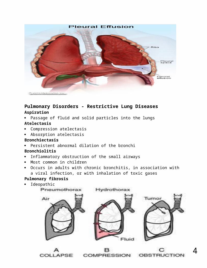
Pleural Abnormalities Pleural effusion
o Transudative effusion o Exudative effusiono Hemothoraxo Empyema
Infected pleural effusiono Chylothorax
4
Pulmonary Disorders - Restrictive Lung DiseasesAspiration Passage of fluid and solid particles into the lungsAtelectasis Compression atelectasis Absorption atelectasisBronchiectasis Persistent abnormal dilation of the bronchiBronchiolitis Inflammatory obstruction of the small airways Most common in children Occurs in adults with chronic bronchitis, in association with a viral infection, or with
inhalation of toxic gasesPulmonary fibrosis Ideopathic
Inhalation DisordersToxic gasesPneumoconiosis Silica Asbestos CoalAllergic alveolitis Extrinsic allergic alveolitis (hypersensitivity pneumonitis)Pulmonary edema Excess water in the lungs
Acute respiratory distress syndrome (ARDS) Fulminant form of respiratory failure characterized by acute lung inflammation and
diffuse alveolocapillary injury

5
Injury to the pulmonary capillary endothelium Inflammation and platelet activation Surfactant inactivation Atelectasis Manifestations:
o Hyperventilationo Respiratory alkalosiso Dyspnea and hypoxemiao Metabolic acidosiso Hypoventilationo Respiratory acidosiso Further hypoxemiao Hypotension, decreased cardiac output, death
Evaluation and treatmento Physical examination, blood gases, and radiologic examinationo Supportive therapy with oxygenation and ventilation and prevention of
infectiono Surfactant to improve compliance

6
Pulmonary Disorders - Obstructive Lung Diseases Airway obstruction that is worse with expiration Common signs and symptoms
o Dyspnea and wheezing Common obstructive disorders:
o Asthmao COPDo Emphysema o Chronic bronchitis
Obstructive lung diseases: Asthma Chronic inflammatory disorder of the airways Inflammation results from hyperresponsiveness of the airways Can lead to obstruction and status asthmaticus Symptoms include expiratory wheezing, dyspnea, and tachypnea Peak flow meters, oral corticosteroids, inhaled beta-agonists, and anti-
inflammatories used to treat

7
Obstructive lung diseases: chronic bronchitis Hypersecretion of mucus and chronic productive cough that lasts for at least 3
months of the year and for at least 2 consecutive years Inspired irritants increase mucus production and the size and number of mucous
glands The mucus is thicker than normal Bronchodilators, expectorants, and chest physical therapy used to treat
Obstructive lung diseases: emphysema Abnormal permanent enlargement of the gas-exchange airways accompanied by
destruction of alveolar walls without obvious fibrosis Loss of elastic
recoil Centriacinar
emphysema Panacinar
emphysema
How COPD develops Smoking
causes increased mucus production and bronchial inflammation
Nicotine paralyzes the mucociliary escalator o Mucociliary escalator traps mucus, bacteria, irritants
Nicotine blocks protein inhibitors which will eventually dissolve the alveoli Pathophysiology
o Involves all four parts of the respiratory tract Bronchi Bronchioles Alveoli Parenchyma
o Specific Pathophysiology Increased resistance to airflow Loss of elastic recoil Decreased expiratory flow rate Alveolar walls frequently break because of the increased resistance
of air flows The hyper inflated lungs flatten the curvature of the diaphragm and
enlarge the rib cage The altered configuration of the chest cavity places the respiratory
muscles, including the diaphragm, at a mechanical disadvantage and impairs their force-generating capacity
Consequently, the metabolic work of breathing increases, and dyspnea increases

8
Two types of COPD Type A – Pink Puffers
o Have mostly emphysemao Need to breathe rapidly to exchange O2 and CO2 o Have prominent dyspnea, the fast puffing keeps them from becoming
cyanotico Most of the lung is perfused with blood exchange is not efficient because of
fewer alveoli Type B – Blue Bloaters
o Have mostly chronic bronchitis with bronchiolar obstruction and non-ventilated alveoli
o Results in shunting of cyanotic blood away from the area where there is no air in the lungs
o Results in pulmonary hypertension which leads to heart failure with peripheral swelling
o Severe dyspnea with any exertion Diagnosis
o Smoker with hacking cough, sputum and dyspnea Type A – thin, dorsal kyphosis, clubbing, pigeon breast (pectus
carinatum) or funnel chest (pectus excavatum) Type B – obese, swollen appearance, cyanotic
o X-ray finding Large lung volumes hyperlucent, flat diapgragm, increased AP
diametero Pulmonary function tests
Airway obstruction and decrease, air trappingo Blood gases
Type A – normal blood gases Type B – marked hypoxemia and CO2 retention
Treatment of COPDo Bronchodilatorso Antibioticso Corticosteroidso Supplemental oxygen therapyo Chest physiotherapy to lose secretionso Surgery to remove diseased lung tissueo Lung transplantation

9
Respiratory tract infectionsTuberculosis
Mycobacterium tuberculosis Acid-fast bacillus Airborne transmission Tubercle formation Caseous necrosis Positive tuberculin skin test (PPD)
Acute bronchitis Acute infection or inflammation of the airways or bronchi Commonly follows a viral illness Acute bronchitis causes similar symptoms to pneumonia but does not demonstrate
pulmonary consolidation and chest infiltrates Abscess formation and cavitation
o Abscesso Consolidationo Cavitation
Pulmonary embolus Occlusion of a portion of the pulmonary vascular bed by a thrombus, embolus, tissue
fragment, lipids, or an air bubble Pulmonary emboli commonly arise from the deep veins in the thigh Virchow triad

10
o Venous stasis, hypercoagulability, and injuries to the endothelial cells that line the vessels
Occurs when a blood clot is from the deep venous system travels to the lungso Usually involves veins of legs, arms and pelvis (pregnancy)
Three conditions are put you at risko Increased coagulation of blood
Stress, surgery, injury, heart attack, severe illnesso Stasis or stagnation of blood flow
Seen in conditions of immobility such as prolonged bed rest long car rides of plane flights in cramped position
o Damage to vessel wall or venous valves Stasis-induced phlebitis, soft-tissue injury, bad ankle sprain
Pathophysiologyo Pulmonary infarction of distal tissues occurs in a small number of caseso Hemorrhage and edema of tissues distal to the clot is more commono Vasoconstriction of pulmonary blood vessels occurs
This causes a release of serotonin an vasoconstrictive amines which cause more constriction
o Low blood pH causes even more constrictiono Right sided heart failure followed by left sided blood flow followed by syncope
and sudden death S & S
o Sudden dyspneao Pleuritic chest pain with hemoptysiso Can have syncope followed by death
Diagnosiso Normal chest x-rayo Perfusion lung scan shows absence of perfusion to involved arterieso Pulmonary arteriography – “gold standard”o Contrast CTo Decreased blood gases and increased pH
Treatmento tPA – tissue plasminogen activator if potentially life threatening embolismo Complete bed resto Anticoagulation with heparin in ICUo Coumadin anticoagulation for six monthso Vena caval filter surgery
PE prophylaxiso Most common secondary cause of hospital deathso Lower extremity anti-embolism device with compression during surgery are
after heart attack or sever illnesso Low dose heparin during surgeryo Graduated compression support hose for patients with deep venous
insuficiency

11
Pulmonary vascular disorders: Pulmonary hypertension Mean pulmonary artery pressure 5 to 10 mm Hg above normal or above 20 mm Hg Primary pulmonary hypertension
o Idiopathic Diseases of the respiratory system and hypoxemia are more common causes Classifications:
o Pulmonary arterial hypertensiono Pulmonary venous hypertensiono Pulmonary hypertension due to a respiratory disease or hypoxemiao Pulmonary hypertension due to thrombotic or embolic diseaseo Pulmonary hypertension due to diseases of the pulmonary vasculature
Pulmonary vascular disorders: Cor pulmonale Pulmonary heart disease
o Right ventricular enlargemento Secondary to pulmonary hypertensiono Pulmonary hypertension creates chronic pressure overload in the right
ventricle
Malignancies of the Respiratory Tract Lip
o Most common form—exophytico Stages
Laryngealo Carcinoma of the true vocal cords (most common)o Supraglottico Subglottic
Lung (bronchogenic)o Most common cause is cigarette smokingo Heavy smokers have a 20 times’ greater chance of developing lung cancer
than nonsmokers

12
o Smoking is related to cancers of the larynx, oral cavity, esophagus, and urinary bladder
o Environmental or occupational risk factors are also associatedo Lung Types:
Non-small cell cancer: Squamous cell carcinoma Adenocarcinoma Large cell carcinoma
Small cell cancer—from neuroendocrine tissue so see ectopic hormone secretion (paraneoplastic); large size