bilateral multiple pulmonary artery aneurysms associated with cavitary pulmonary tuberculosis...
TRANSCRIPT
CASE REPORT Open Access
Bilateral multiple pulmonary arteryaneurysms associated with cavitarypulmonary tuberculosis: a case reportPedro Pallangyo1*, Frederick Lyimo2, Smita Bhalia1, Hilda Makungu2, Bashir Nyangasa3, Flora Lwakatare2,Pal Suranyi4 and Mohamed Janabi1
Abstract
Background: Pulmonary artery aneurysms constitute <1% of aneurysms occurring in the thoracic cavity. Congenitalcardiac defects are responsible for the majority (>50%) of cases, however, pulmonary artery aneurysm is a raresequelae of pulmonary tuberculosis reported in about 5% of patients with chronic cavitary tuberculosis onautopsy. The natural history of this potentially fatal condition remains poorly understood and guidelines foroptimal management are controversial.
Case presentation: A 24-year-old man, a nursing student of African descent, was referred to us from an up-country regional hospital with a 4-week history of recurrent episodes of breathlessness, awareness of heartbeatsand coughing blood 3 weeks after completing a 6-month course of anti-tuberculosis drugs. A physical examinationrevealed conjuctival and palmar pallor but there were no stigmata of connective tissue disorders, systemic vasculitidesor congenital heart disease. An examination of the cardiovascular system revealed accentuated second heart sound(S2) with early diastolic (grade 1/6) and holosystolic (grade 2/6) murmurs at the pulmonic and tricuspid areasrespectively. Blood tests showed iron deficiency anemia, prolonged bleeding time, and mild hyponatremia. Achest radiograph revealed bilateral ovoid-shaped perihilar opacities while a computed tomography scan showedbilateral multiple pulmonary artery pseudoaneurysms with surrounding hematoma together with adjacent cysticchanges, consolidations, and tree-in-bud appearance. Our patient refused to undergo surgery and died of aneurismalrupture after 9 days of hospitalization.
Conclusions: The presence of intractable hemoptysis among patients with tuberculosis even after completion of anti-tuberculosis course should raise an index of suspicion for pulmonary artery aneurysm. Furthermore, despite of its rarity,early recognition and timely surgical intervention of pulmonary artery aneurysm is crucial to reducing morbidity andpreventing the attributed mortality.
Keywords: Pulmonary artery aneurysm, Pulmonary hypertension, PA aneurysm, PAA, tuberculosis, TB
BackgroundFocal dilatation (>4 cm) [1–3] of the pulmonary arterialsystem is referred to as pulmonary artery aneurysm (PAA).Owing to its asymptomatic course in the majority of cases,this rare entity used to be an autopsy finding with preva-lence rates ranging between 0.001% and 0.007% [4–7]. Withthe advent of a two-dimensional (2D)-echocardiography
(ECHO), computed tomography (CT) scan and magneticresonance imaging (MRI), more cases are now diagnosedoften incidentally among living patients [8–13]. Nonethe-less, the natural history of this potentially fatal condition re-mains poorly understood and guidelines for optimalmanagement are controversial [3, 14–18].Causes of PAA are numerous and diverse in pathogen-
esis, however, congenital cardiac defects (patent ductusarteriosus, ventricular and atrial septal defects) are impli-cated in about 50% of cases [19, 20]. Other causes includeinfections (tuberculosis, syphilis, mycotic aneurysms),
* Correspondence: [email protected] of Cardiovascular Medicine, Jakaya Kikwete Cardiac Institute,P.O Box 65141, Dar es Salaam, TanzaniaFull list of author information is available at the end of the article
© The Author(s). 2017 Open Access This article is distributed under the terms of the Creative Commons Attribution 4.0International License (http://creativecommons.org/licenses/by/4.0/), which permits unrestricted use, distribution, andreproduction in any medium, provided you give appropriate credit to the original author(s) and the source, provide a link tothe Creative Commons license, and indicate if changes were made. The Creative Commons Public Domain Dedication waiver(http://creativecommons.org/publicdomain/zero/1.0/) applies to the data made available in this article, unless otherwise stated.
Pallangyo et al. Journal of Medical Case Reports (2017) 11:196 DOI 10.1186/s13256-017-1360-x
systemic vasculitides (Behcet’s disease, giant cell arteritis),connective tissue disorders (Marfan’s syndrome, Hughes-Stovin syndrome), degenerative diseases (atherosclerosis),chest trauma, and idiopathic PAA [3, 4, 8, 9, 14, 16–24].The clinical manifestations of PAA are largely nonspecificbut dyspnea, palpitations, chest pain, cough, andhemoptysis are frequently reported in symptomatic pa-tients [1–28]. Radiological imaging is essential in estab-lishing the diagnosis as the nonspecific clinical findingsare inevitably inconclusive. We report a case of bilateralmultiple pulmonary artery aneurysms in a 24-year-oldmale nursing student from Tanzania.
Case presentationA 24-year-old man, a nursing student of African descent,was referred to us from an up-country regional hospitalfor further investigations and expert management. Hispast medical history was unremarkable and he deniedany history of tobacco or intravenous drug use (IVDU),chest trauma, sexually transmitted infection (STI) oropen tuberculosis (TB) contact. He was diagnosed withpulmonary TB based on constitutional symptoms andchest X-ray findings, and had completed a 6-monthcourse of anti-TB medications (isoniazid, rifampicin, pyr-azinamide, and ethambutol) 7 weeks prior his visit toour institution. He was somewhat symptom-free forabout 3 weeks when he developed recurrent episodes ofbreathlessness, awareness of heartbeats and coughingblood, which had gradually worsened and persisted forabout 4 weeks prior this index visit.On examination, he was a sick-looking but oriented
and well-kempt young man. He had a blood pressure of92/57 mmHg, pulse rate of 121 beats/minute, respiratoryrate of 19 breaths/minute and temperature of 36.7 °C.His body mass index (BMI) was 21.2 kg/m2 (weight 59kg and height 1.67 m). A physical examination revealedconjuctival and palmar pallor but there were no stigmataof connective tissue disorders, systemic vasculitides orcongenital heart disease. A respiratory system examin-ation revealed bilateral symmetrical chest movements;however, dullness and reduced breath sounds were notedon the mammary and inframammary regions bilaterallyon percussion and auscultation respectively. An examin-ation of the cardiovascular system revealed accentuatedsecond heart sound (S2) with early diastolic (grade 1/6)and holosystolic (grade 2/6) murmurs at the pulmonicand tricuspid areas respectively.Hematological and biochemical tests revealed iron de-
ficiency anemia [hemoglobin (Hb) 8.18 g/dL, mean cor-puscular volume (MCV) 61.8 fL, mean corpuscularhemoglobin (MCH) 19.2 pg/cell and red cell distributionwidth (RDW) 21.9%], prolonged bleeding time [pro-thrombin time (PT) 14.6 s and partial thromboplastintime (PTT) 32.7 s], and mild hyponatremia [sodium
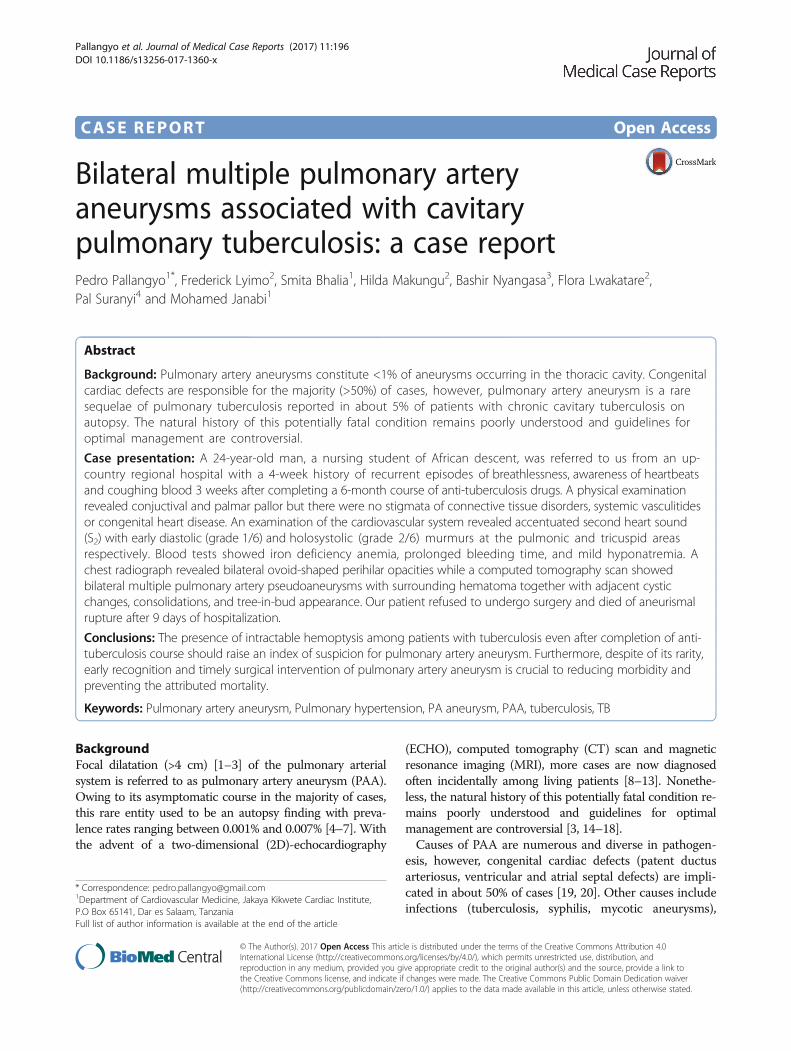
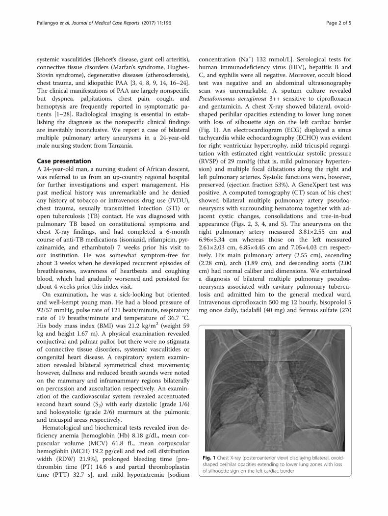
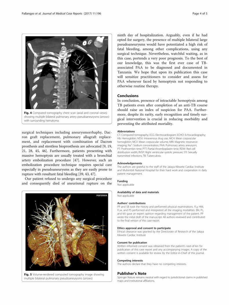
concentration (Na+) 132 mmol/L]. Serological tests forhuman immunodeficiency virus (HIV), hepatitis B andC, and syphilis were all negative. Moreover, occult bloodtest was negative and an abdominal ultrasonographyscan was unremarkable. A sputum culture revealedPseudomonas aeruginosa 3++ sensitive to ciprofloxacinand gentamicin. A chest X-ray showed bilateral, ovoid-shaped perihilar opacities extending to lower lung zoneswith loss of silhouette sign on the left cardiac border(Fig. 1). An electrocardiogram (ECG) displayed a sinustachycardia while echocardiography (ECHO) was evidentfor right ventricular hypertrophy, mild tricuspid regurgi-tation with estimated right ventricular systolic pressure(RVSP) of 29 mmHg (that is, mild pulmonary hyperten-sion) and multiple focal dilatations along the right andleft pulmonary arteries. Systolic functions were, however,preserved (ejection fraction 53%). A GeneXpert test waspositive. A computed tomography (CT) scan of his chestshowed bilateral multiple pulmonary artery pseudoa-neurysms with surrounding hematoma together with ad-jacent cystic changes, consolidations and tree-in-budappearance (Figs. 2, 3, 4, and 5). The aneurysms on theright pulmonary artery measured 3.81×2.55 cm and6.96×5.34 cm whereas those on the left measured2.61×2.03 cm, 6.85×4.45 cm and 7.05×4.03 cm respect-ively. His main pulmonary artery (2.55 cm), ascending(2.28 cm), arch (1.89 cm), and descending aorta (2.00cm) had normal caliber and dimensions. We entertaineda diagnosis of bilateral multiple pulmonary pseudoa-neurysms associated with cavitary pulmonary tubercu-losis and admitted him to the general medical ward.Intravenous ciprofloxacin 500 mg 12 hourly, bisoprolol 5mg once daily, tadalafil (40 mg) and ferrous sulfate (270
Fig. 1 Chest X-ray (posteroanterior view) displaying bilateral, ovoid-shaped perihilar opacities extending to lower lung zones with lossof silhouette sign on the left cardiac border
Pallangyo et al. Journal of Medical Case Reports (2017) 11:196 Page 2 of 5

mg) plus folic acid (300 mcg) were initiated. Moreover,our patient was counseled regarding surgery and prog-nosis of his condition; however, he refused to undergoany surgical procedure. After 9 days of hospitalization,our patient died of aneurismal rupture. Due to religiousbeliefs, relatives of the deceased refused an autopsy andone was not done.
DiscussionPulmonary artery aneurysms constitute <1% of aneurysmsoccurring in the thoracic cavity [29]. The cause could beidiopathic but many medical conditions including con-genital heart defects, connective tissue disorders, systemicvasculitides, and infections are often associated with PAA[3, 4, 8, 9, 14, 16–24]. Clinical presentation is invariablynonspecific and roughly depends on the underlying eti-ology, location, and size of the aneurysm [9]. Owing to itslargely vague presentation [4–7], noninvasive imagingtechniques are crucial in reaching the diagnosis [8–13];
however, pulmonary angiography remains the gold stand-ard diagnostic modality [2, 8, 19, 21, 23, 30–32]. About20–60% of patients with PAA will die from aneurysm rup-ture while other serious complications including airwaycompression and intravascular thrombosis are not uncom-mon [9, 33–38].Hemoptysis in a tuberculosis (TB) setting is relatively
common, usually self-limiting or controlled by anti-TBdrugs. Nevertheless, presence of a massive or several ep-isodes of minor hemoptysis in TB is life-threatening,likely originating from the arterial system, and requireearly and aggressive intervention [39, 40]. Rasmussen’saneurysm is a rare sequalae of pulmonary TB resultingfrom gradual weakening of the pulmonary artery wallfrom the adjacent tubercular cavity leading to thinningand pseudoaneurysm formation [36–39]. It has been re-ported in approximately 5% of autopsy series involving pa-tients with chronic cavitary tuberculosis [39–44]. Despiteabsence of clear guidelines for treatment of PAA, several
Fig. 2 Computed tomography chest scan (coronal and axial views) showing bilateral pulmonary artery pseudoaneurysms with adjacent cysticchanges (arrows), consolidations and tree-in-bud appearance
Fig. 3 Computed tomography chest scan (axial and coronal views) showing multiple bilateral pulmonary artery pseudoaneurysms (arrows) withsurrounding hematoma
Pallangyo et al. Journal of Medical Case Reports (2017) 11:196 Page 3 of 5
surgical techniques including aneurysmorrhaphy, Dac-ron graft replacement, pulmonary allograft replace-ment, and replacement with combination of Dacronprosthesis and stentless bioprosthesis are advocated [9, 19,21, 28, 45, 46]. Furthermore, patients presenting withmassive hemoptysis are usually treated with a bronchialartery embolization procedure [47]. However, such anembolization procedure technique requires special careespecially in pseudoaneurysms as they are easily prone torupture with resultant fatal bleeding [39, 43, 47].Our patient refused to undergo any surgical procedure
and consequently died of aneurismal rupture on the
ninth day of hospitalization. Arguably, even if he hadopted for surgery, the presence of multiple bilateral largepseudoaneurysms would have potentiated a high risk offatal bleeding, among other complications, using anysurgical technique. Nevertheless, watchful waiting, as inthis case, portends a very poor prognosis. To the best ofour knowledge, this was the first ever case of TB-associated PAA to be diagnosed and documented inTanzania. We hope that upon its publication this casewill sensitize practitioners to consider and assess forPAA whenever faced by hemoptysis not responding tootherwise routine therapy.
ConclusionsIn conclusion, presence of intractable hemoptysis amongTB patients even after completion of an anti-TB courseshould raise an index of suspicion for PAA. Further-more, despite its rarity, early recognition and timely sur-gical intervention is crucial in reducing morbidity andpreventing the attributed mortality.
AbbreviationsCT: Computed tomography; ECG: Electrocardiogram; ECHO: Echocardiography;Hb: Hemoglobin; IVDU: Intravenous drug use; MCH: Mean corpuscularhemoglobin; MCV: Mean corpuscular volume; MRI: Magnetic resonanceimaging; Na+: Sodium concentration; PAA: Pulmonary artery aneurysm;PT: Prothrombin time; PTT: Partial thromboplastin time; RDW: Red celldistribution width; RVSP: Right ventricular systolic pressure; STI: Sexuallytransmitted infections; TB: Tuberculosis
AcknowledgementsThe authors are grateful to the staff of the Jakaya Kikwete Cardiac Instituteand Muhimbili National Hospital for their hard work and cooperation in dailypatient management.
FundingNot applicable
Availability of data and materialsNot applicable
Authors’ contributionsPP and SB took the history and performed physical examinations. FLy HM,FLw, and PS performed and interpreted all the imaging modalities. BN, PL,and MJ gave an expert opinion regarding management of the patient. PPwrote the initial draft of the manuscript. All authors reviewed and contributedto the final version of this case report.
Ethics approval and consent to participateEthical clearance was granted by the Directorate of Research of the JakayaKikwete Cardiac Institute
Consent for publicationWritten informed consent was obtained from the patient’s next-of-kin forpublication of this case report and any accompanying images. A copy of thewritten consent is available for review by the Editor-in-Chief of this journal.
Competing interestsThe authors declare that they have no competing interests.
Publisher’s NoteSpringer Nature remains neutral with regard to jurisdictional claims in publishedmaps and institutional affiliations.
Fig. 4 Computed tomography chest scan (axial and coronal views)showing multiple bilateral pulmonary artery pseudoaneurysms (arrows)with surrounding hematoma
Fig. 5 Volume-rendered computed tomography image showingmultiple bilateral pulmonary pseudoaneurysms (arrows)
Pallangyo et al. Journal of Medical Case Reports (2017) 11:196 Page 4 of 5
Author details1Department of Cardiovascular Medicine, Jakaya Kikwete Cardiac Institute,P.O Box 65141, Dar es Salaam, Tanzania. 2Department of Radiology,Muhimbili National Hospital, P.O Box 65000, Dar es Salaam, Tanzania.3Department of Cardiovascular Surgery, Jakaya Kikwete Cardiac Institute, P.OBox 65141, Dar es Salaam, Tanzania. 4Department of Radiology andRadiological Science, Medical University of South Carolina, 25 CourteneyDrive, MSC 226, Charleston, SC 29425, USA.
Received: 15 May 2017 Accepted: 22 June 2017
References1. Barbour DJ, Roberts WC. Aneurysm of the pulmonary trunk unassociated
with intracardiac or great vessel left-to-right shunting. Am J Cardiol. 1987;59:192–4.
2. Chetty KG, McGovern J, Mahutte CK. Hilar mass in a patient with chest pain.Chest. 1996;109(6):1643–4.
3. Araújo I, Escribano P, Lopez-Gude MJ, et al. Giant pulmonary arteryaneurysm in a patient with vasoreactive pulmonary hypertension: a casereport. BMC Cardiovasc Disord. 2011;11:64.
4. Serasli E, Antoniadou M, Steiropoulos P, et al. Low-pressure pulmonaryartery aneurysm presenting with pulmonary embolism: a case series. J MedCase Rep. 2011;5:163.
5. Seguchi M, Wada H, Sakakura K, et al. Idiopathic pulmonary artery aneurysm.Circulation. 2011;124(14):369–70.
6. Ting P, Jugdutt BI, Le Tan J. Large pulmonary artery aneurysm associatedwith Marfan syndrome. Int J Angiol. 2012;19(1):48–50.
7. Deterling Jr RA, Clagett T. Aneurysm of the pulmonary artery; review of theliterature and report of a case. Am Heart J. 1947;34:471–99.
8. Nguyen ET, Silva CIS, Seely JM, Chong S, Lee KS, Müller NL. Pulmonaryartery aneurysms and pseudoaneurysms in adults: findings at CT andradiography. AJR. 2007;188(2):126–34.
9. Sargur R, Murthy KAS. Pulmonary artery aneurysm - report of a case andreview of literature. J Cardiovasc Med Cardiol. 2015;2(1):29–31.
10. Wu WS. Images in cardiovascular medicine. Huge calcified pulmonaryarterial aneurysm. Circulation. 2003;107:2280–1.
11. Janssens F, Verswijvel G, Colla P, Smits J, Gubbelmans H, et al. Proximalpulmonary artery aneurysm. JBR-BTR. 2003;86:83–5.
12. Khalil A, Parrot A, Fartoukh M, et al. Images in cardiovascular medicine.Large pulmonary artery aneurysm rupture in Hughes-Stovin syndrome:multidetector computed tomography pattern and endovascular treatment.Circulation. 2006;114:380–1.
13. Cherwek H, Amundson S. Images in clinical medicine. Pulmonary-arteryaneurysm. N Engl J Med. 2003;348:e1.
14. Singh V, Khare R, Chandra S, Dwivedi SK. Giant pulmonary artery aneurysmin a patient with rheumatic mitral stenosis. Heart Views. 2014;15:89–92.
15. Mayoral-Campos V, de Benito-Arévalo JL, Varea-Sanz MA. Pulmonary arteryaneurysm. Arch Bronconeumol. 2013;49:551–2.
16. Baztarrica GP, Bevacqua F, Porcile R. Pulmonary artery aneurysm. Rev EspCardiol. 2010;63:240–1.
17. Vistarini N, Aubert S, Gandjbakhch I, Pavie A. Surgical treatment of apulmonary artery aneurysm. Eur J Cardiothorac Surg. 2007;31(6):1139–41.
18. Garcia A, Byrne JG, Bueno R, et al. Aneurysm of the main pulmonary artery.Ann Thorac Cardiovasc Surg. 2008;14:399–401.
19. Shih HH, Kang PL, Lin CY, Lin YH. Main pulmonary artery aneurysm. J ChinMed Assoc. 2007;70(10):453–5.
20. Shankarappa RK, Moorthy N, Chandrasekaran D, Nanjappa MC. Giantpulmonary artery aneurysm secondary to primary pulmonary hypertension.Tex Heart Ins J. 2010;37:244–5.
21. Fazlinejad A, Vojdanparast M, Esfehani RJ, et al. Giant idiopathic pulmonaryartery aneurysm: an interesting incidental finding. Case Rep Vasc Med. 2014;2014:251373.
22. Hammad AM, Al-Qahtani SM, Al-Zahrani MA. Huge pulmonary arteryaneurysm. Can Respir J. 2009;16(3):93–5.
23. Theodoropoulos P, Ziganshin BA, Tranquilli M, Elefteriades JA. Pulmonaryartery aneurysms: four case reports and literature review. Int J Angiol. 2013;22(3):143–8.
24. Chiu P, Irons M, van de Rijn M, et al. Giant pulmonary artery aneurysm in apatient with Marfan syndrome and pulmonary hypertension. Circulation.2016;133:1218–21.
25. Muthialu N, Raju V, Muthubaskaran V, et al. Idiopathic pulmonary arteryaneurysm with pulmonary regurgitation. Ann Thoracic Surg. 2010;90(6):2049–51.
26. van Rens MTM, Westermann CJJ, Postmus PE, Schramel FM. Untreatedidiopathic aneurysm of the pulmonary artery; long-term follow-up. RespirMed. 2000;94(4):404–5.
27. Arslan S, Kalkan ME, Gündoǧdu F, Kantarci M. Idiopathic pulmonary arteryaneurysm in a patient presenting with chest pain. Turk Kardiyoloji DernegiArsivi. 2009;37(4):253–5.
28. Agarwal S, Chowdhury UK, Saxena A, et al. Isolated idiopathic pulmonaryartery aneurysm. Asian Cardiovasc Thorac Ann. 2002;10(2):167–9.
29. Ritter CO, Weininger M, Machann M, et al. Non-invasive imaging in a rarecase of main pulmonary artery aneurysm. Respir Med. 2008;102(5):790–2.
30. Lopez-Candales A, Kleiger RE, Aleman-Gomez J, Kouchoukos NT, BotneyMD. Pulmonary artery aneurysm: review and case report. Clin Cardiol. 1995;18:738–40.
31. Bartter T, Irwin RS, Nash G. Aneurysms of the pulmonary arteries. Chest.1988;94:1065–75.
32. Nair KS, Cobanoglu AM. Idiopathic main pulmonary artery aneurysm. AnnThorac Surg. 2001;71:1688–90.
33. Ungaro R, Saab S, Almond CH, Kumar S. Solitary peripheral pulmonary arteryaneurysms. Pathogenesis and surgical treatment. J Thorac Cardiovasc Surg.1976;71(4):566–71.
34. Butto F, Lucas JRV, Edwards JE. Pulmonary arterial aneurysm. A pathologicstudy of five cases. Chest. 1987;91(2):237–41.
35. Sakuma M, Demachi J, Suzuki J, Nawata J, Takahashi T, Shirato K. Proximalpulmonary artery aneurysms in patients with pulmonary arteryhypertension: complicated cases. Intern Med. 2007;46(21):1789–93.
36. Hernández V, Ruiz-Cano MJ, Escribano P, Sánchez MA. Complications ofproximal pulmonary artery aneurysm in patients with severe pulmonaryarterial hypertension. Rev Esp Cardiol. 2010;63(5):617–8.
37. Kussman BD, Geva T, McGowan FX. Cardiovascular causes of airwaycompression. Paediatr Anesth. 2004;14:60–74.
38. Arena V, De Giorgio F, Abbate A, Cappelli A, De Mercurio D, Carbone A.Fatal pulmonary arterial dissection and sudden death as initial manifestationof primary hypertension case report. Cardiovasc Pathol. 2004;13:349–71.
39. Chatterjee K, Colaco B, Colaco C, et al. Rasmussen's aneurysm: a forgottenscourge. Respir Med Case Rep. 2015;16:74–6.
40. Keeling AN, Costello R, Lee MJ. Rasmussen's aneurysm: a forgotten entity?Cardiovasc Interv Radiol. 2008;31(1):196–200.
41. Santelli ED, Katz DS, Goldschmidt AM, Thomas HA. Embolization of multipleRasmussen aneurysms as a treatment of hemoptysis. Radiology. 1994;193:396–8.
42. Remy J, Smith M, Lemaitre L, Marache P, Fournier E. Treatment of massivehemoptysis by occlusion of a Rasmussen aneurysm. Am J Roentgenol. 1980;135:605–6.
43. Shih SY, Tsai IC, Chang YT, et al. Fatal haemoptysis caused by a rupturedRasmussen's aneurysm. Thorax. 2011;66(6):553–4.
44. Raviglione MC, O’Brien RJ. Tuberculosis. In: Kasper DL, Braunwald E, FauciAS, Hauser SL, Longo DL, Jameson L, editors. Harrison’s principles of internalmedicine. 16th ed. New York: McGraw Hill; 2005. p. 953–66.
45. Kuwaki K, Morishita K, Sato H, et al. Surgical repair of the pulmonary trunkaneurysm. Euro J Cardiothoracic Surg. 2000;18(5):535–9.
46. Casselman F, Deferm H, Peeters P, Vanermen H. Aneurysm of the leftpulmonary artery: surgical allograft repair. Ann Thorac Surg. 1995;60:1423–5.
47. Sapra R, Sharma G, Minz AK. Rasmussen's aneurysm: a rare and forgottencause of hemoptysis. Indian Heart J. 2015;67 Suppl 3:53–6.
Pallangyo et al. Journal of Medical Case Reports (2017) 11:196 Page 5 of 5