vestibular examination tools – mu pt 8390, fall 2012vestibular tests & measures: study guide...
TRANSCRIPT
Vestibular Examination Tools (and handouts) – MU PT 8390, Fall 2012 -- a subset of Instruments and Questionnaires available online: http://web.missouri.edu/~proste/tool/vest/ Performance based instruments:
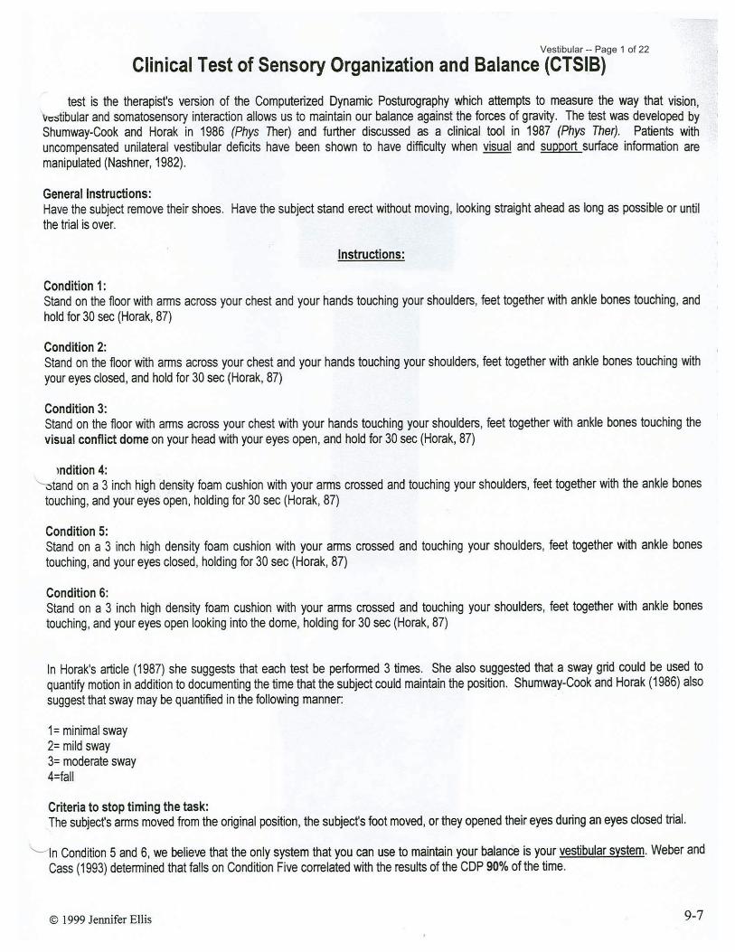
1. CTSIB - Clinical Test of Sensory Integration on Balance "Foam & Dome" 2. Fukuda Step Test 3. Motion Sensitivity Score (also known as the Motion Sensitivity Quotient, MSQ) 4. Dynamic Gait Index (DGI) … see Geriatric Exam Tool Kit 5. Functional Gait Assessment (FGA) … see Geriatric Exam Tool Kit
Questionnaires:
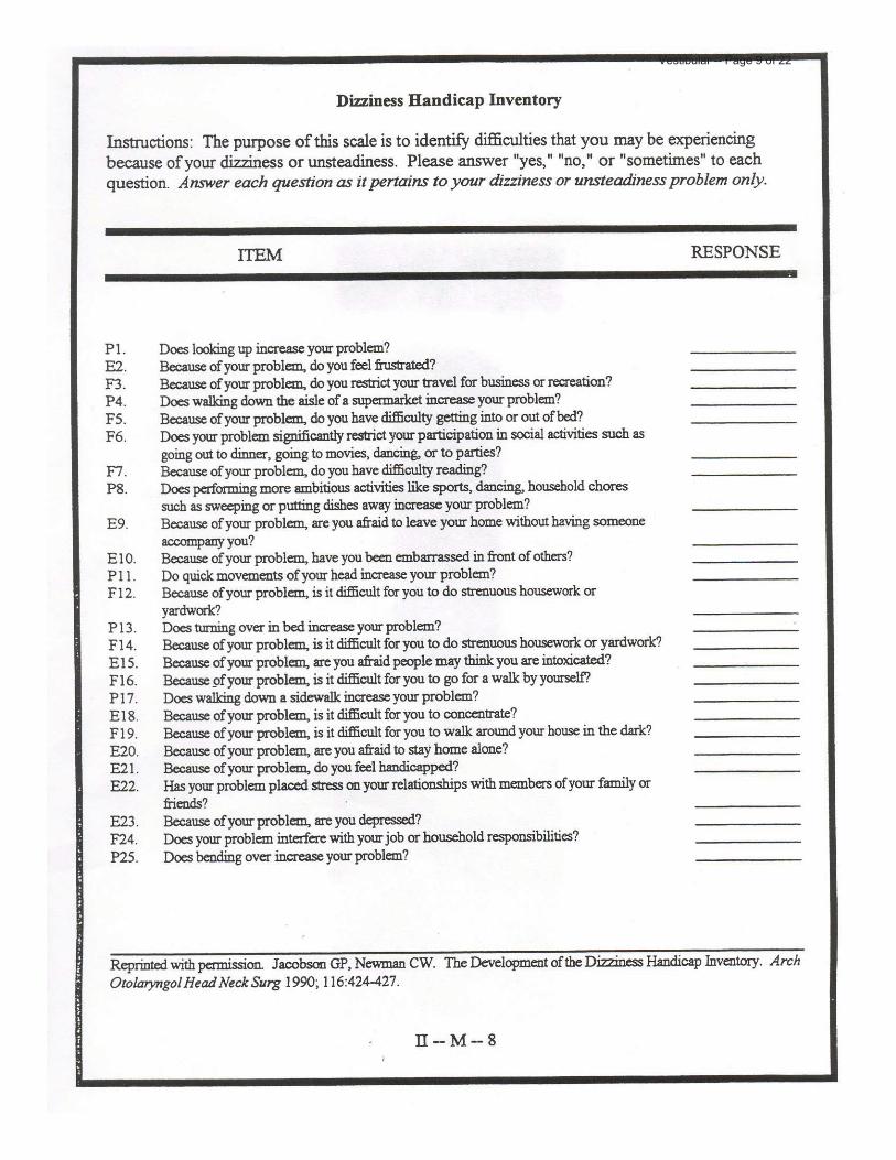
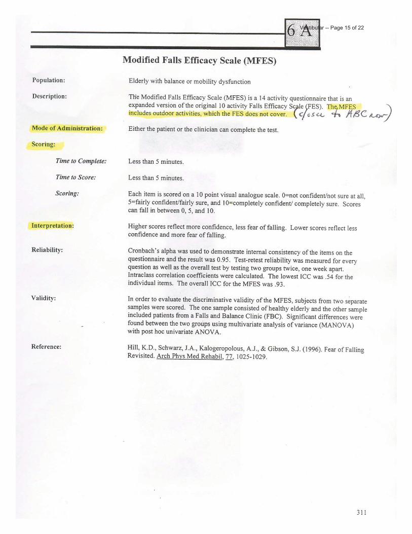
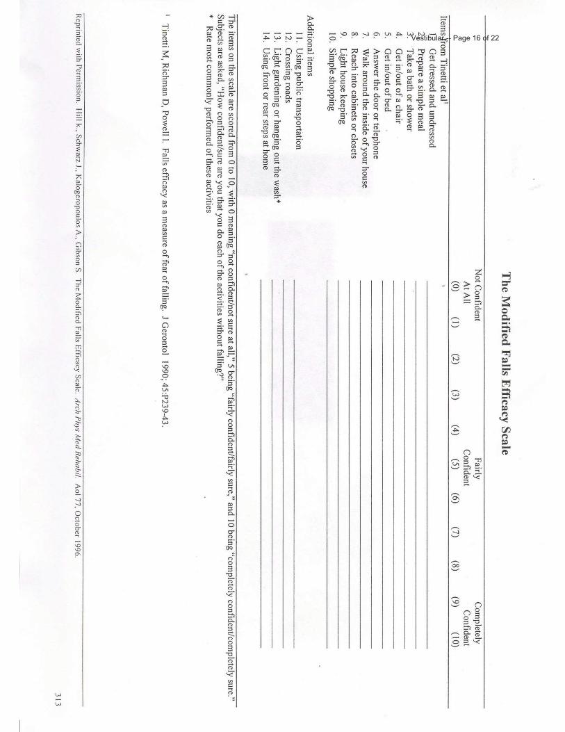
6. Activities specific Balance Confidence scale – ABC … see Geriatric Exam Tool Kit 7. Dizziness Handicap Inventory (DHI) 8. Geriatric Depression Scale 9. Modified Falls Efficacy Scale (MFES)
Handouts: (more to come during the vestibular unit)
10. Dizziness symptomology
11. BPPV Treatment Algorighm - Herdman
12. Intertpreting Nystagmus
13. Algorithm for differential diagnosis - Dunphy
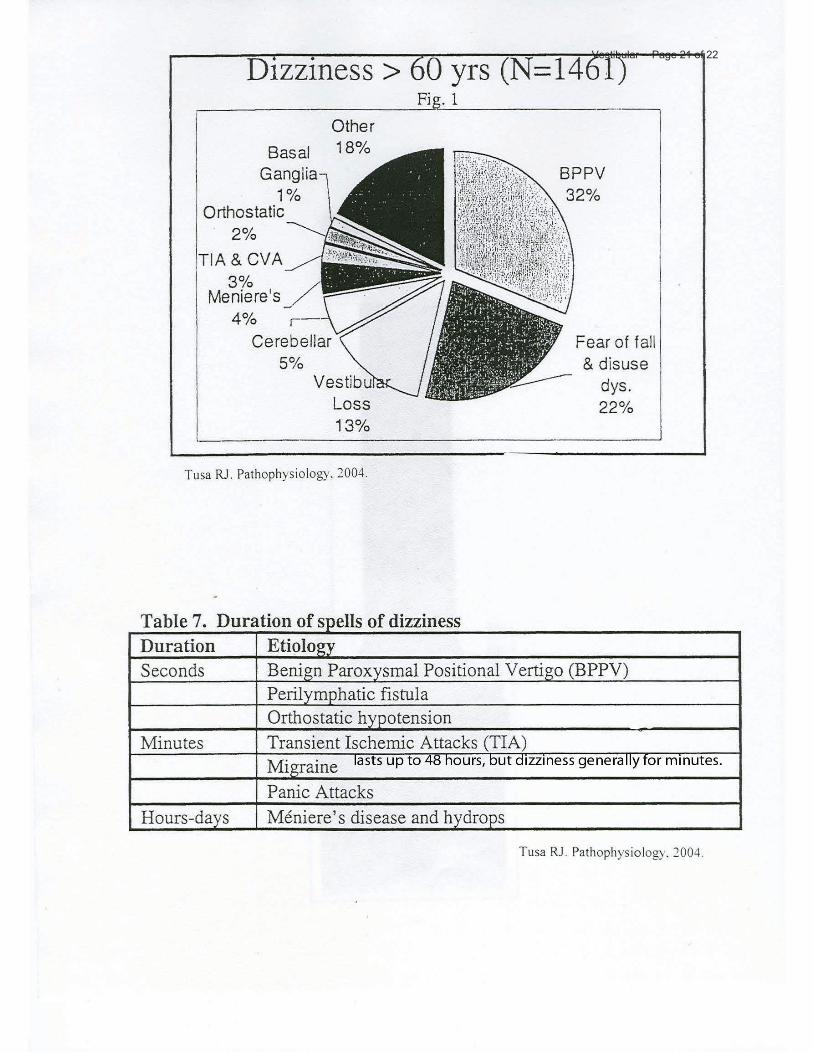
14. Epidemiology & duration of Vertigo
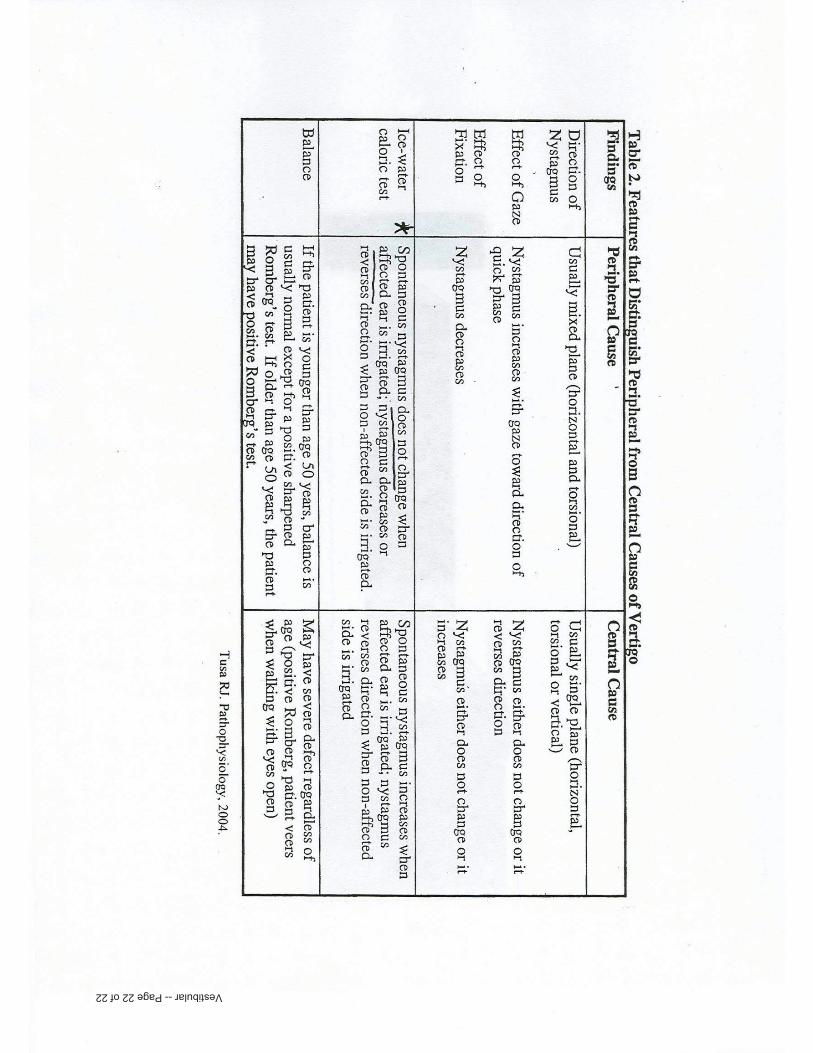
15. Central vs. Peripheral Lesion 16. Vestibular Tests & Measures Study Guide: how to perform them

Vestibular -- Page 1 of 22

Vestibular -- Page 2 of 22

Vestibular -- Page 3 of 22
Positive Fukuda: distance traveled is > 50 cm. (19.7 in.).Herdman, S.J. (2007). Vestibular Rehabilitation. Philadelphia: FA Davis, 3rd ed.

Vestibular -- Page 4 of 22
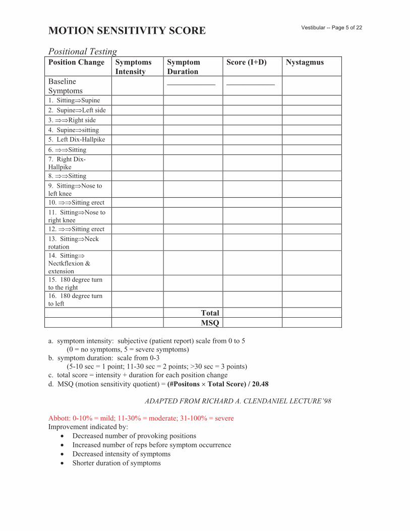
MOTION SENSITIVITY SCORE
Positional Testing Position Change Symptoms
IntensitySymptomDuration
Score (I+D) Nystagmus
BaselineSymptoms
____________ ____________
1. Sitting�Supine 2. Supine�Left side 3. ��Right side 4. Supine�sitting 5. Left Dix-Hallpike 6. ��Sitting 7. Right Dix-Hallpike
8. ��Sitting 9. Sitting�Nose to left knee
10. ��Sitting erect 11. Sitting�Nose to right knee
12. ��Sitting erect 13. Sitting�Neckrotation
14. Sitting�Nectkflexion & extension
15. 180 degree turn to the right
16. 180 degree turn to left
TotalMSQ
a. symptom intensity: subjective (patient report) scale from 0 to 5 (0 = no symptoms, 5 = severe symptoms) b. symptom duration: scale from 0-3 (5-10 sec = 1 point; 11-30 sec = 2 points; >30 sec = 3 points) c. total score = intensity + duration for each position change d. MSQ (motion sensitivity quotient) = (#Positons � Total Score) / 20.48
ADAPTED FROM RICHARD A. CLENDANIEL LECTURE’98
Abbott: 0-10% = mild; 11-30% = moderate; 31-100% = severeImprovement indicated by:
� Decreased number of provoking positions � Increased number of reps before symptom occurrence � Decreased intensity of symptoms � Shorter duration of symptoms
Vestibular -- Page 5 of 22

Vestibular -- Page 6 of 22

Vestibular -- Page 7 of 22

Vestibular -- Page 8 of 22
Vestibular -- Page 9 of 22

Vestibular -- Page 10 of 22

Vestibular -- Page 11 of 22

Vestibular -- Page 12 of 22

Vestibular -- Page 13 of 22

Vestibular -- Page 14 of 22
Vestibular -- Page 15 of 22
Vestibular -- Page 16 of 22

Dizziness Symptomology
Subjective complaint
Mechanism Etiology
“room spinning” (compensates by squinting or closing eyes)
Vertigo Vestibular Definition: Illusory sensation of motion of self or environment
BPPV (canalithiasis or cupulolithiasis) Unilateral Peripheral Hypofunction, ie asymmetry of tonic
firing (could be secondary to chronic Meniere’s) UVL: Unilateral Peripheral lesion: viral (labyrinthitis),
trauma, vascular, perilymph fistula Unilateral central lesion (to vestibular nuclei in Pons): CVA,
MS, CHI Very loud noise – Tullios Migraine headache Drugs
“going to faint … light headed” “I woke up on the floor.”
Syncope Vascular Metabolic
Othrostatic hypotension e.g. drug SE Hypoglycemia Vertebrobasilar Insufficiency Anemia (internal bleeding or B-12 deficiency) Cardiac Pump Failure, drop in cardiac output, valve stenosis Arrhythmias: A-Fib (may be benign); PVC (too frequent or
bigeminy, trigeminy, couplets, V-tach …) Anxiety attack, hyperventilation Hypothroid condition Severe HTN – brain attack Carotid sinus hypersensitivity (palpation triggers Valsalva
with hypotensive episode)
“lost my balance … no reason”
Dysequilibrium
BVL (ototoxicity, bilateral infections, age-related degeneration, meningitis)
Chronic UVL Ototoxicity (gentamicin, an antibiotic destroys hair cells) Central lesion to vestibular cortex (parietal lobe) Surgery for acoustic neuroma that damages vestib n., or a
Vestibular neurectomy as treatment for severe Meniere’s Peripheral Neuropathy (diabetic, alcoholic, pernicious
anemia) with insensate feet (also impaired skin, joint, and muscle propioceptors)
Impaired neuromuscular control / reflexes, righting reactions, reaction time, nerve conduction velocity (aging)
LE weakness, (esp with decr DF ROM – loss of ankle strategy)
Cerebellar lesion, ataxia
Oscilopsia: blurring with head movement Adapted from: O’Sullivan, S.B. and Schmitz T.J. (Eds.). (2007). Physical rehabilitation: assessment and treatment (5th ed.). Philadelphia: F. A. Davis Company. p.1004.


Vestibular -- Page 19 of 22

Vestibular -- Page 20 of 22
Vestibular -- Page 21 of 22
Vestibular -- Page 22 of 22
Vestib
ular T
ests & M
easures: S
tud
y Gu
ide
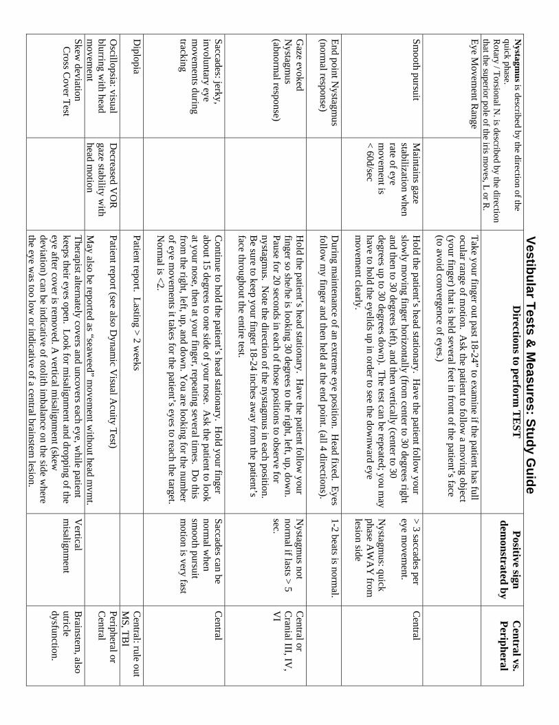
Nystagm
us is described by the direction of the
quick phase. R
otary / Torsional N
. is described by the direction that the superior pole of the iris m
oves, L or R
.
Direction
s to perform
TE
ST
P
ositive sign
dem
onstrated
by
Cen
tral vs. P
eriph
eral
Eye M
ovement R
ange
Take your finger out past 18-24” to exam
ine if the patient has full ocular range of m
otion. Ask the patient to follow
a moving object
(your finger) that is held several feet in front of the patient’s face (to avoid convergence of eyes.)
Sm
ooth pursuit
Maintains gaze
stabilization when
rate of eye m
ovement is
< 60d/sec
Hold the patient’s head stationary. H
ave the patient follow your
slowly m
oving finger horizontally (from center to 30 degrees right
and then to 30 degrees left), and then vertically (center to 30 degrees up to 30 degrees dow
n). The test can be repeated; you m
ay have to hold the eyelids up in order to see the dow
nward eye
movem
ent clearly.
> 3 saccades per
eye movem
ent. N
ystagmus: quick
phase AW
AY
from
lesion side
Central
End point N
ystagmus
(normal response)
D
uring maintenance of an extrem
e eye position. Head fixed. E
yes follow
my finger and then held at the end point. (all 4 directions).
1-2 beats is normal.
Gaze evoked
Nystagm
us (abnorm
al response)
H
old the patient’s head stationary. Have the patient follow
your finger so she/he is looking 30 degrees to the right, left, up, dow
n. P
ause for 20 seconds in each of those positions to observe for nystagm
us. Note the direction of the nystagm
us in each position. B
e sure to keep your finger 18-24 inches away from
the patient’s face throughout the entire test.
Nystagm
us not norm
al if lasts > 5
sec.
Central or
Cranial III, IV
, V
I
Saccades: jerky,
involuntary eye m
ovements during
tracking
C
ontinue to hold the patient’s head stationary. Hold your finger
about 15 degrees to one side of your nose. Ask the patient to look
at your nose, then at your finger, repeating several times. D
o this from
the right, left, up, and down. Y
ou are looking for the number
of eye movem
ents it takes for the patient’s eyes to reach the target. N
ormal is <
2.
Saccades can be
normal w
hen sm
ooth pursuit m
otion is very fast
Central
Diplopia
P
atient report. Lasting >
2 weeks
C
entral: rule out M
S, T
BI
Oscillopsia: visual
blurring with head
movem
ent
Decreased V
OR
gaze stability w
ith head m
otion
Patient report (see also D
ynamic V
isual Acuity T
est) M
ay also be reported as “seaweed” m
ovement w
ithout head mvm
t.
P
eripheral or C
entral
Skew
deviation C
ross Cover T
est
Therapist alternately covers and uncovers each eye, w
hile patient keeps their eyes open. L
ook for misalignm
ent and dropping of the eye after cover is rem
oved. A vertical m
isalignment (skew
deviation) can be indicative of otolith im
balance on the side where
the eye was too low
or indicative of a central brainstem lesion.
Vertical
misalignm
ent B
rainstem, also
utricle dysfunction.
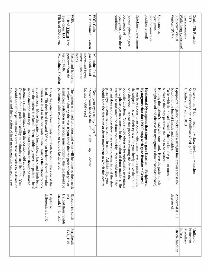
O
cular Tilt R
eaction O
TR
(can accom
pany W
allenberg syndrome)
O
bservation: Triad =
head tilt + skew
deviation + torsion
See illustration on: O
’Sullivan 4th ed. p.832
O’S
ullivan 5th ed. p.1013
U
nilateral brainstem
M
edullary infarct
Subjective V
isual V
ertical (SV
V)
E
quipment: 5 gallon bucket w
ith a straight line drawn across the
bottom of the bucket (inside and outside).
Task: w
ith their head “inside” the bucket, the patient turns the bucket so that they perceive the line to be vertical.
Abnorm
al if > 2
degrees off U
tricle function
Spontaneous
nystagmus
(not movem
ent or position related)
H
olding the patient’s head with one hand. H
ave the patient look straight ahead and observe for nystagm
us (slow phase/fast phase).
Horizon
tal Nystagm
us th
at stops w
gaze fixation =
Perip
heral
Nystagm
us th
at does N
OT
stop w
ith gaze fixation
= C
entral
Optokinetic nystagm
us (norm
al physiological occurrence of nystagm
us under these conditions)
If you have access to an optokinetic drum
, have the patient follow
the striped lines with their eyes w
hile you slowly m
ove the drum in
one direction. Repeat this procedure rotating the drum
in the opposite direction. Y
ou should observe for optokinetic nystagmus
(slow phase eye m
ovements in the direction of drum
rotation). Be
careful to not rotate the drum too quickly. Y
ou should note if the patient does not produce slow
phase eye movem
ents or if the slow
phase eye movem
ents are saccadic in nature. Additionally, you
should note the direction of drum m
ovement in w
hich this occurs.
VO
R G
ain
1. Maintained F
ixation M
aintains fixed gaze w
ith head m
ovement (eyes
move opposite to
head).
“Keep your eyes on m
y finger.” “M
ove your head to the left … right …
up … dow
n” (at rate >
60d / sec)
VO
R
2. Head T
hru
st Test.
(eyes open: EO
) T
ilt head 30d down.
Faster and harder to
perform than the
test of VO
R
Maintained F
ixation
The patient w
ill need to understand what w
ill be done so their neck is relaxed during the test. If you noted that the patient had pain or significant restriction in cervical spine m
obility, this test should be perform
ed with extrem
e caution or should be deferred. G
rasp the patient’s head firmly w
ith both hands on the side of their head. T
ilt their head forward 30 so that horizontal sem
i-circular canals are level in the horizontal plane. Instruct the patient to look at your nose. M
ove the patient’s head slowly back and forth being
sure the patient is relaxed. Then, suddenly m
ove the patient’s head in one direction and stop. T
he head movem
ent should be moved
through a small am
plitude with the position held at the end.
Observe for the patient’s ability to m
aintain visual fixation. You
should note if the patient makes corrective saccades to re-fixate on
your nose and the direction of head movem
ent that caused the re-
Saccade (to catch
up) L
sided thrust yields saccade? =
L lesion
Helpful to
differentiate L / R
Peripheral:
UV
L, B
VL

fixation saccades, e.g. if a thrust to the L yields a saccade to re-
fixate on your nose a Left UV
L is indicated. N
ote: If you are uncomfortable m
oving the person’s head from
center to an eccentric position, try moving the person’s head from
an eccentric position to center
VO
R
3. Head S
hakin
g Induced N
ystagmus.
(eyes closed – EC
) T
ilt head 30d down.
E
yes are closed and with 30
º neck flexion (horizontal SC
C
position). I shake their head vigorously (2 Hz) L
&R
for 20 cycles. S
top and then they open their eyes (best viewed w
ith frenzels).
Horiz. N
ystagmus =
Peripheral U
VL
V
ertical Nystagm
us = C
entral
VO
R
4. VO
R C
ancellation T
ilt head 30d down.
Cerebellum
has to inhibit the V
OR
Gain
during VO
R
Cancellation
T
ilt their head forward 30 S
itting with their arm
s extended in front of them
, thumbs up (in “shooters position”). T
hey maintain gaze
on thumbs w
hile twisting / rotating their trunk and arm
s as a unit to the left and right. T
heir head moves w
ith target (thumbs),
canceling VO
R G
ain A
lternate: Grasp the patient’s head firm
ly with both hands on the
side of their head. Tilt their head forw
ard 30 while you m
ove in the sam
e direction that you move the patient’s head w
ith your face rem
aining directly in front of the patient’s face.
Saccades,
Nystagm
us C
entral: C
erebellar
VO
R
5. Dynam
ic Visual
Acuity T
est – DV
A
Tilt head 30d dow
n.
Passive T
est H
ave the patient wear their glasses if they need distance correction.
Depending on the type of acuity chart being utilized, have the
patient sit the appropriate distance from the chart. (T
he ET
DR
S
charts are designed to be viewed from
a distance of 4 meters to
provide Snellen equivalent acuity ratios or LogM
AR
values as noted on the chart). H
ave the patient read to the lowest line that
they can until they cannot correctly identify all the letters on a given line. N
ote the line where this occurs and/or the num
ber of optotypes the patient incorrectly identifies. N
ow, standing behind the patient, grasp the patient’s head firm
ly w
ith both hands on the side of their head, tilt their head forward
30 so that horizontal semi-circular canals are level in the
horizontal plane. While m
oving their head side to side at a frequency of 2 H
z (2 complete side to side cycles per second – use
metronom
e if available) have the patient read to the lowest line that
they can until they can not correctly identify all the letters on a given line. N
ote the line where this occurs and/or the num
ber of optotypes the patient incorrectly identifies. K
eep the range of m
otion of the head movem
ents small so as to not restrict the visual
field, which m
ay occur with patients w
ho wear glasses.
If “lose” >2 lines compared to static = oscillopsia.
If lose >3 lines = Vestibular hypofunction.
H
oriz. SC
C

Po
sition
al Man
euvers
1. H
allpike-Dix T
est (test unaffected side first, if obvious from
history)
Test of P
osterior and A
nterior S
CC
+
Post. S
CC
sign will
be Upbeating (cranial)
nystagmus (63%
) +
Ant. S
CC
sign will
be Dow
nbeating (caudal) nystagm
us (12%
)
Criteria for positive H
PD
sign: (example below
is for R side lesion)
1. torsional/linear-rotary nystagm
us; reproduced by provocative positioning w
ith affected R ear dow
n 2.
brief latency of 5-15 seconds before the start of nystagm
us. 3.
nystagmus of b
rief duration, (toward the lesion
i.e. R torsion)
4. reversal of nystagm
us direction on return to upright position (aw
ay from lesion
i.e. L torsion)
5. response dim
inishes with repetition of m
aneuver (fatigability)
Vertigo,
Nystagm
us:
<
60s C
analithiasis
> 60s:
Cupulolithiasis
(fatigues)
Persistent:
possibly Central
Peripheral:
misplaced or
adhered otoconia
2. Roll T
est (S
ee O’S
ullivan 5th ed. p.1010)
Test of H
orizontal
SC
C (1%
) W
ill be positive to both sides, with one side being w
orse.
Geotropic N
ystagmus =
Canalithiasis
Horizontal C
RT
Ageotropic N
ystamus =
Cupulolithiasis
Brandt D
aroff
P
eripheral: m
isplaced or adhered otoconia
3. Vertebral A
rt. Test
M
aneuver: Sit w
ith knees on elbows and chin in hand. L
ook up to the (right) for 30 seconds.
M
aneuver: Sitting w
ith (passive) cervical extension and rotation, holding 30 sec. (M
agee p.154)
Vertigo, nystagm
us, headache, visual disturbance central signs.
Fu
nctio
nal T
ests M
otion Sensitivity
In
strum
ents:
Motion Sensitivity Score (V
estibular System
Evaluation &
Training): rolling, sit to stand, etc. (16 item
s), with
vertigo rated for duration and intensity. B
alance & M
obility
Instru
men
ts:
Functional R
each, Multidim
ensional Reach
B
erg Balance
T
inetti Balance &
Gait
T
UG
, and the Five-T
imes Sit to Stand
P
referred Gait Speed
D
ynamic G
ait Index: 4-item &
Functional G
ait Assessm
ent
Fukuda
P
erturbation Tests (hips, sternum
)
Clinical T
est of Sensory Integration and Balance (C
TSIB
)
Qu
estionn
aires:
Dizziness H
andicap Inventory
Modified F
alls Efficacy Scale
A
ctivities-specific Balance
Confidence (A
BC
) Scale
Physical A
ctivity Scale for the E
lderly (PA
SE)
C
ognition: o
M
ini MentalState E
xam
o
The B
lessed Orientation-
Mem
ory-Concentration
(BO
MC
) Test
o
Geriatric D
epression Scale P
ortions adapted from H
erdman S
J. Vestibular T
esting & R
ehabilitation Com
petency Course, N
otes, Em
ory University &
AP
TA
. March, 2004.
Abbott C
., Prost E
., Aug. 2010.