verapamil, diltiazem and nifedipine interactions with calmodulin stimulated (ca2+ + mg2+)-atpase
TRANSCRIPT
Biochemical Pharmacology, Vol. 37. No. 5, pp. 917-920, 1988. OOMi-2952/88 $3.00 + 0.W Printed in Great Britain. @ 1988. Pergamon Journals Ltd.
VERAPAMIL, DILTIAZEM AND NIFEDIPINE INTERACTIONS WITH CALMODULIN STIMULATED
(Caz+ + Mg2+)-ATPase*
HENRY C. KIM and BEAT U. RAESS~
Department of Pharmacology, Indiana University School of Medicine, Evansville Center for Medical Education, Evansville, IN 47732, U.S.A.
(Receiued 19 May 1987; accepted 13 July 1987)
Abstract-The functional interactions of the three prototype Ca*+ antagonists, verapamil, diltiaxem and nifedipine, were examined in relation to the cahnodulin regulated plasma membrane Ca*+ pump ATPase. For this we used low ionic strength derived, calmodulin depleted, human red cell ghost membranes. Exogenously added calmodulin activated basal (Ca2+ + MgZ+)-ATPase in a concentration- dependent manner. Half-maximal activation by 6 nM calmodulin was antagonized by lo-’ M verapamil and low3 M diltiaxem 25.1 and 12.1% respectively. The inhibition appeared to be specific for calmodulin activation since basal activity was not affected by these agents. Nifedipine had no effects on basal or calmodulin stimulated (Ca*+ + Mg2+)-ATPase activity. Unlike dihydropyridine modulation of verapamil and diltiaxem binding at high affinity channel sites, nifedipine in this system did not alter the inhibitory responses of verapamil and diltiaxem. The calmodulin directed antagonism of the two drugs was shown to be strictly additive over a full range of calmodulin concentrations and appeared to change predominantly the V_ and, to a lesser degree, the affinity of calmodulin for the (Ca2+ + Mg2+)-ATPase. It is concluded that this model system provides evidence for additional functional discrepancies among the various classes of Ca*+ antagonists.
Human red cells extrude Ca2+ against a large con- centration gradient. This adenosine-triphosphate (ATP) dependent calcium pump mechanism is expressed biochemically as a (Ca2+ + Mg2+)- ATPase activity [l]. The enzyme, which is located in the plasma membrane, plays a vital role in the regulation of intracellular Ca2+ not only in red cells, but most likely in all animal cells. In addition to calcium, the ubiquitous protein calmodulin regulates both plasma membrane (Ca2+ + Mg2+)-ATPase and Ca2+ transport. Various types of drugs that bind to calmodulin, the enzyme, or both have been shown to alter these processes. Among these drugs are certain Ca2+ antagonists that, besides acting on the Ca2+ influx channels, have been shown to inhibit calmodulin-dependent functions including plasma membrane (Ca2+ + Mg2+)-ATPase activity [2-S].
The binding of Ca2+ antagonists to various sites involved in the regulation of intracellular Ca2+ makes it plausible that certain combinations of anta onists
s may interact to produce opposing or greate than additive effects. The idea that the pharmacological effects of structurally dissimilar Ca2+ antagonists are mediated by different sites of action at the voltage- operated influx channel is supported by the findings that verapamil binds allosterically to inhibit [3H]nitrendipine in rat cerebral cortex and heart, whereas diltiazem, which is chemically and struc- turally diverse from the dihydropyridines and ver-
* This work was supported, in part, by a grant from the American Heart Association, Indiana Affiliate and NIH Training Grant T35HLW584.
t Corresponding author: Beat U. Raess, Ph.D., Depart- ment of Pharmacology, Indiana University School of Medi- cine, P.O. Box 3287, Evansville, IN 47732.
apamil, appears to enhance [3H]nitrendipine binding [6,7]. Since it is not known whether or not similar interactions exist at secondary binding sites, such as the plasma membrane (Ca2+ + Mg2+)-ATPase, we designed this study to examine the effects of three structurally diverse Ca2+ antagonists and their func- tional interactions specifically on factors regulating the basal and calmodulin stimulated plasma mem- brane (Ca2+ + Mg2+)-ATPase activity in the human red blood cell.
EXPERIMENTAL PROCEDURES
Materials. Outdated (41- to 66-day-old) packed human erythrocytes were obtained from the local American Red Cross blood bank facility. Verapamil, diltiazem, and nifedipine were supplied by Knoll Pharmaceuticals (Whippany, NJ), Marion Lab- oratories, Inc. (Kansas City, MO), and Pfizer, Inc. (New York, NY) respectively. Crystalline disodium adenosine-S’triphosphate salt was obtained from Boehringer Mannheim GmbH, West Germany. The remaining reagents, including human erythrocyte calmodulin, were purchased through the Sigma Chemical Co. (St. Louis, MO).
Membranepreparation. Erythrocytes were washed three times, with 4 vol. of ice-cold isotonic NaCl (154 mM), and after each centrifugation (twice at 1100 gill,, and once at 3000 g,, for 5 min) the buffy coat was aspirated. Washed cells were hemolyzed in 14 vol. of an ice-cold 20 mM imidazole, 0.1 mM ethyleneglycolbis (amino - ethylether) tetra - acetate (EGTA) solution (pH 7.4) and then centrifuged for 20 min at 39,200 g,,,. After the supematant fraction was discarded, the membrane pellet was washed three times. The membranes were washed two more
917
918 H. C. KIM and B. U. RAESS
times in 12 vol. of ice-cold 20 mM imidazole, pH 7.4, and centrifuged for 20 min at 39,200 g,,. A final wash with 8 vol. of ice-cold 40 mM histidine-imidaz- ole, pH 7.1, was performed at the above specified conditions. The rinsed membrane pellets were recon- stituted with 40 mM histidine-imidazole to yield a protein concentration of 6 to 8.5 mg/ml. Membranes were stored at 4” for subsequent addition to the ATPase assay at a concentration of 0.2 mg/ml. Mem- brane proteins were determined by a modification [8] of the method of Lowry et al. [9].
ATPase activity msay. The standard ATPase assay medium contained the following: 18 mM histidine- imidazole buffer at pH 7.1, 3 mM MgClz, 15 mM KCl,80mMNaC1,0.1mMEGTA,0.1mMouabain, and 0.2 mM CaClr. Calmodulin concentrations from 0.9 to 36 nM were prepared from lyophilized human red cell derived calmodulin. Drug concentrations from 10e5 to 10e3 M for bothverapamil and diltiazem were prepared by using distilled deionized water. In the case of nifedipine, ethanol was used to achieve concentrations of 10e7 to lop5 M. Ethanol (0.25%) solvent control experiments were without effect on the ATPase. Because of the photo-sensitive decom- position of nifedipine in solution, experiments with this drug were conducted under conditions of mini- mal light exposure. ATPase reactions were initiated by the addition of 1.0 mM ATP. Assay tubes were incubated in a shaker bath at 37”. Reactions were stopped after 60 min by adding 2% sodium dodecyl sulfate. Inorganic phosphate was measured by a Technicon Autoanalyzer using a modified Fiske- Subbarow method [8]. (Mg*+)-ATPase activities were measured in the absence of calcium in the medium. The basal (Ca*+ + Mg*+)-ATPase activi- ties depended on the presence of 19pM free Ca*+ (confirmed by an ion-selective electrode), and cal- modulin stimulated (Ca*+ + Mg*+)-ATPase activi- ties were defined from the addition of exogenous calmodulin.
Calculutiom. Statistical treatment of the data con- sisted of Student’s t-test and multiple regression analysis as tests of significance in the interaction experiments [lo]. All data points were generated
from at least two independent experiments done in duplicate. Error bars were omitted when smaller than the symbols and/or for clarity; however, for every set of measurements, the standard error of the mean was never greater than 55%.
RESULTS
Experiments were conducted in two phases. The initial phase of experiments was designed to test combinations of the three compounds for possible drug interactions. For the second group of experi- ments, calmodulin response curves were examined to clarify the exact nature of the drug interaction, if any.
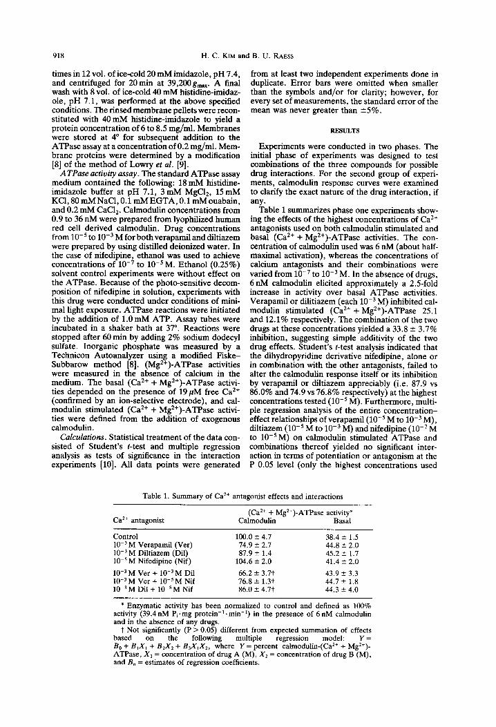
Table 1 summarizes phase one experiments show- ing the effects of the highest concentrations of Ca*+ antagonists used on both calmodulin stimulated and basal (Ca*+ + Mg*+)-ATPase activities. The con- centration of calmodulin used was 6 nM (about half- maximal activation), whereas the concentrations of calcium antagonists and their combinations were varied from 10e7 to 10m3 M. In the absence of drugs, 6 nM calmodulin elicited approximately a 2.5fold increase in activity over basal ATPase activities. Verapamil or dilitiazem (each 10m3 M) inhibited cal- modulin stimulated (Ca*+ + Mg*+)-ATPase 25.1 and 12.1% respectively. The combination of the two drugs at these concentrations yielded a 33.8 2 3.7% inhibition, suggesting simple additivity of the two drug effects. Student’s r-test analysis indicated that the dihydropyridine derivative nifedipine, alone or in combination with the other antagonists, failed to alter the calmodulin response itself or its inhibition by verapamil or diltiazem appreciably (i.e. 87.9 vs 86.0% and 74.9 vs 76.8% respectively) at the highest concentrations tested (10e5 M). Furthermore, multi- ple regression analysis of the entire concentration- effect relationships of verapamil ( 10e5 M to 10e3 M) , diltiazem (10T5 M to 10e3 M) and nifedipine (10e7 M to 10v5 M) on calmodulin stimulated ATPase and combinations thereof yielded no significant inter- action in terms of potentiation or antagonism at the P 0.05 level (only the highest concentrations used
Table 1. Summary of Car+ antagonist effects and interactions
Ca*+ antagonist (Ca*+ + Mg*+)-ATPase activity*
Calmodulin Basal
Control lo-’ M Verapamil (Ver) lo-’ M Diltiaxem (Dil) 10m5 M Nifedipine (Nif)
10e3 M Ver + lo-’ M Dil low3 M Ver + 10e5 M Nif lo-’ M Dii + 10e5 M Nif
100.0 2 4.7 38.4 2 1.5 74.9 k 2.7 44.8 +- 2.0 87.9 r 1.4 45.2 f 1.7
104.6 k 2.0 41.4 2 2.0
66.2 f 3.7t 43.9 _’ 3.3 76.8 f 1.3t 44.7 5 1.8 86.0 t 4.77 44.3 -+ 4.0
* Enzymatic activity has been normalized to control and defined as 100% activity (39.4 nM Pi. mg protein-‘. min-‘) in the presence of 6 nM calmodulin and in the absence of any drugs.
t Not significantly (P > 0.05) different from expected summation of effects based on the following multiple regression model: Y= B. + BrX, + B2X2 + BJx,X2, where Y =-percent &lmodulin-(Ca*+ + Mgr+)- ATPase, X1 = concentration of drug A (M), Xz = concentration of drug B (M), and E, = estimates of regression coefficients.
Calcium antagonist inhibition of (Ca*+ + Mg’+)-ATPase 919
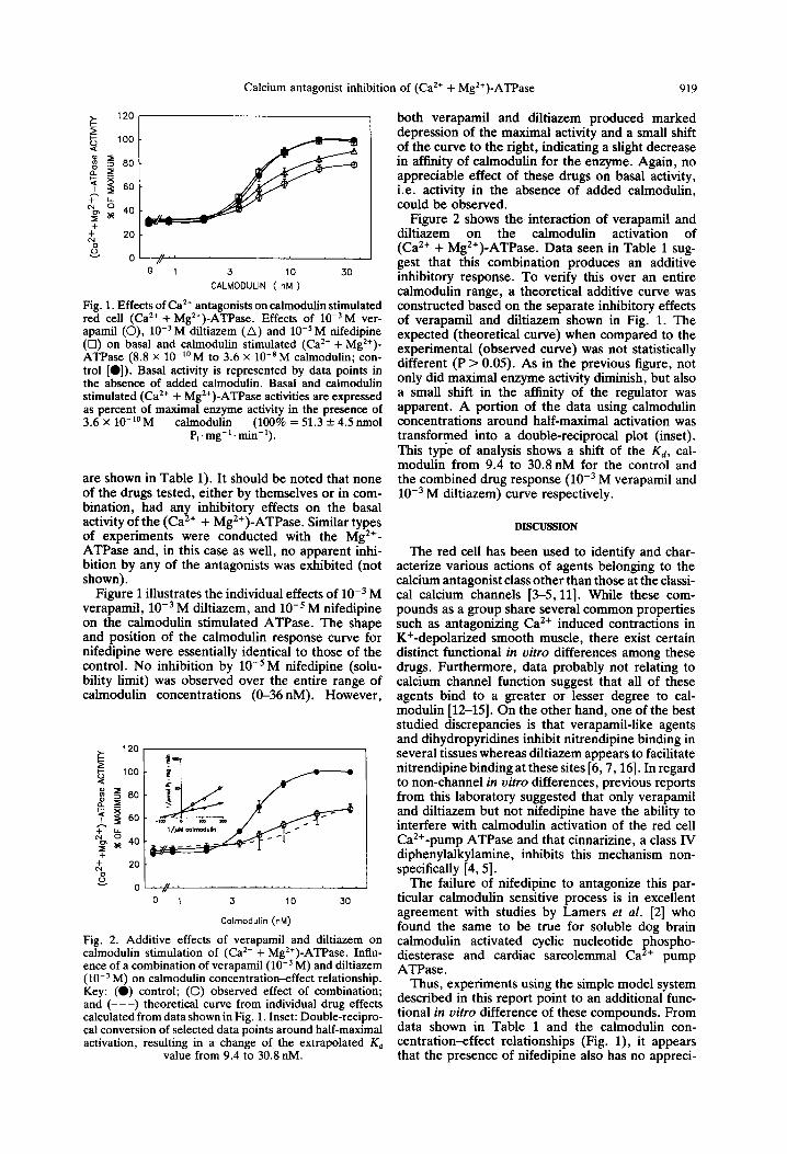
0’. , 3 10 30
CALMODULIN ( nM )
Fin. 1. Effects of Ca*+ antagonists on calmodulin stimulated red cell (Ca*+ + Mg*+)-AkPase. Effects of lo-‘M ver- aoamil (0). 10m3 M diltiazem t Al and 10e5 M nifedioine (b) on ‘b&l and calmodulin kmulated (Ca*+ + M&)- ATPase (8.8 x lo-“M to 3.6 x lo-*M calmodulin; con- trol [O]). Basal activity is represented by data points in the absence of added calmodulin. Basal and calmodulin stimulated (Ca2+ + Mg2+)-ATPase activities are expressed as percent of maximal enzyme activity in the presence of 3.6 x lo-‘OM calmodulin (100% = 51.3 + 4.5 nmol
Pi . mg- ’ . min- ‘) .
are shown in Table 1). It should be noted that none of the drugs tested, either by themselves or in com- bination, had any inhibitory effects on the basal activity of the (Ca*+ + Mg2+)-ATPase. Similar types of experiments were conducted with the Mg2+- ATPase and, in this case as well, no apparent inhi- bition by any of the antagonists was exhibited (not shown).
Figure 1 illustrates the individual effects of 10m3 M verapamil, 10m3 M diltiazem, and 10e5 M nifedipine on the calmodulin stimulated ATPase. The shape and position of the calmodulin response curve for nifedipine were essentially identical to those of the control. No inhibition by 10e5 M nifedipine (solu- bility limit) was observed over the entire range of calmodulin concentrations (O-36 nM). However,
-7Y 0 1 3 10 30
Calmodulin (M)
Fig. 2. Additive effects of verapamil and diltiazem on calmodulin stimulation of (Ca’+ + Mg2+)-ATPase. Influ- ence of a combination of verapamil (10e3 M) and diltiaxem (W3 M) on calmodulin concentration-effect relationship. Kev: (0) control; (0) observed effect of combination: and (L-1) theoretidal’curve from individual drug effects calculated from data shown in Fig. 1, Inset: Double-recipro- cal conversion of selected data points around half-maximal activation, resulting in a change of the extrapolated Kd
value from 9.4 to 30.8 nM.
both verapamil and diltiazem produced marked depression of the maximal activity and a small shift of the curve to the right, indicating a slight decrease in affinity of calmodulin for the enzyme. Again, no appreciable effect of these drugs on basal activity, i.e. activity in the absence of added calmodulin, could be observed.
Figure 2 shows the interaction of verapamil and diltiazem on the calmodulin activation of (Ca’+ + Mg*+)-ATPase. Data seen in Table 1 sug- gest that this combination produces an additive inhibitory response. To verify this over an entire calmodulin range, a theoretical additive curve was constructed based on the separate inhibitory effects of verapamil and diltiazem shown in Fig. 1. The expected (theoretical curve) when compared to the experimental (observed curve) was not statistically different (P > 0.05). As in the previous figure, not only did maximal enzyme activity diminish, but also a small shift in the affinity of the regulator was apparent. A portion of the data using calmodulin concentrations around half-maximal activation was transformed into a double-reciprocal plot (inset). This type of analysis shows a shift of the Kd, cal- modulin from 9.4 to 30.8nM for the control and the combined drug response (10e3 M verapamil and 10e3 M diltiazem) curve respectively.
DISCUSSION
The red cell has been used to identify and char- acterize various actions of agents belonging to the calcium antagonist class other than those at the classi- cal calcium channels [3-5,111. While these com- pounds as a group share several common properties such as antagonizing Ca*+ induced contractions in K+-depolarized smooth muscle, there exist certain distinct functional in vitro differences among these drugs. Furthermore, data probably not relating to calcium channel function suggest that all of these agents bind to a greater or lesser degree to cal- modulin [12-1.51. On the other hand, one of the best studied discrepancies is that verapamil-like agents and dihydropyridines inhibit nitrendipine binding in several tissues whereas diltiazem appears to facilitate nitrendipine binding at these sites [6,7,16]. In regard to non-channel in vitro differences, previous reports from this laboratory suggested that only verapamil and diltiazem but not nifedipine have the ability to interfere with calmodulin activation of the red cell Ca*+-pump ATPase and that cinnarizine, a class IV diphenylalkylamine, inhibits this mechanism non- specifically [4,5].
The failure of nifedipine to antagonize this par- ticular calmodulin sensitive process is in excellent agreement with studies by Lamers et al. [2] who found the same to be true for soluble dog brain calmodulin activated cyclic nucleotide diesterase and cardiac sarcolemmal
fhospho- Ca + pump
ATPase . Thus, experiments using the simple model system
described in this report point to an additional func- tional in vitro difference of these compounds. From data shown in Table 1 and the calmodulin con- centration-effect relationships (Fig. l), it appears that the presence of nifedipine also has no appreci-

920 H. C. KIM and B. U. RAESS
able effect on either verapamil or diltiazem inhibition In summary, while verapamil and diltiazem anta- of calmodulin stimulated (CaZ+ + Mg2+)-ATPase. gonized calmodulin activation, the dihydropyridine In other words, unlike with channel related binding sites, in this situation 10e5M nifedipine does not
representative nifedipine, either alone or in com- bination with class I and III representatives, failed to
appear to interfere with verapamil or diltiazem action affect (Ca2+ + Mg2+)-ATPase. Thus, the apparent in a functional manner. Rather than the allosteric- modulation of calcium antagonist binding as seen at type modulation of channel binding sites, our results channels in other tissues is not applicable to this suggest that the inhibition of calmodulin stimulated secondary, low affinity target. This is another ATPase by verapamil and diltiazem seen in Fig. 2 example of a ~nctional in vitro difference among is simply the summation of the separate inhibitory slow Ca*+ channel selective antagonists which needs effects of each drug. to be added to their pharmacological spectra.
Whilst the inhibition kinetics for verapamil and diltiazem indicate both V,, and & changes sug- gesting apparent competitive and non-competitive mechanisms, it is not clear whether these agents inhibit stimulation of the calmodulin-dependent pro- cess by interacting with the enzyme itself or by virtue of binding to calmodulin directly or a combination of both. Because of the substantial discrepancy of reported binding constants of these drugs to cal- modulin on the one hand (i.e. PM) [3,16] and the concentrations needed to inhibit the process studied here (i.e. mM), it seems plausible that the binding site of these drugs on calmodulin is not involved in, nor is different from, the site responsible for activating (Ca*+ + Mg2+)-ATPase.
Ac~~o~~e~geme~t-me authors would Iike to thank Dr L. Cote, Department of Statistics, Purdue University, for his helpful advice with the multiple regression analysis.
REFERENCES
1. H. J. Schatzmann, A. Rev. Physioi. 45, 303 (1983). 2. J. M. J. Lamers, K. J. Cvsouw and P. D. Verdouw,
Biochem. Pharmac. 34, 3837 (1985). 3. 0. Scharff and B. Foder. Biockim. bioDhvs. Acra $72.
1 .
29 (1984). 4. M. H. Gersten and B. U. Raess, Clin. Res. 34, 998A
(1986).
Considering the high concentrations needed to bring about a partial inhibition of the calmodulin response makes it difficult to reconcile the effects described here with therapeutic relevance. Clearly, the millimolar concentrations used here are much higher than the typical dose ranges for verapamil or diltiazem (0.01 to 1OpM) and nifedipine (0.001 to 1 ,uM). Unfortunately, when using even higher con- centrations than those employed here, all ATPase activities, including basal (Ca*+ + Mg2+)-ATPase and other membrane associated ATPases, deterio- rate rapidly. This makes it difficult to assess whether these partial inhibitions represent effects via inde- pendent mechanisms or merely the fact that the effects at 10S3 M are suboptimal for each drug. The results presented here are consistent with an earlier proposal [5] suggesting that both verapamil and dil- tiazem interact with a site on the ATPase directly and thus preventing the binding of calmodulin. Alternatively, a direct or indirect interference with steps in the activation process subsequent to the binding of the regulator cannot be excluded either.
5. B. U. Raess and M. H. Gersten, Biochem. Pharmac. 36, 2455 (1987).
6. H. I. Yamamu~, H. Schoemaker, R. G. Boles and W. R. Roeske, Biochem. biophys. Res. Commun. 108,640 (1982).
7. F. J. Ehlert, E. Itoga, W. R. Roeske and H. I. Yama- mura, Biochem. biophys. Res. Commun. 104, 937 (1982).
8. B. U. Raess and F. F. Vincenzi, 1. pharmuc. Meth. 4, 273 (1980).
9. 0. H. Lowry, N. J. Rosebrough, A. L. Farr and R. J. Randall, J. bioL Chem. 193,265 (1951).
10. C. R. Hicks, Fun~ument~l Conce& in the Design of Experiments, Chap. 8. Holt, Rinehart&Winston, New York (1982).
11. J. Striessnig, G. Zernig and H. Glossmann, Eur. J. Pharmac. 108, 329 (1985).
12. S. L. Brostriim, B. Ljung, S. Mardh, S. Forsen and E. Thulin. Namre. Lo& 292. 777 (1981).
13. J. D. Johnson and L. A. Wittenauer, biochem. J. 211, 473 (1983).
14. P. M. Epstein, K. Fiss, R. Hachisu and D. M. Andren- yak, Biochem. biophys. Res. Cummttn. 105, 1142 (1982).
15. M. Spedding, Trends pkarmuc. Sci. 6, 109 (1985). 16. J. D. Johnson, Biopkys. J. 45, 134 (1984).