vegf expression in hrf
TRANSCRIPT

TARGETING NEW
ANGIOGENIC
FACTORS IN
RETINOBLASTOMAJESUS GARCIA
Background
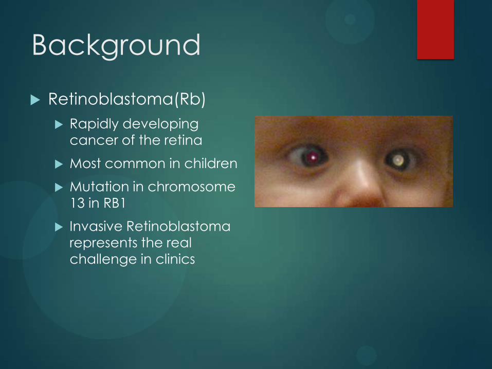
Retinoblastoma(Rb)
Rapidly developing
cancer of the retina
Most common in children
Mutation in chromosome
13 in RB1
Invasive Retinoblastoma
represents the real
challenge in clinics
Background:
Angiogenesis
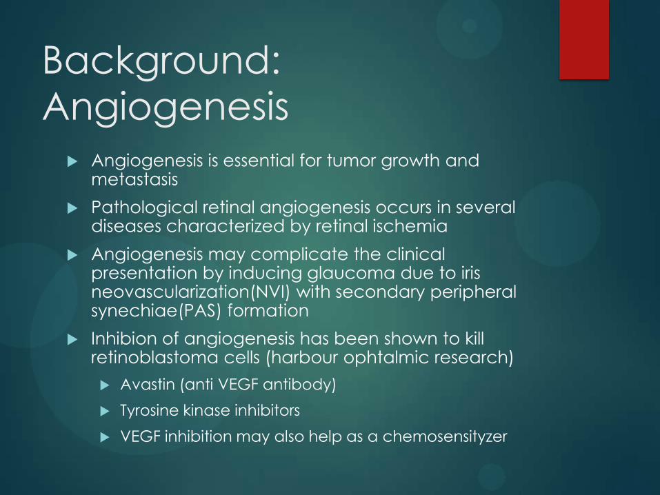
Angiogenesis is essential for tumor growth and metastasis
Pathological retinal angiogenesis occurs in several diseases characterized by retinal ischemia
Angiogenesis may complicate the clinical presentation by inducing glaucoma due to iris neovascularization(NVI) with secondary peripheral synechiae(PAS) formation
Inhibion of angiogenesis has been shown to kill retinoblastoma cells (harbour ophtalmic research)
Avastin (anti VEGF antibody)
Tyrosine kinase inhibitors
VEGF inhibition may also help as a chemosensityzer
Background: VEGF
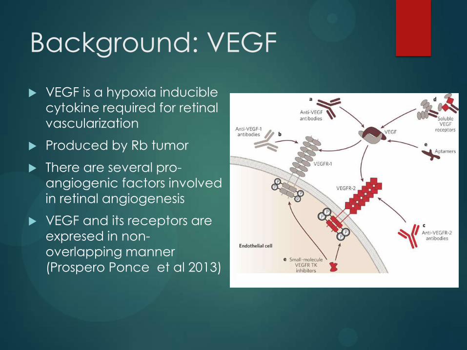
VEGF is a hypoxia inducible
cytokine required for retinal
vascularization
Produced by Rb tumor
There are several pro-
angiogenic factors involved
in retinal angiogenesis
VEGF and its receptors are
expresed in non-
overlapping manner
(Prospero Ponce et al 2013)
High Risk Features
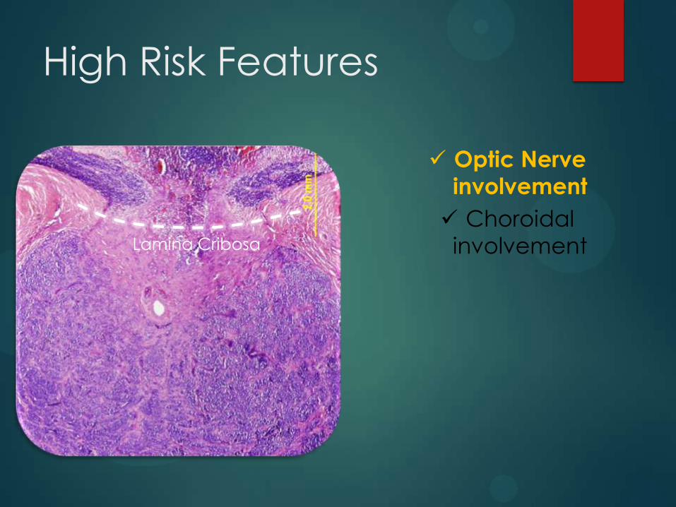
Lamina Cribosa
Optic Nerve
involvement
Choroidal
involvementLamina Cribosa
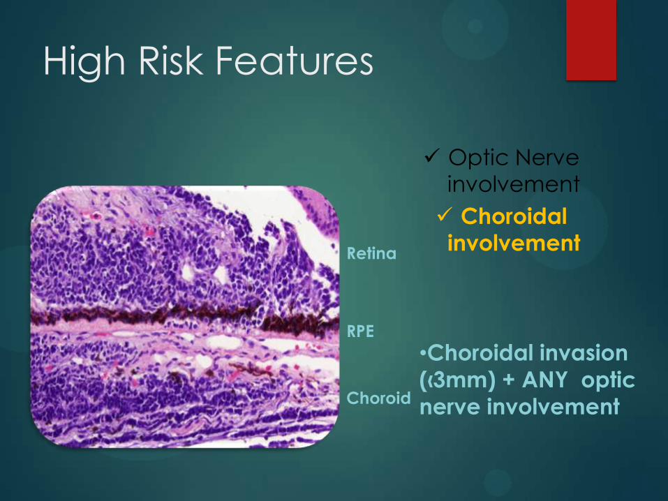
•Choroidal invasion
(‹3mm) + ANY optic
nerve involvement
High Risk Features
RPE
Choroid
Retina
Optic Nerve
involvement
Choroidal
involvement
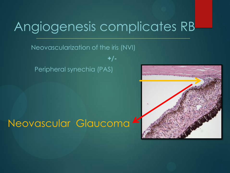
Angiogenesis complicates RB
Neovascularization of the iris (NVI)
+/-
Peripheral synechia (PAS)
Neovascular Glaucoma
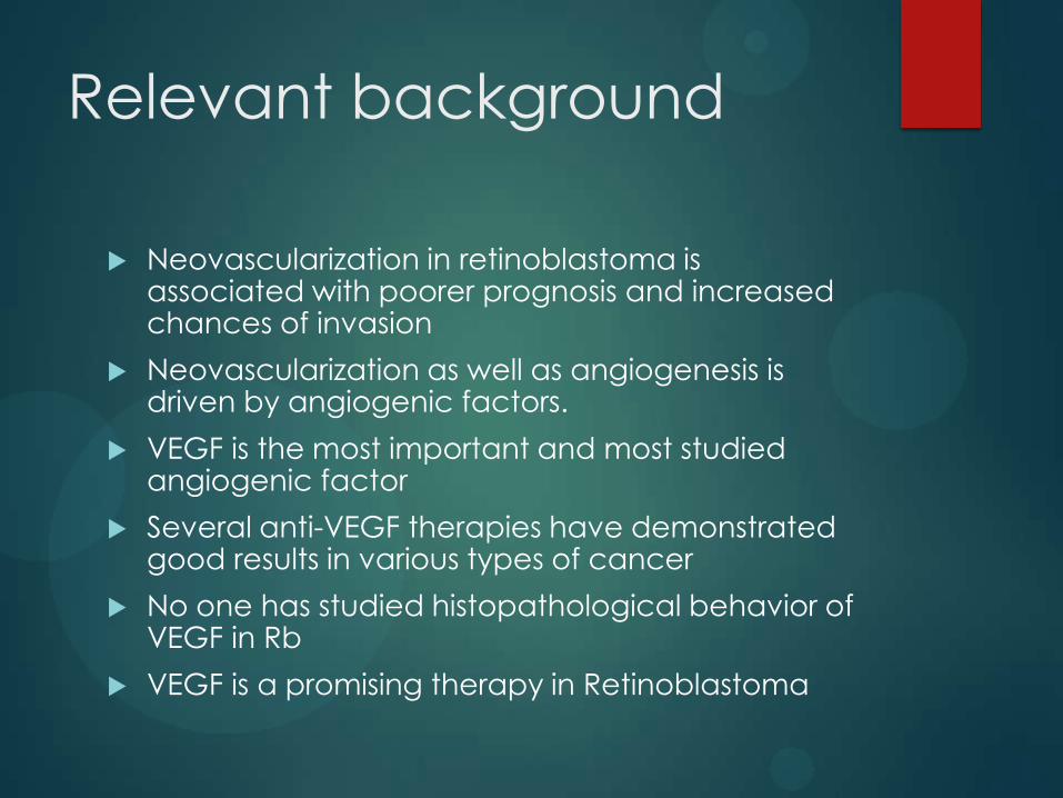
Relevant background
Neovascularization in retinoblastoma is associated with poorer prognosis and increased chances of invasion
Neovascularization as well as angiogenesis is driven by angiogenic factors.
VEGF is the most important and most studied angiogenic factor
Several anti-VEGF therapies have demonstrated good results in various types of cancer
No one has studied histopathological behavior of VEGF in Rb
VEGF is a promising therapy in Retinoblastoma
Hypothesis
HRF tumors will have increased secretion of VEGF
and its receptor VEGFR2
Objective
To analyze the expression of angiogenic factors in
the eye (retina, iris, and tumor ) in
retinoblastoma with high-risk features (HRF) and
non-high risk features (Non-HRF). To correlate the
expression of angiogenic factors in tumors with HRF and the expression of stem cell marker
Sox2. keep order same throughout. If possible,
use this order when discussing your results as well.
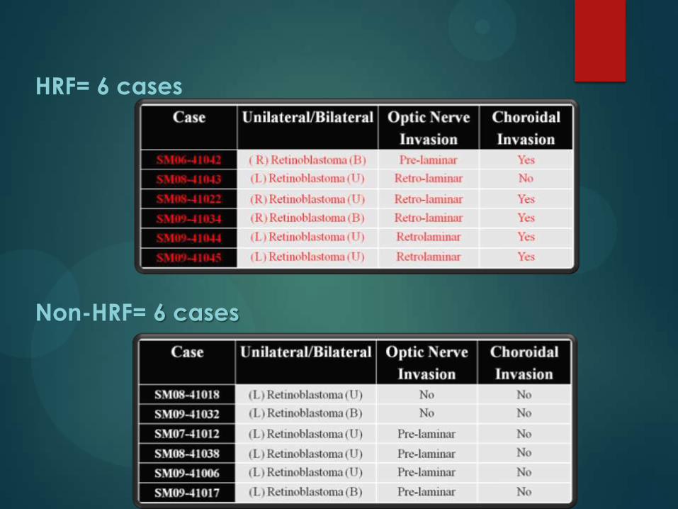
HRF= 6 cases
Non-HRF= 6 cases
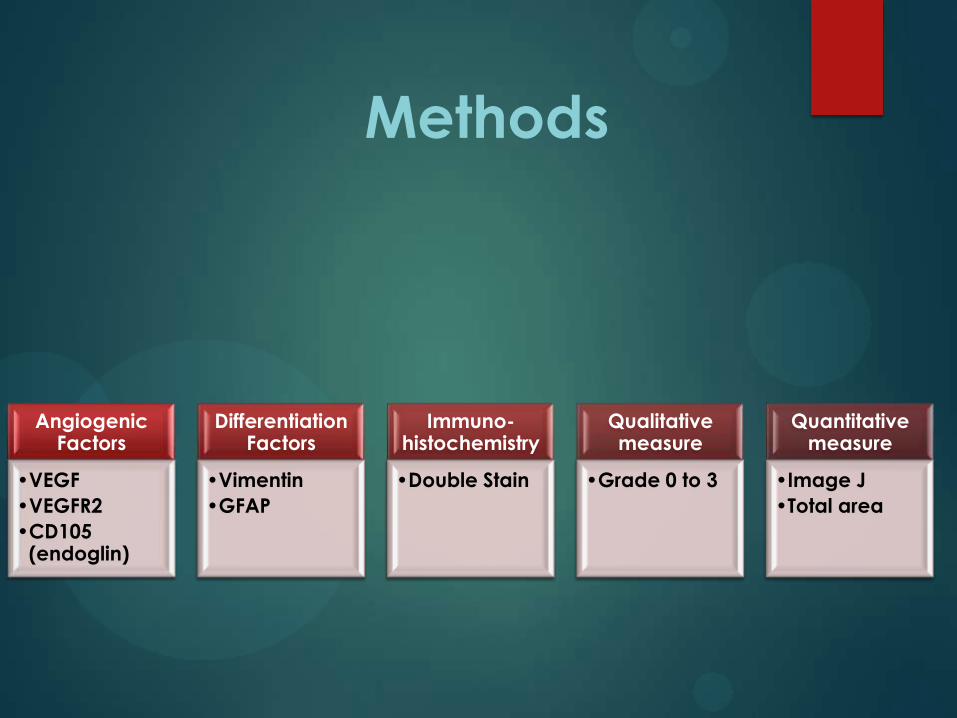
Methods
AngiogenicFactors
•VEGF
•VEGFR2
•CD105 (endoglin)
Differentiation Factors
•Vimentin
•GFAP
Immuno-histochemistry
•Double Stain
Qualitative measure
•Grade 0 to 3
Quantitative measure
•Image J
•Total area

Methods
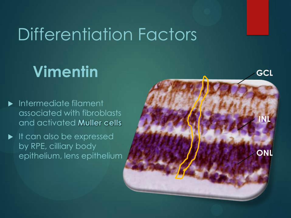
Differentiation Factors
Vimentin
Intermediate filament
associated with fibroblasts
and activated Muller cells
It can also be expressed
by RPE, cilliary body
epithelium, lens epithelium
GCL
INL
ONL
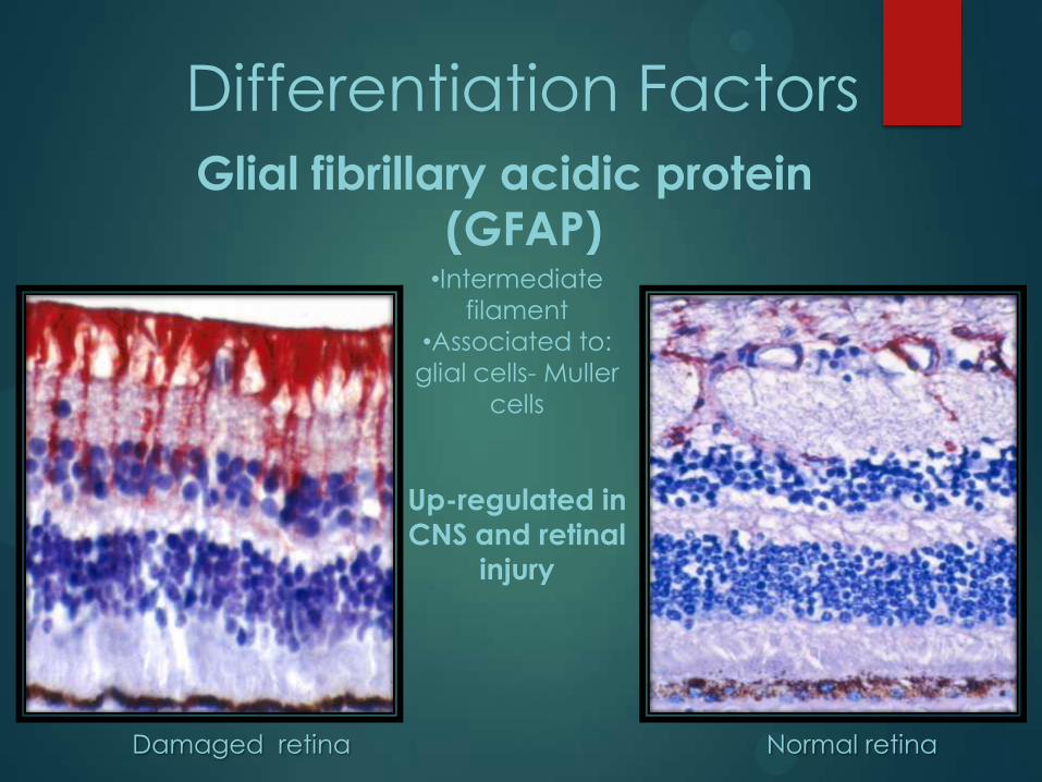
Differentiation Factors
Glial fibrillary acidic protein
(GFAP)
Normal retinaDamaged retina
•Intermediate
filament
•Associated to:
glial cells- Muller
cells
Up-regulated in
CNS and retinal
injury
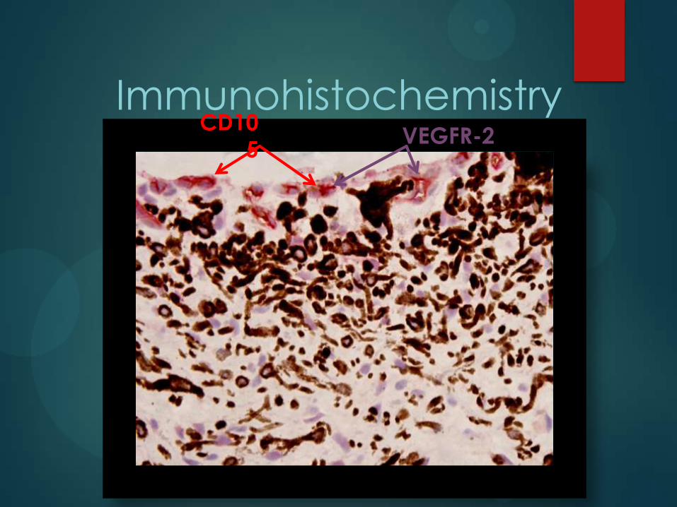
ImmunohistochemistryCD10
5VEGFR-2
Iris. 40X magnification
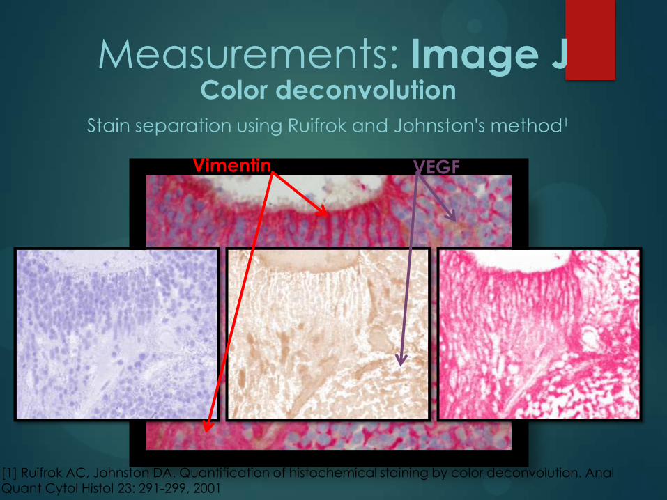
Color deconvolution
Stain separation using Ruifrok and Johnston's method1
Measurements: Image J
[1] Ruifrok AC, Johnston DA. Quantification of histochemical staining by color deconvolution. Anal Quant Cytol Histol 23: 291-299, 2001
Vimentin VEGF

Analyzing particles
Measurements: Image J
8 bit
Binary

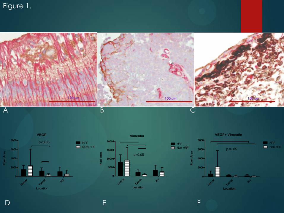
ResultsANGIOGENIC FACTORS AND HRF IN RB
A B C
D E F
100 μm100 μm100 μm
VEGF
Ret
ina
Tumor
Iris
0
2000
4000
6000
8000HRF
NON-HRF
p<0.05
Location
Pix
el
Are
a
Vimentin
Ret
ina
Tumor
Iris
0
5000
10000
15000
20000HRF
Non-HRF
p<0.05
Location
Pix
el
Are
a
VEGF+ Vimentin
Ret
ina
Tumor
Iris
0
2000
4000
6000
8000HRF
Non-HRFp<0.05
LocationP
ixel
Are
a
*
Figure 1.
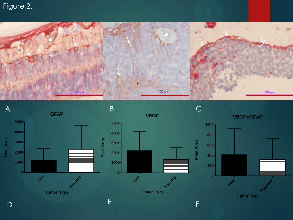
A B C
D E F
100 μm
GFAP
HRF
Non-H
RF
0
1000
2000
3000
4000
5000
Tumor Type
Pix
el
Are
a
VEGF
HRF
Non-H
RF
0
1000
2000
3000
4000
5000
Tumor Type
Pix
el
Are
a
VEGF+GFAP
HRF
Non-H
RF
0
200
400
600
800
1000
Tumor TypeP
ixel
Are
a
**
Figure 2.
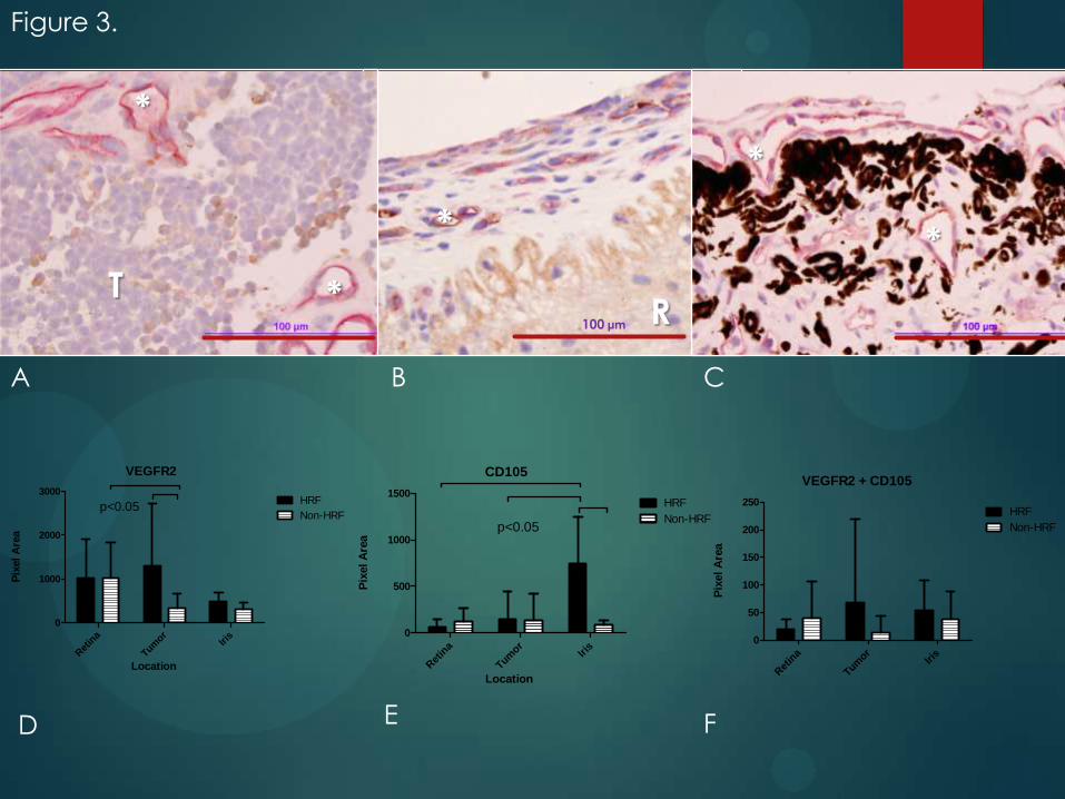
A B C
D E F
100 μm
VEGFR2
Ret
ina
Tumor
Iris
0
1000
2000
3000HRF
Non-HRFp<0.05
Location
Pix
el
Are
a
CD105
Ret
ina
Tumor
Iris
0
500
1000
1500HRF
Non-HRFp<0.05
Location
Pix
el
Are
a
VEGFR2 + CD105
Ret
ina
Tumor
Iris
0
50
100
150
200
250HRF
Non-HRF
Pix
el
Are
a
Figure 3.
*
*
*
*
*
TR

Figure 4.
A
F
C
ED
B
G
Summary
HRF tumor s express more VEGF and VEGFR2 than
non-HRF
Comparison with other stainings indicate that
VEGF secretion might be done by tumor cells
Neovascularization occures more in the iris
This expression is correlated with the expression of
stem cell marker SOX2
Conclusions
HRF tumors seem to be more “stem like”
They might regulate their invasiveness through a
VEGF feedback loop
Temporal expression of VEGF receptors should be
considered
Anti-VEGF therapy might be a promising therapy
for Rb and its side effects
Acknowledgements
Patricia Chevez-Barrios, MD
Rebecca Penland
Magdalena Arredondo
Claudia M. Prospero, MD