vascular pathology atherosclerosis, hypertension, vasculitis
TRANSCRIPT
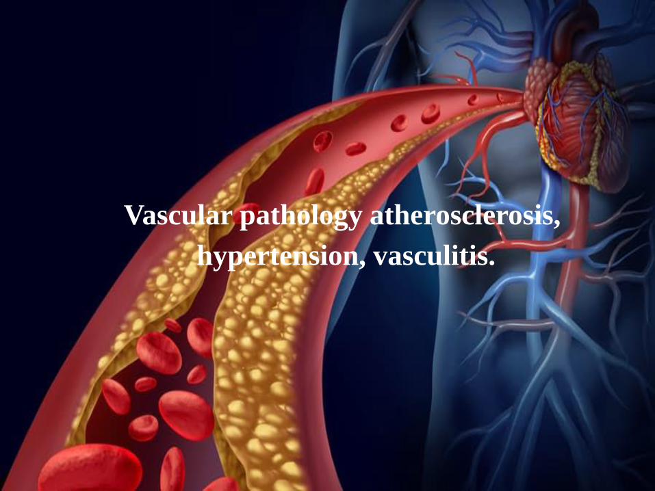
Vascular pathology atherosclerosis,
hypertension, vasculitis.
Vascular pathology atherosclerosis, hypertension, vasculitis
I. Microspecimens:
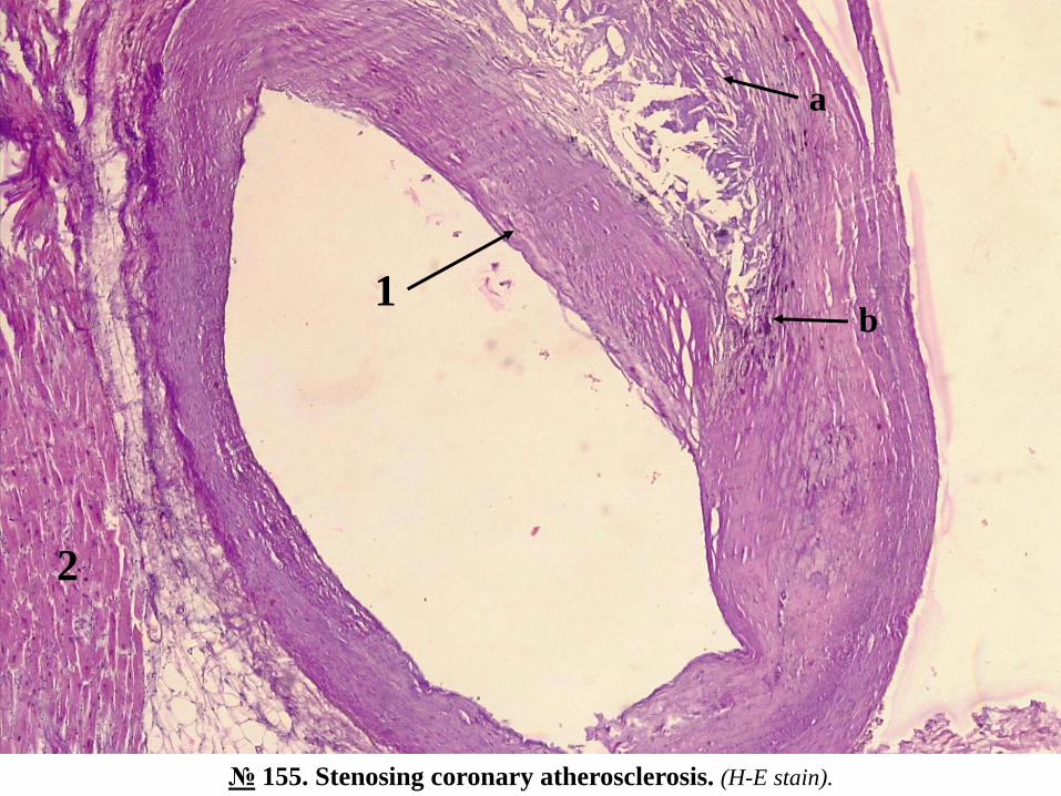
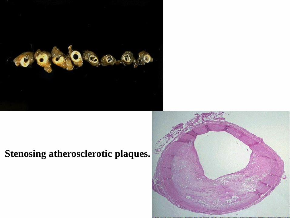
№ 155. Stenosing coronary atherosclerosis. (H-E stain).
Indications:
1. Stenosing atherosclerotic plaque in the artery wall.
a. cholesterol crystals;
b. calcium deposits.
2. Adjacent heart muscle.
Cross section through the subepicardial coronary artery with the underlying myocardium. Grossly, it can be seenthat the wall of the artery is unevenly thickened, the lumen stenotic, in some parts blue-violet calcium deposits.Atherosclerotic plaque - focal, eccentric thickening of the wall, separated from the lumen of the vessel by the fibrouscapsule - a thick, dense layer of collagen fibers with diffuse hyaline, stained homogeneous eosinophil; in the centerof the plate deposits of optically empty aciculate crystals empty of cholesterol, amorphous masses of lipids andweakly eosinophilic tissue detritus, around foam cells, fibrosis, granular deposits of calcium, lymphocytes, plasmacells. Myocardium with normal structure.
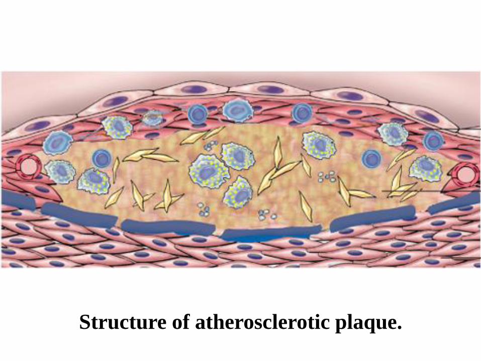
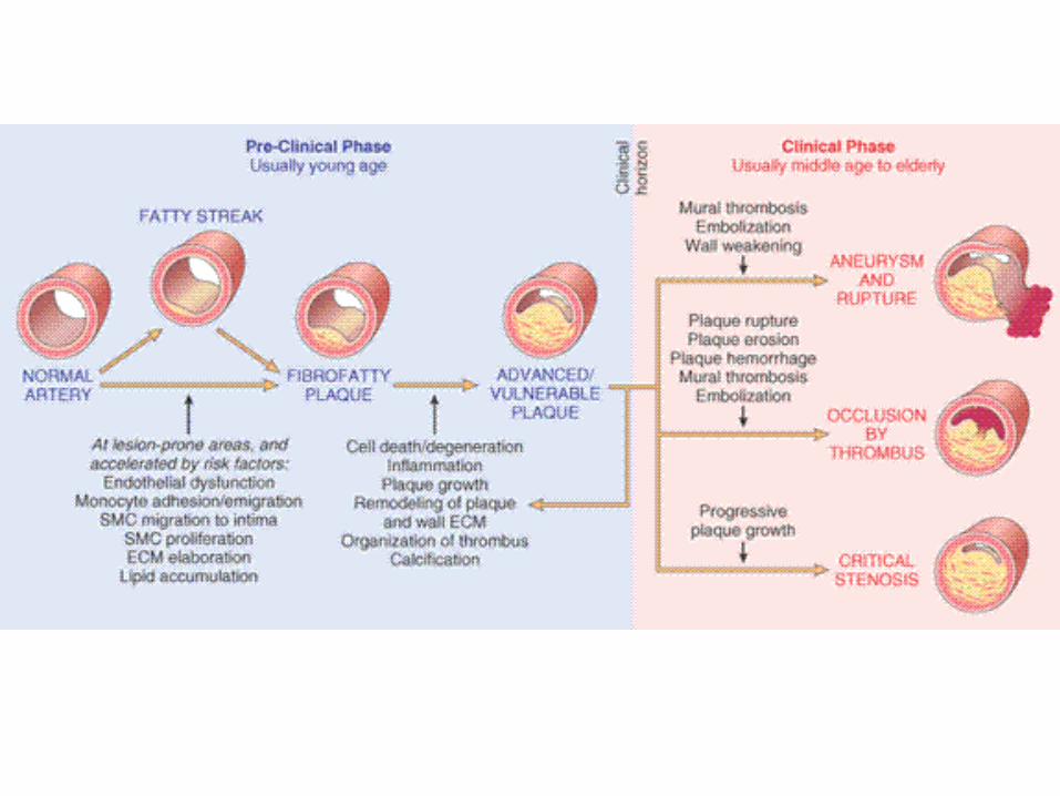
Atherosclerosis is manifested morphologically by the appearance in the arteries of large and medium caliber offocal thickening, dense of the intima, which stenoses the lumen, called atherosclerotic plaques, fibrous plaques,fibrolipids or atheromas. Microscopically the atheroma has the following structure: the luminal surface is covered bythe fibrous capsule, made up of collagen fibers with diffuse hyaline, under the capsule the center or necrotic nucleusformed by necrotic residues, intra- and extracellular lipids (especially cholesterol and cholesterol esters), cells foamy(macrophages and smooth muscle cells, containing lipids), collagen, fragments of disintegrated elastic fibers, fibrinand other plasma proteins, macrophages, lymphocytes, calcium salts. At the periphery of the atheroma,neovascularization processes (neoformation of blood vessels) are revealed. Deeper than the necrotic center is theatrophied and fibrous middle sheath. These components of the atheroma can be in different proportions. In "stable"atheromas, the fibrous capsule is thickened, dense, the necrotic center and inflammation are pronounced, the fibrosisprocess predominates. In “unstable”, “vulnerable” atheromas, the capsule is thin, fine, the necrotic center rich inlipids, active inflammation, the plaques being susceptible to erosions, ulcerations, thrombosis, hemorrhages, whichleads to acute ischemia of the tributary areas of the vessel. In coronary artery atherosclerosis stenosis leads tochronic ischemia and diffuse cardiosclerosis, and acute ischemia - to myocardial infarction.
№ 155. Stenosing coronary atherosclerosis. (H-E stain).
1b
2
a
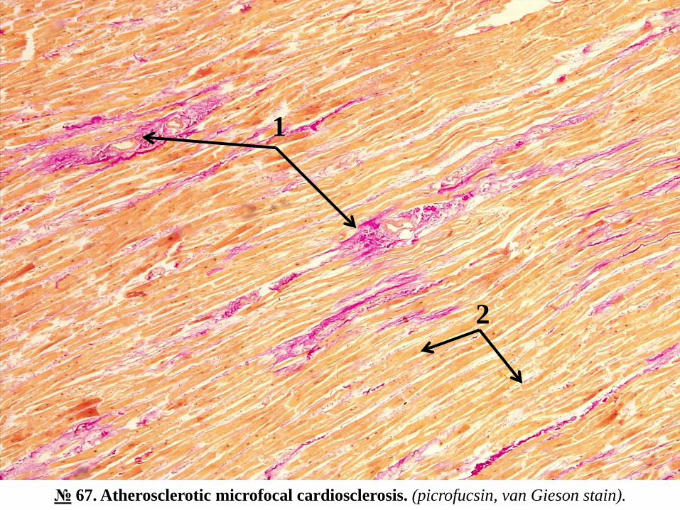
№ 67. Atherosclerotic microfocal cardiosclerosis. (picrofucsin, van Gieson stain).
Indications:
1. Collagen fibers (colored in red).
2. Muscle fibers (colored in yellow).
Microscopically reveals multiple bundles of red collagen fibers, of different thickness, locatedamong the myocardial fibers, predominantly perivascular, most cardiomyocytes have a normalappearance, cytoplasm colored yellow, some slightly atrophied.
Diffuse cardiosclerosis is a process of diffuse excessive proliferation of connective tissue in the heartwall. It is the morphological substrate of chronic ischemic heart disease, including ischemic heartdisease. The main causative factor is stenotic atherosclerosis of the coronary arteries, chronic ischemiacausing dystrophic and atrophic lesions of cardiomyocytes and proliferation of fibroconjunctival tissue.The process of sclerosis is more pronounced perivascular, around the small-caliber arteries. Possiblecomplications: congestive heart failure, heart and conduction disturbances.
Diffuse cardiosclerosis can also develop from interstitial myocarditis, eg in rheumatism, diphtheria,influenza, measles, sepsis.
№ 67. Atherosclerotic microfocal cardiosclerosis. (picrofucsin, van Gieson stain).
2
1
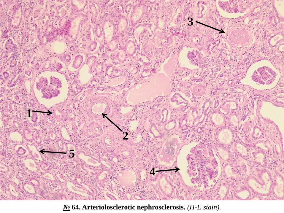
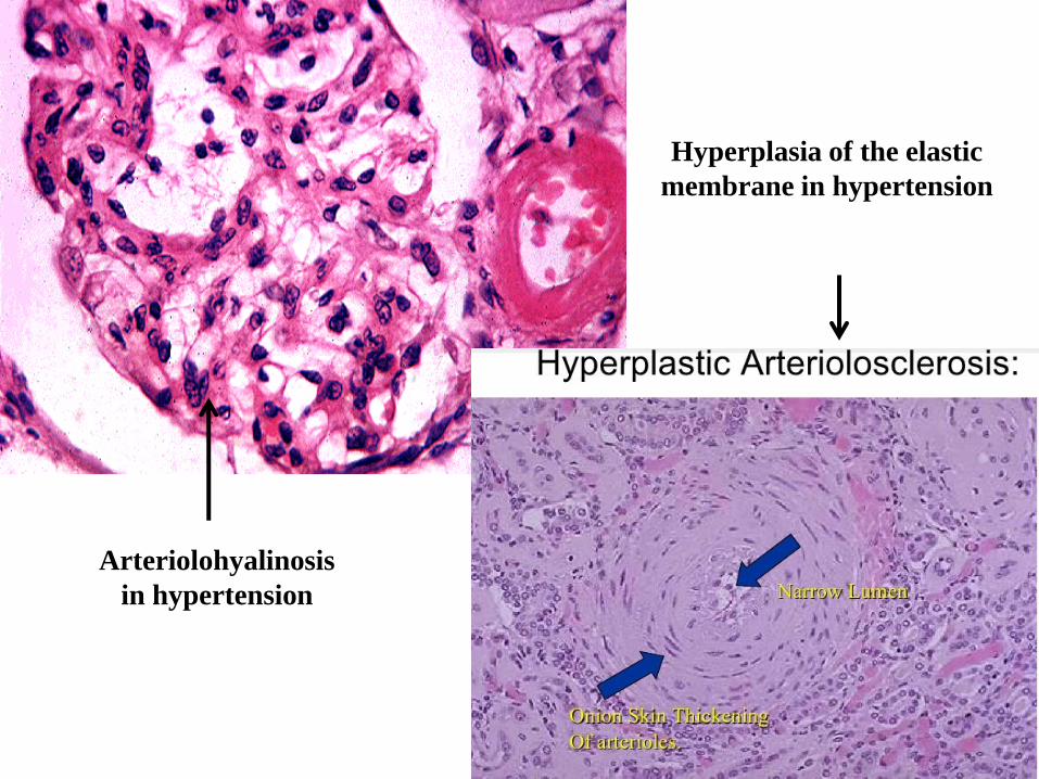
№ 64. Arteriolosclerotic nephrosclerosis. (H-E stain).
Indications:
1. Arteriole with hyalinosis of wall and lumen stenosis.
2. Medium caliber artery with hyperplasia of internal elastic membrane (elastofibrosis).
3. Hyalinated glomerulus with obliteration of capsule cavity.
4. Hyperplased glomerulus.
5. Atrophied tube with thin wall and dilated lumen.
Microscopically, it represents a pronounced atrophy of the renal parenchyma and diffuse sclerosis. In thearterioles the lumen is stenotic, the walls thickened, with diffuse hyaline, homogeneously coloredeosinophilic; in small and medium-sized arteries the walls are thickened due to hyperplasia of the innerelastic membrane and hypertrophy of smooth muscle cells, stenotic lumen; many glomeruli are completelyacellular, with diffuse hyaline, obliterated capsule; the glomeruli are also reduced in size, atrophied, othershyperplasia (compensatory hyperplasia), most of the renal tubes are atrophied, with thin, dilated walls, in thelumen of some tubes eosinophilic protein masses.
Arteriolosclerotic nephrosclerosis is a manifestation of chronic (benign) hypertension. In the kidneys thereis a progressive process of parenchymal atrophy and replacement with fibroconjunctival tissue (sclerosis),caused by damage to the arterioles and small and medium-sized arteries, which occur during the evolution ofhypertension. The target of hypertension is the arterioles, in which hyaline arteriolosclerosis develops(arterioles hyaline). Hyalinosis occurs after the infiltration of the arterial walls with plasma proteins, whichturn into dense, structured masses of hyaline, which leads to atrophy of smooth muscle cells and the gradualtransformation of the arteriole into a narrow tube, with homogeneous wall, structured, dense, covered fromthe inside with endothelial cells. These changes cause ischemia of the glomeruli, their atrophy and sclerosis(glomerulosclerosis), atrophy of the tubes adhering to that glomerulus. Finally, diffuse atrophy of the renalparenchyma and nephrosclerosis occur. The kidney shrinks in size, becomes dense, acquires a shriveledappearance, with a granular surface due to the alternation of small foci of depression (atrophy and sclerosis)with prominent foci of compensatory hypertrophy of intact nephrons. Nephrosclerosis and shrinkage ofkidney result in progressive chronic renal failure.
№ 64. Arteriolosclerotic nephrosclerosis. (H-E stain).
1
3
2
5
4
II. Macrospecimens:
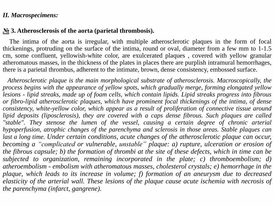
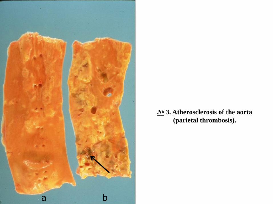
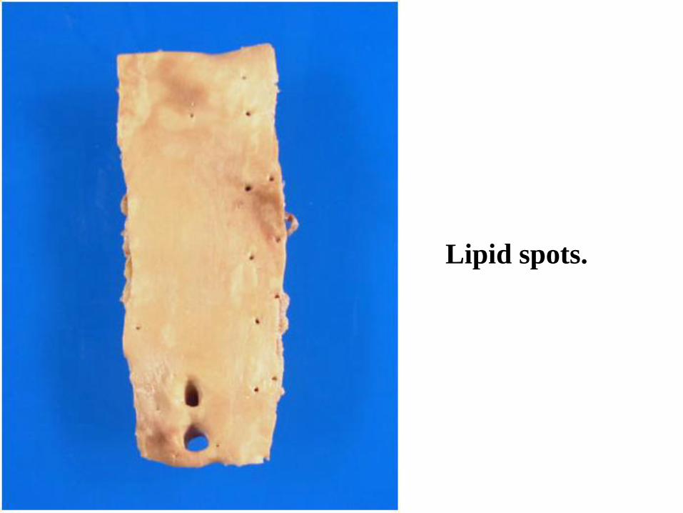
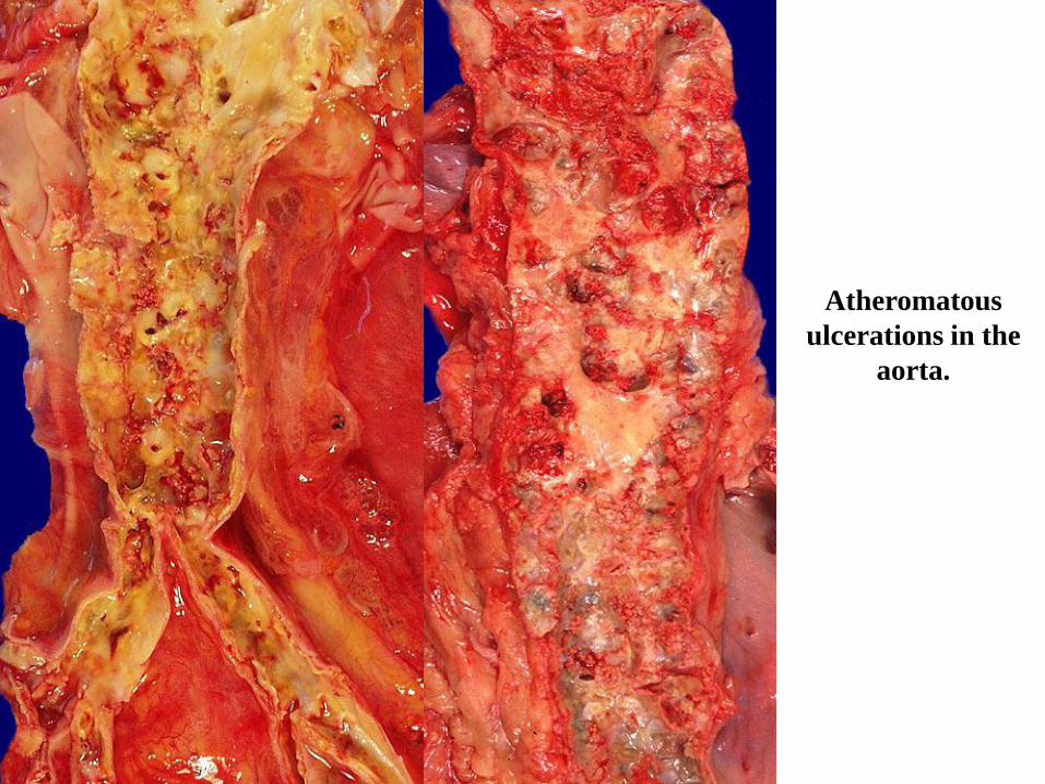
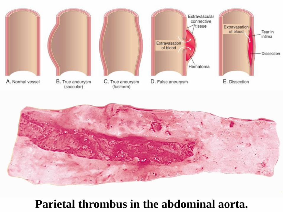
№ 3. Atherosclerosis of the aorta (parietal thrombosis).
The intima of the aorta is irregular, with multiple atherosclerotic plaques in the form of focalthickenings, protruding on the surface of the intima, round or oval, diameter from a few mm to 1-1.5cm, some confluent, yellowish-white color, are exulcerated plaques , covered with yellow granularatheromatous masses, in the thickness of the plates in places there are purplish intramural hemorrhages,there is a parietal thrombus, adherent to the intimate, brown, dense consistency, embossed surface.
Atherosclerotic plaque is the main morphological substrate of atherosclerosis. Macroscopically, theprocess begins with the appearance of yellow spots, which gradually merge, forming elongated yellowlesions - lipid streaks, made up of foam cells, which contain lipids. Lipid streaks progress into fibrousor fibro-lipid atherosclerotic plaques, which have prominent focal thickenings of the intima, of denseconsistency, white-yellow color, which appear as a result of proliferation of connective tissue aroundlipid deposits (liposclerosis), they are covered with a caps dense fibrous. Such plaques are called"stable". They stenose the lumen of the vessel, causing a certain degree of chronic arterialhypoperfusion, atrophic changes of the parenchyma and sclerosis in those areas. Stable plaques canlast a long time. Under certain conditions, acute changes of the atherosclerotic plaque can occur,becoming a “complicated or vulnerable, unstable” plaque: a) rupture, ulceration or erosion ofthe fibrous capsule; b) the formation of thrombi at the site of these defects, which in time can besubjected to organization, remaining incorporated in the plate; c) thromboembolism; d)atheroembolism - embolism with atheromatous masses, cholesterol crystals; e) hemorrhage in theplaque, which leads to its increase in volume; f) formation of an aneurysm due to decreasedelasticity of the arterial wall. These lesions of the plaque cause acute ischemia with necrosis ofthe parenchyma (infarct, gangrene).
№ 3. Atherosclerosis of the aorta
(parietal thrombosis).
a b
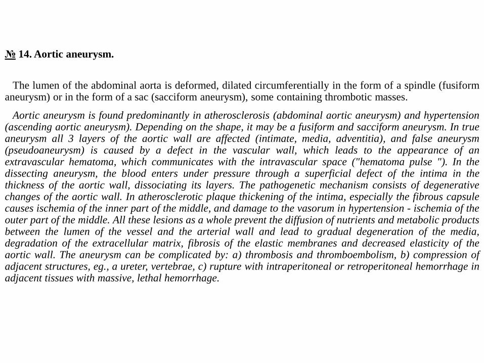
№ 14. Aortic aneurysm.
The lumen of the abdominal aorta is deformed, dilated circumferentially in the form of a spindle (fusiformaneurysm) or in the form of a sac (sacciform aneurysm), some containing thrombotic masses.
Aortic aneurysm is found predominantly in atherosclerosis (abdominal aortic aneurysm) and hypertension(ascending aortic aneurysm). Depending on the shape, it may be a fusiform and sacciform aneurysm. In trueaneurysm all 3 layers of the aortic wall are affected (intimate, media, adventitia), and false aneurysm(pseudoaneurysm) is caused by a defect in the vascular wall, which leads to the appearance of anextravascular hematoma, which communicates with the intravascular space ("hematoma pulse "). In thedissecting aneurysm, the blood enters under pressure through a superficial defect of the intima in thethickness of the aortic wall, dissociating its layers. The pathogenetic mechanism consists of degenerativechanges of the aortic wall. In atherosclerotic plaque thickening of the intima, especially the fibrous capsulecauses ischemia of the inner part of the middle, and damage to the vasorum in hypertension - ischemia of theouter part of the middle. All these lesions as a whole prevent the diffusion of nutrients and metabolic productsbetween the lumen of the vessel and the arterial wall and lead to gradual degeneration of the media,degradation of the extracellular matrix, fibrosis of the elastic membranes and decreased elasticity of theaortic wall. The aneurysm can be complicated by: a) thrombosis and thromboembolism, b) compression ofadjacent structures, eg., a ureter, vertebrae, c) rupture with intraperitoneal or retroperitoneal hemorrhage inadjacent tissues with massive, lethal hemorrhage.
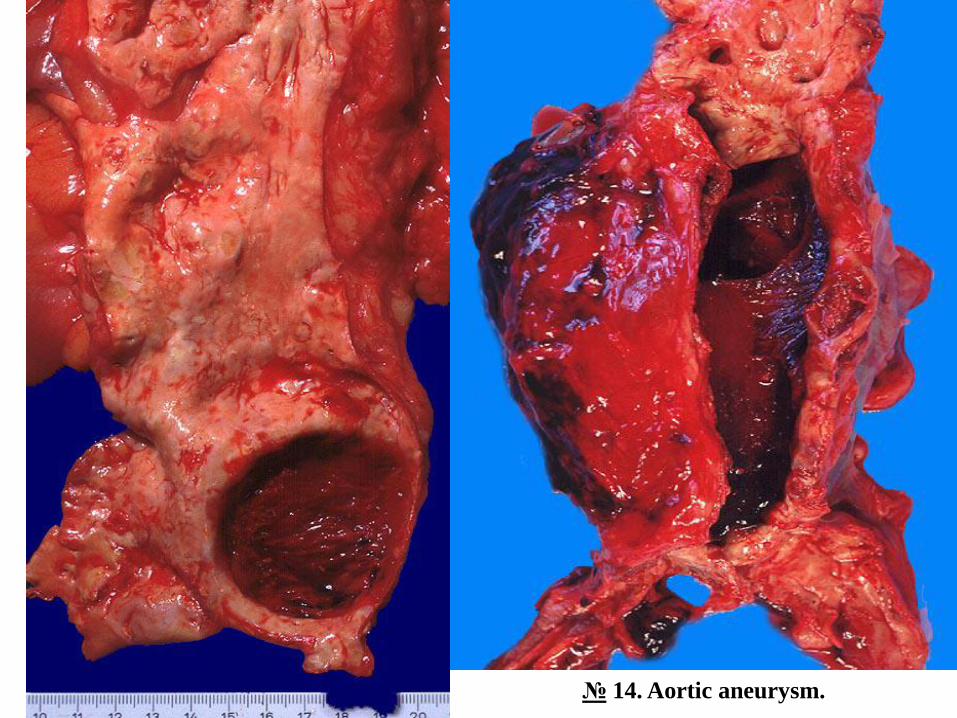
№ 14. Aortic aneurysm.
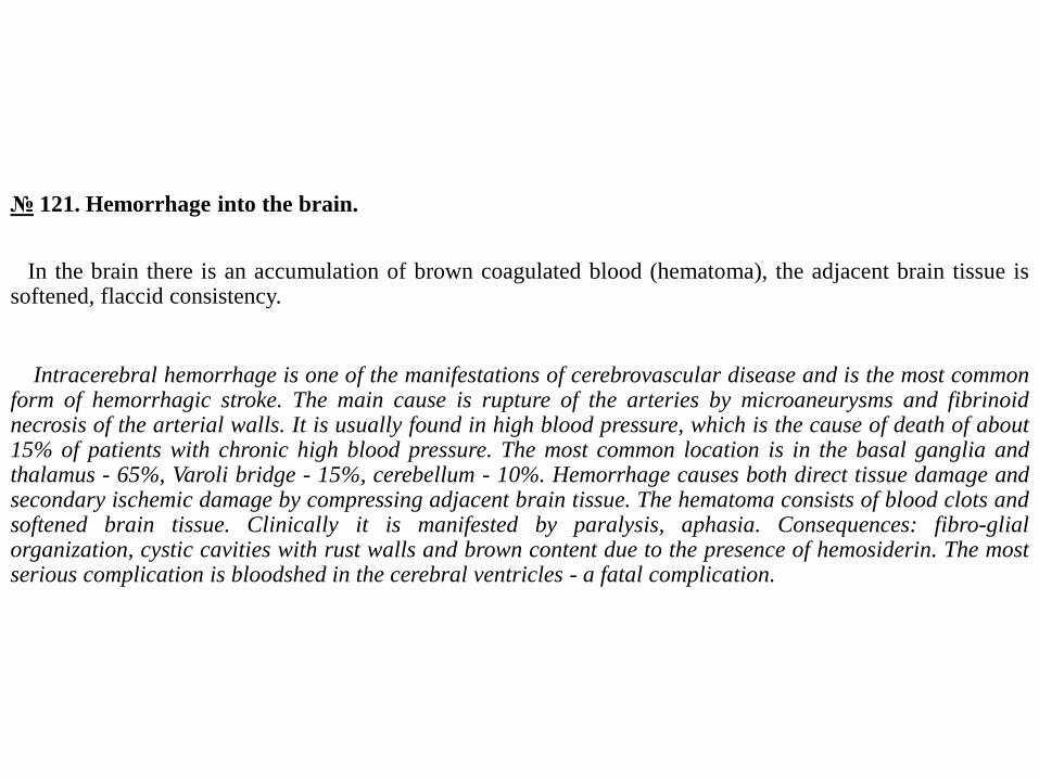
№ 121. Hemorrhage into the brain.
In the brain there is an accumulation of brown coagulated blood (hematoma), the adjacent brain tissue issoftened, flaccid consistency.
Intracerebral hemorrhage is one of the manifestations of cerebrovascular disease and is the most commonform of hemorrhagic stroke. The main cause is rupture of the arteries by microaneurysms and fibrinoidnecrosis of the arterial walls. It is usually found in high blood pressure, which is the cause of death of about15% of patients with chronic high blood pressure. The most common location is in the basal ganglia andthalamus - 65%, Varoli bridge - 15%, cerebellum - 10%. Hemorrhage causes both direct tissue damage andsecondary ischemic damage by compressing adjacent brain tissue. The hematoma consists of blood clots andsoftened brain tissue. Clinically it is manifested by paralysis, aphasia. Consequences: fibro-glialorganization, cystic cavities with rust walls and brown content due to the presence of hemosiderin. The mostserious complication is bloodshed in the cerebral ventricles - a fatal complication.
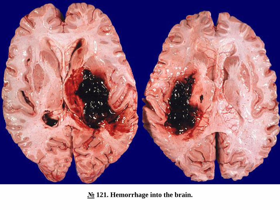
№ 121. Hemorrhage into the brain.
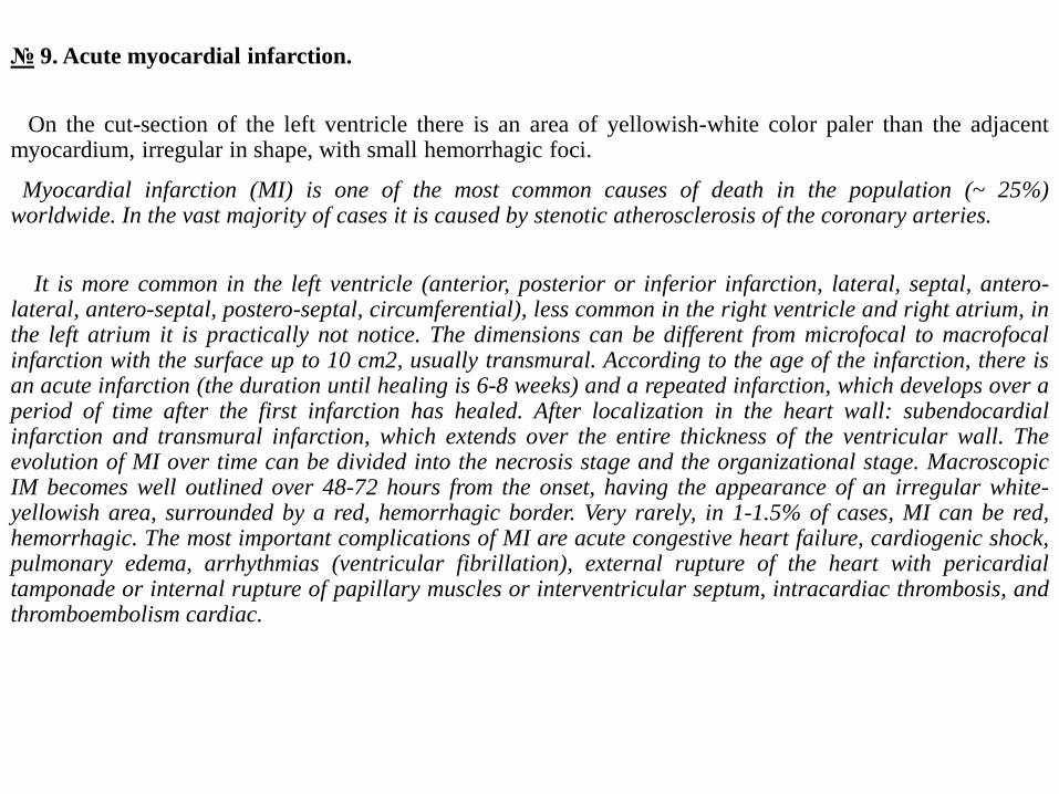
№ 9. Acute myocardial infarction.
On the cut-section of the left ventricle there is an area of yellowish-white color paler than the adjacentmyocardium, irregular in shape, with small hemorrhagic foci.
Myocardial infarction (MI) is one of the most common causes of death in the population (~ 25%)worldwide. In the vast majority of cases it is caused by stenotic atherosclerosis of the coronary arteries.
It is more common in the left ventricle (anterior, posterior or inferior infarction, lateral, septal, antero-lateral, antero-septal, postero-septal, circumferential), less common in the right ventricle and right atrium, inthe left atrium it is practically not notice. The dimensions can be different from microfocal to macrofocalinfarction with the surface up to 10 cm2, usually transmural. According to the age of the infarction, there isan acute infarction (the duration until healing is 6-8 weeks) and a repeated infarction, which develops over aperiod of time after the first infarction has healed. After localization in the heart wall: subendocardialinfarction and transmural infarction, which extends over the entire thickness of the ventricular wall. Theevolution of MI over time can be divided into the necrosis stage and the organizational stage. MacroscopicIM becomes well outlined over 48-72 hours from the onset, having the appearance of an irregular white-yellowish area, surrounded by a red, hemorrhagic border. Very rarely, in 1-1.5% of cases, MI can be red,hemorrhagic. The most important complications of MI are acute congestive heart failure, cardiogenic shock,pulmonary edema, arrhythmias (ventricular fibrillation), external rupture of the heart with pericardialtamponade or internal rupture of papillary muscles or interventricular septum, intracardiac thrombosis, andthromboembolism cardiac.
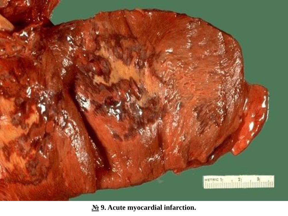
№ 9. Acute myocardial infarction.
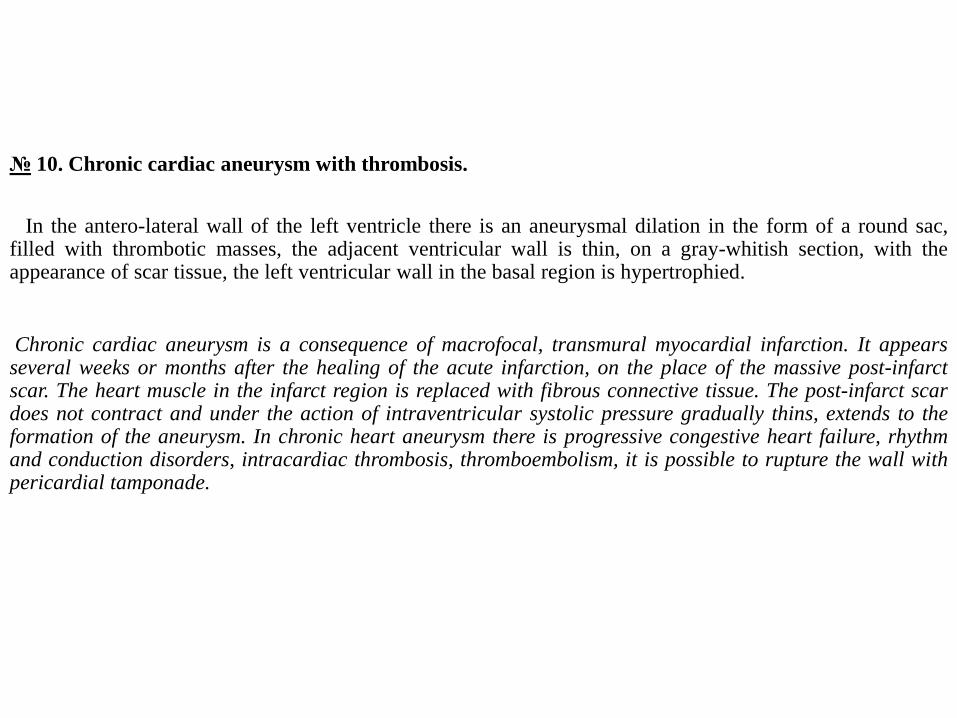
№ 10. Chronic cardiac aneurysm with thrombosis.
In the antero-lateral wall of the left ventricle there is an aneurysmal dilation in the form of a round sac,filled with thrombotic masses, the adjacent ventricular wall is thin, on a gray-whitish section, with theappearance of scar tissue, the left ventricular wall in the basal region is hypertrophied.
Chronic cardiac aneurysm is a consequence of macrofocal, transmural myocardial infarction. It appearsseveral weeks or months after the healing of the acute infarction, on the place of the massive post-infarctscar. The heart muscle in the infarct region is replaced with fibrous connective tissue. The post-infarct scardoes not contract and under the action of intraventricular systolic pressure gradually thins, extends to theformation of the aneurysm. In chronic heart aneurysm there is progressive congestive heart failure, rhythmand conduction disorders, intracardiac thrombosis, thromboembolism, it is possible to rupture the wall withpericardial tamponade.
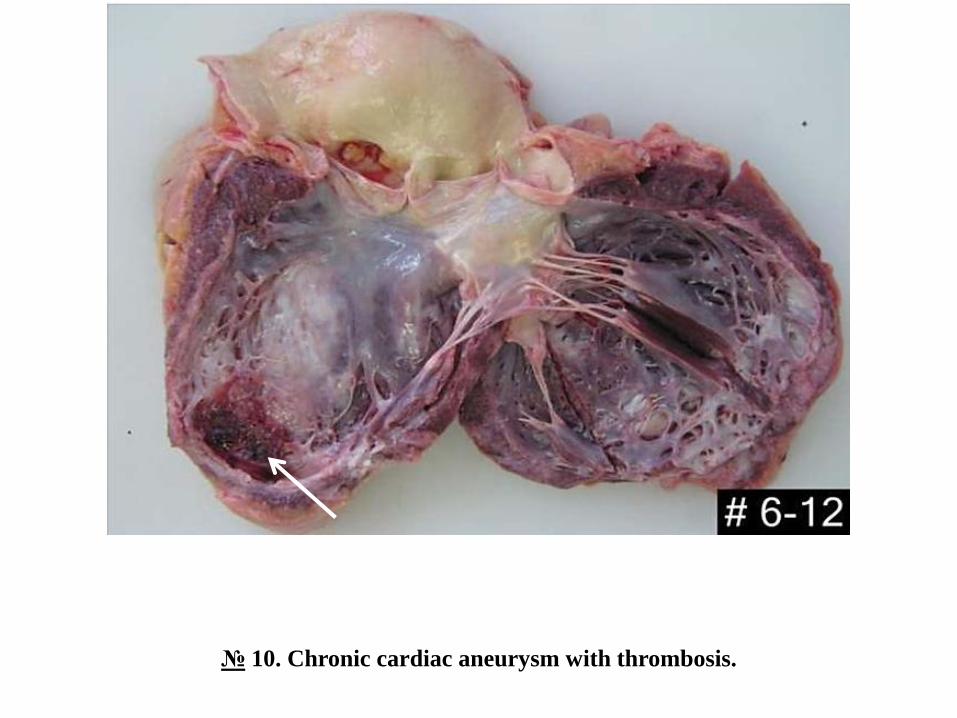
№ 10. Chronic cardiac aneurysm with thrombosis.
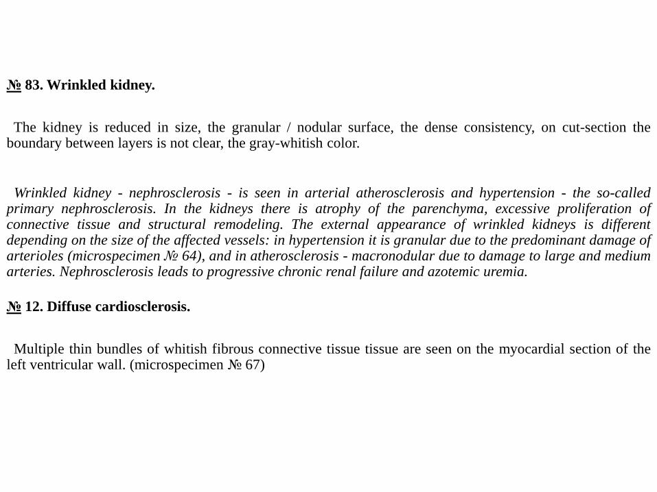
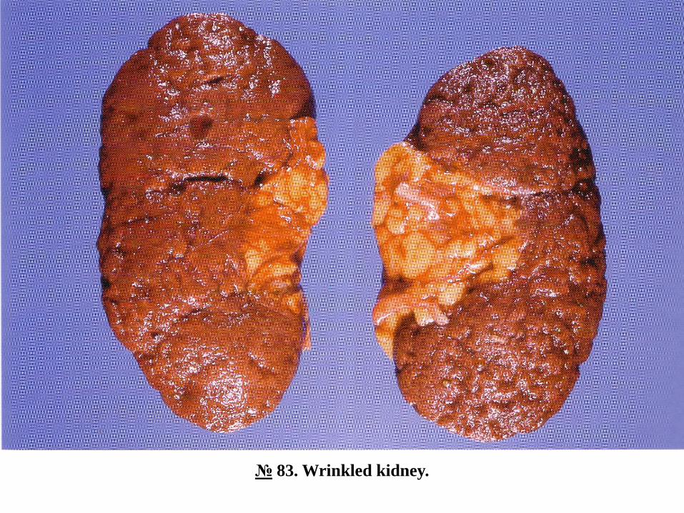
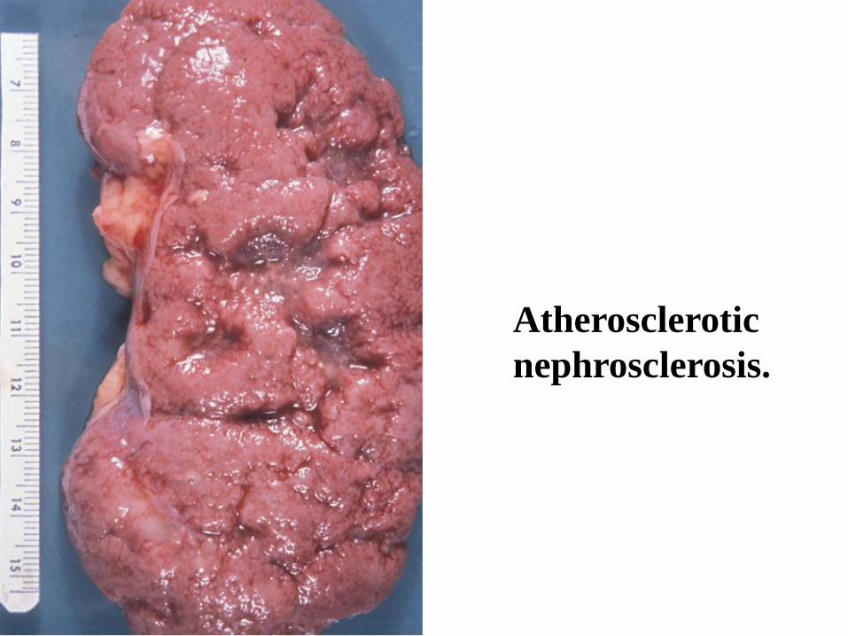
№ 83. Wrinkled kidney.
The kidney is reduced in size, the granular / nodular surface, the dense consistency, on cut-section theboundary between layers is not clear, the gray-whitish color.
Wrinkled kidney - nephrosclerosis - is seen in arterial atherosclerosis and hypertension - the so-calledprimary nephrosclerosis. In the kidneys there is atrophy of the parenchyma, excessive proliferation ofconnective tissue and structural remodeling. The external appearance of wrinkled kidneys is differentdepending on the size of the affected vessels: in hypertension it is granular due to the predominant damage ofarterioles (microspecimen № 64), and in atherosclerosis - macronodular due to damage to large and mediumarteries. Nephrosclerosis leads to progressive chronic renal failure and azotemic uremia.
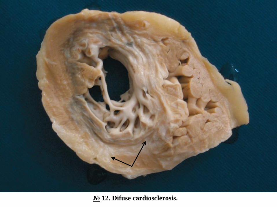
№ 12. Diffuse cardiosclerosis.
Multiple thin bundles of whitish fibrous connective tissue tissue are seen on the myocardial section of theleft ventricular wall. (microspecimen № 67)
№ 83. Wrinkled kidney.
№ 12. Difuse cardiosclerosis.
Lipid spots.
Atheromatous
ulcerations in the
aorta.
Stenosing atherosclerotic plaques.
Structure of atherosclerotic plaque.
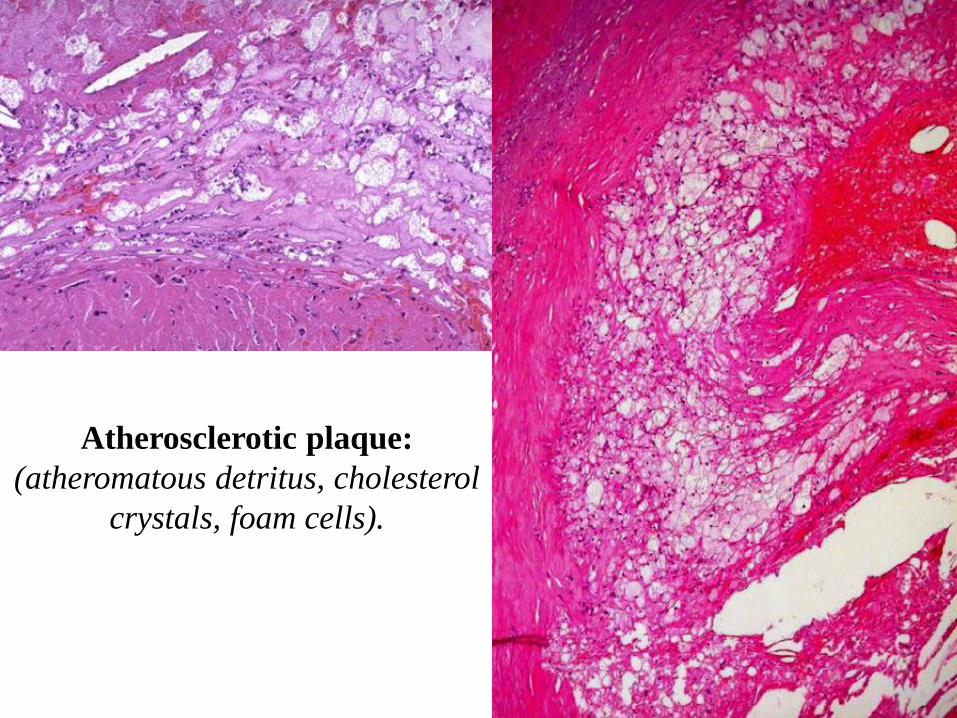
Atherosclerotic plaque:
(atheromatous detritus, cholesterol
crystals, foam cells).

Brain atrophy.
Norma
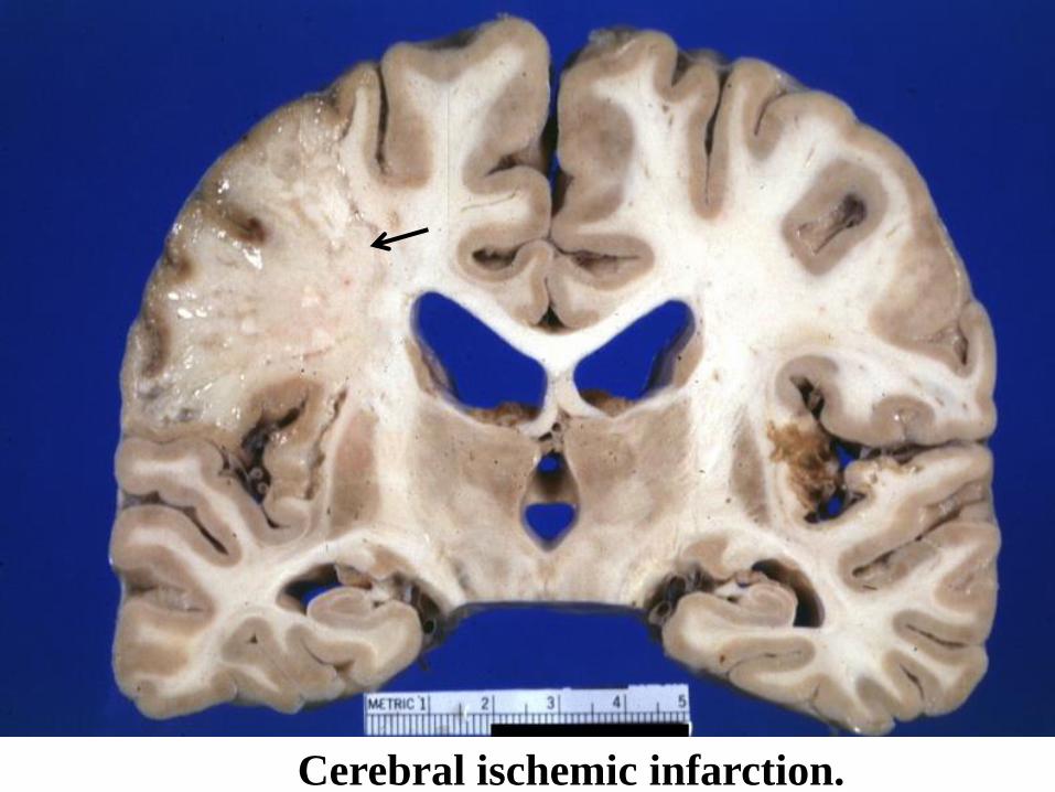
Cerebral ischemic infarction.
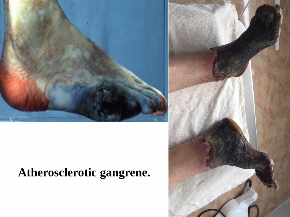
Atherosclerotic gangrene.
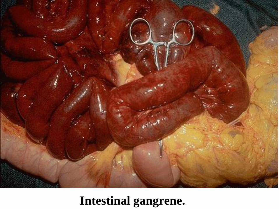
Intestinal gangrene.
Parietal thrombus in the abdominal aorta.
Atherosclerotic
nephrosclerosis.
Arteriolohyalinosis
in hypertension
Hyperplasia of the elastic
membrane in hypertension
Fibrinoid necrosis of the arterial wall in malignant hypertension
(hypertensive crisis).
Fibrinoid necrosis
Thrombosis
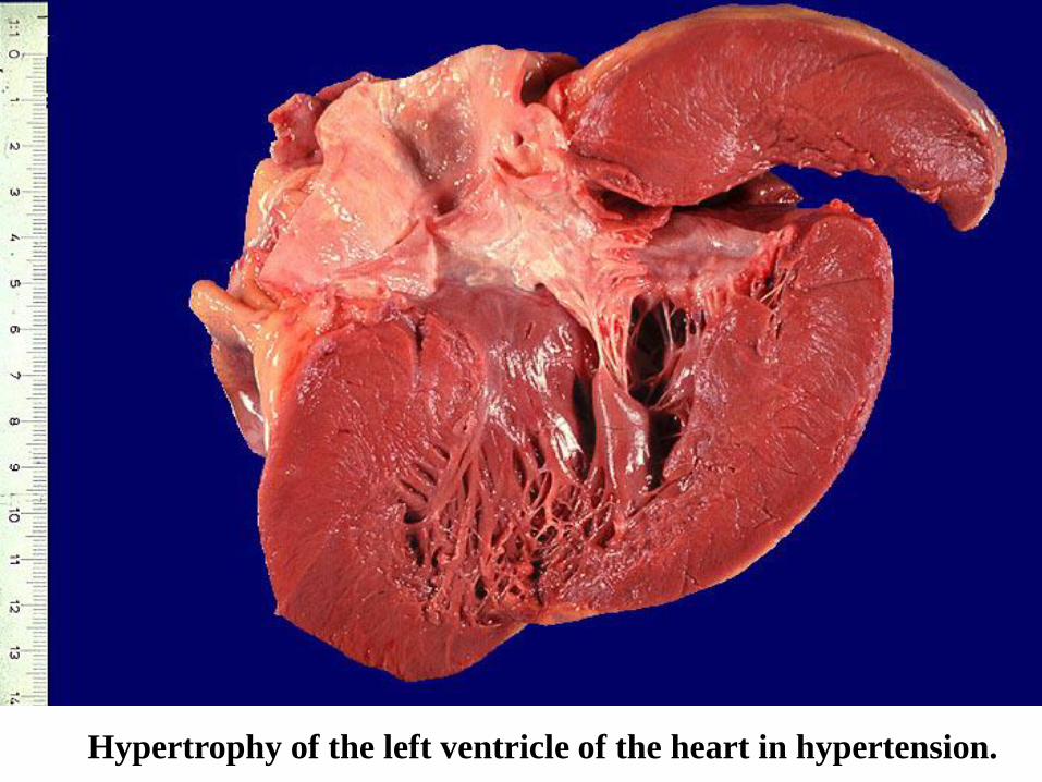
Hypertrophy of the left ventricle of the heart in hypertension.
Diseases of BLOOD VESSELS
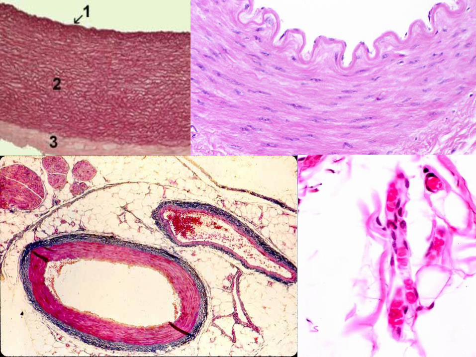
COMPONENTS• Intima, Media, Adventitia, M>A or A>M
• ENDOTHELIUM
• INTERNAL ELASTIC LAMINA
• ECM: Elastin (~aging), collagen, mucopolysaccharides
• Smooth Muscle
•Connective Tissue
• Fat
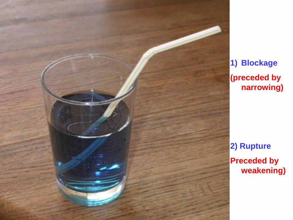
1) Blockage
(preceded by
narrowing)
2) Rupture
Preceded by
weakening)
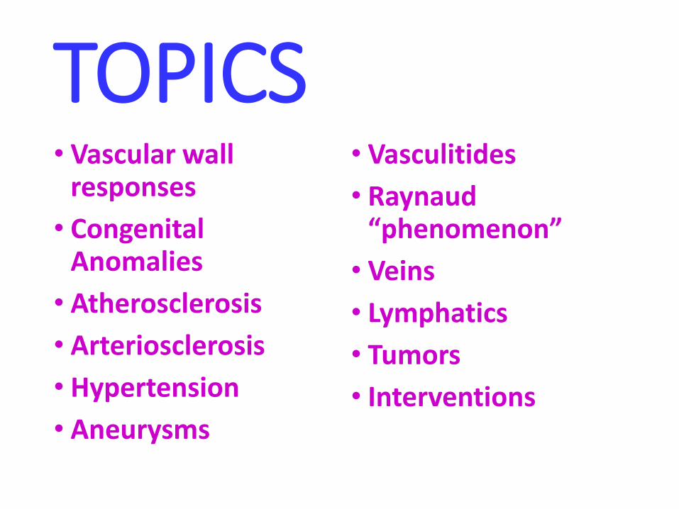
TOPICS• Vascular wall
responses
• Congenital Anomalies
• Atherosclerosis
• Arteriosclerosis
• Hypertension
• Aneurysms
• Vasculitides
• Raynaud “phenomenon”
• Veins
• Lymphatics
• Tumors
• Interventions
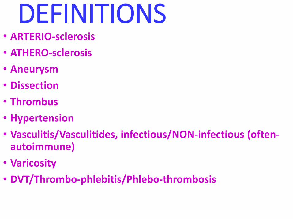
DEFINITIONS• ARTERIO-sclerosis
• ATHERO-sclerosis
• Aneurysm
• Dissection
• Thrombus
• Hypertension
• Vasculitis/Vasculitides, infectious/NON-infectious (often-autoimmune)
• Varicosity
• DVT/Thrombo-phlebitis/Phlebo-thrombosis
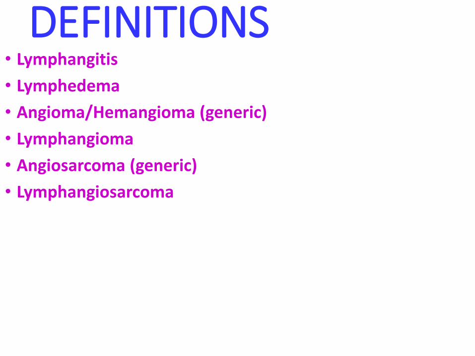
DEFINITIONS• Lymphangitis
• Lymphedema
• Angioma/Hemangioma (generic)
• Lymphangioma
• Angiosarcoma (generic)
• Lymphangiosarcoma
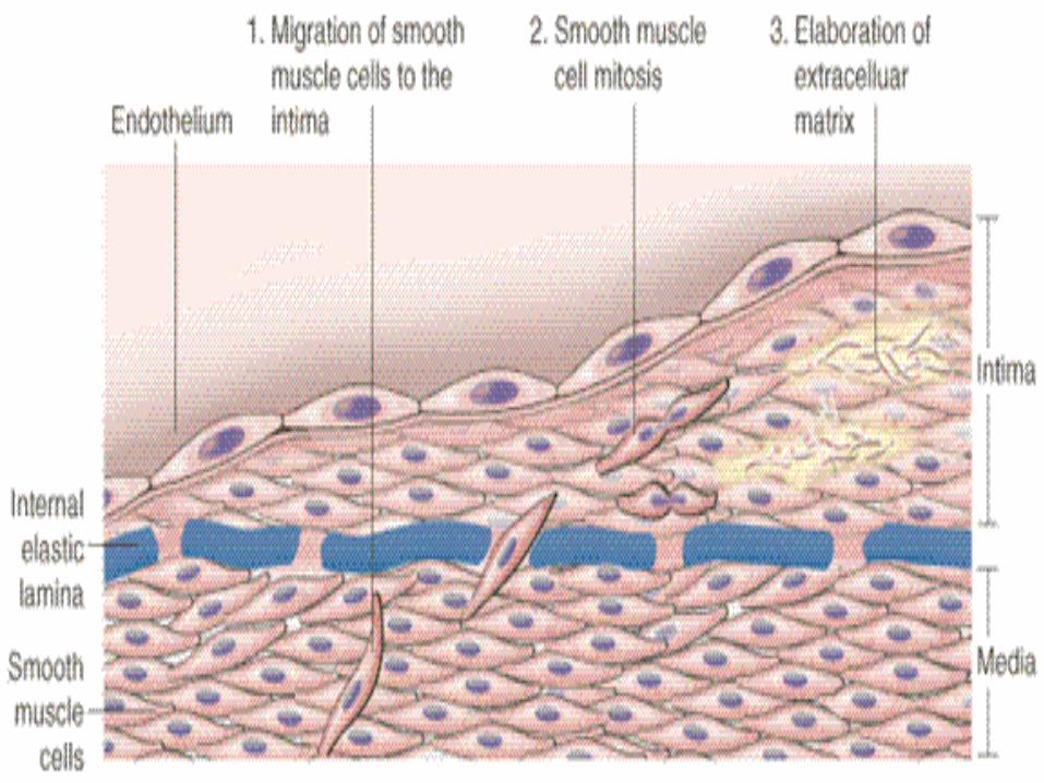
NON-Specific Vascular WallResponse to Injury
•Endothelial “activation”
•Smooth Muscle cell roles
•Development, Growth, Remodeling
•Intimal “thickening”
ENDOTHELIAL CELLS• Recall Jeckyl/Hyde concept: maintain hemostasis/cause
thrombosis
• Maintenance of Permeability Barrier
• Elaboration of Anticoagulant, Antithrombotic, Fibrinolytic Regulators
• Elaboration of Prothrombotic Molecules
• Extracellular Matrix Production (collagen, proteoglycans)
• Modulation of Blood Flow and Vascular Reactivity
• Regulation of Inflammation and Immunity
• Regulation of Cell Growth
• Oxidation of LDL
ENDOTHELIAL CELL“ACTIVATORS” (Δ?)
•Cytokines
•Bacterial Products
•Hemodynamic Forces
•Lipid Products
•Viruses
•Complement
•Hypoxia
VASCULAR SMOOTH MUSCLE
• Vasoconstriction
• Vasodilatation
• Make ECM:• Collagen
• Elastin
• Proteoglycans
• Regulated by:• PROMOTORS: PDGF, endothelin, thrombin, etc.
• INHIBITORS: Heparan SO4, NO, TGF-β
Vessel Growth & Remodeling
• The sum total of all the factors and processes involved in tissue injury and the body’s ability to grow vessels, develop new pathways, and re-perfuse areas in response to tissue and/or blood vessel injury.
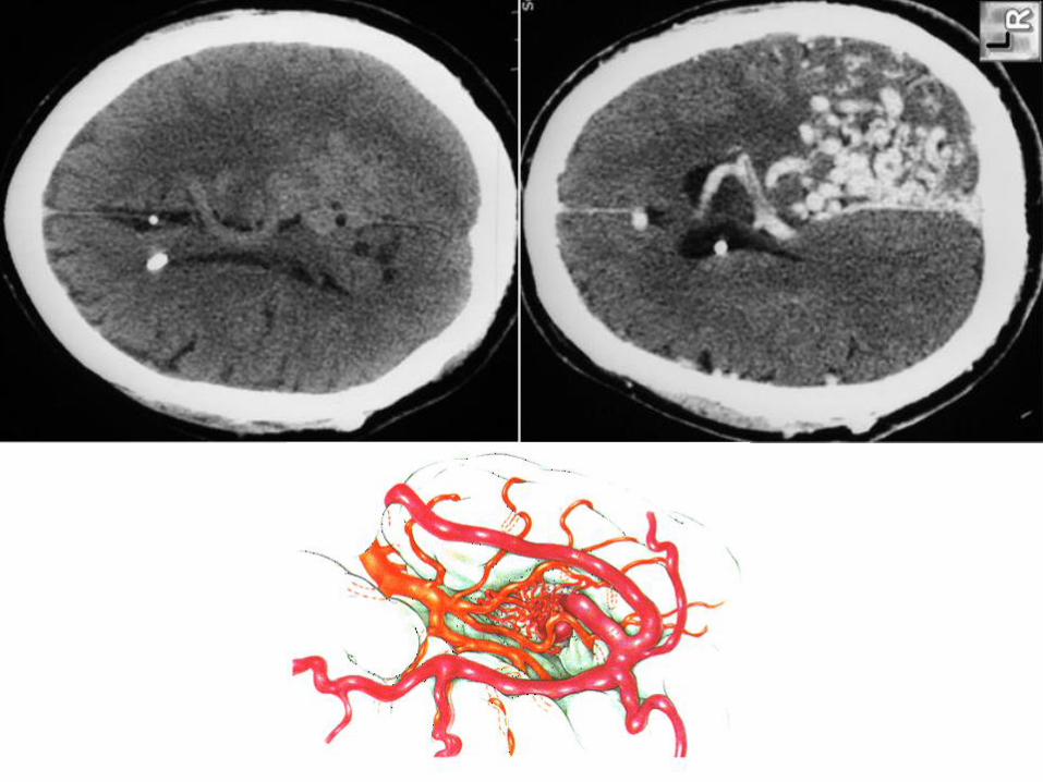
CONGENITAL ANOMALIES• Arteriovenous fistulas
• Also called ArterioVenous Malformation (AVM)
• Common factor is abnormal communication between high pressure arteries and low pressure veins
• Usually congenital (malformation), but can be acquired by trauma or inflammation
• Most often described in the brain as an AVM
• Often asymptomatic or with hemorrhage or pressure effects
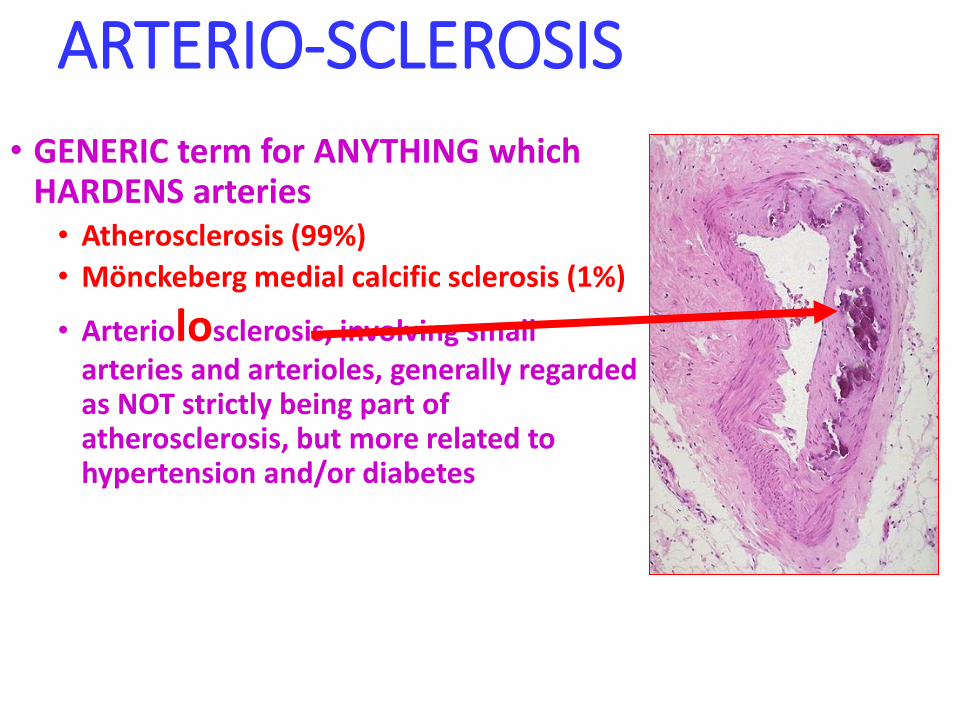
ARTERIO-SCLEROSIS
• GENERIC term for ANYTHING which HARDENS arteries• Atherosclerosis (99%)
• Mönckeberg medial calcific sclerosis (1%)
• Arteriolosclerosis, involving small arteries and arterioles, generally regarded as NOT strictly being part of atherosclerosis, but more related to hypertension and/or diabetes

ATHEROSCLEROSIS(classical)
•Etiology/Risk Factors
•Pathogenesis
•Morphology
•Clinical Expression
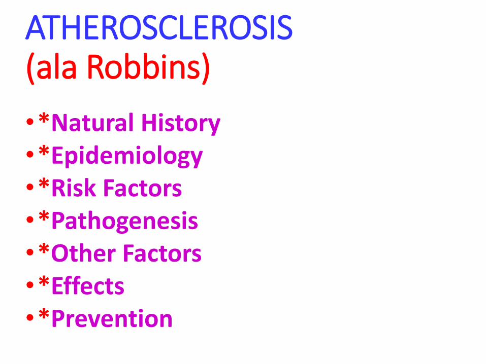
ATHEROSCLEROSIS(ala Robbins)
•*Natural History•*Epidemiology•*Risk Factors•*Pathogenesis•*Other Factors•*Effects•*Prevention

*NATURAL HISTORY
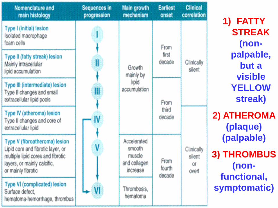
1) FATTY
STREAK
(non-
palpable,
but a
visible
YELLOW
streak)
2) ATHEROMA
(plaque)
(palpable)
3) THROMBUS
(non-
functional,
symptomatic)
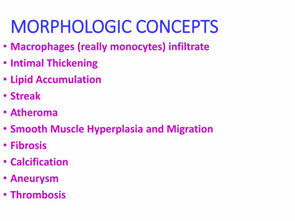
MORPHOLOGIC CONCEPTS• Macrophages (really monocytes) infiltrate
• Intimal Thickening
• Lipid Accumulation
• Streak
• Atheroma
• Smooth Muscle Hyperplasia and Migration
• Fibrosis
• Calcification
• Aneurysm
• Thrombosis
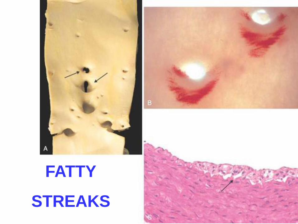
FATTY
STREAKS
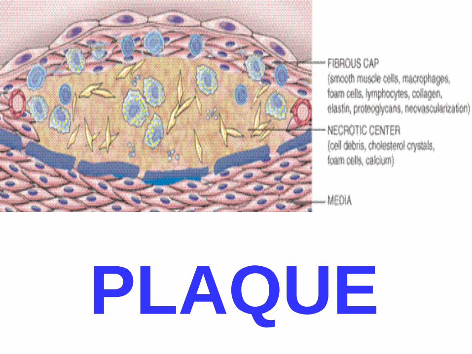
PLAQUE
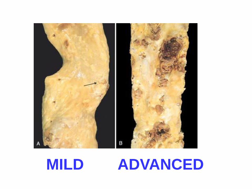
MILD ADVANCED
ADVANCED FEATURES
•RUPTURE
•ULCERATION
• EROSION
•ATHEROEMBOLI
•HEMORRHAGE
• THROMBOSIS
•ANEURYSM
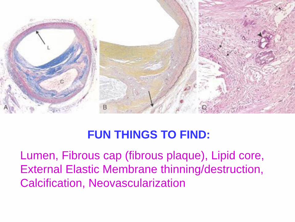
FUN THINGS TO FIND:
Lumen, Fibrous cap (fibrous plaque), Lipid core,
External Elastic Membrane thinning/destruction,
Calcification, Neovascularization
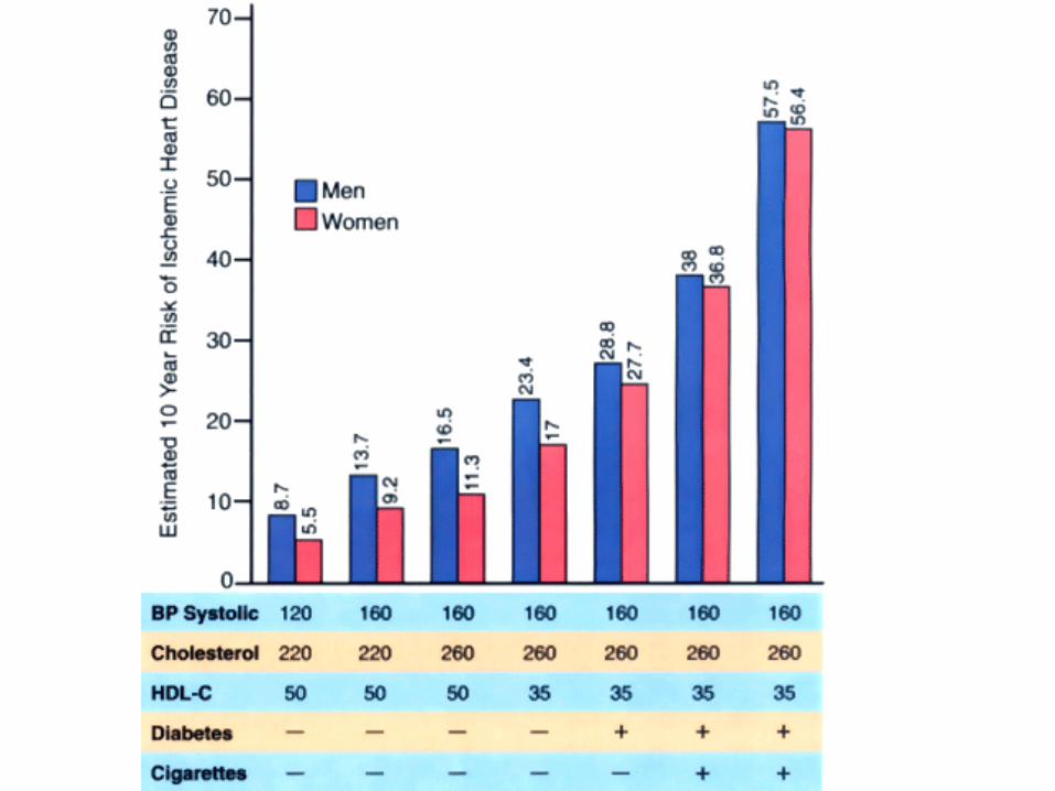
*EPIDEMIOLOGY & RISK FACTORS
Epid./RiskFactors
•Related to “development” of nation
•US highest
•50-70% DECREASE 1963→2000. Why?
•AGE
• SEX, M>F until menopause, estrogen “protection”
•GENETICS
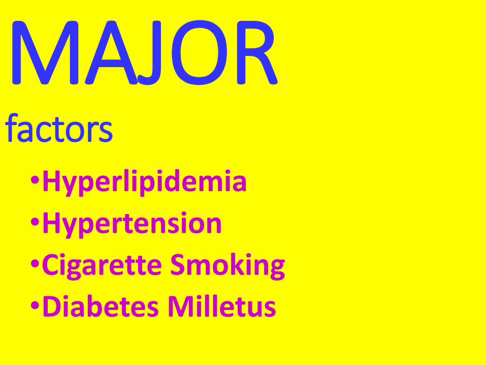
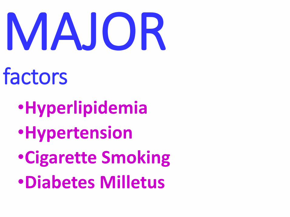
MAJORfactors•Hyperlipidemia
•Hypertension
•Cigarette Smoking
•Diabetes Milletus
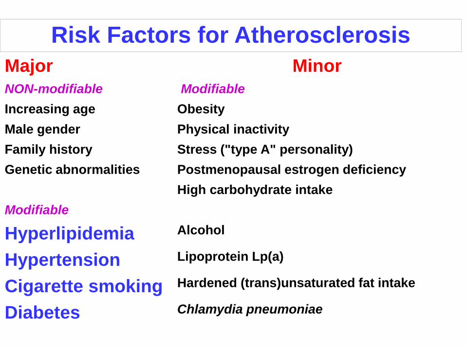
Risk Factors for Atherosclerosis
Major Minor
NON-modifiable Modifiable
Increasing age Obesity
Male gender Physical inactivity
Family history Stress ("type A" personality)
Genetic abnormalities Postmenopausal estrogen deficiency
High carbohydrate intake
Modifiable
Hyperlipidemia Alcohol
Hypertension Lipoprotein Lp(a)
Cigarette smoking Hardened (trans)unsaturated fat intake
Diabetes Chlamydia pneumoniae
MAJORfactors•Hyperlipidemia
•Hypertension
•Cigarette Smoking
•Diabetes Milletus
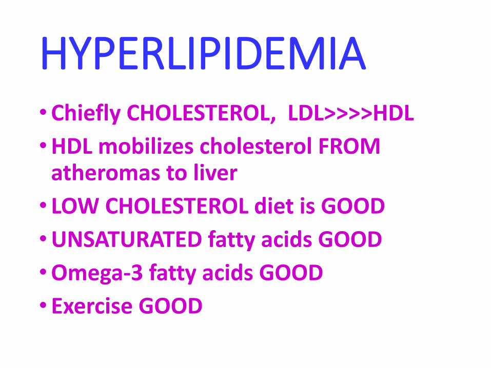
HYPERLIPIDEMIA•Chiefly CHOLESTEROL, LDL>>>>HDL
•HDL mobilizes cholesterol FROM atheromas to liver
• LOW CHOLESTEROL diet is GOOD
•UNSATURATED fatty acids GOOD
•Omega-3 fatty acids GOOD
• Exercise GOOD
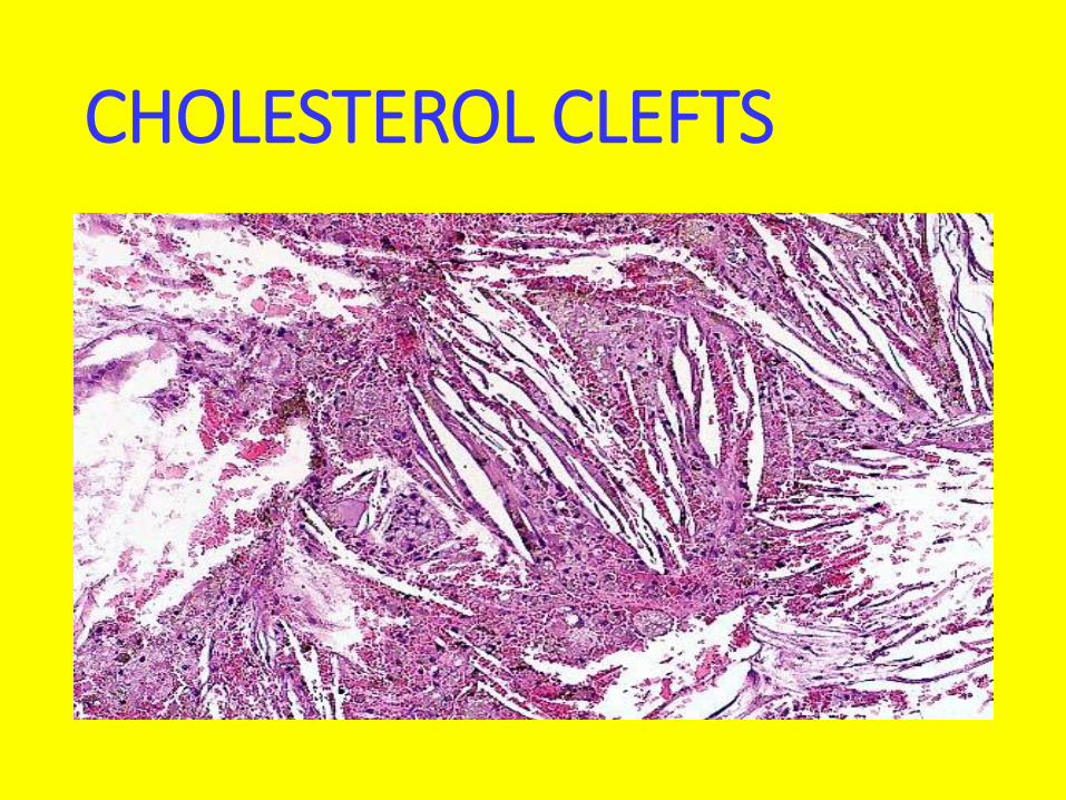
CHOLESTEROL CLEFTS
HYPERTENSION•HYPERTENSION causes ATHEROSCLEROSIS. Why?
•ATHEROSCLEROSIS causes HYPERTENSION. Why?
CIGARETTES•What more needs to be said?
DIABETES•If there was one disease which I could challenge you to, as a dare, to PROVE to me that was
NOT EXACTLY THE SAMEas atherosclerosis, it would be DIABETES! Any takers?
NON major factors• Homocysteinuria/homocysteinemia, related to low B6 and
folate intake
• Coagulation defects
• Lipoprotein Lp(a), independent of cholesterol. Lp(a) is an altered form of LDL
• Inadequate exercise, Type “A” personality, obesity(independent of diabetes)
• Protective effect of moderate alcohol? Medical LIE, sponsored by the booze industry and alcoholic physicians!
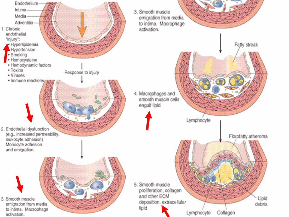
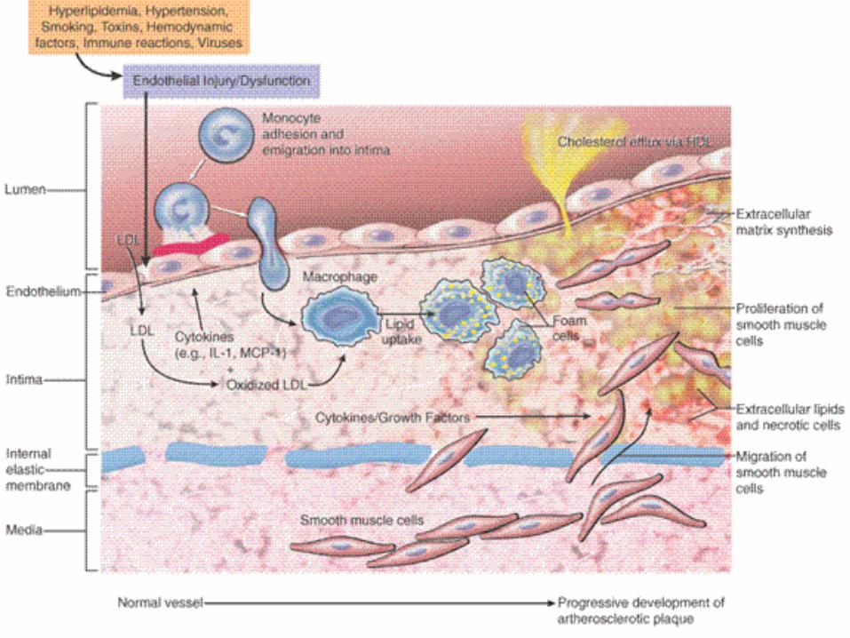
PATHOGENESIS•“atherosclerosis is a chronic inflammatory response of the arterial wall initiated by injury to the endothelium”
PATHOGENESIS SAGA• Chronic endothelial injury→
• LDL, Cholesterol in arterial WALL→
• OXIDATION of lipoproteins→
• Monocytes migrate→ endothelium→*
• Platelet adhesion and activation→
• Migration of SMOOTH MUSCLE from media to intima to activate macrophages (foam cells)→
• Proliferation of SMOOTH MUSCLE and ECM→
• Accumulation of lipids in cells and ECM
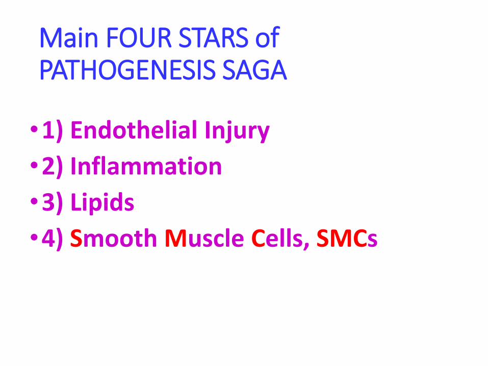
Main FOUR STARS of PATHOGENESIS SAGA
•1) Endothelial Injury
•2) Inflammation
•3) Lipids
•4) Smooth Muscle Cells, SMCs
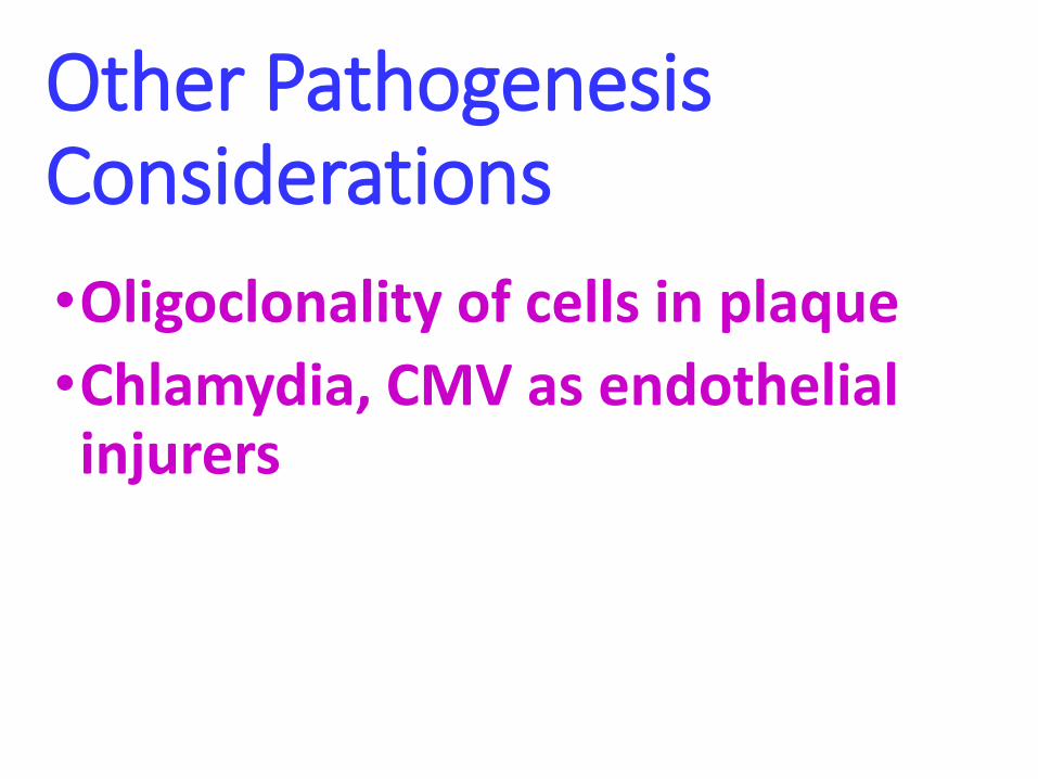
Other Pathogenesis Considerations
•Oligoclonality of cells in plaque
•Chlamydia, CMV as endothelial injurers
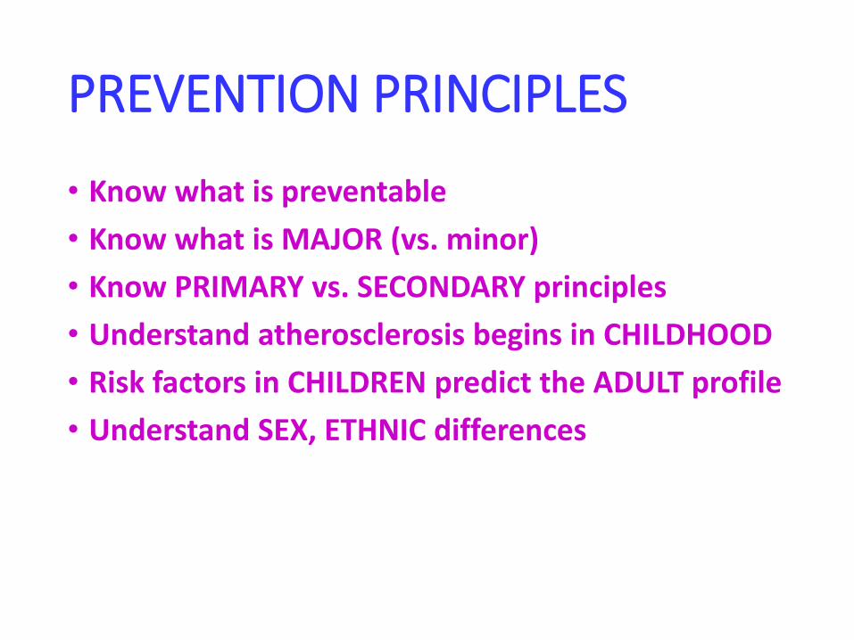
PREVENTION PRINCIPLES
• Know what is preventable
• Know what is MAJOR (vs. minor)
• Know PRIMARY vs. SECONDARY principles
• Understand atherosclerosis begins in CHILDHOOD
• Risk factors in CHILDREN predict the ADULT profile
• Understand SEX, ETHNIC differences
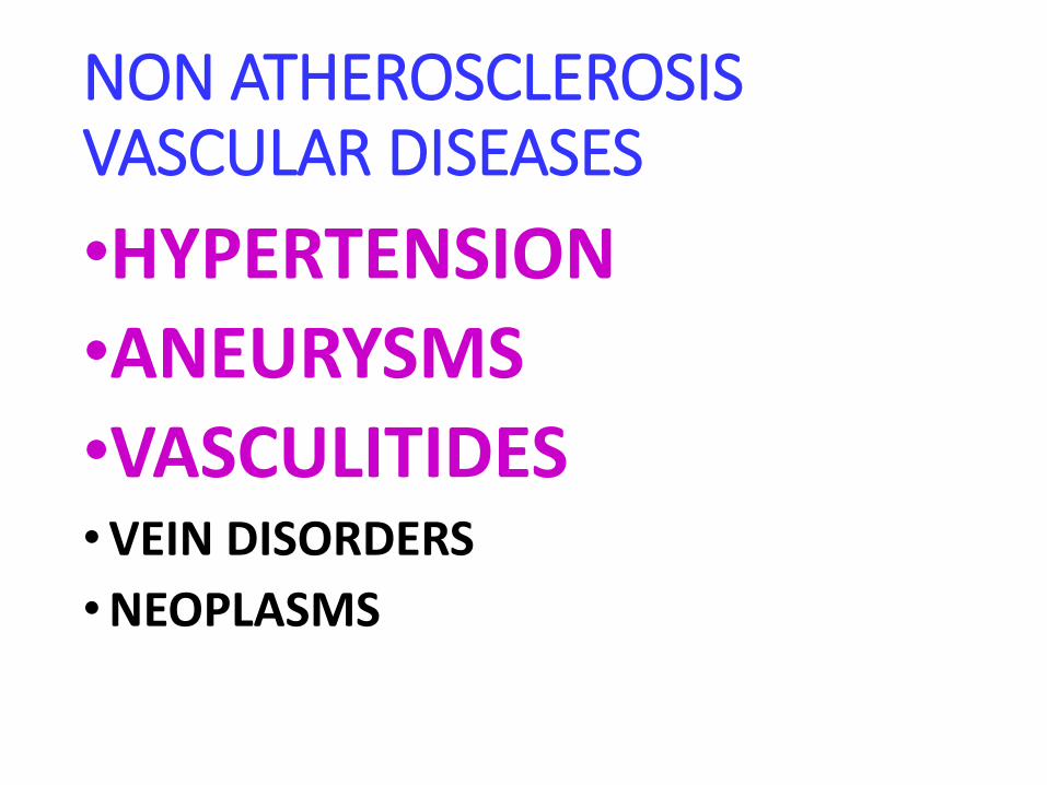
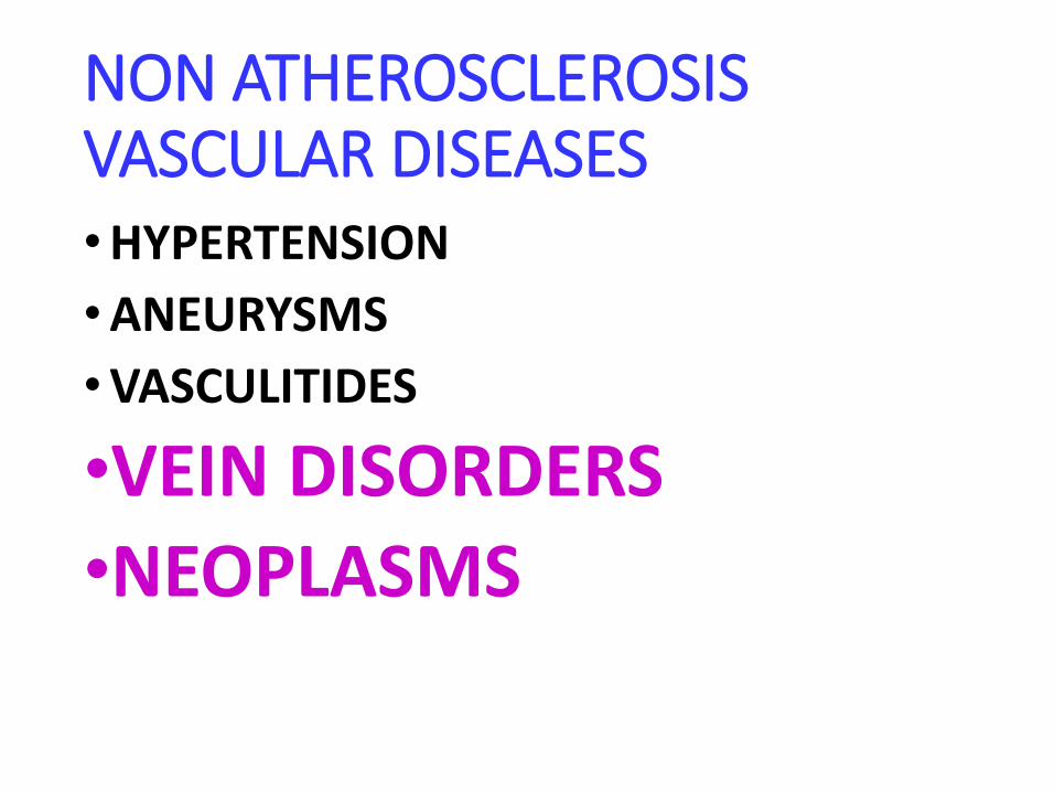
NON ATHEROSCLEROSISVASCULAR DISEASES
•HYPERTENSION
•ANEURYSMS
•VASCULITIDES•VEIN DISORDERS
•NEOPLASMS
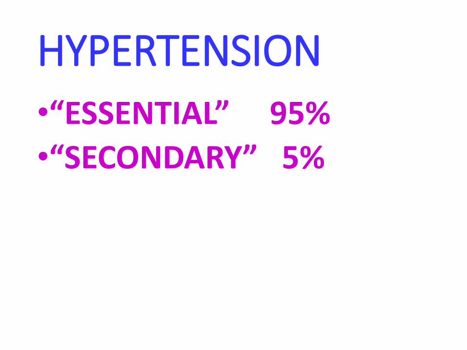
HYPERTENSION
•“ESSENTIAL” 95%
•“SECONDARY” 5%
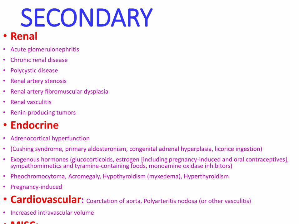
SECONDARY• Renal• Acute glomerulonephritis
• Chronic renal disease
• Polycystic disease
• Renal artery stenosis
• Renal artery fibromuscular dysplasia
• Renal vasculitis
• Renin-producing tumors
• Endocrine• Adrenocortical hyperfunction
• (Cushing syndrome, primary aldosteronism, congenital adrenal hyperplasia, licorice ingestion)
• Exogenous hormones (glucocorticoids, estrogen [including pregnancy-induced and oral contraceptives], sympathomimetics and tyramine-containing foods, monoamine oxidase inhibitors)
• Pheochromocytoma, Acromegaly, Hypothyroidism (myxedema), Hyperthyroidism
• Pregnancy-induced
• Cardiovascular: Coarctation of aorta, Polyarteritis nodosa (or other vasculitis)
• Increased intravascular volume
• MISC: Increased cardiac output, Rigidity of the aorta, Neurologic, Psychogenic, Increased
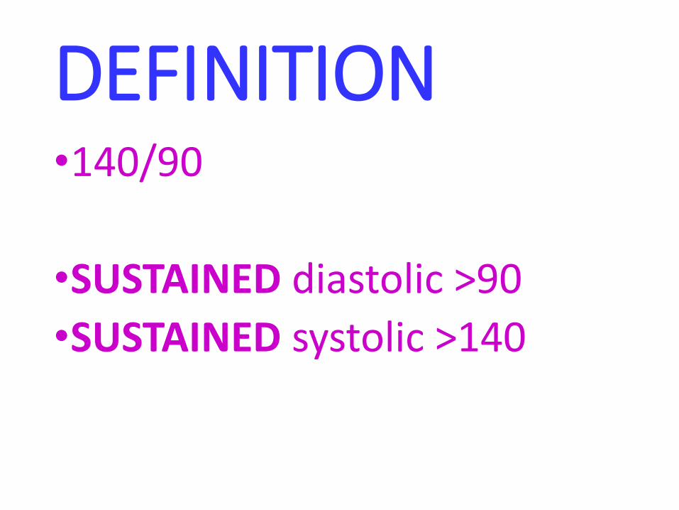
DEFINITION•140/90
•SUSTAINED diastolic >90
•SUSTAINED systolic >140
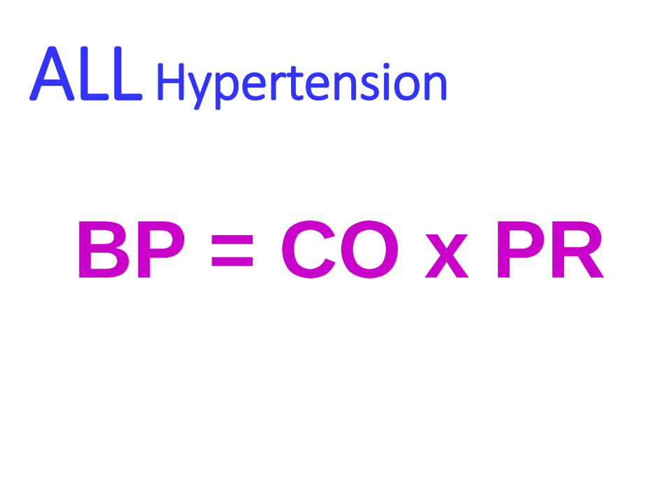
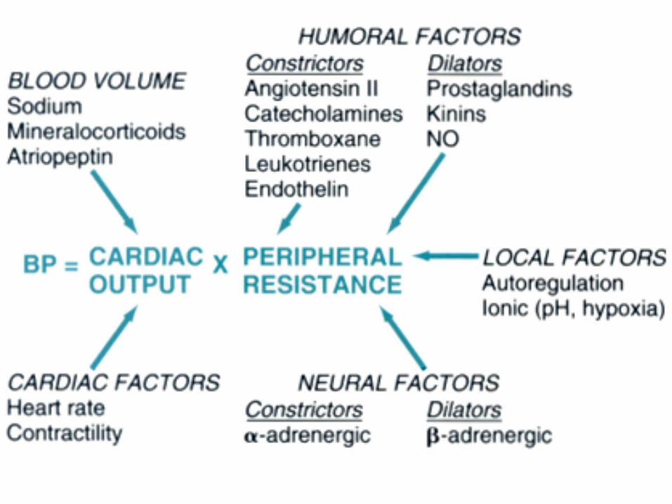
BP = CO x PR
ALL Hypertension
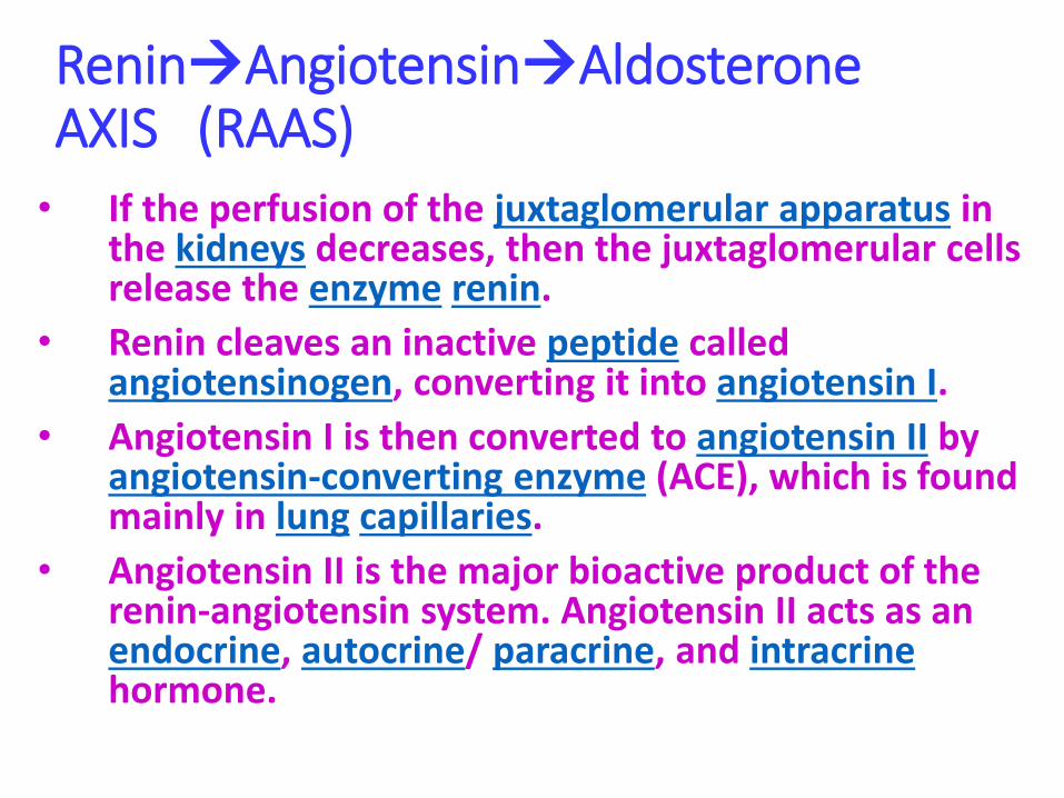
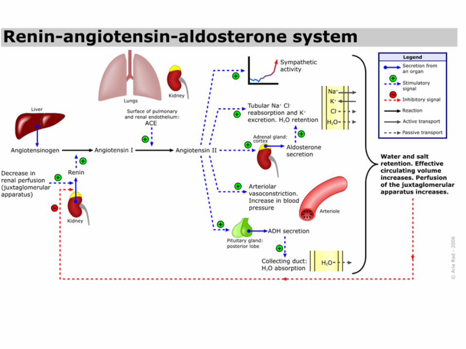
Renin→Angiotensin→AldosteroneAXIS (RAAS)
• If the perfusion of the juxtaglomerular apparatus in the kidneys decreases, then the juxtaglomerular cells release the enzyme renin.
• Renin cleaves an inactive peptide called angiotensinogen, converting it into angiotensin I.
• Angiotensin I is then converted to angiotensin II by angiotensin-converting enzyme (ACE), which is found mainly in lung capillaries.
• Angiotensin II is the major bioactive product of the renin-angiotensin system. Angiotensin II acts as an endocrine, autocrine/ paracrine, and intracrinehormone.
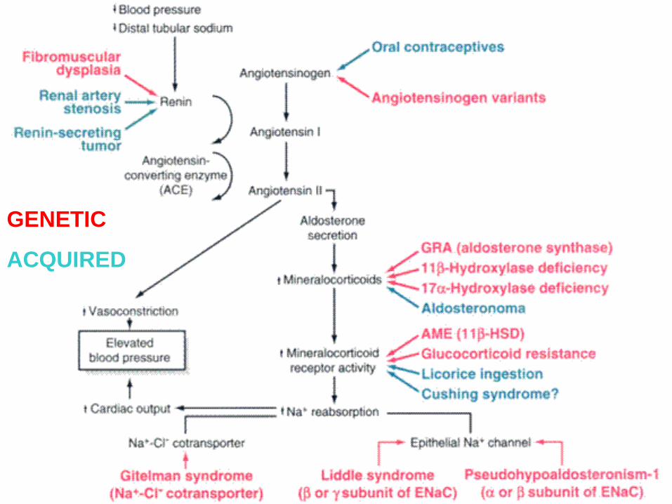
GENETIC
ACQUIRED
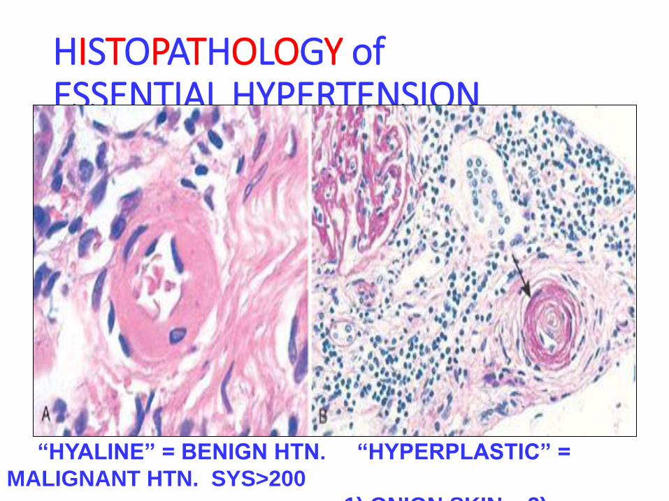
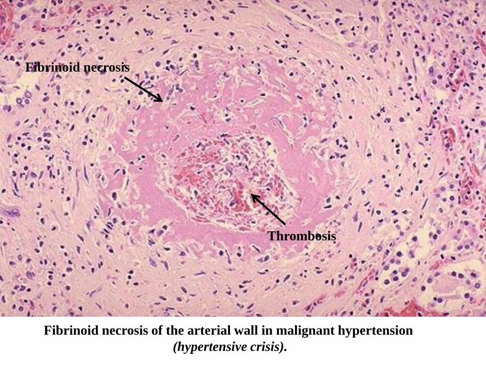
HISTOPATHOLOGY ofESSENTIAL HYPERTENSION
“HYALINE” = BENIGN HTN. “HYPERPLASTIC” =
MALIGNANT HTN. SYS>200
1) ONION SKIN 2)
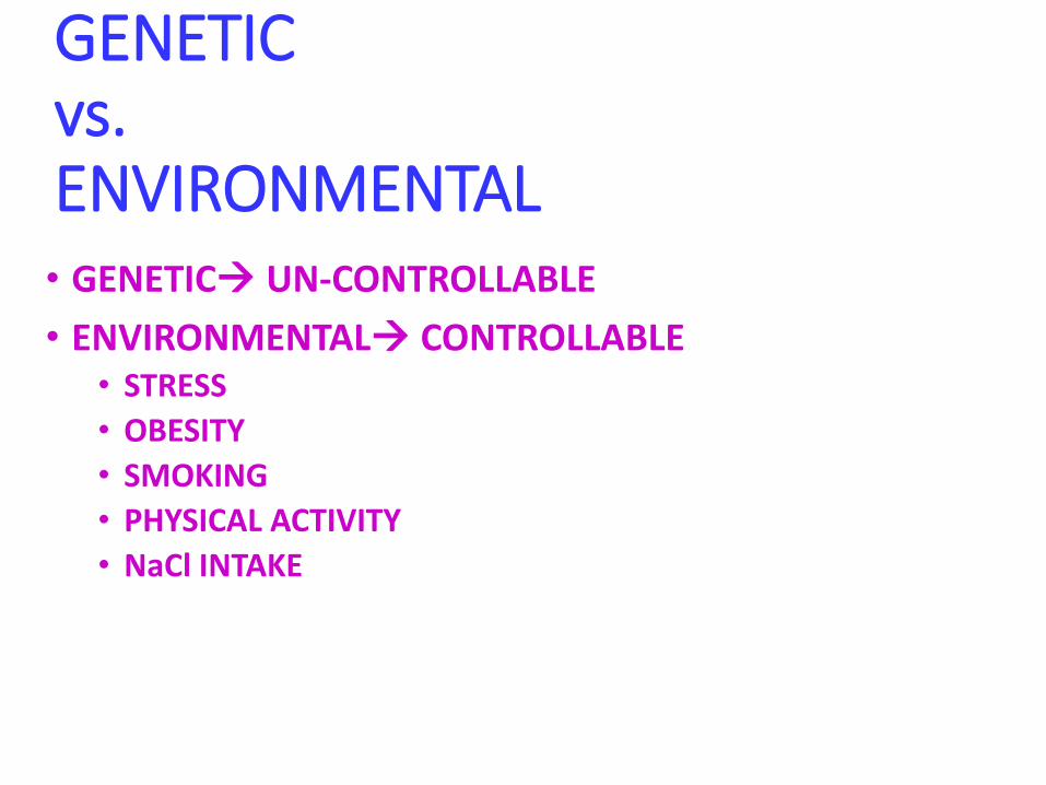
GENETICvs.ENVIRONMENTAL• GENETIC→ UN-CONTROLLABLE
• ENVIRONMENTAL→ CONTROLLABLE• STRESS
• OBESITY
• SMOKING
• PHYSICAL ACTIVITY
• NaCl INTAKE
ANEURYSMS• TRUE vs. FALSE
• ATHEROSCLEROTIC
• NON-ATHEROSCLEROTIC• CONGENITAL
• LUETIC (SYPHILITIC)
• TRAUMATIC
• “MYCOTIC” (MIS-leading term)
• 2° to VASCULITIS
• SACCULAR (i.e., “Berry”) vs. FUSIFORM
• DISSECTION vs. NON-DISSECTION

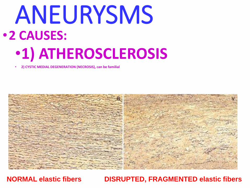
ANEURYSMS•2 CAUSES:
•1) ATHEROSCLEROSIS• 2) CYSTIC MEDIAL DEGENERATION (NECROSIS), can be familial
NORMAL elastic fibers DISRUPTED, FRAGMENTED elastic fibers
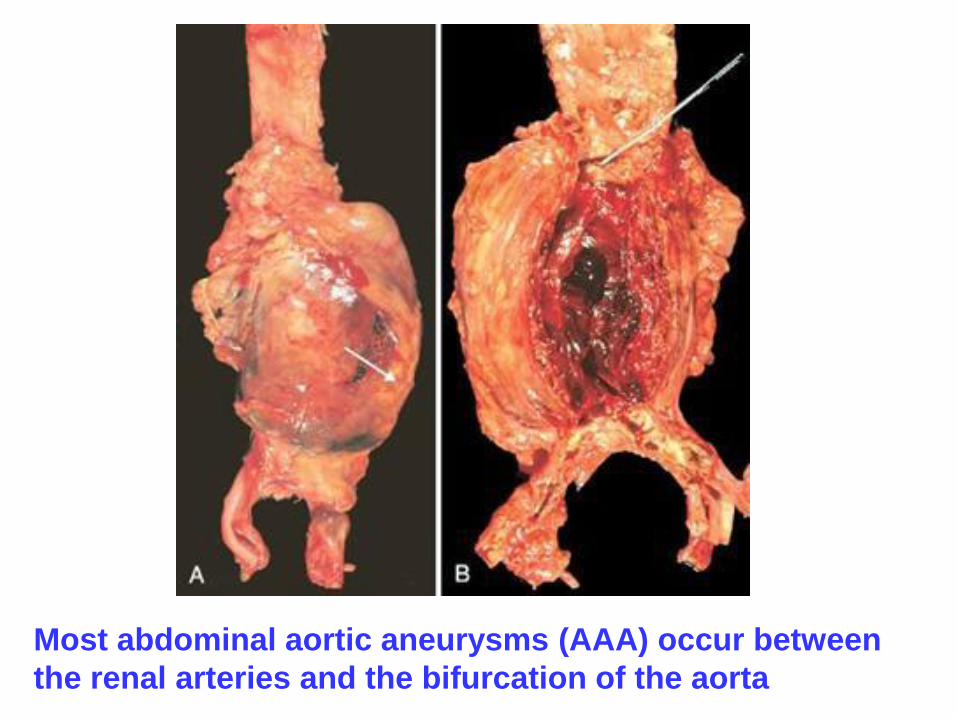
Most abdominal aortic aneurysms (AAA) occur between
the renal arteries and the bifurcation of the aorta
ANEURYSMS(sequelae)
•RUPTURE•OBSTRUCTION• EMBOLISM•COMPRESSION• URETER• SPINE
•MASS EFFECT
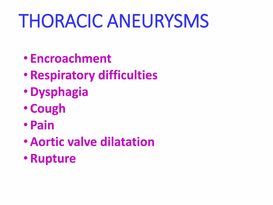
THORACIC ANEURYSMS
• Encroachment•Respiratory difficulties•Dysphagia•Cough•Pain•Aortic valve dilatation•Rupture
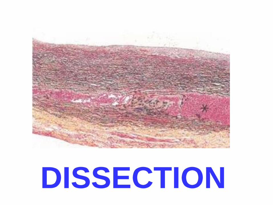
DISSECTION
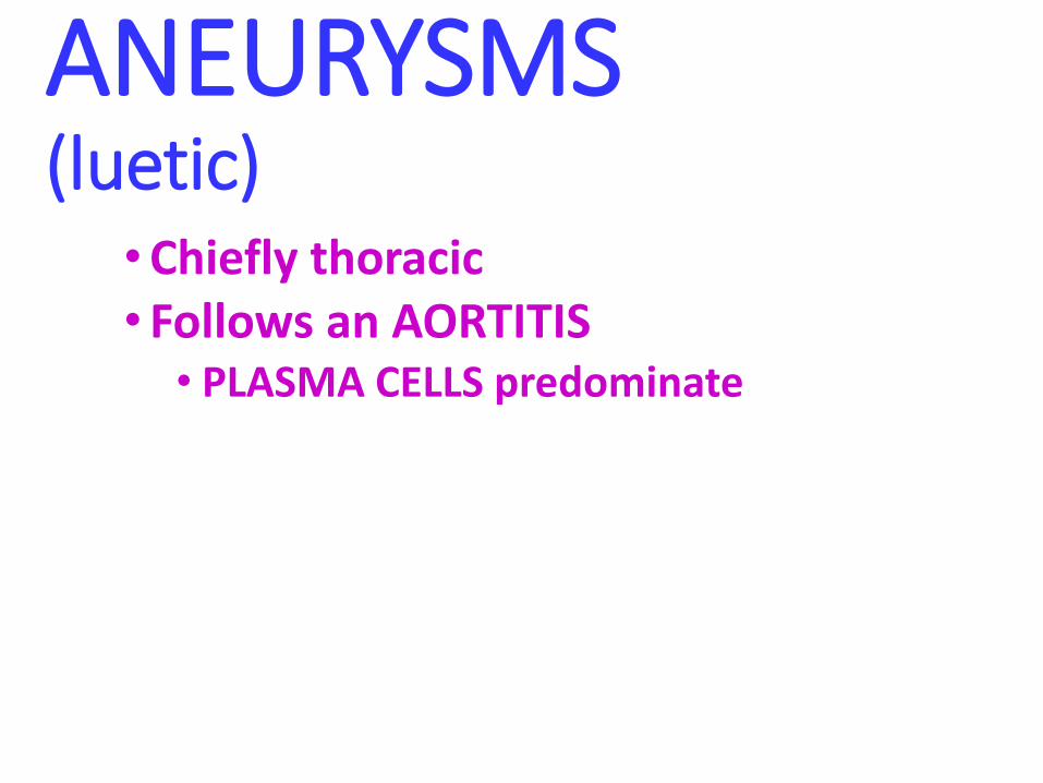
ANEURYSMS(luetic)
•Chiefly thoracic• Follows an AORTITIS• PLASMA CELLS predominate
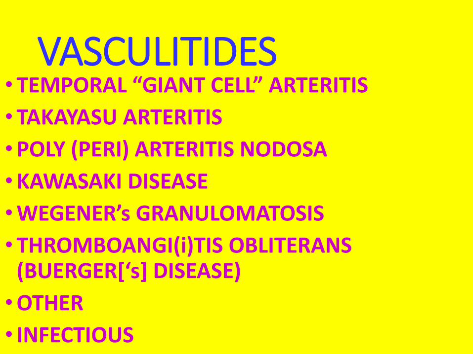
VASCULITIDES• TEMPORAL “GIANT CELL” ARTERITIS
• TAKAYASU ARTERITIS
•POLY (PERI) ARTERITIS NODOSA
•KAWASAKI DISEASE
•WEGENER’s GRANULOMATOSIS
• THROMBOANGI(i)TIS OBLITERANS (BUERGER[‘s] DISEASE)
•OTHER
• INFECTIOUS
VASCULITIDES• Chiefly arterial
• Infectious (5%) vs. Non-infectious (95%)
• NON-infectious are generally “AUTO”-IMMUNE. Why?
• Persistent findings:• Immune complexes
• ANTI-NEUTROPHIL AB’s (Wegener’s, “Temporal”)
• ANTI-ENDOTHELIAL CELL AB’s (Kawasaki)
• Often DRUG related (Hypersensitivity, e.g.)
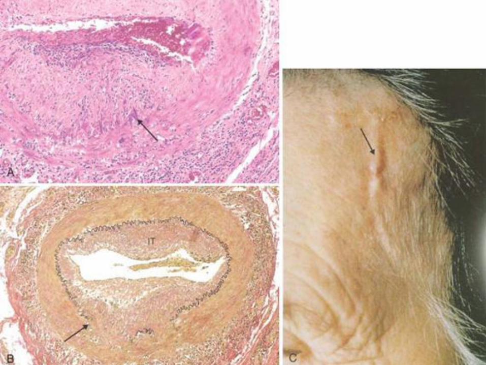
“TEMPORAL” ARTERITISaka, Giant Cell Arteritis, GCA• ADULTS
• Mainly arteries of the head and temporal arteries are the most visibly, palpably, and surgically accessible
• BLINDNESS most feared sequelae
• GRANULOMATOUS WALL inflammation diagnostic
• OFTEN associated with marked ESR elevation to be then known as POLYMYALGIA RHEUMATICA
• Anti-NEUTROPHIL AB’s often POSITIVE
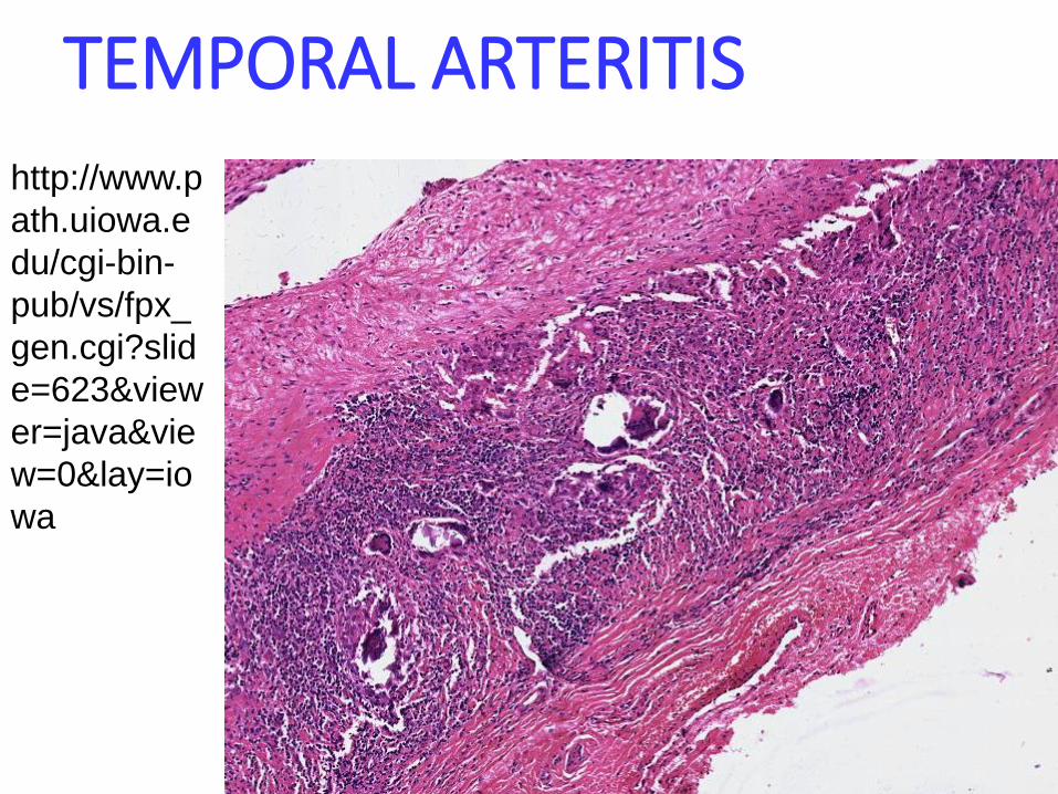
TEMPORAL ARTERITIS
http://www.p
ath.uiowa.e
du/cgi-bin-
pub/vs/fpx_
gen.cgi?slid
e=623&view
er=java&vie
w=0&lay=io
wa
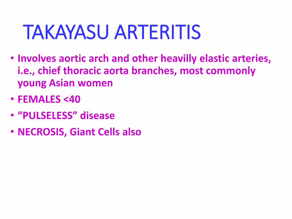
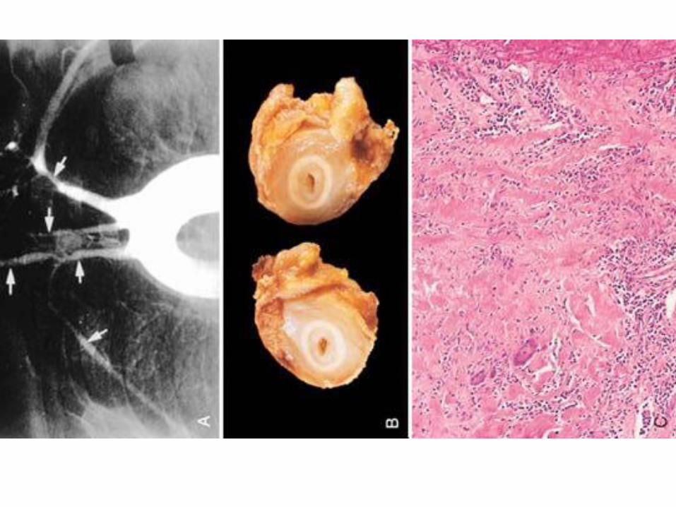
TAKAYASU ARTERITIS• Involves aortic arch and other heavilly elastic arteries,
i.e., chief thoracic aorta branches, most commonly young Asian women
• FEMALES <40
• “PULSELESS” disease
• NECROSIS, Giant Cells also
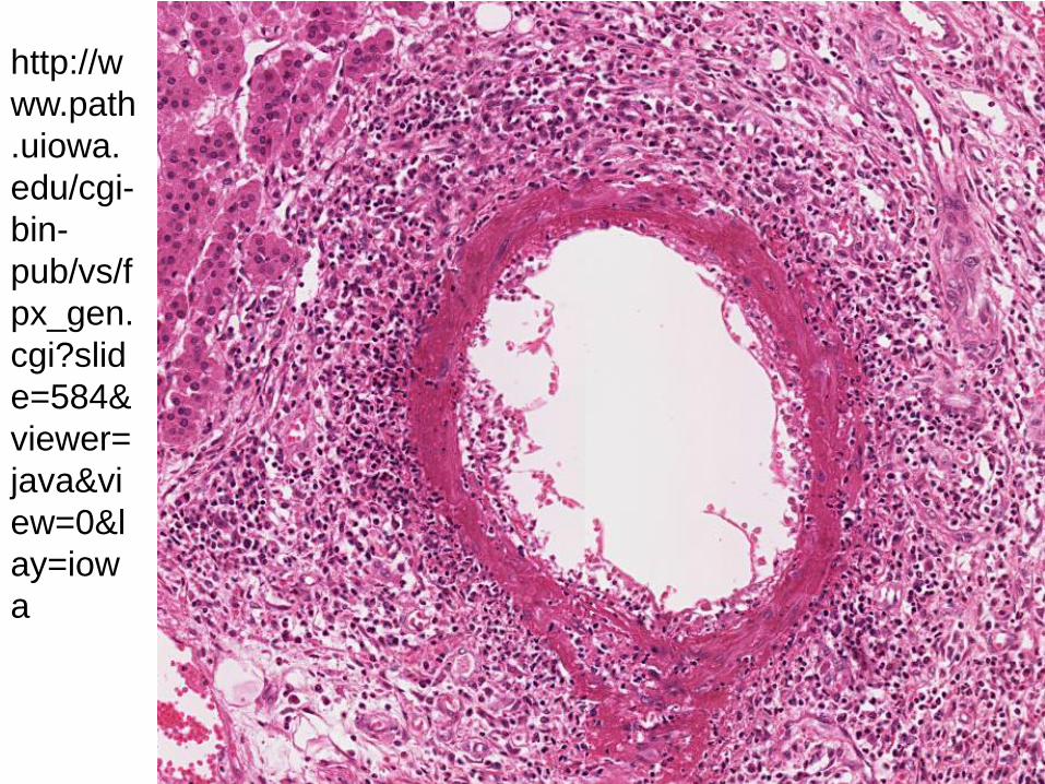
POLY-(Peri-) ARTERITIS NODOSA (PAN)
• ANY MEDIUM or SMALL artery
• OFTEN visceral arteries
• Infarcts, aneurysms, ischemia
• CLASSICAL AUTOIMMUNE disease
• SEGMENTAL, TRANSMURAL, NECROTIZING (fibrinoid) inflammation
http://w
ww.path
.uiowa.
edu/cgi-
bin-
pub/vs/f
px_gen.
cgi?slid
e=584&
viewer=
java&vi
ew=0&l
ay=iow
a
KAWASAKI DISEASE
• CHILDREN <4
• CORONARY ARTERIES
• LEADING cause of ACQUIRED heart disease in children
• USA and JAPAN
• Fatal in only 1%
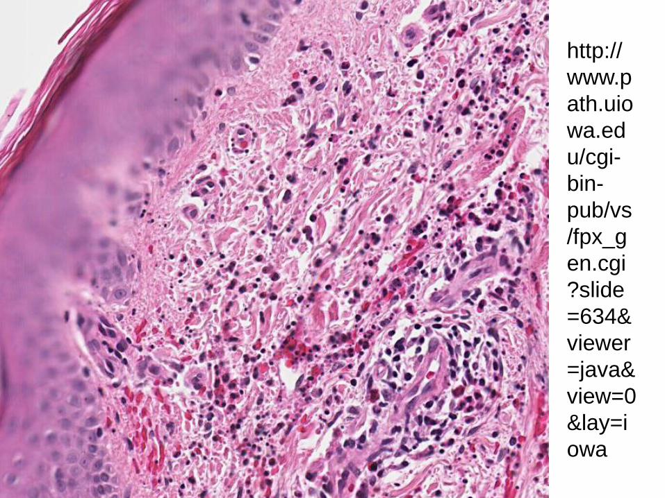
MICROSCOPIC POLYANGIITISHYPERSENSITIVITY VASCULITISLEUKOCYTOCLASTIC VASCULITIS
•SMALL VESSELS OF ALL TYPES, e.g., capillaries and veins too
•FRAGMENTED NEUTROPHILS• aka, LEUKOCYTOCLASIA
• aka, NUCLEAR “DUST”
• Most are ALLERGIC reactions to allergens like penicillin or strep
• DERMATOLOGIST’s DISEASE
http://
www.p
ath.uio
wa.ed
u/cgi-
bin-
pub/vs
/fpx_g
en.cgi
?slide
=634&
viewer
=java&
view=0
&lay=i
owa
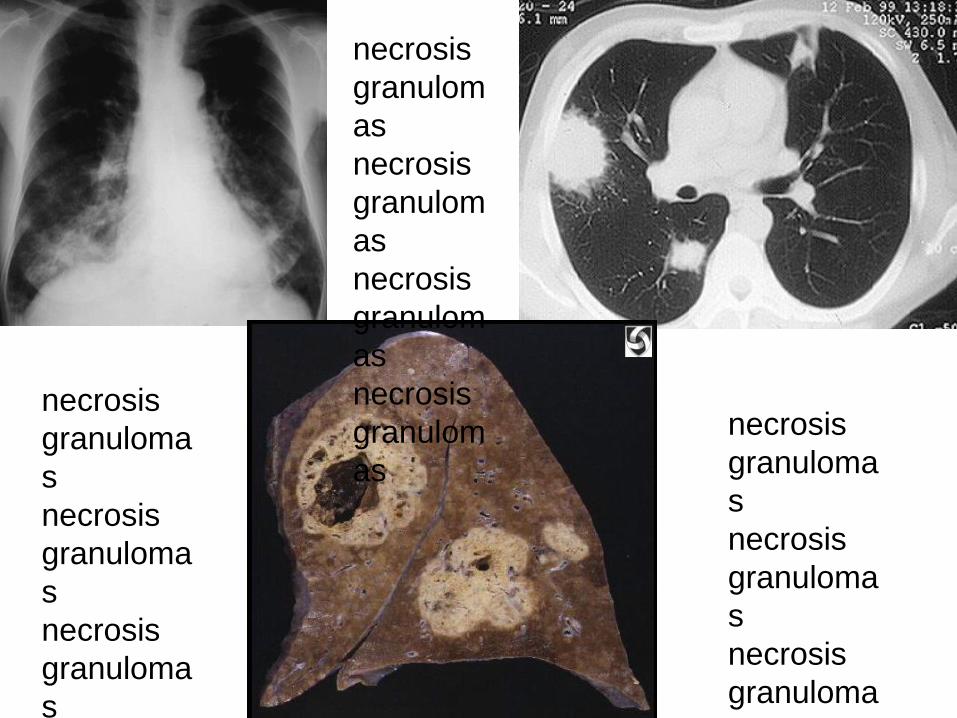
WEGENER GRANULOMATOSIS
• M>F, often in 40’s
• ACUTE NECROTIZING GRANULOMAS OF UPPER an LOWER respiratory tract
• NECROTIZING GRANULOMATOUS VASCULITIS of SMALL vessels of ALL types
• Often renal involvement, “crescentic” glomerulonephritis
• ANTI-NEUTROPHIL-CYTOPLASMIC-AB’s usually present
necrosis
granuloma
s
necrosis
granuloma
s
necrosis
granuloma
s
necrosis
necrosis
granuloma
s
necrosis
granuloma
s
necrosis
granuloma
s
necrosis
granulom
as
necrosis
granulom
as
necrosis
granulom
as
necrosis
granulom
as
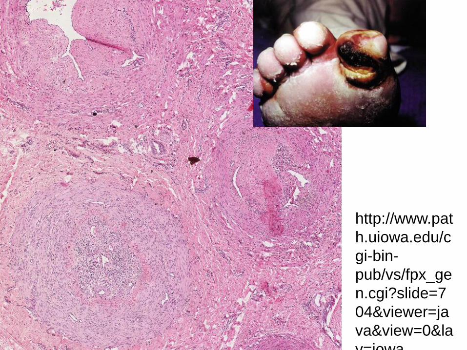
THROMBOANGIITIS OBLITERANSBUERGER(‘s) Disease
• 100% caused by cigarette smoking
• MEN>>>F, 30’s, 40’s
• Often arteries are 100% obliterated, hence the name “obliterans”
• EXTREMITIES most often involved
http://www.pat
h.uiowa.edu/c
gi-bin-
pub/vs/fpx_ge
n.cgi?slide=7
04&viewer=ja
va&view=0&la
y=iowa
OTHER VASCULITIDES
•SLE
•RHEUMATOID ARTHRITIS
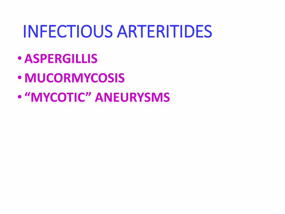
INFECTIOUS ARTERITIDES
•ASPERGILLIS
•MUCORMYCOSIS
• “MYCOTIC” ANEURYSMS
NON ATHEROSCLEROSISVASCULAR DISEASES•HYPERTENSION
•ANEURYSMS
•VASCULITIDES
•VEIN DISORDERS
•NEOPLASMS
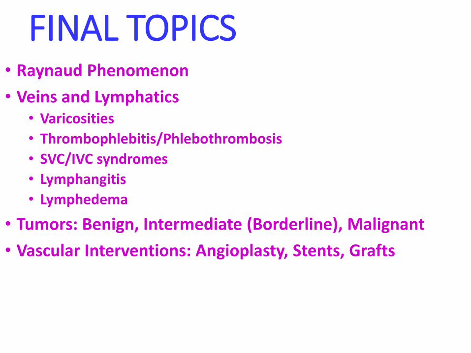
FINAL TOPICS• Raynaud Phenomenon
• Veins and Lymphatics• Varicosities
• Thrombophlebitis/Phlebothrombosis
• SVC/IVC syndromes
• Lymphangitis
• Lymphedema
• Tumors: Benign, Intermediate (Borderline), Malignant
• Vascular Interventions: Angioplasty, Stents, Grafts
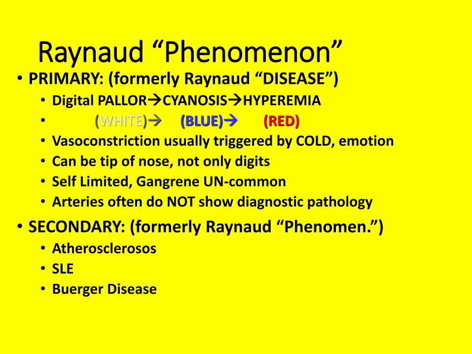
Raynaud “Phenomenon”• PRIMARY: (formerly Raynaud “DISEASE”)
• Digital PALLOR→CYANOSIS→HYPEREMIA
• (WHITE)→ (BLUE)→ (RED)
• Vasoconstriction usually triggered by COLD, emotion
• Can be tip of nose, not only digits
• Self Limited, Gangrene UN-common
• Arteries often do NOT show diagnostic pathology
• SECONDARY: (formerly Raynaud “Phenomen.”)• Atherosclerosos
• SLE
• Buerger Disease
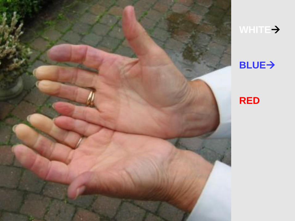
WHITE→
BLUE→
RED
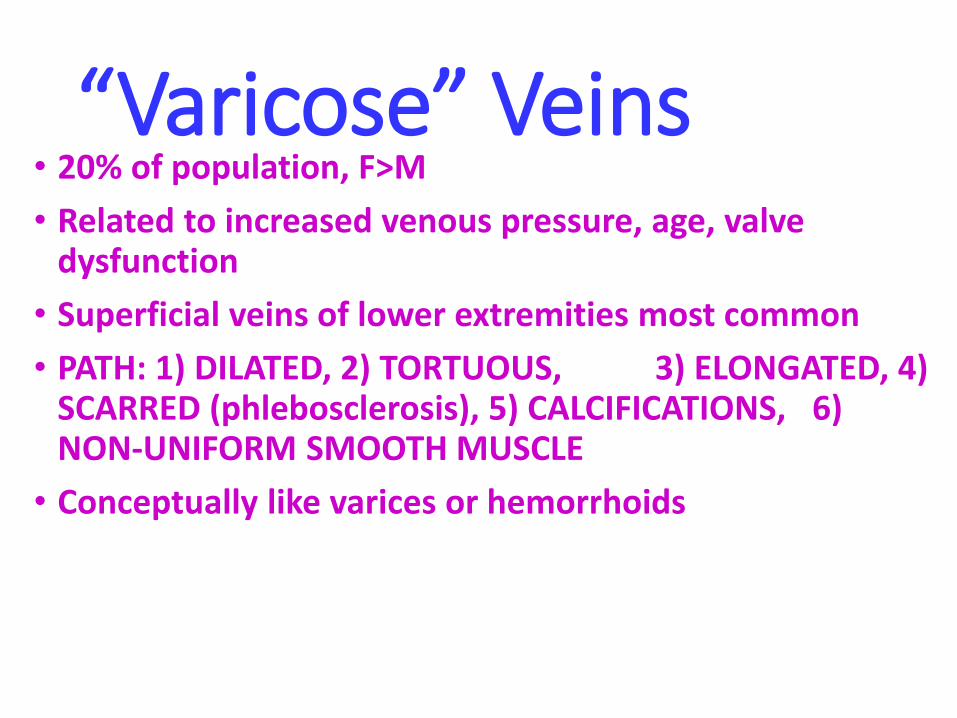
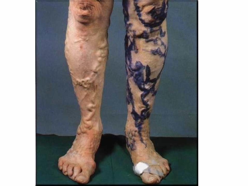
“Varicose” Veins• 20% of population, F>M
• Related to increased venous pressure, age, valve dysfunction
• Superficial veins of lower extremities most common
• PATH: 1) DILATED, 2) TORTUOUS, 3) ELONGATED, 4) SCARRED (phlebosclerosis), 5) CALCIFICATIONS, 6) NON-UNIFORM SMOOTH MUSCLE
• Conceptually like varices or hemorrhoids
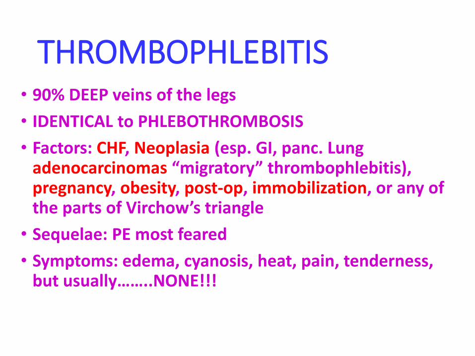
THROMBOPHLEBITIS• 90% DEEP veins of the legs
• IDENTICAL to PHLEBOTHROMBOSIS
• Factors: CHF, Neoplasia (esp. GI, panc. Lung adenocarcinomas “migratory” thrombophlebitis), pregnancy, obesity, post-op, immobilization, or any of the parts of Virchow’s triangle
• Sequelae: PE most feared
• Symptoms: edema, cyanosis, heat, pain, tenderness, but usually……..NONE!!!
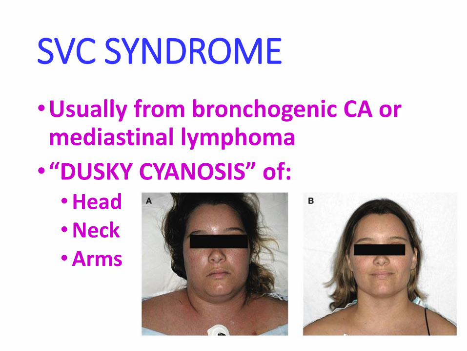
SVC SYNDROME
•Usually from bronchogenic CA or mediastinal lymphoma
•“DUSKY CYANOSIS” of:•Head•Neck•Arms
IVC SYNDROME• Secondary to:
• NEOPLASMS (external compression)
• ASCENDING THROMBOSIS from FEMORALS, ILIACS
• AAA, Gravid uterus
• Bilateral leg edema
• Massive proteinuria if renal veins involved (like nephrotic syndrome)
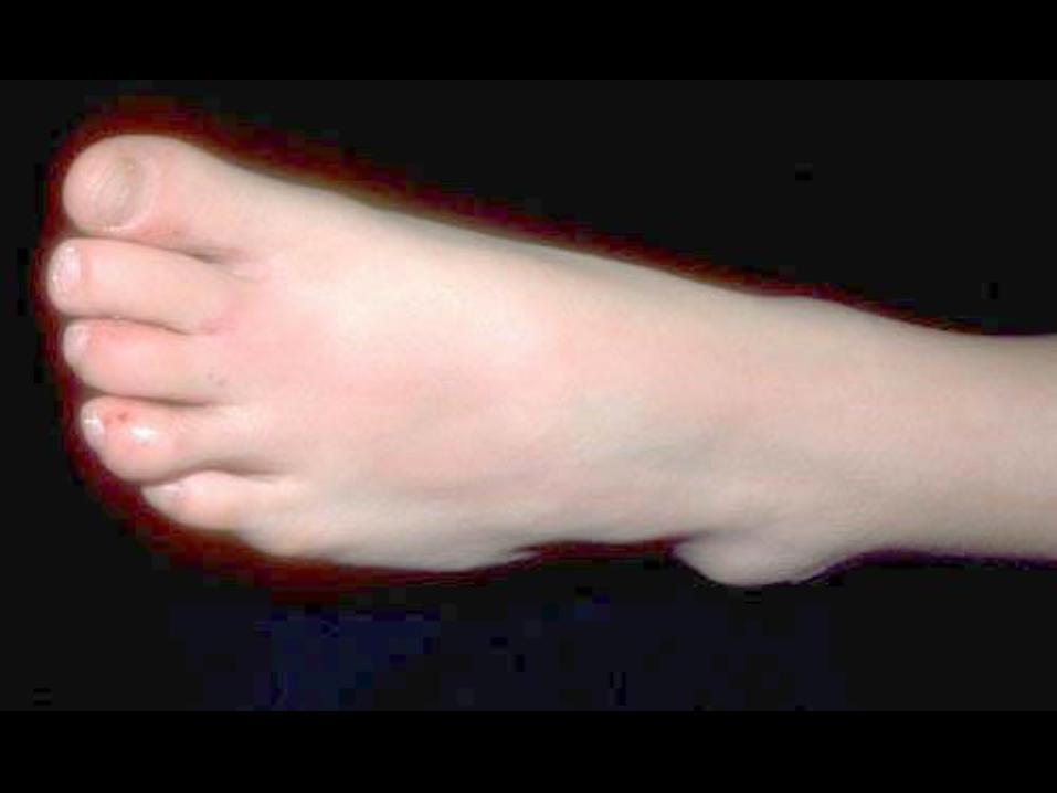
LYMPHANGITIS• From regional infections
• Group-A beta-hemolytic strep most common
• Lymphatics dilated, filled with WBCs
• Cellulitis usually present too
• Lymphadenitis also usually follows
• If lymph nodes cannot filter (process) antigens
enough→ septicemia
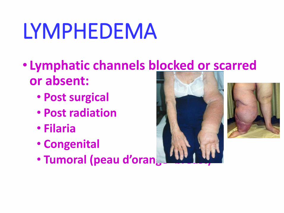
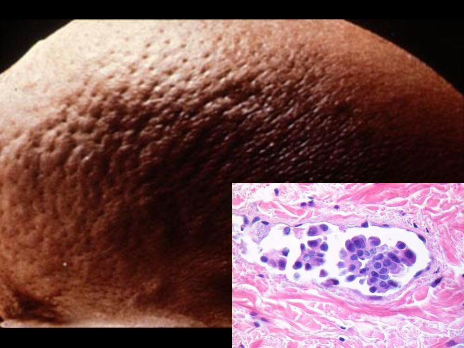
LYMPHEDEMA
• Lymphatic channels blocked or scarred or absent:• Post surgical• Post radiation• Filaria• Congenital• Tumoral (peau d’orange- breast)
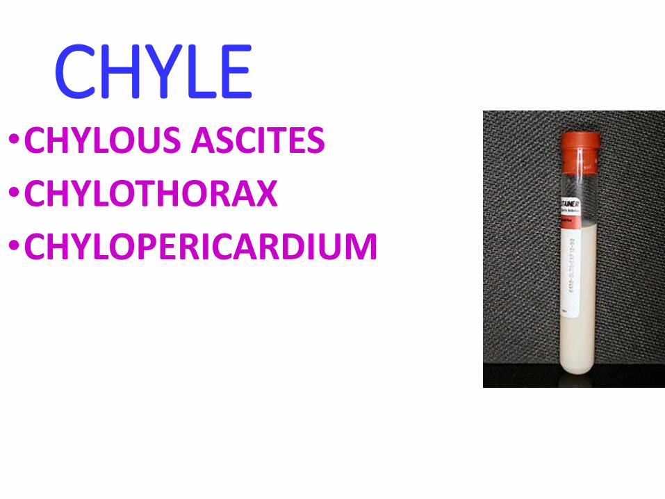
CHYLE•CHYLOUS ASCITES
•CHYLOTHORAX
•CHYLOPERICARDIUM
Vascular TUMORS
•BENIGN (NEVER metastasize, in fact some are not even TRUE neoplasms, but hamartomas)
• INTERMEDIATE (rarely metastasize)
•MALIGNANT (FREQUENT and EARLY metastases, like any other sarcoma→ lung)
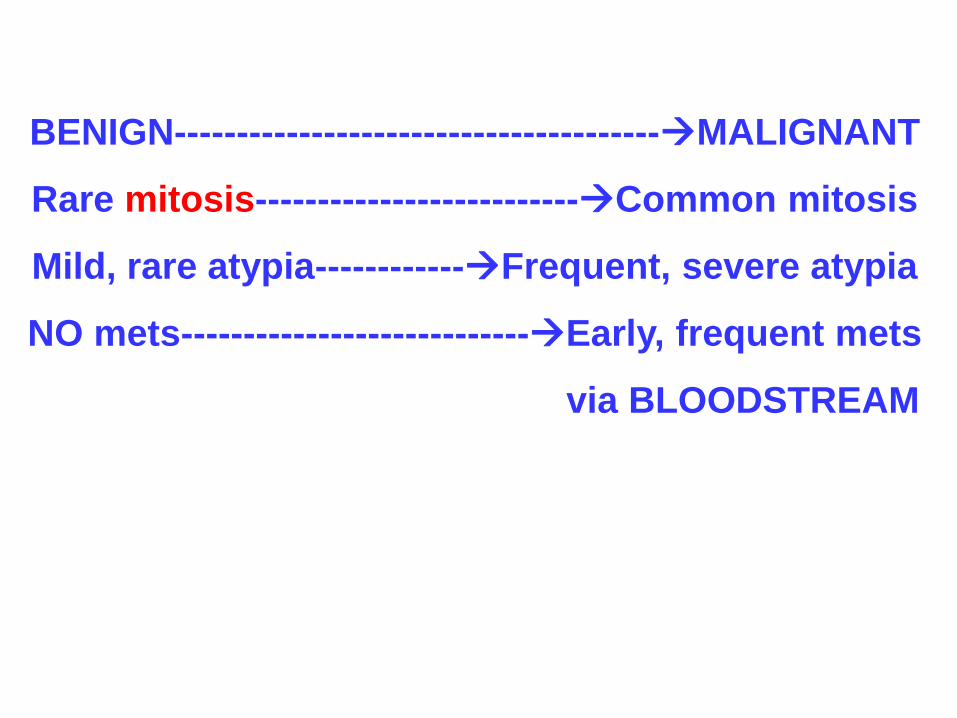
BENIGN---------------------------------------→MALIGNANT
Rare mitosis--------------------------→Common mitosis
Mild, rare atypia------------→Frequent, severe atypia
NO mets----------------------------→Early, frequent mets
via BLOODSTREAM
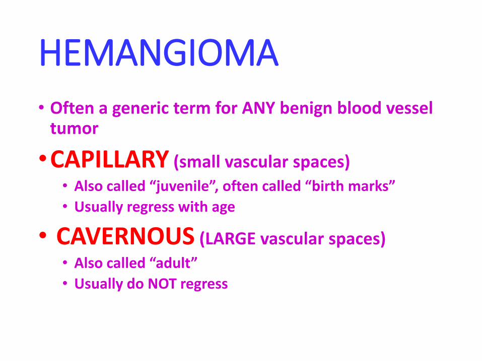
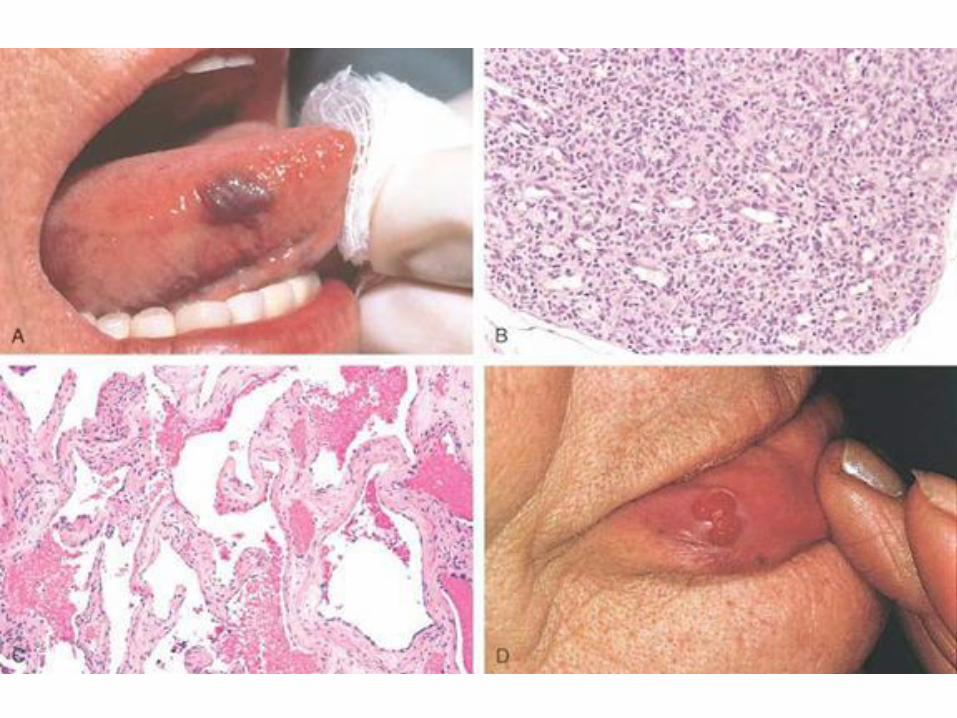
HEMANGIOMA• Often a generic term for ANY benign blood vessel
tumor
•CAPILLARY (small vascular spaces)
• Also called “juvenile”, often called “birth marks”
• Usually regress with age
• CAVERNOUS (LARGE vascular spaces)
• Also called “adult”
• Usually do NOT regress
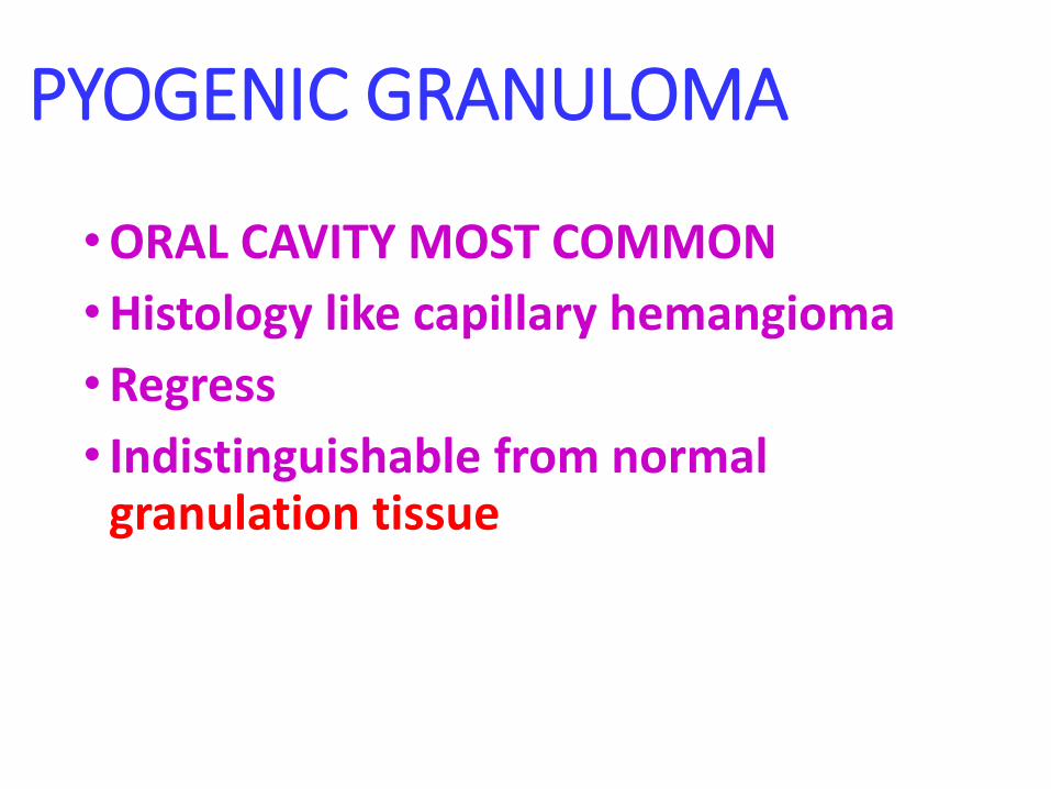
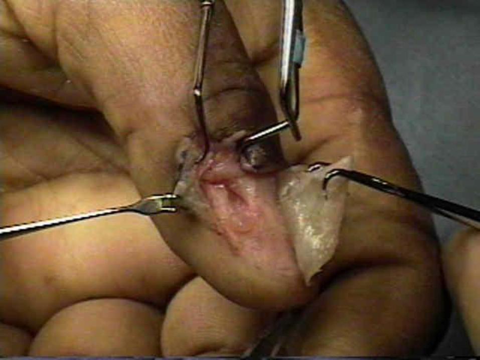
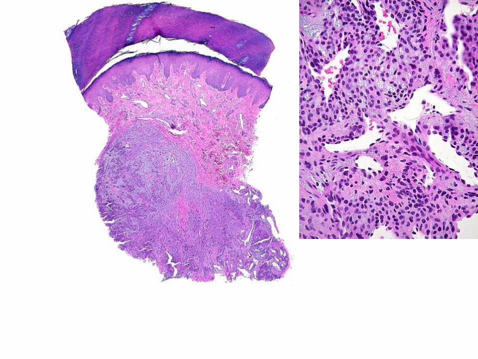
PYOGENIC GRANULOMA
•ORAL CAVITY MOST COMMON
•Histology like capillary hemangioma
•Regress
• Indistinguishable from normal granulation tissue
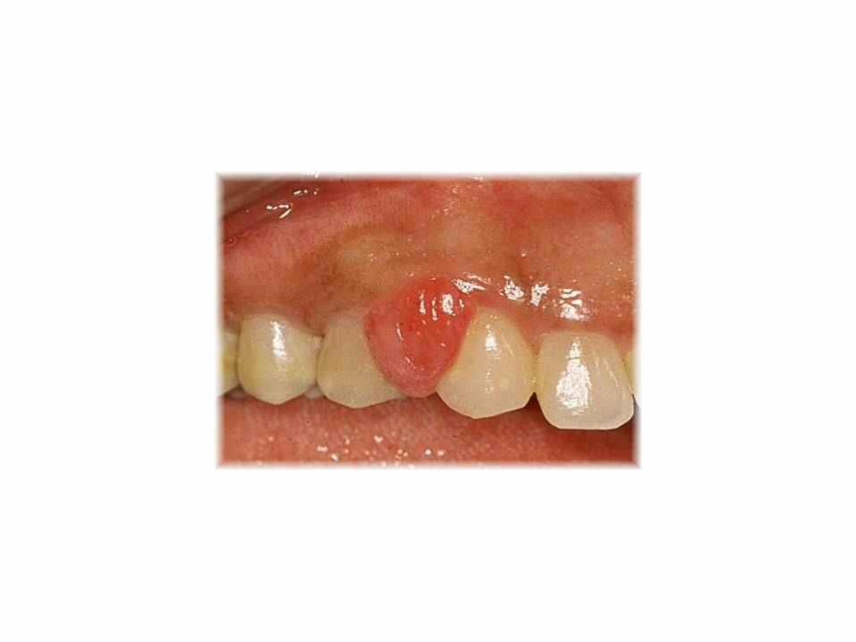
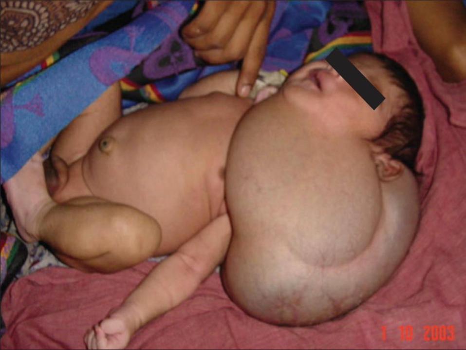
LYMPHANGIOMA• Small 1-2 mm
•90% Head and neck region in kids <2
•Generally……RARE
•When large size and/or spaces present often called “CYSTIC HYGROMA”
GLOMUS TUMORGLOMANGIOMA
•1 cm
•Most commonly under nail
•Painful
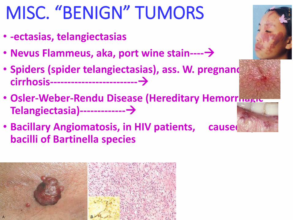
MISC. “BENIGN” TUMORS• -ectasias, telangiectasias
• Nevus Flammeus, aka, port wine stain----→
• Spiders (spider telangiectasias), ass. W. pregnancy, cirrhosis-------------------------→
• Osler-Weber-Rendu Disease (Hereditary Hemorrhagic Telangiectasia)-------------→
• Bacillary Angiomatosis, in HIV patients, caused by bacilli of Bartinella species
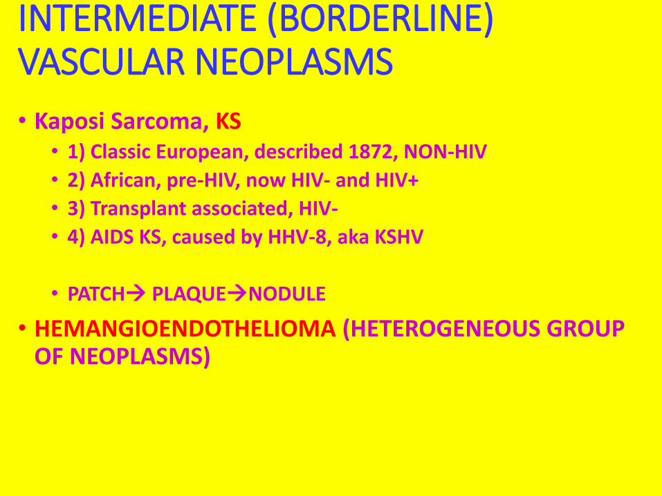
INTERMEDIATE (BORDERLINE)VASCULAR NEOPLASMS
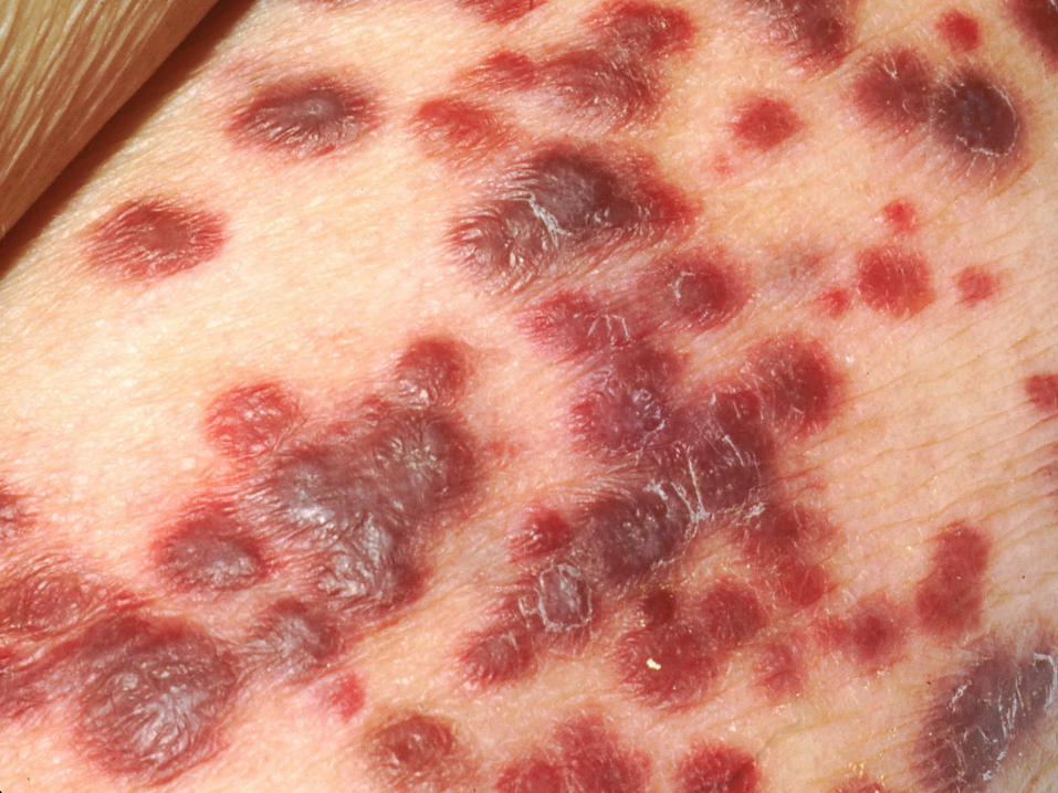
• Kaposi Sarcoma, KS• 1) Classic European, described 1872, NON-HIV
• 2) African, pre-HIV, now HIV- and HIV+
• 3) Transplant associated, HIV-
• 4) AIDS KS, caused by HHV-8, aka KSHV
• PATCH→ PLAQUE→NODULE
• HEMANGIOENDOTHELIOMA (HETEROGENEOUS GROUP OF NEOPLASMS)
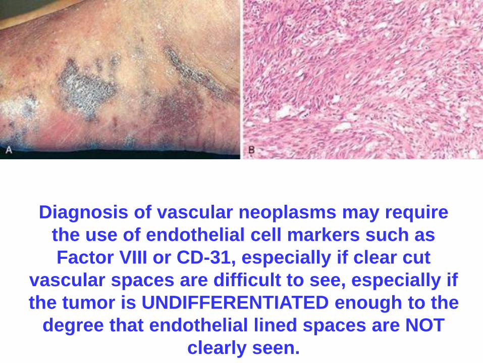
Diagnosis of vascular neoplasms may require
the use of endothelial cell markers such as
Factor VIII or CD-31, especially if clear cut
vascular spaces are difficult to see, especially if
the tumor is UNDIFFERENTIATED enough to the
degree that endothelial lined spaces are NOT
clearly seen.
MALIGNANT VASCULAR TUMORS
•ANGIOSARCOMA• May not look “vascular” at all
• Severe atypia
• Frequent and often bizarre mitoses
• Behave as any sarcoma might, i.e., early pulmonary metastases
•HEMANGIOPERICYTOMA• HETEROGENOUS group of disorders
• Most commonly arising in pelvic retroperitoneum
VASCULAR INTERVENTIONS
• ANGIOPLASTY
• STENTS
• GRAFTS• Autologous (saphenous v., internal mammary a.)
• Synthetic (Teflon)

ANGIOPLASTIES•Plaque fracture (crackling sound)
•Dissection
•Arterial dilatation initially
•Restenosis ~ 6 months
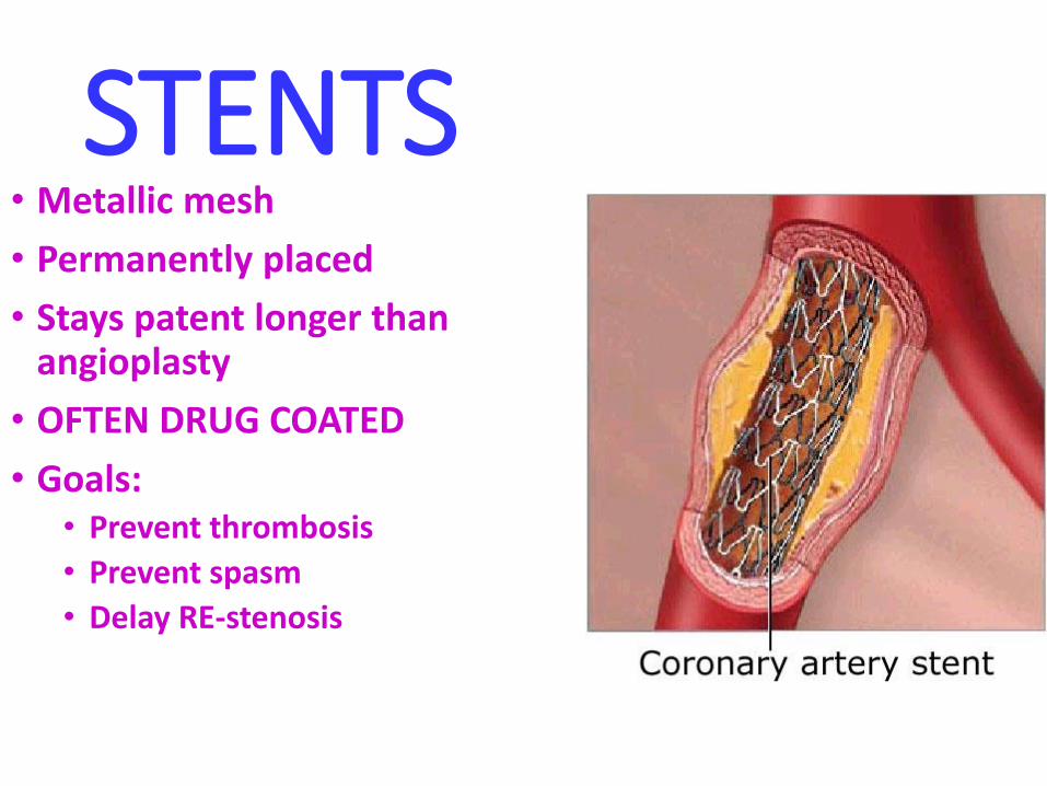
STENTS• Metallic mesh
• Permanently placed
• Stays patent longer than angioplasty
• OFTEN DRUG COATED
• Goals:• Prevent thrombosis
• Prevent spasm
• Delay RE-stenosis
GRAFTS• 400,000 CABG grafts per year in USA
• Saphenous v. vs. Internal mammary a. (internal thoracic a.)
• 50% patent after 10 years, for saphenous v.
• 90% patent after 10 years, for mammary a.
• Endothelial and smooth muscle migration and proliferation key factors for success