van de graaff: human vi. maintenance of the 19. urinary
TRANSCRIPT

Van De Graaff: Human Anatomy, Sixth Edition
VI. Maintenance of the Body
19. Urinary System © The McGraw−Hill Companies, 2001
Urinary System
Clinical Case StudyA 17-year-old male was involved in a knife fight in which he sustained two stab wounds to theanterior abdomen. He was brought to the emergency room where he complained of mild ab-dominal pain and an urgent need to urinate. Although neither of the stab wounds was bleedingexternally, examination by the surgeon revealed signs of moderate hemorrhagic shock. Onewound was 3 cm below the right costal margin at the midclavicular line; the other was just me-dial to the right anterior superior iliac spine. The surgeon immediately ordered preparations foran emergency exploratory laparotomy. She noted that the patient’s urinary bladder was quitefull and chided the intern for not having placed a urinary catheter. Placement of the catheteryielded a brisk flow of bright red blood.
How would you explain the phenomenon of hematuria (blood in the urine) in this case?Which of the two stab wounds is most likely associated with the hematuria? Regarding theblood draining into the catheter, trace and explain its course of flow. Begin at a point in the ab-dominal aorta, and end the course with drainage into the catheter. Assuming that the surgeonwould be prompted to remove the kidney in order to quickly control hemorrhage, what possibleanatomical variant should she keep in mind?
Hints: Study the positions of the kidneys within the abdominal cavity and note the location ofthe supportive and protective serous membrane. Carefully examine the specific location ofurine production and the route of urine passage through the urinary system.
Introduction to the Urinary System 676
Kidneys 676Ureters, Urinary Bladder,
and Urethra 684
CLINICAL CONSIDERATIONS 688
Developmental Exposition: The Urinary System 689
Clinical Case Study Answer 694Chapter Summary 695Review Activities 696
19
FIGURE: The placement of a urinarycatheter is a common procedure for patientswho have abdominal trauma or abdominalsurgery. A laboratory assessment (urinalysis)of the collected urine may help reveal theextent of the trauma or a patient’s progress inhealing.
Van De Graaff: Human Anatomy, Sixth Edition
VI. Maintenance of the Body
19. Urinary System © The McGraw−Hill Companies, 2001
INTRODUCTION TO THE URINARY SYSTEMThe urinary system maintains the composition and properties ofthe body fluid that establishes the internal environment of thebody cells. The end product of the urinary system is urine, whichis voided from the body during micturition.
Objective 1 List the functions of the urinary system.
Objective 2 Identify the arteries that transport blood to theurinary system for filtration.
The urinary system, along with the respiratory, digestive, and in-tegumentary systems, excretes substances from the body. For thisreason, these systems are occasionally referred to as excretory sys-tems. In the process of cellular metabolism, nutrients taken in bythe digestive system and oxygen from inhaled air are used to syn-thesize a variety of substances while providing energy needed forbody maintenance. Metabolic processes, however, produce cellu-lar wastes that must be eliminated if homeostasis is to be main-tained. Just as the essential nutrients are transported to the cellsby the blood, the cellular wastes are removed through the circu-latory system to the appropriate excretory system. Carbon diox-ide is eliminated through the respiratory system; excessive water,salts, nitrogenous wastes, and even excessive metabolic heat areremoved through the integumentary system; and various diges-tive wastes are eliminated through the digestive system.
The urinary system is the principal system responsible forwater and electrolyte balance. Electrolytes are compounds thatseparate into ions when dissolved in water. Electrolyte balance isachieved when the number of electrolytes entering the bodyequals the number leaving. Hydrogen ions, for example, aremaintained in precise concentration so that an acid-base, or pH,balance exists in the body.
A second major function of the urinary system is the excre-tion of toxic nitrogenous compounds—specifically, urea and cre-atinine. Other functions of the urinary system include theelimination of toxic wastes that may result from bacterial actionand the removal of various drugs that have been taken into thebody. All of these functions are accomplished through the forma-tion of urine by the kidneys.
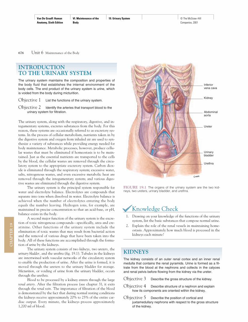
The urinary system consists of two kidneys, two ureters, theurinary bladder, and the urethra (fig. 19.1). Tubules in the kidneysare intertwined with vascular networks of the circulatory systemto enable the production of urine. After the urine is formed, it ismoved through the ureters to the urinary bladder for storage.Micturition, or voiding of urine from the urinary bladder, occursthrough the urethra.
Blood to be processed by a kidney enters through the largerenal artery. After the filtration process (see chapter 3), it exitsthrough the renal vein. The importance of filtration of the bloodis demonstrated by the fact that during normal resting conditionsthe kidneys receive approximately 20% to 25% of the entire car-diac output. Every minute, the kidneys process approximately1,200 ml of blood.
Knowledge Check1. Drawing on your knowledge of the functions of the urinary
system, list the basic substances that compose normal urine.2. Explain the role of the renal vessels in maintaining home-
ostasis. Approximately how much blood is processed in thekidneys each minute?
KIDNEYSThe kidney consists of an outer renal cortex and an inner renalmedulla that contains the renal pyramids. Urine is formed as a fil-trate from the blood at the nephrons and collects in the calycesand renal pelvis before flowing from the kidney via the ureter.
Objective 3 Describe the gross structure of the kidney.
Objective 4 Describe structure of a nephron and explainhow its components are oriented within the kidney.
Objective 5 Describe the position of cortical andjuxtamedullary nephrons with respect to the gross structureof the kidney.
676 Unit 6 Maintenance of the Body
CH
AP
TE
R 1
9
Inferiorvena cava
Kidney
Abdominalaorta
Ureter
Urinary bladder
Urethra
FIGURE 19.1 The organs of the urinary system are the two kid-neys, two ureters, urinary bladder, and urethra.
Van De Graaff: Human Anatomy, Sixth Edition
VI. Maintenance of the Body
19. Urinary System © The McGraw−Hill Companies, 2001
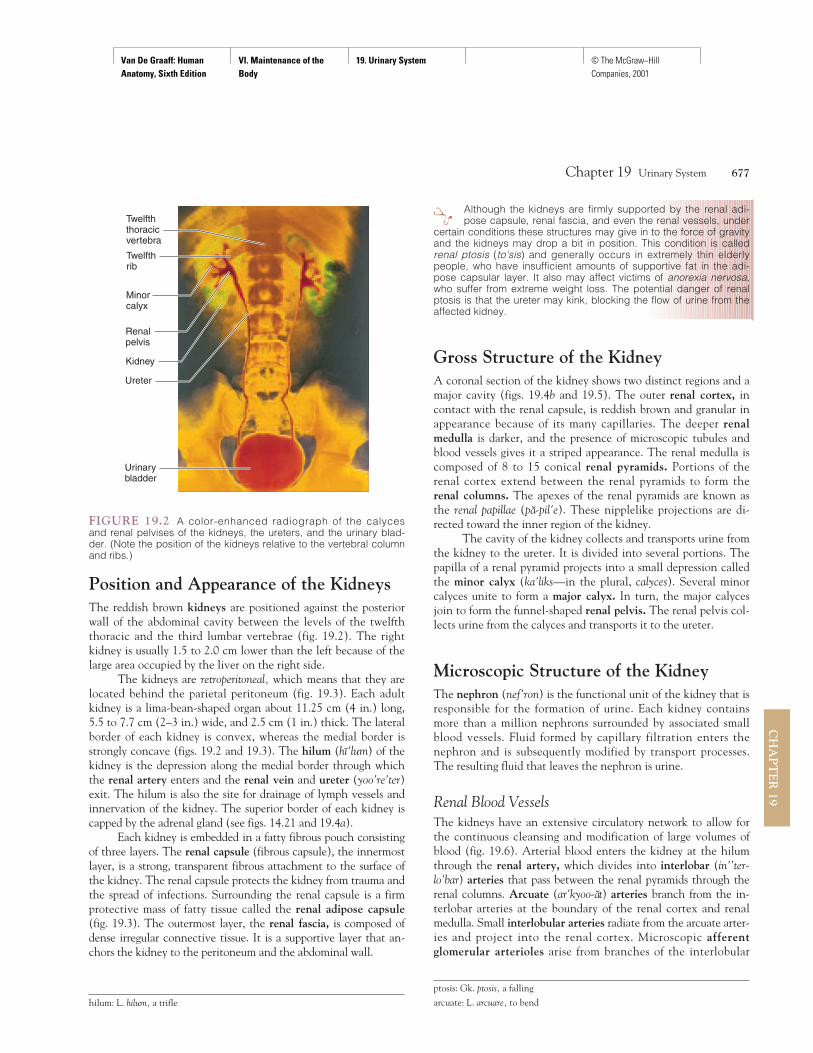
Position and Appearance of the KidneysThe reddish brown kidneys are positioned against the posteriorwall of the abdominal cavity between the levels of the twelfththoracic and the third lumbar vertebrae (fig. 19.2). The rightkidney is usually 1.5 to 2.0 cm lower than the left because of thelarge area occupied by the liver on the right side.
The kidneys are retroperitoneal, which means that they arelocated behind the parietal peritoneum (fig. 19.3). Each adultkidney is a lima-bean-shaped organ about 11.25 cm (4 in.) long,5.5 to 7.7 cm (2–3 in.) wide, and 2.5 cm (1 in.) thick. The lateralborder of each kidney is convex, whereas the medial border isstrongly concave (figs. 19.2 and 19.3). The hilum (hı'lum) of thekidney is the depression along the medial border through whichthe renal artery enters and the renal vein and ureter (yoo're'ter)exit. The hilum is also the site for drainage of lymph vessels andinnervation of the kidney. The superior border of each kidney iscapped by the adrenal gland (see figs. 14.21 and 19.4a).
Each kidney is embedded in a fatty fibrous pouch consistingof three layers. The renal capsule (fibrous capsule), the innermostlayer, is a strong, transparent fibrous attachment to the surface ofthe kidney. The renal capsule protects the kidney from trauma andthe spread of infections. Surrounding the renal capsule is a firmprotective mass of fatty tissue called the renal adipose capsule(fig. 19.3). The outermost layer, the renal fascia, is composed ofdense irregular connective tissue. It is a supportive layer that an-chors the kidney to the peritoneum and the abdominal wall.
Although the kidneys are firmly supported by the renal adi-pose capsule, renal fascia, and even the renal vessels, under
certain conditions these structures may give in to the force of gravityand the kidneys may drop a bit in position. This condition is calledrenal ptosis (to'sis) and generally occurs in extremely thin elderlypeople, who have insufficient amounts of supportive fat in the adi-pose capsular layer. It also may affect victims of anorexia nervosa,who suffer from extreme weight loss. The potential danger of renalptosis is that the ureter may kink, blocking the flow of urine from theaffected kidney.
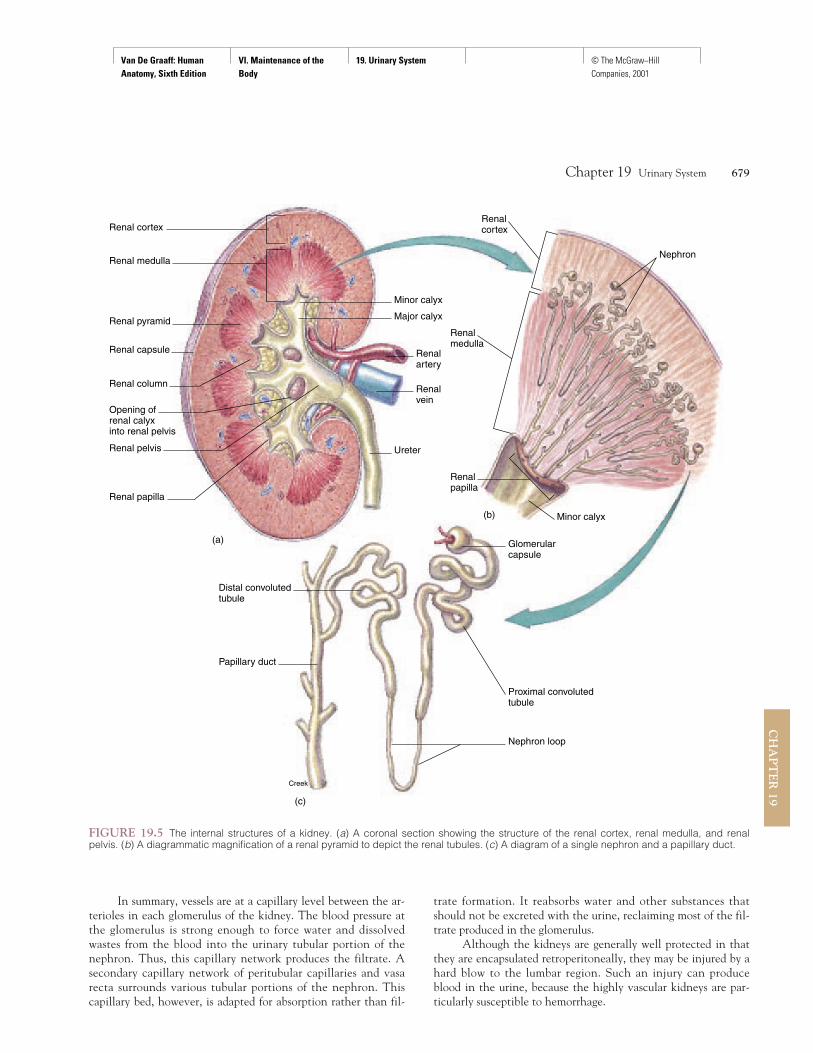
Gross Structure of the KidneyA coronal section of the kidney shows two distinct regions and amajor cavity (figs. 19.4b and 19.5). The outer renal cortex, incontact with the renal capsule, is reddish brown and granular inappearance because of its many capillaries. The deeper renalmedulla is darker, and the presence of microscopic tubules andblood vessels gives it a striped appearance. The renal medulla iscomposed of 8 to 15 conical renal pyramids. Portions of therenal cortex extend between the renal pyramids to form therenal columns. The apexes of the renal pyramids are known asthe renal papillae (pa-pil'e). These nipplelike projections are di-rected toward the inner region of the kidney.
The cavity of the kidney collects and transports urine fromthe kidney to the ureter. It is divided into several portions. Thepapilla of a renal pyramid projects into a small depression calledthe minor calyx (ka'liks—in the plural, calyces). Several minorcalyces unite to form a major calyx. In turn, the major calycesjoin to form the funnel-shaped renal pelvis. The renal pelvis col-lects urine from the calyces and transports it to the ureter.
Microscopic Structure of the KidneyThe nephron (nef'ron) is the functional unit of the kidney that isresponsible for the formation of urine. Each kidney containsmore than a million nephrons surrounded by associated smallblood vessels. Fluid formed by capillary filtration enters thenephron and is subsequently modified by transport processes.The resulting fluid that leaves the nephron is urine.
Renal Blood VesselsThe kidneys have an extensive circulatory network to allow forthe continuous cleansing and modification of large volumes ofblood (fig. 19.6). Arterial blood enters the kidney at the hilumthrough the renal artery, which divides into interlobar (in''ter-lo'bar) arteries that pass between the renal pyramids through therenal columns. Arcuate (ar'kyoo-at) arteries branch from the in-terlobar arteries at the boundary of the renal cortex and renalmedulla. Small interlobular arteries radiate from the arcuate arter-ies and project into the renal cortex. Microscopic afferentglomerular arterioles arise from branches of the interlobular
Chapter 19 Urinary System 677
CH
AP
TE
R 19
Twelfththoracicvertebra
Twelfthrib
Minorcalyx
Renalpelvis
Kidney
Ureter
Urinarybladder
FIGURE 19.2 A color-enhanced radiograph of the calyces and renal pelvises of the kidneys, the ureters, and the urinary blad-der. (Note the position of the kidneys relative to the vertebral columnand ribs.)
hilum: L. hilum, a trifle
ptosis: Gk. ptosis, a falling
arcuate: L. arcuare, to bend
Van De Graaff: Human Anatomy, Sixth Edition
VI. Maintenance of the Body
19. Urinary System © The McGraw−Hill Companies, 2001
arteries. The afferent glomerular arterioles transport the blood intoball-shaped capillary networks, the glomeruli (glo-mer'yu-li), whichproduce a blood filtrate that enters the urinary tubules. The bloodremaining in the glomerulis leaves through efferent glomerulararterioles. This blood vessel arrangement is unique because bloodusually flows out of a capillary bed into venules rather than into
other arterioles. From the efferent glomerular arterioles, the bloodenters either the peritubular capillaries surrounding the convo-luted tubules or the vasa recta surrounding the ascending and de-scending tubules (fig. 19.7). From these capillary networks, theblood is drained into veins that parallel the course of the arteriesin the kidney. These are the interlobular veins, arcuate veins,and interlobar veins. The interlobar veins descend between therenal pyramids, converge, and then leave the kidney as a singlerenal vein that empties into the inferior vena cava.
678 Unit 6 Maintenance of the Body
CH
AP
TE
R 1
9
Vertebra
Spleen
Abdominalaorta
Pancreas
Inferiorvena cava
Renalcapsule
Parietalperitoneum
Renal fascia
Kidney
Renaladipose capsule
Stomach
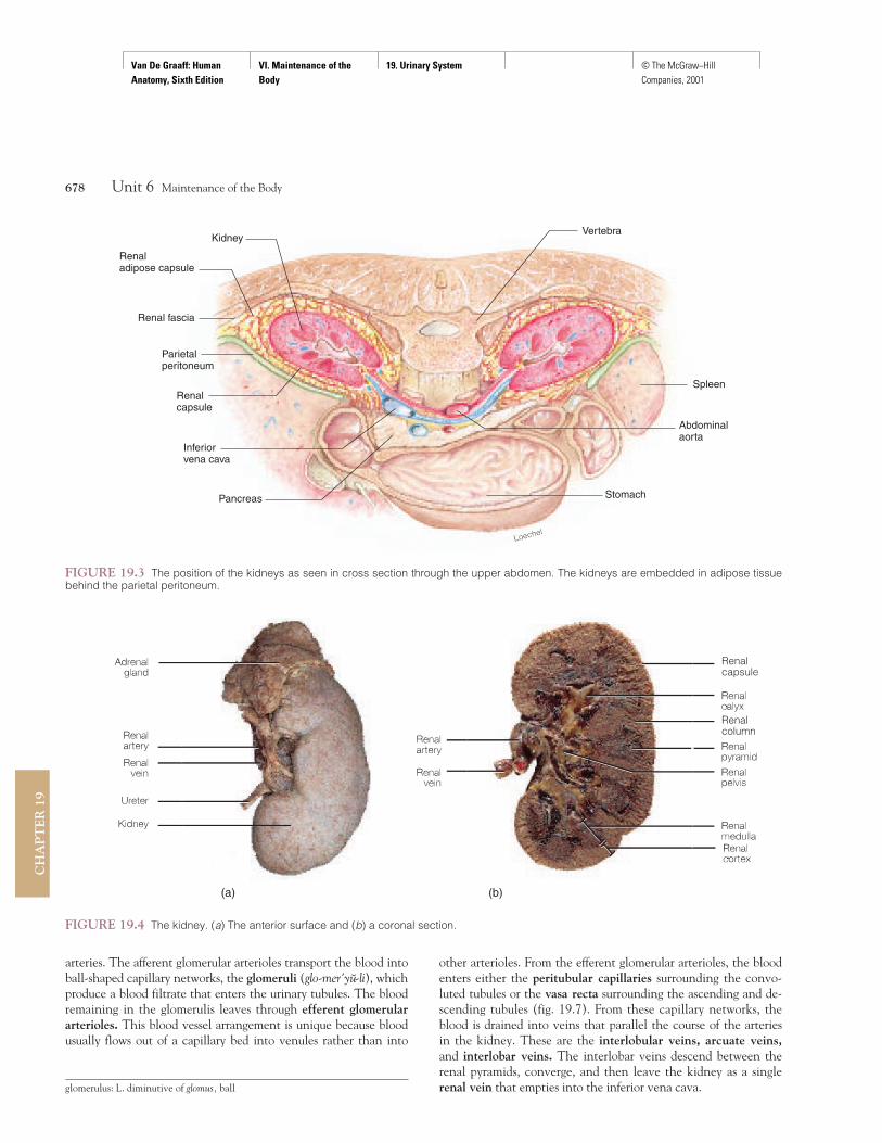
FIGURE 19.3 The position of the kidneys as seen in cross section through the upper abdomen. The kidneys are embedded in adipose tissuebehind the parietal peritoneum.
Renalcapsule
Renalcolumn
FIGURE 19.4 The kidney. (a) The anterior surface and (b) a coronal section.
(a) (b)
glomerulus: L. diminutive of glomus, ball
Van De Graaff: Human Anatomy, Sixth Edition
VI. Maintenance of the Body
19. Urinary System © The McGraw−Hill Companies, 2001
In summary, vessels are at a capillary level between the ar-terioles in each glomerulus of the kidney. The blood pressure atthe glomerulus is strong enough to force water and dissolvedwastes from the blood into the urinary tubular portion of thenephron. Thus, this capillary network produces the filtrate. Asecondary capillary network of peritubular capillaries and vasarecta surrounds various tubular portions of the nephron. Thiscapillary bed, however, is adapted for absorption rather than fil-
trate formation. It reabsorbs water and other substances thatshould not be excreted with the urine, reclaiming most of the fil-trate produced in the glomerulus.
Although the kidneys are generally well protected in thatthey are encapsulated retroperitoneally, they may be injured by ahard blow to the lumbar region. Such an injury can produceblood in the urine, because the highly vascular kidneys are par-ticularly susceptible to hemorrhage.
Chapter 19 Urinary System 679
CH
AP
TE
R 19
Renal cortex
Minor calyx
Major calyx
Renalartery
Renalmedulla
Renalcortex
Nephron
Renalpapilla
Minor calyx
Glomerularcapsule
Proximal convolutedtubule
Nephron loop
Papillary duct
Distal convolutedtubule
(c)
Creek
(b)
Renalvein
Ureter
Renal medulla
Renal pyramid
Renal capsule
Renal column
Opening ofrenal calyx into renal pelvis
Renal pelvis
(a)
Renal papilla
FIGURE 19.5 The internal structures of a kidney. (a) A coronal section showing the structure of the renal cortex, renal medulla, and renalpelvis. (b) A diagrammatic magnification of a renal pyramid to depict the renal tubules. (c) A diagram of a single nephron and a papillary duct.

Van De Graaff: Human Anatomy, Sixth Edition
VI. Maintenance of the Body
19. Urinary System © The McGraw−Hill Companies, 2001
NephronThe tubular nephron consists of a glomerular capsule, proximalconvoluted tubule, descending limb of the nephron loop (loop ofHenle), ascending limb of the nephron loop, and distal convolutedtubule (fig. 19.7).
The glomerular (Bowman's) capsule surrounds theglomerulus. The glomerular capsule and its associated glomerulusare located in the renal cortex of the kidney and together consti-tute the renal corpuscle (fig. 19.8). The glomerular capsule con-tains an inner visceral layer of epithelium, in contact with theglomerular capillaries and an outer parietal layer. The space be-tween these two layers, called the capsular space, is where theglomerular filtrate collects.
The glomerular epithelium contains tiny pores called fen-estrae (fe-nes'tre) that permit the filtrate to pass from the bloodinto the glomerular capsular space (fig. 19.8). Although the fen-estrae are large, they are still small enough to prevent the passageof blood cells, platelets, and most plasma proteins into the
680 Unit 6 Maintenance of the Body
CH
AP
TE
R 1
9
FIGURE 19.6 The principal arteries and veins of a kidney.
Efferent glomerulararterioleAfferent glomerulararteriole
Glomerular capsule
FIGURE 19.7 A simplified illustration of blood flow from a glomerulus to an efferent glomerular arteriole, to the peritubular capillaries, and tothe venous drainage of the kidney.
Bowman’s capsule: from Sir William Bowman, English anatomist, 1816–92

Van De Graaff: Human Anatomy, Sixth Edition
VI. Maintenance of the Body
19. Urinary System © The McGraw−Hill Companies, 2001
Chapter 19 Urinary System 681
CH
AP
TE
R 19
Efferent glomerular arterioleAfferent glomerular arteriole
Blood flowPrimary process Capillary
endothelium
Fenestrae
Filtration
Loechel
Podocyte Slit pores
Pedicel
Internal wall of glomerular capsule
Proximal convoluted tubule
External wall ofglomerular capsule
Blood flow
Glomerulus
(a)
Slit pore Pedicel Primary processof podocyte
Glomerulus
FIGURE 19.8 The structure of a glomerulus. (a) A renal corpuscle is composed of a glomerulus and a glomerular (Bowman’s) capsule. Notethat the diameter of the efferent glomerular arteriole carrying blood away from the glomerulus is smaller than that of the afferent glomerular arteri-ole transporting blood into the glomerulus. This is one of the contributing factors in the maintenance of high blood pressure within the glomerulus.The first step of urine formation is the filtration through the glomerular membrane (arrows) into the glomerular space of the glomerular capsule.(b) A scanning electron micrograph of a glomerulus (8,000×).
(b)
Van De Graaff: Human Anatomy, Sixth Edition
VI. Maintenance of the Body
19. Urinary System © The McGraw−Hill Companies, 2001
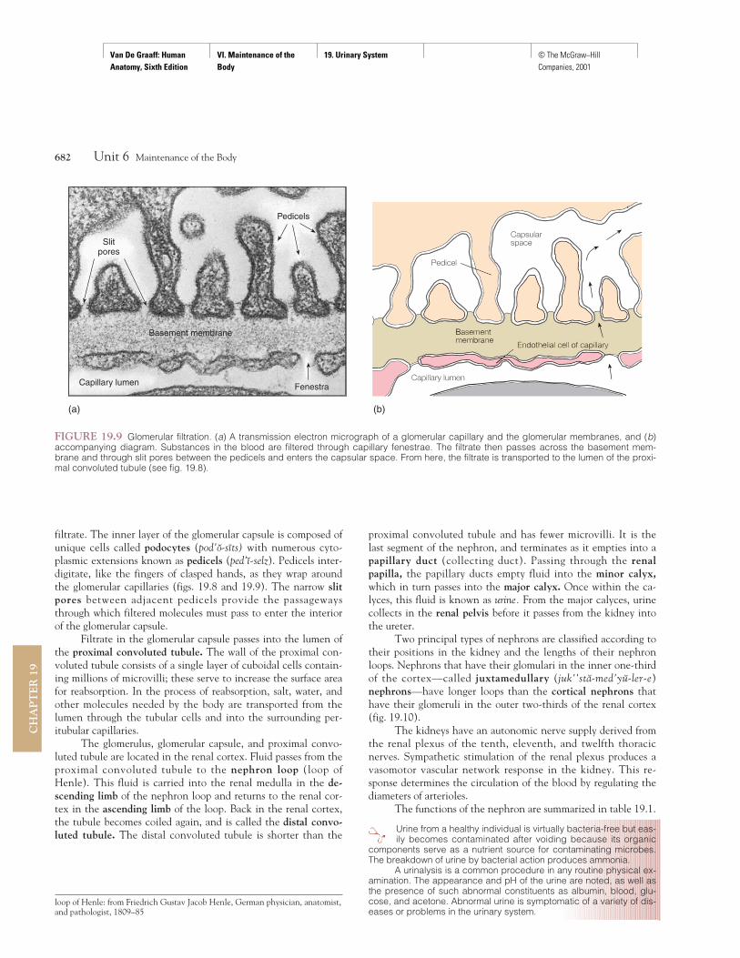
filtrate. The inner layer of the glomerular capsule is composed ofunique cells called podocytes (pod'o-sıts) with numerous cyto-plasmic extensions known as pedicels (ped'ı-selz). Pedicels inter-digitate, like the fingers of clasped hands, as they wrap aroundthe glomerular capillaries (figs. 19.8 and 19.9). The narrow slitpores between adjacent pedicels provide the passagewaysthrough which filtered molecules must pass to enter the interiorof the glomerular capsule.
Filtrate in the glomerular capsule passes into the lumen ofthe proximal convoluted tubule. The wall of the proximal con-voluted tubule consists of a single layer of cuboidal cells contain-ing millions of microvilli; these serve to increase the surface areafor reabsorption. In the process of reabsorption, salt, water, andother molecules needed by the body are transported from thelumen through the tubular cells and into the surrounding per-itubular capillaries.
The glomerulus, glomerular capsule, and proximal convo-luted tubule are located in the renal cortex. Fluid passes from theproximal convoluted tubule to the nephron loop (loop ofHenle). This fluid is carried into the renal medulla in the de-scending limb of the nephron loop and returns to the renal cor-tex in the ascending limb of the loop. Back in the renal cortex,the tubule becomes coiled again, and is called the distal convo-luted tubule. The distal convoluted tubule is shorter than the
proximal convoluted tubule and has fewer microvilli. It is thelast segment of the nephron, and terminates as it empties into apapillary duct (collecting duct). Passing through the renalpapilla, the papillary ducts empty fluid into the minor calyx,which in turn passes into the major calyx. Once within the ca-lyces, this fluid is known as urine. From the major calyces, urinecollects in the renal pelvis before it passes from the kidney intothe ureter.
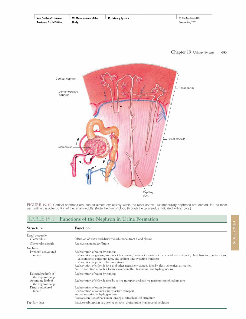
Two principal types of nephrons are classified according totheir positions in the kidney and the lengths of their nephronloops. Nephrons that have their glomulari in the inner one-thirdof the cortex—called juxtamedullary (juk''sta-med'yu-ler-e)nephrons—have longer loops than the cortical nephrons thathave their glomeruli in the outer two-thirds of the renal cortex(fig. 19.10).
The kidneys have an autonomic nerve supply derived fromthe renal plexus of the tenth, eleventh, and twelfth thoracicnerves. Sympathetic stimulation of the renal plexus produces avasomotor vascular network response in the kidney. This re-sponse determines the circulation of the blood by regulating thediameters of arterioles.
The functions of the nephron are summarized in table 19.1.
Urine from a healthy individual is virtually bacteria-free but eas-ily becomes contaminated after voiding because its organic
components serve as a nutrient source for contaminating microbes.The breakdown of urine by bacterial action produces ammonia.
A urinalysis is a common procedure in any routine physical ex-amination. The appearance and pH of the urine are noted, as well asthe presence of such abnormal constituents as albumin, blood, glu-cose, and acetone. Abnormal urine is symptomatic of a variety of dis-eases or problems in the urinary system.
682 Unit 6 Maintenance of the Body
CH
AP
TE
R 1
9
Basement membrane
FenestraCapillary lumen
Pedicels
Slitpores
(a) (b)
FIGURE 19.9 Glomerular filtration. (a) A transmission electron micrograph of a glomerular capillary and the glomerular membranes, and (b)accompanying diagram. Substances in the blood are filtered through capillary fenestrae. The filtrate then passes across the basement mem-brane and through slit pores between the pedicels and enters the capsular space. From here, the filtrate is transported to the lumen of the proxi-mal convoluted tubule (see fig. 19.8).
loop of Henle: from Friedrich Gustav Jacob Henle, German physician, anatomist,and pathologist, 1809–85
Van De Graaff: Human Anatomy, Sixth Edition
VI. Maintenance of the Body
19. Urinary System © The McGraw−Hill Companies, 2001
Chapter 19 Urinary System 683
CH
AP
TE
R 19
FIGURE 19.10 Cortical nephrons are located almost exclusively within the renal cortex. Juxtamedullary nephrons are located, for the mostpart, within the outer portion of the renal medulla. (Note the flow of blood through the glomerulus indicated with arrows.)
TABLE 19.1 Functions of the Nephron in Urine Formation
Structure Function
Renal corpuscleGlomerulus Filtration of water and dissolved substances from blood plasma
Glomerular capsule Receives glomerular filtrate
NephronProximal convoluted Reabsorption of water by osmosis
tubule Reabsorption of glucose, amino acids, creatine, lactic acid, citric acid, uric acid, ascorbic acid, phosphate ions, sulfate ions, calcium ions, potassium ions, and sodium ions by active transport
Reabsorption of proteins by pinocytosisReabsorption of chloride ions and other negatively charged ions by electrochemical attractionActive secretion of such substances as penicillin, histamine, and hydrogen ions
Descending limb of Reabsorption of water by osmosis the nephron loop
Ascending limb of Reabsorption of chloride ions by active transport and passive reabsorption of sodium ionsthe nephron loop
Distal convoluted Reabsorption of water by osmosistubule Reabsorption of sodium ions by active transport
Active secretion of hydrogen ionsPassive secretion of potassium ions by electrochemical attraction
Papillary duct Passive reabsorption of water by osmosis; drains urine from several nephrons

Van De Graaff: Human Anatomy, Sixth Edition
VI. Maintenance of the Body
19. Urinary System © The McGraw−Hill Companies, 2001
Knowledge Check3. Describe the general appearance of the renal cortex and
renal medulla.4. Trace the course of blood through the kidney from the
renal artery to the renal vein.5. Trace the course of tubular fluid from the glomerular cap-
sules to the ureter.6. Draw a diagram of a nephron and label the renal cortex and
renal medulla. Also label the structures within each region.
URETERS, URINARY BLADDER,AND URETHRAUrine is channeled from the kidneys to the urinary bladder by theureters and expelled from the body through the urethra. The mu-cosa of the urinary bladder permits distension, and the muscles ofthe urinary bladder and urethra function in the control of micturition.
Objective 6 Describe the location, structure, and function ofthe ureters.
Objective 7 Describe the gross and histological structureand the innervation of the urinary bladder.
Objective 8 Describe the micturition reflex.
Objective 9 Compare and contrast the structure of the maleurethra with that of the female.
UretersThe ureters (yoo-re'terz), like the kidneys, are retroperitoneal.These tubular organs, each about 25 cm (10 in.) long, begin atthe renal pelvis and course inferiorly to enter the urinary bladderat the posterolateral angles of its base. The thickest portion of aureter, near where it enters the urinary bladder, is approximately1.0 cm (0.4 in.) in diameter.
The wall of the ureter consists of three layers, or tunics.The inner mucosa is continuous with the linings of the renaltubules and the urinary bladder. The mucosa consists of transi-tional epithelium (fig. 19.11). The cells of this layer secrete amucus that coats the walls of the ureter with a protective film.
The middle layer of the ureter is called the muscularis.It consists of inner longitudinal and outer circular layers ofsmooth muscle fibers. In addition, the proximal one-third ofthe ureter contains another longitudinal layer to the outsideof the circular layer. Muscular peristaltic waves move theurine through the ureter. The peristaltic waves are initiatedby the presence of urine in the renal pelvis, and their fre-quency is determined by the volume of urine. The waves,which occur from once every few seconds to once every fewminutes, force urine through the ureter and cause it to spurtinto the urinary bladder.
The outer layer of the ureter is called the adventitia(ad''ven-tish'a) The adventitia is composed of loose connectivetissue that covers and protects the underlying layers. In addition,extensions of the connective tissue anchor the ureter in place.
The arterial supply of the ureter comes from several sources.Branches from the renal artery serve the superior portion. Thetesticular (or ovarian) artery (also called gonadal artery) suppliesthe middle portion, and the superior vesicular artery serves thepelvic region. The venous return is through corresponding veins.
A urinary stone (calculus) may develop in any organ of the uri-nary system. A renal stone (“kidney stone”) is one that forms in
a kidney (fig. 19.12). A renal stone may obstruct the ureter andgreatly increase the frequency of peristaltic waves in an attempt topass through. The pain from a lodged urinary stone is extreme andextends throughout the pelvic area. A lodged urinary stone alsocauses a sympathetic ureterorenal reflex that results in constriction ofrenal arterioles, thus reducing the production of urine in the kidneyon the affected side.
684 Unit 6 Maintenance of the Body
CH
AP
TE
R 1
9
Lumen
Transitionalepithelium
Mucosa
Muscularis
Adventitia
FIGURE 19.11 A photomicrograph of a ureter in cross section.(Note the transitional epithelium lining the lumen.)
FIGURE 19.12 A renal stone (kidney stone) placed next to a dimefor size comparison. Factors contributing to renal stone formationmay include the ingestion of excessive mineral salts, a decrease inwater intake, and overactivity of the parathyroid glands. A renal stonegenerally consists of calcium oxalate, calcium phosphate, and uricacid crystals.
calculus: L. calculus, small stone
Van De Graaff: Human Anatomy, Sixth Edition
VI. Maintenance of the Body
19. Urinary System © The McGraw−Hill Companies, 2001
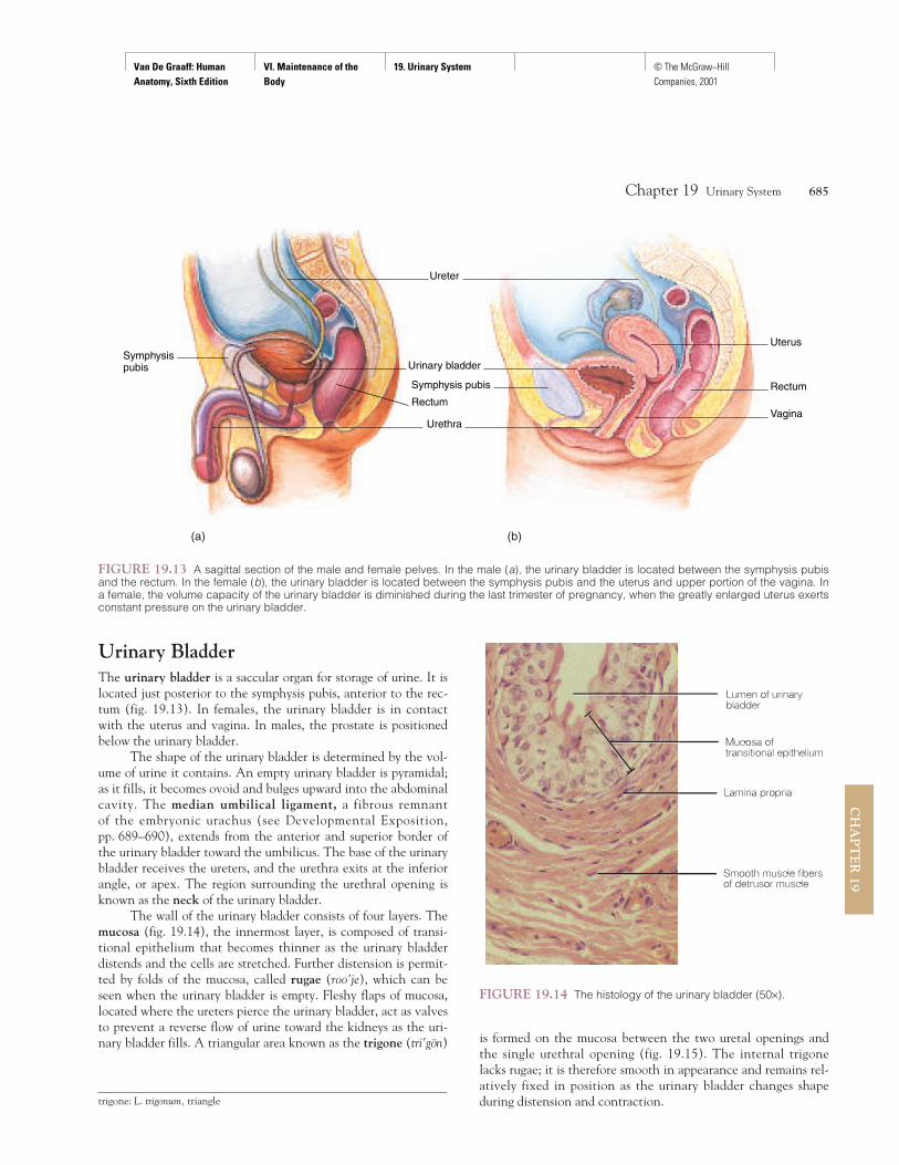
Urinary BladderThe urinary bladder is a saccular organ for storage of urine. It islocated just posterior to the symphysis pubis, anterior to the rec-tum (fig. 19.13). In females, the urinary bladder is in contactwith the uterus and vagina. In males, the prostate is positionedbelow the urinary bladder.
The shape of the urinary bladder is determined by the vol-ume of urine it contains. An empty urinary bladder is pyramidal;as it fills, it becomes ovoid and bulges upward into the abdominalcavity. The median umbilical ligament, a fibrous remnant of the embryonic urachus (see Developmental Exposition,pp. 689–690), extends from the anterior and superior border ofthe urinary bladder toward the umbilicus. The base of the urinarybladder receives the ureters, and the urethra exits at the inferiorangle, or apex. The region surrounding the urethral opening isknown as the neck of the urinary bladder.
The wall of the urinary bladder consists of four layers. Themucosa (fig. 19.14), the innermost layer, is composed of transi-tional epithelium that becomes thinner as the urinary bladderdistends and the cells are stretched. Further distension is permit-ted by folds of the mucosa, called rugae (roo'je), which can beseen when the urinary bladder is empty. Fleshy flaps of mucosa,located where the ureters pierce the urinary bladder, act as valvesto prevent a reverse flow of urine toward the kidneys as the uri-nary bladder fills. A triangular area known as the trigone (tri'gon)
Chapter 19 Urinary System 685
CH
AP
TE
R 19
Symphysispubis
Uterus
Rectum
Vagina
Urinary bladder
Symphysis pubis
Rectum
Urethra
Ureter
(a) (b)
FIGURE 19.13 A sagittal section of the male and female pelves. In the male (a), the urinary bladder is located between the symphysis pubisand the rectum. In the female (b), the urinary bladder is located between the symphysis pubis and the uterus and upper portion of the vagina. Ina female, the volume capacity of the urinary bladder is diminished during the last trimester of pregnancy, when the greatly enlarged uterus exertsconstant pressure on the urinary bladder.
FIGURE 19.14 The histology of the urinary bladder (50×).
trigone: L. trigonum, triangle
is formed on the mucosa between the two uretal openings andthe single urethral opening (fig. 19.15). The internal trigonelacks rugae; it is therefore smooth in appearance and remains rel-atively fixed in position as the urinary bladder changes shapeduring distension and contraction.
Van De Graaff: Human Anatomy, Sixth Edition
VI. Maintenance of the Body
19. Urinary System © The McGraw−Hill Companies, 2001
686 Unit 6 Maintenance of the Body
CH
AP
TE
R 1
9
Urinarybladder
Prostatic partof urethra
Membranouspart of urethra
Spongy partof urethra
Corpuscavernosum
penis
Bulbourethralgland
Urinarybladder
Urethra
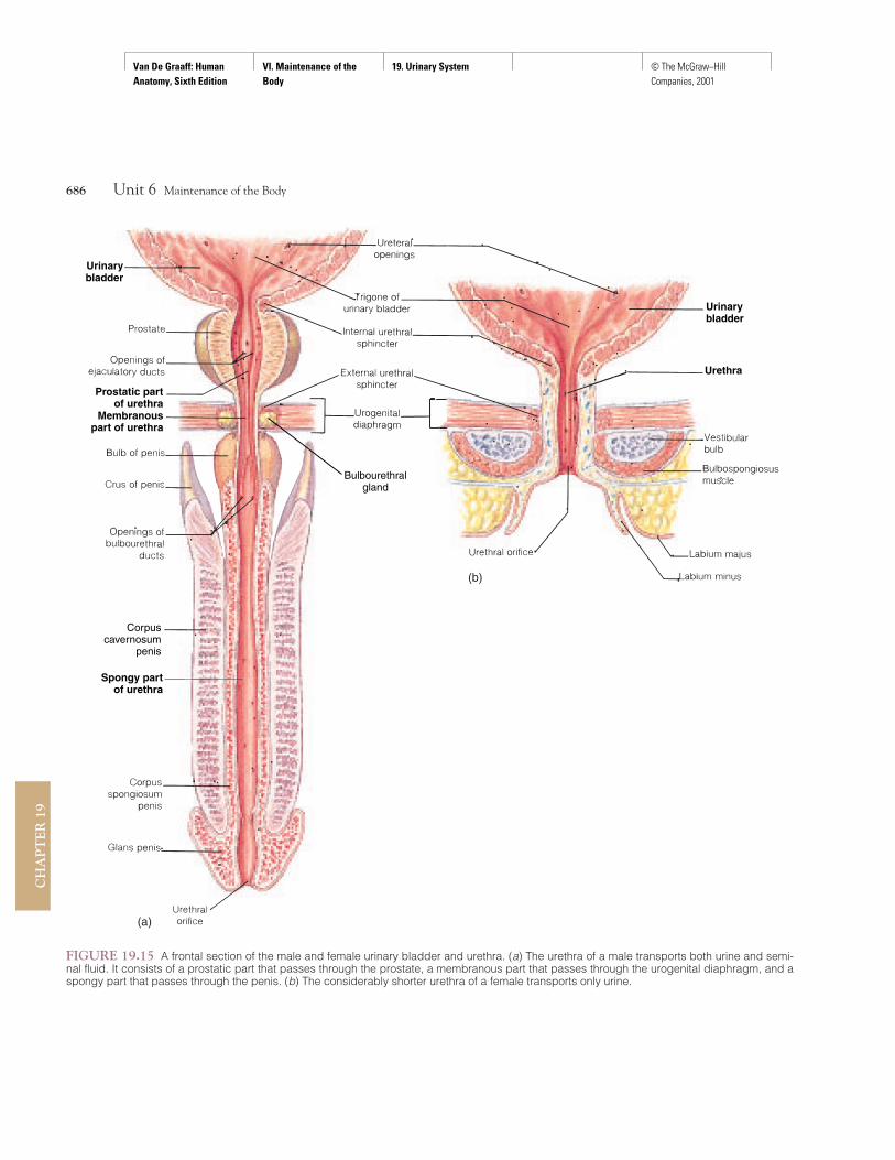
FIGURE 19.15 A frontal section of the male and female urinary bladder and urethra. (a) The urethra of a male transports both urine and semi-nal fluid. It consists of a prostatic part that passes through the prostate, a membranous part that passes through the urogenital diaphragm, and aspongy part that passes through the penis. (b) The considerably shorter urethra of a female transports only urine.
(a)
(b)
Van De Graaff: Human Anatomy, Sixth Edition
VI. Maintenance of the Body
19. Urinary System © The McGraw−Hill Companies, 2001
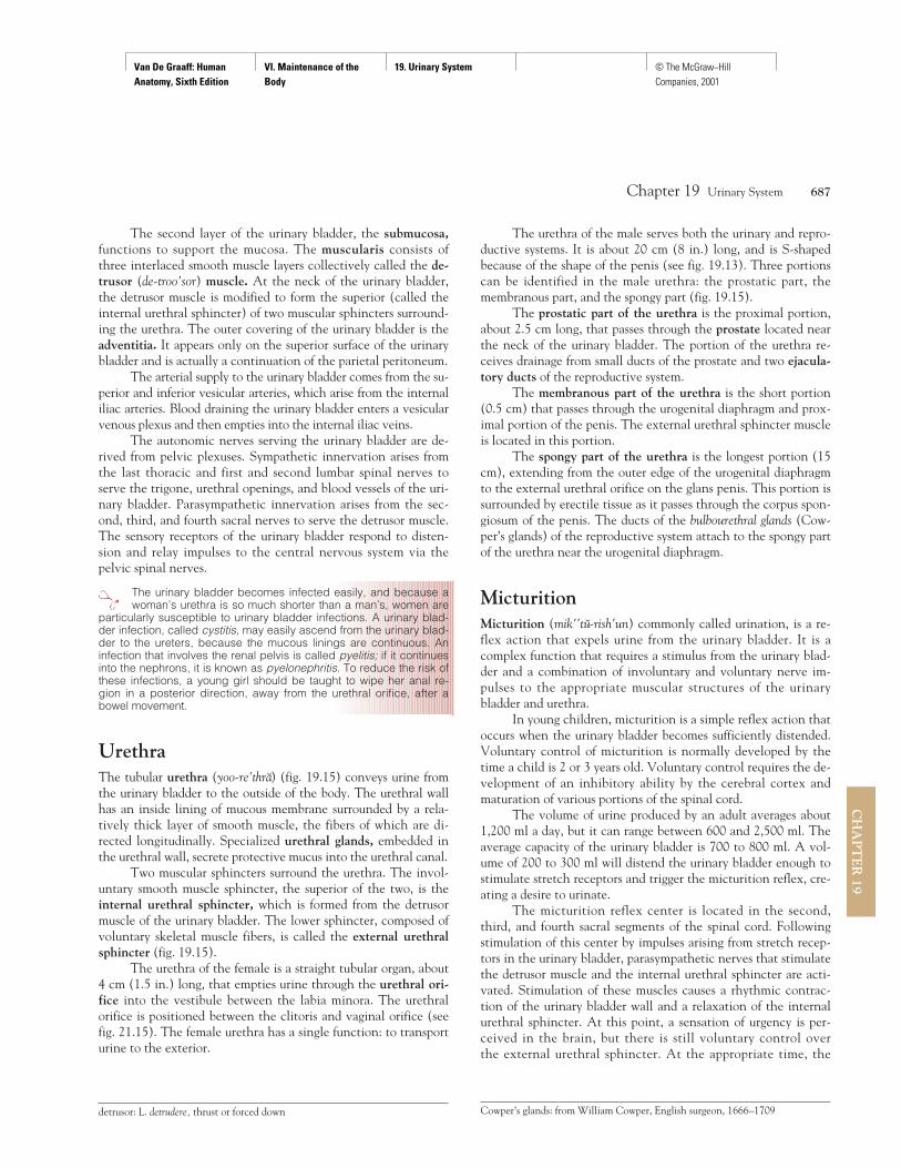
The second layer of the urinary bladder, the submucosa,functions to support the mucosa. The muscularis consists ofthree interlaced smooth muscle layers collectively called the de-trusor (de-troo'sor) muscle. At the neck of the urinary bladder,the detrusor muscle is modified to form the superior (called theinternal urethral sphincter) of two muscular sphincters surround-ing the urethra. The outer covering of the urinary bladder is theadventitia. It appears only on the superior surface of the urinarybladder and is actually a continuation of the parietal peritoneum.
The arterial supply to the urinary bladder comes from the su-perior and inferior vesicular arteries, which arise from the internaliliac arteries. Blood draining the urinary bladder enters a vesicularvenous plexus and then empties into the internal iliac veins.
The autonomic nerves serving the urinary bladder are de-rived from pelvic plexuses. Sympathetic innervation arises fromthe last thoracic and first and second lumbar spinal nerves toserve the trigone, urethral openings, and blood vessels of the uri-nary bladder. Parasympathetic innervation arises from the sec-ond, third, and fourth sacral nerves to serve the detrusor muscle.The sensory receptors of the urinary bladder respond to disten-sion and relay impulses to the central nervous system via thepelvic spinal nerves.
The urinary bladder becomes infected easily, and because awoman’s urethra is so much shorter than a man’s, women are
particularly susceptible to urinary bladder infections. A urinary blad-der infection, called cystitis, may easily ascend from the urinary blad-der to the ureters, because the mucous linings are continuous. Aninfection that involves the renal pelvis is called pyelitis; if it continuesinto the nephrons, it is known as pyelonephritis. To reduce the risk ofthese infections, a young girl should be taught to wipe her anal re-gion in a posterior direction, away from the urethral orifice, after abowel movement.
UrethraThe tubular urethra (yoo-re'thra) (fig. 19.15) conveys urine fromthe urinary bladder to the outside of the body. The urethral wallhas an inside lining of mucous membrane surrounded by a rela-tively thick layer of smooth muscle, the fibers of which are di-rected longitudinally. Specialized urethral glands, embedded inthe urethral wall, secrete protective mucus into the urethral canal.
Two muscular sphincters surround the urethra. The invol-untary smooth muscle sphincter, the superior of the two, is theinternal urethral sphincter, which is formed from the detrusormuscle of the urinary bladder. The lower sphincter, composed ofvoluntary skeletal muscle fibers, is called the external urethralsphincter (fig. 19.15).
The urethra of the female is a straight tubular organ, about4 cm (1.5 in.) long, that empties urine through the urethral ori-fice into the vestibule between the labia minora. The urethralorifice is positioned between the clitoris and vaginal orifice (see fig. 21.15). The female urethra has a single function: to transporturine to the exterior.
The urethra of the male serves both the urinary and repro-ductive systems. It is about 20 cm (8 in.) long, and is S-shapedbecause of the shape of the penis (see fig. 19.13). Three portionscan be identified in the male urethra: the prostatic part, themembranous part, and the spongy part (fig. 19.15).
The prostatic part of the urethra is the proximal portion,about 2.5 cm long, that passes through the prostate located nearthe neck of the urinary bladder. The portion of the urethra re-ceives drainage from small ducts of the prostate and two ejacula-tory ducts of the reproductive system.
The membranous part of the urethra is the short portion(0.5 cm) that passes through the urogenital diaphragm and prox-imal portion of the penis. The external urethral sphincter muscleis located in this portion.
The spongy part of the urethra is the longest portion (15cm), extending from the outer edge of the urogenital diaphragmto the external urethral orifice on the glans penis. This portion issurrounded by erectile tissue as it passes through the corpus spon-giosum of the penis. The ducts of the bulbourethral glands (Cow-per’s glands) of the reproductive system attach to the spongy partof the urethra near the urogenital diaphragm.
MicturitionMicturition (mik''tu-rish'un) commonly called urination, is a re-flex action that expels urine from the urinary bladder. It is acomplex function that requires a stimulus from the urinary blad-der and a combination of involuntary and voluntary nerve im-pulses to the appropriate muscular structures of the urinarybladder and urethra.
In young children, micturition is a simple reflex action thatoccurs when the urinary bladder becomes sufficiently distended.Voluntary control of micturition is normally developed by thetime a child is 2 or 3 years old. Voluntary control requires the de-velopment of an inhibitory ability by the cerebral cortex andmaturation of various portions of the spinal cord.
The volume of urine produced by an adult averages about1,200 ml a day, but it can range between 600 and 2,500 ml. Theaverage capacity of the urinary bladder is 700 to 800 ml. A vol-ume of 200 to 300 ml will distend the urinary bladder enough tostimulate stretch receptors and trigger the micturition reflex, cre-ating a desire to urinate.
The micturition reflex center is located in the second,third, and fourth sacral segments of the spinal cord. Followingstimulation of this center by impulses arising from stretch recep-tors in the urinary bladder, parasympathetic nerves that stimulatethe detrusor muscle and the internal urethral sphincter are acti-vated. Stimulation of these muscles causes a rhythmic contrac-tion of the urinary bladder wall and a relaxation of the internalurethral sphincter. At this point, a sensation of urgency is per-ceived in the brain, but there is still voluntary control overthe external urethral sphincter. At the appropriate time, the
Chapter 19 Urinary System 687
CH
AP
TE
R 19
detrusor: L. detrudere, thrust or forced down Cowper’s glands: from William Cowper, English surgeon, 1666–1709
Van De Graaff: Human Anatomy, Sixth Edition
VI. Maintenance of the Body
19. Urinary System © The McGraw−Hill Companies, 2001
conscious activity of the brain activates the motor nerve fibers(S4) to the external urethral sphincter via the pudendal nerve(S2, S3, and S4), causing the sphincter to relax and urinationto occur.
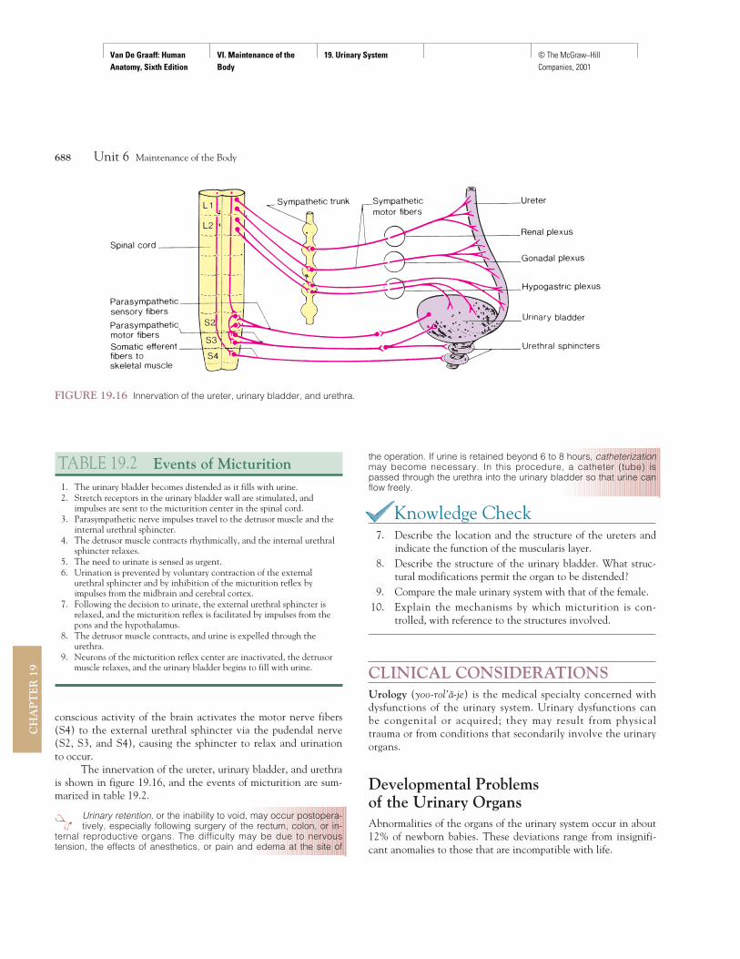
The innervation of the ureter, urinary bladder, and urethrais shown in figure 19.16, and the events of micturition are sum-marized in table 19.2.
Urinary retention, or the inability to void, may occur postopera-tively, especially following surgery of the rectum, colon, or in-
ternal reproductive organs. The difficulty may be due to nervoustension, the effects of anesthetics, or pain and edema at the site of
the operation. If urine is retained beyond 6 to 8 hours, catheterizationmay become necessary. In this procedure, a catheter (tube) ispassed through the urethra into the urinary bladder so that urine canflow freely.
Knowledge Check7. Describe the location and the structure of the ureters and
indicate the function of the muscularis layer.8. Describe the structure of the urinary bladder. What struc-
tural modifications permit the organ to be distended?9. Compare the male urinary system with that of the female.
10. Explain the mechanisms by which micturition is con-trolled, with reference to the structures involved.
CLINICAL CONSIDERATIONSUrology (yoo-rol'a-je) is the medical specialty concerned withdysfunctions of the urinary system. Urinary dysfunctions canbe congenital or acquired; they may result from physicaltrauma or from conditions that secondarily involve the urinaryorgans.
Developmental Problems of the Urinary OrgansAbnormalities of the organs of the urinary system occur in about12% of newborn babies. These deviations range from insignifi-cant anomalies to those that are incompatible with life.
688 Unit 6 Maintenance of the Body
CH
AP
TE
R 1
9
FIGURE 19.16 Innervation of the ureter, urinary bladder, and urethra.
TABLE 19.2 Events of Micturition1. The urinary bladder becomes distended as it fills with urine.2. Stretch receptors in the urinary bladder wall are stimulated, and
impulses are sent to the micturition center in the spinal cord.3. Parasympathetic nerve impulses travel to the detrusor muscle and the
internal urethral sphincter.4. The detrusor muscle contracts rhythmically, and the internal urethral
sphincter relaxes.5. The need to urinate is sensed as urgent.6. Urination is prevented by voluntary contraction of the external
urethral sphincter and by inhibition of the micturition reflex byimpulses from the midbrain and cerebral cortex.
7. Following the decision to urinate, the external urethral sphincter isrelaxed, and the micturition reflex is facilitated by impulses from thepons and the hypothalamus.
8. The detrusor muscle contracts, and urine is expelled through theurethra.
9. Neurons of the micturition reflex center are inactivated, the detrusormuscle relaxes, and the urinary bladder begins to fill with urine.
Van De Graaff: Human Anatomy, Sixth Edition
VI. Maintenance of the Body
19. Urinary System © The McGraw−Hill Companies, 2001
689
The Urinary System
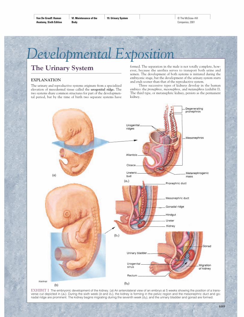
EXPLANATIONThe urinary and reproductive systems originate from a specializedelevation of mesodermal tissue called the urogenital ridge. Thetwo systems share common structures for part of the developmen-tal period, but by the time of birth two separate systems have
formed. The separation in the male is not totally complete, how-ever, because the urethra serves to transport both urine andsemen. The development of both systems is initiated during theembryonic stage, but the development of the urinary system startsand ends sooner than that of the reproductive system.
Three successive types of kidneys develop in the humanembryo: the pronephros, mesonephros, and metanephros (exhibit I).The third type, or metanephric kidney, persists as the permanentkidney.
Developmental Exposition
EXHIBIT I The embryonic development of the kidney. (a) An anterolateral view of an embryo at 5 weeks showing the position of a trans-verse cut depicted in (a1). During the sixth week (b and b1), the kidney is forming in the pelvic region and the mesonephric duct and go-nadal ridge are prominent. The kidney begins migrating during the seventh week (b2), and the urinary bladder and gonad are formed.
(a)
(b)
(a1)
(b1)
(b2)

Van De Graaff: Human Anatomy, Sixth Edition
VI. Maintenance of the Body
19. Urinary System © The McGraw−Hill Companies, 2001
690
EXHIBIT II The development of the matanephric kidney (a) at4 weeks, (b) 5 weeks, (c) 7 weeks, and (d) at birth. (d1) A magni-fied view of the arrangement of papillary ducts within a papilla.
(a)
(b)
(c)
(d)
The pronephros (pro-nef'ros) develops during the fourthweek after conception and persists only through the sixth week.Of the three developmental kidneys, it is the most superior in po-sition on the urogenital ridge and is connected to the embryoniccloaca by the pronephric duct. Although the pronephros is non-functional and degenerates in humans, most of its duct is used bythe mesonephric kidney (exhibit I), and a portion of it is impor-tant in the formation of the metanephros.
The mesonephros (mez''o-nef'ros) develops toward theend of the fourth week as the pronephros becomes vestigial.The mesonephros forms from an intermediate portion of the uro-genital ridge and functions throughout the embryonic period ofdevelopment.
Although the metanephros (met''a-nef'ros) begins its for-mation during the fifth week, it does not become functional untilimmediately before the start of the fetal stage of development, atthe end of the eighth week. The paired metanephric kidneys con-tinue to form urine throughout fetal development. The urine isexpelled through the urinary system into the amniotic fluid.
The metanephros develops from two mesodermal sources(exhibit II). The glomerular part of the kidney forms from a spe-cialized caudal portion of the urogenital ridge called themetanephrogenic mass. The tubular drainage portion of the kid-neys forms as a diverticulum that emerges from the wall of themesonephric duct near the cloaca. This diverticulum, known asthe ureteric (yoo''re-ter'ik) bud, expands into the metanephro-genic mass to form the drainage pathway for urine. The stalk ofthe ureteric bud develops into the ureter, whereas the expandedterminal portion forms the renal pelvis, calyces, and papillaryducts. A combination of the ureteric bud and metanephrogenicmass forms the other tubular channels within the kidney.
Once the metanephric kidneys are formed, they begin tomigrate from the pelvis to the upper posterior portion of the ab-domen. The renal blood supply develops as the kidneys becomepositioned in the posterior body wall.
The development of the kidneys illustrates the conceptthat ontogeny (embryonic development) recapitulates
phylogeny (evolution). This means that the development of thethree kidney types follows an evolutionary pattern. The larvae of afew of the more primitive vertebrates have functional pronephrickidneys. Adult fishes and amphibians have mesonephric kidneys,whereas adult reptiles, birds, and mammals have metanephrickidneys.
The urinary bladder develops from the urogenital sinus,which is connected to the embryonic umbilical cord by the fetalmembrane called the allantois (exhibit I). By the twelfth week,the two ureters are emptying into the urinary bladder, the urethrais draining, and the connection of the urinary bladder to the al-lantois has been reduced to a supporting structure called the ura-chus (yoo'ra-kus).
Occasionally a patent urachus is present in a newborn andis discovered when urine is passed through the umbilicus,
especially if there is a urethral obstruction. Usually, however, it re-mains undiscovered until an enlarged prostate in an elderly maleobstructs the urethra and forces urine through the patent urachusand out the umbilicus (navel).
(d1)
Van De Graaff: Human Anatomy, Sixth Edition
VI. Maintenance of the Body
19. Urinary System © The McGraw−Hill Companies, 2001
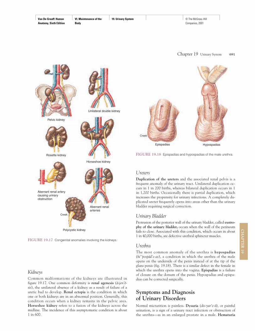
KidneysCommon malformations of the kidneys are illustrated in figure 19.17. One common deformity is renal agenesis (a-jen'e-sis), the unilateral absence of a kidney as a result of failure of auretic bud to develop. Renal ectopia is the condition in whichone or both kidneys are in an abnormal position. Generally, thiscondition occurs when a kidney remains in the pelvic area.Horseshoe kidney refers to a fusion of the kidneys across themidline. The incidence of this asymptomatic condition is about1 in 600.
UretersDuplication of the ureters and the associated renal pelvis is afrequent anomaly of the urinary tract. Unilateral duplication oc-curs in 1 in 200 births, whereas bilateral duplication occurs in 1in 1,200 births. Occasionally there is partial duplication, whichincreases the propensity for urinary infections. A completely du-plicated ureter frequently opens into areas other than the urinarybladder requiring surgical correction.
Urinary BladderProtrusion of the posterior wall of the urinary bladder, called exstro-phy of the urinary bladder, occurs when the wall of the perineumfails to close. Associated with this condition, which occurs in about1 in 40,000 births, are defective urethral sphincter muscles.
UrethraThe most common anomaly of the urethra is hypospadias(hi''pospad'e-as), a condition in which the urethra of the maleopens on the underside of the penis instead of at the tip of theglans penis (fig. 19.18). There is a similar defect in the female inwhich the urethra opens into the vagina. Epispadias is a failureof closure on the dorsum of the penis. Hypospadias and epispa-dias can be corrected surgically.
Symptoms and Diagnosis of Urinary DisordersNormal micturition is painless. Dysuria (dis-yur'e-a), or painfulurination, is a sign of a urinary tract infection or obstruction ofthe urethra—as in an enlarged prostate in a male. Hematuria
Chapter 19 Urinary System 691
CH
AP
TE
R 19
Pelvic kidney
Creek
Unilateral double kidney
Rosette kidney
Aberrant renal arterycausing urinary obstruction
Horseshoe kidney
Aberrant renalarteries
Polycystic kidney
FIGURE 19.17 Congenital anomalies involving the kidneys.
Creek
Epispadias Hypospadias
FIGURE 19.18 Epispadias and hypospadias of the male urethra.
Van De Graaff: Human Anatomy, Sixth Edition
VI. Maintenance of the Body
19. Urinary System © The McGraw−Hill Companies, 2001
means blood in the urine and is usually associated with trauma.Bacteriuria means bacteria in the urine, and pyuria is the termfor pus in the urine, which may result from a prolonged infec-tion. Oliguria is a scanty output of urine, whereas polyuria is anexcessive output. Low blood pressure and kidney failure are twocauses of oliguria. Uremia (yoo-re'me-a) is a condition in whichsubstances ordinarily excreted in the urine accumulate in theblood. Enuresis (en''yu-re'sis), or incontinence, is the inability tocontrol micturition. It may be caused by psychological factors orby structural impairment.
The palpation and inspection of urinary organs is an impor-tant aspect of physical assessment. The right kidney is palpable inthe supine position; the left kidney usually is not. The distendedurinary bladder is palpable along the superior pelvic rim.
The urinary system may be examined using radiographictechniques. An intravenous pyelogram (pi'e-logram) (IVP) per-mits radiographic examination of the kidneys following the in-jection of radiopaque dye. In this procedure, the dye that hasbeen injected intravenously is excreted by the kidneys so thatthe renal pelvises and the outlines of the ureters and urinarybladder can be observed in a radiograph.
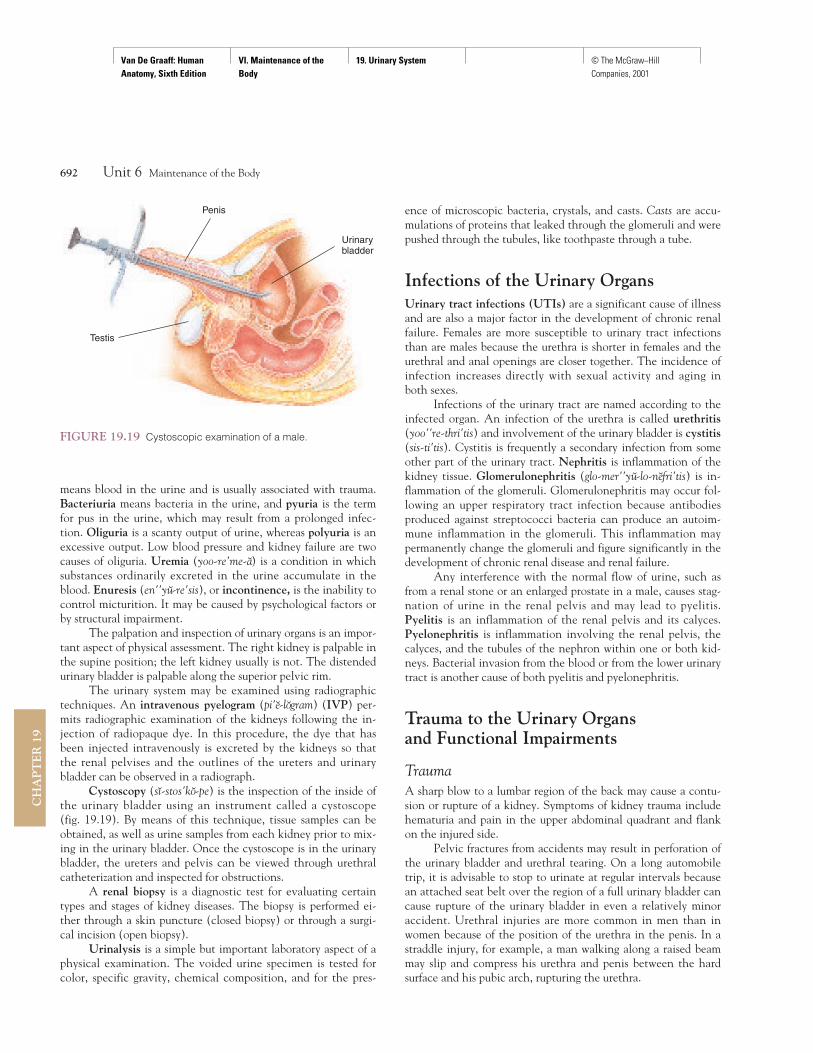
Cystoscopy (sı-stos'ko-pe) is the inspection of the inside ofthe urinary bladder using an instrument called a cystoscope (fig. 19.19). By means of this technique, tissue samples can beobtained, as well as urine samples from each kidney prior to mix-ing in the urinary bladder. Once the cystoscope is in the urinarybladder, the ureters and pelvis can be viewed through urethralcatheterization and inspected for obstructions.
A renal biopsy is a diagnostic test for evaluating certaintypes and stages of kidney diseases. The biopsy is performed ei-ther through a skin puncture (closed biopsy) or through a surgi-cal incision (open biopsy).
Urinalysis is a simple but important laboratory aspect of aphysical examination. The voided urine specimen is tested forcolor, specific gravity, chemical composition, and for the pres-
ence of microscopic bacteria, crystals, and casts. Casts are accu-mulations of proteins that leaked through the glomeruli and werepushed through the tubules, like toothpaste through a tube.
Infections of the Urinary OrgansUrinary tract infections (UTIs) are a significant cause of illnessand are also a major factor in the development of chronic renalfailure. Females are more susceptible to urinary tract infectionsthan are males because the urethra is shorter in females and theurethral and anal openings are closer together. The incidence ofinfection increases directly with sexual activity and aging inboth sexes.
Infections of the urinary tract are named according to theinfected organ. An infection of the urethra is called urethritis(yoo''re-thri'tis) and involvement of the urinary bladder is cystitis(sis-ti'tis). Cystitis is frequently a secondary infection from someother part of the urinary tract. Nephritis is inflammation of thekidney tissue. Glomerulonephritis (glo-mer''yu-lo-nefri'tis) is in-flammation of the glomeruli. Glomerulonephritis may occur fol-lowing an upper respiratory tract infection because antibodiesproduced against streptococci bacteria can produce an autoim-mune inflammation in the glomeruli. This inflammation maypermanently change the glomeruli and figure significantly in thedevelopment of chronic renal disease and renal failure.
Any interference with the normal flow of urine, such asfrom a renal stone or an enlarged prostate in a male, causes stag-nation of urine in the renal pelvis and may lead to pyelitis.Pyelitis is an inflammation of the renal pelvis and its calyces.Pyelonephritis is inflammation involving the renal pelvis, thecalyces, and the tubules of the nephron within one or both kid-neys. Bacterial invasion from the blood or from the lower urinarytract is another cause of both pyelitis and pyelonephritis.
Trauma to the Urinary Organs and Functional Impairments
TraumaA sharp blow to a lumbar region of the back may cause a contu-sion or rupture of a kidney. Symptoms of kidney trauma includehematuria and pain in the upper abdominal quadrant and flankon the injured side.
Pelvic fractures from accidents may result in perforation ofthe urinary bladder and urethral tearing. On a long automobiletrip, it is advisable to stop to urinate at regular intervals becausean attached seat belt over the region of a full urinary bladder cancause rupture of the urinary bladder in even a relatively minoraccident. Urethral injuries are more common in men than inwomen because of the position of the urethra in the penis. In astraddle injury, for example, a man walking along a raised beammay slip and compress his urethra and penis between the hardsurface and his pubic arch, rupturing the urethra.
692 Unit 6 Maintenance of the Body
CH
AP
TE
R 1
9
Penis
Urinarybladder
Testis
FIGURE 19.19 Cystoscopic examination of a male.
Van De Graaff: Human Anatomy, Sixth Edition
VI. Maintenance of the Body
19. Urinary System © The McGraw−Hill Companies, 2001
ObstructionThe urinary system can become obstructed anywhere along thetract. Calculi (stones) are the most common cause, but blockagecan also come from trauma, strictures, tumors or cysts, spasms orkinks of the ureters, or congenital anomalies. If not corrected, anobstruction causes urine to collect behind the blockage and gen-erate pressure that may cause permanent functional andanatomic damage to one or both kidneys. As a result of pressurebuildup in a ureter, a distended ureter, or hydroureter, develops.Dilation in the renal pelvis is called hydronephrosis.
Urinary stones (calculi) are generally the result of infec-tions or metabolic disorders that cause the excretion of largeamounts of organic and inorganic substances (see fig. 19.12). Asthe urine becomes concentrated, these substances may crystalizeand form granules in the renal calyces. The granules then serveas cores for further precipitation and development of larger cal-culi. This becomes dangerous when a calculus grows largeenough to cause an obstruction. The calculus also causes intensepain when it passes through the urinary tract.
Renal Failure and HemodialysisRenal output of 50 to 60 cc of urine per hour is considerednormal. If the output drops to less than 30 cc per hour, it mayindicate renal failure—the loss of the kidney’s ability tomaintain fluid and electrolyte balance and to excrete wasteproducts. Renal failure can be either acute or chronic. Acuterenal failure is the sudden loss of kidney function caused byshock and hemorrhage, thrombosis, or other physical trauma
to the kidneys. The kidneys may sustain a 90% loss of theirnephrons through tissue death and still continue to functionwithout apparent difficulty. If a patient suffering acute renalfailure is stabilized, the nephrons have an excellent capacityto regenerate.
A person with chronic renal failure cannot sustain lifeindependently. Chronic renal failure is the end result of kid-ney disease in which the kidney tissue is progressively de-stroyed. As renal tissue continues to deteriorate, the optionsfor sustaining life are hemodialysis (he''mo-di-al'-ısis) or kidneytransplantation.
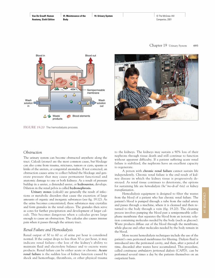
Hemodialysis equipment is designed to filter the wastesfrom the blood of a patient who has chronic renal failure. Thepatient’s blood is pumped through a tube from the radial arteryand passes through a machine, where it is cleansed and then re-turned to the body through a vein (fig. 19.20). The cleaningprocess involves pumping the blood past a semipermeable cello-phane membrane that separates the blood from an isotonic solu-tion containing molecules needed by the body (such as glucose).Waste products diffuse out of the blood through the membrane,while glucose and other molecules needed by the body remain inthe blood.
More recent hemodialysis techniques include the use of thepatient’s own peritoneal membranes for filtering. Dialysis fluid isintroduced into the peritoneal cavity, and then, after a period oftime, discarded after wastes have accumulated. This procedure,called continuous ambulatory peritoneal dialysis (CAPD), can beperformed several times a day by the patients themselves on anoutpatient basis.
Chapter 19 Urinary System 693
CH
AP
TE
R 19
Wasteproducts
Blood elements
Blood in Blood out
Semipermeablemembrane
Washsolution
in
Washsolution
out
FIGURE 19.20 The hemodialysis process.

Van De Graaff: Human Anatomy, Sixth Edition
VI. Maintenance of the Body
19. Urinary System © The McGraw−Hill Companies, 2001
Urinary IncontinenceThe inability to voluntarily retain urine in the urinary bladder isknown as urinary incontinence. It has a number of causes and maybe temporary or permanent. Emotional stress is a cause of tempo-rary incontinence in adults. Permanent incontinence may resultfrom neurological trauma, various urinary diseases, tissue damagewithin the urinary bladder or urethra, or weakness of the pelvicfloor muscles. Remarkable advances have been made in treatingpermanent urinary incontinence through the implantation of anartificial urethral sphincter.
694 Unit 6 Maintenance of the Body
CH
AP
TE
R 1
9
Clinical Case Study AnswerThe hematuria experienced by the patient is probably the result of lacer-ation caused by the upper abdominal knife wound. The lower rightquadrant stab most likely did not violate the urinary tract. The course ofblood seen in the catheter begins and proceeds as follows: abdominalaorta → right renal artery → smaller parenchymal artery → through thelacerated vessel(s) → into the lacerated urinary collecting system, eitherat the calyx or the renal pelvis or proximal right ureter → urinary blad-der → into catheter. During the operation, the surgeon should keep inmind that in 2% to 4% of the population only one kidney is present. If,therefore, she is prompted to remove the damaged kidney, she shouldfirst confirm the presence of a second functioning kidney. If a secondkidney is not present, every effort should be made to correct the prob-lem without performing a nephrectomy, which would consign the pa-tient to chronic hemodialysis or kidney transplant.
CLINICAL PRACTICUM 19.1A 41-year-old male presents in your officebecause his urine has a pink tinge. He alsoreports an increase in his frequency of urina-tion. The patient denies fever, chills, pain,or foul odor from his urine.
The physical exam is normal. A routineurinalysis reveals red blood cells too numerousto count, no white blood cells, and no bacte-ria. You order an intravenous urogram (IVU)(a), which is an injection of intravenous con-trast material for an exam of the urinary sys-tem, and CT scan (b). (B = bladder.)
Q U E S T I O N S1. What is the large dark mass (filling
defect) seen in the urinary bladder(indicated with the letter B) on theIVU? Is it the source of the bleeding?
2. On the CT, white contrast fills thelumen of the urinary bladder (indicatedwith an arrow). Why does the patienthave increased frequency of urination?
3. Transitional carcinoma has a 5–10%incidence of synchronous tumors.Synchronous tumors are tumors of thesame cell type occuring at the sametime in separate locations. What otherstructures must be evaluated for thepresence of tumors?
(a)
(b)

Van De Graaff: Human Anatomy, Sixth Edition
VI. Maintenance of the Body
19. Urinary System © The McGraw−Hill Companies, 2001
Chapter 19 Urinary System 695
CH
AP
TE
R 19
CLINICAL PRACTICUM 19.2A 41-year-old female presents at the emer-gency room with left lumbar pain that attimes radiates to the left groin. She describesthe pain as intermittent and crampy, but de-nies any fever or blood in her urine.
Physical exam shows a nontender ab-domen and no evidence of costovertebralangle tenderness. Routine urinalysis showsminimal red blood cells, no white bloodcells, and no bacteria. You order an intra-venous urogram.
Q U E S T I O N S1. On the precontrast image, what is the
density indicated by the arrow?2. What effect does this have on the
ureter as demonstrated by thepostcontrast image?
3. Why is the patient having intermittentpain?
Postcontrast image
Precontrast image
Chapter Summary
Introduction to the Urinary System(p. 676)
1. The urinary system consists of twokidneys, two ureters, the urinary bladder,and the urethra.
2. The urinary system maintains thecomposition and properties of body fluid,which establishes the extracellularenvironment. The end product of theurinary system is urine, which is voidedfrom the body during micturition.
Kidneys (pp. 676–684)1. The kidneys are retroperitoneal, embedded
in a renal adipose capsule.2. Each kidney is contained by a renal
capsule and divided into an outer renalcortex and an inner renal medulla.(a) The renal medulla is composed of
renal pyramids separated by renalcolumns.
(b) The renal papillae empty urine intothe minor calyces and then into themajor calyces, which drain into therenal pelvis. From there, urine flowsthrough the ureter.
3. Each kidney contains more than a millionmicroscopic functional units callednephrons.(a) Filtration occurs in the glomeruli,
which receive blood from afferentglomerular arterioles.
(b) Glomerular blood is drained byefferent glomerular arterioles thatdeliver blood to peritubular capillariessurrounding the nephron tubules.
(c) The glomerular capsules and distalconvoluted tubules are located in therenal cortex.
(d) The nephron loops are located in therenal medulla.
(e) Filtrate from the distal convolutedtubules are drained into papillaryducts that extend through the renalmedulla to empty urine into thecalyces.
Ureters, Urinary Bladder, and Urethra(pp. 684–688)
1. Urine is channeled from the kidneys tothe urinary bladder by the ureters andexpelled from the urinary bladder through
the urethra. The detrusor muscle of theurinary bladder and the sphincter musclesof the urethra are used in the control ofmicturition.(a) Each ureter contains three layers: the
mucosa, muscularis, and adventitia.(b) The lumen of the urinary bladder is
lined by transitional epithelium,which is folded into rugae. Thesestructures enhance the ability of theurinary bladder to distend.
(c) The urethra has an internal sphincterof smooth muscle and an externalsphincter of skeletal muscle.
(d) The male urethra conducts urineduring urination and seminal fluidduring ejaculation. The femaleurethra is much shorter than that of amale and conducts only urine.
(e) The male urethra is composed ofprostatic, membranous, and spongyportions.
2. Micturition is controlled by reflex centersin the second, third, and fourth sacralsegments of the spinal cord.
Van De Graaff: Human Anatomy, Sixth Edition
VI. Maintenance of the Body
19. Urinary System © The McGraw−Hill Companies, 2001
696 Unit 6 Maintenance of the Body
CH
AP
TE
R 1
9
Review Activities
Objective Questions1. Which of the following statements about
the renal pyramids is false?(a) They are located in the renal
medulla.(b) They contain glomeruli.(c) They contain papillary ducts.(d) They are supported by renal columns.
2. Renal vessels and the ureter attach at theconcave medial border of the kidneycalled(a) the renal (c) the calyx.
pelvis. (d) the hilum.(b) the urachus.
3. The renal medulla of the kidney contains(a) glomerular capsules.(b) glomeruli.(c) renal pyramids.(d) adipose capsules.
4. Urine flowing from the papillary ductsenters directly into(a) the renal calyces.(b) the ureter.(c) the renal pelvis.(d) the distal convoluted tubules.
5. Which of the following statementsconcerning the kidneys is false?(a) They are retroperitoneal.(b) They each contain 8 to 15 renal
pyramids.(c) They each have two distinct
regions—the renal cortex and renalmedulla.
(d) They are positioned between thethird and fifth lumbar vertebrae.
6. A renal stone (calculus), would mostlikely cause stagnation of urine in whichportion of the urinary system?(a) the urinary bladder(b) the renal column(c) the ureter(d) the renal pelvis(e) the urethra
7. Distention of the urinary bladder ispossible because of the presence of(a) rugae.(b) the trigone.(c) the adventitia.(d) the transitional epithelium.(e) both a and d.
8. The detrusor muscle is located in(a) the kidneys.(b) the ureters.
(c) the urinary bladder.(d) the urethra.
9. The internal urethral sphincter isinnervated by(a) sympathetic neurons.(b) parasympathetic neurons.(c) somatic motor neurons.(d) all of the above.
10. Which of the following statements aboutmetanephric kidneys is true?(a) They become functional at the end of
the eighth week.(b) They are active throughout fetal
development.(c) They are the third pair of kidneys to
develop.(d) All of the above are true.
Essay Questions1. Describe the location of the kidneys in
relation to the abdominal cavity and theperitoneal membranes.
2. Diagram the kidney structures that can beidentified in a coronal section.
3. Describe how the kidney is supportedagainst the posterior abdominal wall. Howis this support related to the conditioncalled renal ptosis?
4. Trace a drop of blood from an interlobularartery through a glomerulus and into aninterlobular vein. List in order all thevessels through which the blood passes.How do structural differences in afferentand efferent glomerular arterioles ensurethe high blood pressure needed for filtrateformation?
5. In a male, trace the path of urine from thesite of filtration at the renal corpuscle tothe outside of the body. List in order allthe structures through which the urinepasses.
6. What is a nephron? Describe the twotypes of nephrons found in a kidney. Whyare nephrons considered the functionalunits of the urinary system?
7. Describe the mechanism involved in thepassage of urine from the renal pelvis tothe urinary bladder.
8. Describe the urinary bladder with regardto position, histological structure, bloodsupply, and innervation.
9. Compare and contrast the urethra in themale and female.
10. What is the micturition reflex? Discussthe physiological and functional events ofa voluntary micturition response.
11. What is a metanephrogenic mass? Aureteric bud? Discuss the sequentialdevelopment of these embryonicstructures to form the urinary system.How can a greater knowledge of thisdevelopment process lead to a betterunderstanding of congenitalabnormalities?
12. Briefly describe the purpose of cytoscopyand urinalysis.
13. List four common congenitalmalformations of the urinary system.Which of these require surgicalcorrection?
14. Define dysuria, hematuria, bacteriuria,pyuria, oliguria, polyuria, uremia, andenuresis.
Critical-Thinking Questions1. Why is it more accurate to refer to the
kidneys and associated structures as theurinary system rather than the excretorysystem?
2. Treatment with sulfa medications such asGantrisin (sulfisoxazole) and broad-spectrum antibiotics such as tetracyclineor ampicillin usually clear up thesymptoms of cystitis very quickly. What isthe danger of discontinuing the prescribedmedication as soon as the symptoms are gone?
3. The neighborhood day-care center won’taccept children who are still in diapers.You’ve tried to toilet train your 15-month-old boy, but you haven’t madeany progress at all. Should you persist inyour efforts, or would it be better to wait?Explain.
4. What functions of a real kidney does anartificial kidney (dialysis machine) fail toduplicate?
5. Your friend’s baby is due next month, andshe is constantly running to the bathroomto urinate. Can you explain why?
6. Explain why a male should be particularlyconcerned if he has difficulty voidingurine.
Visit our Online Learning Center at http://www.mhhe.com/vdgfor chapter-by-chapter quizzing, additional study resources, and related web links.