vaginal delivery - chapter 27 of williams obstetrics 24th edition
TRANSCRIPT


VAGINAL DELIVERYA Morning Report by OB Intern Baring

WHAT IS THE NATURAL CULMINATION OF THE
SECOND STAGE OF LABOR?
?QUESTION:

Controlled vaginal delivery of a healthy neonate with
minimal trauma to the mother
ANSWER:
Vaginal Delivery
-Vaginal delivery is the preferred route of delivery for most fetuses
-Certain clinical settings may favor cesarean delivery
-A malpresenting fetus or multifetal gestation may be delivered vaginally but require special techniques
Route of Delivery-Spontaneous vaginal vertex delivery poses the lowest risk of most maternal and fetal comorbidity
-Compared with cesarean delivery, spontaneous vaginal delivery has lower associated rates of: +maternal infection +hemorrhage +anesthesia complications +peripartum hysterectomy
- Pelvic floor disorders may be increased in those initial undergoing NSVD
-Pelvic floor protection advantages gained from Cesarean delivery are lost as women age

-The end of second-stage labor is heralded as: +the perineum begins to distend +the overlying skin becomes stretched +the fetal scalp is seen through the separating labia
-Increased perineal pressure from the fetal head creates reflexive bearing- down efforts, which are encouraged when appropriate
-Preparations are made for delivery
Preparation for Delivery

-Some considerations that arise during labor are reiterated +the bladder is palpated > if distended > catheterization +continued attention is given to FHR monitoring +antibiotic prophylaxis against infective endocarditis not to be given in most women w/ cardiac conditions except those with cyanotic heart disease or prosthetic valves or both.
-Pushing positions may vary, but for delivery > dorsal lithotomy position is the most widely used and often the most satisfactory
-For better exposure > leg holders or stirrups are used
Preparation for Delivery
-No increased rates of perineal lacerations with stirrup use compared to without their use
-With positioning, legs are not separated too widely or placed one higher than the other
-Within the leg holder, the popliteal region should rest comfortably in the proximal portion and the heel in the distal portion
-The legs are not strapped into the stirrups, thereby allowing quick flexion of the thighs backward onto the abdomen should shoulder dystocia develop
Preparation for Delivery

-Legs may cramp during the second stage, in part, because of pressure by the fetal head on pelvic nerves
-Cramping may be relieved by: +repositioning the affected leg +brief massage
-Preparation for delivery includes vulvar and perineal cleansing
-Sterile drapes may be placed in such a way that only the immediate area around the vulva is exposed
Preparation for Delivery

-By the time of perineal distention, the position of the presenting occiput is usually known
-Molding and caput formation have precluded accurate identification
-Most cases, presentation is directly occiput anterior or is rotated slightly oblique
-In perhaps 5 percent, persistent occiput posterior is identified
-Rarely, the vertex will be presenting in the occiput transverse position
Occiput Anterior Position

-Vulvovaginal opening is dilated by the fetal head to gradually form an ovoid and finally, an almost circular opening *CROWNING - encirclement of the largest head diameter by the vulvar ring
-Perineum thins; may undergo spontaneous laceration *unless an episiotomy is done
-Anus becomes greatly stretched, and the anterior wall of the rectum may be easily seen through it
Occiput Anterior Position
THE NEXT STEP
Delivery of the Head

Occiput Anterior Position
Episiotomy? -Increases the risk of a tear into the external anal sphincter, the rectum, or both -Anterior tears involving the urethra and labia are more common in women in whom an episiotomy is avoided -Do not routinely perform episiotomy
Delivery of the Head
Occiput Anterior Position
How to Limit Vaginal Laceration?-Intrapartum perineal massage - widens the introitus for head passage STEPS: 1st: The perineum is grasped in the midline by both hands using the thumb and opposing fingers 2nd: Outward and lateral stretch against the perineum is then repeatedly applied
Delivery of the Head
Occiput Anterior Position
-When the head distends the vulva and perineum enough to open the vaginal introitus to a diameter of 5 cm or more, a gloved hand may be used to support the perineum -The other hand is used to guide and control the fetal head to avoid expulsive delivery -Slow delivery of the head may decrease lacerations
Delivery of the Head

Occiput Anterior Position
-If expulsive efforts are inadequate or expeditious delivery is needed, the modified Ritgen maneuver may be employed STEPS: 1st: Gloved fingers beneath a draped towel exert forward pressure on the fetal chin through the perineum just in front of the coccyx 2nd: The other hand presses superiorly against the occiput -The Ritgen maneuver allows controlled fetal head delivery -It also favors neck extension so that the head passes through the introitus and over the perineum with its smallest diameters
Delivery of the Head

The occiput is being kept close to the symphysis by moderate pressure on the fetal chin at the tip of the maternal coccyx
Occiput Anterior PositionDelivery of the Head: Modified Ritgen Maneuver

Moderate upward pressure is applied to the fetal chin by the posterior hand covered with a sterile towel, while the suboccipital region of the fetal head is held against the symphysis.
Occiput Anterior PositionDelivery of the Head: Modified Ritgen Maneuver

Occiput Anterior Position
-Following delivery of the fetal head, a finger should be passed across the fetal neck to determine whether it is encircled by one or more umbilical cord loops -A nuchal cord is found in approximately 25 percent of deliveries and ordinarily causes no harm *if an umbilical cord coil is felt: loose: slipped over the head applied too tightly: the loop should be cut between two clamps
Delivery of the Shoulders

The umbilical cord, if identified around the neck, is readily slipped over the head
Occiput Anterior PositionDelivery of the Shoulders

Occiput Anterior Position
-Following its delivery, the fetal head falls posteriorly, bringing the face almost into contact with the maternal anus -The occiput promptly turns toward one of the maternal thighs, and the head assumes a transverse position *This external rotation indicates that the bisacromial diameter has rotated into the antero-posterior diameter of the pelvis
Delivery of the Shoulders

Occiput Anterior Position
-Most often, the shoulders appear at the vulva just after external rotation and are born spontaneously -If delayed, extraction aids controlled delivery -The sides of the head are grasped with two hands, and gentle downward traction is applied until the anterior shoulder appears under the pubic arch -Next, by an upward movement, the posterior shoulder is delivered. *abrupt or powerful force is avoided to avert brachial plexus injury
Delivery of the Shoulders

Occiput Anterior Position
Gentle downward traction to effect descent of the anterior shoulder After the delivery of the anterior shoulder completed > Gentle upward traction to deliver the posterior shoulder
Delivery of the Shoulders

Occiput Anterior Position
-The rest of the body almost always follows the shoulders without difficulty -With prolonged delay, its birth may be hastened by moderate traction on the head and moderate pressure on the uterine fundus -Hooking the fingers in the axillae is avoided > can injure upper extremity nerves and produce paralysis -Traction, furthermore, should be exerted only in the direction of the long axis of the neonate; applied obliquely, it causes neck bending and excessive brachial plexus stretching
Delivery of the Shoulders

Occiput Anterior Position
-The umbilical cord is cut between two clamps placed 6 to 8 cm from the fetal abdomen -Umbilical cord clamp is applied 2 to 3 cm from its insertion into the fetal abdomen *using for example the Double Grip Umbilical Clamp (Hollister)
Clamping the Cord
Occiput Anterior Position
-TERM: delay in umbilical cord clamping for up to 60 seconds may: +increase total body iron stores +expand blood volume +decrease anemia incidence *valuable in populations in which iron deficiency is prevalent -PRETERM: delayed cord clamping for 30-60 seconds has benefits: +higher red cell volume +decreased need for blood transfusion +better circulatory stability +lower rates of intraventricular hemorrhage and necrotizing enterocolitis
Clamping the Cord

Other Positions-Persistent occiput posterior
-Occiput transverse

WHEN DOES THE
THIRD STAGE OF LABOR BEGIN AND WHEN DOES IT END?
?QUESTION:

Third-stage labor begins immediately after fetal birth
and ends with placental delivery
ANSWER:

Delivery of the Placenta
-Delivery of an intact placenta -Avoidance of uterine inversion or postpartum hemorrhage
Goals

Delivery of the Placenta-Immediately after newborn birth, uterine fundal size and consistency are examined... +uterus remains firm, no unusual bleeding: -watchful waiting until the placenta separates -massage is not employed -fundus is frequently palpated -umbilical cord traction must not be used

Delivery of the Placenta-Signs of Placental Separation: +sudden gush of blood into the vagina +globular and firmer fundus +lengthening of the umbilical cord as the placenta descends into the vagina +rise of the uterus into the abdomen *concurrently, the placenta, having separated, passes down into the lower uterine segment and vagina *appear within 1 minute after delivery; usually within 5 minutes

Delivery of the Placenta-Once the placenta has detached from the uterine wall, it should be determined that the uterus is firmly contracted -Mother may be asked to bear down, and the intraabdominal pressure often expels the placenta into the vagina -After ensuring that the uterus is contracted firmly, pressure is exerted by a hand wrapped around the fundus to propel the detached placenta into the vagina - The umbilical cord is kept slightly taut but is not pulled

Delivery of the Placenta-Concurrently, the heel of the hand exerts downward pressure between the symphysis pubis and the uterine fundus -Once the placenta passes through the introitus, pressure on the uterus is relieved -The placenta is then gently lifted away

Delivery of the Placenta
The hand is not trying to push the fundus of the uterus through the birth canal! As the placenta leaves the uterus and enters the vagina, the uterus is elevated by the hand on the abdomen while the cord is held in position.

Delivery of the Placenta
The placenta is removed from the vagina by lifting the cord

Delivery of the Placenta
Membranes that were somewhat adhered to the uterine lining are separated by gentle traction with a ring forceps
Manual Delivery of the Placenta
-Occasionally, the placenta will not separate promptly; common with preterm delivery (Dombrowski, 1995) -If there is brisk bleeding and the placenta cannot be delivered spontaneously, manual removal of the placenta is indicated

-One hand grasps the fundus
-The other hand is inserted into the uterine cavity, and the fingers are swept from side to side as they are advanced
-When the placenta has become detached, it is grasped and removed
Manual Delivery of the Placenta

Management of the 3rd Stage of Labor-Practices within the 3rd stage of labor may be considered as either: +Physiological or Expectant Management +Active management

Management of the 3rd Stage of Labor
Physiological or expectant management -involves waiting for placental separation signs and allowing the placenta to deliver either spontaneously or aided by nipple stimulation or gravity (World Health Organization, 2012)
Active management -consists of: +early cord clamping +controlled cord traction during placental delivery +immediate administration of prophylactic uterotonics *goal - limit postpartum hemorrhage -may include uterine massage

Management of the 3rd Stage of LaborActive management -Uterotonics appear to be the most important factor to decrease postpartum blood loss -Choices include: +oxytocin (Pitocin) - first-line agent +misoprostol (Cytotec) +carboprost (Hemabate) +The Ergots - second-line agents -ergonovine (Ergotrate) -methylergonovine (Methergine)

Management of the 3rd Stage of LaborActive management -Uterotonics may be given before or after placental expulsion without increasing rates of postpartum hemorrhage, placental retention, or third-stage labor length (Soltani, 2010) -If given before delivery of the placenta > may entrap an undiagnosed, undelivered second twin *abdominal palpation should confirm no additional fetuses

Management of the 3rd Stage of LaborUterotonics: OXYTOCIN -Synthetic oxytocin is identical to that produced by the posterior pituitary -Action is noted at approximately 1 minute, and it has a mean half-life of 3 to 5 minutes -Not given Intravenously as a large bolus -Given as a dilute solution by continuous intravenous infusion or as an intramuscular injection

Management of the 3rd Stage of Labor
-Despite the routine use of oxytocin, no standard prophylactic dose is established for its use following either vaginal delivery or CS -Standard practice: +Intravenous infusion -add 20 units (2 mL) of oxytocin per liter of infusate *administered after delivery of the placenta at a rate of 10 to 20 mL/min (200 to 400 mU/min) for a few minutes until the uterus remains firmly contracted and bleeding is controlled *reduced to 1 to 2 mL/ min until the mother is ready for transfer from the RR to the postpartum unit +Intramuscular: 10 units is given

Management of the 3rd Stage of LaborUterotonics: ERGOT ALKALOIDS -Ergonovine and Methylergonovine have similar activity levels in myometrium -Powerful stimulants of myometrial contraction exerting an effect which may persist for hours -Require very specific storage conditions -DANGEROUS to the fetus when given before delivery -Side effects: transient maternal hypertension, N/V, headache, tinnitus, painful uterine contractions

Management of the 3rd Stage of LaborUterotonics: MISOPROSTOL -This prostaglandin E1 analogue has proved inferior to oxytocin for postpartum hemorrhage prevention (Tunçalp, 2012) -In resource-poor settings that lack oxytocin, misoprostol is suitable for hemorrhage prophylaxis and is given as a single oral 600-µg dose -Side effects include: +shivering - 30 percent +fever - 5 percent -Unlike other prostaglandins, nausea or diarrhea is infrequent

WHAT IS THE FOURTH STAGE OF LABOR?
?QUESTION:

The hour immediately following delivery of the
placenta has been designated by some as the critical fourth stage of labor
ANSWER:
Why Critical?-Lacerations are repaired -Postpartum hemorrhage despite uterotonics as the result of uterine atony is most likely -Hematomas may expand
-Uterine tone and the perineum should be frequently evaluated -Maternal blood pressure and pulse be recorded immediately after delivery and every 15 minutes for the first 2 hours -Placenta, membranes, and umbilical cord should be examined for completeness and for anomalies
What can be done?

Birth Canal Lacerations
-Lower genital tract lacerations may involve the cervix, vagina, or perineum -Perineal tears may follow any vaginal delivery and are classified by their depth -Third and fourth-degree lacerations are considered higher-order lacerations and are associated with greater blood loss, puerperal pain, and wound disruption or infection risk

First Degree
Second Degree
Third Degree
Fourth Degree

First Degree
-Involve the fourchette, perineal skin, and vaginal mucous membrane but not the underlying fascia and muscle
-These included periurethral lacerations, which may bleed profusely

Second Degree
-Involve, in addition, the fascia and muscles of the perineal body but
not the anal sphincter
-These tears may be midline, but
often extend upward on one or both sides of the
vagina, forming an irregular triangle

Third Degree
-Extend farther to involve the external
anal sphincter

Fourth Degree
-Extend completely through the rectal mucosa to expose its lumen and thus involves disruption of both the external and
internal anal sphincters

Episiotomy-The word episiotomy derives from the Greek episton—pubic region—plus –tomy—to cut -Definitions: +Episiotomy - incision of the pudendum—the external genital organs +Perineotomy - incision of the perineum -Incision may be made in the midline, creating a median or midline episiotomy or begin off the midline and directed laterally and downward away from the rectum, termed a mediolateral episiotomy

Episiotomy
-Should be considered for indications such as: +shoulder dystocia +breech delivery +macrosomic fetuses +operative vaginal deliveries +persistent occiput posterior positions +other instances in which failure to perform will result in significant perineal rupture

Episiotomy
Midline VS Mediolateral
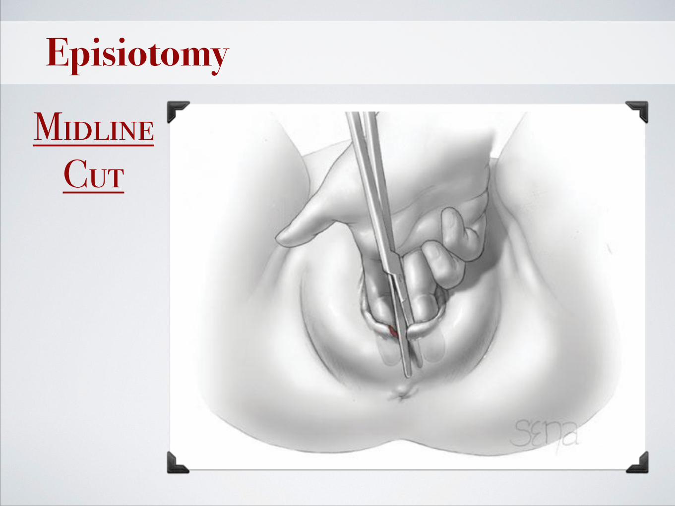
Episiotomy
Midline Cut
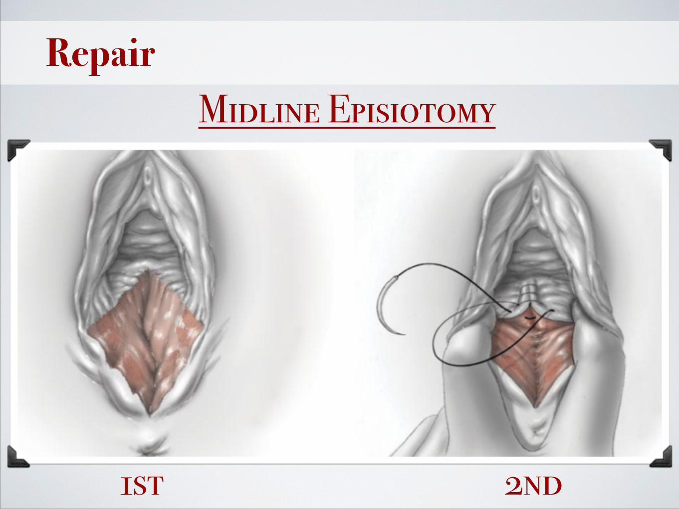
RepairMidline Episiotomy
1st 2nd
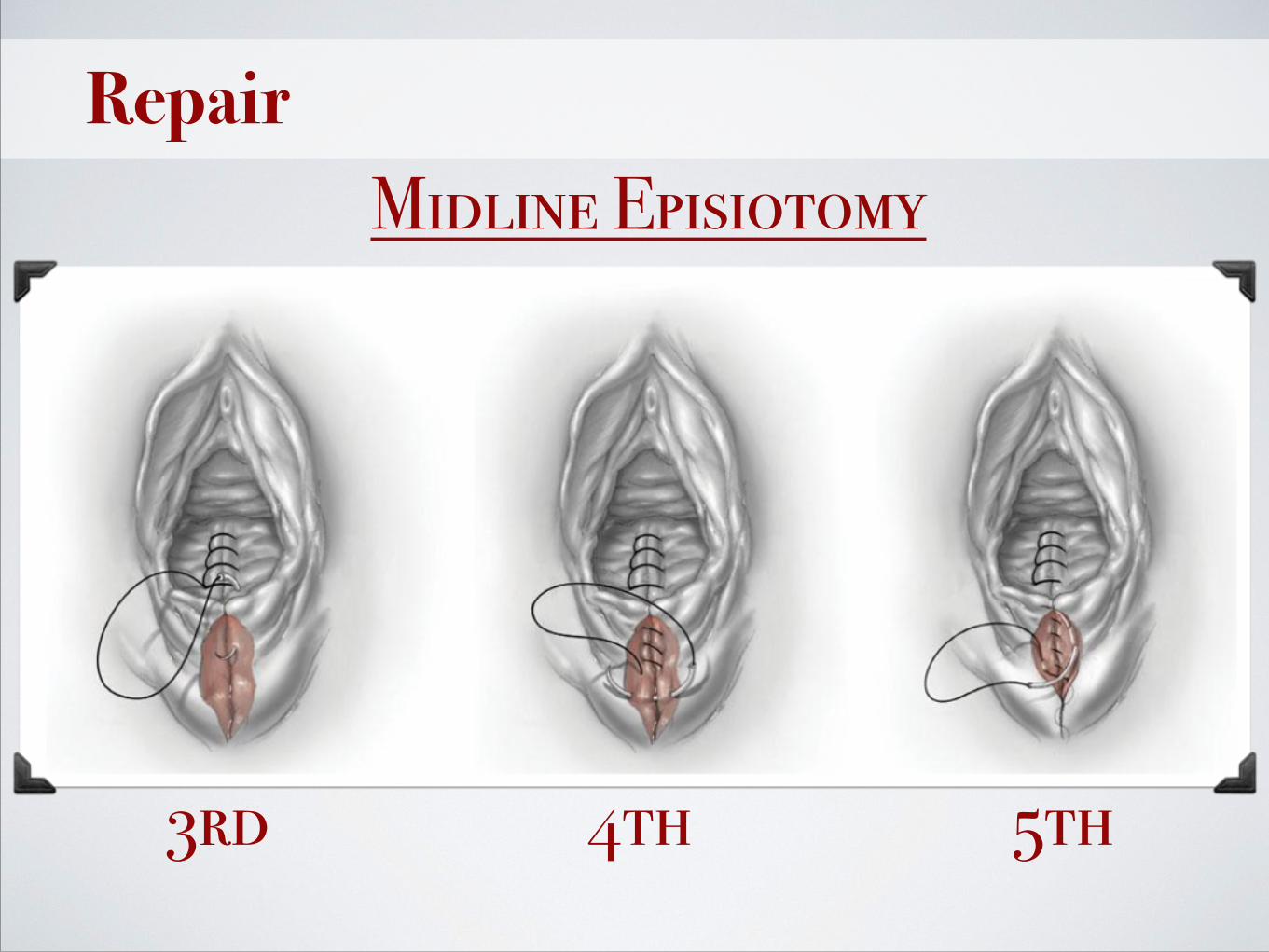
RepairMidline Episiotomy
3rd 4th 5th

Repair
Mediolateral Episiotomy

Repair
Fourth-degree Laceration
1st 2nd

Repair
Fourth-degree Laceration
3rd

Repair
Fourth-degree Laceration
4th

Repair
Fourth-degree Laceration
5th

Thank YounnO P
