utr of the rice osmac1 mrna enabling the sufficient ... · a long 5˜ utr of the rice osmac1 mrna...
TRANSCRIPT
Plant Biotechnology 29, 43–49 (2012)
Copyright © 2012 The Japanese Society for Plant Cell and Molecular Biology
A long 5′ UTR of the rice OsMac1 mRNA enabling the sufficient translation of the downstream ORF
Hiroshi Teramura1, Yusuke Enomoto1, Hiromi Aoki1, Tadamasa Sasaki1, Hiroaki Shimada1,2,*1 Department of Biological Science and Technology, Tokyo University of Science, Noda, Chiba 278-8510, Japan; 2 Research Center for RNA Science, Tokyo University of Science, Noda, Chiba 278-8510, Japan* E-mail: [email protected] Tel: +81-4-7124-1501 Fax: +81-4-7122-1499
Received November 25, 2011; accepted December 9, 2011 (Edited by T. Demura)
Abstract Untranslated regions (UTRs) of mRNA are involved in many posttranscriptional regulatory pathways. �e rice OsMac1 mRNA had a 5′ UTR of more than 500 nucleotides (nt), containing a CU-rich region and three upstream open reading frames (uORFs) preceding the downstream ORF. �e expected GFP-fusion protein was detected at the cell membrane, indicating the occurrence of translation of the downstream ORF. �e 5′ UTR contained three splicing variants that were generated by alternative splicing. A reporter analysis using β-glucuronidase indicated that only the longest one, UTRc, showed a signi�cant ability for the e�cient translation of the downstream ORF, whereas other splicing variants showed low level of translational e�ciency of the corresponding ORF. �ese results suggested that the additional 38-nt sequence unique to UTRc localized between the CU-rich region and the uORFs may be involved in the increased translational e�ciency of the downstream ORF located a�er the long 5′ UTR.
Key words: Alternative splicing, membrane protein, rice (Oryza sativa), 5′ untranslated region, upstream open reading frame (uORF).
Gene expression is regulated precisely to maintain the homeostasis of the cell function. This process is achieved via multiple processes, including transcription, translation and posttranslational modification. In particular, translation is important for the determination of the accumulation of proteins. �e initiation of the translation in eukaryotic cells involves a large number of factors, some of which are involved in the initial binding of the ribosome 40S subunit of the ribosome to the capped 5′ end of the mRNA, as well as in the binding of the initiator tRNA. A�er the initial binding, the complex containing the small ribosomal subunit scans the mRNA until it recognizes the �rst initiation codon, AUG. At this point, initiation factors are released and 60S subunit of the ribosome joins the complex, to start protein synthesis (Kozak 1999; Kozak 2007).
Recent studies revealed that translation is regulated by several mechanisms. Global regulation of translation involves the modulation of the activity of general factors, whereas mRNA-speci�c control is mediated by regulatory proteins or by noncoding RNAs. In particular, untranslated regions (UTRs) of mRNA are o�en involved in many posttranscriptional regulatory pathways that control mRNA localization, and stability and translation
e�ciency (Chatterjee and Pal 2009; Pesole et al. 2001).Many 5′ UTRs of mRNAs contain multiple AUG
codons, which suggests that the �rst-AUG rule is not followed in a remarkable fraction of mRNAs (Grillo et al. 2010; Pesole et al. 2001). �e regulation of the translation of speci�c eukaryotic mRNAs is sometimes mediated by small upstream open-reading-frames (uORFs) that limit the access of the small subunit to a downstream ORF (Holcik and Pestova 2007). The yeast GCN4 gene encodes the transcription factor Gcn4, the mRNA of which carries four uORFs in its 5′ leader sequence. �e translation of GCN4 is activated by amino acid starvation. In this case, it is predicted that reduction in the levels of the ternary complex containing the methionyl initiator tRNA, eIF2, and GTP enables the bypassing of the start codons of uORFs by scanning ribosomes (Hinnebusch et al. 2004; Nielsen et al. 2004). Translational control by uORF located in the 5′ UTR has also been demonstrated, such as is the case for the Arabidopsis transcription factor bZIP11, the yeast AP1-like transcription factor Yap2, and the plant S-adenosylmethionine decarboxylase (AdoMetDC) mRNAs (Hanfrey et al. 2005; Rahmani et al. 2009; Vilela et al. 1999).
Original Paper
DOI: 10.5511/plantbiotechnology.11.1209a
�is article can be found at http://www.jspcmb.jp/Published online February 20, 2012

44 A 5′UTR increasing a translational e�ciency
Copyright © 2012 The Japanese Society for Plant Cell and Molecular Biology
We found a long 5′ UTR in a rice mRNA encoding a membrane protein. �is 5′ UTR composed of three variants that were derived from the alternative splicing. Among them, one of the 5′ UTR showed signi�cantly higher translational e�ciency of the downstream ORF than those observed by the others. Here, we describe the characteristics of the 5′ UTR, and its e�ect on the translation of the downstream ORF.
Materials and methods
Plant materialsRice (Oryza sativa L. cv. Nipponbare) was used as the wild-type plant, and was grown in a greenhouse.
PCR and real-time quantitative RT-PCRGenomic DNA was prepared according to the method of Murray and �ompson (1980). PCR was performed using Blend-Taq thermostable DNA polymerase (Toyobo, Osaka, Japan). Total RNA was prepared from each tissue as described previously (Imamura et al. 2007). First-strand cDNA was synthesized from 1 μg of total RNA using the ReverTra Ace cDNA synthesis kit (Toyobo) with an oligo-dT(20) primer. Amplification of the entire cDNA of the AP004685 gene (termed OsMac1) was performed using primers, 5′-CACCGCACGCTACGCCTACG-3′ and 5′-AATGCCAGGGAGGTAAAAGGA-3′. The amplified fragments were cloned into pENTRTM/D-TOPO (Invitrogen, Carlsbad, CA, USA). Real-time quantitative PCR was performed as described previously (She et al. 2010) using an ABI PRISM 7000 Sequence Detection System (Applied Biosystems, CA, USA) with a SYBR Green real-time PCR mix (Toyobo). Primer sets (either 5′-CACATCTCCCTCAAGGATC-3′, 5 ′ - A C A T C T C C C T C A A G G T T G - 3 ′ , o r 5′-TCACATCTCCCTCAAGCTA-3′ as a forward primer and 5′-CACGGTAGTATTCAACTGCTTG-3′ as a reverse primer) were used for measurement of the levels of the splice variants (UTRa, UTRb, and UTRc, respectively) of the OsMac1 transcripts. �e levels of β-glucuronidase (GUS) transcripts was measured using the primer set, 5′-GCCGATGCAGATATTCGTA-3′ and 5′-CCATCACTTCCTGATTATTGA-3′. The level of the Actin1 mRNA (AK100267) was used as a positive control for the normalization of data. �e primer set 5′-CCCTCCTGAAAGGAAGTACAGTGT-3′ and 5′-GTCCGAAGAATTAGAAGCATTTCC-3′ was used for detection of Actin1 transcript.
Transient expression of the GFP fusion gene in onion epidermal cellsThe region covering the ORF of the entire OsMac1 transcript and that covering UTRc and the entire OsMac1 ORF located a�er this sequence were ampli�ed using
the primer sets, 5′-CACCATGGAATTGGCAGAGC-3′ (orf-fw) and 5′-TTTCTCCCTAGTAATCCATC-3′ (orfns-rv), and 5′-CACCGCACGCTACGCCTACG-3′ (utr-fw) and orfns-rv, respectively, from the OsMac1 cDNA containing UTRc as its 5′ UTR. �e nucleotide sequence of orfns-rv was designed to remove the termination codon. �e ampli�ed fragment was entered into pENTRTM/D-TOPO (Invitrogen) and was fused to the GFP gene using a pGWB5 plasmid (Nakagawa et al. 2007), via an LR clonase (Invitrogen) reaction. �e GFP fusion gene was introduced into onion epidermal cells via particle bombardment (Bio-Rad, Richmond, CA, USA) and transient expression was detected according to von Arnim et al. (1998) using a confocal laser-scanning microscope (LSM 510 META; Carl Zeiss AG, Oberkochen, Germany).
Determination of GUS activity and estimation of translational e�ciencyGUS activity was determined according to Jefferson et al. (1987). A plasmid harboring the GUS gene as a reporter gene was introduced into rice callus using the Agrobacterium-mediated transformation method (Hiei et al. 1994). Transgenic callus grown on 2N6 plates (Fujimura et al. 1985) supplemented with 50 mg l−1 Hygromycin B and 250 mg l−1 Claforan were transferred into R2 medium (Ling and Robinson 1997) supplemented with 50 mg l−1 Hygromycin B and 250 mg l−1 Claforan, and cultured for 10 days at 30°C, with medium exchange performed every 5 days. GUS activity was analyzed by staining for 15 min to 24 h at 37°C using a reaction solution containing 50 mM sodium phosphate bu�er, pH 7.0, with 5% (v/v) methanol, 0.1% (v/v) Triton X-100 and 1 mM 5-bromo-4-chroro-2-indolyl-β-glucronide. To measure GUS activity, grown callus cells were homogenized in bu�er (50 mM sodium phosphate bu�er, pH 7.0, with 10 mM EDTA, 0.1% (v/v) Triton X-100, 0.1% (v/v) sodium N-lauroyl sarcosylate, 5 mM (±)-Dithiothreitol, and 20% (v/v) methanol). A�er cell debris was removed by centrifugation at 12,000 rpm for 5 min at 4°C, GUS activity was determined via reaction using 1 mM 4-methylumbelliferyl glucuronide as a substrate at 37°C for 30 min. Translational e�ciency was estimated as the relative value of GUS activity relative to the level of the reporter mRNA, which was determined using real-time quantitative RT-PCR. �e data were analyzed statistically using Student’s t test.
Construction of reporter genesThe GUS gene, which was used as a reporter, was obtained from pBI221 (Jefferson et al. 1987), from which the coding region was amplified using primers that corresponded to the regions of the initiation and termination of the ORF (5′-CACCCTCGAGAGATTAGCCTTTTCAATTTC-3′
H. Teramura et al. 45
Copyright © 2012 The Japanese Society for Plant Cell and Molecular Biology
and 5′-TGAATTCCCGATCTAGTAACAT-3′), and then inserted into pENTRTM/D-TOPO (Invitrogen) to generate pENTR-35S-GUS.
An expression plasmid containing each UTR followed by the GUS gene was obtained as follows: UTRa, UTRb, and UTRc were amplified from the corresponding cDNAs using the PCR primer set, 5′-CCCTCTAGAGACGCTACGCCTACGCGGGAG-3′ and 5′-GGGTCTAGATGCCAATTCCATTGTTCTCTC -3′. �ese sequences were inserted into the XbaI site preceding the GUS gene in pENTR-35S-GUS, after digestion of the ampli�ed fragments with XbaI. Binary plasmids were produced using the resultant plasmids (pENTR-UTRa, pENTR-UTRb, and pENTR-UTRc, respectively) and pGWB1 (Nakagawa et al. 2007) via LR clonase reaction.
Results
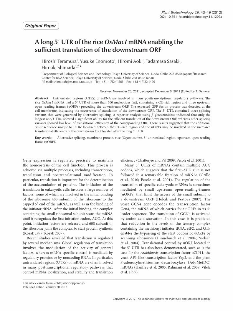
A novel gene containing a long 5′UTRWe found a novel gene (AP004685; Os06g0726600) upstream of the gene that encodes the rice starch branching enzyme 1 (Sbe1), which is located on chromosome 6. This gene encoded a protein that exhibited local homology to the MAP-kinase activating protein and was named as OsMac1. �e corresponding cDNA, which comprised 2,467 base-pairs, was registered in the database (acc. no. AK111844). �e transcript of this gene had a long 5′ UTR composed of the 526 nt preceding the main ORF. �is region contained a CU-rich region encompassing approximately 250 nt at the 5′ region, and three short ORFs (uORFs) at the 3′ region (Figure 1). �ese uORFs comprised 81, 78, and 72 nt, respectively, and encoded short peptides showing no homology to known proteins.
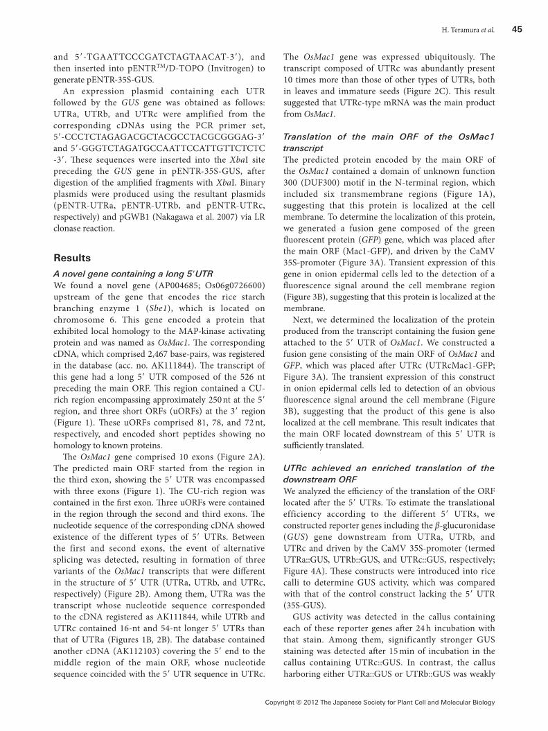
�e OsMac1 gene comprised 10 exons (Figure 2A). The predicted main ORF started from the region in the third exon, showing the 5′ UTR was encompassed with three exons (Figure 1). �e CU-rich region was contained in the �rst exon. �ree uORFs were contained in the region through the second and third exons. �e nucleotide sequence of the corresponding cDNA showed existence of the different types of 5′ UTRs. Between the first and second exons, the event of alternative splicing was detected, resulting in formation of three variants of the OsMac1 transcripts that were di�erent in the structure of 5′ UTR (UTRa, UTRb, and UTRc, respectively) (Figure 2B). Among them, UTRa was the transcript whose nucleotide sequence corresponded to the cDNA registered as AK111844, while UTRb and UTRc contained 16-nt and 54-nt longer 5′ UTRs than that of UTRa (Figures 1B, 2B). �e database contained another cDNA (AK112103) covering the 5′ end to the middle region of the main ORF, whose nucleotide sequence coincided with the 5′ UTR sequence in UTRc.
The OsMac1 gene was expressed ubiquitously. The transcript composed of UTRc was abundantly present 10 times more than those of other types of UTRs, both in leaves and immature seeds (Figure 2C). �is result suggested that UTRc-type mRNA was the main product from OsMac1.
Translation of the main ORF of the OsMac1 transcriptThe predicted protein encoded by the main ORF of the OsMac1 contained a domain of unknown function 300 (DUF300) motif in the N-terminal region, which included six transmembrane regions (Figure 1A), suggesting that this protein is localized at the cell membrane. To determine the localization of this protein, we generated a fusion gene composed of the green �uorescent protein (GFP) gene, which was placed a�er the main ORF (Mac1-GFP), and driven by the CaMV 35S-promoter (Figure 3A). Transient expression of this gene in onion epidermal cells led to the detection of a �uorescence signal around the cell membrane region (Figure 3B), suggesting that this protein is localized at the membrane.
Next, we determined the localization of the protein produced from the transcript containing the fusion gene attached to the 5′ UTR of OsMac1. We constructed a fusion gene consisting of the main ORF of OsMac1 and GFP, which was placed a�er UTRc (UTRcMac1-GFP; Figure 3A). �e transient expression of this construct in onion epidermal cells led to detection of an obvious �uorescence signal around the cell membrane (Figure 3B), suggesting that the product of this gene is also localized at the cell membrane. �is result indicates that the main ORF located downstream of this 5′ UTR is su�ciently translated.
UTRc achieved an enriched translation of the downstream ORFWe analyzed the e�ciency of the translation of the ORF located a�er the 5′ UTRs. To estimate the translational efficiency according to the different 5′ UTRs, we constructed reporter genes including the β-glucuronidase (GUS) gene downstream from UTRa, UTRb, and UTRc and driven by the CaMV 35S-promoter (termed UTRa::GUS, UTRb::GUS, and UTRc::GUS, respectively; Figure 4A). �ese constructs were introduced into rice calli to determine GUS activity, which was compared with that of the control construct lacking the 5′ UTR (35S-GUS).
GUS activity was detected in the callus containing each of these reporter genes a�er 24 h incubation with that stain. Among them, significantly stronger GUS staining was detected a�er 15 min of incubation in the callus containing UTRc::GUS. In contrast, the callus harboring either UTRa::GUS or UTRb::GUS was weakly
46 A 5′UTR increasing a translational e�ciency
Copyright © 2012 The Japanese Society for Plant Cell and Molecular Biology
Figure 1. Structure of the OsMac1 mRNA. (A) Schematic representation of the OsMac1 mRNA. Open boxes and a �lled box show the uORFs (uORF1, uORF2, and uORF3) and downstream ORF encoding a protein that is homologous to a MAP-kinase activation protein, respectively. �e triangle indicates the region of the additional 38-nt sequence found in UTRc, and the hatched boxes in the downstream ORF show the location of the transmembrane regions (TMRs) located in the DUF300 domain included in the predicted protein. (B) Nucleotide sequence of the UTRc of the OsMac1 mRNA. �e CU-rich sequences in the 5′ region are underlined. �e open box indicates the additional 38-nt sequence. uORFs and the downstream ORF are indicated by arrows placed below the nucleotide sequence. Regions between �lled triangles a and c, and those between b and c, lack UTRa and UTRb, respectively. �e open triangle shows the position of the splicing site located between the second and third exons.
Figure 2. Alternative splicing in the 5′ UTR of the OsMac1 mRNA. (A) Genomic structure of the OsMac1 gene. Filled boxes indicate exons. �e site of alternative splicing is shown. (B) Schematic representation of the alternative splicing of UTRa, UTRb, and UTRc. Spliced regions are indicated by broken lines. (C) Expression of each variant of the OsMac1 mRNA in the 5th leaf and in developing seeds at 5 days a�er �owering (DAF) and 10 DAF. �e level of these transcripts is shown as a relative value to that observed for Actin1; these level were determined using real-time quantitative RT-PCR. Error bars indicate SD (n=5).

H. Teramura et al. 47
Copyright © 2012 The Japanese Society for Plant Cell and Molecular Biology
stained for GUS to a lesser extent than that detected in the callus transformed by 35S-GUS (Figure 4B). �e di�erence in GUS activity depended on the gene introduced.
We determined the efficiency of translation of the downstream ORF, which was estimated as the ratio of the GUS activity to the amount of these mRNAs in the cell. �e callus transformed with UTRc::GUS exhibited a translational efficiency that was ∼15-fold higher than that detected in the callus harboring the control gene. The transformants containing UTRa::GUS or UTRb::GUS exhibited a very low translational e�ciency, which was approximately half of that detected in 35S-GUS (Figure 4C). A similar amount of mRNA was detected in all callus lines using real-time quantitative RT-PCR (qRT-PCR), suggesting that nearly equal transcription occurred from each fusion gene. These results indicate that UTRc possesses a speci�c region that increases the translational e�ciency of the downstream ORF located a�er the long 5′ UTR. As UTRc was 38 nt longer than UTRb (Figure 1), it is expected that this
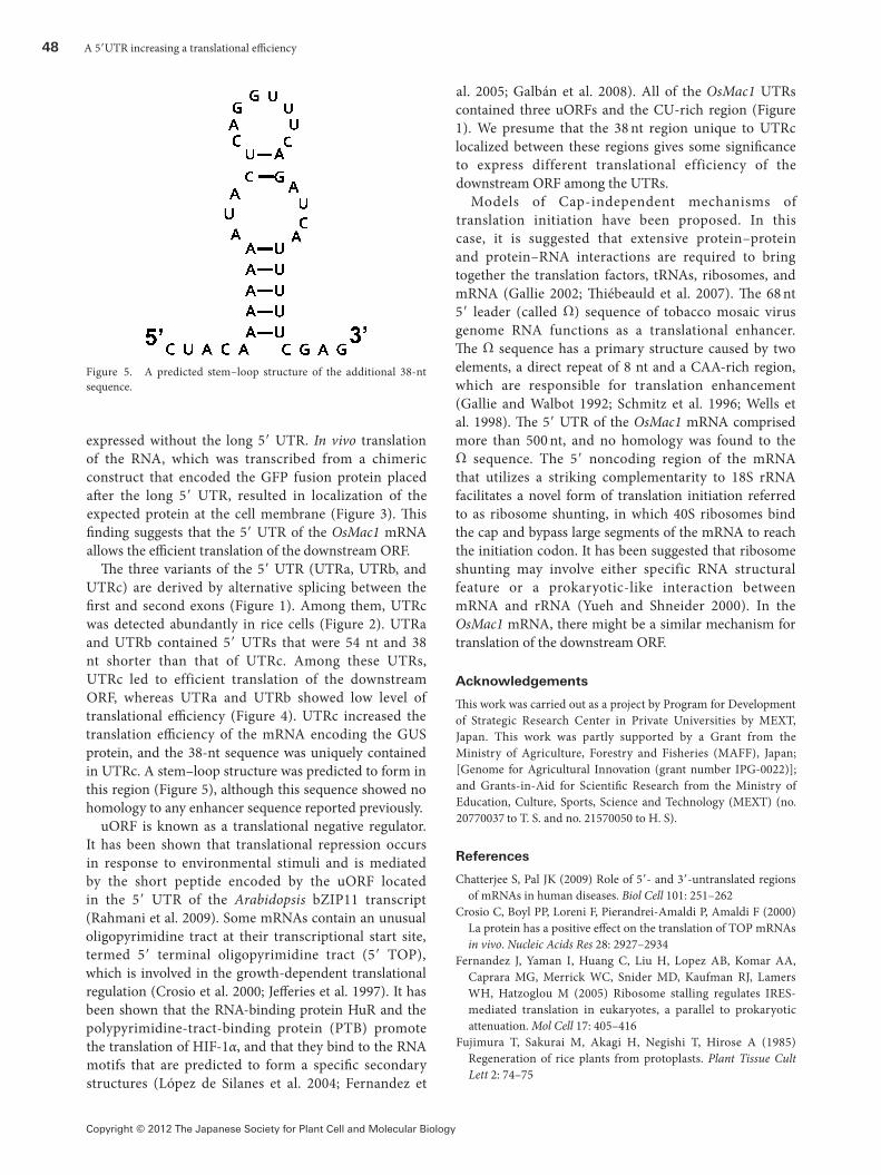
additional 38-nt region is involved in the e�ciency of the translation of the downstream ORF. �e nucleotide sequence of the additional 38-nt sequence predicted to form a stem-loop structure (Figure 5). �is sequence showed no homology to any known cis elements involved in translation.
Discussion
The rice OsMac1 mRNA possesses a long 5′ UTR preceding the main ORF, which includes a CU-rich region and three uORFs that are predicted to a�ect on the translational efficiency of the downstream ORF (Figure 1). �e protein encoded by the downstream ORF was localized at the cell membrane, as its GFP fusion protein was detected at the cell membrane when it was
Figure 3. Translation of the main ORF of the OsMac1 mRNA. (A) Schematic representation of the structure of the expression plasmids, which were constructed for transient expression in onion epidermal cells. Mac1-GFP indicates the fusion gene consisting of the entire OsMac1 sequence with sGFP driven by the CaMV 35S promoter (35S-pro), and UTRcMac1-GFP contained the fusion gene placed a�er UTRc. �e control plasmid contained sGFP. Ter, NOS terminator. (B) Detection and subcellular localization of the translated OsMac1-GFP fusion proteins in onion epidermal cells. Fluorescence images of GFP and di�erential interference contrast macroscopic images (DIC) are shown. Genes were introduced using microprojectile methods. Scale bar = 100 μm.
Figure 4. Translational e�ciency of the UTRs. (A) Structure of the reporter GUS genes placed downstream of the UTRs. �e position of the additional 38-nt sequence is indicated by a triangle. �e regions around this sequence in UTRa, UTRb, and UTRc are highlighted below. Broken lines indicate the nucleotides missing in UTRa and UTRb, via the alternative splicing. 35S-pro, CaMV 35S-promoter; ter, NOS-terminator. (B) GUS-activity staining in the callus cells harboring each gene. �e upper panels show the cell lines a�er 15 min of staining. Scale bar=500 μm. (C) Translational e�ciency is shown as the values of GUS activity relative to the amount of its mRNA in the transformed callus cells. �e translational e�ciency of UTRc::GUS was set at 1.0. Error bars indicate the SD (n=5). An asterisk indicates a signi�cant di�erence between the translational e�ciency of the individual UTR and the 35S-GUS at P<0.05.
48 A 5′UTR increasing a translational e�ciency
Copyright © 2012 The Japanese Society for Plant Cell and Molecular Biology
expressed without the long 5′ UTR. In vivo translation of the RNA, which was transcribed from a chimeric construct that encoded the GFP fusion protein placed a�er the long 5′ UTR, resulted in localization of the expected protein at the cell membrane (Figure 3). �is �nding suggests that the 5′ UTR of the OsMac1 mRNA allows the e�cient translation of the downstream ORF.
�e three variants of the 5′ UTR (UTRa, UTRb, and UTRc) are derived by alternative splicing between the �rst and second exons (Figure 1). Among them, UTRc was detected abundantly in rice cells (Figure 2). UTRa and UTRb contained 5′ UTRs that were 54 nt and 38 nt shorter than that of UTRc. Among these UTRs, UTRc led to efficient translation of the downstream ORF, whereas UTRa and UTRb showed low level of translational e�ciency (Figure 4). UTRc increased the translation e�ciency of the mRNA encoding the GUS protein, and the 38-nt sequence was uniquely contained in UTRc. A stem–loop structure was predicted to form in this region (Figure 5), although this sequence showed no homology to any enhancer sequence reported previously.
uORF is known as a translational negative regulator. It has been shown that translational repression occurs in response to environmental stimuli and is mediated by the short peptide encoded by the uORF located in the 5′ UTR of the Arabidopsis bZIP11 transcript (Rahmani et al. 2009). Some mRNAs contain an unusual oligopyrimidine tract at their transcriptional start site, termed 5′ terminal oligopyrimidine tract (5′ TOP), which is involved in the growth-dependent translational regulation (Crosio et al. 2000; Je�eries et al. 1997). It has been shown that the RNA-binding protein HuR and the polypyrimidine-tract-binding protein (PTB) promote the translation of HIF-1α, and that they bind to the RNA motifs that are predicted to form a speci�c secondary structures (López de Silanes et al. 2004; Fernandez et
al. 2005; Galbán et al. 2008). All of the OsMac1 UTRs contained three uORFs and the CU-rich region (Figure 1). We presume that the 38 nt region unique to UTRc localized between these regions gives some signi�cance to express different translational efficiency of the downstream ORF among the UTRs.
Models of Cap-independent mechanisms of translation initiation have been proposed. In this case, it is suggested that extensive protein–protein and protein–RNA interactions are required to bring together the translation factors, tRNAs, ribosomes, and mRNA (Gallie 2002; �iébeauld et al. 2007). �e 68 nt 5′ leader (called Ω) sequence of tobacco mosaic virus genome RNA functions as a translational enhancer. �e Ω sequence has a primary structure caused by two elements, a direct repeat of 8 nt and a CAA-rich region, which are responsible for translation enhancement (Gallie and Walbot 1992; Schmitz et al. 1996; Wells et al. 1998). �e 5′ UTR of the OsMac1 mRNA comprised more than 500 nt, and no homology was found to the Ω sequence. The 5′ noncoding region of the mRNA that utilizes a striking complementarity to 18S rRNA facilitates a novel form of translation initiation referred to as ribosome shunting, in which 40S ribosomes bind the cap and bypass large segments of the mRNA to reach the initiation codon. It has been suggested that ribosome shunting may involve either specific RNA structural feature or a prokaryotic-like interaction between mRNA and rRNA (Yueh and Shneider 2000). In the OsMac1 mRNA, there might be a similar mechanism for translation of the downstream ORF.
Acknowledgements
�is work was carried out as a project by Program for Development of Strategic Research Center in Private Universities by MEXT, Japan. This work was partly supported by a Grant from the Ministry of Agriculture, Forestry and Fisheries (MAFF), Japan; [Genome for Agricultural Innovation (grant number IPG-0022)]; and Grants-in-Aid for Scienti�c Research from the Ministry of Education, Culture, Sports, Science and Technology (MEXT) (no. 20770037 to T. S. and no. 21570050 to H. S).
References
Chatterjee S, Pal JK (2009) Role of 5′- and 3′-untranslated regions of mRNAs in human diseases. Biol Cell 101: 251–262
Crosio C, Boyl PP, Loreni F, Pierandrei-Amaldi P, Amaldi F (2000) La protein has a positive e�ect on the translation of TOP mRNAs in vivo. Nucleic Acids Res 28: 2927–2934
Fernandez J, Yaman I, Huang C, Liu H, Lopez AB, Komar AA, Caprara MG, Merrick WC, Snider MD, Kaufman RJ, Lamers WH, Hatzoglou M (2005) Ribosome stalling regulates IRES-mediated translation in eukaryotes, a parallel to prokaryotic attenuation. Mol Cell 17: 405–416
Fujimura T, Sakurai M, Akagi H, Negishi T, Hirose A (1985) Regeneration of rice plants from protoplasts. Plant Tissue Cult Lett 2: 74–75
Figure 5. A predicted stem–loop structure of the additional 38-nt sequence.

H. Teramura et al. 49
Copyright © 2012 The Japanese Society for Plant Cell and Molecular Biology
Galbán S, Kuwano Y, Pullmann R Jr, Martindale JL, Kim HH, Lal A, Abdelmohsen K, Yang X, Dang Y, Liu JO, Lewis SM, Holcik M, Gorospe M (2008) RNA-binding proteins HuR and PTB promote the translation of hypoxia-inducible factor 1α. Mol Cell Biol 28: 93–107
Gallie DR, Walbot V (1992) Identi�cation of the motifs within the tobacco mosaic virus 5′-leader responsible for enhancing translation. Nucleic Acids Res 20: 4631–4638
Gallie DR (2002) Protein-protein interactions required during translation. Plant Mol Biol 50: 949–970
Grillo G, Turi A, Licciulli F, Mignone F, Liuni S, Ban� S, Gennarino VA, Horner DS, Pavesi G, Picardi E, Pesole G (2010) UTRdb and UTRsite (RELEASE 2010): a collection of sequences and regulatory motifs of the untranslated regions of eukaryotic mRNAs. Nucleic Acids Res 38(Database issue): D75–D80
Hanfrey C, Elliott KA, Franceschetti M, Mayer MJ, Illingworth C, Michael AJ (2005) A dual upstream open reading frame-based autoregulatory circuit controlling polyamine-responsive translation. J Biol Chem 280: 39229–39237
Hiei Y, Ohta S, Komari T, Kumashiro T (1994) Efficient transformation of rice (Oryza sativa L.) mediated by Agrobacterium and sequence analysis of the boundaries of the T-DNA. Plant J 6: 271–282
Hinnebusch AG, Asano K, Olsen DS, Phan L, Nielsen KH, Valásek L (2004) Study of translational control of eukaryotic gene expression using yeast. Ann NY Acad Sci 1038: 60–74
Holcik M, Pestova TV (2007) Translation mechanism and regulation: old players, new concepts. Meeting on translational control and non-coding RNA. EMBO Rep 8: 639–643
Imamura T, Kusano H, Kajigaya Y, Ichikawa M, Shimada H (2007) A rice dihydrosphingosine C4 hydroxylase (DSH1) gene, which is abundantly expressed in the stigmas, vascular cells and apical meristem, may be involved in fertility. Plant Cell Physiol 48: 1108–1120
Jefferies HB, Fumagalli S, Dennis PB, Reinhard C, Pearson RB, Thomas G (1997) Rapamycin suppresses 5’TOP mRNA translation through inhibition of p70s6k. EMBO J 16: 3693–3704
Je�erson RA, Kavanagh TA, Bevan MW (1987) GUS fusions: beta-glucuronidase as a sensitive and versatile gene fusion marker in higher plants. EMBO J 6: 3901–3907
Kozak M (1999) Initiation of translation in prokaryotes and eukaryotes. Gene 234: 187–208
Kozak M (2007) Some thoughts about translational regulation: forward and backward glances. J Cell Biochem 102: 280–290
Ling MM, Robinson BH (1997) Approaches to DNA mutagenesis: an overview. Anal Biochem 254: 157–178
López de Silanes I, Zhan M, Lal A, Yang X, Gorospe M (2004) Identi�cation of a target RNA motif for RNA-binding protein HuR. Proc Natl Acad Sci USA 101: 2987–2992
Murray MG, Thompson WF (1980) Rapid isolation of high molecular weight plant DNA. Nucleic Acids Res 8: 4321–4325
Nakagawa T, Kurose T, Hino T, Tanaka K, Kawamukai M, Niwa Y, Toyooka K, Matsuoka K, Jinbo T, Kimura T (2007) Development of series of gateway binary vectors, pGWBs, for realizing e�cient construction of fusion genes for plant transformation. J Biosci Bioeng 104: 34–41
Nielsen KH, Szamecz B, Valásek L, Jivotovskaya A, Shin B-S, Hinnebusch AG (2004) Functions of eIF3 downstream of 48S assembly impact AUG recognition and GCN4 translational control. EMBO J 23: 1166–1177
Pesole G, Mignone F, Gissi C, Grillo G, Licciulli F, Liuni S (2001) Structural and functional features of eukaryotic mRNA untranslated regions. Gene 276: 73–81
Rahmani F, Hummel M, Schuurmans J, Wiese-Klinkenberg A, Smeekens S, Hanson J (2009) Sucrose control of translation mediated by an upstream open reading frame-encoded peptide. Plant Physiol 150: 1356–1367
Schmitz J, Prüfer D, Rohde W, Tacke E (1996) Non-canonical translation mechanisms in plants: e�cient in vitro and in planta initiation at AUU codons of the tobacco mosaic virus enhancer sequence. Nucleic Acids Res 24: 257–263
She K-C, Kusano H, Koizumi K, Yamakawa H, Hakata M, Imamura T, Fukuda M, Naito N, Tsurumaki Y, Yaeshima M, Tsuge T, Matsumoto K, Kudoh M, Itoh E, Kikuchi S, Kishimoto N, Yazaki J, Ando T, Yano M, Aoyama T, Sasaki T, Satoh H, Shimada H (2010) A novel factor FLOURY ENDOSPERM2 is involved in regulation of rice grain size and starch quality. Plant Cell 22: 3280–3294
Thiébeauld O, Pooggin MM, Ryabova LA (2007) Alternative translation strategies in plant viruses. Plant Viruses 1: 1–20
Vilela C, Ramirez CV, Linz B, Rodrigues-Pousada C, McCarthy JEG (1999) Post-termination ribosome interactions with the 5′ UTR modulate yeast mRNA stability. EMBO J 18: 3139–3152
von Arnim AG, Deng X-W, Stacey MG (1998) Cloning vectors for the expression of green �uorescent protein fusion proteins in transgenic plants. Gene 221: 35–43
Wells DR, Tanguay RL, Le H, Gallie DR (1998) HSP101 functions as a speci�c translational regulatory protein whose activity is regulated by nutrient status. Genes Dev 12: 3236–3251
Yueh A, Schneider RJ (2000) Translation by ribosome shunting on adenovirus and hsp70 mRNAs facilitated by complementarity to 18S rRNA. Genes Dev 14: 414–421