uterine transplant, birth of baby vincent as shared by brannstrorm snd team
TRANSCRIPT
UTERINE TRANSPLANT
A BOON
?Dr. Vijayalakshmi.G.Pillai, M.B.B.S, DGO, MRCOG(London)Member of Royal College of Obstetricians and Gynaecologists
Member Of American Society of Reproductive MedicineMember European Society of human reproduction and Embryology
Chief Medical Officer - Head of Dept Obstetrics Gynaecology and Infertility, VIJAYALAKSHMI MEDICAL CENTRE, VENNALA, KOCHI

Reproductive biology breakthrough?
It seems science has a solution for every reproductive woe…. World's first child , baby Vincent born after uterus transplantation
Date: October 4, 2014: Hip Hip Hurray Hurray!!!!!.....

Uterus Transplant, Latest Fertility Feat..?......Skeptical?
• Isn’t this the best thing that happened after Louis Brown?............. Happy to be part of this world-order of Reproductive Biologists!!!
• “Media celebrations”, doomed or just: excitement or contemptuous?
• Baby Born From Transplanted Uterus: Should We Be Doing This?..... How many should be repeating this feat in this world?

“Join the first ever Workshop on Uterine transplantation in Human organized by the Uterus Transplantation Research Project from the
University of Gothenburg, Sweden. The workshop is free of charge for congress participants”. Program:
Part 1 Part 2
Inclusion/exclusion criteria of UTx recipients and preop. InvestigationMats Brännström
IVF treatment in UTx patientsLars.B. Nilsson
Inclusion/exclusion criteria of UTx donors and preop. InvestigationLiza Johannesson
Surgical technique for uterine harvest in deceased donorMichael Olausson
Surgical technique for uterine harvest in live donor & videoMats Brännström/ Lennart Wiman
Surgical technique for UTx in recipient & videoMichael Olausson/ Lennart Wiman
Detailed results of Gotenburgh UTx trialMats Brännström
Rejection detection in UTx and treatmentsLiza Johannesson Institutional and personal requirements before for human UTx Mats Brännström
Exit strategies at UTxLiza Johannesson
Future of human UTxMats Brännström
Introduction
Patient groups with absolute uterine factor infertilityLars.B. Nilsson
Alternatives to UTx Lars.B. Nilsson
The complicated ethics of UTxMichael Olausson
Research on UTx, human experience and UTx training requirementsMats Brännström
Immunosuppression and risks in organ transplantationMichael Olausson
Immunosuppression and pregnancyLiza Johannesson
Live or deceased uterus donorLiza Johannesson
Coffee break

“Our demonstration of a live birth after uterus transplantation in a woman born with no uterus has eradicated the diagnosis of absolute uterine factor infertility.”- Professor Mats Brannstrom, Sweden
Womb transplant birth worth the risk, says mother

H/O Uterine Transplant in Human being
In Turkey, 2011, the first uterus transplant from a deceased donor by doctors at Akdeniz University Hospital in Antalya.
The 21-year-old, Derya Sert, with MRHK. The world's first uterus transplant surgery gaining long-term function
In April 2013, they announced that Derya Sert was pregnant. She had had her pregnancy terminated in its 8th week with missed abortion.
First performed in 2000 by the doctors in Saudi Arabia on a 26-year-old who had CS hysterectomy. The transplanted uterus failed after 99 days.Int J Gynaecol Obstet. 2002 Mar;76(3):245-51.Transplantation of the human uterus. Fageeh W, Raffa H, Jabbad H, Marzouki A.
The donor, a 46-year-old with multiloculated ovarian cysts, underwent a modified hysterectomy to preserve tissue and vascular integrity. Immunosuppression by oral cyclosporine A, azathioprine and prednisolone.

World’s First Mother Daughter WombTransplant: September 22,
2012 • Two Swedish women have
received new wombs donated by their mothers in the first mother-to-daughter uterine transplants.
• ”The mothers who donated their uteruses are already up and walking and are going to be able to go home within a few days”…….
Both recipients have delivered healthy children to date.

A few animal models
Baboon uterus ex vivo after the two ovarian arteries have been joined to create one larger vessel (A) and two ovarian veins have been joined to create a large vein (V).
Schematic outline of end-to-side vascular anastomoses of the vessels of a baboon utero-tubal-ovarian graft to the external iliac vessels.
Schematic representation of the autotransplanted sheeputerus. (a) grafted uterine horn, (b) grafted ovary, (c) ovarian artery, (d) round ligament, (e) anterior branch of internal iliac artery and utero-ovarian vein, (f) recipient external iliac artery and vein, (g) vaginal anastomosis.

Schematic outline of end-to-side vascular anastomosis between the common iliac artery of a rat uterine transplant and the aorta of the recipient rat.
Schematic outline of end-to-side vascular anastomosis between the anterior branch of the left internal iliac artery of a sheep. Uterine graft and the vena cava and aorta in mouse.

Modern studies published on main aspects of Utx.
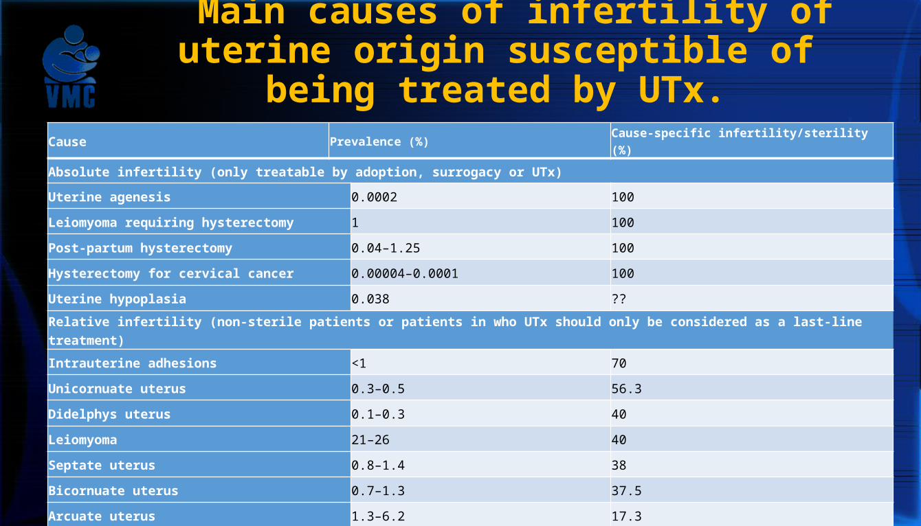
Main causes of infertility of uterine origin susceptible of being
treated by UTx.Cause Prevalence (%) Cause-specific infertility/sterility (%)
Absolute infertility (only treatable by adoption, surrogacy or UTx)
Uterine agenesis 0.0002 100
Leiomyoma requiring hysterectomy 1 100
Post-partum hysterectomy 0.04–1.25 100
Hysterectomy for cervical cancer 0.00004–0.0001 100
Uterine hypoplasia 0.038 ??
Relative infertility (non-sterile patients or patients in who UTx should only be considered as a last-line treatment)
Intrauterine adhesions <1 70
Unicornuate uterus 0.3–0.5 56.3
Didelphys uterus 0.1–0.3 40
Leiomyoma 21–26 40
Septate uterus 0.8–1.4 38
Bicornuate uterus 0.7–1.3 37.5
Arcuate uterus 1.3–6.2 17.3

The present case Prologue: 4 generations of revered motherhood
• The available motherhood options for AUFI: adoption or gestational surrogacy.
• Surrogacy is not allowed in many countries: ?ethical, ? legal, or ? religious reasons.
• The team from Gothenburg: sufficient preclinical research for > a decade on several animal models, from rodents to non-human primates.
• The first clinical trial of transplantation: 9 women who received uteri from live donors, including own mothers.
• Seven (7) women began menses in the first 2–3 months and the grafts remained viable.

Patient
• 35-year-old, MRHK syndrome, O+ve, BMI 21 kg/m², underwent uterus transplantation at Sahlgrenska University Hospital (Gothenburg, Sweden)approved by the regional ethics board.
• She was also born with only one kidney and had
vaginal and uterine aplasia.
• The donor, recipient, and her male partner had given their written informed consent.
• Neovagina had been created by self-dilatation. • Rejection risks, surgical complications at caesarean
section, or side-effects of immunosuppression were well informed.

The donor
• Blood group O+, a close family friend, healthy non-smoker and BMI of 20 kg/m².
• 61 years, Para 2, with two previous vaginal deliveries, at 26 years and 29 years of age.
• Menopaused around 7 years before.
• To ascertain menstrual functionality of the uterus and to possibly increase uterine artery blood flow preoperatively, she was treated with sequential OCP for 3 months.
• The HLA mismatch between donor and recipient was 3/2 and no HLA antibodies were present.
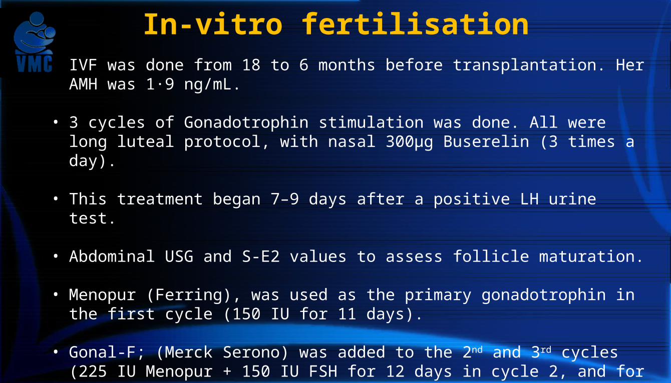
In-vitro fertilisation• IVF was done from 18 to 6 months before transplantation. Her AMH was
1·9 ng/mL. • 3 cycles of Gonadotrophin stimulation was done. All were long luteal
protocol, with nasal 300μg Buserelin (3 times a day). • This treatment began 7–9 days after a positive LH urine test. • Abdominal USG and S-E2 values to assess follicle maturation. • Menopur (Ferring), was used as the primary gonadotrophin in the first
cycle (150 IU for 11 days).
• Gonal-F; (Merck Serono) was added to the 2nd and 3rd cycles (225 IU Menopur + 150 IU FSH for 12 days in cycle 2, and for 14 days in cycle 3).
• Triggered by injection of 250μg rhCG ( Ovitrelle; Merck Serono).
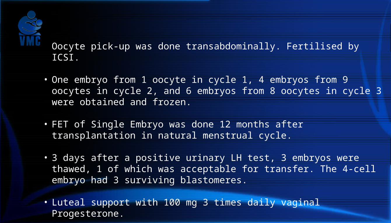
• Oocyte pick-up was done transabdominally. Fertilised by ICSI. • One embryo from 1 oocyte in cycle 1, 4 embryos from 9 oocytes in
cycle 2, and 6 embryos from 8 oocytes in cycle 3 were obtained and frozen.
• FET of Single Embryo was done 12 months after transplantation in
natural menstrual cycle.
• 3 days after a positive urinary LH test, 3 embryos were thawed, 1 of which was acceptable for transfer. The 4-cell embryo had 3 surviving blastomeres.
• Luteal support with 100 mg 3 times daily vaginal Progesterone.
• ASA 75 mg was started from ET day. • She conceived in first cycle of Thaw-ET.

Surgery, Donor
• Procedure : isolation of the uterus with bilateral, long venous and arterial vascular pedicles.
• Complex surgery, due to extensive
vascular dissection to include the distal parts of internal iliac veins and arteries.
• In this case, 2 large uterine veins on each
side converged into one major uterine vein that drained into the internal iliac veins.

• On left side, one of these veins passed over the ureter and the other under. Hence one of these veins had to be transected to keep an intact ureter.
• After surgical isolation, the uterus was flushed bilaterally through the arterial ends with cold histidine–tryptophan–ketoglutarate solution (Custodiol-HTK; NordMedica AS).
• The vascular ends of the graft were trimmed and the left-sided vein that had been divided was anastomosed end-to-end by a continuous suture (8-0 polypropylene).

Surgery, Recipient
• 1 h before final graft retrieval from donor, surgery to prepare the recipient was initiated in an adjacent OR.
• Through a midline incision, the external iliac vessels were dissected and prepared for anastomosis.
• The vaginal vault was separated from the bladder and rectum.
• Sutures to be used for uterine fixation, were placed bilaterally through the round ligaments , uterosacral ligaments, and paravaginal connective tissues.

• The uterus was brought into the pelvis and end-to-side vascular anastomoses done to connect the uterine veins to the external iliac veins (with 8-0 polypropylene sutures) and the anterior divisions of the internal iliac arteries to the external iliac arteries (with 7-0 polypropylene sutures) on both sides.
• The anastomoses were created in the sequence of left venous, left arterial, right venous, and right arterial.
• The uterus was then attached to the orthotopic position

• Then, fixation of uterus to the ligaments and suturing the bladder peritoneum on the uterine graft on top of the recipient’s bladder to provide extra structural support.
• Next, opened the blood flow to the uterus and ascertained that good pulses existed distal to the arterial anastomosis sites.
• Uterine tissue changed from pale to reddish.
• The skin-to-skin durations of surgeries were 10 h 7 min for the donor and 4 h 55 min for the recipient.
• The total ischaemic time of the uterine graft was 2 h 19 min (cold ischaemia: 1 h 6 min; warm ischaemia: 1 h 13 min).

• A retroperitoneal haematoma was diagnosed in the recipient on the day 2 postop & she was transfused with two units of leukocyte-reduced packed RBCs.
• To establish that blood flow through the uterine arteries continued during the first post-transplantation days, a 20-MHz Doppler probe was placed with a silicon cuff (Cook-Schwartz Doppler probe; Cook Medical) around the left uterine artery.
• The signal was transduced through a thin cable, which was exteriorised through the midline incision. The probe could then be easily pulled out after the 3-day observation period.
• Both were discharged from the hospital after 6 days of postop care.

Immunosuppression and follow-up
• Induction immunosuppression: IV anti-thymocyte globulin (Thymoglobulin) (Genzyme, Cambridge), 2·5 mg/kg just before surgery and 12 h later. One dose of 500 mg IV methylprednisolone (Solu-Medrol); Pfizer) just before uterine reperfusion.
• Maintenance immunosuppression: oral tacrolimus of 5–10 ng/mL (Prograf/Advagraf) (Astellas Pharma) and oral mycophenolatemofetil aiming at trough levels of 40–60 mg. h/L (Cellcept) (Roche, Basel, Switzerland) for the first 10 months post-surgery. Azathioprine 2 mg/kg per day (Imurel) (Orion Pharma) used later instead of mycophenolatemofetil after 10 months, to avoid the potentially teratogenic effects.
• Prednisolone 5 mg. orally after 6 months transplant because of repeated rejection episodes.

Follow up
Clinical visits and lab exams, initially twice weekly during the first postop month & then every 2 weeks in months 2–6. Subsequently, monthly. With visual inspection of transplanted cervix, bacterial culture from the canal, and occasional cervical biopsies at follow-ups. Ultrasound scans to assess uterine size, and endometrial thickness and echogenicity. Doppler uterine artery flow velocity waveforms on both sides. Biopsies of cervix at pre-determined time-points (at 1,2,4 weeks and thereafter monthly) or when there was any abnormal symptoms. HPV was checked for any CIN. Routine clinical blood investigations of pregnancy as per organ transplant patient protocol of the Sahlgrenska University hospital. Followed up in high-risk pregnancy unit every 2-3 weeks including fetal growth and Doppler chart.

Results
• First menses post-transplant occurred spontaneously after 43 days. • The blood flow doppler (velocity waveforms) of the uterine arteries were
similar on the left and right side and were within the low to normal range. • 2 mild rejection episodes, after 9 days and then 6 months 24 days & one
borderline episode at 2 months and 28 days, diagnosed by cervical biopsy without clinical symptoms, reversed by corticosteroids.
• HPV subtype 31 detected at 8 months 12 days post transplantation. Treated by mini-conisation, with subsequent biopsy showing no dysplasia.

Pregnancy:
• Creatinine levels, with a single kidney, were raised during pre-pregnancy (median 94 μmol/L) and were further elevated during pregnancy (106 μmol/L).
• USG showed a slight hydronephrosis in the single right
kidney at 27 weeks.
• She was working full time until the day before delivery. • Fetal growth and estimated weight were normal by
USG. • Uterine artery and Umbilical artery Doppler remained
normal. • The patient was admitted with headache and Pre-
eclampsia at 31w 5d. (BP- 180/120, albuminuria, low Platelet count, Breech presentation), and CTG after 10 h, showed variable decelerations.

• Received 2 doses Betamethasone, 12 h.
• CS was done 16 h after admission under spinal anesthesia. A midline vertical incision. Mild adhesions noted and a lower segment transverse CS was done after opening bladder peritoneum and uterines identified.
• The placenta weighed 375 g. HPE of the placenta showed normal findings except, villi showing PE changes (villi of small caliber, increased fibrin deposits and signs of fibrin thrombi).
• The uterus contracted on IV 10 U oxytocin. The
uterine incision was sutured two-layered. • A small myometrial biopsy was taken from the
fundus and the histology of this was normal.

The baby -Vincent
• The birth-weight of the neonate was 1775 g, APGAR scores were 9, 9, 10 and the umbilical artery pH was 7·21.
• The baby was discharged on day 16 in good health.
• The first postnatal week was uneventful and the baby was in good condition, requiring only phototherapy and room air.

Discussions
• This livebirth after uterus transplantation in a woman born with no uterus has eradicated the diagnosis of absolute uterine factor infertility.
• This also comes after more than a
decade of intensive animal research in this specialty by several groups worldwide.
• Unlike other organ transplantation, this is temporary and uterus can be removed soon after a live birth or as CS hysterectomy.
• Immunosuppressant side-effects can thereby be limited to short period in life.

Conclusion
The ethical issues of uterus transplantation are complex in its, specific facets of non-maleficence, autonomy, beneficence, justice, and dignity.
About 3% of all infertile couples have uterus factor infertility
12000-15000 potential uterus transplantation patients in United Kingdom (Sieunaire et al, Int Surg,2005;90:249)
2000-3000 potential uterus transplantation patients in the Nordic countries.
We don’t have a real statistics.

Implications of this success story:
• Despite remarkable advances in infertility treatment, major forms of uterine factor infertility have remained untreatable.
• This success has eradicated the diagnosis of absolute uterine factor infertility.
• Face, larynx, hand transplantation have also now reached the stage as established clinical procedures, after non-vital tissues or organs which after transplantation, would have the chance to substantially increase an individual’s quality of life…..

VIJAYALAKSHMI MEDICAL CENTRE
THANK YOU