use of micronized purified flavonoid fraction together ... · use of micronized purified flavonoid...
TRANSCRIPT

ORIGINAL RESEARCH
Use of Micronized Purified Flavonoid FractionTogether with Rivaroxaban Improves Clinicaland Ultrasound Outcomes in Femoropopliteal VenousThrombosis: Results of a Pilot Clinical Trial
Kirill Lobastov . Ilya Schastlivtsev . Victor Barinov
Received: October 25, 2018 / Published online: December 11, 2018� The Author(s) 2018
ABSTRACT
Introduction: The aim of this study was toassess the impact of adding long-term micro-nized purified flavonoid fraction (MPFF) tostandard treatment of femoropopliteal deepvein thrombosis (DVT).Methods: This pilot, comparative, open-labelstudy with blinded outcome assessor enrolledpatients with a first episode of femoropoplitealDVT confirmed by duplex ultrasound scanning(DUS). All participants were randomly allocatedto one of two treatment groups: (1) control thatreceived a standard treatment with oralrivaroxaban, and (2) experimental that involvedadditional treatment with MPFF 1000 mg/day.Both drugs were used for 6 months. Patientswere followed for the whole treatment periodand underwent DUS every 2 months to deter-mine the degree of recanalization by popliteal(PV), femoral vein (FV), and common femoralvein (CFV) compressibility. Thrombi extensionwere assessed by the modified Marder score. Atthe end of the follow-up period, patients wereassessed with Villalta and venous clinical
severity scales (VCSS). Patients with a Villaltascore C 5 were diagnosed with post-thromboticsyndrome (PTS).Results: Sixty patients were randomized to thecontrol or MPFF groups (n = 30 in each group).There were 40 men and 20 women with a meanage ± SD of 56.3 ± 13.4 years. Clinicallyunprovoked DVT was recognized in 65% ofcases and left side localization in 45%. Themean baseline Marder score was 15.0 ± 4.8 and11.1 ± 4.3 in the experimental and controlgroups, respectively (p = 0.002). At 6 months,the mean Villalta score in the MPFF group wassignificantly lower compared with control(2.9 ± 2.7 versus 5.8 ± 3.0; p\ 0.0001). PTSwas diagnosed in six patients (20%) and 17patients (57%) in the experimental and controlgroups respectively (p = 0.007). A significantdifference between the groups was also observedfor the VCSS value (2.3 ± 1.9 versus 4.9 ± 1.9;p\0001). After 6 months of treatment theMarder score decreased to 0.8 ± 1.6 and2.8 ± 3.5 in the experimental and controlgroups, respectively (p = 0.006). In the MPFFgroup, there was a greater reduction in theMarder score (p\ 0.0001) and more rapid rateof recanalization for the FV (p\ 0.0001), with anon-significant trend for the CFV (p = 0.130)and PV (p = 0.204) compared with the controlgroup. Full recanalization of the PV at 6 monthswas observed in 24 patients (80%) who hadreceived MPFF, and only 17 patients (57%) inthe control group (p = 0.095).
Enhanced digital features To view enhanced digitalfeatures for this article go to https://doi.org/10.6084/m9.figshare.7378130.
K. Lobastov (&) � I. Schastlivtsev � V. BarinovPirogov Russian National Research MedicalUniversity, Moscow, Russian Federatione-mail: [email protected]
Adv Ther (2019) 36:72–85
https://doi.org/10.1007/s12325-018-0849-z
Conclusion: The addition of MPFF to standardtherapy for DVT in the form of oral rivaroxabancan reduce the incidence of PTS at 6 months inpatients with proximal DVT and increase thespeed of deep vein recanalization.Funding: Les Laboratoires Servier funded thearticle processing fees, editorial assistance, andopen access fee for this manuscript.
Keywords: Cardiology; Deep vein thrombosis;Micronized purified flavonoid fraction; Post-thrombotic syndrome; Recanalization; Rivarox-aban
INTRODUCTION
Deep vein thrombosis (DVT) is a significantmedical and social problem. According to offi-cial statistics, the incidence of DVT in the Rus-sian Federation remained stable throughout theperiod 2012–2016 accounting for 1.5–1.6 casesper 1000 population per year [1–3], which isslightly higher than figures obtained in pub-lished epidemiological studies [4–8]. The mainlong-term issue after an episode of DVT is theoccurrence of post-thrombotic syndrome (PTS),which significantly affects the patients’ qualityof life. The incidence of PTS in the 10–15 yearsafter the primary thrombotic episode reaches19–42%, and severe forms of the disease withthe presence of trophic disorders can be detec-ted in 3–4% of patients [9–12]. The symptoms ofPTS usually develop within the first 6 monthsfollowing the primary DVT, but can occur yearsafter the initial event [9].
The pathogenetic mechanisms leading to thedevelopment of PTS may involve lesions of thedeep vein valves with the formation of bloodreflux, incomplete recanalization of venoussegments with the persistence of residualvenous obstruction, or a combination of bothmechanisms [13]. The most important compo-nent is the presence of residual venousobstruction, which increases the risk of devel-oping PTS by 1.6–2.0-fold [14–18]. Therecanalization of veins is a complex biologicalprocess. One of its integral components is theimmune inflammatory reaction, which isresponsible for clearance of thrombotic masses
from the lumen of the vessel and venous walllesion [19, 20]. Previous studies have shownthat increased levels of inflammatory markers,in particular C-reactive protein, interleukins-1and -6, intercellular adhesion molecule-1(ICAM-1), and a reduced level of anti-inflam-matory cytokines (interleukin-10) are associatedwith a higher risk of PTS development [21–25].This raises the possibility that pharmacologicalmodulation of the inflammatory reaction mayprotect the venous wall from excessive leuko-cyte aggression, while retaining the natural roleof immune cells in the process of vesselrecanalization.
Among the potential pharmacological agentswith the ability to target the inflammatoryreaction directly in the venous wall, micronizedpurified flavonoid fraction (MPFF), an oralagent, is of particular interest. Previous experi-mental studies have demonstrated its promi-nent endothelial protective properties in theform of reduced rolling, adhesion, and migra-tion of leukocytes in the settings of reperfusioninjury and secondary venous hypertension[26–29]. It has also been shown to suppress theinflammatory reaction in patients with chronicvenous disease (CVD) [30], including those whohave undergone sclerotherapy [31]. To test thehypothesis that long-term treatment with MPFFmay affect the natural course of DVT, this studyevaluated the impact of MPFF use in the treat-ment of patients with femoropopliteal DVT.
METHODS
This pilot, single-center, open-label compara-tive clinical trial with blinded outcome assess-ment recruited patients over 18 years of agewith a first episode of clinically provoked orunprovoked DVT in the femoropopliteal vas-cular segment with possible involvement of thecalf veins and the common femoral vein. DVTlocation was confirmed by duplex ultrasoundscan (DUS). All procedures performed in studiesinvolving human participants were in accor-dance with local ethics committee of the Clin-ical Hospital No. 1 of the PresidentsAdministration of Russian Federation and withthe 1964 Helsinki declaration and its later
Adv Ther (2019) 36:72–85 73

amendments or comparable ethical standards.Informed consent was obtained from all indi-vidual participants included in the study. At theauthors’ institution it is not obligatory to reg-ister pilot and unfunded studies; therefore thistrial is not registered.
Exclusion criteria were suspected or con-firmed pulmonary embolism; bilateral DVTlocation; extension of the thrombosis into theiliac veins above the inguinal fold; contraindi-cations to therapy with anticoagulants or MPFF;verified malignant tumor at the time of DVTmanifestation; known severe thrombophilia(deficiency of protein C, protein S, antithrom-bin III, antiphospholipid syndrome); use ofparenteral anticoagulants for 7 days or morefrom the time of DVT verification; inability touse compression stockings; any intervention forDVT (inferior vena cava filter implantation,thrombectomy, or catheter-guided thromboly-sis); continued use of other drugs affecting thehemostatic system (except acetylsalicylic acid ata dose of up to 100 mg per day); and poorcompliance.
The study was carried out at the facilities ofthe Clinical Hospital No. 1 of the PresidentsAdministration of the Russian Federation in2017–2018. In accordance with the internalprotocol of the institution, all patients withsuspected DVT entering the emergency depart-ment were assessed using a two-level Wellsscore and a D-dimer evaluation [32]. In patientswith a high clinical probability of DVT or acombination of low clinical probability for DVTwith an increased level of D-dimer, anticoagu-lant therapy was initiated with low molecularweight heparin (LMWH), followed by DUS.Patients with diagnosed femoropoplitealvenous thrombosis were assessed for fulfillmentof the inclusion and exclusion criteria. Aftersigning informed consent, patients were ran-domly allocated to one of two groups based onthe number on the patient’s medical recordform: those whose last digit was even wereassigned to the MPFF group, and those whoselast digit was odd were assigned to the controlgroup; if the last digit was zero, the penultimatedigit was used. In the case of premature com-pletion of recruitment to either group, all
eligible patients were intentionally allocated tothe second group.
During the period from hospital admissionto treatment allocation, all patients receivedtherapeutic doses of LMWH (enoxaparin 1 mg/kg twice daily). After randomization, patients inboth groups were switched to the oral antico-agulant rivaroxaban at a dosage of 15 mg twicedaily for up to 3 weeks from the administrationof the first dose of LMWH. Thereafter theyreceived a dosage of 20 mg once daily for up to6 months. The first dose of rivaroxaban wasadministered instead of LMWH at the time ofthe next scheduled injection. In addition, dur-ing the first 3 days following the DVT diagnosis,patients in both groups applied individualizedmedical compression in the form of thigh-highstockings with a pressure of 23–32 mmHg (class2 compression according to RAL-GZ 387 stan-dard). Matching the size of the stocking to thediameter of the limb was controlled 3 weeks and3 months after the start of treatment. Patientswere advised to wear the stockings 24 h a dayfor the first week and then during the day forthe next 6 months. In the MPFF group, patientsreceived MPFF 500 mg twice daily for 6 monthsin addition to standard treatment. The first doseof MPFF was administered immediately aftertreatment allocation and in parallel with theswitch to the oral form of the anticoagulant.
Patients were followed for 6 months withclinical and ultrasound evaluation every2 months. At baseline, in addition to standardclinical data, the affected lower limb was eval-uated for the presence of pre-existing CVD, andif confirmed, the clinical class according to theCEAP (Clinical, Etiological, Anatomical, andPathological) classification was determined.CEAP clinical classes C1 (telangiectases), C2(varicose veins), C4 (skin pigmentation,eczema, lipodermatosclerosis or athrophieblanche), C5 (healed venous ulcer), and C6(active venous ulcer) were confirmed objec-tively on the basis of the presence of corre-sponding signs of the disease. CEAP clinicalclass C3 (presence of leg edema) was confirmedon the basis of the patient’s medical history(complaints of edema of the lower limb or needfor compression therapy prior to the manifes-tation of thrombosis), as at the time of physical
74 Adv Ther (2019) 36:72–85
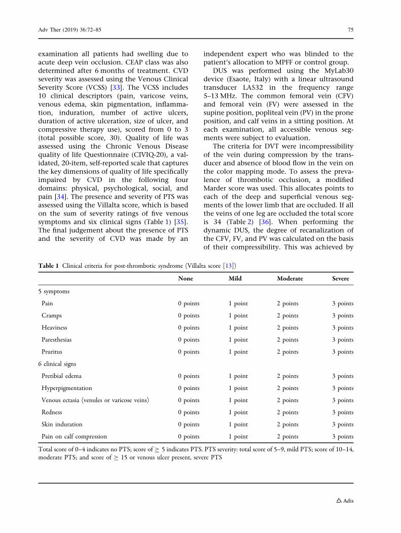
examination all patients had swelling due toacute deep vein occlusion. CEAP class was alsodetermined after 6 months of treatment. CVDseverity was assessed using the Venous ClinicalSeverity Score (VCSS) [33]. The VCSS includes10 clinical descriptors (pain, varicose veins,venous edema, skin pigmentation, inflamma-tion, induration, number of active ulcers,duration of active ulceration, size of ulcer, andcompressive therapy use), scored from 0 to 3(total possible score, 30). Quality of life wasassessed using the Chronic Venous Diseasequality of life Questionnaire (CIVIQ-20), a val-idated, 20-item, self-reported scale that capturesthe key dimensions of quality of life specificallyimpaired by CVD in the following fourdomains: physical, psychological, social, andpain [34]. The presence and severity of PTS wasassessed using the Villalta score, which is basedon the sum of severity ratings of five venoussymptoms and six clinical signs (Table 1) [35].The final judgement about the presence of PTSand the severity of CVD was made by an
independent expert who was blinded to thepatient’s allocation to MPFF or control group.
DUS was performed using the MyLab30device (Esaote, Italy) with a linear ultrasoundtransducer LA532 in the frequency range5–13 MHz. The common femoral vein (CFV)and femoral vein (FV) were assessed in thesupine position, popliteal vein (PV) in the proneposition, and calf veins in a sitting position. Ateach examination, all accessible venous seg-ments were subject to evaluation.
The criteria for DVT were incompressibilityof the vein during compression by the trans-ducer and absence of blood flow in the vein onthe color mapping mode. To assess the preva-lence of thrombotic occlusion, a modifiedMarder score was used. This allocates points toeach of the deep and superficial venous seg-ments of the lower limb that are occluded. If allthe veins of one leg are occluded the total scoreis 34 (Table 2) [36]. When performing thedynamic DUS, the degree of recanalization ofthe CFV, FV, and PV was calculated on the basisof their compressibility. This was achieved by
Table 1 Clinical criteria for post-thrombotic syndrome (Villalta score [13])
None Mild Moderate Severe
5 symptoms
Pain 0 points 1 point 2 points 3 points
Cramps 0 points 1 point 2 points 3 points
Heaviness 0 points 1 point 2 points 3 points
Paresthesias 0 points 1 point 2 points 3 points
Pruritus 0 points 1 point 2 points 3 points
6 clinical signs
Pretibial edema 0 points 1 point 2 points 3 points
Hyperpigmentation 0 points 1 point 2 points 3 points
Venous ectasia (venules or varicose veins) 0 points 1 point 2 points 3 points
Redness 0 points 1 point 2 points 3 points
Skin induration 0 points 1 point 2 points 3 points
Pain on calf compression 0 points 1 point 2 points 3 points
Total score of 0–4 indicates no PTS; score of C 5 indicates PTS. PTS severity: total score of 5–9, mild PTS; score of 10–14,moderate PTS; and score of C 15 or venous ulcer present, severe PTS
Adv Ther (2019) 36:72–85 75

measuring the veins’ diameters without com-pression and at maximum compression withthe transducer at the narrowest point of eachvenous segment. The degree of recanalizationwas calculated with the following formula:[d (no compression) - d (maximal compres-sion)]/d (no compression) 9 100%. All mea-surements were repeated three times, and anaverage value was used for calculations. Allultrasound studies were performed by a spe-cialist blinded to the patient’s allocation to theMPFF or control group.
The primary study endpoint was the pres-ence of PTS signs at 6 months, determined by ablinded expert. For the diagnosis of PTS, theVillalta score was used. The criteria for a PTSdiagnosis were a Villalta score of C 5; severe PTSwas defined as a score of C 15 or the presence ofan active venous ulcer.
The secondary study endpoints includedsevere PTS; symptomatic or asymptomaticrecurrence of DVT; symptomatic pulmonaryembolism; change in CEAP clinical class; CVD
severity assessed by VCSS, quality of life assessedby CIVIQ-20; complete recanalization of vessels;change in degree of recanalization for the CFV,FV, and PV; change in thrombus extension bythe Marder score; and the occurrence of bleed-ing and/or other adverse events associated withtherapy.
Symptomatic recurrence of DVT was definedas an increase in edema, pain, or hyperemia ofthe affected lower limb, or the occurrence of thesame signs on the intact leg. The asymptomaticrecurrence of DVT was defined as the occur-rence of signs of total occlusion in a previouslyrecanalized venous segment. A completerecanalization was the clearance of thromboticmasses by 80% or more in the venous lumen(residual vein occlusion less than 20%).
Bleeding related to anticoagulant therapywas classified as major (according to the Inter-national Society on Thrombosis and Hemostasiscriteria) [37], clinically relevant non-major (anyepisode not fulfilling the criteria of majorbleeding, but resulting in withdrawal of anti-coagulant therapy and/or requiring a specificmedical intervention and/or leading to anunscheduled visit to the doctor), or minor (anyepisode not fulfilling the criteria of major orclinically relevant non-major bleeding).
Patients’ general health status was assessed ateach follow-up visit with an emphasis on iden-tifying possible adverse events of therapy.
Statistical Methods
As this was a pilot study with an exploratorynature the minimum sample size was not cal-culated. Statistical analysis was carried out usingIBM SPSS Statistics v.19 software package. Allabsolute values are presented as the mean valuewith the standard deviation (mean ± SD). Thedistribution was verified by a Kol-mogorov–Smirnov test. If a normal distributionwas confirmed, comparisons were performedusing the t test for independent samples forcontinuous variables, or the two-sided Fisher’sexact test and a Chi-square test for categoricalvariables. Comparisons of mean values withtime were performed by assessment of within-and between-subject effects, as well as their
Table 2 Ultrasound quantification of deep vein throm-bosis (modified Marder score [36])
Venous segment examined Score
Iliac vein 8
Common femoral vein 4
Femoral vein 4
Deep femoral vein 2
Popliteal vein 2
Tibial trunk 2
Peroneal trunk 2
Tibial vein (both of the pair) 1 (2)
Peroneal vein (both of the pair) 1 (2)
Gastrocnemius vein (both medial and lateral) 1 (2)
Soleal vein or other calf muscle vein 1
Great saphenous vein trunk 2
Small saphenous vein trunk 1
Maximal score for one limb 34
76 Adv Ther (2019) 36:72–85

within-subject interaction using the generallinear model for repeated measurements (GLM-RM). Differences were considered statisticallysignificant when the p value was less than 0.05.
RESULTS
During the inclusion period, 132 patients withsuspected DVT were admitted to the hospitaland thrombosis was confirmed in 104 cases. Ofthese, 68 patients fulfilled the selection criteriaand 8 patients refused to participate. Theremaining 60 patients were allocated to one ofthe two treatment groups (n = 30 in eachgroup), and all patients completed the study(Fig. 1).
The clinical characteristics of the groups arepresented in Table 3. Patients in the controlgroup receiving standard treatment had ahigher rate of CVD prior to the occurrence ofDVT, in particular CEAP clinical class C2–C4.
Patients in the two groups were comparable interms of the prevalence of lesions of the mainvessels in the femoropopliteal venous segment,although the total extent of the pathologicalprocess as determined by the Marder score washigher in the MPFF group (Table 3). These dif-ferences were associated primarily with thegreater involvement of calf veins in patientstreated with MPFF.
The results for the primary and secondaryendpoints are summarized in Table 4. PTS wasreported in 56.7% patients after 6 months ofanticoagulant therapy with rivaroxaban andcompression therapy and in 20% of patientsrandomized to MPFF plus standard therapy(p = 0.007). No severe forms of PTS were repor-ted in either group. The mean score on theVillalta scale and the mean VCSS value werealso lower in patients in the MPFF group. TheCIVIQ-20 global index score revealed animproved quality of life in the MPFF group.
Fig. 1 CONSORT flowchart of the study
Adv Ther (2019) 36:72–85 77

Progression of pre-existing CVD wasobserved in one patient in the MPFF group(transition from C2 to C4 class) and six patientsin the control group (transition from C2 to C4class in one patient, and transition from C2 toC3 class in five patients). Thus, the signs of CVDprogression were observed in 20% of patientsreceiving standard therapy for DVT and only in3.3% of patients with additional use of MPFF.
The recurrence of a venous thromboembolicevent was observed in one patient from thecontrol group, who had a symptomatic recur-rent DVT on the contralateral limb, whichrequired a temporary switch to LMWH followedby therapy with vitamin K antagonists. Nomajor bleedings were observed in either group.Clinically relevant, non-major bleedings wererepresented by one case of hemorrhoidalbleeding in the MPFF group and two cases of
macrohematuria in the control group. In addi-tion, two cases of minor bleeding were reported(one in each group): rectal bleeding in the MPFFgroup and epistaxis in the control group.
In terms of other adverse effects, threepatients in the MPFF group experienced a milddyspeptic disorder, which did not requiretreatment discontinuation.
Rates of recanalization in the main venoussegments are presented in Fig. 2. In bothgroups, there was a significant trend towardsprogressive clearance of thrombotic massesfrom the vessel lumen (p\0.0001 for within-subject effect ‘‘time’’). There were no significantdifferences in the rate of recanalization in thepopliteal vein between the two groups, andcomplete recovery of vascular patency wasachieved in 80% of patients in the MPFF groupand 57% of patients in the control group
Table 3 Characteristics of patients in the MPFF and control groups
Parameter MPFF (n = 30) Control (n = 30) p value
Age (years, mean ± SD) 55.2 ± 14.9 57.5 ± 11.9 0.518
Male (%) 56.7 76.7 0.170
Clinically unprovoked DVT (%) 56.7 73.3 0.279
Duration of symptoms by time of hospital admission (days, mean ± SD) 4.3 ± 3.7 4.2 ± 2.6 0.936
Time from hospital admission to allocation (days, mean ± SD) 3.9 ± 1.2 3.9 ± 1.7 0.931
History of CVD (%) 50.0 73.4 0.110
Class C0 23.3 23.3 0.014
Class C1 26.7 3.3
Class C2 33.3 46.8
Class C3 16.7 13.3
Class C4 0.0 13.3
Lesions of the left lower extremity (%) 50.0 40.0 0.604
Thrombus extension by Marder score (mean ± SD) 15.0 ± 4.8 11.1 ± 4.3 0.002
CFV lesion (%) 43.3 33.3 0.596
FV lesion (%) 83.3 66.7 0.233
PV lesion (%) 100.0 100.0 1.000
DVT deep vein thrombosis, SD standard deviation, CVD chronic venous disease, CFV common femoral vein, FV femoralvein, PV popliteal vein
78 Adv Ther (2019) 36:72–85

(p = 0.095). The recanalization of the femoralvein, in turn, occurred significantly faster inpatients receiving MPFF (p\0.0001 for within-subject interaction ‘‘time 9 group’’); however,
the rate of achieving complete recovery of vas-cular patency was not significantly different:92% in the MPFF group vs. 75% in the controlgroup (p = 0.214).
Table 4 Rates of the primary and secondary endpoints in the MPFF and control groups
Endpoint MPFF (n = 30) Control (n = 30) p value
Primary
Diagnosis of PTS 6 (20.0%) 17 (56.7%) 0.007
Secondary
Diagnosis of severe PTS 0 0 –
Villalta score, mean ± SD 2.9 ± 2.7 5.8 ± 3.0 \ 0.0001
VCSS value, mean ± SD 2.3 ± 1.9 4.9 ± 1.9 \ 0.0001
CIVIQ-20 score, mean ± SD 24.1 ± 4.6 31.6 ± 8.5 \ 0.0001
Progression in CEAP clinical class 1 (3.3%) 6 (20.0%) 0.057
Recurrence of VTE 0 1 (3.3%) 1.000
Bleeding 2 (6.7%) 3 (10.0%) 1.000
Major 0 0 –
Clinically relevant non-major 1 (3.3%) 2 (6.7%) 1.000
Minor 1 (3.3%) 1 (3.3%) 1.000
Other adverse events 3 (10.0%) 0 0.237
SD standard deviation, PTS post-thrombotic syndrome, CEAP Clinical, Etiological, Anatomical, and Pathological classi-fication, CIVIQ-20 Chronic Venous Disease quality of life Questionnaire, VCSS Venous Clinical Severity Score, VTEvenous thromboembolic event
Fig. 2 Rates of recanalization for the main venous segments. Generalized linear model repeated measures: p1 within-subjecteffect ‘‘time’’, p2 within-subject interaction ‘‘time 9 group’’, p3 between-subject effect ‘‘group’’
Adv Ther (2019) 36:72–85 79
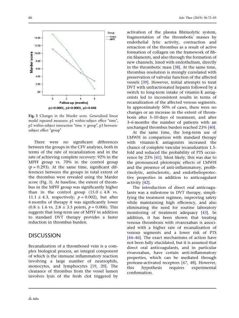
There were no significant differencesbetween the groups in the CFV analyses, both interms of the rate of recanalization and in therate of achieving complete recovery: 92% in theMPFF group vs. 70% in the control group(p = 0.293). At the same time, significant dif-ferences between the groups in total extent ofthe thrombus were revealed using the Marderscore (Fig. 3). At baseline, the extent of throm-bus in the MPFF group was significantly higherthan in the control group (15.0 ± 4.8 vs.11.1 ± 4.3, respectively; p = 0.002), but after6 months of therapy it was significantly lower(0.8 ± 1.6 vs. 2.8 ± 3.5 points, p = 0.006). Thissuggests that long-term use of MPFF in additionto standard DVT therapy provides a fasterreduction in thrombus burden.
DISCUSSION
Recanalization of a thrombosed vein is a com-plex biological process, an integral componentof which is the immune inflammatory reactioninvolving a large number of neutrophils,monocytes, and lymphocytes [19, 20]. Theclearance of thrombus from the vessel lumeninvolves lysis of the fresh clot triggered by
activation of the plasma fibrinolytic system,fragmentation of the thrombotic masses byendothelial lytic activity, contraction andretraction of the thrombus as a result of activeformation of collagen on the framework of fib-rin filaments, and also through the formation ofnew channels, lined with endothelium, directlyin the thrombotic mass [38]. At the same time,thrombus resolution is strongly correlated withpreservation of valvular function of the affectedvessels [39]. However, initial attempts to treatDVT with unfractionated heparin followed by aswitch to long-term intake of vitamin K antag-onists led to inconsistent results in terms ofrecanalization of the affected venous segments.In approximately 50% of cases, there were nochanges or an increase in the extent of throm-bosis after 5–10 days of treatment, and after3–6 months the number of patients with anunchanged thrombus burden reached 23% [40].
At the same time, the long-term use ofLMWH in comparison with standard therapywith vitamin K antagonists increased thechance of complete vascular recanalization 1.5-fold and reduced the probability of PTS occur-rence by 23% [41]. Most likely, this was due tothe pronounced pleiotropic effects of LMWHand the presence of anti-inflammatory, profib-rinolytic, antisclerotic, and endothelioprotec-tive properties in addition to anticoagulantactivity [42].
The introduction of direct oral anticoagu-lants was a milestone in DVT therapy, simpli-fying the treatment regimen, improving safetywhile maintaining high efficiency, and alsoeliminating the need for routine laboratorymonitoring of treatment adequacy [43]. Inaddition, it has been shown that treatingvenous thrombosis with rivaroxaban is associ-ated with a higher rate of recanalization ofvenous segments and a lower risk of PTS[44–46]. The exact mechanisms of action havenot been fully elucidated, but it is assumed thatdirect oral anticoagulants, and in particularrivaroxaban, have certain anti-inflammatoryproperties, which can be mediated throughprotease-activated receptors [47, 48]. However,this hypothesis requires experimentalconfirmation.
Fig. 3 Changes in the Marder score. Generalized linearmodel repeated measures: p1 within-subject effect ‘‘time’’,p2 within-subject interaction ‘‘time 9 group’’, p3 between-subject effect ‘‘group’’
80 Adv Ther (2019) 36:72–85

When assessing the effect of anticoagulanttherapy on the completeness of recanalizationof thrombosed vessels, a particular difficulty isthe interpretation of the clinical significance ofresidual venous obstruction. Thus, Piovella et al.and Siragusa et al. consider residual thrombus as‘‘clinically significant’’ if it occupies more than40% of the vessel lumen [49, 50]. At the sametime, Prandoni et al. in their early studiesdefined residual obstruction in the femor-opopliteal segment to be significant if veinthickness at maximum compression exceeded2–3 mm [51, 52]. In more recent studies evalu-ating the degree of vascular recanalization inpatients receiving direct oral anticoagulants,the cutoff for vein thickness was increased to4 mm [46]. Given this uncertainty, the rate ofresidual venous obstruction detection, as well asits impact on disease outcome, can vary sub-stantially from study to study. In this study, weassumed a clinically significant residualobstruction at 20% of the vessel cross-sectionaldiameter, which is approximately 1–2 mm forthe femoropopliteal segment and correspondsto the early criteria of Prandoni et al. [51, 52].When this cutoff value was used, the rate ofresidual venous obstruction diagnosis onrivaroxaban monotherapy was 43% for thepopliteal vein, 25% for the femoral vein, and30% for the common femoral vein. These fig-ures are significantly higher than that obtainedusing a 4-mm cutoff value, at which the rate ofresidual obstruction diagnosis was only 21.1%[46].
When long-term use of rivaroxaban wascombined with MPFF, the rate of residualvenous obstruction was reduced to 20% for thepopliteal vein, and 8% for the femoral vein andcommon femoral vein, which indicates a sig-nificant improvement in the recanalizationprocess with the addition of flavonoid. Thebenefits of MPFF were even more pronouncedwhen analyzed by changes in the Marder scorereflecting the total extent of thrombus.
The addition of MPFF resulted in a morerapid and complete clearance of thromboticmasses from the blood vessels, and as a result,by the end of the 6-month follow-up period thescores in the MPFF group were significantlylower than in the control group. The observed
differences can be explained by the known anti-inflammatory activity of MPFF. Previousresearch in patients with CVD has shown thatMPFF treatment for 60 days decreased levels ofcellular adhesion molecules (ICAM-1 by 32%and VCAM by 29%) in the regional venousblood [30]. In women with CVD class CEAP C1,significant increases in levels of C-reactive pro-tein, histamine, interleukin-1, tumor necrosisfactor-alpha, and vascular endothelial growthfactor have been observed in blood samplestaken from the vessel supplying the telangiec-tasia 10 days after microsclerotherapy [31].When sclerotherapy is performed with adjunc-tive MPFF the activity of these inflammatorymarkers is significantly reduced. Further studiesare now warranted to confirm whether MPFFcan also affect levels of inflammatory biomark-ers in venous thrombosis.
A further difficulty when assessing long-termDVT outcomes is objective confirmation of thepresence of PTS. In previous studies, the rate ofPTS diagnosis by the Villalta scale in patientstreated with rivaroxaban was 25–45% for amean follow-up period of about 2 years [44, 45].In our study, the rate of PTS diagnosis after6 months of rivaroxaban monotherapy washigher at 56.7%. This may be due to the pecu-liarities of the Villalta scale, which has a highsensitivity to the presence of pre-existing CVD.It is known that the presence of primary venousinsufficiency prior to the development of DVTraises the risk of PTS occurrence 1.5–3.2-fold[13]. Moreover, the Villalta scale has beenreported to result in a misdiagnosis of PTS in42% of patients who do not have specific post-thrombotic changes in the deep veins [53]. Thehigh sensitivity of this tool to pre-existing CVDcan result in a wide dispersion in the rates ofregistered long-term adverse DVT outcomes. Inour study, about three-quarters of the patientsin the control group had a primary lesion of thesuperficial veins before the development ofthrombosis, which could have had an impacton the high prevalence of PTS. At the sametime, around half the patients in the MPFFgroup also had pre-existing CVD. However,signs of disease progression in terms of a wors-ening in CEAP clinical class to more severeforms were much less common with the use of
Adv Ther (2019) 36:72–85 81

the MPFF (3.3% vs. 20%). This apparent trendmay be associated with the ability of MPFF toreverse the symptoms and signs of CVD, andthus have a positive effect on the preventionand treatment of early forms of PTS [54].
The current study has demonstrated thebenefits of using MPFF together with rivaroxa-ban for the long-term treatment of DVT. Theaddition of MPFF not only allowed the rate ofPTS to be reduced, but also acceleratedrecanalization of affected venous segments. Atthe same time, the proposed therapeuticapproach was associated with a high safetyprofile; no serious events associated with long-term intake of MPFF were recorded, and all theidentified adverse events were mild in natureand did not result in treatment discontinuation.
This preliminary research had a pilot studydesign and was subject to certain limitations.First, the small sample size did not allowadjustments to be made for differences betweentreatment groups, in particular the extent ofthrombus and the presence of pre-existing CVD.When planning further studies, appropriaterandomization methods should be used toeliminate any source of bias in treatmentassignments, patient recruitment should becarefully balanced for pre-existing CVD orexclude participants with this condition. Otherimportant limitations were the absence of aplacebo control and the short follow-up period.These limitations must be overcome in subse-quent studies. Larger randomized controlledtrials are now required to establish the superi-ority of the combined regimen before routineuse in clinical practice can be recommended.
CONCLUSION
Preliminary data demonstrate that long-termtherapy for DVT with a combination ofrivaroxaban and MPFF reduced the rate of PTSdevelopment compared with rivaroxabanmonotherapy and accelerated recanalization ofaffected venous segments. These results nowneed to be confirmed in larger randomized,double-blind, placebo-controlled studies.
ACKNOWLEDGEMENTS
We thank the participants of the study. Thestudy was carried out at the facilities of theClinical Hospital No. 1 of the PresidentsAdministration of the Russian Federation in2017–2018.
Funding. The open access fee, article pro-cessing charges, and editorial assistance for theproduction of this article were funded by LesLaboratoires Servier. The study was conductedwithout any financial or technical support. Allauthors had full access to all of the data in thisstudy, contributed to the concept and inter-pretation of the literature, were involved in thedrafting and revising of the paper and approvalof the final submitted version, and take com-plete responsibility for the integrity of the dataand accuracy of the data analysis.
Editorial Assistance. Editorial assistance forEnglish language editing was provided by JennyGrice, Bieuzy les Eaux, France and funded by LesLaboratoires Servier.
Authorship. All named authors meet theInternational Committee of Medical JournalEditors (ICMJE) criteria for authorship for thisarticle, take responsibility for the integrity ofthe work as a whole, and have given theirapproval for this version to be published.
Disclosures. Kirill Lobastov, Ilya Schastlivt-sev and Victor Barinov have nothing to disclose.
Compliance with Ethics Guidelines. Allprocedures performed in studies involvinghuman participants were in accordance withlocal ethics committee of the Clinical HospitalNo. 1 of the Presidents Administration of Rus-sian Federation and with the 1964 Helsinkideclaration and its later amendments or com-parable ethical standards. Informed consent wasobtained from all individual participants inclu-ded in the study.
Data Availability. The datasets generatedand/or analyzed during the current study are
82 Adv Ther (2019) 36:72–85
available from the corresponding author onreasonable request.
Open Access. This article is distributedunder the terms of the Creative CommonsAttribution-NonCommercial 4.0 InternationalLicense (http://creativecommons.org/licenses/by-nc/4.0/), which permits any noncommer-cial use, distribution, and reproduction in anymedium, provided you give appropriate creditto the original author(s) and the source, providea link to the Creative Commons license, andindicate if changes were made.
REFERENCES
1. O,oaz pa,okedaevocnm dceuo yacekeybz Poccbb d2012 uoly. Cnanbcnbxecrbe vanepbaks, Xacnm II 2013.[The general prevalences of diseases in the totalpopulation of Russia in 2012. Statistical Materials,Part II 2013 (In Russian)]. http://www.rosminzdrav.ru/documents/8029-statisticheskaya-informatsiya-2012. Accessed 19 Oct 2018.
2. Pa,okedaevocnm dceuo yacekeybz Poccbb d 2014 uoly.Cnanbcnbxecrbe vanepbaks, Xacnm II 2015 [Theprevalences of diseases in the total population ofRussia in 2014. Statistical Materials, Part II 2015 (InRussian)]. https://www.rosminzdrav.ru/documents/9479-statisticheskaya-informatsiya-za-2014. Acces-sed 19 Oct 2018.
3. Pa,okedaevocnm dceuo yacekeybz Poccbb d 2016 uoly.Cnanbcnbxecrbe vanepbaks Xacnm II 2017. [Theprevalences of diseases in the total population ofRussia in 2016. Statistical Materials, Part II 2017 (InRussian)]. https://www.rosminzdrav.ru/ministry/61/22/stranitsa-979/statisticheskie-i-informatsionnye-materialy/statisticheskiy-sbornik-2016-god. Accessed 19 Oct 2018.
4. Hippisley-Cox J, Coupland C. Development andvalidation of risk prediction algorithm (QThrom-bosis) to estimate future risk of venous throm-boembolism: prospective cohort study. BMJ.2011;343:d4656.
5. Oger E. Incidence of venous thromboembolism: acommunity-based study in Western France. EPI-GETBP Study Group. Groupe d’Etude de la Throm-bose de Bretagne Occidentale. J Thromb Haemost.2000;83(5):657–60.
6. Naess IA, Christiansen SC, Romundstad P, Can-negieter SC, Rosendaal FR, Hammerstrom J. Inci-dence and mortality of venous thrombosis: a
population-based study. J Thromb Haemost.2007;5(4):692–9.
7. Van Beek E, Buller H, Ten Cate J. Epidemiology ofvenous thromboembolism. A textbook of vascularmedicine. London: Arnold; 1996. p. 471–88.
8. Silverstein MD, Heit JA, Mohr DN, Petterson TM,O’Fallon WM, Melton LJ 3rd. Trends in the inci-dence of deep vein thrombosis and pulmonaryembolism: a 25-year population-based study. ArchIntern Med. 1998;158(6):585–93.
9. Prandoni P, Lensing AW, Cogo A, et al. The long-term clinical course of acute deep venous throm-bosis. Ann Intern Med. 1996;125(1):1–7.
10. Mohr DN, Silverstein MD, Heit JA, Petterson TM,O’fallon WM, Melton LJ. The venous stasis syn-drome after deep venous thrombosis or pulmonaryembolism: a population-based study. Mayo ClinProc. 2000;75(12):1249–56.
11. Franzeck UK, Schalch I, Jager KA, Schneider E,Grimm J, Bollinger A. Prospective 12-year follow-upstudy of clinical and hemodynamic sequelae afterdeep vein thrombosis in low-risk patients (Zurichstudy). Circulation. 1996;93(1):74–9.
12. Beyth RJ, Cohen AM, Landefeld CS. Long-termoutcomes of deep-vein thrombosis. Arch InternMed. 1995;155(10):1031–7.
13. Kahn S, Comerota A, Cushman M. Council onClinical Cardiology, Council on Cardiovascular andStroke Nursing. The postthrombotic syndrome:evidence-based prevention, diagnosis, and treat-ment strategies: a scientific statement from theAmerican Heart Association. Circulation.2014;130(18):1636–61.
14. Prandoni P, Frulla M, Sartor D, Concolato A, Giro-lami A. Vein abnormalities and the post-thromboticsyndrome. J Thromb Haemost. 2005;3(2):401–2.
15. Vedovetto V, Dalla Valle F, Milan M, Pesavento R,Prandoni P. Residual vein thrombosis and trans-popliteal reflux in patients with and without thepost-thrombotic syndrome. Thromb Haemost.2013;110(4):854–5.
16. Galanaud J, Holcroft C, Rodger M, et al. Predictorsof post-thrombotic syndrome in a population witha first deep vein thrombosis and no primary venousinsufficiency. J Thromb Haemost.2013;11(3):474–80.
17. Tick L, Kramer M, Rosendaal F, Faber W, Doggen C.Risk factors for post-thrombotic syndrome inpatients with a first deep venous thrombosis.J Thromb Haemost. 2008;6(12):2075–81.
Adv Ther (2019) 36:72–85 83

18. Comerota AJ, Grewal N, Martinez JT, et al. Post-thrombotic morbidity correlates with residualthrombus following catheter-directed thrombolysisfor iliofemoral deep vein thrombosis. J Vasc Surg.2012;55(3):768–73.
19. Budnik I, Brill A. Immune factors in deep veinthrombosis initiation. Trends Immunol.2018;39:610–23.
20. Henke PK, Wakefield T. Thrombus resolution andvein wall injury: dependence on chemokines andleukocytes. Thromb Res. 2009;123:S72–8.
21. Rabinovich A, Cohen JM, Cushman M, et al.Inflammation markers and the risk of post throm-botic syndrome: results from the Bio-Sox Study.Blood. 2013;122(21):36.
22. Rabinovich A, Cohen JM, Cushman M, et al. Asso-ciation between inflammation biomarkers, ana-tomic extent of deep venous thrombosis, andvenous symptoms after deep venous thrombosis.J Vasc Surg Venous Lymphat Disord.2015;3(4):347–53.
23. Roumen-Klappe E, Janssen M, Van Rossum J, et al.Inflammation in deep vein thrombosis and thedevelopment of post-thrombotic syndrome: aprospective study. J Thromb Haemost.2009;7(4):582–7.
24. Shbaklo H, Holcroft CA, Kahn SR. Levels ofinflammatory markers and the development of thepost-thrombotic syndrome. J Thromb Haemost.2009;101(03):505–12.
25. Bouman A, Smits J, Ten Cate H, Ten Cate-Hoek A.Markers of coagulation, fibrinolysis and inflamma-tion in relation to post-thrombotic syndrome.J Thromb Haemost. 2012;10(8):1532–8.
26. Korthuis RJ, Gute DC. Adhesion molecule expres-sion in postischemic microvascular dysfunction:activity of a micronized purified flavonoid fraction.J Vasc Res. 1999;36(Suppl 1):15–23.
27. Takase S, Lerond L, Bergan JJ, Schmid-SchonbeinGW. The inflammatory reaction during venoushypertension in the rat. Microcirculation.2000;7(1):41–52.
28. Takase S, Pascarella L, Lerond L, Bergan JJ, Schmid-Schonbein GW. Venous hypertension, inflamma-tion and valve remodeling. Eur J Vasc EndovascSurg. 2004;28(5):484–93.
29. Maria das Gracas C, Cyrino FZ, de Carvalho JJ,Blanc-Guillemaud V, Bouskela E. Protective effectsof micronized purified flavonoid fraction (MPFF) ona novel experimental model of chronic venous
hypertension. Eur J Vasc Endovasc Surg.2018;55(5):694–702.
30. Shoab SS, Porter JB, Scurr JH, Coleridge-Smith PD.Effect of oral micronized purified flavonoid fractiontreatment on leukocyte adhesion molecule expres-sion in patients with chronic venous disease: a pilotstudy. J Vasc Surg. 2000;31(3):456–61.
31. Bogachev VY, Boldin BV, Lobanov VN. Benefits ofmicronized purified flavonoid fraction as adjuvanttherapy on inflammatory response after scle-rotherapy. Int Angiol. 2018;37(1):71–8.
32. Wells PS, Anderson DR, Rodger M, et al. Evaluationof D-dimer in the diagnosis of suspected deep-veinthrombosis. N Eng J Med. 2003;349(13):1227–35.
33. Vasquez MA, Munschauer CE. Venous ClinicalSeverity Score and quality-of-life assessment tools:application to vein practice. Phlebology.2008:259–75.
34. CIVIQ users’ guide. http://www.civiq-20.com.Accessed 19 Oct 2018.
35. Villalta S, Bagatella P, Piccioli A, Lensing A, Prins M,Prandoni P. Assessment of validity and repro-ducibility of a clinical scale for the post-thromboticsyndrome [abstract]. Haemostasis. 1994(24 suppl1):158a.
36. Marder VJ, Soulen RL, Atichartakarn V, et al.Quantitative venographic assessment of deep veinthrombosis in the evaluation of streptokinase andheparin therapy. J Lab Clin Med.1977;89(5):1018–29.
37. Schulman S, Kearon C. Definition of major bleedingin clinical investigations of antihemostatic medici-nal products in non-surgical patients. J ThrombHaemost. 2005;3(4):692–4.
38. Henke PK, Wakefield TW, Diaz JA. Acute andchronic venous thrombosis: pathogenesis and newinsights. Handbook of venous and lymphatic dis-orders. Boca Raton: CRC; 2017. p. 121–36.
39. Meissner MH, Caps MT, Bergelin RO, Manzo RA,Strandness DE Jr. Propagation, rethrombosis andnew thrombus formation after acute deep venousthrombosis. J Vasc Surg. 1995;22(5):558–67.
40. Egermayer P. The effects of heparin and oral anti-coagulants on thrombus propagation and preven-tion of the postphlebitic syndrome: a critical reviewof the literature. Prog Cardiovasc Dis.2001;44(1):69–80.
41. Hull RD, Liang J, Townshend G. Long-term low-molecular-weight heparin and the post-thrombotic
84 Adv Ther (2019) 36:72–85
syndrome: a systematic review. Am J Med.2011;124(8):756–65.
42. Poredos P, Jezovnik M. Heparin promotes recanal-ization of venous thrombotic occlusions. IntAngiol. 2018;37(4):261–8.
43. Roussin A. Effective management of acute deepvein thrombosis: direct oral anticoagulants. IntAngiol. 2015;34(1):16–29.
44. Utne K, Dahm A, Wik H, Jelsness-Jørgensen L,Sandset P, Ghanima W. Rivaroxaban versus war-farin for the prevention of post-thrombotic syn-drome. Thromb Res. 2018;163:6–11.
45. Jeraj L, Jezovnik M, Poredos P. Rivaroxaban versuswarfarin in the prevention of post-thromboticsyndrome. Thromb Res. 2017;157:46–8.
46. Prandoni P, Ageno W, Mumoli N, et al. Recanal-ization rate in patients with proximal vein throm-bosis treated with the direct oral anticoagulants.Thromb Res. 2017;153:97–100.
47. Rothmeier AS, Ruf W. Protease-activated receptor 2signaling in inflammation. Semin Immunopathol.2012;34(1):133–4.
48. Ramacciotti E, Volpiani GG, Davila R, Resende VA,Silveira FM, Fareed J. Early recanalization of veins inpatients with DVT treated with rivaroxaban: do weneed to move from bedside to bench again? IntAngiol. 2018;37(1):1–3.
49. Piovella F, Crippa L, Barone M, et al. Normalizationrates of compression ultrasonography in patients
with a first episode of deep vein thrombosis of thelower limbs: association with recurrence and newthrombosis. Haematologica. 2002;87(5):515–22.
50. Siragusa S, Malato A, Anastasio R, et al. Residualvein thrombosis to establish duration of anticoag-ulation after a first episode of deep vein thrombosis:the Duration of Anticoagulation based on Com-pression UltraSonography (DACUS) study. Blood.2008;112(3):511–5.
51. Prandoni P, Lensing AW, Prins MH, et al. Residualvenous thrombosis as a predictive factor of recur-rent venous thromboembolism. Ann Intern Med.2002;137(12):955–60.
52. Prandoni P, Prins MH, Lensing AW, et al. Residualthrombosis on ultrasonography to guide the dura-tion of anticoagulation in patients with deepvenous thrombosis: a randomized trial. Ann InternMed. 2009;150(9):577–85.
53. Trinh F, Paolini D, Fish J, Kasper G, Lurie F. Use ofVillalta score for defining post-thrombotic diseasemay lead to false-positive diagnosis in 42% ofpatients with primary chronic venous disease.J Vasc Surg Venous Lymphat Disord. 2018;6(2):291.
54. Kakkos SK, Nicolaides AN. Efficacy of micronizedpurified flavonoid fraction (Daflon�) on improvingindividual symptoms, signs and quality of life inpatients with chronic venous disease: a systematicreview and meta-analysis of randomized double-blind placebo-controlled trials. Int Angiol.2018;37(2):143–54.
Adv Ther (2019) 36:72–85 85