use of dominant-negative hrpa mutants to dissect hrp pilus assembly
TRANSCRIPT

1
USE OF DOMINANT-NEGATIVE HRPA MUTANTS TO DISSECT HRP PILUS ASSEMBLY AND TYPE III SECRETION IN PSEUDOMONAS SYRINGAE PV. TOMATO*
Yong Hoon Lee‡§¶, Olatomirin O. Kolade‡||¶, Kinya Nomura‡, Dennis N. Arvidson||,and Sheng Yang He‡||**
From the ‡Department of Energy Plant Research Laboratory, ||Department of Microbiology and Molecular Genetics, and **Department of Plant Biology, Michigan State University, East Lansing, MI, 48824, USA, and
§Department of Plant Pathology, National Institute of Agricultural Science and Technology, Rural Development Administration, Seodundong, Suwon, 441-707, Korea
Running title: Type III secretion in Pseudomonas syringaeAddress correspondence to: Sheng Yang He, Department of Energy Plant Research Laboratory, Michigan State University, East Lansing, MI, 48824, Tel: 517 353 9181; Fax: 517 353 9168; E-mail: [email protected]
The Hrp pilus plays an essential role in the long-distance type III translocation of effector proteins from bacteria into plant cells. HrpA is the structural subunit of the Hrp pilus in Pseudomonas syringae pv. tomato (Pst) DC3000. Little is known about the molecular features in the HrpA protein for pilus assembly or for transporting effector proteins. From previous collections of nonfunctional HrpA derivatives that carry random pentapeptide insertions or single amino acid mutations, we identified several dominant-negative mutants that blocked the ability of wild-type Pst DC3000 to elicit host responses. The dominant-negative phenotype was correlated with the disappearance of the Hrp pilus in culture and inhibition of the wild-type HrpA protein to self-assemble in vitro. Dominant-negative HrpA mutants can be grouped into two functional classes, one class exerted a strong dominant-negative effect on the secretion of effector proteins AvrPto and HopPtoM in culture, and the other did not. The two classes of mutant HrpA proteins carry pentapeptide insertions in discrete regions, which are interrupted by insertions without a dominant-negative effect. These results enable prediction of possible subunit-subunit interaction sites in the assembly of the Hrp pilus, and suggest the usefulness of dominant-negative mutants in the dissection of the role of the wild-type HrpA protein in various stages of type III translocation: protein exit across the bacterial cell wall, the assembly and/or stabilization of the Hrp pilus in the extracellular space, and Hrp pilus-mediated long-distance transport beyond the bacterial cell wall.
The bacterial type III secretion system (TTSS) is a long-distance protein transport system, moving bacterial effector proteins from the bacterial cytoplasm into a eukaryotic cell (1-4). During this
long-distance transport, several physical barriers must be traversed: the bacterial cell wall, the host extracellular matrix layer (e.g., plant cell wall or animal mucous layer/glycocalyx), and the host plasma membrane. The TTSS has adapted to such long-distance transport by assembling extracellular needle/pilus-like appendages of various lengths (3, 4). For example, the TTSS of mammalian pathogenic bacteria assembles a needle complex, which consists of a base structure embedded in the bacterial cell wall and a primarily extracellular hollow needle of 6 to 8 nm in diameter and 50 to 80 nm in length (5-10). In enteropathogenic Escherichia coli (EPEC), the needle is connected with another extracellular filament called the EspA filament (11, 12). The EspA filament is 12 nm in diameter and can be several micrometers long (13, 14). The TTSS of plant pathogenic bacteria assembles an extracellular appendage called the Hrp pilus, which is 6 to 8 nm wide and several micrometers long (15-20). The needle, EspA filament, and Hrp pilus are believed to be tunnels linking the type III “secreton” embedded in the bacterial cell wall and a type III “translocon” in the host plasma membrane. The secreton allows the exit of effector proteins across the bacterial cell wall, which can be studied in culture as a separate step, whereas the translocon allows the translocation of effectors into the host cell. In vivo, type III secretion/translocation likely occurs as a continuous process through the secreton, the needle/EspA filament/Hrp pilus, and the translocon.
The structural subunits of Hrp pili in Pst strain DC3000 and R. solanacearum are HrpA and HrpY proteins, respectively (15, 17). The hrpA and hrpYgenes are required for bacterial causation of TTSS-associated host responses: disease in susceptible plants and the hypersensitive response (HR) in resistant plants (15, 17). A comprehensive mutational study of type III secretion (hrp) genes in R. solanacearum showed a close correlation
JBC Papers in Press. Published on March 29, 2005 as Manuscript M500972200
Copyright 2005 by The American Society for Biochemistry and Molecular Biology, Inc.
by guest on February 11, 2018http://w
ww
.jbc.org/D
ownloaded from

2
between Hrp pilus assembly, type III protein secretion, and bacterial elicitation of TTSS-associated plant responses (20). Microscopicstudies show that newly secreted effector proteins are localized near the tip of the Hrp pilus, suggesting that the Hrp pilus serves as a conduit for long-distance transport of effector proteins in the extracellular space (21, 22).
Little is known about the molecular details of subunit-subunit interactions during the assembly of TTSS needle/pilus or exact mechanisms by which these filaments function in long-distance protein transport. Dominant-negative mutants are powerful tools for understanding the structure, assembly, and function of protein complexes in biological systems. We focused this study on the identification and characterization of dominant-negative mutants of HrpA. We reasoned that identification and characterization of dominant-negative mutants of HrpA will not only improve our understanding of HrpA function in pilus assembly and type III protein transport, but also create an opportunity for the determination of the HrpA tertiary structure. Currently, no high-resolution x-ray crystal structure of a type III conduit protein in the final conformation has been reported. This is likely due to the technical problem that these proteins preferentially form multimeric superstructures rather than three-dimensional crystals. We reasoned that a subset of dominant-negative mutant HrpA monomers may be partially defective in assembly and more readily crystallize.
In a previous study, Taira et al. (23) created a comprehensive set of pentapeptide insertions in the Pst DC3000 HrpA protein and identified 21 nonfunctional HrpA mutants with insertions located in the C-terminal half of the HrpA protein. These mutant HrpA proteins could not complement a PstDC3000 hrpA mutant either for production of the extracellular Hrp pilus or elicitation of plant responses. In another study, Wei et al. (24) reported several nonfunctional HrpA proteins caused by single amino acid mutations. The biochemical defects of all these mutant HrpA proteins are not understood. In this study, we found that several pentapeptide-induced nonfunctional HrpA proteins, when expressed from a plasmid, exerted a strong dominant-negative effect on the function of the wild-type HrpA protein in vitro and in vivo. A detailed analysis of these dominant-negative HrpA mutants led to the identification of several specific regions within the HrpA protein that differentially affect the ability of wild-type Pst DC3000 to elicit TTSS-associated plant responses, assemble/stabilize
the extracellular pilus, and/or secrete effector proteins across the bacterial cell wall in culture.
EXPERIMENTAL PROCEDURES
Bacterial Strains and Growth Conditions ThePst strain used in this research was DC3000. pHRPA derivatives carrying single amino acid mutations were reported previously (24). The hrpAplasmids carrying random 15-bp insertions were kindly provided by Dr. Suvi Taira (23). The hrpAgene inserts in the latter hrpA plasmids were amplified by PCR using oligonucleotides ATATAGGATCCTGCAAAGACGCTGGAACC (EcoRI underlined) and ATATAGAATTCGGGGTACCTCCTCAAGGTAGCGGCCCCCTC (BamHI underlined) and cloned into pUCP19 (25). The resulting plasmids were introduced into Pst DC3000 by electroporation. The truncated avrRpt280-255 gene (26) was cloned into the XbaI-HindIII site of pUCP19. The hrpAgene was amplified by PCR using primers TGAATTCTTGCAAAGACGCTGGAACCG (EcoRI site underlined) and AATCTAGAGTAACTGATACCTTTAGCG (XbaIsite underlined) and fused to the 5’ end of avrRpt280-
255.The bacteria were grown in Luria-Bertani (LB)
medium (27) at 30oC or in hrp-inducing fructose minimal media (MM) (28) at 20oC. Antibiotics used were rifampicin (50 g/mL) and ampicillin (100 g/mL).
Hypersensitive Response and Disease Assay Bacteria were grown to an OD600 of about 0.6 in
LB media. Bacteria were pelleted, then resuspended in sterilized distilled water to an OD600of 0.2 (approximately 1x10⁸ cfu/mL; for HR assay) or 0.002 (approximately 1x10⁶ cfu/mL; for disease assay). Bacterial suspensions were infiltrated into fully expanded leaves of tobacco (Nicotiana tabacum L. cv. Samsun NN) or Arabidopsis thaliana (accession Col-0) using a sterile, needleless plastic syringe. The HR symptom in tobacco leaves, characterized by tissue collapse in the infiltrated area, was observed 20 h after inoculation. Disease necrosis in Arabidopsis leaves was monitored over a 4-day period.
Assay for Secretion of Effector Proteins in Culture Bacteria were grown in LB broth until OD600 = 0.6 and collected by centrifugation. The cells were resuspended in hrp-inducing MM or LB and incubated with shaking at 20oC for 12 h. Cultures were separated into cell and supernatant fractions by centrifugation at 14,000xg. The cell and
by guest on February 11, 2018http://w
ww
.jbc.org/D
ownloaded from

3
supernatant fractions were concentrated 5 and 50 times, respectively. The proteins in these fractions were separated on 15% SDS-PAGE gels (29) and transferred to Immobilon-P membrane (Millipore Corp.). Immunoblot was performed with primary antibodies against HrpA, AvrPto, HopPtoM or ShcM and a secondary alkaline-phosphatase-conjugated antibody (Sigma).
Scanning and Transmission Electron Microscopic Analyses The methods for both electron microscopic analyses were essentially the same as described by Flegler et al. (30), except for a few modifications. Briefly, scanning electron microscopy (SEM) was performed on bacterial cells grown on carbon/formvar-coated nickel grids (300 mesh). A 10- L droplet of bacterial suspension, adjusted to an OD600 of 0.1 in hrp-inducing MM, was placed on a grid and incubated at 20oC for 14 h. Bacteria were fixed in 4% glutaraldehyde with 0.1 M cacodylate buffer (pH 7.4) and dehydrated in a series (25%, 50%, 75% and 95%) of ethanol, critical-point dried using liquid as the transitional fluid, and sputter coated with gold (~7 nm thick). At each step the bacteria were treated as gently as possible to preserve the integrity of surface structures. The mounted bacteria were viewed with a JEOL 6300F SEM. Images were captured and stored electronically.
For examination of self-assembly of purified HrpA proteins into pilus-like structures, the pH of the HrpA solution (pH 5.0) was raised to 7.8 by adding one tenth volume of 0.2 M Tris-HCl (pH 8.0; filtered). After 20-min incubation at room temperature, a 10- l drop of the sample was applied to a grid. Three minutes later, excess sample was removed using a piece of filter paper. The grid was dried in a fume hood for 5 min, stained with 1% phosphotungstic acid (pH 6.5), air-dried again, and examined using a JEOL 100-CEX transmission electron microscope (TEM) at an accelerating voltage of 100 kV (18). To investigate the effect of a mutant HrpA protein on the self-assembly of the wild-type HrpA protein, we mixed one part of wild type HrpA protein with one part of the same concentration (ca. 2 mg/mL) or 10-, 25-, or 50-fold diluted mutant HrpA protein. The mixtures were treated as described above and examined with a TEM.
Overproduction and Purification of HrpA Protein Wild-type and mutant hrpA genes were amplified using oligonucleotides 5’GGA ATTCATATGGTCGCATTTGCAGGAT3’ and 5’GGGTAACGCCAGGGTTTT3’, and cloned into the NdeI and EcoRI sites of pET28a (Novagen).
HrpA proteins with the N-terminal 6xHis tag were overexpressed in E. coli BL21 (DE3) and purified using an NTA column (Qiagen) following the manufacturer’s instructions. The elution buffer contained 50 mM NaH2PO4, 500 mM NaCl, and 250 mM imidazole. The HrpA protein preparations were concentrated using Amicon concentrators (molecular weight cutoff of 3,000 daltons) to approximately 2 mg/mL. The N-terminal His-tag was removed using the thrombin cleavage capture kit (Novagen). The resulting HrpA protein solutions were stored in 20 mM sodium acetate buffer (pH 5.0).
RESULTS
Dominant-Negative Effect of HrpA-AvrRpt280-255on Pst DC3000 In a previous study (31), we attempted to identify new type III effector genes in Pst DC3000 using the type III translocation reporter AvrRpt280-255 (26, 32). As expected, DC3000(pAVRPTO-AVRRPT280-255) caused RPS2-dependent HR, suggesting that this effector is translocated into the Arabidopsis cell (30). However, Pst DC3000(pHRPA-AVRRPT2) gave an unexpected result. Not only was there no RPS2-dependent HR (data not shown), but this strain also did not give an HR in the nonhost tobacco nor did it cause disease in the host Arabidopsis (Fig. 1A). In these experiments, all fusion proteins were expressed from plasmid pUCP19 (33).
To determine whether the negative effect of the HrpA-AvrRpt280-255 fusion on the ability of DC3000 to elicit host responses was caused by an overexpression of HrpA from pUCP18/19, we analyzed pHRPA, which expresses only HrpA from pUCP18 (15, 24). In contrast to pHRPA-AVRRPT280-255, pHRPA did not have a dominant-negative effect on the ability of DC3000 to elicit the HR or to cause disease (Fig. 1A). Furthermore, none of the other type III effector-AvrRpt280-255 fusions we made in pUCP18 had a negative effect on the ability of DC3000 to elicit host responses (30, 34). Thus, it appears that a negative effect on the ability of DC3000 to carry out TTSS-associated functions is unique to the fusion between AvrRpt2 and HrpA, even though both proteins are normal TTSS substrates.
To determine the molecular basis of the effect of the addition of AvrRpt280-255 to the C-terminus of HrpA, we examined the secretion of TTSS substrates across the bacterial cell wall in culture. Three substrates were monitored: HrpA, HopPtoM (34) and AvrPto (24). We found that none of these
by guest on February 11, 2018http://w
ww
.jbc.org/D
ownloaded from

4
three TTSS substrates was detectable in the culture medium of Pst DC3000(pHRPA-AVRRPT280-255),whereas wild-type DC3000 and DC3000 carrying pUCP18 (the vector control) or pHRPA secreted all three proteins (Fig. 1B). In DC3000(pHRPA-AVRRPT280-255) the HrpA-AvrRpt2 fusion itself was detected in the cell, but not secreted to the medium (Fig. 1B). These results suggest that the dominant-negative effect of the HrpA-AvrRpt280-255fusion results from HrpA-AvrRpt280-255-mediated poisoning of the secreton structure and blockage of the exit of TTSS substrates across the bacterial cell wall. Thus, addition of a normal TTSS substrate protein, AvrRpt2, to the C-terminus of HrpA causes a dominant-negative effect on the function of the TTSS.
Identification of HrpA Mutants that Exhibit Dominant-Negative Effects To determine whether mutations within the HrpA protein could also produce dominant-negative derivatives, we used the elicitation of the HR in the nonhost tobacco as a rapid method of screening of the three previously reported mutant HrpA proteins carrying single amino acid mutations (24) and twenty-one mutant HrpA proteins carrying random pentapeptide insertions (23). DC3000, DC3000(pUCP18), and DC3000(pHRPA) were used as positive controls in this screen. All three positive control strains elicited a robust HR in tobacco leaves (Fig. 2A). In contrast, eleven nonfunctional HrpA proteins carrying pentapeptide insertion F1, 2, 4, 5, 6, 9, 12, 13, 18, 19, or 20 (insertion #203, 204, 287, 211, 213, 379, 246, 248, 272, 288 and 289, respectively, in Ref. 23) exerted a strong dominant-negative effect, preventing DC3000 from eliciting an HR (Fig. 2). Three nonfunctional HrpA proteins containing single amino acid mutations, and six nonfunctional HrpA proteins carrying pentapeptide insertion F7, 8, 10, 11, 16, or 17 (insertion #224, 239, 234, 245, 260 and 268, respectively, in Ref. 23) had no dominant-negative effect (Fig. 2B). Four nonfunctional HrpA proteins carrying insertion F3, 14, 15, or 22 (insertion #208, 254, 258 and 378, respectively, in Ref. 23) gave an intermediate dominant-negative effect. According to the HR phenotype, we grouped the analyzed nonfunctional HrpA proteins into class I (F1, 2, 4, 5, 6, 9, 12, 13, 18, 19, and 20), class II (F7, 8, 10, 11, 16, and 17), and class III (F3, 14, 15, and 22). Class III mutants were not studied further because of the significant variability in the HR phenotype in different experiments.
Dominant-Negative Effect on the Secretion of HrpA and Effector Proteins in Culture To determine whether the dominant-negative
pentapeptide insertional HrpA mutants, like pHRPA-AVRRPT280-255, also poison the secreton function, we examined the secretion of HrpA, AvrPto, and HopPtoM in culture. The three proteins were produced in similar amounts in DC3000 and DC3000 carrying the mutant hrpAplasmids (Fig. 3), suggesting that the dominant-negative effect was not exerted at the gene expression level. DC3000 containing class II mutants (e.g., F7) secreted HrpA, AvrPto, and HopPtoM normally to the outside of the cell (Fig. 3). However, DC3000 containing pHRPA-AVRRPT280-255 or some of the class I mutants (e.g., F4) secreted much reduced levels of HrpA and no detectable AvrPto and HopPtoM, whereas DC3000 containing other class I mutants (e.g., F5) secreted all three proteins. There was a tight correlation between secretion of HrpA and effector proteins AvrPto and HopPtoM; however, the dominant-negative effect on the secretion of AvrPto and HopPtoM seemed more severe than on the secretion of HrpA. The cytoplasmic marker protein ShcM was detected only in the cell fraction, indicating no nonspecific leakage in these experiments (Fig. 3). Taken together, these results suggest that the dominant-negative effect of seven class I mutants (F2, 4, 6, 13, 18, 19, and 20; designated class IA hereinafter), like pHRPA-AVRRPT280-255, can be attributed to the poisoning of the secreton structure in the bacterial cell wall. However, the dominant-negative effect of four class I mutants (F1, 5, 9, and 12; designated class IB hereinafter) on HR elicitation cannot be explained by poisoning of the secreton structure.
Dominant-Negative Effect on the Production of Extracellular Hrp Pili in vivo We next examined a possible dominant-negative effect of pentapeptide-induced mutant HrpA proteins on the formation of the Hrp pilus in culture. Because of the time-consuming nature of electron microscopic experiments, we selected two HrpA mutants from each representative group for these experiments: F4 and F6 for class IA, F5, and F9 for class IB, and F7 and F8 for class II. We also examined DC3000(pHRPA-AVRRPT280-255). Wild-type DC3000 was used as a control. Hrp pili were readily detectable on the grids of DC3000 and of DC3000 containing class II mutants F7 or F8 (Fig. 4). In contrast, long Hrp pili were not detectable on the grids of DC3000(pHRPA-AVRRPT2) or DC3000 containing class IA and IB mutants F4, 5, 6, or 9 (Fig. 4). Thus, despite their different effects on the secreton function, both class IA and class IB HrpA mutants prevented DC3000 from forming the
by guest on February 11, 2018http://w
ww
.jbc.org/D
ownloaded from

5
wild-type level of extracellular Hrp pili in culture (Fig. 4) and from eliciting the HR (Fig. 2).
Dominant-Negative Effect on the Self-Assembly of Wild-Type HrpA Protein in vitro The dominant-negative effect of class IA mutants on both the secreton function and Hrp pilus assembly suggests that the two processes are intertwined. How the secreton function and Hrp pilus assembly are connected, temporarily and/or spatially, is not known, but understanding this connection is crucial to the elucidation of the type III secretion mechanism in plant pathogenic bacteria. The secreton function and Hrp pilus assembly may represent two sequential steps. In this scenario the Hrp pilus is not part of the secreton structure. The dominant-negative effect of class IA mutant HrpA proteins on the assembly of the Hrp pilus is indirect, resulting from competitive inhibition of the secretion of wild-type HrpA through the secreton by mutant HrpA proteins. Alternatively, the Hrp pilus is part of the secreton structure. In this scenario, the dominant-negative effect of class IA mutant HrpA proteins is exerted directly at the level of Hrp pilus assembly, which is integral to the secreton construction. One way to determine which of these two scenarios is more accurate is to examine whether class IA mutants directly affect Hrp pilus assembly in vitro.
Roine et al. (16) developed an experimental protocol for HrpA self-assembly into pilus-like structures in vitro. To determine whether dominant-negative HrpA mutants can self assemble into a pilus, we expressed wild-type and mutant HrpA proteins in E. coli BL21 (DE3) as 6xHis fusions and purified fusion proteins using NTA affinity columns. There was no noticeable difference in the level of expression in E. coli or the stability in vitro between wild-type HrpA and mutant HrpA proteins. The N-terminal 6xHis tag was then removed by thrombin cleavage and the resulting HrpA proteins were used immediately for pilus assembly experiments. As reported previously (16), wild-type HrpA protein was assembled into long pilus-like structures (Fig 5). The pH was critical for self-assembly in vitro. Wild-type HrpA protein forms pilus-like structures only when the pH of the solution is above 7.0 (16; this study). In solutions of a lower pH (e.g., pH 5.0), no pilus-like structure could be found (data not shown). None of the examined mutant HrpA proteins, from class IA (F4, F6), IB (F5, F9) or II (F7, F8), were able to form long pili by themselves. Instead, aggregates of variable shapes were observed and most of them were amorphic (Fig. 5). The longest filamentous structures were found in
samples of F5 and F9 (Fig. 5). This analysis suggests that all six nonfunctional mutant HrpA proteins analyzed here are affected in assembly, albeit to different degrees, with F5 and F9 slightly less strongly affected than other mutants.
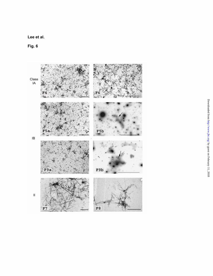
We next determined whether dominant-negative HrpA mutants are able to interfere with the self-assembly of wild-type HrpA into pili. Each of the six mutant HrpA proteins was mixed with the wild-type HrpA protein in a 1:1 ratio in pH 5.0. The pH of the solution was then raised to 7.8 to allow formation of pilus-like structures. We found that class II mutants F7 and F8 had little effect on the self-assembly of the wild-type HrpA protein (Fig. 6). In contrast, class IA (F4 and F6) and IB mutants (F5 and F9) strongly interfered with the ability of the wild-type HrpA to self-assemble into long pilus-like filaments (Fig. 6). However, the 1:1 mixtures of wild-type and mutant F5 or F9 HrpA proteins occasionally allowed formation of short filaments, whereas no such short filaments were formed in the mixtures of wild-type and mutant F4 or F6 HrpA proteins (Fig. 6). In 1:10 (mutant/wild-type) and 1:25 mixtures, class IB mutants F5 and F9 also had less effect on the self-assembly of wild-type HrpA protein, compared with class IA mutants F4 and F6 (data not shown). In the 1:50 mixture, both class IA and IB mutants had no effect on the self-assembly of wild-type HrpA protein. Taken together, these results suggest that the dominant-negative effects of both class IA and class IB mutants are caused by a direct interference, albeit to different degrees, with the ability of wild-type HrpA to assemble into pili.
Clustering of Pentapeptide Insertions with Similar Dominant-Negative Phenotypes When the locations of the pentapeptide insertions that gave distinct phenotypes were examined, we found a striking correlation between the insertion sites and the corresponding phenotypes (Fig. 2). All seven class IA mutants carry insertions near the C-terminus (between amino acids 104-112; indicated by diamonds). These insertions gave the strongest dominant-negative effect, preventing DC3000 from eliciting an HR in tobacco, secreting effector proteins in culture, forming extracellular Hrp pilus, and interfering with the ability of the wild-type HrpA protein to self assemble in vitro. The four class IB mutants carry insertions between amino acids 72 and 80 (indicated by triangles). Their dominant-negative effects were similar to class IA mutants, except that they did not affect the secretion of HrpA, AvrPto, or HopPtoM in culture. The four class II mutants contain insertions at residue 57, between residues 97 and 99, or at the C-terminal
by guest on February 11, 2018http://w
ww
.jbc.org/D
ownloaded from

6
residue (indicated by circles); they did not have any dominant-negative effect. Finally, the four class III mutants carry insertions between amino acid residues 84 and 91 (indicated by squares). These mutants had variable effects on the ability of DC3000 to elicit an HR in tobacco.
DISCUSSION
Assembly of extracellular filaments is a key feature of the TTSS in both mammalian and plant pathogenic bacteria. Genetic, molecular, and microscopic studies have firmly established the requirement of these filaments for type III secretion/translocation. However, little is known about the molecular details of subunit-subunit interactions during filament assembly or exact mechanisms by which these filaments function in long-distance protein transport. In this study, from collections of previously isolated nonfunctional HrpA mutants of Pst DC3000, we identified several dominant-negative mutants. Characterization of the dominant-negative HrpA mutants enabled us to gain insights into the involvement of specific regions of HrpA in the assembly and function of the HrpA protein in various stages of type III secretion.
Importance of the Hrp Pilus Our analysis of the dominant-negative HrpA mutant proteins reaffirms the importance of the Hrp pilus in plant-P. syringae interactions. We found that all examined dominant-negative HrpA mutants that block wild-type Pst DC3000 from eliciting TTSS-mediated plant responses also prevent the production of extracellular Hrp pili (Fig. 4). This result is consistent with the previous observation that the hrpA mutant, which lacks the Hrp pilus structural gene, is defective in type III secretion as well as in the elicitation of all TTSS-mediated host responses (15, 24).
Towards Understanding the Molecular Defects of Nonfunctional HrpA Proteins and Possible Subunit-Subunit Interaction Sites The large collection of the nonfunctional HrpA mutants isolated by Taira and colleagues (23) provided a crucial resource for the identification of the dominant-negative mutants in this research. Prior to this study, the specific molecular defects of these nonfunctional HrpA proteins were not understood. We analyzed six representative mutants and found that none of them were able to self-assemble in vitro (Fig. 5). This result therefore provides an explanation for the molecular defects of at least a subset of these nonfunctional HrpA proteins. It
remains to be determined whether other HrpA mutants are also defective in assembly.
Detailed analysis of the dominant-negative effects of HrpA mutants enabled us to reach a deeper understanding of the structure-function relationship of the HrpA protein. We found that the most upstream pentapeptide insertion, F7, produces a nonfunctional HrpA protein that cannot interfere with the assembly of wild-type HrpA or exert a dominant-negative effect. Unlike single amino acid substitution mutations, pentapeptide insertions not only disturb local structures, but if they are inserted into an -helix, they may also alter the orientation of the downstream -helix and other secondary structures. -helices seem to dominate the C-terminal portion of the HrpA protein based on computer prediction (Fig. 7), and the F7 insertion is located within a major putative -helix (Fig. 7A). Thus, the F7 insertion likely causes a major structural change, rendering it unable to engage in a subunit-subunit interaction with the wild-type HrpA protein.
Class IB mutants (e.g., F5 and F9) are located downstream of the F7 insertion in the same putative
-helix (Fig. 7). These mutants appear to be less affected in structure, as indicated by their ability to interfere with the self-assembly of wild-type HrpA in vitro and to exert dominant-negative effects on HR elicitation and assembly of extracellular Hrp pili. Therefore, unlike the F7 insertion mutant, the class IB mutants appear to contain at least one major subunit-subunit interaction site. This subunit-subunit interaction site, presumably located between the F7 insertion and class IB (Figs. 2B and 7) insertions, is probably responsible for the unproductive interaction between class IB mutants and wild-type HrpA protein, resulting in the observed dominant-negative effect. We considered the possibility that the different dominant-negative effects by the F7 insertion and class IA insertions are caused by different pentapeptide insertion sequences. However, the pentapeptide sequences of four class IB mutants are dissimilar to each other (MRPHA, VRPQQ, CGRTQ, and NAAAM; 23), whereas the F7 insertion sequence is somewhat similar to that (CGRTS; 23) of the F1 insertion, making it unlikely that the pentapeptide insertion sequences are responsible for the observed different dominant-negative phenotypes.
Class IA (e.g, F4 and F6) and additional class II (e.g., F8) mutants are located further downstream (Fig. 7A). Like the F7 insertion, F8 and the nearby class II insertions did not exert a dominant-negative
by guest on February 11, 2018http://w
ww
.jbc.org/D
ownloaded from

7
effect. Presumably, these class II insertions destroy another major subunit-subunit interaction site, thus rendering the mutant HrpA protein unable to interact with wild-type HrpA protein. The finding of class II insertions (F7 vs. F8) in two discrete regions suggests that we may have identified a pair of complementary subunit-subunit interaction sites that mediate the association of two adjacent HrpA proteins (Fig. 7B). Such complementary subunit-subunit interactions were elucidated recently at the atomic level for another TTSS-associated extracellular filament, the flagellum. It was shown that the upper half of the C-terminal -helix of one flagellin subunit interacts with the lower half of the C-terminal -helix of an adjacent flagellin subunit (35). Considering the apparently identical subunit arrangements in the needle (36), the EspA filament (37), and the flagellum (35), it would not be surprising that general features of subunit-subunit interactions would be conserved in all TTSS-associated filaments.
The Secreton Function and Hrp pilus Assembly Previous genetic evidence favors a functional relationship between the Hrp pilus in plant pathogenic bacteria and the needle in mammalian pathogenic bacteria (4, 38). For example, mutations in the structural gene of the Hrp pilus or the needle completely block the secreton function (i.e., secretion of effector proteins across the bacterial cell wall) (5-10, 17, 24). Our identification of the class IA dominant-negative HrpA mutants, which block the ability of DC3000 to assemble the Hrp pilus and to secrete effector proteins across the bacterial cell wall, further supports a role of the HrpA protein in the secreton function. Genetic and microscopic image analyses show that the needle is an integral part of the secreton and is needed to keep the secreton open (5-10, 39). If the Hrp pilus is the functional equivalent of the needle, the base of the Hrp pilus would also be an integral part of the secreton structure in plant pathogenic bacteria, which could explain why the HrpA protein is required for the secretion of effector proteins across the bacterial cell wall. The strong dominant-negative effect of class IA mutant HrpA proteins on the self-assembly of wild-type HrpA in vitro supports the hypothesis that Hrp pilus assembly is an integral part of the secreton construction in the bacterial cell wall.
In contrast with class IA mutants, class IB HrpA mutants (e.g., F5 and F9) block the ability of PstDC3000 to assemble the extracellular Hrp pili and to elicit TTSS-associated host responses but not the secretion of effector proteins across the bacterial
cell wall. This important observation suggests that even with a normal level of secretion of effector proteins across the bacterial cell wall, an extracellular Hrp pilus is still necessary for PstDC3000 to elicit all TTSS-associated responses. Therefore, isolation of class IA and IB dominant-negative mutants enabled us, for the first time, to separate two Hrp pilus-mediated steps during type III secretion: secretion across the bacterial cell wall and long-distance transport beyond the bacterial cell wall. The different effects of class IA and IB mutants on the secreton function may lie in the relative timing and/or strengths of the dominant-negative effects of the two classes of mutants. The dominant-negative effects of class 1A mutants may be exerted earlier and/or more strongly on the assembly of the secreton, so that Hrp pilus/secreton assembly does not reach the outer surface of the bacterial cell wall. On the other hand, the dominant-negative effects of class IB mutants may be exerted later and/or more weakly, so that Hrp pilus/secreton assembly proceeds to the outer surface of the bacterial cell wall, allowing secretion of effector proteins. Weaker dominant-negative effects of class IB mutants are supported by the observation that class IB mutants F5 and F9 had less severe interference with the self-assembly of wild-type HrpA proteins, and presumably the construction of the secreton in the cell wall, than class IA mutants F4 and F6 (Figs. 6 and 7).
An alternative, albeit less likely, explanation for the isolation of class IB mutants is that the Hrp pilus is functionally related to the EspA filament of EPEC. The EPEC espA mutant is defective in the transport of effector proteins into the host cell, but is normal in the secretion of effector proteins across the bacterial cell wall (13, 14). In this regard, class IB mutants pheno-copy the espA mutant. However, the possibility of an exact functional equivalency between the Hrp pilus and the EspA filament does not agree well with the existing experimental evidence, because the EspA filament functions only in long-distance transport but not in secretion across the bacterial cell wall, whereas the Hrp pilus functions in both processes. It is possible that the HrpA protein adopts two different molecular forms in these two processes: a polymerized pilus form (functionally equivalent to the EspA filament) needed for long-distance protein transport in the extracellular space, and an intracellular monomer form in the secreton structure (functionally equivalent to the needle complex). In this scenario, the class IB mutant-mediated interference with the assembly of the wild-type HrpA protein (Fig. 6)
by guest on February 11, 2018http://w
ww
.jbc.org/D
ownloaded from

8
would have a dominant-negative effect only on pilus-mediated long-distance transport in the extracellular space, but not on the ability of a monomeric HrpA protein to conduct the secreton function in the cell wall. However, this scenario cannot explain why class IA mutants have a strong dominant-negative effect on the secreton function.
Implications for Future Determination of the HrpA Structure Because structural subunits of bacterial supramolecular appendages have an inherent tendency to polymerize, rather than to form three-dimensional crystals, it is very difficult to determine the structures of such proteins in their final functional conformations. Crystallization of the structural subunits of bacterial appendages has generally required using derivatives lacking the ability to assemble into a superstructure. For example, the structure of the F41 fragment of flagellin lacks 52 N-terminal residues and 44 C-terminal resides (40). Crystallization of Type IV pilin subunits required the removal of an N-terminal region or the use of detergent (41-44). The self-assembly of EspA and Type I pilin was prevented from by co-crystallizing with its cognate chaperone
or by adding a small "donor strand" peptide to an N-terminally truncated protein (45-48). Our identification of the dominant-negative, assembly-defective HrpA derivatives may facilitate future crystallization and structural determination of the HrpA protein.
To conclude, we identified a collection of useful dominant-negative HrpA mutants of Pst DC3000 in this study. By examining the various dominant-negative phenotypes exerted by these HrpA mutants, we identified putative subunit-subunit interaction sites during HrpA assembly into the Hrp pilus and gained further insight into the role of HrpA in type III protein secretion. Besides the general significance of the understanding of the function of the Hrp pilus in type III secretion/translocation in plant pathogenic bacteria, the results presented here should facilitate future studies aimed at determining and interpreting the tertiary structure of the HrpA protein. We suggest that a similar approach be applied to the study of other TTSS-associated appendages, such as the needle and the EspA filament in mammalian pathogenic bacteria.
REFERENCES
1. Galán, J. E., and Collmer, A. (1999) Science 284,1322-1328 2. Cornelis, G. R., and Van Gijsegem, F. (2000) Annu. Rev. Microbiol. 54, 735-774 3. He, S.Y., and Jin, Q. (2003) Curr. Opinion. Microbiol. 6, 5-19 4. Romantschuk, M., Roine, E., and Taira, S. (2001) Eur. J. Plant Pathol. 107, 153– 160. 5. Kubori, T., Matsushima, Y., Nakamura, D., Uralil, J., Lara-Tejero, M., Sukhan, A., Galán, J. E., and
Aizawa, S.-I. (1998) Science 280, 602-605 6. Kimbrough, T. G., and Miller, S. I. (2000) Proc. Natl. Acad. Sci. USA 97, 11008-11013 7. Blocker, A., Jouihri, N., Larquet, E., Gounon, P., Ebel, F., Parsot, C., Sansonetti, P., and Allaoui, A. (2001)
Mol. Microbiol. 39, 652-663 8. Hoiczyk, E., and Blobel, G. (2001) Proc. Natl. Acad. Sci. USA 98, 4669-4674 9. Tamano, K., Aizawa, S.-I., Katayama, E., Nonaka, T., Imajoh-Ohmi, A., Kuwae, A., Nagai, S., and
Sasakawa, C. (2000) EMBO J. 19, 3876-3887 10. Journet, L., Agrain, C, Broz, P., and Cornelis, G. R. (2003). Science 302, 1757-1760 11. Daniell, S. J., Kocsis, E., Morris, E., Knutton, S., Booy, F. P., and Frankel, G. (2003) Mol. Microbiol. 49,
301-308 12. Sekiya, K., Ohishi, M., Ogino, T., Tamano, K., Sasakawa, C., and Abe, A. (2001) Proc. Natl. Acad. Sci.
USA 98, 11638-11643 13. Knutton, S., Rosenshine, I., Pallen, M. J., Nisan, I., Neves, B. C., Bain, C., Wolff, C., Dougan, G., and
Frankel, G. (1998) EMBO J. 17, 2166-2176 14. Ebel, F., Podzadel, T., Rohde, M., Kresse, A. U., Kramer, S., Deibel, C., Guzman, C. A., and Chakraborty,
T. (1998) Mol. Microbiol. 30, 147-161 15. Roine, E., Wei, W., Yuan, J., Nurmiaho-Lassila, E.-L., Kalkkinen, N., Romantschuk, M., and He, S. Y.
(1997) Proc. Natl. Acad. Sci. USA 94, 3459-3464 16. Roine, E., Saarinen, J., Kalkkinen, N., and Romantschuk, M. (1997) FEBS Lett. 417, 168-172 17. Van Gijsegem, F., Vasse, J., Camus, J. Marenda, M., and Boucher, C. (2000) Mol. Microbiol. 36, 249-260 18. Jin, Q., Hu, W., Brown, I., McGhee, G., Hart, P., McGhee, G., Hart, P., Jones, A. L., and He, S. Y. (2001)
by guest on February 11, 2018http://w
ww
.jbc.org/D
ownloaded from

9
Mol. Microbiol. 40, 1129-1139 19. Buttner, D., and Bonas, U. (2002) EMBO J. 21, 5313-5322 20. Van Gijsegem, F., Vasse, J., De Rycke, R., Castello, P., and Boucher, C. (2002) Mol. Microbiol. 44, 935-
946 21. Jin, Q., and He, S. Y. (2001) Science 294, 2556-2558 22. Li, C. M., Brown, I., Mansfield, J., Stevens, C., Boureau, T., Romantschuk, M., and Taira, S. (2002)
EMBO J. 21, 1909-1915 23. Taira, S., Jarno, T., Elina, R., Eeva-Liisa, N.-L., Harri, S., and Martin, R. (1999) Mol. Microbiol. 34, 737-
744 24. Wei, W., Plovanich-Jones, A., Deng, W.-L., Collmer, A., Huang, H.-C., and He, S. Y. (2000) Proc. Natl.
Acad. Sci. USA 97, 2247-2252 25. Schweizer, H. P. (1991) Gene 97, 109-112 26. Mudgett, M. B., Chesnokova, O., Dahlbeck, D., Clark, E. T., Rossier O. Bonas U., and Staskawicz B. J.
(2000) Proc. Natl. Acad. Sci. USA 97, 13324-13329 27. Sambrook, J., Fritsch, E., and Maniatis, T. (1989) Molecular Cloning: a Laboratory Manual, 2nd ed.
Cold Spring Harbor, NY, Cold Spring Harbor Laboratory Press 28. Huynh, T. V., Dahlbeck, D., and Staskawicz, B. J. (1989) Science 245, 1374-1377 29. Laemmli, U. (1970) Nature 227, 680-685 30. Flegler, S. L., Heckman, J. W., and Klomparens, K. L. (1993) Scanning and Transmission Electron
Microscopy: An Introduction. pp. 43-168, Oxford press, Oxford. 31. Zwiesler-Vollick, J., Plovanich-Jones, A., Nomura, K., Bandyopadhyay, S., Joardar, V., Kunkel, B. N.,
and He, S. Y. (2002) Mol. Microbiol. 45, 1207–1218 32. Guttman, D. S., and Greenberg, J. T. (2001) Mol. Plant-Microbe Interact. 14, 145-155 33. Keen, N. T., Tamaki, S., Kobayashi, D., and Trollinger, D. (1988) Gene 70, 191-19734. Badel, J. L., Nomura, K., Bandyopadhyay, S., Shimizu, R., Collmer, A., and He, S. Y. (2003) Mol.
Microbiol. 49, 1239-1251 35. Yonekura, K., Maki-Yonekura, S., and Namba, K. (2003) Nature 424, 643-650 36. Cordes, F. S., Komoriya, K., Larquet, E., Yang, S., Egelman, E. H., Blocker, A., and Lea, S. M. (2003) J.
Biol. Chem. 278, 17103-17107 37. Daniell, S. J., Kocsis, E., Morris, E., Knutton, S., Booy, F. P., and Frankel, G. (2003) Mol. Microbiol. 49,
301-308 38. He, S. Y. (1997) Trends Microbiol. 5, 489-495 39. Marlovits, T. C., Kubori, T., Sukhan, A., Thomas, D. R., Galan, J. E., and Unger, V. M. (2004) Science
306, 1040-1042 40. Samatey, F. A., K. Imada, S. Nagashima, F. Vonderviszt, T. Kumasaka, M. Yamamoto, and K. Namba.
(2001) Nature 410, 331-337 41. Craig, L., Taylor, R. K., Pique, M. E., Adair, B. D., Arvai, A. S., Singh, M., Lloyd, S. J., Shin, D. S.,
Getzoff, E. D., Yeager, M., Forest, K. T., and Tainer, J. A. (2003) Mol. Cell 11, 1139-1150 42. Hazes, B., Sastry, P. A., Hayakawa, K., Read R. J., and Irvin, R. T. (2000) J. Mol. Biol. 299, 1005-1017 43. Keizer, D. W., Slupsky, C. M., Kalisiak, M., Campbell, A. P., Crump, M. P., Sastry, P. A., Hazes, B., Irvin,
R. T., and Sykes, B. D. (2001) J. Biol. Chem. 276, 24186-24193 44. Parge, H. E., Forest, K. T., Hickey, M. J., Christensen, D. A., Getzoff, E. D., and Trainer, J. A. (1995)
Nature 378, 32-38 45. Yip, C. K., Finlay, B. B., and Strynadka, N. C. (2005) Nat. Struct. Mol. Biol. 12, 75-81. 46. Choudhury, D., Thompson, A., Stojanoff, V., Langermann, S., Pinkner, J., Hultgren, S. J., and Knight, S.
D. (1999) Science 285, 1061-1066 47. Sauer, F. G., Futterer, K., Pinkner, J. S., Dodson, K. W., Hultgren, S. J., and Waksman, G.. (1999) Science
285, 1058-1061 48. Sauer, F. G., Pinkner, J. S., Waksman, G.., and Hultgren, S. J. (2002) Cell 111, 543-551
FOOTNOTES
*This research was supported by grants from the United States Departments of Agriculture and Energy (to S.
by guest on February 11, 2018http://w
ww
.jbc.org/D
ownloaded from

10
Y. H.) and Michigan State University Intramural Grant Program (to D. N. A. and S. Y. H.). ¶These authors contributed equally to this study.
FIGURE LEGENDS
Fig. 1. Dominant-negative effects of pHRPA-AVRRPT280-255 on the ability of Pst DC3000 to cause plant responses and secretion of effector proteins. A, Bacterial suspensions of OD600 = 0.2 (approximately 1x10⁸
cfu/mL) and OD600 = 0.002 (approximately 1x10⁶ cfu/mL) in sterilized distilled water were infiltrated into fully expanded leaves of tobacco and Arabidopsis, respectively. The HR symptom in nonhost tobacco leaves, as indicated by tissue collapse in the upper leaf panels, was observed 20 h after inoculation. Disease chlorosis and necrosis in host Arabidopsis leaves (lower leaf panels) were recorded 4 days after inoculation. B, Bacteria grown in hrp-inducing minimal medium or LB for 12 h at 20oC were separated into cell and supernatant fractions. The cell and supernatant fractions were concentrated 5 and 50 times, respectively. Proteins in these fractions were separated on a 15% SDS-PAGE gel, followed by immunoblotting with a primary antibody against HrpA, AvrPto, or HopPtoM, and a secondary alkaline-phosphatase-conjugated IgG antibody. MM: Pst DC3000 in hrp-inducing medium; LB: Pst DC3000 in LB medium. Other strains are the same as in A. In the cell fraction of the HrpA protein blot, pHRPA-AVRRPT280-255 fusion protein was marked by an asterisk. In the cell fraction of the HopPtoM blot, a cross-reacting protein present in all lanes migrated slightly faster than HopPtoM (indicated by arrow).
Fig. 2. Dominant-negative effects of pHRPA derivatives carrying pentapeptide insertions on the ability of Pst DC3000 to cause the HR in nonhost tobacco and/or disease in host Arabidopsis leaves. A, PstDC3000 ( ) or Pst DC3000 carrying pHRPA-F4, -5, -6, -7, -8, or -9 was infiltrated into tobacco or Arabidopsis leaves. The HR and disease assay conditions were identical to those described in Fig. 1A. B, A diagram of the C-terminal portion (nucleotides [nt] 241-436, amino acid [aa] residues 50-113) of HrpA (adapted from Taira et al. [23]). The 21 pentapeptide insertions (F1-20, F22) are indicated by different symbols based on their dominant-negative phenotypes: Triangle ( ), dominant-negative mutants that block Pst DC3000 to cause the HR and disease, but allow effector secretion in culture; rectangle ( ), mutants that have inconsistent dominant-negative phenotypes; circle ( ), mutants that do not have a dominant-negative phenotype; diamond ( ), mutants that block Pst DC3000 to cause the HR and disease and to secrete effector proteins in culture.
Fig. 3. Immunoblot analysis of the effects of HrpA mutants on the ability of DC3000 to produce and/or secrete effector proteins in culture. Conditions for bacterial growth and immunoblotting were identical to those described in Fig. 1B. The cytosolic chaperone ShcM was used as a cytoplasmic protein control. All HrpA mutants were examined. Results from representative strains are shown here. In the cell fractions of the AvrPto and HopPtoM blots, arrows indicate AvrPto and HopPtoM, to distinguish them from cross-reacting proteins present in all lanes.
Fig. 4. Scanning electron micrographs of Pst DC3000 cells expressing representative mutant HrpA proteins. Bacteria were grown on carbon/formvar-coated nickel grids in hrp-inducing MM at 20oC for 14 h. Bacteria were then fixed in 4% glutaraldehyde, dehydrated, critical-point dried, and sputter coated with gold (~7 nm thick). The mounted bacteria were viewed with a JEOL 6300F SEM. Bar represents 1 m.
Fig. 5. Transmission electron micrographs showing the ability of wild-type and mutant HrpA to self-assemble into pilus-like structures. The pilus assembly was induced by increasing the pH of the HrpA solution (600 g/mL) from 5.0 to 7.8. The sample was stained with 1% phosphotungstic acid (pH 6.5), and examined using a JEOL 100-CEX TEM at an accelerating voltage of 100 kV. The short filamentous structures occasionally observed in F5 and F9 samples are shown in F5b and F9b. Bar represents 1 m.
Fig. 6. Transmission electron micrographs showing the dominant-negative effects of mutant HrpA proteins on the self-assembly of wild-type HrpA protein. The wild-type and mutant HrpA protein
by guest on February 11, 2018http://w
ww
.jbc.org/D
ownloaded from

11
solutions (600 g/mL) were mixed at the ratio 1:1. The mixtures were treated as described in Fig. 5 and examined with a TEM. Bar represents 1 m. Arrows in F5b and F9b panels indicate short filaments occasionally observed only in the WT/F5 and WT/F9 mixtures. These short filaments are more clearly discernible under a higher magnification. In the mixture of wild-type HrpA and wild-type HrpA, long pilus-like filaments were formed as in Fig. 5.
Fig. 7. A model for the effects of various classes of pentapeptide insertions on the HrpA subunit-subunit interaction. A, The primary HrpA amino acid sequence is shown with secondary structures predicted by the nnPredict (http://www.cmpharm.ucsf.edu/~nomi/nnpredict.html) and PROF (http://www.predictprotein.org/doc/methodsPP.html#PX_about_profsec) programs. Representative pentapeptide insertions are shown on top. A putative HrpA secondary structure is drawn below. Wavy lines indicate unstructured regions; whereas the cylindrical bars indicate putative -helices. The N-terminus (ca. 50 residues) is dispensable for the function and self-assembly of HrpA (16, 23). All 21 pentapeptide insertions that produce nonfunctional HrpA are located within the C-terminal portion of the HrpA protein (23). B, Two adjacent wild-type HrpA subunits (in grey) are shown. The relative positions of the two adjacent HrpA subunits are drawn according to the helical arrangement of the flagellin subunits (35). Short lines between the two subunits indicate putative subunit-subunit interaction sites, with thicker lines indicating stronger interactions. C, Two mutant HrpA subunits (in pink) are shown with representative insertions and the C-terminal AvrRpt2 fusion indicated. Note that F7 and F8 are drawn in complementary positions. D-G, Hypothetical abnormal interactions of mutant HrpA subunits with the wild-type HrpA subunit. Note that class 1A (F6) and class IB (F5) insertions, and C-terminal AvrRpt2 fusion each destroys at least one weak interaction site. The remaining strong interaction site apparently allows a dominant-negative (DN) effect. In contrast, class II insertions (F7 and F8) destroy the strong interaction site, preventing a dominant-negative effect. In the HrpA-AvrRpt2 fusion, AvrRpt2 fused to the C-terminus of HrpA appears to prevent complete subunit-subunit interactions, mimicking the effect of class IA pentapeptide insertions.
by guest on February 11, 2018http://w
ww
.jbc.org/D
ownloaded from

Lee et al.
Fig. 1
by guest on February 11, 2018http://w
ww
.jbc.org/D
ownloaded from

Lee et al.
Fig. 2
by guest on February 11, 2018http://w
ww
.jbc.org/D
ownloaded from

Lee et al.
Fig. 3
by guest on February 11, 2018http://w
ww
.jbc.org/D
ownloaded from

Lee et al.
Fig. 4
.
by guest on February 11, 2018http://w
ww
.jbc.org/D
ownloaded from

Lee et al.
Fig. 5
by guest on February 11, 2018http://w
ww
.jbc.org/D
ownloaded from
Lee et al.
Fig. 6
by guest on February 11, 2018http://w
ww
.jbc.org/D
ownloaded from

1 10 20 30 40 50 60 70 80 90 100 110
MVAFAGLTSKLTNLGNSAVGGVGGALQGVNTVASNATLQKNILLGTGDSLSVDAQAKASKESDANGAKLIAMQAQETMKKQTMDVLNAIQAGKEDSTNKKISATATNAKGISY
--EHH---HHHHH----EE------EE-EHEH---HHHHHEEEE------EHHHHHH-----HH--HHHHHHHHHHHHHH--HHHHHHHH----------EEHHH--------
HHHHH HHHHHHHHHHHHHHHHH HHHHHHHHHH EEEEEEHHHHH HHHHHHH HHHHHHHHHHHH
F5F9 F4 F6F7 F8
nnPredict
PROF
HrpA
A
IBII II IA
B
E
F6
HrpA-AvrRpt2
G
F8
F5
D
F7
F
F8F6
F5
C
HrpA-AvrRpt2
F7
DN
DN
No DN
No DN
Lee et al.,Fig. 7
by guest on February 11, 2018 http://www.jbc.org/ Downloaded from

Yang HeYong Hoon Lee, Olatomirin O. Kolade, Kinya Nomura, Dennis N. Arvidson and Sheng
secretion in Pseudomonas syringae pv. tomatoUse of dominant-negative HrpA mutants to dissect Hrp pilus assembly and type III
published online March 29, 2005J. Biol. Chem.
10.1074/jbc.M500972200Access the most updated version of this article at doi:
Alerts:
When a correction for this article is posted•
When this article is cited•
to choose from all of JBC's e-mail alertsClick here
by guest on February 11, 2018http://w
ww
.jbc.org/D
ownloaded from